Article

Occurrence and Distribution of Major Viruses Infecting Eggplant in Lebanon and Molecular Characterization of a Local Potato Virus X Isolate

1
CNR Istituto per la Protezione Sostenibile delle Piante, via Amendola 122/D, 70126 Bari, Italy
2
LARI Department of Plant Protection, Tal Amara, Zahlé P.O. Box 287, Lebanon
*
Authors to whom correspondence should be addressed.
Submission received: 19 December 2020 / Revised: 29 January 2021 / Accepted: 1 February 2021 / Published: 5 February 2021

Abstract

:
This research was carried out in order to evaluate the presence and distribution of viral infections causing severe disease in eggplant plants collected from different districts in Bekaa valley, Lebanon. Most infected plants showed virus-like symptoms consisting predominantly of leaf blotch, mottling chlorotic and ring spots; leaf twisting and plant dwarf were also observed in the visited fields. Symptomatic and asymptomatic plants were collected and screened by ELISA test for the presence of several different pathogenic viruses potentially present in the area. Results showed that potato virus Y (PVY) was the most prevalent virus found by ELISA (detected in the 15.3% of the tested plants), followed by eggplant mottled dwarf virus (EMDV, 2.9%) and cucumber mosaic virus (CMV, 1.2%), while tomato spotted wilt virus (TSWV), alfalfa mosaic virus (AMV) and pepper mottle virus (PepMoV) were not detected. Biological indexing of symptomatic ELISA-negative plants, followed by electron microscopy, indicated the presence of virus-like particles of the genus Potexvirus, which was subsequently confirmed as potato virus X (PVX) by RT-PCR and Sanger sequencing. PVX was found in 35.3% of the tested plants, all sampled in the northern Bekaa area. In a phylogenetic analysis, the partial coat protein gene sequence of a selected Lebanese isolate, PVX-AK1, clustered together with other PVX isolates from Asia. Furthermore, the 124-aa sequence of PVX-AK1 shared 100% identity with PVX-UK3, an isolate which is known as avirulent in potato genotypes carrying either Nx or Rx resistance genes. This work revealed a picture of the previously uninvestigated phytosanitary status of eggplant crops in an important horticultural area of Lebanon.

1. Introduction

In Lebanon, about a quarter of the cultivated land is used for vegetable production, whether irrigated, rain fed or protected. Vegetable crops are grown in the coastal plains, Bekaa valley and medium elevation mountains. Bekaa valley is the largest agricultural area in Lebanon, which provides the biggest share of the country’s agricultural production [1]. Vegetables include a wide variety of tuber and bulb crops (potato, garlic, onion, radish and turnip), leafy vegetables (lettuce, cabbage, cauliflower, spinach, mint, parsley and others), fruit vegetables (tomato, cucumber, eggplant, watermelon and melon), leguminous crops (green peas, fava beans and other types of beans) and corn, which is cultivated both for human consumption and as a fodder crop [2]. However, Solanaceae plants, including potato, tomato, pepper, tobacco and eggplant, are widely cultivated in different regions of the country with temperate climate and are commonly found in many households. Eggplant (Solanum melongena) is a very important vegetable crop in Lebanon which has developed a high popularity and has become one of the most consumed vegetables. Eggplant cultivation has started to spread recently in the Lebanon, especially in the Bekaa region due to the demand of the local market. The total cultivated area reached 20,700 Ha in 2010, representing about 12% of the total area used for growing fruit vegetables [3]. “Sawad leil” and “Negra”, are the most cultivated eggplant varieties, yielding an average of 80–100 t/ha. Other introduced varieties such as “Humsi”—a Syrian local variety—as well as “Valentine” and “Daisy” varieties, have been introduced, yielding an average of 30–40 t/ha.
Solanaceae plants are exposed to many pathogens, including threatening fungi, bacteria and viruses [4,5]. Plant viruses are among the principal disease-inducing agents affecting solanaceous crops as they are transmitted through vegetative propagation, by contact between infected and healthy plants and by different vectors, including insects and nematodes [6]. Compared to other cultivated Solanaceae species, no viral emergences have been mentioned in eggplants in Lebanon, whereas only few studies have been carried out on potato and tomato viruses [7,8,9,10]. However, widespread diffusion of eggplant-infecting viruses in bordering countries and in the Mediterranean basin represent a serious risk not to be underestimated. Eggplant is naturally infected by several viruses causing disease epidemics and representing increased agronomic threats. Table 1 summarizes information about all viruses naturally infecting the eggplant plant.
Some of these viruses, such as the cucumber mosaic virus (CMV, [17]), tomato spotted wilt orthotospovirus (TSWV, [26]) and eggplant mottled dwarf alphanucleorhabdovirus (EMDV, [27]) are polyphagous and have worldwide diffusion, while for others, e.g., potato virus Y (PVY, [14]) and pepino mosaic virus (PepMV, [22]) the host range is restricted to solanaceous species. Several studies have described the occurrence of Potato virus X in eggplant. The most recent one was reported from Iran, where potato virus X (PVX) was found in all surveyed fields in both eggplants and peppers (Capsicum annuum) [31].
Potato virus X is the type species in the genus Potexvirus in the family Alphaflexiviridae that groups viruses phylogenetically related by replication mechanisms, structural proteins and genome type and organization [23]. PVX is basically mechanically transmitted (e.g., seed grading machines, contact by plants etc.) and its natural hosts are mainly limited to the Solanaceae (such as, potato, tomato, eggplant and cape gooseberry), although there are some susceptible plants in other families, e.g., in Amaranthaceae and Chenopodiaceae. PVX is ranked among viruses with important economic impacts, causing diseases in commercially important crops (with 50% yield reductions in some potato cultivars; [32]) and its effects can be worsened by co-infection with other viruses, in particular Potato virus Y (PVY; a phenomenon also known as “synergism”; [33]). The damage caused by PVX vary according to plant species and the presence of mixed infections with other viruses in the same plant. For example, PVX in potato and tomato plants develops mild local lesions and varying degrees of leaf deformation and mosaic symptoms, while these symptoms can be more severe when the infection is mixed with PVX + PVY [34,35]. The full genomic RNA sequences (6.4 kb) of several PVX strains were obtained about thirty years ago [36,37,38] and since then, due to its high stability and replication rate, PVX is often used as a vector for heterologous gene expression and studies on RNA silencing in plants [39].
In 2018, mosaic and leaf distortion symptoms of alleged viral origin were observed in eggplants, mainly in the northern region, whereas a more limited distribution of similar symptoms was reported in central and western regions of the Bekaa valley. Therefore, the aims of the present study were: (i) to establish the etiology of the observed disease phenotypes and to characterize viruses possibly associated with the symptoms; (ii) to identify viral agents previously not detected in similar surveys in Lebanon; and (iii) to verify the possibility that some severe disease symptoms could be induced by mixed infections of two or more viruses.

2. Materials and Methods

2.1. Plant Material

During the 2018 growing season (spanning approximately June to October) several eggplant smallholders and other rural farmers, reported symptoms possibly associated with viral infections (leaf mottling, mosaic, stunting, ring and/or chlorotic spots) in the frame of a national survey conducted on vegetable crops in Lebanon. Visited fields were selected based on information provided by Lebanese Agricultural Research Institute (LARI), Tal Amara, Zahle, where many farmers made inquiries into unusual symptoms of possible biotic and/or abiotic diseases that had been noticed in their fields. With the exception to Hermel district (northern Bekaa), where three visited fields were smaller than 0.5 ha of surface area, the area of each selected field in the other districts was ≈ 0.5 ha. Within each selected field, four dots were placed on four sides to create a virtual square or rectangle in which samples would be collected. Plant samples were then collected from this space following the “W” pattern scheme, taking into account that the approved planting distance between plants is 0.5 m. Following this scheme, all plants were counted and then the incidence of symptomatic infected plants was calculated. Moreover, sampling distance between individual plants was approx. 30–40 m. A total of 170 leaf samples were collected from symptomatic and asymptomatic eggplant plants in nine commercial fields from six locations in Bekaa valley. The sources of samples included: Hermel (50 samples/3 fields), Qaa (40 samples/2 fields), Labweh (20 samples/1 field), Kab Elias (20 samples/1 field), Mansoura (20 samples/1 field) and Ammiq (20 samples/1 field), (Figure 1). All collected samples were kept at 4 °C until being tested in laboratory.

2.2. Serological Assay

All collected samples were tested by the double-antibody sandwich enzyme-linked immunosorbent assay (DAS-ELISA) using specific antibodies immediately available in the laboratory. These included serological assays to alfalfa mosaic virus (AMV), PVY, CMV, eggplant mottled dwarf virus (EMDV), tomato spotted wilt virus (TSWV) and PepMoV, (Loewe Biochemica GmdH, Sauerlach, Germany), which were used for ELISA tests following the manufacturer’s instructions. Positive and negative controls supplied with the kits were added in each test. Briefly, the 96-well ELISA plates were coated with 200 μL of anti PVY, CMV, EMDV, TSWV, AMV and PepMoV IgGs diluted 1:200 in coating buffer and then the plates were incubated at 37 °C for 4 h. Leaf midveins and petioles from 6–8 eggplant leaves were macerated in the presence phosphate buffered saline (PBS) with the detergent Tween 20 (1/10 w/v) using an automated Homex 6 apparatus (Bioreba, Switzerland). Two hundred microliters of the homogenized samples in PBS buffer were loaded in duplicate in the microplates and then plates were kept overnight at 4 °C before the addition of alkaline phosphatase-conjugated anti PVY, CMV, EMDV, TSWV, AMV and PepMoV IgGs diluted 1:200. Plates were then incubated at 37 °C for 4 h prior to the addition of the substrate (1 mg/mL p-nitrophenyl phosphate in diethanolamine buffer, pH 9.8). Absorbance values were recorded at 405 nm with a microplate reader (Bio-Tek Instruments, Winooski, VT, USA). Samples with DAS-ELISA values at least three times more than the mean value of the healthy control samples were considered as positive [40].

2.3. Biological Assay

Leaf tissues from six symptomatic eggplant plants collected from the northern Bekaa region and that were negative for the viruses tested in ELISA, in addition to other three healthy plants were macerated using a mortar and pestle in inoculation buffer (50 mM Na2HPO412H2O; 50 mM KH2PO4, 0.44% (w/v) sodium diethyldithiocarbamate; pH 7) and extracts were mechanically inoculated on six- to eight-week-old Nicotiana benthamiana indicator plants grown on soil and maintained in the greenhouse at 24 °C. After dusting the carborundum onto the surface of leaves, plant sap from each macerated sample was rubbed onto the surface of leaves of two replicates N. benthamiana plants then inoculated leaves were rinsed with distilled water. Twenty days later the presence of virus was checked by the induced symptoms in inoculated plants. Leaf tissues from indicator plants then were sampled and were kept at −20 °C for further molecular analysis.

2.4. Electron Microscopy

Electron microscopy analyses were carried out on some negative ELISA-test leaves from eggplants plants to investigate the nature of the symptomatology. To analyze for possible virus particles, leaf dips from symptomatic eggplant tissues were processed and observed using a Philips Morgagni 282D electron microscope. For thin sectioning, tissue fragments were excised from the same leaves of symptomatic eggplants and healthy control plants. All samples were processed according to standard procedures [41]. Briefly, samples were fixed in 4% glutaraldehyde in 0.05 M potassium phosphate buffer (pH 7.2) for 2 h and then were post-fixed at 4 °C in 1% osmium tetroxide in the same buffer for 2 h. Overnight bulk staining in 0.5% aqueous uranyl acetate, dehydration in graded ethanol dilutions, and embedding in TAAB Spurr (Agar Scientific, Stansted, UK) resin followed. Thin sections were stained with lead citrate before observations with a Philips Morgagni 282D (FEI Company, Hillsboro, OR, USA) transmission electron microscope at 60 KV accelerating voltage.

2.5. Total Nucleic Acids Extraction, cDNA Synthesis and PCR

Midribs and petioles of symptomatic and asymptomatic eggplant leaves were cut and then ground in liquid nitrogen. Total RNA was extracted from 200 mg of the ground tissues using Qiagen RNeasy plant mini kits (QIAGEN) following the manufacturer’s instruction. The RNA was finally eluted in 50 μL of nuclease-free water. The quality and quantity of RNA were analyzed and quantified using a NanoDrop 2000 spectrophotometer (Thermo Fisher Scientific, Waltham, MA, USA). Complementary DNA (cDNA) was synthesized using 500 ng of RNA, 1 µL of hexamer random primers (ROCHE Diagnostics, Mannheim, Germany) (0.5 µg/µL) and 4 µL RNase-free water. The mix was denatured by heating at 70 °C for 5 min, spun for 10 s, and immediately chilled on ice for 5 min, then mixed with 2 µL DTT (0.1 M), 1 µL dNTPs (10 mM), 200 units of Moloney murine leukemia virus (M-MLV) reverse-transcriptase enzyme (Thermo Fisher Scientific), 5 µL M-MLV (5×) first strand buffer and then the final volume was adjusted to 25 µL using nuclease-free water. The mixture was incubated at 39 °C for 1 h, then 70 °C for 10 min and finally at 4 °C before use or stored at −20 °C. Samples were screened by RT-PCR for the presence of PVX using specific primers designed that can amplify 562 bp from the PVX coat protein (CP) gene: PVX-sense: 5′-TAGCACAACACAGGCCACAG-3′ and PVX- antisense: 5′-GGCAGCATTCATTTCAGCTTC-3′ [42]. The primers used were synthesized by Macrogen (Korea). PCR reaction was carried out in a 50 μL mixture containing 25 µL DreamTaq Green PCR Master Mix 2× (Thermo Fisher Scientific), 0.8 µL of 10 pmol/µL from each forward and reverse primer, 4µL cDNA of each sample and the final volume made up with nuclease-free water. Gradient PCR was performed using the following parameters; one cycle at 95 °C for 5 min as heating step of Taq DNA polymerase, 35 cycles at 95 °C for 30 s (denaturation), 56 °C for 30 s (annealing) and 72 °C for 1 min (extension), followed by final extension at 72 °C for 10 min. PCR products were analyzed by electrophoresis on a 1.2% agarose gel and stained by GelRed (Biotium, Fremont, CA USA). All steps above were also repeated for the confirmation of the presence of PVX in N. benthamiana indicator plants previously collected and kept at −20 °C.

2.6. Cloning, Sequencing and Phylogenetic Analysis

PCR amplicons generated from different PVX isolates were subsequently purified using a PCR Purification Kit (Qiagen, Valencia, CA, USA) and ligated into the pSC-A-amp/kan vector using the Strataclone kit ( Agilent Technologies, Santa Clara, CA, USA) according to the manufacturer’s instructions. Resulting plasmids were used for transforming the competent cells provided with the kit, using a heat shock technique. White colony formation on Luria–Bertani (LB) agar plates containing 50 μg/μL X-Gal were individually selected and cultured overnight in LB (37 °C/220 rpm). Plasmid extractions were carried out using Plasmid DNA Extraction mini (Thermo Fisher Scientific). Selected clones were subjected to automated sequencing (Macrogen, Seoul, Korea) in the forward and reverse direction using primers M13FOR/REV. Bioinformatics tools used for sequence analyses and alignments for nucleotides homology included NCBI-BLAST tool (http://blast.ncbi.nlm.nih.gov/Blast.cgi) and CLUSTAL-X program [43]. Construction of the phylogenetic tree from the aligned sequences was conducted and bootstrapped with 1000 node replications using Mega X [44]. A total of 17 PVX isolates sequences distributed all over the world were obtained from NCBI database and were used to analyze the phylogenetic relationships with the Lebanese PVX isolate. The CP alignment with strain PVX-UK3 (GenBank acc. no. M95516.1) was inferred by the SerialCloner v. 2.6 tool (https://serial-cloner.en.softonic.com/).

3. Results

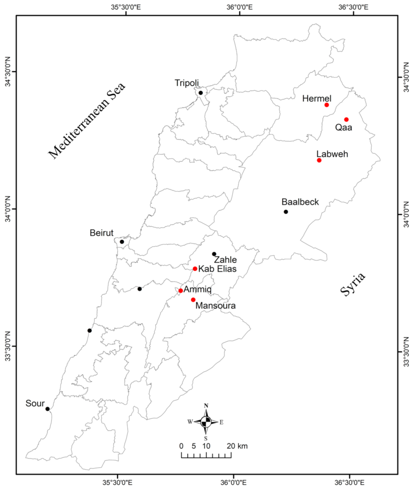
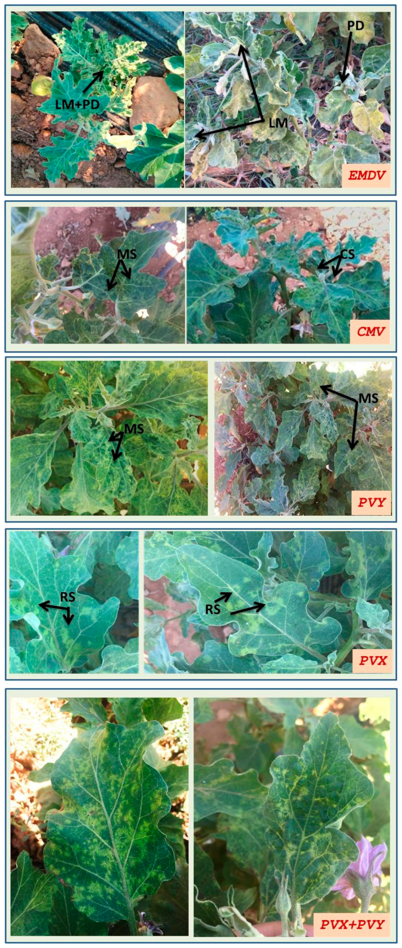
3.1. Survey, Visual Inspections and Virus Distribution in Bekaa Valley

After receiving warning reports from farmers about symptoms of possible viral origin in different areas of the Bekaa Valley, Lebanon, action was taken in order to verify the diffusion and the severity of diseases in eggplant crops. A total of nine fields in the major vegetable-growing areas of the Bekaa valley were surveyed during the late summer of 2018. The average temperature and rainfall during the growing season were 25 °C and 800 mm in southern Bekaa and 20 °C and 200 mm in extreme northern Bekaa. Although some of the symptoms could have been mistakenly attributed to viral infection, we took visual inspection data as an approximate starting point of the following survey based on specific diagnosis. As shown in Table 2, the incidence of visually-estimated virus infection from each visited district/field and the results were as follows: (northern Bekaa): 35% in Hermel, 30% in Qaa and 20% in Labweh; (central Bekaa): 10% in Kab Elias; (western Bekaa): 10% in Mansoura and Ammi. The results of ELISA test showed that 33 eggplant plants out of 170 (19.4%) in the Bekaa valley were infected with EMDV, PVY, or CMV. On the other hand, the other tested viruses (AMV, TSWV and PepMoV) were not detected in any surveyed areas. For each detected virus, negative controls were used in each microplate, therefore, the range of absorbance values of negative controls, depending on the antiserum used varied from 0.068 to 0.082 at 405 nm. The absorbance values of the positive samples were 0.372–0.923 for PVY, 0.388–0.820 for EMDV and 0.429–0.735 for CMV. Overall, the results obtained by ELISA demonstrated that PVY was the most prevalent virus found in eggplant leaf samples, identified in 15.3% tested plants, followed by EMDV and CMV (2.9 and 1.2%, respectively). Moreover, all visited fields were infected with PVY, while infection with CMV was limited to one field in Mansoura district and EMDV was found in three different locations. Among the visited areas, virus prevalence was the highest in the northern Bekaa (Hermel, Qaa and Labweh) showing 18, 25 and 15% ELISA-positive samples, respectively. In central Bekaa, 15% of the collected samples were infected in Kab Elias district, whereas in western Bekaa, Mansoura and Ammiq reported 25 and 15% of virus infections, respectively. Table 2 summarizes the prevalence of virus infection by visual observation and ELISA test for all visited fields. No cases of mixed infections in a same individual plant were detected by ELISA.

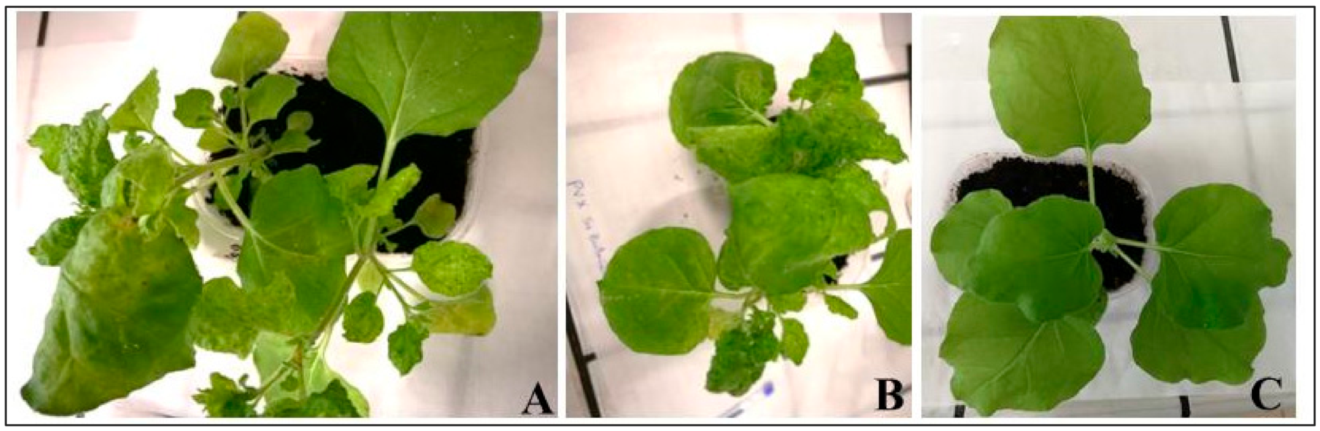
3.2. Biological Assay

Six symptomatic eggplant plants that were negative for the viruses tested in ELISA, were further investigated for possible viral infection by mechanical inoculation in N. benthamiana plants. All six N. benthamiana plants reacted positively to the mechanical inoculation by developing typical viral symptoms in inoculated and in systemically infected leaves, whereas mock plants did not display any type of symptoms. After 20 days post inoculation, systemic symptoms on N. benthamiana were clearly visualized and included chlorotic spots, leaf deformation, systemic mild mosaic and irregular white lesions on small leaves and intermediate leaf rugosity (Figure 2). The success of mechanical transmission indicated the presence of one or more viruses, but since symptoms were rather aspecific, further diagnostic analyses were performed.

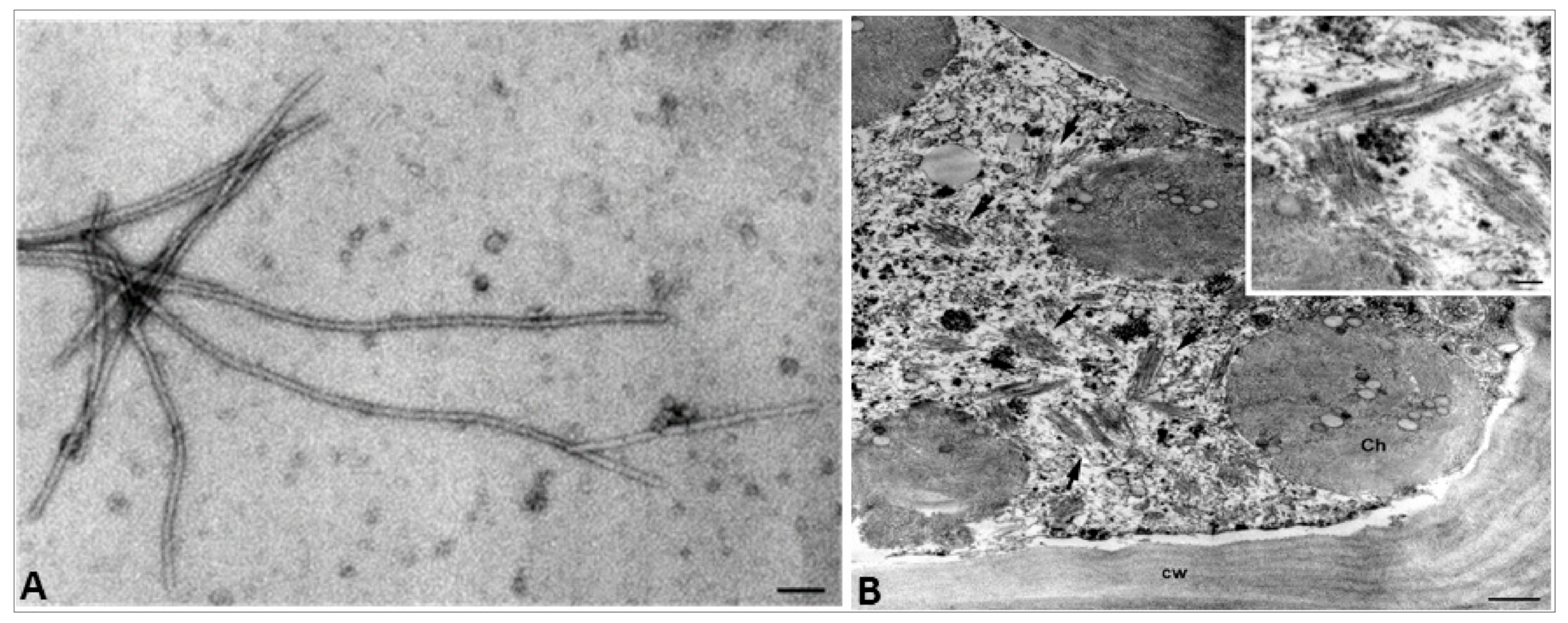
3.3. Electron Microscopy

Transmission electron microscopy (TEM) analyses were carried out on some negative ELISA-test leaves from eggplants plants to investigate the origin of the symptomatology. To analyze possible virus particles, leaf dips and embedding from symptomatic plants, eggplant tissues were processed and observed under TEM. Elongated virus-like particles were found in leaf dip preparation from symptomatic eggplant plants that resembled those of the family Flexiviridae. Particles were flexuous, 450–600 nm in length and 12–13 nm in diameter (Figure 3A). Thin sections of the same sample showed cytoplasmic laminated aggregates of virus particles inside mesophyll cells resembling the cytopathology of the genus Potexvirus (Figure 3B). No alterations were found in the cells of the healthy control plant. Therefore, TEM analysis suggested the presence of viral particles belonging to a possible potexvirus, previously not investigated by ELISA with specific antisera.

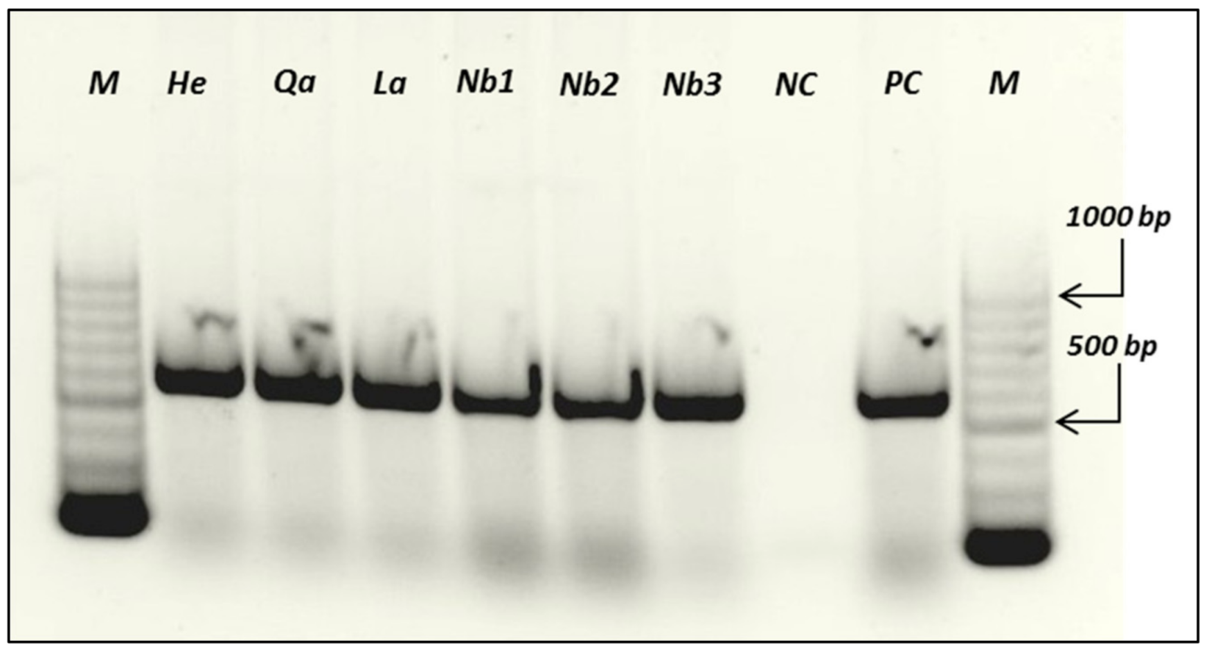
3.4. Molecular Detection of PVX by RT-PCR

In order to confirm the additional presence of a potexvirus, predicted by biological assays and TEM observations, a molecular diagnostic assay was performed. Since PVX was the most probable potexvirus candidate species expected to infect eggplant fields, a specific primer pair for this virus was first employed. A total of 85 samples from 170 plants collected in northern, central and western Bekaa (Table 3) were tested. Selected samples included: (i) 35 symptomatic plants that tested negative by ELISA; (ii) 15 plants found to be infected by ELISA, in order to check the presence of mixed infections with PVX; and (iii) 35 symptomless eggplant plants from the same field. Overall, a total of 30 out of 85 eggplant plants (35.3%), reacted positively in RT-PCR and were therefore found to be infected with PVX. A 562 bp PCR product, corresponding to a fragment of the PVX CP gene, was amplified from the naturally infected eggplant samples as well as from N. benthamiana plants used in the biological assays (Figure 4). Infections with PVX were found only in samples collected from the northern Bekaa region. The highest number of infected plants (60%) was sampled in the Hermel and Labweh districts, followed by the Qaa district (45%). Out of the 35 symptomatic plants used in RT-PCR, only 27 were positive to PVX, while the rest (three) of the positive samples were retrieved from three asymptomatic plants collected from the same region. RT-PCR revealed mixed infections occurring in two samples from the Qaa district previously found to be infected with PVY in ELISA. It cannot be excluded that other viruses could be present in the eight plants that showed virus-like symptoms and tested negative to PVX and all the other viruses searched for by ELISA. Table 3 provides a summary of the PVX infections by RT-PCR.

3.5. Association of Virus Infection with Plant Symptoms

According to the results obtained from both serological and molecular tests, most infected plants in the field showed virus-like symptoms on leaves. Observed symptoms consisted of leaf twisting; chlorotic spots; mosaic symptoms consisting of leaf blotches and mottling of the infected leaves; ring spots consisting of light green circles symptoms on the upper surface of the infected leaves. In some cases, infected plants revealed various types of mosaic symptoms that—to a certain degree—did not show clear differences among infecting viruses, especially when infected with either PVY or CMV. However, the two plants collected from Mansoura district, infected with CMV, showed mosaic symptoms, mottling and discoloration spots that evolved in light necrotic lesions arranged parallel to the nerves of the leaves. These plants were collected from the edge of the unique visited field at Mansoura. On the other hand, deformed leaves of varying intensity, in addition to chlorotic or yellow vein discolorations turning into a generalized chlorotic mottling, were clearly observed in plants infected with EMDV. These plants were also accompanied by mild to severe stunting. Moreover, systemic mosaic symptoms consisting of chlorotic spots and mottling were generally observed on plants found to be infected with PVY. Plants infected by PVX showed chlorotic ring spots with mosaic patterns, mostly consisting of irregular patches distributed among the interveinal areas. Plants co-infected with PVX and PVY incited more severe symptoms on leaves than plants infected by either virus individually. In fact, plants with mixed infections showed both chlorotic ring spots and severe mosaic symptoms (Figure 5).

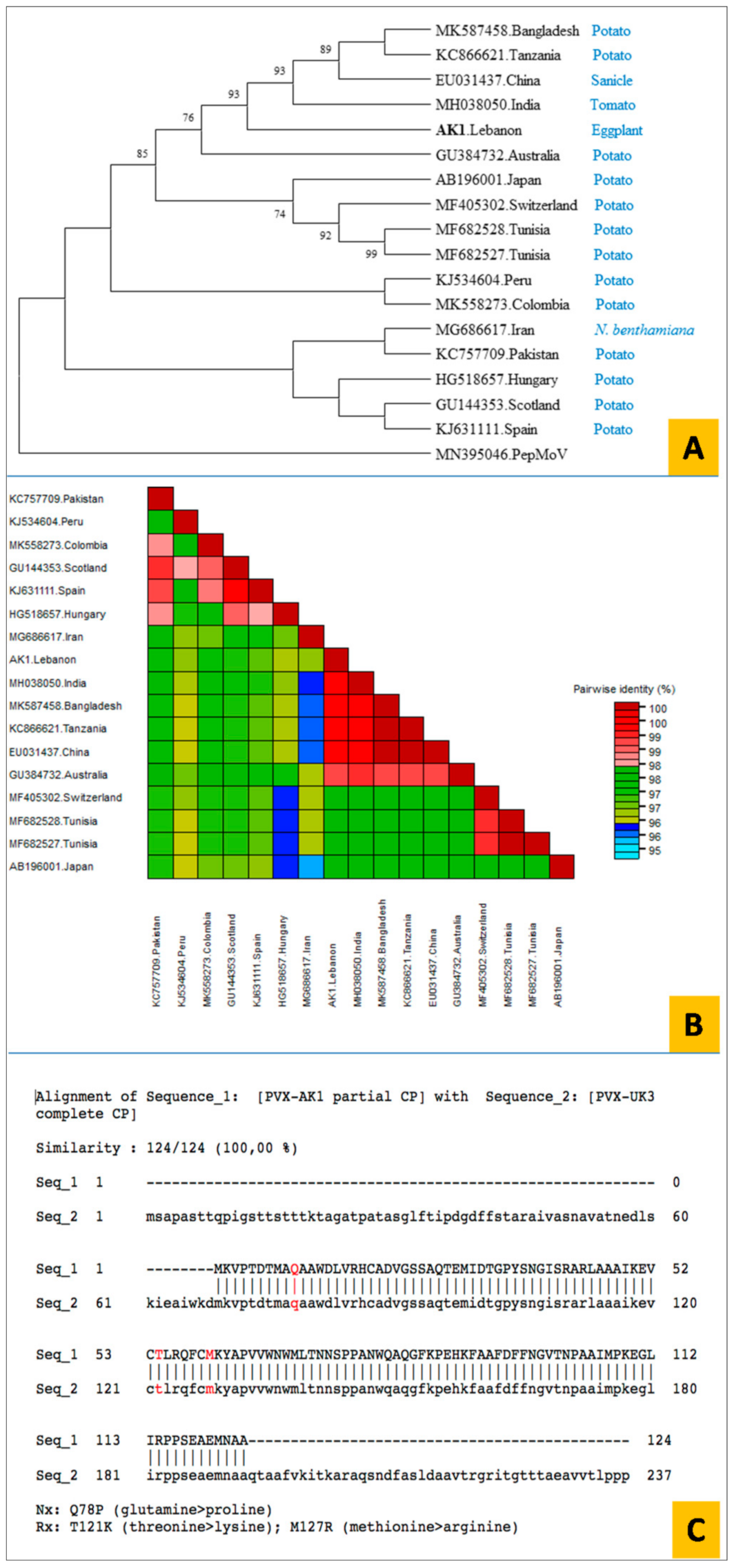
3.6. Cloning and Sequence Analysis of the PVX CP Gene

A PCR-amplified DNA fragment of 562 bp, corresponding to the partial sequence of the PVX CP gene from the infected areas of the Bekaa valley was analyzed. Sequences from three eggplant isolates were obtained and showed 100% identity among them at the nucleotide level. A representative isolate from Lebanon (PVX-AK1) was submitted to the GenBank database with the accession number (LR877715). The BLAST search revealed a high similarity of PVX-AK1 (99.4 and 99.3%) with isolates from India and Bangladesh (accession numbers MH038050 and MK587458, respectively). Three main clusters were reflected by phylogenetic analyses conducted with 16 different PVX isolates available from GenBank, in which the Lebanese isolate slightly separated from a group containing partial CP sequences of PVX isolates retrieved from Tanzania, China, Bangladesh and India (Figure 6A). These results were confirmed by a sequence dramatic tool (SDT) with a graphical interface that can show pairwise identity scores using a color-coded pairwise identity matrix, which enables a comprehensive comparison between sequences under study [45] (Figure 6B).
Since some PVX isolates may show amino acidic (aa) CP sequences variants at specific positions that determine a virulent phenotype on potato genotypes carrying either Nx or Rx resistance genes, we compared the partial PVX-AK1 CP sequence with that of PVX-UK3 (GenBank acc. no. M95516.1). The latter PVX isolate is avirulent on potato genotypes carrying either Nx or Rx resistance genes. As shown in Figure 6C, the 124-aa sequence of the two isolates shared 100% identity. Therefore, it can be inferred that PVX-resistant potato cultivars would also be effective against this newly characterized Lebanese isolate, since no other aa residues able to induce resistance-breaking effects have been reported so far.

4. Discussion

In field and greenhouse conditions, eggplant is much less susceptible to viruses than other Solanaceous crops, such as tomato and pepper [46]. Evidence of spots of diseased eggplant fields in the Bekaa valley, Lebanon, led to a survey program aiming to identifying the prevalence and diffusion of viral pathogens. Only a few studies have reported on the incidence of viruses in cultivated eggplant in the Mediterranean environment, and none of them have assessed the situation in Lebanon. As found in other Mediterranean areas, e.g., Turkey [47], PVY was the most prevalent virus, since it was identified in over 15% of the collected samples, and none of the visited areas were PVY-free. The occurrence and distribution throughout the area of EMDV was recorded, too. This virus is more polyphagous than PVY, since it has been reported in a range of solanaceous species (eggplant, tomato, potato, pepper) and other crops (e.g., cucumber and tobacco), ornamental and wild plants. EMDV is reportedly widespread in the Mediterranean basin [48,49]. To the best of our knowledge, this is the first report of EMDV in eggplant in Lebanon, a novelty that will require future monitoring for assessing the spread and the consequent damage that might occur due to this virus. Another polyphagous virus, CMV, was observed with a lower incidence than that of other viruses. This virus was found only in western Bekaa, where pepper cultivation already suffers infections by this virus (E. Choueiri, unpublished data). These three viruses are vectored by winged insects in the open field. PVY and CMV are non-persistently transmitted by aphids, while EMDV is transmitted very specifically only by the leafhopper Agallia vorobjevi Dl. (fam. Cicadellidae) [50]. In principle, in the case of non-persistent transmission, control strategies based on vector insect control are considered largely ineffective, whereas in the case of EMDV, persistent-circulative transmission by leafhoppers may be prevented by proper vector control strategies [51]. Our results, with novel data on the presence of damaging viral pathogens in Lebanese horticulture, offer important information for more efficient, science-based disease control programs in the field.
Rapid diagnostic techniques for the timely detection of emerging or novel plant viruses are of great importance for agriculture. Our study demonstrates, once again, the importance of TEM for providing informative descriptions of viral particles and ultrastructural features induced by viruses within infected cells for clear identification, in our case, of PVX as an additional disease agent in eggplant [52,53]. PVX is a major viral pathogen with worldwide distribution. In this study, a previously unreported diffusion of PVX on eggplants was found in the northern Bekaa, with an infection rate ranging between 45–60% of the tested plants. This is similar to reports from a similar survey on PVX diffusion in horticultural crops in Iran [31]. Interestingly, three of thirty-five asymptomatic eggplant plants also tested positive to molecular assays. Early diffusion of PVX in symptomless infections may have determined a previously underestimated presence of this virus in northern Bekaa fields [54]. The introduction and spread of PVX is largely due to movement of personnel and equipment. Cultural practices, such as transplanting, manual harvesting, as well as plant-to-plant contact, contribute to the spread of this viral disease [4]. Consequently, roguing of infected plants, sanitation of equipment and tools and trading with only certified eggplant seedlings particularly for local varieties can prevent further spread of PVX in eggplants and other crops.
Although there is no evidence that PVX may act as the principal cause of severe disease in eggplant, monitoring its diffusion and prevalence may help to identify susceptible cultivars and avoid increased PVX accumulation in areas where crops at risk (e.g., potato) are grown. It is particularly noteworthy that in the present survey PVY–PVX mixed infection was also recorded in eggplant, as was previously reported for potato [55], tobacco [56] and tomato [35]. These two viruses are known to incite synergistic pathogenetic effects, leading to exacerbated symptoms that were, in fact, also observed also in eggplant plants. Thus, it can be expected that, in eggplant, the interaction between PVX and potyviruses may lead to an increased PVX titer, in comparison with singly infected plants, as reported in other host plants, including potato [57,58]. In potato—where PVX can induce particularly severe diseases—and other solanaceous crops, specific management strategies have been suggested to limit the severity of diseases caused by mixed infections [59]. Besides synergy, another link between pathogenic viruses in eggplants and a possible problem for potato cultivation, is the upsurge of viral resistance-breaking isolates. In potato, PVX control in the long term is granted by the availability of commercial genotypes carrying either Rx or Nx resistance genes [60]. The PVX CP is the viral determinant of the outcome of interactions between the virus and potato lines carrying either resistance genes. We found that aa mutations in the PVX CP that are responsible for the resistance-breaking phenotypes, namely a glutamine to proline substitution at position 78 for Nx and two substitutions (threonine to lysine and methionine to arginine at positions 121 and 127, respectively) for Rx [61,62], were absent in the CP sequence of the Lebanese isolate PVX-AK1. This finding was not unexpected, since resistance-breaking variants have been found only in South American PVX isolates [63], that were shown to be distantly related to PVX-AK1 and other Asian isolates in the phylogenetic analysis presented here.

5. Conclusions

In conclusion, the current study not only presented novel results about eggplant-infecting viruses in Lebanon, but it is also the first study showing that PVX is diffused in some important areas of the country where eggplant is grown. In general, the incidence of viral pathogens was relatively high, especially in northern Bekaa, the major eggplant-growing area in Lebanon. The results indicate that, at least for some tested viruses, growers must act in order to reduce further virus diffusion and thereby improve their production capabilities. Since some of the identified viruses, particularly PVY and PVX, can also incite disease in other solanaceous crops, and are well known as severe pathogens in potato, disease management strategies will be necessary in areas where these two crops are grown in close proximity.

Author Contributions

Investigation, E.C. and F.J.; methodology, R.A.K., E.C. and A.D.S.; resources, E.C. and F.J.; software, R.A.K., and F.C.; supervision, E.C., M.S. and R.A.K.; validation, M.S. and F.C.; writing—original draft, R.A.K., E.C. and F.C.; writing—review and editing, F.C. All authors have read and agreed to the published version of the manuscript.

Funding

This research received no external funding.

Institutional Review Board Statement

Not applicable.

Informed Consent Statement

Not applicable.

Data Availability Statement

Not applicable.

Acknowledgments

Authors would like to thank Pasquale Saldarelli and the three anonymous reviewers for their valuable comments and suggestions to improve the quality of the paper. We also thank Maroun El Moujabber and Yara El Khoury for their technical support in Lebanon.

Conflicts of Interest

The authors declare that they have no competing interest.

References

  1. Ministry of Agriculture (MoA) of Lebanon, FAO. Agriculture in Lebanon 2008–2009; Technical Report; Ministry of Agriculture/Food and Agriculture Organization: Beirut, Lebanon, 2010.
  2. Nassif, M.H. Groundwater Governance in Central Bekaa, Lebanon. In IWMI Project Report, Groundwater Governance in the Arab World–Taking Stock and Addressing Challenges; Report No. 9; IWMI: Beirut, Lebanon, 2016. [Google Scholar]
  3. Anonymous. Résultats Globaux du Module de Base du Recensement de l’agriculture 2010; Projet Observatoire Libanais de Développement Agricole: Beirut, Lebanon, 2012; p. 140. [Google Scholar]
  4. Beemster, A.B.R.; Rozendaal, A. Potato viruses: Properties and symptoms. In Viruses of Potatoes and Seed-Potato Production; De Borkz, J.A., Ed.; Pubdoc: Wageningen, The Netherlands, 1972; pp. 115–143. [Google Scholar]
  5. Hančinský, R.; Mihálik, D.; Mrkvová, M.; Candresse, T.; Glasa, M. Plant viruses Infecting solanaceae family members in the cultivated and wild environments: A review. Plants 2020, 9, 667. [Google Scholar] [CrossRef]
  6. Banttari, E.E.; Ellis, J.P.; Kurana, S.M.P. Diseases caused by viruses and virus-like pathogens. In Potato Health Management; Rowe, R.C., Ed.; APS PRESS: St. Paul, MN, USA, 1993; pp. 127–133. [Google Scholar]
  7. Abou-Jawdeh, Y.; Sobh, H.; Saad, A.T. Incidence of potato virus diseases and their significance for a seed certification program in Lebanon. Phytopathol. Mediterr. 2001, 40, 1113–1118. [Google Scholar]
  8. Choueiri, E.; El-Zammar, S.; Jreijiri, F.; Saad, A.T.; Afram, M.A.; Varveri, C. Records of potato viruses in Lebanon. J. Plant Pathol. 2002, 84, 139. [Google Scholar]
  9. Choueiri, E.; El-Zammar, S.; Jreijiri, F.; Mnayer, D.; Massaad, R.; Saad, A.T.; Hanna, L.; Varveri, C. Phytosanitary status of potato in Bekaa valley in Lebanon. Bull. OEPP 2004, 34, 117–121. [Google Scholar] [CrossRef]
  10. Abou-Jawdah, Y.; El Mohtar, C.; Sobh, H. First report of tomato spotted wilt virus on tomatoes in Lebanon. Plant Dis. 2006, 90, 378. [Google Scholar] [CrossRef] [PubMed]
  11. Fidan, H.; Sarıkaya, P. Tomato chlorosis virus and tomato yellow leaf curl virus causing mixed infection in protected eggplant (S. melongena) crops in Turkey. Acta Scientiarum polonorum. Hortorum. Cultus. 2020, 19, 81–89. [Google Scholar]
  12. Mansour, A.; Almusa, A.; Vetten, H.V.; Lesemann, D.E. Properties of a Cowpea mild Mottle virus (CPMMV) isolate from eggplant in Jordan and evidence for biological and serological differences between CPMMV Isolates from leguminous and solanaceous hosts. J. Phytopathol. 2008, 146, 539–547. [Google Scholar] [CrossRef]
  13. Dombrovsky, A.; Sapkota, R.; Lachman, O.; Pearlsman, M.; Antignus, Y. A new aubergine disease caused by a whitefly-borne strain of tomato mild mottle virus (TomMMoV). Plant Pathol. 2013, 62, 750–759. [Google Scholar] [CrossRef]
  14. Sadeghi, M.S.; Behjatnia, S.A.A.; Masumi, M.; Izadpanah, K. Characterisation of a strain of Potato virus Y causing eggplant mosaic in southern Iran. Australas Plant Pathol. 2008, 37, 79–86. [Google Scholar] [CrossRef]
  15. Tsai, W.S.; Abdourhamane, I.K.; Kenyon, L. First Report of Pepper veinal mottle virus associated with mosaic and mottle diseases of tomato and pepper in mali. Plant Dis. 2010, 94, 378. [Google Scholar] [CrossRef]
  16. Rakib, A.; Mustafa, A.A.; Adhab, A.; Ismail, K.A.H. Eggplant blister mottled virus (EBMV): A possible new potyvirus characterized from Iraq. J. Gen. Mol. Virol. 2011, 3, 49–52. [Google Scholar]
  17. Bagewadi, B.; Hossain, M.S.; Fayad, A.; Naidu, R.A. First report of cucumber mosaic virus from eggplant (S. melongena) in Bangladesh. Plant Dis. 2015, 99, 293. [Google Scholar] [CrossRef] [PubMed]
  18. Rui, P.H.; Jiang, L.; Li, S.; Jiang, X.Z.; Zhao, Q.Q.; Ying Feng, J.; Jiang, T. First report of broad bean wilt virus 2 infection in eggplant in China. J. Plant Pathol. 2020, 102, 543. [Google Scholar] [CrossRef] [Green Version]
  19. Sokhansanj, Y.; Rakhshandehroo, F.; Pourrahim, R. First report of tomato ringspot virus on eggplant in Iran. J. Plant Pathol. 2012, 94, S4.94. [Google Scholar] [CrossRef]
  20. Sastry, K.S.; Nayudu, M.V. Ringspot symptoms of eggplant, incited by Tobacco ring spot virus/Sintomi di maculatura anulare su Melanzana causati dal virus delta maculatura anulare del Tobacco. Phytopathol. Mediterr. 1976, 15, 60–62. [Google Scholar]
  21. Ozdemir, S.; Erilmez, S.; Payland, I.C. First report of Alfalfa mosaic virus in eggplant in Turkey. J. Plant Pathol. 2011, 93, S4.82. [Google Scholar]
  22. Blystad, D.A.; van der Vlugt, R.; Alfaro-Fernández, A.; del Carmen Córdoba, M.; Bese, G.; Hristova, D.; Pospieszny, H.; Mehle, N.; Ravnikar, M.; Tomassoli, L.; et al. Host range and symptomatology of Pepino mosaic virus strains occurring in Europe. Eur. J. Plant Pathol. 2015, 143, 43–56. [Google Scholar] [CrossRef] [Green Version]
  23. Martelli, G.P.; Adams, M.J.; Kreuze, J.F.; Dolja, V.V. Family Flexiviridae: A case study in virion and genome plasticity. Ann. Rev. Phytopathol. 2007, 45, 73–100. [Google Scholar] [CrossRef] [Green Version]
  24. He, Y.H.; Huang, K.; Yao, L.Z.; Li, W.P.; Liu, C.M.; Chen, L.; Chen, H.R.; Chen, S.Y.; Wang, J.G. First report of tobacco mosaic virus infecting dutch eggplant (Solanum aculeatissimum) in China. Plant Dis. 2019, 103, 2973. [Google Scholar] [CrossRef]
  25. Nawaz, H.H.; Umer, M.; Bano, S.; Usmani, A.; Naseer, M. A Research review on tomato bushy stunt virus disease complex. J. Nat. Sci. Res. 2014, 4, 18–23. [Google Scholar]
  26. Kamberoglu, M.A.; Caliskan, A.F.; Alan, B. First report of tomato spotted wilt virus on eggplant in Turkey. J. Plant Pathol. 2009, 91, 231. [Google Scholar]
  27. Martelli, G.P. Bacilliform particles associated with mottled dwarf of eggplant (S. melongena L.). J. Gen. Virol. 1969, 5, 319–320. [Google Scholar] [CrossRef]
  28. Brioso, P.S.T.; Pimentel, J.P.; Louro, R.P.; Kitajima, E.W.; Oliveira, D.E. Andean potato mottle virus—Characterization of a strain naturally infecting eggplant (S. melongena). Fitopatol. Bras. 1993, 18, 526–533. [Google Scholar]
  29. Briand, J.P.; Bouley, J.P.; Witz, J. Self-assembly of eggplant mosaic virus protein. Virology 1997, 76, 664–669. [Google Scholar] [CrossRef]
  30. Dombrovsky, A.; Pearlsman, M.; Lachman, O.; Antignus, Y. Characterization of a new strain of eggplant mottled crinkle virus (EMCV) infecting eggplants in Israel. Phytoparasitica 2009, 37, 477–483. [Google Scholar] [CrossRef]
  31. Ravanbod, E.; Rakhshandehroo, F.; Golnaraghi, A. Survey of potato virus X in vegetable fields of Iran. J. Plant Pathol. 2018, 100, 137. [Google Scholar] [CrossRef] [Green Version]
  32. Hahm, Y.; Slack, S.A.; Slattery, R.J. Reinfection of potato seed stocks with potato virus S and potato virus X in Wisconsin. Am. Potato J. 1981, 58, 117–125. [Google Scholar] [CrossRef]
  33. Syller, J. Facilitative and antagonistic interactions between plant viruses in mixed infections. Mol. Plant Pathol. 2012, 13, 204–216. [Google Scholar] [CrossRef]
  34. Nie, X.; Singh, M. Response of potato, tobacco and Physalis floridana plants to mixed infection with PVX, PVYNTN and PVY strains. Can. J. Plant Pathol. 2013, 35, 390–401. [Google Scholar] [CrossRef]
  35. Liang, Z.; Dickison, V.; Singh, M.; Xiong, X.; Nie, X. Studies of tomato plants in response to infections with PVX and different PVY isolates reveal a remarkable PVX-PVYNTN synergism and diverse expression profiles of genes involved in different pathways. Eur. J. Plant Pathol. 2016, 144, 55–71. [Google Scholar] [CrossRef]
  36. Skryabin, K.G.; Kraev, A.S.; Morozov, S.Y.; Rozanov, M.; Chernov, B.K.; Lukasheva, L.I.; Atabekovet, J.G. The nucleotide sequence of potato virus X RNA. Nucleic Acids Res. 1988, 16, 10929–10930. [Google Scholar] [CrossRef]
  37. Huisman, M.J.; Linthorst, H.J.; Bol, J.F.; Cornelissen, J.C. The complete nucleotide sequence of potato virus X and its homologies at the amino acid level with various plus-stranded RNA viruses. J. Gen. Virol. 1988, 69, 1789–1798. [Google Scholar] [CrossRef]
  38. Orman, B.E.; Celnik, R.M.; Mandel, A.M.; Torres, H.N.; Mentaberry, A.N. Complete cDNA sequence of a South American isolate of potato virus X. Virus Res. 1990, 16, 293–305. [Google Scholar] [CrossRef]
  39. Scholthof, K.G.; Adkins, S.; Czosnek, H.; Palukaitis, P.; Jacquot, E.; Hohn, T.; Hohn, B.; Saunders, K.; Candresse, T.; Ahlquist, P.; et al. Top 10 plant viruses in molecular plant pathology. Mol. Plant Pathol. 2011, 12, 938–954. [Google Scholar] [CrossRef]
  40. Clark, M.F.; Adams, A.N. Characteristics of microplate method of enzyme-linked immunosorbent assay for detection of plant viruses. J. Gen.Virol. 1977, 34, 475–483. [Google Scholar] [CrossRef] [PubMed]
  41. Martelli, G.P.; Russo, M. Use of thin sectionning for the visualization and identification of plant viruses. J. Virol. Methods 1984, 8, 143–224. [Google Scholar]
  42. Nie, X.; Singh, R.P. A novel usage of random primers for multiplex RT-PCR detection of virus and viroid in aphids, leaves, and tubers. J. Virol. Methods 2001, 91, 37–49. [Google Scholar] [CrossRef]
  43. Thompson, J.D.; Gibson, T.J.; Plewniak, F.; Jeanmougin, F.; Higgins, D.G. The CLUSTAL_X windows interface: Flexible strategies for multiple sequence alignment aided by quality analysis tools. Nucleic Acids Res. 1997, 25, 4876–4882. [Google Scholar] [CrossRef] [Green Version]
  44. Kumar, S.; Stecher, G.; Li, M.; Knyaz, C.; Tamura, K. MEGA X: Molecular evolutionary genetics analysis across computing platforms. Mol. Biol. Evol. 2018, 35, 1547–1549. [Google Scholar] [CrossRef]
  45. Muhire, B.M.; Varsani, A.; Martin, D.P. SDT: A virus classification tool based on pairwise sequence alignment and identity calculation. PLoS ONE 2014, 9, e108277. [Google Scholar] [CrossRef] [PubMed]
  46. Daunay, M.C.; Caranta, C.; Pédron, F.; Deulvot, C.; Dussault, M.; Moretti, A.; Jullian, E.; Ruffel, S.; Moquet, F.; Majde, M.; et al. Heredity of Tomato mosaic virus (ToMV) and Potato virus Y (PVY) resistance in eggplant (S. melongena L.). In Proceedings of the XIIth Meeting on Genetics and Breeding of Capsicum and Eggplant, Noordwijkerhout, The Netherlands, 17–19 May 2004; pp. 154–160. [Google Scholar]
  47. Colak-Ates, A.; Fidan, H.; Ozarslandan, A.; Ata, A. Determination of the resistance of certain eggplant lines against Fusarium wilt, potato Y potyvirus and root-knot nematode using molecular and classic methods. Fresenius Environ. Bull. 2018, 27, 7446–7453. [Google Scholar]
  48. Pappi, P.G.; Chaintoutis, S.C.; Dovas, C.I.; Efthimiou, K.E.; Katis, N.I. Development of one-tube real-time qRT-PCR and evaluation of RNA extraction methods for the detection of eggplant mottled dwarf virus in different species. J. Virol. Methods 2015, 212, 59–65. [Google Scholar] [CrossRef] [PubMed]
  49. Babaie, G.; Kouhi-Habibi, M.; Massah, A.; Dizadji, A.; Izadinejad, L.; Simon, A. Complete genome sequence and genome analysis of eggplant mottled dwarf virus-Iranian isolate. J. Phytopathol. 2015, 163, 331–341. [Google Scholar] [CrossRef]
  50. Babaie, G.; Izadpanah, K. Vector transmission of eggplant mottled dwarf virus in Iran. J. Phytopathol. 2003, 151, 679–682. [Google Scholar] [CrossRef]
  51. Dietzgen, R.G.; Mann, K.S.; Johnson, K.N. Plant virus-insect vector interactions: Current and potential future research directions. Viruses 2016, 8, 303. [Google Scholar] [CrossRef]
  52. Zechmann, B.; Zellnig, G. Rapid diagnosis of plant virus diseases by transmission electron microscopy. J. Virol. Methods 2009, 162, 163–169. [Google Scholar] [CrossRef]
  53. Richert-Pöggeler, K.R.; Franzke, K.; Hipp, K.; Kleespies, R.G. Electron microscopy methods for virus diagnosis and high resolution analysis of viruses. Front. Microbiol. 2019, 9, 3255. [Google Scholar] [CrossRef]
  54. Roossinck, M.J.; García-Arenal, F. Ecosystem simplification, biodiversity loss and plant virus emergence. Curr. Opin. Virol. 2015, 10, 56–62. [Google Scholar] [CrossRef]
  55. De Bokx, J.A. Potato virus Y. In Compendium of Potato Diseases; Hooker, W.J., Ed.; American Phytopathological Society: St. Paul, MN, USA, 1986; pp. 70–71. [Google Scholar]
  56. Rochow, W.F.; Ross, A.F. Virus multiplication in plants doubly infected by potato viruses X and Y. Virology 1955, 1, 10–27. [Google Scholar] [CrossRef]
  57. Vance, V.B. Replication of Potato virus X RNA is altered in coinfections with Potato virus Y. Virology 1991, 182, 486–494. [Google Scholar] [PubMed]
  58. Gonzalez-Jara, P.; Tenllado, F.; Martinez-Garcia, B.; Atencio, F.A.; Barajas, D.; Vargas, M.; Diaz-Ruiz, J.; Diaz-Ruiz, J.R. Host-dependent differences during synergistic infection by Potyviruses with Potato virus X. Mol. Plant Pathol. 2004, 5, 29–35. [Google Scholar] [CrossRef]
  59. Hameed, A.; Iqbal, Z.; Asad, S.; Mansoor, S. Detection of multiple Potato viruses in the field suggests synergistic interactions among Potato viruses in Pakistan. Plant Pathol. J. 2014, 30, 407–415. [Google Scholar] [CrossRef] [Green Version]
  60. Cockerham, G. Genetical studies on resistance to potato viruses X and Y. Heredity 1970, 25, 309–348. [Google Scholar] [CrossRef]
  61. Goulden, M.G.; Köhm, B.A.; Santa Cruz, S.; Kavanagh, T.A.; Baulcombe, D.C. A feature of the coat protein of potato virus X affects both induced virus resistance in potato and viral fitness. Virology 1993, 197, 293–302. [Google Scholar] [CrossRef] [PubMed]
  62. Santa Cruz, S.; Baulcombe, D. Analysis of potato virus X coat protein genes in relation to resistance conferred by the genes Nx, Nb and Rx1 of potato. J. Gen. Virol. 1995, 76, 2057–2061. [Google Scholar] [CrossRef]
  63. Querci, M.; Baulcombe, D.C.; Goldbach, R.W.; Salazar, L.F. Analysis of the resistance-breaking determinants of potato virus X (PVX) strain HB on different potato genotypes expressing extreme resistance to PVX. Phytopathology 1995, 85, 1003–1010. [Google Scholar] [CrossRef]
Figure 1. Schematic map of Lebanon showing the geographic origin (red spots) of the 170 collected eggplant leaf samples from Bekaa valley.
Agriculture 11 00126 g001
Figure 2. Symptoms developed on Nicotiana benthamiana plants mechanically inoculated with extracts from naturally infected eggplants leaves. (A,B): Systemic mild mosaic and irregular white lesions on small leaves and intermediate leaf rugosity; (C): Mock control.
Agriculture 11 00126 g002
Figure 3. (A): Negatively stained virus particles in leaf dip preparation from symptomatic eggplant. Bars = 100 nm; (B) Ultrastructural aspects of infected eggplant mesophyll cells showing virus aggregates (black arrows). (Ch) Chloroplast; (cw) cell wall. Inset shows enlargement of virus aggregates. Bar = 500 nm, inset 100 nm.
Agriculture 11 00126 g003
Figure 4. The 1.2% Agarose gel electrophoresis analysis of 562 bp of the PVX coat protein (CP) gene amplified by RT-PCR from three eggplant samples from (He): Hermel, (Qa): Qaa, (La): Labweh, and three mechanically inoculated Nicotiana benthamiana plants (Nb1–3); (NC) negative control, (PC) positive control and (M) marker GeneRuler 100 bp DNA Ladder (Thermo Scientific).
Agriculture 11 00126 g004
Figure 5. Disease symptoms on eggplant plants from Bekaa valley naturally infected with different viruses during the growing season of 2018. Symptoms consisted of leaf mottling (LM); plant dwarf (PD); chlorotic spots (CS); mosaic symptoms (MS) and ring spots (RS). Each detected virus in this study is indicated in Red. CMV, EMDV and PVY were found by ELISA test; PVX was detected by RT-PCR.
Agriculture 11 00126 g005
Figure 6. (A) Phylogenetic analysis of a 562 bp fragment of the CP gene of Potato virus X including the sequence from the northern Bekaa valley in Lebanon. Sequences selected from different countries aligned with Lebanese sequence were included in the phylogenetic tree which was constructed by Mega X program [44] by using the maximum-likelihood method with Kimura’s two-parameter model and 1000 replicates bootstrap value (only node value above 70% are shown) A sequence of PepMoV (accession number MN395046) was used as an outgroup to root the tree. The natural host of each selected PVX isolate is shown in blue. (B) Sequence dramatic tool (SDT) Color-coded pairwise identity matrix generated by alignment of 562 bp fragment from CP gene of potato virus X from Lebanon and another 17 isolates distributed worldwide. Each colored square represents a percentage identity score between two sequences (one indicated horizontally to the left and the other vertically at the bottom). The colored scale key indicates the correspondence between the colors appeared in the matrix and pairwise identities between isolates. (C) Comparison of the amino acid sequences of PVX-AK1 and PVX-UK3 coat proteins. Amino acids responsible for virulence or avirulence on potato genotypes carrying Nx and Rx resistance genes are shown in red. Mutations leading to resistance-breaking phenotypes are indicated below.
Agriculture 11 00126 g006
Table 1. List of viruses naturally infecting eggplant plants and their natural transmission modes.
FamilyGenusSpecies Natural TransmissionReference
GeminiviridaeBegomovirusTomato yellow leaf curl virusTYLCVWhiteflies[11]
ClosteroviridaeCrinivirusTomato chlorosis virusToCVWhiteflies[11]
BetaflexiviridaeCarlavirusCowpea mild mottle virusCPMMVWhiteflies[12]
PotyviridaeIpomovirusTomato mild mottle virusTomMMoVWhiteflies[13]
PotyviridaePotyvirusPotato virus YPVYAphids[14]
PotyviridaePotyvirusPepper veinal mottle virusPVMVAphids[15]
PotyviridaePotyvirusEggplant blister mottled virusEBMVAphids[16]
BromoviridaeCucumovirusCucumber mosaic virusCMVAphids[17]
SecoviridaeFabavirusBroad bean wilt virusBBWVAphids[18]
SecoviridaeNepovirusTomato ringspot virusToRSVNematodes[19]
SecoviridaeNepovirusTobacco ringspot virusTRSVNematodes[20]
BromoviridaeAlfamovirusAlfalfa mosaic virusAMVAphids/seeds[21]
AlphaflexiviridaePotexvirusPepino mosaic virusPepMVContact/seeds[22]
AlphaflexiviridaePotexvirusPotato virus XPVXContact/seeds[23]
VirgaviridaeTobamovirusTobacco mosaic virusTMVContact/seeds[24]
TombusviridaeTombusvirusTomato bushy stunt virusTBSVSeed[25]
TospoviridaeOrthotospovirusTomato spotted wilt orthotospovirusTSWVThrips[26]
RhabdoviridaeAlphanucleorhabdovirusEggplant mottled dwarf alphanucleorhabdovirusEMDVLeafhoppers[27]
SecoviridaeComovirusAndean potato mottle virusAPMoVBeetles/contact[28]
TymoviridaeTymovirusEggplant mosaic virusEMVBeetles[29]
TombusviridaeTombusvirusEggplant mottled crinkle virusEMCVUnknown[30]
Table 2. Prevalence of virus infections revealed by visual inspection and ELISA tests in eggplants plants collected from north, center and western areas of Bekaa valley.
ELISA-Positive Samples
LocationNo. FieldsMean Virus Incidence (%visual)No. TestedNo. Detected EMDVPepMoVTSWVAMVCMVPVYELISA Positive (%)
Hermel335509 2 0000718%
Qaa23040102 0000825%
Labweh120 2030 0000315%
Kab Elias110203 10000215%
Mansoura11020500002325%
Ammiq1102030 0000315%
Total9 170335 000226
(19.4%)(2.9%) (1.2%)(15.3%)
Table 3. Prevalence of PVX infections revealed by RT-PCR test in eggplants plants different regions in Bekaa valley.
RegionLocationNo. TestedNo. PVX+RT-PCR Detection
Northern BekaaHermel251560%
Qaa20945%
Labweh10660%
Central BekaaKab Elias1000.0
Western BekaaMansoura1000.0
Ammiq1000.0
Total 853035.3%
Publisher’s Note: MDPI stays neutral with regard to jurisdictional claims in published maps and institutional affiliations.

