Article

Optimization of Agrobacterium Mediated Genetic Transformation in Paspalum scrobiculatum L. (Kodo Millet)

1
Department of Botany, University of Rajasthan, Jaipur, Rajasthan 302 004, India
2
Department of Bioscience, Manipal University Jaipur, Rajasthan 303 007, India
3
Sri Venkateswara College, Delhi University-South Campus, New Delhi 110 021, India
4
Amity Institute of Biotechnology, Amity University Rajasthan, Jaipur, Rajasthan 303 007, India
*
Author to whom correspondence should be addressed.
Submission received: 10 March 2021 / Revised: 13 May 2021 / Accepted: 18 May 2021 / Published: 28 May 2021

Abstract

:
An efficient and reproducible protocol for Agrobacterium tumefaciens mediated genetic transformation was developed for kodo millet (Paspalum scrobiculatum L.) by optimizing various parameters. Agrobacterium strains EHA 105 and LBA 4404 harboring plasmids pCNL 56 and pCAMBIA 2300, respectively, provided the highest transformation efficiency. Addition of acetosyringone (AS) in infection medium (200 µM-EHA 105, 250 µM-LBA 4404) and co-cultivation medium (50 µM) increased the transformation efficiency. Transient and stable expression of gus gene was confirmed with histochemical assay of infected embryos and leaves of transformed plants, respectively. The best GUS response was obtained by pretreatment of callus with an antinecrotic mixture (10 mg/L Cys + 5 mg/L Ag + 2.5 mg/L As) at infection time of 20 min followed by co-cultivation for 3 days (EHA 105) and 5 days (LBA 4404) in dark. Regenerated transgenic plants were obtained after 8 to 10 weeks of selection on callus induction medium (NAA 0.5 mg/L, BAP 1 mg/L) containing 50 mg/L Kan + 250 mg/L Cef and were rooted for 2 weeks on MS medium containing PAA (1 mg/L) and phytagel. The plantlets established in greenhouse showed normal growth. Therefore, the protocol developed in the present study can be used for development of improved varieties of kodo millet.

1. Introduction

Millet is a generic term used for unrelated small seeded forage grass cereals with coarse grains consisting of 10 genera, and are used for food, feed and forage worldwide [1,2]. They are staple food crops in developing nations of Africa and Asia and are known as a poor man’s cereal or orphan crops [3].
Millets serve as excellent alternatives for cereals due to having an equivalent nutritional level, with the advantage of growing with minimum inputs [4,5]. They are often treated as nutri-cereals due to their rich protein and mineral profile along with high vitamin, calcium, non-starchy polysaccharide and fiber contents and a lower glycine index [6,7] and are considered as ideal components in different food formulations intended for specific target groups [8,9]. As per the Food and Agriculture Organization Corporate Statistical Database (FAOSTAT), the demand for cereals will increase to up to 2.89 billion tonnes by year 2030, while their production is expected to decline due to decreased irrigation, a lack of availability of large cultivable lands and other abiotic environmental threats.
Paspalum scrobiculatum L. (kodo millet) is one of the highly valued small millets. It is widely cultivated in South Asian countries such as India, the Philippines and Vietnam, with India being one of the largest producers [10,11]. The high dietary fiber, vitamin and essential amino acid content of the plant make it an excellent substitute for cereals [12]. The plant also possesses antidiabetic and high antioxidant activity among millets [6,13]. However, susceptibility to the stress and the long maturity season of kodo millet have restricted its widespread use compared to other cereals. Considering the economic importance of the crop and the limitation associated with its incorporation into a balanced diet with other conventional cereals, it has become imperative to develop strategies for enhanced yield and increase abiotic stress tolerance in kodo millet [1,5].
Agrobacterium is commonly used for the transformation of monocotyledons due to its wide host range, stable T-DNA integration, relatively low transgene copy number and minimal equipment use [14]. Various factors including the type of explant, Agrobacterium strain, plasmid, promoter and selectable marker gene(s) along with time for infection and co-cultivation contribute to the development of successful transformation procedures [15,16]. Historically, monocots have been more difficult than dicots to transform using Agrobacterium, largely due to anatomical differences, limited wound response and production of vir gene inducing compounds [17]. Various attempts have been made to optimize regeneration and transformation of major millets including pearl millet, finger millet, foxtail millet and others [3,18]. The genus Paspalum is comprised of 350 different species and is one of the largest genera of family Poaceae, yet reports of successful genetic transformation only exist for Paspalum notatum [19,20] and P. vaginatum [21].
A successful and reproducible protocol for in vitro regeneration and multiplication of Paspalum scrobiculatum has already been reported in our previous study [3]. Therefore, in this current study, an efficient protocol for Agrobacterium mediated genetic transformation in kodo millet was developed. As per our knowledge, the present study is the first report of genetic transformation of such an important millet.

2. Materials and Methods

2.1. Plant Material and Preparation of Explants

Seeds of different varieties of P. scrobiculatum L. (RK 15, GPUK-3, RBK-155, RK-390-25, TNAU 86) were procured from All India Coordinated Millet Improvement Project (AICMIP), University of Agricultural Sciences, GKVK, Bangalore. The seeds were dehusked, rinsed with 20% (v/v) Extran (Merck, Mumbai, India) for 5 min and were surface sterilized with 0.1% (w/v) HgCl2 (Merck, Mumbai, India) solution for 3 min followed by thorough rinsing (3 times) with sterile distilled water.

2.2. Screening of Potent Varieties

Sterilized seeds of all 5 varieties were cultured on callus induction medium composed of MS [22] basal salts supplemented with 3 mg/L (w/v) 2,4-dichlorophenoxyacetic acid (2,4-D) (Duchefa, Netherlands), 3% (w/v) sucrose (Loba chemicals, India) and 0.8% (w/v) agar (Bacteriological Grade, Merck, India) as gelling agent, with pH adjusted to 5.8 ± 0.05. The cultures were then maintained in culture chamber at 26 ± 1 °C and 24 μmol·m−2·s−1 light intensity with a photoperiod of 16/8 h for 6 weeks. Total 25 seeds with 5 seeds per flask were used for each treatment. Callus regeneration potential of all genotypes was recorded to identify the most responsive genotype, which was used for further experiments of gene transformation.
The calli of best responding variety were maintained through subsequent subcultures on callus maintenance medium (MS + 0.2 mg/L 2,4-D) every 4 weeks.

2.3. Antibiotic Sensitivity Test

Prior to optimization of the genetic transformation protocol using Agrobacterium, antibiotic tolerance capacity of the callus was estimated through a previously reported method [23]. All the antibiotics were procured from HiMedia, Mumbai, India. Callus regeneration media was augmented with varying concentrations of kanamycin (Kan) (0, 10, 20, 50 and 100 mg/L) and its effect on callus proliferation was recorded. The experiment was performed in triplicates for each concentration such that each plate contained 10 calli pieces (i.e., n = 30).

2.4. Bacterial Strain and Inoculum Preparation

Two Agrobacterium tumefaciens strains (EHA 105 and LBA 4404) harboring plasmid pCNL 56 (15.9 Kb), and pCAMBIA 2300 (8.7 Kb), respectively, were procured from IARI, New Delhi, India. Both plasmids consisted of a gusA reporter gene and nptII selectable marker gene controlled with a CaMV 35S promoter (Figure 1).
The culture medium types and antibiotics used are as follows (Table 1):
The suspension cultures were incubated overnight in incubator shaker (Scigenics, Chennai, India) at 28 °C and 220 rpm.

2.5. Infection and Co-Cultivation

Preculture was prepared by transferring 2–3 week old calli (age after the initiation) on callus maintenance medium and incubating in culture chamber for 3–5 days prior to infection. For infection and co-cultivation, the bacterial cultures with varying cell densities were centrifuged, and the pellets were dissolved in MS liquid medium. Varied concentrations of acetosyringone (AS) (100 μM, 150 μM, 200 μM, 250 μM and 300 μM) with or without surfactants (Tween-20 and Pluronic acid F-68) were added and the medium was termed as the ‘infection medium’. Calli with or without pretreatment with antinecrotic mixture (Table 2), were inoculated in the infection medium and were agitated gently for different time intervals (5–25 min) at varied temperatures. The calli pieces were then transferred to the ‘co-cultivation medium’ (MS + 0.2 mg/L 2,4-D) containing varied AS concentrations (0–200 μM) and were incubated in the dark at lower temperatures (20 °C, 22 °C, 24 °C, 26 °C and 28 °C) for different time durations (0–5 days).

2.6. Selection and Regeneration of Transformed Plantlets

After co-cultivation, calli pieces were washed with liquid MS medium containing 250 mg/L cefatoxime (Cef) and transferred onto ‘callus maintenance medium’ and incubated in a growth chamber. After a week of resting phase, the calli pieces were transferred onto the first selection medium (callus maintenance medium + 50 mg/L Kan + 250 mg/L Cef) for primary selection, followed by a subculture on second selection medium (callus maintenance medium) two weeks later, for secondary selection. In the third selection stage, the calli pieces were transferred on ‘regeneration medium’ composed of MS + 0.5 mg/L 1-naphthaleneacetic acid (NAA) + 1 mg/L 6-benzylaminopurine (BAP) (without antibiotics). The transgenic shoots developed within 3–4 weeks and were then transferred to a ‘rooting medium’ containing MS + 0.5 mg/L phenylacetic acid (PAA) (without antibiotics). The transgenic plantlets were then successfully acclimatized and hardened under greenhouse conditions.

2.7. GUS Histochemical Assay

For selection of transformants, GUS histochemical assay was performed as per Jefferson et al. [24] for both calli and regenerated plantlets, in which the presence of gus reporter gene was demonstrated through histochemical ß-glucuronidase assay in successfully transformed plantlets. GUS activity at each selection step was performed for calli and the regenerated plantlets. The plant segments were treated with 70% (v/v) ethanol for chlorophyll removal and were dipped in X-gluc staining solution (100 mg/mL X-gluc + 0.1 M sodium phosphate buffer + 20% Triton X) overnight at 28 °C.
The percentage of GUS expression was calculated as the ratio of number of GUS positive calli to the total number of calli infected, and the transformation efficiency was calculated as the ratio of number of kanamycin positive plants to the total number of transformed plants as reported by Sharma, et al. [25]. The assay was performed in triplicates and transformation efficiency for each was reported as mean of the three replicates ± Standard error (SE).

3. Result and Discussion

3.1. Selection of Variety

For establishment of a positive tissue culture experiment, the best responding variety of the plant must be used. Acknowledging this, 5 different varieties of Paspalum scrobiculatum L. were screened for their callus regeneration potential. All the varieties developed two types of calli: (i) nodular, creamish, imperforated, firm embryogenic and (ii) watery and yellowish non-embryogenic callus. Callus induction from immature seeds was observed on MS medium supplemented with 3 mg/L 2,4-D (Figure 2a). The 2 weeks old, watery and translucent callus was transferred on maintenance medium composed of MS + 0.2 mg/L 2,4-D (Figure 2b,c). After successful induction from the seeds, the callus showed very slow growth. This was in line with the previous report on embryogenic callus morphology, which suggested that both non-embryogenic and embryogenic callus are formed upon callus initiation. The non-embryogenic callus is sometimes the first to appear but lacks morphogenic ability and serves as nurse tissue [26]. Calli with different morphological and regeneration potential from same explant have been reported in barley as well [27].
The variety RK-390–25 was the least responding in terms of the percentage of callus produced, while GPUK-3 comparatively showed higher callus formation and shoot regeneration efficiency with maximum response on MS + 3 mg/L 2,4-D (Table 3). Hence, variety GPUK-3 was found to be best in terms of callus response (%) and was chosen for further investigations.

3.2. Antibiotic Sensitivity

The dose of antibiotic had a direct impact on the callus growth and proliferation. Callus growth began to wither, and the survival of callus slowed down with the introduction of even smaller concentrations of Kan (10 mg/L). Higher concentrations (50 mg/L) of Kan resulted in browning of the calli pieces within 10 days of inoculation (Figure 3). Callus turned brownish-black at a 100 mg/L concentration. Since extremely low doses of antibiotic in the medium might not be able to control the bacterial growth, 50 mg/L Kan was considered to be optimal for further experiments.
We optimized the protocol for cultivar of barley and reported 50 mg/L Kan for profuse growth of the calli. Apart from Kan, hygromycin is also reportedly used in several transformation protocols, but the monocots were found to be sensitive against it [28].

3.3. Optimization of Different Parameters for Efficient A. tumefaciens Mediated Transformation

3.3.1. Agrobacterium Strain(s)

The experiment consisted of two Agrobacterium strains i.e., EHA 105 harboring pCNL 56 and LBA 4404 harboring pCAMBIA 2300. Plasmids pCNL 56 and pCAMBIA 2300 demonstrated maximum GUS expression percentages of 73% and 62%, respectively. The use of CaMV 35S promoter has been widely established for successful monocot transformation protocols [5,29].

3.3.2. Effect of Cell Density

The Agrobacterium strains (EHA 105 and LBA 4404) were grown to cell densities of 0.5, 0.8, 1.0 and 1.5 at 28 °C and 220 rpm. The bacterial suspension with cell densities of 0.8 (EHA 105) and 1.0 (LBA 4404) after 16 h and 18 h, respectively were found to be optimal for infection in terms of the highest percentage of embryogenic calli showing GUS activity (Figure 4a). Maximum transformation efficiency levels of 15.5% and 14.4% were obtained with cell densities of 0.8 (EHA 105) and 1.0 (LBA 4404) (Table A1). This was in agreement with findings on finger millet [25] and foxtail millet [30,31], while a cell density beyond 1.0 resulted in overgrowth of the bacterium, which in turn affected the viability of the infected calli. Hiei, et al. [32] reported that the overgrowth of Agrobacterium negates the flourishing of the callus culture and a particular density helps in adhering of bacteria to the explant surface, thereby allowing it to penetrate the plant cell.

3.3.3. Effect of Pretreatment with Antinecrotic Mixture

The antinecrotic mixture AM 3 remarkably improved the GUS response (EHA 105—77.3%, LBA 4404—74%) and transformation efficiency (EHA 105–22.5%, LBA 4404—16.2%) against the untreated plants for both of the strains (Figure 4b, Table A2). Enríquez-Obregón, et al. [33] reported that the treatment with such formulations improves the cell survival and enhances the competence for foreign gene inclusion. The inclusion of L-Cysteine in antinecrotic mix or co-cultivation media enhances the transformation frequency and improves the viability of transformants by reducing the extent of cellular oxidation of explant tissues [34,35,36,37]. Similarly, ascorbic acid and silver nitrate has also been reported to reduce browning of transformed callus and Agrobacterium growth on the transformed tissues, respectively.

3.3.4. Effect of Surfactants

Augmentation of infection medium with surfactants helps with better attachment of bacteria, ensuring proper T-DNA delivery by reducing the surface tension. The GUS response enhanced when surfactants i.e., Tween-20 and Pluronic acid F-68 were added in the infection medium. Higher concentration of Tween-20 (0.5%) resulted in enhancement of GUS expression and transformation efficiency for both EHA 105 & LBA 4404 (Figure 4c, Table A6) Pluronic acid F-68 addition also increased the percentage of explants (80%) with blue coloration infected with either of both the strains (Figure 4d). The maximum transformation efficiency with Pluronic acid F-68 (0.5%) was observed to be 44% and 40% for EHA 105 and LBA 4404, respectively (Table A7). The study concorded with the findings of Ziemienowicz [38], who concluded that the addition of surfactants to suspensions of Agrobacterium increases the transformation efficiency. Pretreatment with such agents has been also been reported to increase the GUS expression efficiency in Hordeum vulgare [39].

3.3.5. Effect of Infection Time

The calli infected for 5 min did not display any overgrowth of the bacterial culture when transferred to co-cultivation medium, while those infected for 25 min proliferated the calli and resulted in enhanced GUS activity, but profuse Agrobacterium growth was observed around callus during co-cultivation. The calli pieces infected with Agrobacterium strains for 20 min resulted in maximum (EHA 105—72%; LBA 4404—74%) transient GUS expression (Figure 4e) and transformation efficiency (EHA 105—24.8%, LBA 4404—20%) (Table A3). Infecting the calli for more than 20 min led to a sharp decline in transformation efficiency, ultimately ceasing the calli growth, while a lower time interval did not allow the bacteria to attach to the explant surface sufficiently. Similarly, Ceasar and Ignacimuthu [40] reported that increasing infection time beyond a certain threshold resulted in sharp decline in the viability of the explant.

3.3.6. Effect of Acetosyringone

Millets are unable to produce phenolic compounds that facilitate Agrobacterium infection, hence supplementation of medium with phenolic compound(s) is a critical step in such transformation experiments. Addition of 200 µM and 250 µM AS in the infection medium proved to be efficient for EHA 105 and LBA 4404, respectively (Figure 4f). Similarly, transformation efficiency also increased significantly with the addition of 200 µM (27.7%) and 250 µM (23.3%) for EHA 105 and LBA 4404, respectively (Table A4). Moreover, the addition of 50 µM AS to co-cultivation medium of both strains further facilitated maximum GUS expression (Figure 4g) and transformation efficiency (EHA 105—30%, LBA 4404—23.3%) (Table A5). The transient GUS expression drastically dropped without AS addition in the medium and subsequent regeneration of the transformed plantlets could be achieved properly in many monocotyledonous species. Tyagi, et al. [41] and Ceasar and Ignacimuthu [40] also emphasized the use of AS in co-cultivation medium for induction of vir genes and extension of host range for the bacteria in rice cultivars and finger millet, respectively. Cheng, et al. [42] reported that higher concentrations of AS diminished the calli growth while a very low concentration administered in the medium did not allow proper regeneration of the callus. Further, co-cultivation without AS has resulted in low gfp expression and no GUS expression in monocots, thereby affecting the transformation efficiency [23].

3.3.7. Effect of Co-Cultivation Time and Temperature

Co-cultivation of calli for 3 days and 5 days was optimum for EHA 105 and LBA 4404 mediated transformation, respectively. Increasing the duration of co-cultivation beyond 5 days led to decline in GUS expression of the explants and witnessed overgrowth of Agrobacterium, ultimately leading to callus necrosis and death. Almost two folds increase in callus growth was observed when infected calli were co-cultivated at lower temperature (24 °C) than the standard incubation temperature (26 ± 1 °C) for in vitro cultures (Figure 4h). Similarly, maximum transformation efficiency of 22.8% (EHA 105) and 16.1% (LBA 4404) was also noted at incubation temperature of 24 °C. A sharp decrease in both transient GUS activity and transformation efficiency was noted beyond this temperature and duration of co-cultivation (Figure 4h, Table A8). Lower temperature ranges are essential as the frequency of T-DNA transfer is affected by virB and virD4 encoded proteins, whose optimal functioning is influenced by incubation temperatures [43]. The administration of lower temperature range in monocot Agrobacterium transformation has been reviewed extensively by Dillen, et al. [44], Cheng, Lowe, Spencer, Ye and Armstrong [42] and Hiei, et al. [45] and it corresponds with our present findings.

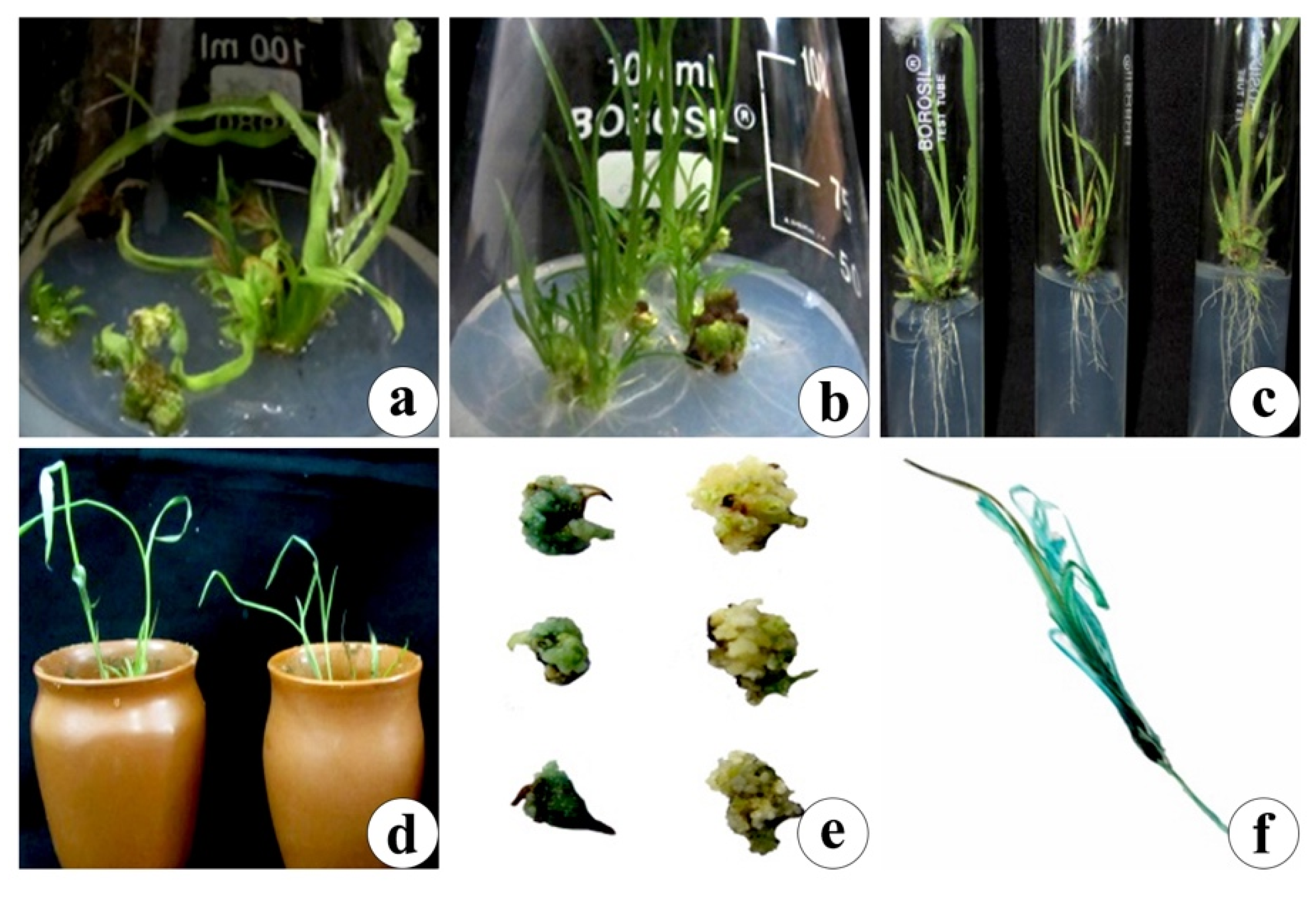
3.4. Co-Cultivation, Selection and Regeneration

The infected calli were transferred again to the callus maintenance medium, on which the non-transformed calli showed necrosis and poor growth. The selection medium ensured presence of antibiotics (Kan and Cef) at increasing concentrations at each step which was in agreement with studies by Karthikeyan, et al. [46], who reported improved efficiency of transformation by 15.33% in Indica rice (Oryza sativa L.) cultivar ADT 43. Calli that successfully endured the shock were transferred to the regeneration medium (Figure 5a,b) and successively to the rooting medium (Figure 5c). The regenerated plantlets were entrenched in greenhouse environment (Figure 5d).

3.5. GUS Histochemical Assay, Transformation Efficiency and Field Transfer

After co-cultivation and regeneration, histochemical GUS assay activity was performed on infected callus and plantlets, respectively. Distinct blue coloration confirmed the activity of gus gene, indicating proper insertion of the T-DNA. A noticeable color difference between transformed and non-transformed calli could clearly be seen (Figure 5e). While the non-transformed calli and plantlets failed to obtain any color, the transformed calli and plantlets showed a striking blue color when incubated at 37 °C overnight, ratifying the transformation process (Figure 5e,f). Maximum transformation efficiency (44%) was obtained with strain EHA 105 when pretreated with surfactant (Pluronic acid F-68, 0.5%). These findings concorded with those reported for other millets [47].
The putative transformants generated were then successfully hardened under greenhouse conditions and their seeds were collected on maturity.

4. Conclusions

To summarize, the present report is the first attempt to develop an efficient protocol for modification of this ignored millet crop genetically. The protocol was designed by optimizing several parameters, which influence Agrobacterium infection. Profuse callus growth was found on MS medium enriched with 3.0 mg/L 2,4-D. EHA 105 and LBA 4404 strains were selected to obtain maximum transformation efficiency. Pretreatment with antinecrotic mixture, the addition of surfactants and lower infection temperature significantly improved transformation efficiency. The calli pieces co-cultivated at 24 °C for 3 days (EHA 105) and 5 days (LBA 4404), respectively, further improved transformation. The barrier for monocot transformation was overcome by the addition of phenolic supplement and acetosyringone in both infection and co-cultivation media. The transformation was confirmed with GUS histochemical assay. This protocol can be used eventually to transform this orphan crop with quality improving genes and traits that impart abiotic resistance to kodo millet, so it can thrive well in the era of nutritionally challenged soils and threatened environment conditions, becoming an excellent alternate for the counterpart cereals.

Author Contributions

Conceptualization, S.L.K. and S.K. (Sumita Kachhwaha); methodology, R.B. and P.P.A.; validation, A.K.-C. and P.P.A.; formal analysis, R.J.; resources, S.K. (Sumita Kachhwaha); writing—original draft preparation, R.B., P.P.A. and R.J.; writing—review and editing, A.K.-C. and S.K. (Sumita Kachhwaha); visualization, S.K. (Sumita Kachhwaha); supervision, S.L.K. All authors have read and agreed to the published version of the manuscript.

Funding

This research received no external funding.

Institutional Review Board Statement

Not applicable.

Informed Consent Statement

Not applicable.

Acknowledgments

Ritika Bhat and Prem Prakash Asopa thank CSIR for the awarding of SRF.

Conflicts of Interest

The authors declare no conflict of interest. The funders had no role in the design of the study; in the collection, analyses, or interpretation of data; in the writing of the manuscript, or in the decision to publish the results.

Appendix A

Table A1. Effect of cell density on transformation efficiency of callus derived from seeds of P. scrobiculatum L.
Cell Density (OD600)Callus InfectedKan Resistant CalliKan Resistant PlantsTransformation
Efficiency (%)
EHALBAEHALBAEHALBAEHALBA
0.590902218535.53.3
0.890903526141015.511.1
1909020322132.214.4
1.59090950202.22
Table A2. Effect of pretreatment with Antinecrotic mix on transformation efficiency of callus derived from seeds of P. scrobiculatum L.
Antinecrotic
Mixture (AM)
Callus InfectedKan Resistant CalliKan Resistant PlantsTransformation
Efficiency (%)
EHALBAEHALBAEHALBAEHALBA
AM 18080169657.56.2
AM 28080221710912.511.2
AM 380803632181322.516.2
Table A3. Effect of infection time (mins) on transformation efficiency of callus derived from seeds of P. scrobiculatum L.
Infection Time (min)Callus InfectedKan Resistant CalliKan Resistant PlantsTransformation
Efficiency (%)
EHALBAEHALBAEHALBAEHALBA
5105105200000
1010510516128107.69.5
151051052118171316.112.38
201051053228262124.7620
25105105610322.81.9
Table A4. Effect of AS concentration in infection medium on transformation efficiency of callus derived from seeds of P. scrobiculatum L.
AS concentration (µM)Callus InfectedKan Resistant CalliKan Resistant PlantsTransformation
Efficiency (%)
EHALBAEHALBAEHALBAEHALBA
10090902719828.82.2
1509090362517918.810
20090904232251827.720
25090902538142115.523.3
30090906110000
Table A5. Effect of AS concentration in co-cultivation medium on transformation efficiency of callus derived from seeds of P. scrobiculatum L.
AS concentration (µM)Callus InfectedKan Resistant CalliKan Resistant PlantsTransformation
Efficiency (%)
EHALBAEHALBAEHALBAEHALBA
09090211512613.36.6
2590903832211923.321.1
509090514327213023.3
10090902236141715.518.8
2009090116313.31.1
Table A6. Effect of Tween- 20 on transformation efficiency of callus derived from seeds of P.scrobiculatum L.
Tween-20 Conc. (%)Callus InfectedKan Resistant CalliKan Resistant PlantsTransformation
Efficiency (%)
EHALBAEHALBAEHALBAEHALBA
0 (Control)7575126425.32.6
0.017575261516921.312
0.0575753327221829.324
0.17575383124213228
0.575754239322942.638.6
Table A7. Effect of Pluronic acid F-68 on transformation efficiency of callus derived from seeds of P. scrobiculatum L.
Pluronic Acid F-68 Conc. (%)Callus InfectedKan Resistant CalliKan Resistant PlantsTransformation
Efficiency (%)
EHALBAEHALBAEHALBAEHALBA
0 (Control)7575147536.64
0.0175752918171022.613.3
0.0575753528251933.325.3
0.175754133282337.330.6
0.57575454033304440
Table A8. Effect of incubation temperature on transformation efficiency of callus derived from seeds of P. scrobiculatum L.
Incubation
Temp. (°C)
Callus InfectedKan Resistant CalliKan Resistant PlantsTransformation
Efficiency (%)
EHALBAEHALBAEHALBAEHALBA
2010510571201.90
22105105231613812.37.6
241051053627241722.816.1
26105105211012411.43.8
28105105000000

References

  1. Kothari, S.; Kumar, S.; Kothari, A.; Watanabe, K. Applications of biotechnology for improvement of millet crops: Review of progress and future prospects. Plant Biotechnol. 2005, 22, 81–88. [Google Scholar] [CrossRef]
  2. Pokharia, A.; Kharakwal, J.; Srivastava, A. Archaeobotanical evidence of millets in the Indian subcontinent with some observations on their role in the Indus civilization. J. Archaeol. Sci. 2014, 42, 442–455. [Google Scholar] [CrossRef]
  3. Kothari-Chajer, A.; Sharma, M.; Kachhwaha, S.; Kothari, S. Micronutrient optimization results into highly improved in vitro plant regeneration in kodo (Paspalum scrobiculatum L.) and finger (Eleusine coracana (L.) Gaertn.) millets. Plant Cell Tiss. Org. Cult. 2008, 94, 105–112. [Google Scholar] [CrossRef]
  4. Shahidi, F.; Chandrasekara, A. Millet grain phenolics and their role in disease risk reduction and health promotion: A review. J. Funct. Foods 2013, 5, 570–581. [Google Scholar] [CrossRef]
  5. Sood, P.; Singh, R.; Prasad, M. Millets genetic engineering: The progress made and prospects for the future. Plant Cell Tiss. Org. Cult. 2019, 137, 421–439. [Google Scholar] [CrossRef]
  6. Geervani, P.; Eggum, B. Nutrient composition and protein quality of minor millets. Plant Foods Hum. Nutr. 1989, 39, 201–208. [Google Scholar] [CrossRef]
  7. Chaudhary, N.; Vyas, S. Effect of germination on proximate composition and anti nutritional factor of millet (ragi) based premixes. Int. J. Food Nutr. Sci. 2014, 3, 72–77. [Google Scholar]
  8. Taylor, J.; Schober, T.; Bean, S. Novel food and non-food uses for sorghum and millets. J. Cereal Sci. 2006, 44, 252–271. [Google Scholar] [CrossRef]
  9. Michaelraj, P.; Shanmugam, A. A study on millets based cultivation and consumption in India. Int. J. Mark. Financ. Serv. Manag. Res. 2013, 2, 49–58. [Google Scholar]
  10. de Wet, J.; Brink, D.; Rao, K.; Mengesha, M. Diversity in kodo millet, Paspalum scrobiculatum. Econ. Bot. 1983, 37, 159–163. [Google Scholar] [CrossRef] [Green Version]
  11. Hegde, P.; Rajasekaran, N.; Chandra, T. Effects of the antioxidant properties of millet species on oxidative stress and glycemic status in alloxan-induced rats. Nutr. Res. 2005, 25, 1109–1120. [Google Scholar] [CrossRef]
  12. Devakumar, C.; Chhonkar, P. Integration of Millets in Fortified Foods, 54th ed.; National Academy of Sciences: New Delhi, India, 2012. [Google Scholar]
  13. Dwivedi, S.; Upadhyaya, H.; Senthilvel, S.; Hash, C.; Fukunaga, K.; Diao, X.; Santra, D.; Baltensperge, D.; Prasad, M. Millets: Genetic and genomic resources. Plant Breed. Rev. 2012, 35, 247–375. [Google Scholar]
  14. Nadolska-Orczyk, A.; Orczyk, W.; Przetakiewicz, A. Agrobacterium-mediated transformation of cereals—From technique development to its application. Acta Physiol. Plant. 2000, 22, 77–88. [Google Scholar] [CrossRef]
  15. Tzfira, T.; Citovsky, V. Agrobacterium-mediated genetic transformation of plants: Biology and biotechnology. Curr. Opin. Biotechnol. 2006, 17, 147–154. [Google Scholar] [CrossRef] [PubMed]
  16. Dosad, S.; Chawla, H. In vitro plant regeneration and transformation studies in millets: Current status and future prospects. Indian J. Plant Physiol. 2016, 21, 239–254. [Google Scholar] [CrossRef]
  17. Sood, P.; Bhattacharya, A.; Joshi, R.; Gulati, A.; Chanda, S.; Sood, A. A method to overcome the waxy surface, cell wall thickening and polyphenol induced necrosis at wound sites-the major deterrents to Agrobacterium mediated transformation of bamboo, a woody monocot. J. Plant Biochem. Biot. 2014, 23, 69–80. [Google Scholar] [CrossRef]
  18. Plaza-Wüthrich, S.; Tadele, Z. Millet improvement through regeneration and transformation. Biotechnol. Mol. Biol. Rev. 2012, 7, 48–61. [Google Scholar] [CrossRef]
  19. Smith, R.; Grando, M.; Li, Y.; Seib, J.; Shatters, R. Transformation of bahiagrass (Paspalum notatum Flugge). Plant Cell Rep. 2002, 20, 1017–1021. [Google Scholar] [CrossRef]
  20. Gondo, T.; Tsuruta, S.; Akashi, R.; Kawamura, O.; Hoffmann, F. Green, herbicide-resistant plants by particle inflow gun-mediated gene transfer to diploid bahiagrass (Paspalum notatum). J. Plant Physiol. 2005, 162, 1367–1375. [Google Scholar] [CrossRef]
  21. Wu, X.; Shi, H.; Chen, X.; Liu, Y.; Guo, Z. Establishment of Agrobacterium-mediated transformation of seashore paspalum (Paspalum vaginatum O. Swartz). Vitr. Cell. Dev. Biol. Plant 2018, 54, 545–552. [Google Scholar] [CrossRef]
  22. Murashige, T.; Skoog, F. A revised medium for rapid growth and bio assays with Tobacco tissue cultures. Physiol. Plant. 1962, 15, 473–497. [Google Scholar] [CrossRef]
  23. Yadav, T.; Kothari, S.; Kachhwaha, S. Optimization of Agrobacterium-Mediated Genetic Transformation and Regeneration of Transgenic Plants in Indian Cultivar of Barley (Hordeum vulgare L. cv. BL 2). Proc. Natl. Acad. Sci. India Sect. B Biol. Sci. 2013, 83, 255–264. [Google Scholar] [CrossRef]
  24. Jefferson, R.; Kavanagh, T.; Bevan, M. GUS fusions: Beta-glucuronidase as a sensitive and versatile gene fusion marker in higher plants. EMBO J. 1987, 6, 3901–3907. [Google Scholar] [CrossRef] [PubMed]
  25. Sharma, M.; Kothari-Chajer, A.; Jagga-Chugh, S.; Kothari, S. Factors influencing Agrobacterium tumefaciens-mediated genetic transformation of Eleusine coracana (L.) Gaertn. Plant Cell Tiss. Org. Cult. 2011, 105, 93–104. [Google Scholar] [CrossRef]
  26. Botti, C.; Vasil, I. Plant regeneration by somatic embryogenesis from parts of cultured mature embryos of Pennisetum americanum (L.) K. Schum. Z. Pflanzenphysiol. 1983, 111, 319–325. [Google Scholar] [CrossRef]
  27. Kachhwaha, S.; Varshney, A.; Kothari, S. Somatic embryogenesis and long term high plant regeneration from barley (Hordeum vulgare L.) using picloram. Cereal Res. Commun. 1997, 25, 117–126. [Google Scholar] [CrossRef]
  28. Chin, D.; Mishiba, K.; Mii, M. Agrobacterium-mediated transformation of protocorm-like bodies in Cymbidium. Plant Cell Rep. 2007, 26, 735–743. [Google Scholar] [CrossRef]
  29. Wang, M.; Li, Z.; Matthews, P.; Upadhyaya, N.; Waterhouse, P. Improved vectors for Agrobacterium tumefaciens-mediated transformation of monocot plants. Int. Symp. Biotechnol. Trop. Subtrop. Species 1997, 461, 401–408. [Google Scholar] [CrossRef]
  30. Wang, M.; Li, P.; Li, C.; Pan, Y.; Jiang, X.; Zhu, D.; Zhao, Q.; Yu, J. SiLEA14, a novel atypical LEA protein, confers abiotic stress resistance in foxtail millet. BMC Plant Biol. 2014, 14, 290. [Google Scholar] [CrossRef]
  31. Sood, P.; Singh, R.K.; Prasad, M. An efficient Agrobacterium-mediated genetic transformation method for foxtail millet (Setaria italica L.). Plant Cell Rep. 2020, 39, 511–525. [Google Scholar] [CrossRef]
  32. Hiei, Y.; Ohta, S.; Komari, T.; Kumashiro, T. Efficient transformation of rice (Oryza sativa L.) mediated by Agrobacterium and sequence analysis of the boundaries of the T-DNA. Plant J. 1994, 6, 271–282. [Google Scholar] [CrossRef] [PubMed] [Green Version]
  33. Enríquez-Obregón, G.; Prieto-Samsónov, D.; Gustavo, A.; Pérez, M.; Selman-Housein, G.; Vázquez-Padrón, R. Agrobacterium-mediated Japonica rice transformation: A procedure assisted by an antinecrotic treatment. Plant Cell Tiss. Org. Cult. 1999, 59, 159–168. [Google Scholar] [CrossRef]
  34. Hensel, G.; Kastner, C.; Oleszczuk, S.; Riechen, J.; Kumlehn, J. Agrobacterium-mediated gene transfer to cereal crop plants: Current protocols for barley, wheat, triticale, and maize. Int. J. Plant Genom. 2009, 2009, 1–9. [Google Scholar] [CrossRef] [PubMed] [Green Version]
  35. Kumar, A.; Reddy, B.; Ramaiah, B.; Sharma, R. Heterosis in white-grained grain mold resistant sorghum hybrids. J. SAT Agric. Res. 2011, 9, 1–6. [Google Scholar]
  36. Bayer, G.; Yemets, A.; Blume, Y. Obtaining the transgenic lines of finger millet Eleusine coracana (L.). with dinitroaniline resistance. Cytol. Genet. 2014, 48, 139–144. [Google Scholar] [CrossRef]
  37. Ramadevi, R.; Rao, K.; Reddy, V. Agrobacterium tumefaciens-mediated genetic transformation and production of stable transgenic pearl millet (Pennisetum glaucum [L.] R. Br.). Vitr. Cell. Dev. Biol. Plant 2014, 50, 392–400. [Google Scholar] [CrossRef]
  38. Ziemienowicz, A. Agrobacterium-mediated plant transformation: Factors, applications and recent advances. Biocatal. Agric. Biotechnol. 2014, 3, 95–102. [Google Scholar] [CrossRef]
  39. Yadav, T.; Kachhwaha, S.; Kothari, S. Efficient in vitro plant regeneration and generation of transgenic plants in barley (Hordeum vulgare L.) using particle bombardment. J. Plant Biochem. Biot. 2013, 22, 202–213. [Google Scholar] [CrossRef]
  40. Ceasar, S.; Ignacimuthu, S. Agrobacterium-mediated transformation of finger millet (Eleusine coracana (L.) Gaertn.) using shoot apex explants. Plant Cell Rep. 2011, 30, 1759–1770. [Google Scholar] [CrossRef]
  41. Tyagi, H.; Rajasubramaniam, S.; Dasgupta, I. Regeneration and Agrobacterium-mediated transformation of a popular indica rice variety, ADT39. Curr. Sci. 2007, 93, 678–683. [Google Scholar]
  42. Cheng, M.; Lowe, B.; Spencer, T.; Ye, X.; Armstrong, C. Factors influencing Agrobacterium-mediated transformation of monocotyledonous species. Vitr. Cell. Dev. Biol. Plant 2004, 40, 31–45. [Google Scholar] [CrossRef]
  43. Fullner, K.; Nester, E. Temperature affects the T-DNA transfer machinery of Agrobacterium tumefaciens. J. Bacteriol. 1996, 178, 1498–1504. [Google Scholar] [CrossRef] [PubMed] [Green Version]
  44. Dillen, W.; De Clereq, J.; Kapila, J.; Zambre, M. The effect of temperature on Agrobacterium tumefaciens-mediated gene transfer to plants. Plant J. 1997, 12, 1459–1463. [Google Scholar] [CrossRef]
  45. Hiei, Y.; Ishida, Y.; Komari, T. Progress of cereal transformation technology mediated by Agrobacterium tumefaciens. Front. Plant Sci. 2014, 5, 1–11. [Google Scholar] [CrossRef] [Green Version]
  46. Karthikeyan, A.; Shilpha, J.; Pandian, S.; Ramesh, M. Agrobacterium-mediated transformation of indica rice cv. ADT 43. Plant Cell Tiss. Org. Cult. 2012, 109, 153–165. [Google Scholar] [CrossRef]
  47. Mundada, P.; Umdale, S.; Ahire, M.; Kumar, S.; Nikam, T. Transgenic Finger Millet [Eleusine coracana (L.) Gaertn.] for Crop Improvement. In Genetically Modified Crops; Springer: Singapore, 2021; pp. 67–77. [Google Scholar]
Figure 1. Genetic map of Agrobacterium tumefaciens strains (a) EHA 105 harboring plasmid pCNL 56, and (b) LBA 4404 harboring plasmid pCAMBIA 2300.
Agronomy 11 01104 g001
Figure 2. Response of GPUK-3 seeds on callus (a) initiation; (b) maintenance and (c) proliferation medium.
Agronomy 11 01104 g002
Figure 3. Antibiotic sensitivity of calli cultured on MS + 0.2 mg/L 2,4-D medium supplemented (a) without (0 mg/L) and with (b) 10 mg/L; (c) 20 mg/L and (d) 50 mg/L kanamycin.
Agronomy 11 01104 g003
Figure 4. Effect of (a) Bacterial density; (b) Composition of antinecrotic mixture; (c) Concentration of Tween-20 in infection medium; (d) Concentration of Pluronic acid F-68 in infection medium; (e) Infection time; Acetosyringone concentration in (f) infection and (g) co-cultivation medium; and (h) Incubation temperature on GUS expression.
Agronomy 11 01104 g004
Figure 5. (a,b) Plantlet Regeneration on MS + 0.5 mg/l NAA + 2 mg/l BAP; (c) Rooting of regenerated shoots on MS + 0.5 mg/l PAA; (d) Regenerated plants transferred to pots and (e,f) GUS histochemical assay.
Agronomy 11 01104 g005
Table 1. Details of culture medium and antibiotics used for growth of different Agrobacterium tumefaciens strains.
StrainCulture MediumAntibiotics
EHA 105 (pCNL 56)Luria-Bertini (LB) broth50 mg/L Kan
LBA 4404 (pCAMBIA 2300)Yeast extract broth (YEB)50 mg/L Kan + 20 mg/L (Rifampicin) Rif + 50 mg/L Streptomycin (Strep)
Table 2. Composition of different types of antinecrotic mixtures (AM) used for pretreatment of the calli.
Antinecrotic Mixture (AM)L-Cysteine
(mg/L)
Silver Nitrate
(mg/L)
Ascorbic Acid
(mg/L)
AM 11051
AM 21052.5
AM 31032.5
Table 3. Screening of morphogenetic competence among different varieties of P. scrobiculatum L. cultured on MS medium containing different concentrations of 2,4-D.
Variety2,4-D Conc.
(mg/L)
Callus Response
(%)
Avg. No. of Regenerated Shoots/Explant
(Mean ± SE)
TNAU 861281.2 ± 0.2
2453.0 ± 0
3624.4 ± 1.1
4202.3 ± 0.8
RK-390-251130
2170
3221.0 ± 0.2
4200
RK 151200
2341.1 ± 0.8
3352.3 ± 0.5
4300
GPUK-31342.1 ± 0.3
2573.2 ± 1.1
3786.8 ± 1.5
4322.5 ± 0
RBK-1551180
2331.5 ± 0.7
3281.3 ± 0.5
4251.2 ± 0.5
Publisher’s Note: MDPI stays neutral with regard to jurisdictional claims in published maps and institutional affiliations.

