Article

Efficient Micropropagation of Genetically Stable Panax ginseng Meyer by Somatic Embryogenesis

1
Department of Herbal Crop Research, National Institute of Horticultural and Herbal Science, Rural Development Administration, Eumseong 27709, Republic of Korea
2
Department of Horticultural Biotechnology, Kyung Hee University, Yongin 17104, Republic of Korea
3
Department of Crop Science and Biotechnology, Dankook University, Cheonan 31116, Republic of Korea
*
Author to whom correspondence should be addressed.
Submission received: 23 March 2023 / Revised: 14 April 2023 / Accepted: 14 April 2023 / Published: 17 April 2023

Abstract

:
Panax ginseng Meyer is a valuable medicinal crop. However, the species’ propagation is limited by its long reproductive cycle and low seed yield. The present study focused on P. ginseng plant regeneration via somatic embryogenesis and evaluated the genetic stability of regenerated plantlets. We assessed the effects of carbon source type and concentration on somatic embryo induction, maturation, and germination. Somatic embryogenesis was optimal in Murashige and Skoog (MS) medium supplemented with 5% sucrose; however, maturation peaked in 1/2 MS containing low concentrations of sucrose ranging from 1 to 2%. Germination and plant regeneration were optimal in germination medium supplemented with 2% sucrose based on high germination rates, efficient plantlet production, and balanced growth characteristics. Molecular marker analysis suggested that the genetic fidelity of the regenerated plants was comparable with that of the control. High-performance liquid chromatography (HPLC) analysis showed that in vitro-grown roots (IGRs) accumulated more ginsenoside than those of the control, but the ginsenoside content of 2 year old IGRs was similar to that of the controls after acclimatization. Our study provides valuable insights into the optimization of genetically stable micropropagation and could promote the distribution of superior P. ginseng cultivars with high product yields and quality.

1. Introduction

Panax ginseng Meyer (Korean or Asian ginseng) is a valuable commercial crop in Korea, the United States, China, Japan, and Vietnam [1]. Triterpenoid saponins, including ginsenoside, are the major pharmaceutical components in ginseng and are mainly classified into dammarane, including protopanaxatriol (PPT) and protopanaxadiol (PPD), and oleanane types [2]. Extensive pharmacological and chemical studies have shown that these compounds have potent therapeutic effects on various diseases, including neurodegenerative [3] and cardiovascular diseases [4], diabetes [5], and cancer [6]. Therefore, the international demand for and economic value of ginseng root are very high.
Since the vegetative propagation of P. ginseng, such as through cuttage, is difficult to establish, conventional propagation depends on seed production [7]. However, over three years of cultivation are required for flowering and setting seeds [8], and adult P. ginseng plants (commonly over 4 years old) can only produce 40–60 seeds per year [9]. These characteristics have inhibited the development and propagation of new cultivars. P. ginseng cv. Cheonryang, developed in 2011, showed high yield characteristics and salt tolerance [10]; however, its agricultural application remains limited by its low propagation efficiency [7]. Micropropagation via somatic embryogenesis may be an effective alternative to enhance the propagation rate of P. ginseng cv. Cheonryang.
In general, the development of in vitro tissue is affected not only by endogenous conditions, including genotype, but also external conditions, such as medium composition [11]. As they provide energy [12] and regulate osmotic pressure [13], carbohydrates are essential components of culture medium that can greatly affect shoot regeneration, somatic embryogenesis, and in vitro rooting [14,15,16].
Regenerated plants derived from tissue culture are often characterized by physiological and genetic alterations, such as gene mutation [17]. Somaclonal variation is useful for creating genetic mutations but highly disadvantageous in the micropropagation of genetically uniform clones [18]. Therefore, even though plants regenerated via direct somatic embryogenesis have a low probability of mutation at the DNA and chloroplast level [19], the evaluation of genetic fidelity—through, for example, molecular markers of DNA sequence variation [20,21]—remains essential to ensuring true-to-type regeneration.
The somatic embryogenesis of P. ginseng has been widely studied; Explants from various organs, such as the root [22], petiole [23], and zygotic embryo [24,25,26,27], have been reported to induce somatic embryos. Furthermore, studies on the effects of the basal medium on the induction, maturation, and germination of somatic embryos have recently been published [28]. However, few studies have assessed the effect of the carbon source in the culture medium; no studies have investigated the post-effect of sucrose concentration in the germination medium on plant regeneration and plantlet formation. Additionally, only a few studies have assessed the genetic fidelity and ginsenoside content of the in vitro-grown roots (IGRs) of P. ginseng derived from somatic embryogenesis.
The present study aimed to describe the effect of carbon source on somatic embryo induction, maturation, germination, and regeneration in P. ginseng cv. Cheonryang. After the successful acclimatization of IGRs, their genetic fidelity was assessed using molecular markers. Further, the ginsenoside content of IGRs before and after acclimatization was compared to that in control 1 year old ginseng roots.

2. Materials and Methods

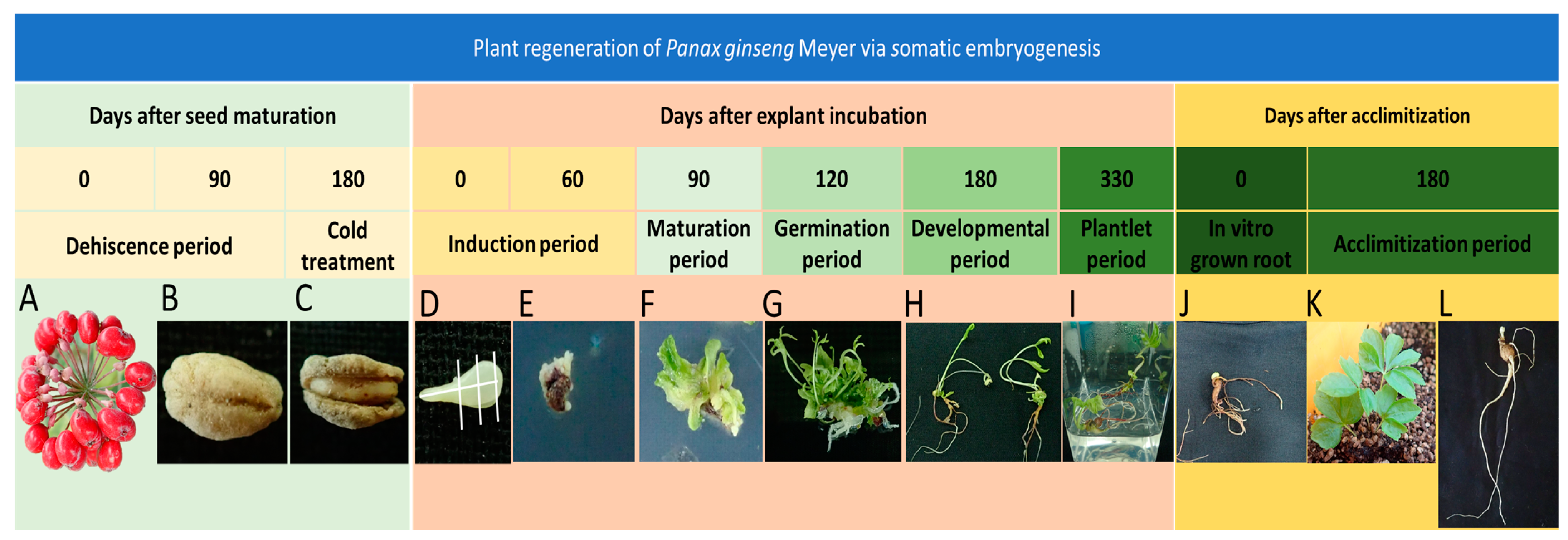
2.1. Plant Materials and Seed Sterilization

The fruits of P. ginseng cv. Cheonryang (Figure 1A) were collected in the field at the Department of Herbal Crop Research, National Institute of Horticultural and Herbal Science, Rural Development Administration (Eumseong, Republic of Korea; 36°94′28.4″ N, 127°74′86.6″ E). Seed dehiscence and cold treatment (Figure 1B,C) were performed as previously described [8]. The seed coat was removed from the matured dehiscent seed. The seeds were soaked in 2% (v/v) sodium hypochlorite for 10 min and surface sterilized by agitation for 5 min to reduce contamination. The seeds were disinfected in 70% (v/v) ethyl alcohol for 1 min and washed with sterile distilled water (dH2O). Secondary sterilization was performed with 2% (v/v) sodium hypochlorite treatment over 20 min and 4–5 washes with sterile dH2O. After disinfection, the seeds were dried for a few minutes to avoid contamination.

2.2. In Vitro Regeneration Protocol and Culture Conditions

Mature zygotic embryos were isolated from the endosperm under aseptic conditions. The top and bottom portions of the embryo were excised (Figure 1D), yielding between four and six explants (approximately 1 cm × 1 cm in size) per seed. Explants were inoculated in somatic embryo induction medium consisting of Murashige and Skoog (MS) basal medium [29] supplemented with various carbon sources (fructose, glucose, maltose, and sucrose) and 8 g/L agar for 60 days without subculture (Figure 1E). Somatic embryos were transferred to the maturation medium (1/2 MS supplemented with various sucrose concentrations and 8 g/L agar) for 30 days (Figure 1F). Thereafter, somatic embryos were transferred to germination medium—1/2 Schenk and Hildebrandt (SH) basal medium [30] supplemented with various sucrose concentrations, 9 g/L agar, and 10 mg/L gibberellic acid (GA3) (Figure 1G). Shoots derived from germination media were subcultured in elongation medium supplemented with 20 g/L sucrose, 5 g/L agar, and 5 g/L activated charcoal in 1/2 SH for root development. Well-developed plantlets (Figure 1H) were transferred to growth medium (1/3 SH medium supplemented with 5 g/L gelrite and 20 g/L sucrose) and grown until the plantlet leaves senesced (Figure 1I). IGRs (Figure 1J) were soaked with 25 mg/L GA3 before being transferred to soil. Most shoots sprouted within 30 days (Figure 1K). After 180 days of cultivation, the shoots were defoliated, and healthy 2 year old IGRs with rhizomes were obtained. (Figure 1L). All basal media, carbon sources, agar, and activated charcoal were obtained from Duchefa Biochemie (Haarlem, The Netherlands). All culture media were adjusted to pH 5.8 before being autoclaved at 121 °C for 15 min. Petri dishes were sealed with parafilm, and cultures were incubated at 23 ± 2 °C under a 16/8 h light/dark cycle using 24 µmol m−2 s−1 white fluorescent light.

2.3. Somatic Embryogenesis

To evaluate the effect of carbon source on somatic embryo induction, 30 g/L each of four carbon source types (fructose, glucose, maltose, and sucrose) were added to induction media before autoclaving. Subsequently, the influence of various sucrose concentrations (10, 30, 50, 70, and 90 g/L) on somatic embryo induction was evaluated. Ten explants were placed in a Petri dish containing somatic embryo induction medium, with five replicates performed per treatment. After 60 days, images were recorded for each treatment using a binocular microscope (S8AP0, Leica, Wetzlar, Germany) to calculate the induction rate and number of somatic embryos as follows: somatic embryo induction rate (%) = number of explants containing somatic embryos/total number of explants × 100; mean number of somatic embryos per explant = ∑ of somatic embryos per explant/number of explants from which at least one somatic embryo was induced.
The embryo formation capacity (EFC) index was calculated as follows [31]: EFC = somatic embryo induction rate × mean number of somatic embryos per explant/100.

2.4. Maturation and Germination of Somatic Embryos

To determine the effect of sucrose concentration on somatic embryo maturation, explants with immature-stage somatic embryos were transferred to maturation medium containing various sucrose concentrations (10, 20, 30, 40, and 50 g/L). We placed ten explants containing ~25 somatic embryos in maturation medium, with three replicates per treatment. After 30 days of culture, somatic embryo stages were classified as globular, heart-shaped, torpedo, or cotyledonary using a binocular microscope (S8AP0, Leica) as previously described [27].
We placed ten explants containing ~50 somatic embryos in germination medium supplemented with various sucrose concentrations (10, 20, 30, and 40 g/L), 9 g/L agar, and 10 mg/L GA3 in 1/2 SH, with five replicates per treatment. After 30 days, images were recorded for each treatment to characterize explant and shoot morphology. Germination rate was calculated as follows: somatic embryo germination rate (%) = number of explants containing shoots/total number of explants × 100.

2.5. Plant Regeneration and Plantlet Conversion

We placed 20 shoots derived from the respective germination treatments in Petri dishes containing elongation medium (1/2 SH, 20 g/L sucrose, 5 g/L agar, and 5 g/L activated charcoal), with five replicates. After 60 days, we investigated the post-effect of germination medium on plant regeneration. Regenerated plants were classified into three morphological types based on the presence of shoots and roots as follows (Figure S1): W types consisted of whole plants with both shoots and roots, S types had only shoots, and R types had only roots with rhizomes.
We investigated the effect of the germination medium on plantlet conversion and morphology. The number of plantlets was calculated as number of W type + number of R type plantlets.
We also evaluated the length, diameter, and weight of the aerial and underground parts of ten plants from each treatment.

2.6. Acclimatization

After treatment with 25 mg/L GA3 for 1 h, hundreds of IGRs (450 ± 50 mg) were transferred to an artificial soil mixture of peat moss and perlite (3:1, v/v) at 25 ± 2 °C in a greenhouse. Simultaneously, P. ginseng cv. Cheonryang seeds were sown to generate control plants for molecular marker and ginsenoside analysis. After 60 days of transplantation, the sprouting rate was calculated as the percentage of the number of sprouted IGRs compared to the number of transplanted IGRs. When the shoots derived from IGRs were defoliated, 2 year old IGRs were harvested.

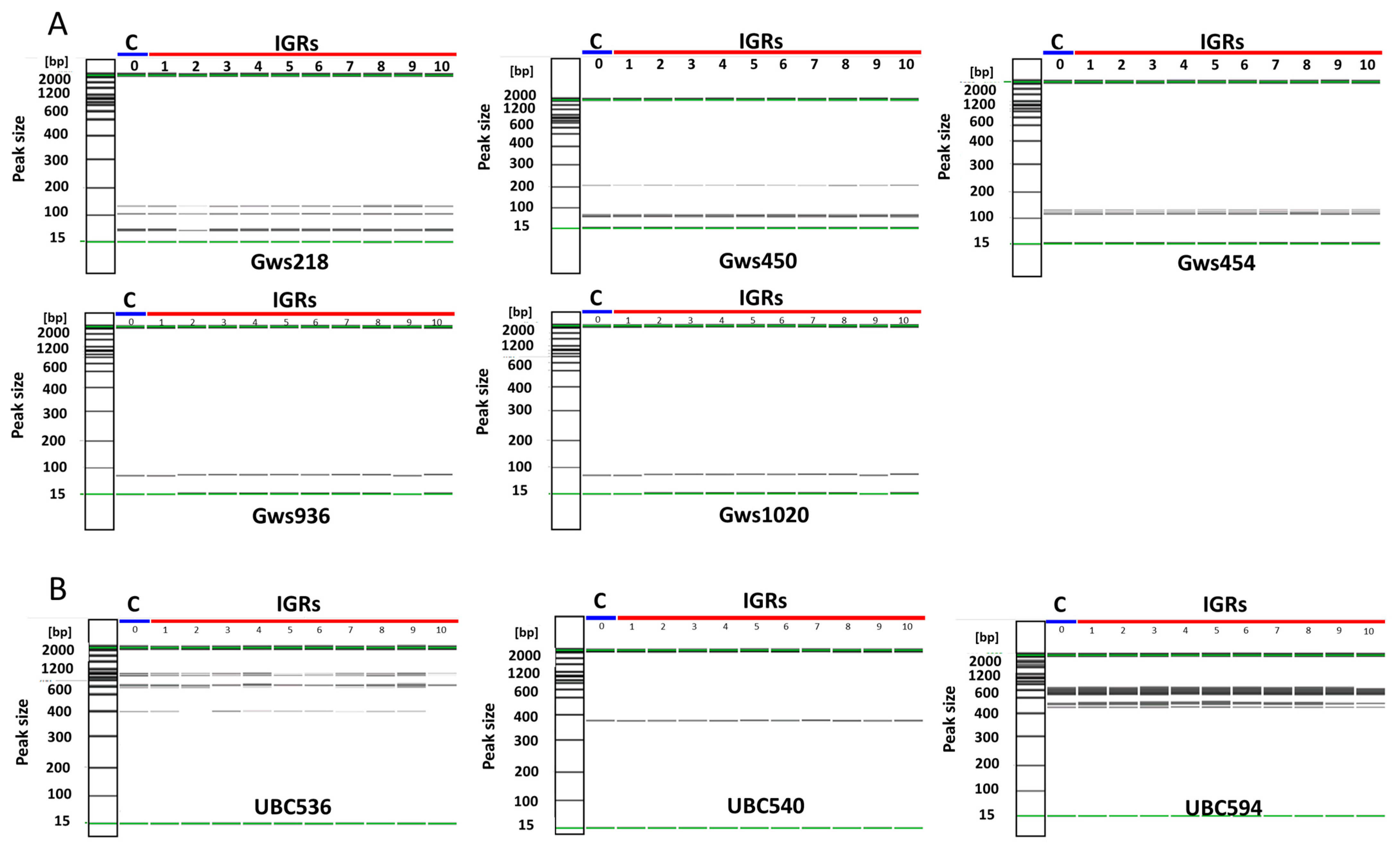
2.7. Determination of Genetic Homogeneity

Based on the method described by Kim et al. [23] and Jang et al. [32], polymerase chain reaction (PCR) was performed using simple sequence repeat (SSR) and random amplified polymorphic DNA (RAPD) primers (Table S1). Genomic DNA was extracted from regenerated and control plant leaves using a plant DNA extraction kit (Qiagen, Germantown, MD, USA) and quantified. The PCR products were analyzed on a QIAxcel (Qiagen) automatic capillary electrophoresis system.

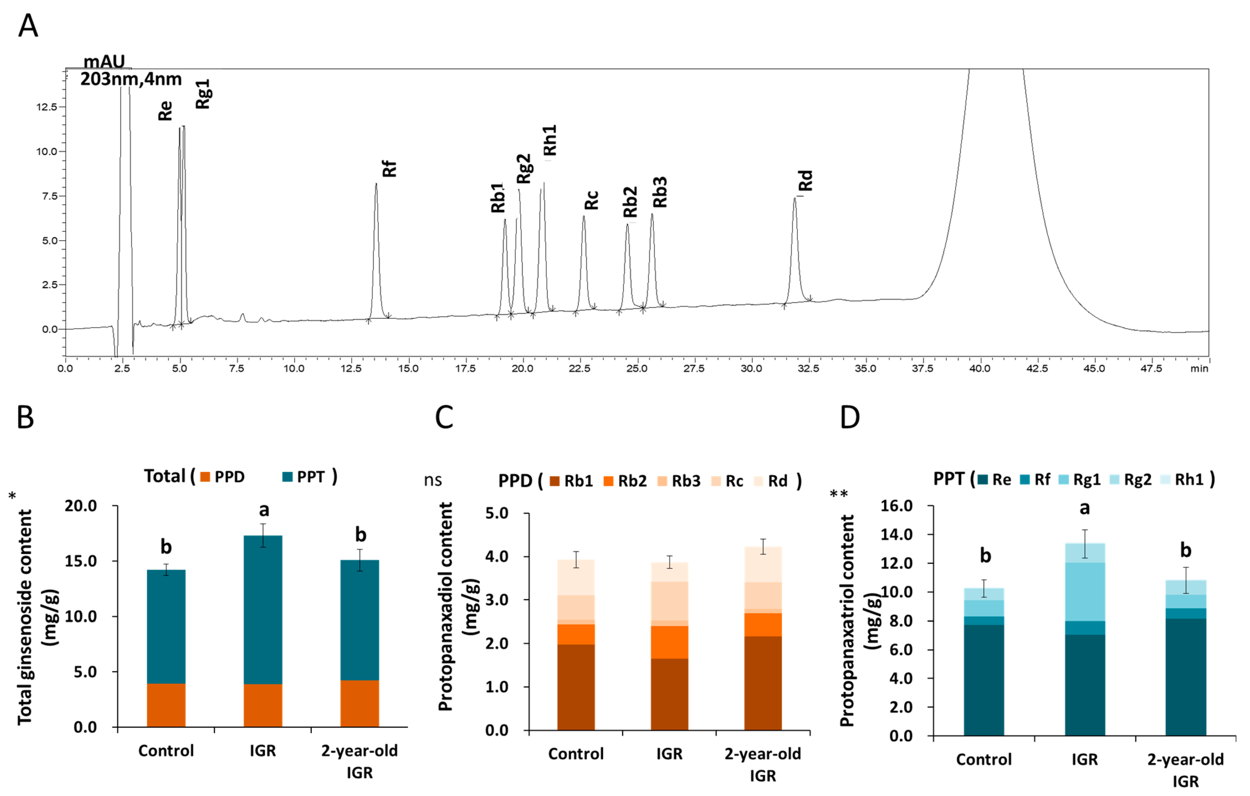
2.8. Ginsenoside Analysis

To analyze the ginsenoside content in IGRs, 2 year old IGRs, and controls (1 year old roots), they were freeze-dried and ground using a mortar and pestle to generate 20 powdered samples per treatment group. We analyzed the levels of ten types of ginsenoside, including PPD (Rb1, Rb2, Rb3, Rc, and Rd) and PPT types (Re, Rf, Rg1, Rg2, and Rh1). Ginsenoside content was calculated as previously described [23]. Each sample was immersed in 70% methyl alcohol and sonicated for 30 min. After centrifugation, the supernatant was purified by filtering through a 0.45 μm membrane filter (Agilent, Santa Clara, CA, USA). The purified sample was injected into a UPLC system (Nexera X2, Shimadzu., Kyoto, Japan) to measure the ginsenoside content in three repeated runs using a Halo RP-amide column (4.6 mm × 150 mm, 2.7 μm; Thermo Fisher Scientific, Wilmington, DE, USA) at 50 °C. The flow rate was set to 0.5–0.8 mL/min, and UV wavelength detection was set to 203 nm.

2.9. Statistical Analysis

All experiments were carried out in a randomized block design and replicated at least three times. Data are presented as the means ± standard error. Differences between groups were analyzed using a one-way analysis of variance (ANOVA) in R ver. 4.1.2 (R Foundation for Statistical Computing, Vienna, Austria) and means were compared using Duncan’s comparison test. Statistical significance was set at p ≤ 0.05.

3. Results

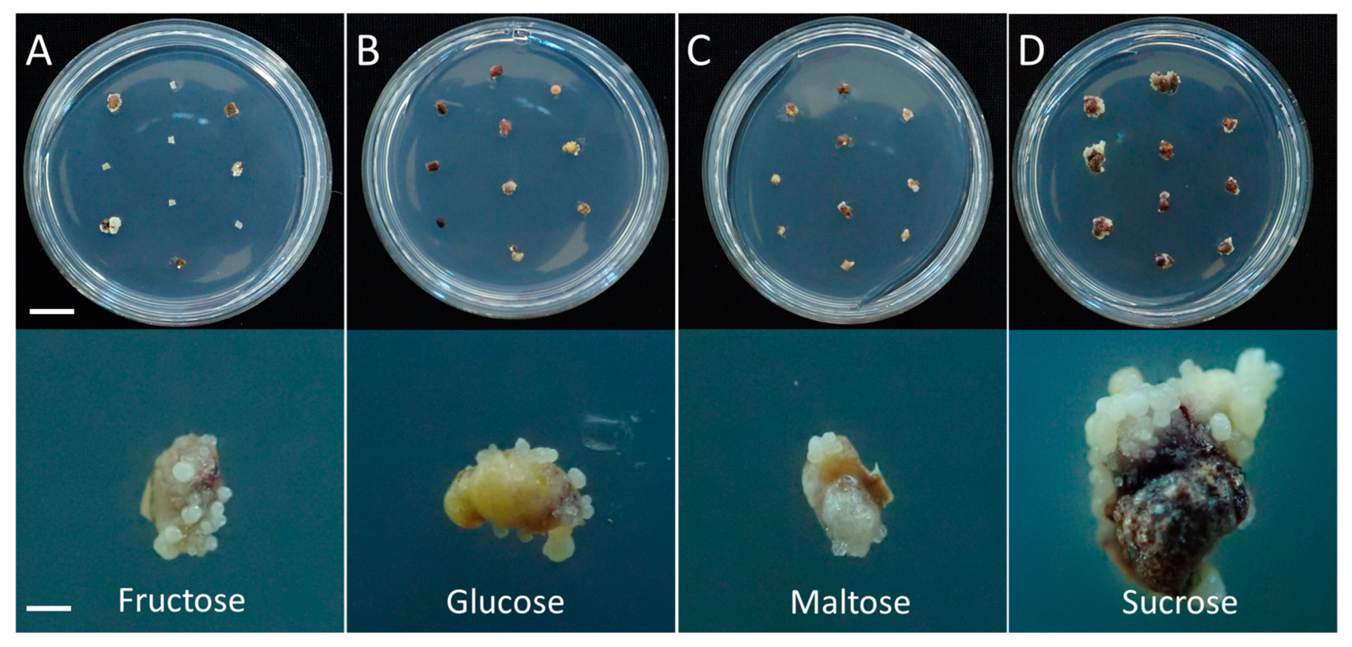
3.1. Effect of Carbon Source on Somatic Embryogenesis

Explants were cultured with four carbon source types (fructose, glucose, maltose, and sucrose) to determine the best carbon source for the somatic embryo induction medium. Somatic embryos were formed in all media, though with significant differences in induction rates and numbers of somatic embryos among carbon source types (Figure 2 and Table 1). The maximum somatic embryo induction rate was observed in medium containing sucrose (93.3%), followed by glucose (72.7%), maltose (66.3%), and fructose (42.3%, p ≤ 0.001). The mean number of somatic embryos was much higher in medium containing sucrose (16.2) than other carbon sources (9.6). The EFC was highest in medium with sucrose (15.1), followed by glucose (7.0), maltose (6.4), and fructose (4.1).
Explants were also cultured on somatic embryo induction media containing different sucrose concentrations. Although somatic embryogenesis occurred in all media regardless of sucrose concentration (Figure S2), clear differences were observed in the intensity of somatic embryo induction (Table 2). The somatic embryo induction rate was greatly affected (p ≤ 0.001) by sucrose concentration and increased from 72.4 to 97.7% for sucrose concentrations ranging from 1% to 5% and decreased for concentrations above 7%. The maximum mean number of somatic embryos was obtained in medium supplemented with 5% sucrose (23.8), followed by 3% (21.5), 7% (12.8), 9% (10.5), and 1% (8.4). The highest EFC index was 21.3 when cultured in a medium containing 5% sucrose, with the second highest achieved with 3% (18.5), and the lowest was observed at 1% (4.0).

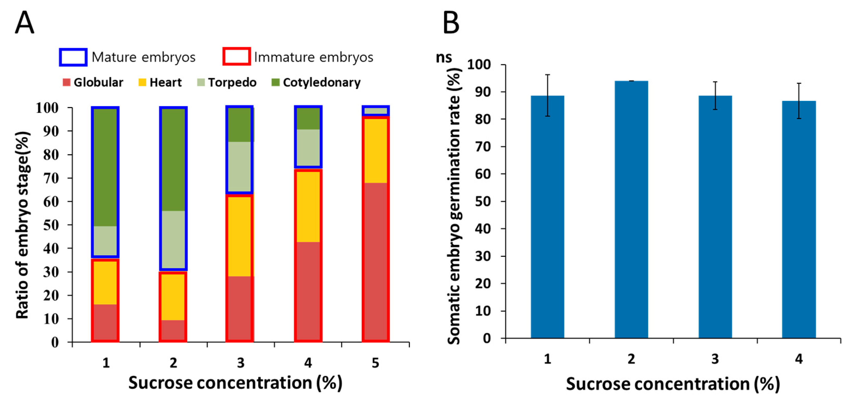
3.2. Effect of Sucrose Concentration on Somatic Embryo Maturation and Germination

Most somatic embryos in induction media were immature and required further maturation. We investigated the development of immature somatic embryos placed in maturation media containing different sucrose concentrations. Even within the same explants, various developmental stages could be observed (Figure 3A). The ratio of somatic embryo stages varied depending on sucrose concentration. The percentage of mature somatic embryos (torpedo and cotyledonary stage) was highest in maturation medium containing 2% sucrose (torpedo 25.3%; cotyledonary 44.0%), followed by 1% sucrose (torpedo 14.7%; cotyledonary 50.7%). As sucrose concentration increased, the proportion of the cotyledonary stage decreased. In addition, at sucrose concentrations above 3%, more than half of somatic embryos remained immature.
We evaluated the effect of sucrose concentration in germination medium on somatic embryo germination rates. Although the maximum germination rate was observed in 2% sucrose (94.0%), no significant differences were observed among treatments (Figure 3B).
The sucrose concentration in germination medium affected the morphology of both explants and shoots (Figure 4). Shoots from medium containing 1% sucrose were weak and occasionally necrotic (Table 3). The most vigorous shoot growth was observed with 2% sucrose; these shoots were also easily detached from explants. However, explants inoculated on germination medium containing over 3% sucrose became hard, making it difficult to separate the shoot from the explant, and the condition of the shoots was poor. We found that, as sucrose concentration increased, red pigment accumulated with secondary somatic embryogenesis in explants.

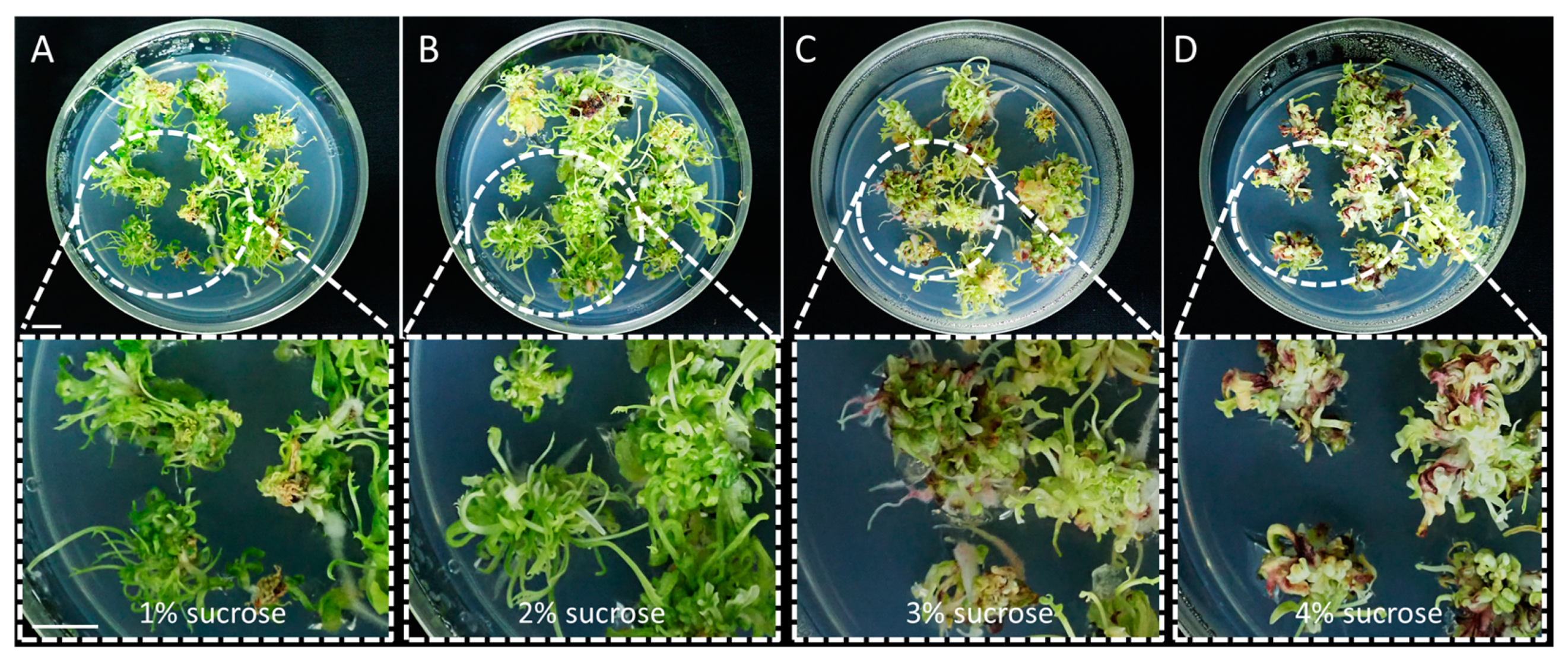
3.3. Effect of Sucrose Concentration on Plant Regeneration

Sucrose concentration in the germination medium continued to affect the plant regeneration. The total number of regenerated plants increased significantly as sucrose concentration in the germination medium decreased (Table 4). The mean number of W types was highest at 1% sucrose, and the number gradually decreased as the sucrose concentration increased. The mean number of S types was also highest at 1% sucrose. However, the mean number of R types was highest at 2% sucrose, followed by 3%. The mean number of plantlets was highest at 2% sucrose, followed by 1% and 3%, with no significant differences among treatments, while the mean number of plantlets was lowest at 4% sucrose.
We investigated the growth characteristics of regenerated plants derived from each sucrose treatment. The length, diameter, and weight of aerial plant parts were significantly higher at 1% sucrose compared to those in the other treatments (Table 5 and Figure S3). Conversely, the growth of underground plant parts derived from 1% sucrose was poor, and there were no significant differences in length among treatments. Root diameter was highest in 3–4% sucrose, but roots were heaviest in 2% sucrose.

3.4. Acclimatization and Determination of Genetic Homogeneity

After several months of cultivation in growth medium, the leaves of plantlets senesced, and thickened IGRs with dormant rhizomes were obtained (Figure 1J). Within 4 weeks after transferring the IGRs to soil, we observed high sprouting rates (90%) coupled with the formation of new leaves. Acclimatized regenerated plants appeared similar to the 2 year old control ginseng plants, despite minor differences in morphological characteristics, such as a higher incidence of multi-stems (Figure 1K).
We assessed the genetic variation in regenerated plants derived from IGRs. Of the molecular markers, three SSR primers (gws218, gws450, and gws454) produced three to four amplicons per marker, and the remaining primers (gws936 and gws1020) formed a single band (Figure 5A). No significant differences were observed in the bands formed between the control and regenerated plants. Three random amplified polymorphic DNA (RAPD) primers (UBC536, UBC540, and UBC592) produced several bands per marker with no significant difference between groups; however, some regenerated plants (numbers two and ten) did not produce a 400 bp amplicon when using the UBC536 primer (Figure 5B).

3.5. Ginsenoside Content Analysis

The contents of ten ginsenosides were compared among IGRs, 2 year old IGRs, and control roots (Figure 6A). Total ginsenoside content was greatly enhanced in IGRs compared to that of the control (Figure 6B). While the content of the PPD type did not vary between groups (Figure 6C), the content of the PPT type was significantly higher in IGRs than in the control (Figure 6D); consequently, the PPD/PPT ratio was also dramatically lower in IGRs compared to the control (Table 6). Among the PPD-type ginsenosides, Rb2, Rb3, and Rc were high and Rbl and Rd were low in IGRs. The IGRs showed enrichment of most PPT-type ginsenosides except Re; the levels of Rf, Rg1, Rg2, and Rh1 in IGRs were 1.3-, 3.6-, 1.3-, and 2-fold higher than other samples, respectively. However, after IGRs had been acclimated, the ginsenoside content of 2 year old IGRs was similar to that of the control, although there was a minor enrichment for each type of ginsenoside.

4. Discussion

An effective protocol is essential for successful plant regeneration via somatic embryogenesis, especially in the micropropagation of new cultivars. Although many previous studies have reported on somatic embryogenesis in P. ginseng [9,24,25,26], detailed protocols for the entire regeneration process, from somatic embryogenesis to genetic stability assessment, are lacking. Additionally, information regarding optimal media conditions at each stage is insufficient. Therefore, the present study sought to comprehensively address the effects of key factors, such as carbon source, on the micropropagation process in P. ginseng cv. Cheonryang (Figure 1).
Appropriate plant growth regulators in culture media are essential for initiating somatic embryogenesis. However, we found that explants derived from a zygotic embryo could produce somatic embryos directly without plant growth regulators, which is consistent with the findings of other studies on P. ginseng [24,25,26] and P. quinquefolius (American ginseng) [33]. Among the various essential components of culture media, carbohydrates are required to provide energy [12], build macromolecules [34], and regulate osmotic pressure [13]. In this study, explants were cultured on somatic embryo induction media supplemented with four different carbon sources (fructose, glucose, maltose, and sucrose). We found that the carbon source greatly affected somatic embryogenesis in P. ginseng (Table 1 and Figure 2); somatic embryo induction was greatest in culture medium supplemented with sucrose. A similar finding was observed by Nhut et al. [15] in the callus culture of P. vietnamensis (Vietnamese ginseng), who found that sucrose had a greater embryogenic effect than glucose and fructose. Several studies have suggested that sucrose is the best choice for cell growth and embryogenesis in many species [35,36]. According to Blanc et al. [37], sucrose hydrolyzes to hexoses and storage compounds more rapidly than other carbon sources, which could promote cell proliferation.
Sucrose is the most commonly used carbohydrate for plant tissue culture and performs various roles as an energy source [12] and regulator of osmotic pressure [13]. Previous studies revealed that adding sucrose to culture medium caused stresses that activated the embryogenesis pathway in somatic cells [38,39]. The recommended sucrose concentration varies depending on the plant species, ranging from 1% to 6%. In the present study, 5% sucrose supplementation resulted in the highest somatic embryo induction, mean number of somatic embryos, and EFC index (Table 2). This is consistent with the result of a previous study on P. ginseng [26]. However, the mean number of somatic embryos and EFC index decreased at sucrose concentrations above 7%, which is similar to the result reported for P. vietnamensis [15]. The negative effects of excessive sucrose concentrations have also been reported in other plant species [40,41]. In contrast, in P. quinquefolius, which is closely related to P. ginseng, the rates of somatic embryogenesis were highest at 7% sucrose [33]. Further studies are needed to compare somatic embryogenesis between P. ginseng and P. quinquefolius under different culture conditions.
Considering that maturation is a major bottleneck for somatic embryogenesis in other species where development is stopped at the globular stage [42,43], somatic embryo maturation is essential for successful regeneration. In the present study, the sucrose concentration in the maturation medium affected somatic embryo development (Figure 3A). At sucrose concentrations higher than 3%, most somatic embryos remained in the immature stage. However, 1–2% sucrose promoted the transition of immature somatic embryos to the mature stage. Kim et al. [9] revealed that more somatic embryos of P. ginseng transitioned from the globular stage to later stages when subcultured in media with 2% sucrose. These results are somewhat distinguishable from several other species wherein increasing sucrose concentrations improved embryo maturation [38,44].
Successful somatic embryo germination is required for efficient micropropagation via somatic embryogenesis. In P. ginseng, GA3 or chilling treatment is essential to stimulate somatic embryo germination [25]. Previous studies recommended 5–10 mg/L GA3 for the germination of Panax genus somatic embryos [22,33,45]. However, few studies have assessed the effects of other media components, such as sucrose concentration in the germination medium [11]. Sucrose is a reducing sugar that is effectively used in most crops at a concentration of 3% [29]. In this study, the germination rate was not affected by the sucrose concentration in germination medium (Figure 3B); however, we observed morphological differences between explants in different sucrose concentrations (Table 3 and Figure 4). At relatively high sucrose concentrations (3% or more), excessive accumulation of red pigment accompanied by secondary embryogenesis was observed. In P. sikkimensis, anthocyanin was the red pigment produced under high sucrose concentrations [46]. Choi et al. [24] also reported that the red pigment accumulated in explants after plasmolysis at a high (1 M) sucrose concentration. The sucrose concentration also affected shoot morphology: at low sucrose concentrations (1%), most shoots were weak and some were necrotic. Shoots derived from germination media containing high sucrose concentrations (3–4%) were not only in poor condition but also showed severe adhesion between the explants and shoots. Yaseen et al. [47] indicated that higher sucrose concentrations in culture media act as stressing agents that increase the concentrations of phenolic compounds, promote tissue necrosis, and decrease shoot regeneration, thereby inhibiting plant growth. In our study, 2% sucrose was optimal for P. ginseng germination, considering the vigorous growth and easy separation of shoots from explants grown in this germination medium.
Rooting remains challenging in the micropropagation of many plant species. Improving root formation is required to promote survival and growth during acclimatization [47]. In the present study, shoots derived from germination media with different sucrose concentrations were cultured in elongation medium for 60 days (Figure 1G) and classified into three distinct types based on morphology (Figure S1). The W type formed normally in IGRs after prolonged culture. The R type sprouted in a month and eventually formed a complete IGR. Consistent with a previous report [48], most S types senesced without rooting. Therefore, W and R types were considered as plantlets in this study.
We found that sucrose concentration in the germination medium had a post-effect on shoot regeneration, rooting, and plantlet formation in P. ginseng. Relatively low sucrose concentrations (1–2%) resulted in higher numbers of regenerated plants (Table 4). Similarly, Takahashi et al. [49] reported that, compared to 3% sucrose, lower sucrose concentrations (0.5–1%) in the culture medium improved shoot regeneration in Actinidia polygama. Although the highest number of S types and maximum mean number of plants were observed at 1% sucrose in the germination medium, the growth of underground plant parts was poorer compared to that of the aerial parts (Table 5 and Figure S3). Similarly, Lee et al. [11] revealed that 1% sucrose in the culture medium promoted the growth of aerial plant parts while inhibiting the growth of underground parts. These results suggest that rooting requires sufficient carbohydrates as it is a fairly energy-intensive process [47]. Sucrose concentrations of more than 3% in the germination medium had a negative effect on the mean number of regenerated plants (Table 4) and growth of aerial parts (Table 5). Previous studies have shown that high sucrose concentrations inhibited shoot growth by increasing osmotic stress [50,51]. Wojtania et al. [16] suggested that above-optimal sucrose concentrations might not only interfere with enzymes that hydrolyze starch into soluble sugars but also enhance the formation of phenolic compounds. Our results indicated that the highest number of plantlets with a balanced development of aerial and underground parts was observed for the germination medium containing 2% sucrose, which is consistent with the results of a previous study on P. ginseng grown in vitro [11]. In Lilium longiflorum, 2% sucrose was also shown to promote both shoot and root formation [52].
Acclimatization is the most important process for achieving successful micropropagation using plant tissue culture since sudden environmental changes are stressful to plants [53]. A previous study showed that the survival rate of regenerated P. ginseng plants was poor (36%) after transfer to soil because regenerated plants grown under aseptic conditions are weak and sensitive to fungal infection [48]. In the present study, acclimatized regenerated plants had a high survival rate (90%); however, their morphology differed from that of conventionally grown P. ginseng shoots (Figure 1K). These results are consistent with those of pioneering studies by Lee et al. [11] and Kim et al. [45], which showed that the high sprouting and survival rates of more than 80% were confirmed when the IGR types were transferred to soil.
Genetic variation in plants regenerated from tissue culture can occur at the DNA sequence level [17]. The present study validated the genetic stability of regenerants in relation to control plants using RAPD and SSR markers. Based on SSR marker analysis, regenerated plants were monomorphic to the control plants (Figure 5A). In the RAPD marker analysis, band patterns from regenerated and control plants were comparable, with negligible differences (Figure 5B). These results indicated that plant regeneration via somatic embryogenesis did not induce any remarkable genetic variation. Similarly, Lee et al. [11] demonstrated that IGRs derived from zygotic embryo cultures showed high genetic fidelity with control plants.
Ginsenoside content is affected by various factors, such as species, developmental stage, and environmental conditions [54]. Previous studies indicated that the ginsenoside content of adventitious roots was lower than that in field-cultivated roots [55,56]. In contrast, the present study showed that ginsenoside levels in IGRs derived from somatic embryogenesis were higher than those in the control roots (Figure 6), which was specifically attributed to the accumulation of PPT-type ginsenosides. The contents of individual ginsenosides also varied between the IGRs and control roots (Table 6), which we presume to be associated with differences inherent to the root types. For instance, IGRs have thickened taproots with a morphology similar to native roots, whereas adventitious roots have an abnormal morphology with only roots. Additionally, the in vitro environment could cause abiotic oxidative stress, thereby promoting ginsenoside accumulation in plant cells [57]. However, the 2 year old acclimatized IGRs showed no significant difference in ginsenoside content compared to the control roots. These findings suggest that the increased ginsenoside content in IGRs is a transient change caused by abiotic stress. To the best of our knowledge, this is the first study to analyze the ginsenoside content of IGRs and 2 year old IGRs of the Panax genus, especially P. ginseng.

5. Conclusions

The present study described a high-efficiency micropropagation protocol, including conditions for optimal tissue regeneration by somatic embryogenesis and assessment of genetic stability, for P. ginseng cv. Cheonryang. It represents a more advanced result than previous studies, as it describes in detail the whole process of micropropagation up to acclimatization of the resulting plantlets. Carbon source type and concentration greatly affected somatic embryogenesis. Somatic embryo induction was maximized in induction media containing 5% sucrose. Sucrose concentrations of 1–2% in maturation media promoted the maturation of immature somatic embryos. Although germination rate was not affected by sucrose concentration, morphological differences for explants and shoots in different sucrose concentrations were observed. The 2% sucrose was the optimal sucrose concentration for the germination medium and produced the highest number of plantlets with balanced growth characteristics. Regenerated plantlets formed thickened IGRs, which likely improved survival rates during acclimatization. Despite minor morphological differences, molecular marker and HPLC analysis revealed that our plant regeneration protocol induced little to no genetic variation. Finally, 2 year old IGRs acclimatized to greenhouse conditions produced similar ginsenoside contents to those of conventionally grown ginseng roots.
Our study provides valuable insights into the optimization of micropropagation procedures and could promote the development of superior P. ginseng cultivars. Future research will be carried out on producing seeds by transplanting IGRs into the field and to confirm the genetic stability of seedlings derived from them.

Supplementary Materials

The following supporting information can be downloaded at https://0-www-mdpi-com.brum.beds.ac.uk/article/10.3390/agronomy13041139/s1. Figure S1: Classification of regenerated plants into three types according to their morphology. (A) W: whole plant, (B) S: shoot only, (C) R: root with rhizome. Scale bar, 1 cm. Figure S2: Somatic embryogenesis according to sucrose concentration in P. ginseng 60 days after inoculation. (A) 1%, (B) 3%, (C) 5%, (D) 7%, and (E) 9% sucrose. Scale bars, upper: 1 cm; lower: 1 mm. Figure S3: Plant regeneration according to sucrose concentration in the germination medium 60 days after transfer to elongation medium. (A) 1%, (B) 2%, (C) 3%, and (D) 4% sucrose. Scale bars, 1 cm. Table S1: List of primers used for detecting the genetic stability of plants regenerated from somatic embryogenesis of P. ginseng.

Author Contributions

Conceptualization, J.-W.L. and Y.-D.P.; methodology, J.-W.L.; software, J.-W.L.; validation, J.-W.L., J.-U.K. and K.-H.B.; formal analysis, J.-W.L., I.-H.J. and K.-H.B.; investigation, J.-W.L. and J.-U.K.; resources, J.-W.L., D.-H.K. and J.-U.K.; data curation, J.-W.L.; writing—original draft preparation, J.-W.L.; writing—review and editing, J.-W.L. and Y.-D.P.; visualization, J.-W.L.; supervision, J.-W.L.; project administration, J.-W.L.; funding acquisition, J.-W.L. All authors have read and agreed to the published version of the manuscript.

Funding

This work was carried out with the support of the Cooperative Research Program for Agriculture Science and Technology Development (project no. PJ01426502) of the Rural Development Administration, Republic of Korea.

Data Availability Statement

Not applicable.

Conflicts of Interest

The authors declare no conflict of interest.

References

  1. Nguyen, V.B.; Giang, V.N.L.; Waminal, N.E.; Park, H.S.; Kim, N.H.; Jang, W.; Lee, J.; Yang, T.J. Comprehensive comparative analysis of chloroplast genomes from seven Panax species and development of an authentication system based on species-unique single nucleotide polymorphism markers. J. Ginseng Res. 2020, 44, 135–144. [Google Scholar] [CrossRef]
  2. Glagoleva, E.S.; Konstantinova, S.E.; Kochkin, D.V.; Ossipov, V.; Titova, M.V.; Popova, E.V.; Nosov, A.M.; Paek, K.Y. Predominance of oleanane-type ginsenoside R0 and malonyl esters of protopanaxadiol-type ginsenosides in the 20-year-old suspension cell culture of Panax japonicus C.A. Meyer. Indus. Crops Prod. 2022, 177, 114417. [Google Scholar] [CrossRef]
  3. Kim, K.H.; Lee, D.; Lee, H.L.; Lim, C.E.; Jung, K.; Kang, K.S. Beneficial effects of Panax ginseng for the treatment and prevention of neurodegenerative diseases: Past findings and future directions. J. Ginseng Res. 2018, 42, 239–247. [Google Scholar] [CrossRef]
  4. Kim, J.H. Pharmacological and medical applications of Panax ginseng and ginsenosides: A review for use in cardiovascular diseases. J. Ginseng Res. 2018, 42, 264–269. [Google Scholar] [CrossRef]
  5. Shao, J.W.; Jiang, J.L.; Zou, J.J.; Yang, M.Y.; Chen, F.M.; Zhang, Y.J.; Lee, J. Therapeutic potential of ginsenosides on diabetes: From hypoglycemic mechanism to clinical trials. J. Funct. Foods 2020, 64, 103630. [Google Scholar] [CrossRef]
  6. Hong, H.; Baatar, D.; Hwang, S.G. Anticancer activities of ginsenosides, the main active components of ginseng. J. Ginseng Res. 2000, 24, 8858006. [Google Scholar] [CrossRef]
  7. Lee, J.W.; Kwon, N.; Kim, J.U.; Bang, K.H.; Jung, S.M.; Lee, S.W.; Kim, D.H.; Kim, Y.C.; Jo, I.H.; Park, Y.D. In vitro micropropagation of commercial ginseng cultivars (Panax ginseng Meyer) via somatic embryogenesis compared to traditional seed production. Horiculturae 2023, 9, 435. [Google Scholar] [CrossRef]
  8. Lee, J.W.; Jo, I.H.; Kim, J.U.; Hong, C.E.; Kim, Y.C.; Kim, D.H.; Park, Y.D. Improvement of seed dehiscence and germination in ginseng by stratification, gibberellin, and/or kinetin treatments. Hortic. Environ. Biotechnol. 2018, 59, 293–301. [Google Scholar] [CrossRef]
  9. Kim, Y.J.; Lee, O.R.; Kim, K.T.; Yang, D.C. High frequency of plant regeneration through cyclic secondary somatic embryogenesis in Panax ginseng. J. Ginseng Res. 2012, 36, 442–448. [Google Scholar] [CrossRef]
  10. Kim, Y.C.; Kim, D.H.; Bang, K.H.; Kim, J.U.; Hyun, D.Y.; Lee, S.W.; Kang, S.W.; Cha, S.W.; Kim, K.H.; Choi, J.K.; et al. A high yielding and salt resistance ginseng variety “Cheonryang”. Korean J. Breed. Sci. 2013, 45, 434–439. [Google Scholar] [CrossRef]
  11. Lee, J.W.; Do, G.R.; Jo, I.H.; Hong, C.E.; Bang, K.H.; Kim, J.U.; Park, Y.D. Zygotic embryo culture is an efficient way to optimize in vitro growth in Panax ginseng. Indus. Crops Prod. 2021, 167, 1134971. [Google Scholar] [CrossRef]
  12. Manchanda, P.; Gosal, S.S. Effect of activated charcoal, carbon sources and gelling agents on direct somatic embryogenesis and regeneration in sugarcane via leaf roll segments. Sugar Tech 2012, 14, 168–173. [Google Scholar] [CrossRef]
  13. Shohael, A.M.; Chakrabarty, D.; Ali, M.B.; Yu, K.W.; Hahn, E.J.; Lee, H.L.; Paek, K.Y. Enhancement of eleutherosides production in embryogenic cultures of Eleutherococcus sessiliflorus in response to sucrose-induced osmotic stress. Process Biochem. 2006, 41, 512–518. [Google Scholar] [CrossRef]
  14. Nhut, D.T.; Huy, N.P.; Binh, N.V.; Nam, N.B.; Thuy, L.N.; Ha, D.T.; Chien, H.X.; Huong, T.T.; Cuong, L.K.; Hien, V.T. Shoot regeneration and micropropagation of Panax vietnamensis Ha et Grushv. from ex vitro leaf-derived callus. Afr. J. Biotechnol. 2011, 10, 19499–19504. [Google Scholar] [CrossRef]
  15. Nhut, D.T.; Vinh, B.V.T.; Hien, T.T.; Huy, N.P.; Nam, N.B.; Chien, H.X. Effects of spermidine, proline and carbohydrate sources on somatic embryogenesis from main root transverse thin cell layers of Vietnamese ginseng (Panax vietnamensis Ha et. Grushv.). Afr. J. Biotechnol. 2012, 11, 1084–1091. [Google Scholar] [CrossRef]
  16. Wojtania, A.; Skrzypek, E.; Marasek-Ciolakowska, A. Soluble sugar, starch and phenolic status during rooting of easy and difficult-to-root magnolia cultivars. Plant Cell Tiss. Organ Cult. 2019, 136, 499–510. [Google Scholar] [CrossRef]
  17. Heydari, H.R.; Chamani, E. High-frequency plant regeneration and genetic fidelity assessment of regenerants by molecular and biochemical markers in Woodland Sage (Salvia nemorosa L.). Indus. Crops Prod. 2022, 187, 115404. [Google Scholar] [CrossRef]
  18. Krishna, H.; Alizadeh, M.; Singh, D.; Singh, U.; Chauhan, N.; Eftekhari, M.; Sadh, R.K. Somaclonal variations and their applications in horticultural crops improvement. 3 Biotech 2016, 6, 54. [Google Scholar] [CrossRef]
  19. Gaj, M.D. Direct somatic embryogenesis as a rapid and efficient system for in vitro regeneration of Arabidopsis thaliana. Plant Cell Tiss. Organ Cult. 2001, 64, 39–46. [Google Scholar] [CrossRef]
  20. Konar, S.; Karmakar, J.; Ray, A.; Adhikari, S.; Bandyopadhyay, T.K. Regeneration of plantlets through somatic embryogenesis from root derived calli of Hibiscus sabdariffa L. (Roselle) and assessment of genetic stability by flow cytometry and ISSR analysis. PLoS ONE 2018, 13, e0202324. [Google Scholar] [CrossRef]
  21. Mamgain, J.; Mujib, A.; Ejaz, B.; Gulzar, B.; Malik, M.Q.; Syeed, R. Flow cytometry and start codon targeted (SCoT) genetic fidelity assessment of regenerated plantlets in Tylophora indica (Burm.f.) Merrill. Plant Cell Tiss. Organ Cult. 2022, 150, 129–140. [Google Scholar] [CrossRef]
  22. Zhang, J.Y.; Sun, H.J.; Song, I.J.; Bae, T.W.; Kang, H.G.; Ko, S.M.; Kwon, Y.I.; Kim, I.W.; Lee, J.C.; Park, S.Y.; et al. Plant regeneration of Korean wild ginseng (Panax ginseng Meyer) mutant lines induced by γ-irradiation (60Co) of adventitious roots. J. Ginseng Res. 2014, 38, 220–225. [Google Scholar] [CrossRef]
  23. Kim, J.Y.; Adhikari, P.B.; Ahn, C.H.; Kim, D.H.; Chang Kim, Y.C.; Han, J.Y.; Kondeti, S.; Choi, Y.E. High frequency somatic embryogenesis and plant regeneration of interspecific ginseng hybrid between Panax ginseng and Panax quinquefolius. J. Ginseng Res. 2019, 43, 38–48. [Google Scholar] [CrossRef]
  24. Choi, Y.E.; Soh, W.Y. Enhanced somatic single embryo formation by plasmolyzing pretreatment from cultured ginseng cotyledons. Plant Sci. 1997, 130, 197–2064. [Google Scholar] [CrossRef]
  25. Choi, Y.E.; Yang, D.C.; Yoon, E.S.; Choi, K.T. High-efficiency plant production via direct somatic single embryogenesis from preplasmolysed cotyledons of Panax ginseng and possible dormancy of somatic embryos. Plant Cell Rep. 1999, 18, 493–499. [Google Scholar] [CrossRef]
  26. Kim, O.T.; Kim, T.S.; In, D.S.; Bang, K.H.; Kim, Y.C.; Choi, Y.E.; Cha, S.W.; Seong, N.S. Optimization of direct somatic embryogenesis from mature zygotic embryos of Panax ginseng C. A. Meyer. J. Plant Biol. 2006, 49, 348–352. [Google Scholar] [CrossRef]
  27. Lee, J.W.; Jo, I.H.; Kim, J.U.; Hong, C.E.; Bang, K.H.; Park, Y.D. Determination of mutagenic sensitivity to gamma rays in ginseng (Panax ginseng) dehiscent seeds, roots, and somatic embryos. Hortic. Environ. Biotechnol. 2019, 60, 721–731. [Google Scholar] [CrossRef]
  28. Lee, J.W.; Kim, J.U.; Bang, K.H.; Kwon, N.; Kim, Y.C.; Jo, I.H.; Park, Y.D. Efficient somatic embryogenesis, regeneration and acclimatization of Panax ginseng Meyer: True-to-type conformity of plantlets as confirmed by ISSR analysis. Plants 2023, 12, 1270. [Google Scholar] [CrossRef]
  29. Murashige, T.; Skoog, F. A revised medium for rapid growth and bioassays with tobacco tissue cultures. Physiol. Plant. 1962, 15, 473–497. [Google Scholar] [CrossRef]
  30. Schenk, R.U.; Hildebrandt, A.C. Medium and techniques for induction and growth of monocotyledonous and dicotyledonous plant cell cultures. Can. J. Bot. 1972, 50, 199–204. [Google Scholar] [CrossRef]
  31. Raomai, S.; Kumaria, S.; Tandon, P. Plant regeneration through direct somatic embryogenesis from immature zygotic embryos of the medicinal plant, Paris polyphylla Sm. Plant Cell Tiss. Organ Cult. 2014, 118, 445–455. [Google Scholar] [CrossRef]
  32. Jang, W.; Jang, Y.; Kim, N.H.; Waminal, N.E.; Kim, Y.C.; Lee, J.W.; Yang, T.J. Genetic diversity among cultivated and wild Panax ginseng populations revealed by high-resolution microsatellite markers. J. Ginseng Res. 2020, 44, 637–643. [Google Scholar] [CrossRef] [PubMed]
  33. Zhou, S.; Brown, D.C.W. High efficiency plant production of North American ginseng via somatic embryogenesis from cotyledon explants. Plant Cell Rep. 2006, 25, 166–173. [Google Scholar] [CrossRef] [PubMed]
  34. Traore, A.; Guiltinan, M. Effects of carbon source and explant type on somatic embryogenesis of four cacao genotypes. Hortic. Sci. 2006, 41, 753–758. [Google Scholar] [CrossRef]
  35. Faisal, M.; Abdel-Salam, E.M.; Alatar, A.A.; Qahtan, A.A. Induction of somatic embryogenesis in Brassica juncea L. and analysis of regenerants using ISSR-PCR and flow cytometer. Saudi J. Biol. Sci. 2021, 28, 1147–1153. [Google Scholar] [CrossRef] [PubMed]
  36. Moon, H.K.; Kim, Y.W.; Hong, Y.P.; Park, S.Y. Improvement of somatic embryogenesis and plantlet conversion in Oplopanax elatus, an endangered medicinal woody plant. SpringerPlus 2013, 2, 428. [Google Scholar] [CrossRef]
  37. Blanc, G.; Lardet, L.; Martin, A.; Jacob, J.L.; Carron, M.P. Differential carbohydrate metabolism conducts morphogenesis in embryogenic callus of Hevea brasiliensis (Müll. Arg.). J. Exp. Bot. 2002, 53, 1453–1462. [Google Scholar] [CrossRef]
  38. Baskaran, P.; Kumari, A.; Van Staden, J. Embryogenesis and synthetic seed production in Mondia whitei. Plant Cell Tiss. Organ Cult. 2015, 121, 205–214. [Google Scholar] [CrossRef]
  39. Karami, O.; Deljou, A.; Esna-Ashari, M.; Ostad-Ahmadi, P. Effect of sucrose concentrations on somatic embryogenesis in carnation (Dianthus caryophyllus L.). Sci. Hortic. 2006, 110, 340–344. [Google Scholar] [CrossRef]
  40. Du, Y.; Cheng, F.; Zhong, Y. Induction of direct somatic embryogenesis and shoot organogenesis and histological study in tree peony (Paeonia sect. Moutan). Plant Cell Tiss. Organ Cult. 2020, 141, 557–570. [Google Scholar] [CrossRef]
  41. Woo, H.A.; Ku, S.S.; Jie, E.Y.; Kim, H.; Kim, H.S.; Cho, H.S.; Jeong, W.J.; Park, S.U.; Min, S.R.; Kim, S.W. Efficient plant regeneration from embryogenic cell suspension cultures of Euonymus alatus. Sci. Rep. 2021, 11, 15120. [Google Scholar] [CrossRef] [PubMed]
  42. Tereso, S.; Zoglauer, K.; Milhinhos, A.; Miguel, C.; Oliveira, M.M. Zygotic and somatic embryo morphogenesis in Pinus pinaster: Comparative histological and histochemical study. Tree Physiol. 2007, 27, 661–669. [Google Scholar] [CrossRef] [PubMed]
  43. Etienne, H.; Bertrand, B.; Georget, F.; Lartaud, M.; Montes, F.; Dechamp, E.; Verdeil, J.L.; Barry-Etienne, D. Development of coffee somatic and zygotic embryos to plants differs in the morphological, histochemical and hydration aspects. Tree Physiol. 2013, 33, 640–653. [Google Scholar] [CrossRef] [PubMed]
  44. Nair, R.R.; Dutta Gupta, S. High-frequency plant regeneration through cyclic secondary somatic embryogenesis in black pepper (Piper nigrum L.). Plant Cell Rep. 2006, 24, 699–707. [Google Scholar] [CrossRef] [PubMed]
  45. Kim, J.Y.; Kim, D.H.; Kim, Y.C.; Kim, K.H.; Han, J.Y.; Choi, Y.E. In vitro grown thickened taproots, a new type of soil transplanting source in Panax ginseng. J. Ginseng Res. 2016, 40, 409–414. [Google Scholar] [CrossRef]
  46. Mathur, A.; Mathur, A.K.; Gangwar, A.; Yadav, S.; Verma, P.; Sangwan, R.S. Anthocyanin production in a callus line of Panax sikkimensis Ban. In Vitro Cell Dev. Biol. Plant 2010, 46, 13–21. [Google Scholar] [CrossRef]
  47. Yaseen, M.; Ahmad, T.; Sablok, G.; Standardi, A.; Hafiz, I.A. Review: Role of carbon sources for in vitro plant growth and development. Mol. Biol. Rep. 2013, 40, 2837–2849. [Google Scholar] [CrossRef]
  48. Choi, Y.E.; Yang, D.C.; Yoon, E.S.; Choi, K.T. Plant regeneration via adventitious bud formation from cotyledon explants of Panax ginseng C. A. Meyer. Plant Cell Rep. 1998, 17, 731–736. [Google Scholar] [CrossRef]
  49. Takahashi, W.; Sugawara, F.; Yamamoto, N.; Bando, E.; Matsushita, J.; Tanaka, O. Plant regeneration in Actinidia polygama Miq. by leaf, stem, and petiole culture with zeatin, and from stem-derived calli on low-sucrose medium. J. For. Res. 2004, 9, 85–88. [Google Scholar] [CrossRef]
  50. Martins, J.P.R.; Verdoodt, V.; Pasqual, M.; De Proft, M. Impacts of photoautotrophic and photomixotrophic conditions on in vitro propagated Billbergia zebrina (Bromeliaceae). Plant Cell Tiss. Organ Cult. 2015, 123, 121–132. [Google Scholar] [CrossRef]
  51. Zahid, N.A.; Jaafar, H.Z.E.; Hakiman, M. Alterations in microrhizome induction, shoot multiplication and rooting of ginger (Zingiber officinale Roscoe) var. Bentong with regards to sucrose and plant growth regulators application. Agronomy 2021, 11, 320. [Google Scholar] [CrossRef]
  52. Nhut, D.T.; Le, B.V.; Fukai, S.; Tanaka, M.; Van, T.T. Effects of activated charcoal, explant size, explant position and sucrose concentration on plant and shoot regeneration of Lilium longiflorum via young stem culture. Plant Growth Regul. 2021, 33, 59–65. [Google Scholar] [CrossRef]
  53. Chandra, S.; Bandopadhyay, R.; Kumar, V.; Chandra, R. Acclimatization of tissue cultured plantlets: From laboratory to land. Biotechnol. Lett. 2010, 32, 1199–1205. [Google Scholar] [CrossRef] [PubMed]
  54. Liu, H.; Gu, H.; Ye, C.; Guo, C.; Zhu, Y.; Huang, H.; Liu, Y.; He, X.; Yang, M.; Zhu, S. Planting density affects Panax notoginseng growth and ginsenoside accumulation by balancing primary and secondary metabolism. Front. Plant Sci. 2021, 12, 628294. [Google Scholar] [CrossRef] [PubMed]
  55. Kim, D.S.; Song, M.; Kim, S.H.; Jang, D.S.; Kim, J.B.; Ha, B.K.; Kim, S.H.; Lee, K.J.; Kang, S.Y.; Jeong, I.Y. The improvement of ginsenoside accumulation on Panax ginseng as a result of γ-irradiation. J. Ginseng Res. 2013, 37, 332–340. [Google Scholar] [CrossRef]
  56. Zhao, Y.; Guo, W.H.; Sun, X.Y.; Li, K.H.; Liu, K.J.; Wang, J.; Wang, Y.; Tan, X.; You, X.L. A culture system for the stable and high-efficiency proliferation of adventitious roots of Panax notoginseng and ginsenoside accumulation. Indus. Crops Prod. 2021, 157, 12882. [Google Scholar] [CrossRef]
  57. Kim, Y.J.; Zhang, D.; Yang, D.C. Biosynthesis and biotechnological production of ginsenosides. Biotechnol. Adv. 2015, 33, 717–735. [Google Scholar] [CrossRef]
Figure 1. Micropropagation of P. ginseng via somatic embryogenesis. (A) Fruits, (B) indehiscent seed after sarcocarp removal, (C) dehiscent seed after dehiscence, (D) mature zygotic embryo before excision, (E) induced somatic embryos after inoculation of induction medium, (F) matured somatic embryos after transfer to maturation medium, (G) germinated somatic embryos after culture of germination medium, (H,I) well-developed regenerated plantlets after several months of in vitro cultivation, (J) in vitro-grown root derived from the in vitro culture process, (K) sprouted in vitro-grown root, and (L) 2 year old IGRs after acclimatization.
Agronomy 13 01139 g001
Figure 2. Somatic embryogenesis of P. ginseng in the presence of four carbon sources after 60 days. (A) Fructose, (B) glucose, (C) maltose, and (D) sucrose. Scale bars, upper: 1 cm; lower: 1 mm.
Agronomy 13 01139 g002
Figure 3. Effect of sucrose concentration on (A) somatic embryo maturation and (B) germination of P. ginseng. Data were recorded 30 days after somatic embryos were transferred to maturation or germination media. Values represent the ratios of embryo stages and means ± standard error of germination rates measured in three independent experiments, each consisting of ten explants. ns: non-significant determined by ANOVA.
Agronomy 13 01139 g003
Figure 4. Somatic embryo germination of P. ginseng according to sucrose concentration in germination medium 30 days after inoculation. (A) 1%, (B) 2%, (C) 3%, and (D) 4% sucrose. Scale bars, 1 cm.
Agronomy 13 01139 g004
Figure 5. Evaluation of genetic fidelity in the regenerated plants by molecular marker analysis. (A) gws218, gws450, gws454, gws936, and gws1020 SSR markers. (B) UBC536, UBC540, and UBC594 RAPD markers. Green lines represent alignment markers. C, control; IGRs, in vitro-grown roots; SSR, simple sequence repeat; RAPD, random amplified polymorphic DNA.
Agronomy 13 01139 g005
Figure 6. Ginsenoside content of control roots, IGRs, and 2 year old IGRs of P. ginseng. (A) Chromatogram of the ten standards. (B) Total ginsenoside content. (C) PPD-type ginsenoside content. (D) PPT-type ginsenoside content. Values represent the means ± standard error of at least three independent experiments, each consisting of 20 roots. ns, *, **: non-significant or significant at p ≤ 0.05 or 0.01 determined by ANOVA. Different letters above each bar represent significant differences (p ≤ 0.05) based on Duncan’s multiple comparison test. IGR, in vitro-grown root; PPD, protopanaxadiol; PPT, protopanaxatriol.
Agronomy 13 01139 g006
Table 1. Effect of carbon source in induction medium on somatic embryogenesis of P. ginseng.
Carbon Source (3%)Somatic Embryo Induction Rate (%) ***Mean Number of Somatic Embryos/Explant **EFC Index
Fructose42.3 ± 4.0 c9.6 ± 2.6 b4.1
Glucose72.7 ± 10.6 b9.6 ± 3.4 b7.0
Maltose66.3 ± 7.7 b9.6 ± 2.8 b6.4
Sucrose93.3 ± 3.5 a16.2 ± 3.7 a15.1
Data were recorded 60 days after inoculation of explants in somatic embryo induction medium. Values represent the means ± standard error of three independent experiments, each consisting of 50 explants. **, ***: significant at p ≤ 0.01 or 0.001 determined by ANOVA. Different letters within each column represent significant differences (p ≤ 0.05) based on Duncan’s multiple comparison test. EFC, embryo formation capacity.
Table 2. Effect of sucrose concentration in induction medium on somatic embryogenesis of P. ginseng.
Sucrose
Concentration (%)
Somatic Embryo
Induction Rate (%) ***
Mean Number of Somatic Embryos/Explant **EFC Index
172.4 ± 8.3 b8.4 ± 4.1 b4.0
391.3 ± 4.5 a21.5 ± 6.3 a18.5
597.7 ± 2.8 a23.8 ± 7.8 a21.3
797.3 ± 4.3 a12.8 ± 7.5 b10.0
965.1 ± 11.7 b10.5 ± 2.3 b5.1
Data were recorded 60 days after inoculation of explants in somatic embryo induction medium. Values represent the means ± standard error of three independent experiments, each consisting of 50 explants. **, ***: significant at p ≤ 0.01 or 0.001 determined by ANOVA. Different letters within each column represent significant differences (p ≤ 0.05) based on Duncan’s multiple comparison test. EFC, embryo formation capacity.
Table 3. Morphological observations of P. ginseng explants and shoots according to sucrose concentration in germination media.
Sucrose Concentration (%)Morphological Characteristics
1Soft explant with light-red pigment accumulation, easily separated shoots, long and weak shoots, occasional shoot necrosis
2Soft explants with light-red pigment accumulation, easily separated shoots, vigorous shoot growth
3Hard explant with red pigment accumulation, difficult to separate shoots, short and poor shoot structure, secondary somatic embryogenesis
4Very hard explant with dark-red pigment accumulation, difficult to separate shoots, short and poor shoot structure, secondary somatic embryogenesis
Morphological observations were recorded 30 days after transferring somatic embryos to germination media.
Table 4. Post-effect of sucrose concentration in the germination medium on plant regeneration.
Sucrose Concentration (%)Mean Number of Regenerated Plants
Total ***W **S *R **Plantlet **
118.6 ± 2.3 a10.3 ± 1.9 a5.5 ± 1.6 a2.8 ± 1.1 bc13.1 ± 2.2 a
215.7 ± 2.8 ab8.6 ± 1.1 ab2.3 ± 1.1 b4.9 ± 1.4 a13.5 ± 2.1 a
313.4 ± 0.7 b6.8 ± 0.8 bc2.5 ± 1.4 b4.1 ± 0.1 ab10.9 ± 0.8 a
47.0 ± 1.4 c4.5 ± 0.6 c1.3 ± 0.4 b1.2 ± 0.9 c5.7 ± 1.2 b
Data were recorded 60 days after transferring the germinated shoots to elongation medium. Values represent the means ± standard error of three independent experiments, each consisting of 100 germinated shoots. *, **, ***: significant at p ≤ 0.05, 0.01, or 0.001 determined by ANOVA. Different letters within each column represent significant differences (p ≤ 0.05) based on Duncan’s multiple comparison test. W, whole plant with shoot and root; S, shoot alone; R, root with rhizome.
Table 5. Effect of sucrose concentration in the germination medium on the growth of shoots and roots.
Sucrose
Concentration (%)
Aerial PartUnderground Part
Shoot Length ***Shoot Diameter ***Shoot Weight **Root Length nsRoot Diameter ***Root Weight **
15.3 ± 1.3 a0.93 ± 0.21 a89.5 ± 60.0 a2.9 ± 0.10.7 ± 0.1 c26.0 ± 13.5 b
22.2 ± 0.4 b0.55 ± 0.10 b47.7 ± 12.6 b3.3 ± 0.31.2 ± 0.3 b59.8 ± 25.2 a
31.9 ± 0.8 b0.52 ± 0.16 b36.6 ± 17.0 b3.7 ± 0.51.6 ± 0.5 a54.7 ± 26.2 a
41.8 ± 0.6 b0.61 ± 0.09 b34.6 ± 14.3 b3.3 ± 0.41.5 ± 0.4 ab54.2 ± 23.2 a
Data were recorded 60 days after transferring shoots to elongation medium. Values represent the means ± standard error of three independent experiments, each consisting of ten plants. ns, **, ***: non-significant or significant at p ≤ 0.01 or 0.001 determined by ANOVA. Different letters within each column represent significant differences (p ≤ 0.05) based on Duncan’s multiple comparison test.
Table 6. Influences of ginsenoside content before and after acclimatization of IGRs as assessed by HPLC analysis.
ControlIGR2-Year-Old IGR
PPD (mg/g) nsRb1 ***1.98 ± 0.11 a1.65 ± 0.05 b2.17 ± 0.12 a
Rb2 ***0.47 ± 0.02 c0.75 ± 0.03 a0.52 ± 0.01 b
Rb3 ***0.09 ± 0.00 c0.12 ± 0.00 a0.11 ± 0.00 b
Rc ***0.57 ± 0.03 b0.89 ± 0.04 a0.62 ± 0.00 b
Rd ***0.82 ± 0.02 a0.45 ± 0.02 b0.82 ± 0.04 a
PPT (mg/g) **Re ns7.72 ± 0.55 a7.04 ± 0.65 a8.14 ± 0.81 a
Rf ***0.59 ± 0.04 c0.94 ± 0.05 a0.70 ± 0.04 b
Rg1 ***1.12 ± 0.08 b4.07 ± 0.28 a0.95 ± 0.02 b
Rg2 ***0.83 ± 0.04 c1.29 ± 0.08 a1.02 ± 0.08 b
Rh1 ***0.04 ± 0.00 c0.08 ± 0.00 a0.04 ± 0.00 b
PPD/PPT **0.38 ± 0.03 a0.29 ± 0.02 b0.39 ± 0.03 a
Total Ginsenoside Content (mg/g) *14.22 ± 0.53 b17.30 ± 1.05 a15.08 ± 0.97 b
HPLC analysis was performed on control roots, IGRs, and 2 year old IGRs. Values represent the means ± standard error of three independent experiments, each consisting of 20 roots. ns, *, **, ***: non-significant or significant at p ≤ 0.05, 0.01, or 0.001 determined by ANOVA. Different letters within each column represent significant differences (p ≤ 0.05) based on Duncan’s multiple comparison test. IGR, in vitro-grown root; HPLC, high-performance liquid chromatography; PPD, protopanaxadiol; PPT, protopanaxatriol.
Disclaimer/Publisher’s Note: The statements, opinions and data contained in all publications are solely those of the individual author(s) and contributor(s) and not of MDPI and/or the editor(s). MDPI and/or the editor(s) disclaim responsibility for any injury to people or property resulting from any ideas, methods, instructions or products referred to in the content.

