Article

Preparation and In Vitro Characterization of Chitosan Nanoparticles and Their Broad-Spectrum Antifungal Action Compared to Antibacterial Activities against Phytopathogens of Tomato

1
Department of Animal Biotechnology, Konkuk University, Seoul 05029, Korea
2
Department of Bioresource and Food Science, Konkuk University, Seoul 05029, Korea
3
Department of Food Science and Biotechnology, Sejong University, 209 Neungdong-ro, Gwangjin-gu, Seoul 05006, Korea
*
Author to whom correspondence should be addressed.
Submission received: 28 November 2018 / Revised: 12 December 2018 / Accepted: 3 January 2019 / Published: 8 January 2019
(This article belongs to the Special Issue Nanotechnology Applications in Agriculture System)

Abstract

:
The present study was to prepare chitosan nanoparticles (CNPs) from chitosan (CS) to evaluate their in vitro antimicrobial activities against phytopathogens of tomato. We prepared and characterized CNPs for their particle size, polydispersity index, and structures. The antifungal properties of CS and CNPs against phytopathogenic fungi namely Colletotrichum gelosporidies, Phytophthora capsici, Sclerotinia sclerotiorum, Fusarium oxysporum, Gibberella fujikuori were investigated. CNPs showed the maximum growth inhibitory effects on mycelial growth of F. oxysporum followed by P. capsici. We also studied antibacterial activities against phytopathogenic bacteria, such as three strains of Erwinia carotovora subsp. carotovora and one strain of Xanthomonas campestris pv. vesicatoria. Our results showed that both CS and CNPs markedly inhibited the growth of the both Xanthomonas and Erwinia strains. From our study, it is evident that both CS and CNPs have tremendous potential against phytopathogens of tomato for further field screening towards crop protection.

Graphical Abstract

1. Introduction

Nanotechnology has been revolutionizing almost all realms in life sciences, especially agriculture. Precision in agriculture was emphasized by many researchers employing nanoparticles against diagnosis and fertilizers application [1]. Chitosan (CS), a naturally occurring linear biopolymer composed of D-glucosamine and N-acetyl glucosamine residues, is derived from the complete or partial deacetylation of chitin [2,3]. In the plant system, chitosan has been reported to induce multifaceted disease resistance [4,5]. CS is a competent elicitor in agriculture mediating plant immunity by microbe-associated molecular patterns [6]. Current research stresses the utility of chitosan nanoparticles (CNPs) in agriculture as a comprehensive strategy towards sustainability in productivity and high yield in plant protection prospects throughout the world in agriculture [7,8,9,10]. NPs prepared from natural sources possess advantages, such as availability of replenishable resources, biocompatibility, biodegradability, and ecological safety. In this regard, chitosan-based nanoparticles are preferably used for various applications owing to their biodegradability, high permeability toward biological membranes, non-toxicity to human, cost-effectiveness, and broad antimicrobial activities [11,12,13,14,15]. Seed treatment with CNPs and as a biofertilizer application efficiently abates fungal infection and aids plant growth [16]. CS polymeric nanoparticles (NPs) are biodegradable and are utilized for the controlled release of NPK fertilizers [17]. CS polymethacrylic acid colloidal suspension helps in sustained release because of a high affinity towards calcium phosphate rather than potassium chloride and urea due to high anionic charges [18]. CNPs also regulate upgraded micronutrient supplementation 1-naphthylacetic acid under various pH and temperature enhancing uptake of plant growth hormones [19]. Cu-CS complemented NPs show diverse antifungal efficacy combating Alternaria alternata, Macrophomina phaseolina, and Rhizoctonia solani [14]. Acetamprid laden alginate-CS nanocapsules are employed for agrochemical supply with efficient delivery and liposome-based application [20]. Further, CNPs significantly enhanced plant immunity [21], demonstrated efficient activity against phytopathogens [22], and produced high yields [23]. CNPs act as carriers for encapsulating herbicides for escalated efficiency and release kinetics with reduced toxicity levels for the combinatorial herbicides, Imazapic and Imazapyr [24]. Thus, CNPs have been regarded as an effective modality compared to other combined applications for improved efficiency and agricultural efficacy pertaining to the arrest of phytopathogens. Nevertheless, CS conventionally has been largely used to increase plant productivity, for crop protection and plant defense, and to enhancement of shelf life of fruits [25]. The potential of chitosan to protect plants from fungal diseases and bacterial diseases has been reported [2,3,4,25,26,27]. The chelating property of chitosan towards various organic and inorganic compounds makes it a suitable biopolymer for improvement in stability, solubility, and biocidal activity. However, the insolubility of bulk chitosan in aqueous media limits its wide spectrum application as an antifungal agent [10,14]. Compared to bulk CS, CNPs imbues versatility in biological activities due to altered physicochemical characteristics, such as size, surface area, cationic nature, active functional groups, and higher encapsulation efficiency, etc., alone and/or through the blending of other components [10,14]. With this important prospect, we undertook the present study to assess the combinatorial research in alienating the effects of CNPs over CS to combat phytopathogens in tomato. Up to date, most research concerning CNPs is concentrated on fungal pathogens. The present report analyzes its effect against various fungal pathogens encountered in tomato as well as bacterial pathogens causing leaf spot and soft rot disease in tomato. Therefore, in the present investigation, CNPs were prepared which were further examined against phytopathogenic bacteria such as Erwinia carotovora subsp. carotovora and Xanthomonas campestris pv. vesicatoria and fungi viz. Colletotrichum gelosporidies, Phytophthora capsici, Sclerotinia sclerotiorum, Fusarium oxysporum, and Gibberella fujikuori.

2. Materials and Methods

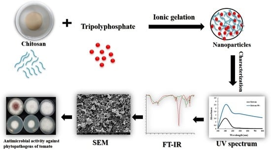
2.1. Preparation of Chitosan Nanoparticles

CNPs were prepared based on the ionic gelation of chitosan (Mw ≈ 190–370 kDa, deacetylation degree ≥75%) with sodium tripolyphosphate (TPP) anions. CS was dissolved at 0.1% level (w/v) in 1% (v/v) acetic acid followed by overnight stirring on a magnetic stirrer at 200 rpm and filtered through a PVDF syringe filter (pore size 0.22 µm). TPP was dissolved at 0.25% level (w/v) in sterile distilled water and filtered through a PVDF membrane syringe filter (pore size 0.22 µm). The cross-linking of chitosan with TPP at equal volume was performed drop by drop under a magnetic stirrer at 700 rpm. The resulting formulation was centrifuged for 10 min at 10,000 rpm, and the pellet was resuspended in sterile distilled water followed by ultra-sonication at 28% pulse ratio for 100 s at 4 °C. Centrifugation followed by ultrasonication was repeated three times, and the precipitated nanoformulation was freeze-dried and stored in a desiccator for further analysis.

2.2. Characterization of Nanoparticles

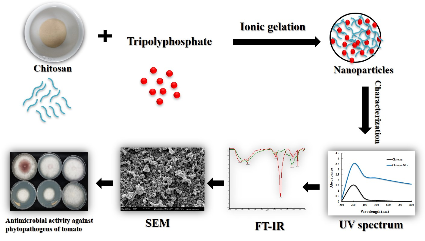
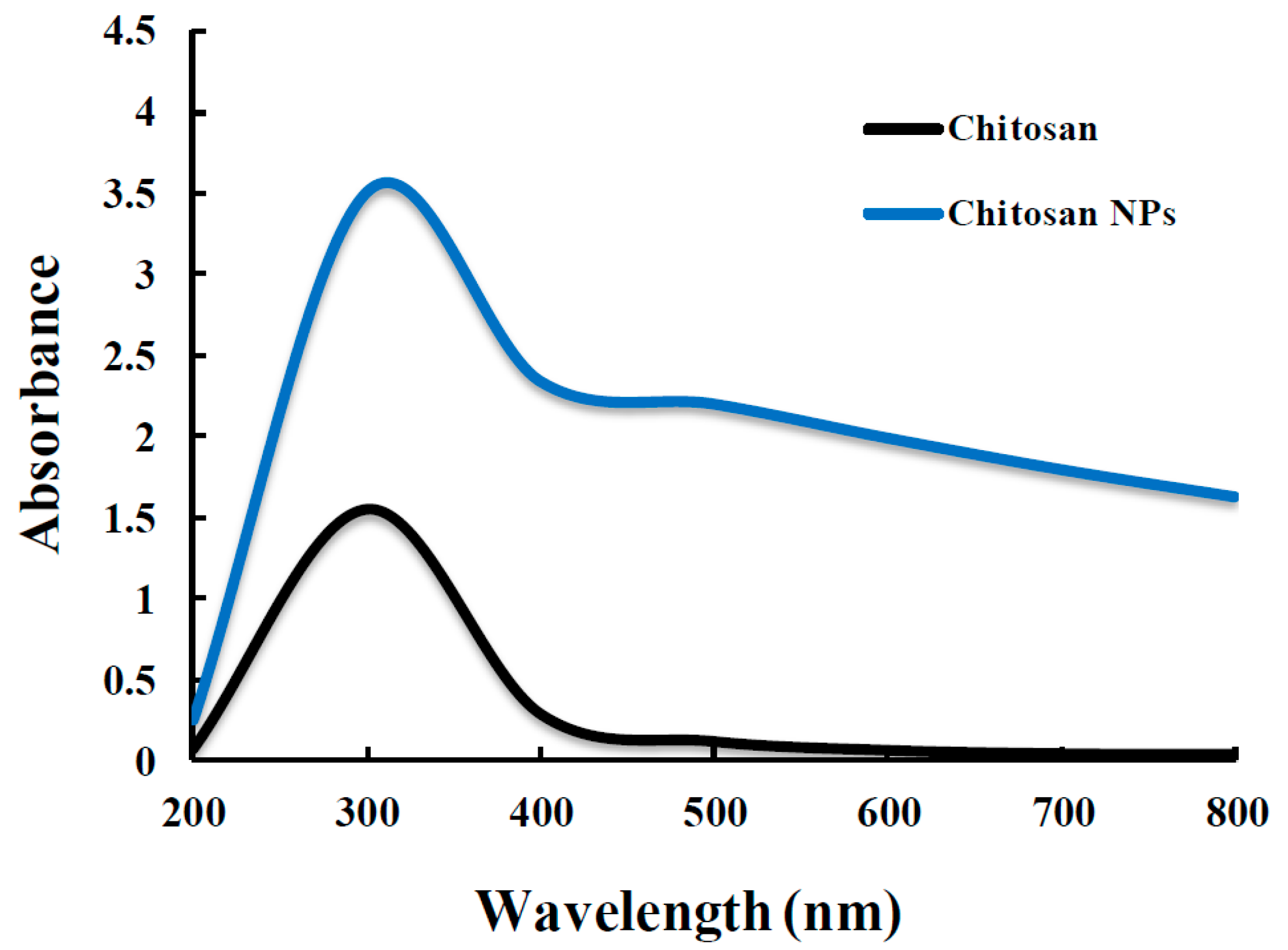
2.2.1. UV-Visible Spectra and Dynamic Light Scattering (DLS) Measurements

UV-visible spectra were recorded using a Shimadzu UV-visible1800 spectrophotometer for the confirmation of nanoparticle formation. Dynamic light scattering (DLS) was used for the measurement of average particle size, and polydispersity index (PDI) on a high-performance particle Zetasizer HPPS-5001 (Malvern, UK). Each sample was analyzed in triplicate at 25 °C at a scattering angle of 90 °C. Pure water was used as a reference for dispersing medium. The results are given as the average particle size obtained from the analysis of three different batches, each of them measured three times.

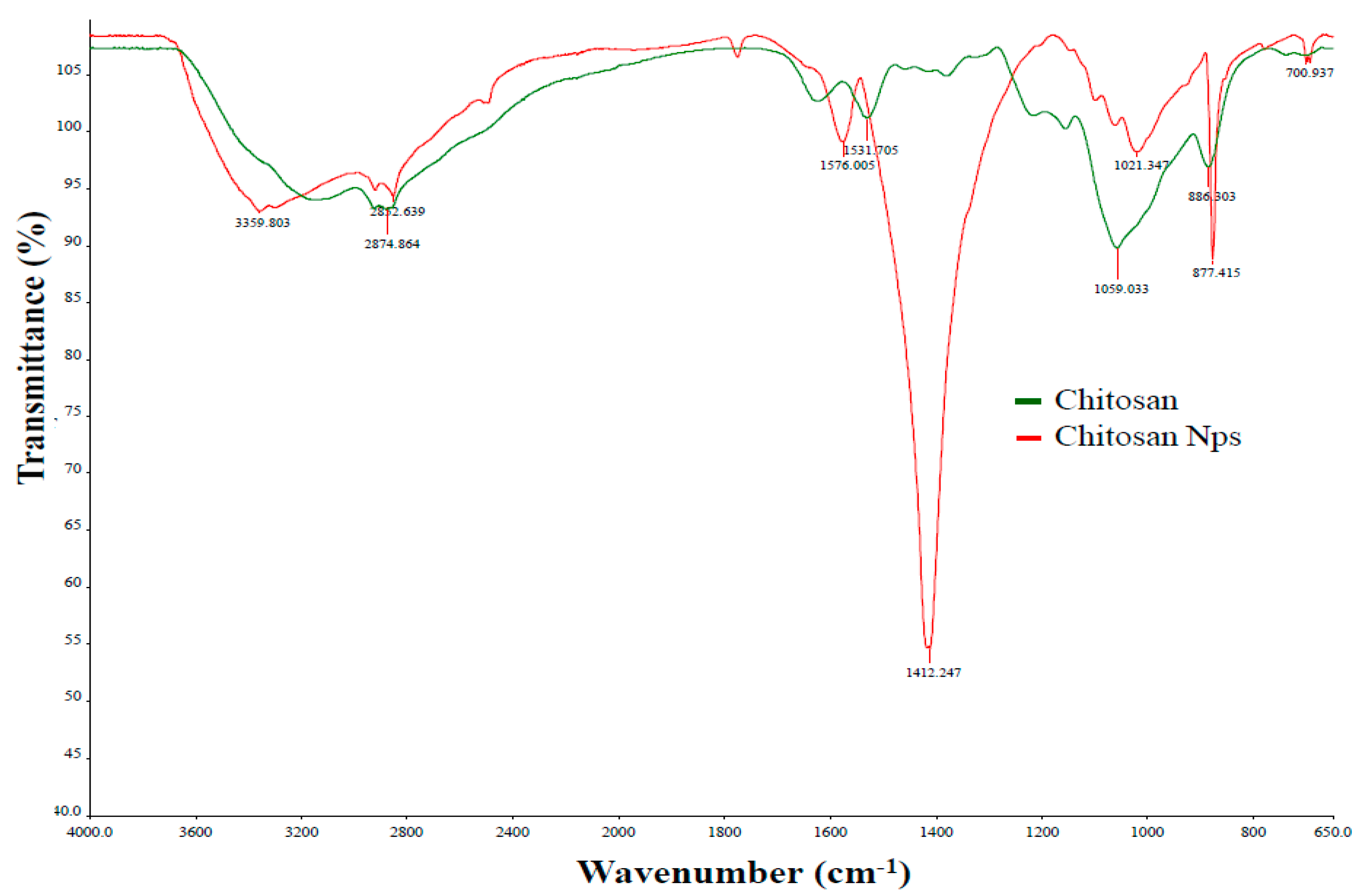
2.2.2. Fourier Transform Infrared (FTIR) Analysis

To confirm the synthesis of nanoparticles, Fourier transform infrared (FTIR) analysis was done. For FTIR, each sample was prepared in potassium bromide (KBr) as a pellet under 1:99 ratio of sample to KBr, and was recorded by ABB FTLA 2000-100 (ABB Co., Quebec, Canada) at a resolution limit of 16 cm−1.

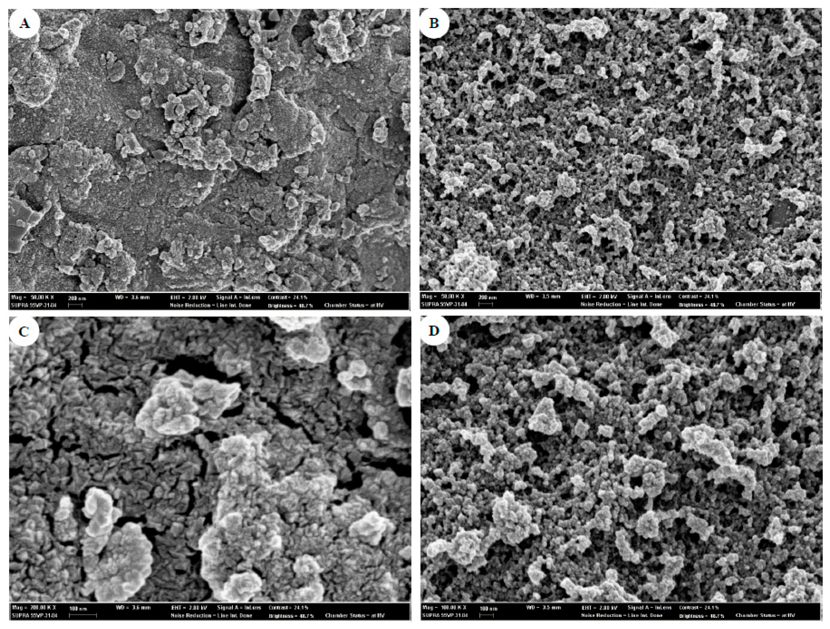
2.2.3. Scanning Electron Microscopy (SEM) Observation

The scanning electron microscope (SEM) was used to study the surface morphology of NPs. The samples were dried by critical point drying (CPD, Emitech) and mounted on aluminium stubs and then coated with gold using a Sputter coater model E-1010 (Emitech). The samples were examined using a scanning electron microscope model S 2700 (Hitachi Ltd, Tokyo, Japan) with 15 kV accelerating voltage.

2.3. Bacterial Strains

2.3.1. Bacterial Strains and Growth Conditions

To obtain the antibacterial activity of CS and CNPs, one strain of X. campestris pv. vesicatoria (KACC1154) and three strains of E. carotovora subsp. carotovora (KACC 113114, 113154, and 133061) that cause leaf spot disease and soft rot disease of Solanum lycopersicum, respectively. The bacterial strains were cultured on nutrient agar medium at 30 °C. After 48 h of incubation, each bacterial suspension was prepared in Luria–Bertani broth (LB).

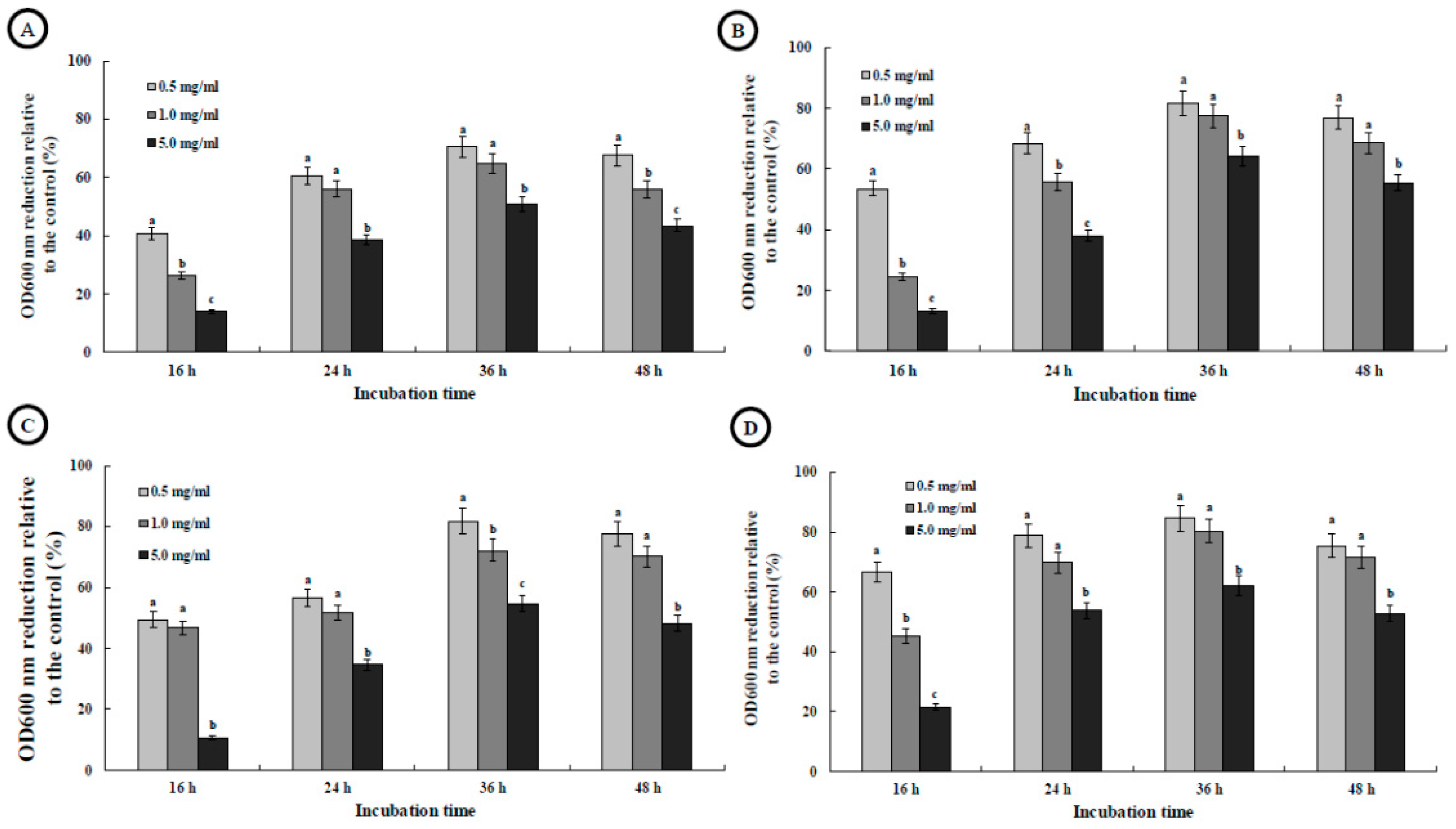
2.3.2. Antibacterial Activities

The inhibition activity of both CS and CNPs against Xanthomonas and Erwinia phytopathogenic bacteria were evaluated by measuring optical density (OD) 600 nm. Xanthomonas and Erwinia strains were cultivated in nutrient agar and incubated at 28 °C. A representative colony was picked off and placed in plus-LB (peptone 10 g, yeast extract powder 5 g, NaCl 5 g, distilled water up to 1000 mL, glucose 1 g, pH 7.2) and incubated overnight on the rotary shaker at 180 rpm, 30 °C. Different concentrations (0.5, 1.0, and 5.0 mg/mL, (w/v)) of CS and CNPs were added to the cultured bacterial suspension and then the mixture was incubated at 30 °C on a rotary shaker at 180 rpm for 48 h while the same volume of sterile distilled water was added to the controls. The absorbance of samples was measured at 600 nm.

2.4. Fungal Strains

2.4.1. Fungal Strains and Growth Conditions

The antifungal properties of CS and CNPs against phytopathogenic fungi namely C. gelosporidies, P. capsici, S. sclerotium, F. oxysporum, and G. fujikori were investigated at various concentrations of CS and CNPs ranging from 0.1 to 5.0 mg/mL. Potato dextrose agar (PDA) medium was prepared and poured in Petri dishes with above-mentioned percentages of various CS and CNPs, separately.

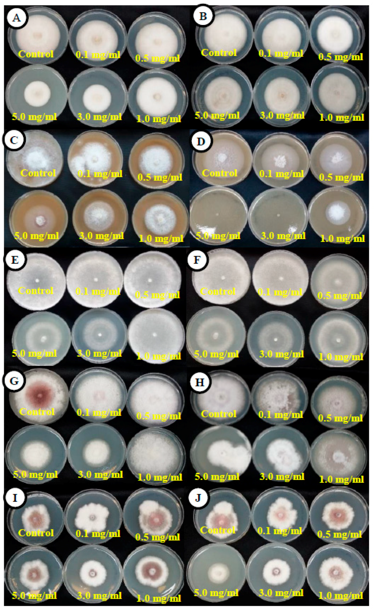
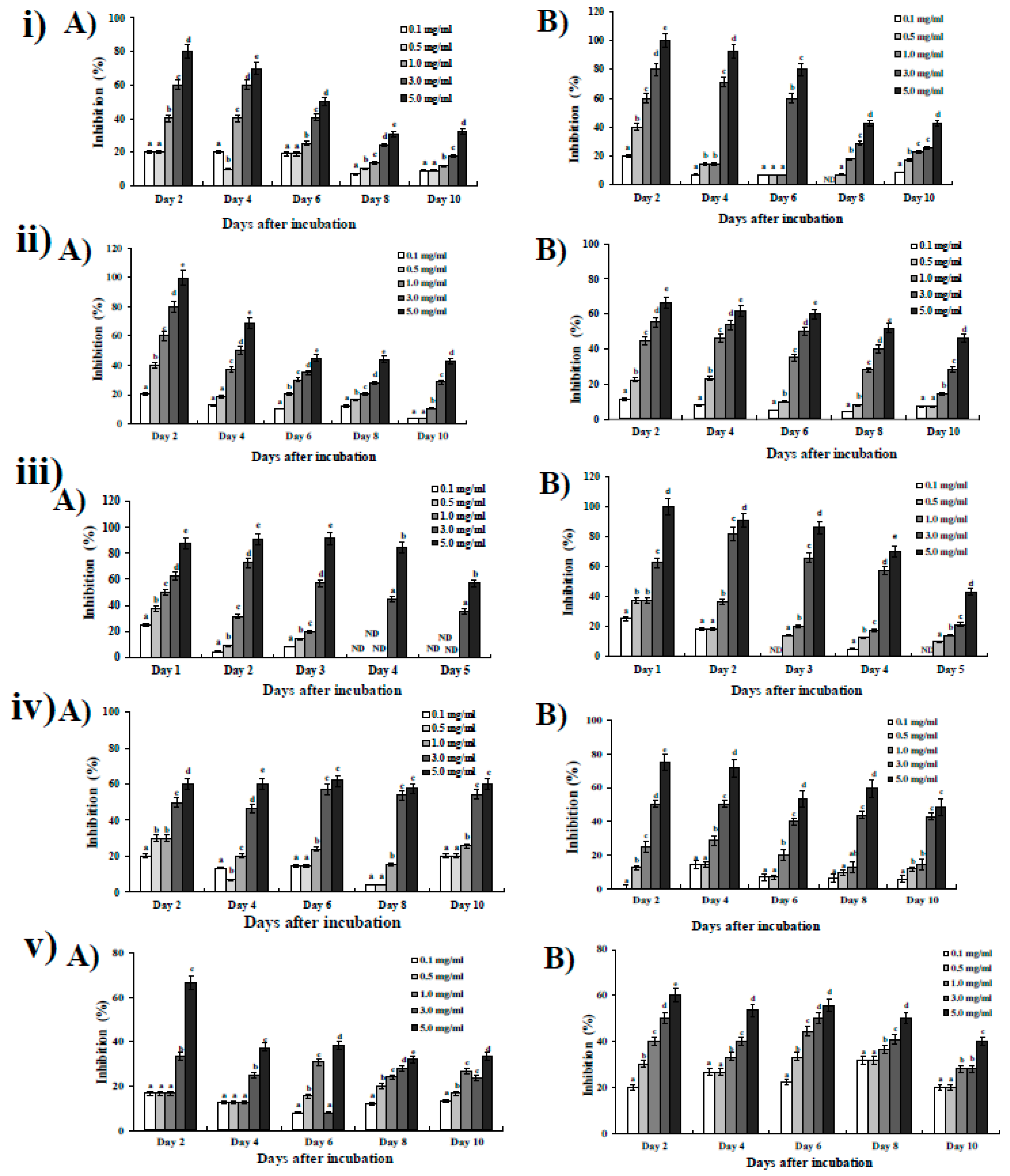
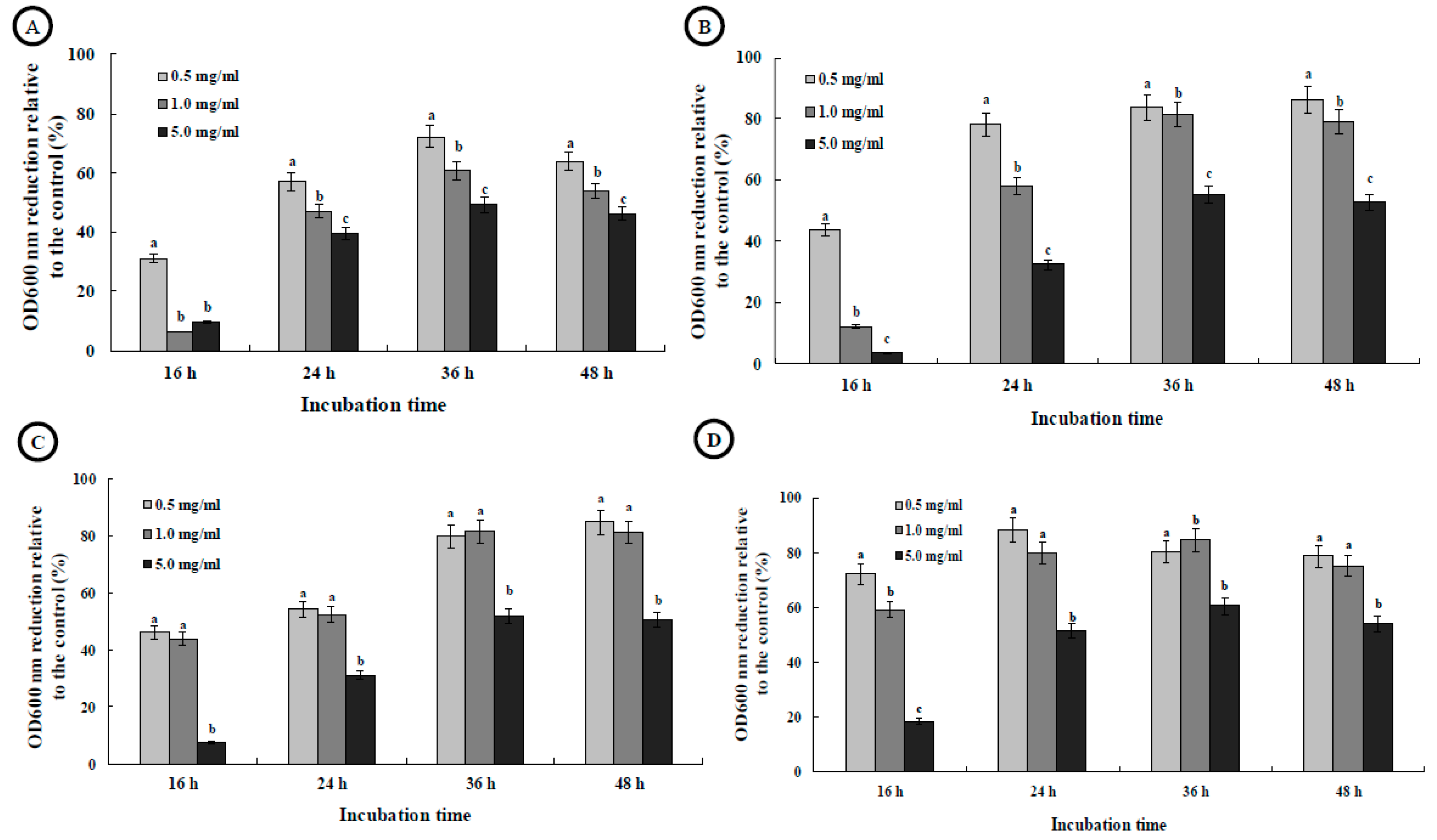
2.4.2. Antifungal Activities

Antifungal assay of CS was conducted for both the radial growth determination of fungi. For radial growth determination, the sterile CS and CNPs solution were added to PDA at a concentration of 0.1, 0.5, 1.0, 3.0, and 5.0 mg/mL, (w/v). Mycelial agar plugs from the actively growing peripheral end of uniform size (diameter, 5.0 mm) were taken from the 7-day old culture of the test pathogens and inoculated in the center of plates supplemented with different concentrations of CS and CNPs. All the Petri dishes were incubated at 28 °C for 10 days, and the observation of radial mycelial growth was recorded when controlling Petri dish was covered with full growth. Wherein, S. sclerotiorum plates were incubated for 5 days, owing to their fast growth. All the treatments consisted of three replications. The inoculated plates were compared with control (without CS and CNPs) to calculate the percentage inhibition rate of mycelia of the pathogen. The percentages of growth inhibition were calculated relative to control using the following formula:
% Inhibition rate = (Mc − Mt) Mc × 100
where Mc is the mycelial growth in control, Mt is the mycelial growth in treatment.

2.5. Statistical Analysis

All of the experiments were conducted in triplicate and results were tabulated as the mean ± standard deviation (SD). Data were analyzed by analysis of variance (ANOVA). Student’s t-test and the probability values of p < 0.05 were considered to be significant.

3. Results and Discussion

The optical properties of biopolymer materials were analysis by UV-Visible spectroscopy and are shown in Figure 1. A UV-Visible spectrum of CS obtained a broad absorption band intensity compared with CNPs (sharp intensity), and the absorption peak wavelength was at 320 nm in the UV region. But in the case of CNPs, a higher intensity level than CS biopolymer was observed, which is due to the formation of NPs. CS capped AuNPs were proved to uptake analytes, such as amorphous carbon nanotubes, copper oxide, and Zinc sulfate, which indirectly shows the ability of CNPs to neutralize the adverse effects of chemical fertilizers [28]. Hence, CNPs, apart from providing plant protection and crop productivity, have a neutralizing effect on the uptake of harmful chemical remnants in the soil. As organic farming is largely stressed these days, green nanotechnology will have positive outcomes without deleterious outcomes to Mother Earth.
The particles size distribution of the CNPs were measured by DLS and is shown in Figure 2. The CNPs showed characteristic chemical changes that occurred during the formation of CNPs from the CS biopolymer source at different experimental conditions. The size of CNPs was in the diameter range of ~100 to 1000 nm, with an early correlation coefficient decay curve and polydispersity index. Escalated antifungal activity against Fusarium Head Blight (FHB) of wheat has been attributed to low molecular weight CNPs, and they could possibly replace the use of chemical fertilizers in the abatement of FHB [29]. The DLS method in the emancipation of antifungal characteristics in broad-spectrum activity in S. lycopersicum also shows a similar effect.
These results were demonstrated by Kheiri et al. [29] under field trails for effective crop protection strategies with the use of CNPs. Earlier, CNPs with a size of less than 400 suppressed wilt disease in tomatoes caused by F. oxysporum f. sp. Lycopersici with a corresponding yield increase [30]. The present study shows the significant scale of outcome even at a range of 100. Hence, the initial DLS analysis intrigues us and can be the basis for future combinatorial analysis for various phytopathogens not only in tomato but also against a variety of crop plants to determine the reliability for CNPs. CS and CNPs functional groups are studied using FTIR spectroscopy. The FTIR spectra of CS and CNPs are shown in Figure 3.
A peak at 3500 to 3300 cm−1 was observed for the main functional group of chitosan and is due to the O-H group of stretching vibrations. The presence of absorption peaks at 1630 and 1531 cm−1 are due to the N-H bending vibration of protonated amino (−NH2) group and C-H bending vibration of the alkyl group. The absorption peaks at 1059 and 886 cm−1 are recognized due to the anti-symmetric stretching vibration of C-O-C bridges and assigned to glucopyranose ring in chitosan matrix. A similar study by Saharan et al. [14] employing Cu-CNPs against pathogenic fungi of tomato showed that spectral peaks for Cu-CNPs were sharp and a shift was observed at 1631 and 1536 cm−1. Whereas, the present results corroborate a minimal variation which suggests that Cu bonding influences the variation. It can be deduced that the nanoencapsulation of CNPs will have varied distribution frequencies that might influence the optimal benefits for agricultural practices. The present study involves CNPs without any further encapsulation. The results affirm that CNPs without any encapsulation can be of significant benefit. Hence, we propose the use of the beneficial nano-encapsulation particle which would boost green agriculture and provide multiple benefits owing to Nanotechnology in Agriculture. On the contrary, chitosan characteristic peaks are shifted to 3359, 2874, 1576, 1412, 1021, 877, and 700 cm−1, when compared with corresponding CNPs. This indicates that an interaction between chitosan and treatment nanoparticles which is an indication of a chemical reaction. However, there need to be further trials for effective field applications. In this regard, it can be concluded that the encapsulation of two herbicides namely, Imazapic and Imazapyr with CNPs is an effective strategy for sustainable agriculture with reduced harm to humans and the environment. The mode of action of this formulation was questioned for a molecular dissection study for affirming the positivity of its role [24]. Although there are several reports pertaining to nanotechnology in progressive agriculture, there are still unanswered questions in the field of CNPs, such as the choice of material for nanoencapsulation which aggregates with CNPs with consideration towards a clear and efficient result for boosting crop production and productivity together with crop protection which has minimal detrimental effects on soil. We have reviewed numerous reviews and researches and believe that the field of Nanotechnology in Agriculture is still underexploited.
The CNPs peak shifted to 1576 cm−1 is due to the wagging of NH2 bond and the strong peak at 1412 cm−1 is due to C-H bending vibration of the alkyl group. The ionic interaction with the treated molecules indicates the conversion of chitosan polymer in the nano form that forms a cross-link with the treated molecules. The surface morphological structure was further examined by SEM analysis. The SEM images of CS and CNPs are displayed at the higher and lower magnification scale of 200 and 100 nm as shown in Figure 4. The morphological structure of CS shows the large particle size and agglomerated state. The SEM images of CNPs clearly show a spherical-like structure and particles in the agglomerated state. Further, the uniformity of the size of the nanoparticles can be observed.
The above results clearly indicate that the CNPs show a highly porous surface due to agglomeration attributes. The porous nature and agglomeration capabilities of the CNPs render them useful as a critical chitosan-based bio-nanopesticide [15]. Agglomeration has been considered as the primary phenomenon for synthesis for novel CNPs for biomedical applications and nanomedicine [31]. The same concept holds for agricultural. Hence, the porous nature can harbor quenching molecules to effectively adsorb harmful chemicals in soil.
This study was carried out to evaluate different concentrations of CS and CNPs against plant pathogenic fungi and bacteria under laboratory conditions. The main concept of this study is dependent on the comparison between the effectiveness of CS and nano-CS concentrations to inhibit phytopathogens. Antifungal effects of CS and CNPs were evaluated against plant pathogenic fungi. The NPs inhibited the radial growth of pathogens at different concentration levels, and all the tested cultures showed a clear significant effect compared with the control treatment (Figure 5 and Figure 6). In the present study, we also found some differences in the antifungal activity in both plate assay and percentage inhibition of CS and CNPs particularly against Colletotrichum gelosporidies and Gibberella fujikuori. These differences in the activity of the fungal species might be due to the presence of some resistance mechanism against these compounds. The differences we found in the percentage of inhibition prove that not all biological systems exhibit similar behavior under the influence of the same external agent.
Management of fungal diseases in food crops is economically important. Recently, a greater emphasis has been given to the development of safe management methods that pose less danger to humans and animals, with a focus on overcoming the deficiencies of synthetic fungicides. CNPs show broad-spectrum activities, such as plant growth promotion, biocide, and plant protection [32].
In this study, CS and CNPs markedly inhibited the growth of the one Xanthomonas strain and three Erwinia strains as seen by measuring the OD value at 600 nm. The reduction in the OD of cell suspension depends on the type of CS and the species of bacteria. CS and CNPs and bacterial species significantly affected the surviving cell numbers (Figure 7 and Figure 8). This result revealed that the growth of X. campestris and E. carotovora was inhibited by CS and CNPs regardless of the kinds of CS and the species of bacteria, which implies that the two kinds of CS were good bactericide for the control of bacterial disease of tomato. Results from this study indicated that the addition of chitosan at 0.5 to 5.0 mg/mL to the two bacterial strains caused a reduction in the OD600nm after 48 h of incubation. The reduction percentage of CS in the OD600nm ranged from 4.4% to 86.97% as compared to the control while the reduction percentage in most of the strains were more than 50.00% (Figure 7).
This result was consistent with the previous result, which found CS had strong antibacterial activity against nine strains of X. arboricola pv. poinsettiicola and X. axonopodis pv. poinsettiicola from different geographic sources based on the colony count method [33]. Similarly, the addition of CNPs to four bacterial strains caused a reduction in the OD600nm after 48 h of incubation. The reduction percentage in the OD600nm ranged from 10.61% to 84.75% as compared to the control while the reduction percentage in strain 13114 was more than 40.00% (Figure 8).
However, in this study, the three Erwinia strains, in general, showed a difference in the sensitivity to CS and CNPs. In particular, the reduction percentage of CS in the OD600nm of strain 13114, 113154, and 133061 was 45.42%, 53.72%, and 51.33% respectively, while the reduction percentage of CNPs in the OD600nm of strain 13114, 113154 and 133061 was 43.31%, 55.37%, and 48.26% respectively, as compared to the corresponding control. In contrast, strain X. campestris 1154 showed susceptibility to both CS and CNPs, which caused the reduction in OD600nm by 54.53% and 53.03%, respectively, as compared to the control. The difference in the sensitivity of bacteria to CS may be attributed to the complexity of interaction between the two kinds of chitosan and these Erwinia and Xanthomonas strains. The above results show prominent antibacterial activities. Cationic properties of CS rather than CNPs induce positively charged quaternary groups to hydroxyl or amino groups that are primarily responsible for antibacterial activities [34]. This aspect clearly depicts that antibacterial activity is an inherent property of CS rather than CNPs. Hence the evidence of broad-spectrum antifungal activity of CNPs and chitosans and their effective antibacterial activity might open up a new perspective in the analysis of CNPs vs. CS when addressing antifungal and antibacterial potentials.

4. Conclusions

In the present work, it was demonstrated that chitosan has significant antifungal activity against the fungi tested, viz. C. gelosporidies, P. capsici, S. sclerotiorum, F. oxysporum, and G. fujikori, and this was much higher than when chitosan nanoparticles were used independently. Thus, CNPs can be effectively used against plant phytopathogenic fungi ensuring a plethora of positive outcomes ranging from antifungal, antibacterial activities, plant growth promotion, biocidal activities, and reduction in harmful effects to humans and environment due to chemical fertilizers. This research also addresses the fact that CNPs are responsible for broad-spectrum activity against phytopathogens of tomato. This opens up new research in the search for an effective combination of CNPs with other elemental forms to ensure broad-spectrum antimicrobial activity affirmatively.

Author Contributions

M.C., J.-W.O. and S.C.C. planned and designed the research; M.C. performed the experiments; M.C. and J.-W.O. wrote the manuscript together with assistance from S.C.C.

Funding

This research received no external funding.

Conflicts of Interest

Authors declare there is no conflict of interest.

References

  1. Duhan, J.S.; Kumar, R.; Kumar, N.; Kaur, P.; Nehra, K.; Duhan, S. Nanotechnology: The new perspective in precision agriculture. Biotechnol. Rep. 2017, 15, 11–23. [Google Scholar] [CrossRef] [PubMed] [Green Version]
  2. Mohamed, N.A.; Sabaa, M.W.; El-Ghandour, A.H.; Abdel-Aziz, M.M.; Abdel-Gawad, O.F. Quaternized N-substituted carboxymethyl chitosan derivatives as antimicrobial agents. Int. J. Biol. Macromol. 2013, 60, 156–164. [Google Scholar] [CrossRef] [PubMed]
  3. Sajomsang, W.; Ruktanonchai, U.R.; Gonil, P.; Warin, C. Quaternization of N-(3-pyridylmethyl) chitosan derivatives: Effects of the degree of quaternization, molecular weight and ratio of N-methylpyridinium and N,N,N-trimethyl ammonium moieties on bactericidal activity. Carbohydr. Polym. 2010, 82, 1143–1152. [Google Scholar] [CrossRef]
  4. El Hadrami, A.; Adam, L.R.; El Hadrami, I.; Daayf, F. Chitosan in plant protection. Mar. Drugs 2010, 8, 968–987. [Google Scholar] [CrossRef] [PubMed]
  5. Li, Z.; Yang, F.; Yang, R. Synthesis and characterization of chitosan derivatives with dual-antibacterial functional groups. Int. J. Biol. Macromol. 2015, 75, 378–387. [Google Scholar] [CrossRef] [PubMed]
  6. Iriti, M.; Faoro, F. Chitosan as a MAMP, searching for a PRR. Plant Signal Behav. 2009, 4, 66–68. [Google Scholar] [CrossRef] [PubMed] [Green Version]
  7. Khot, L.R.; Sankaran, S.; Maja, J.M.; Ehsani, R.; Schuster, E.W. Applications of nanomaterials in agricultural production and crop protection: A. review. Crop Prot. 2012, 35, 64–70. [Google Scholar] [CrossRef]
  8. Park, H.J.; Kim, S.H.; Kim, H.J.; Choi, S.H. A new composition of nanosized silica-silver for control of various plant diseases. J. Plant Pathol. 2006, 22, 295–302. [Google Scholar] [CrossRef]
  9. He, L.; Liu, Y.; Mustapha, A.; Lin, M. Antifungal activity of zinc oxide nanoparticles against Botrytis cinerea and Penicillium expansum. Microbiol. Res. 2011, 166, 207–215. [Google Scholar] [CrossRef]
  10. Shukla, S.K.; Mishra, A.K.; Arotiba, O.A.; Mamba, B.B. Chitosan-based nanomaterials: A state-of-the-art review. Int. J. Biol. Macromol. 2013, 59, 46–58. [Google Scholar] [CrossRef]
  11. Banerjee, T.; Mitra, S.; Kumar Singh, A.; Kumar Sharma, R.; Maitra, A. Preparation, characterization and biodistribution of ultrafine chitosan nanoparticles. Int. J. Pharm. 2002, 243, 93–105. [Google Scholar] [CrossRef]
  12. Qi, L.; Xu, Z.; Jiang, X.; Hu, C.; Zou, X. Preparation and antibacterial activity of chitosan nanoparticles. Carbohydr. Res. 2004, 339, 2693–2700. [Google Scholar] [CrossRef] [PubMed]
  13. Du, W.L.; Niu, S.S.; Xu, Y.L.; Xu, Z.R.; Fan, C.L. Antibacterial activity of chitosan tripolyphosphate nanoparticles loaded with various metal ions. Carbohydr. Polym. 2009, 75, 385–389. [Google Scholar] [CrossRef]
  14. Saharan, V.; Mehrotra, A.; Khatik, R.; Rawal, P.; Sharma, S.S.; Pal, A. Synthesis of chitosan-based nanoparticles and their in vitro evaluation against phytopathogenic fungi. Int. J. Biol. Macromol. 2013, 62, 677–683. [Google Scholar] [CrossRef] [PubMed]
  15. Saharan, V.; Sharma, G.; Yadav, M.; Choudhary, M.K.; Sharma, S.S.; Pal, A.; Raliya, P.; Biswas, R. Synthesis and in vitro antifungal efficacy of Cu-chitosan nanoparticles against pathogenic fungi of tomato. Int. J. Biol. Macromol. 2015, 75, 346–353. [Google Scholar] [CrossRef] [PubMed]
  16. Puoci, F.; Lemma, F.; Spizzirri, U.G.; Cirillo, G.; Curcio, M.; Picci, N. Polymer in agriculture: A review. Am. J. Agri. Biol. Sci. 2008, 3, 299–314. [Google Scholar] [CrossRef]
  17. Corradini, E.; Moura, M.R.; Mattoso, L.H.C. A preliminary study of the incorporation of NPK fertilizer into chitosan nanoparticles. eXPRESS Polym. Lett. 2010, 4, 509–515. [Google Scholar] [CrossRef]
  18. Hasaneen, M.N.A.; Abdel-Aziz, H.M.M.; El-Bialy, D.M.A.; Omer, A.M. Preparation of chitosan nanoparticles for loading with NPK fertilizer. Afr. J. Biotechnol. 2014, 13, 3158–3164. [Google Scholar]
  19. Tao, S.; Pang, R.; Chen, C.; Ren, X.; Hu, S. Synthesis, characterization and slow release properties of O-naphthylacetyl chitosan. Carbohydr. Polym. 2012, 88, 1189–1194. [Google Scholar] [CrossRef]
  20. Kumar, S.; Chauhan, N.; Gopal, M.; Kumar, R.; Dilbaghi, N. Development and evaluation of alginate-chitosan nanocapsules for controlled release of acetamiprid. Int. J. Biol. Macromol. 2015, 81, 631–637. [Google Scholar] [CrossRef]
  21. Chandra, S.; Chakraborty, N.; Dasgupta, A.; Sarkar, J.; Panda, K.; Acharya, K. Chitosan nanoparticles: A positive modulator of innate immune responses in plants. Sci. Rep. 2015, 5, 15195. [Google Scholar] [CrossRef] [PubMed] [Green Version]
  22. Cota, A.O.; Cortez, R.M.O.; Burgos, H.A.; Ezquerra, B.J.M.; Plascencia, J.M. Controlled release matrices and micro/nanoparticles of chitosan with antimicrobial potential: Development of new strategies for microbial control in agriculture. J. Sci. Food Agric. 2013, 93, 1525–1536. [Google Scholar] [CrossRef] [PubMed]
  23. Saharan, V.; Kumaraswamy, R.V.; Choudhary, R.C.; Kumari, S.; Pal, A.; Raliya, R.; Biswas, P. Cu-chitosan nanoparticle mediated sustainable approach to enhance seedling growth in maize by mobilizing reserved food. J. Agric. Food Chem. 2016, 64, 6148–6155. [Google Scholar] [CrossRef] [PubMed]
  24. Maruyama, C.R.; Guilger, M.; Pascoli, M.; Bileshy-José, N.; Abhilash, P.C.; Fraceto, L.F.; de Lima, R. Nanoparticles based on chitosan as carriers for the combined herbicides imazapic and imazapyr. Sci. Rep. 2016, 6, 23854. [Google Scholar] [CrossRef] [PubMed]
  25. Malerba, M.; Cerana, R. Recent advances of chitosan applications in plants. Polymers 2018, 10, 118. [Google Scholar] [CrossRef]
  26. Sajomsang, W.; Gonil, P.; Saesoo, S.; Ovatlarnporn, C. Antifungal property of quaternized chitosan and its derivatives. Int. J. Biol. Macromol. 2012, 50, 263–269. [Google Scholar] [CrossRef] [PubMed]
  27. Bautista-Baños, S.; Hernández-López, M.; Bosquez-Molina, E.; Wilson, C.L. Chitosan controls postharvest anthracnose in bell pepper by activating defense-related enzymes. Crop Prot. 2003, 22, 1087–1092. [Google Scholar]
  28. Sultan, N.M.; Johan, M.R. Synthesis and ultraviolet visible spectroscopy studies of chitosan capped gold nanoparticles and their reactions with analytes. Sci. World J. 2014, 2014, 1–7. [Google Scholar] [CrossRef]
  29. Kheiria, A.; Moosawi Jorfa, S.A.; Malihipourb, A.; Saremic, H.; Nikkhahda, M. Synthesis and characterization of chitosan nanoparticles and their effect on Fusarium head blight and oxidative activity in wheat. Int. J. Biol. Macromol. 2017, 102, 526–538. [Google Scholar] [CrossRef]
  30. Sathiyabama, M.; Charles, R.E. Fungal cell wall polymer-based nanoparticles in protection of tomato plants from wilt disease caused by Fusarium oxysporum f. sp. lycopersici. Carbohydr. Polym. 2015, 133, 400–407. [Google Scholar] [CrossRef]
  31. Ghadi, A.; Mahjoub, S.; Tabandeh, F.; Talebnia, F. Synthesis and optimization of chitosan nanoparticles: Potential applications in nanomedicine and biomedical engineering. Casp. J. Intern. Med. 2014, 5, 156–161. [Google Scholar]
  32. Sathiyabama, M.; Parthasarathy, R. Biological preparation of chitosan nanoparticles and its in vitro antifungal efficacy against some phytopathogenic fungi. Carbohydr. Polym. 2016, 151, 321–325. [Google Scholar] [CrossRef] [PubMed]
  33. Li, B.; Wang, X.; Chen, R.; Huangfu, W.G.; Xie, G.L. Antibacterial activity of chitosan solution against Xanthomonas pathogenic bacteria isolated from Euphorbia pulcherrima. Carbohyd. Polym. 2008, 72, 287–292. [Google Scholar] [CrossRef]
  34. Cheung, R.C.F.; Ng, T.B.; Wong, J.H.; Chan, W.Y. Chitosan: An update on potential biomedical and pharmaceutical applications. Mar. Drugs. 2015, 13, 5156–5186. [Google Scholar] [CrossRef] [PubMed]
Figure 1. UV-visible spectrum of synthesized chitosan and chitosan nanoparticle.
Agronomy 09 00021 g001
Figure 2. (a) Dynamic light scattering (DLS) of the synthesized chitosan nanoparticles. (b) The effective zeta-potential in aqueous solution were measured by particle characterizer.
Agronomy 09 00021 g002
Figure 3. Fourier transform infrared (FTIR) spectrum of chitosan and chitosan nanoparticle.
Agronomy 09 00021 g003
Figure 4. Scanning electron microscope (SEM) images of chitosan (A,C) and chitosan nanoparticles (B,D).
Agronomy 09 00021 g004
Figure 5. Antifungal activity of chitosan nanoparticles (NPs)-Plate assay. (A) Colletotrichum gelosporidies-Chitosan, (B) Colletotrichum gelosporidies-Chitosan NPs, (C) Phytophthora capsici-Chitosan, (D) Phytophthora capsici-Chitosan NPs, (E) Sclerotium sclerotiorum -Chitosan, (F) Sclerotium sclerotiorum-Chitosan NPs, (G) Fusarium oxysporum-Chitosan, (H) Fusarium oxysporum-chitosan NPs, (I) Gibberella fujikori-Chitosan, (J) Gibberella fujikuori-Chitosan NPs. All the Petri dishes were incubated at 28 °C for 10 days whereas Sclerotinia sclerotiorum plates were incubated for 5 days, owing to their fast growth.
Agronomy 09 00021 g005
Figure 6. Antifungal activity of chitosan nanoparticles-Inhibition studies (i) Colletotrichum gelosporidies-A. Chitosan B. Chitosan NPs. (ii) Phytophthora capsici-A. Chitosan B. Chitosan NPs. (iii) Sclerotinia sclerotiorum-A. Chitosan B. Chitosan NPs. (iv) Fusarium oxysporum-A. Chitosan B. Chitosan NPs. (v) Gibberella fujikuori-A. Chitosan B. Chitosan NPs. The percentages of growth inhibition were calculated relative to control.
Agronomy 09 00021 g006
Figure 7. Effect of chitosan on the growth of Xanthomonas campestris pv. vesicatoria and Erwinia cartovora subsp. carotovora pathogenic to Solanum lycopersicum. (A) Erwinia cartovora subsp. carotovora 113114. (B) Erwinia cartovora subsp. carotovora 113154. (C) Erwinia cartovora subsp. carotovora YKB133061. (D) Xanthomonas campestris pv. vesicatoria 11154.
Agronomy 09 00021 g007
Figure 8. Effect of chitosan nanoparticles on the growth of Xanthomonas campestris pv. vesicatoria and Erwinia carotovora subsp. carotovora pathogenic to Solanum lycopersicum. (A) Erwinia carotovora subsp. carotovora 113114. (B) Erwinia carotovora subsp. carotovora 113154. (C) Erwinia carotovora subsp. carotovora YKB133061. (D) Xanthomonas campestris pv. vesicatoria 11154.
Agronomy 09 00021 g008

