Pseudomonas azotoformans Belonging to Pseudomonas fluorescens Group as Causative Agent of Blue Coloration in Carcasses of Slaughterhouse Rabbits

 ,
,  , , and
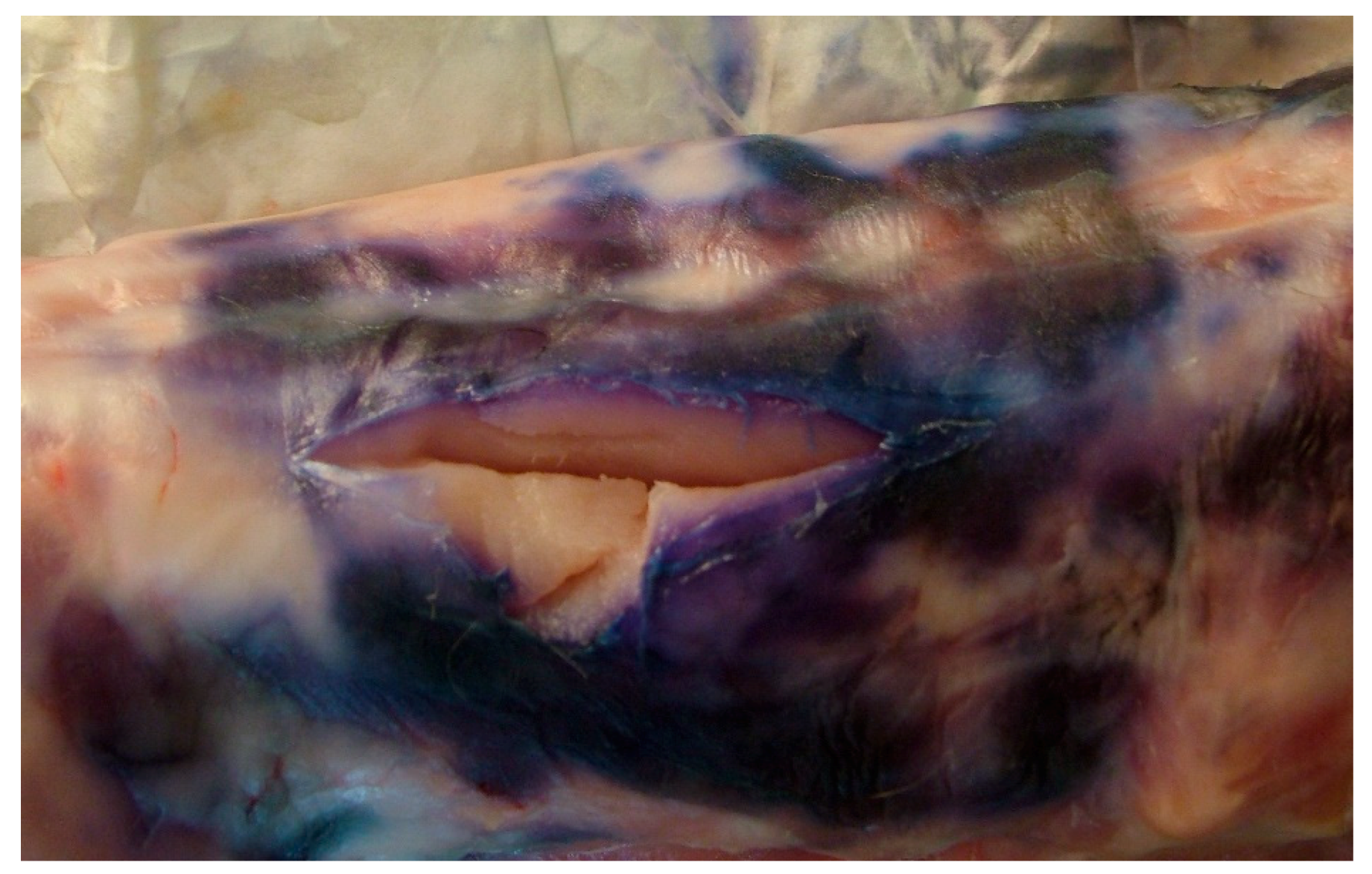
Abstract
:Simple Summary
Abstract
1. Introduction
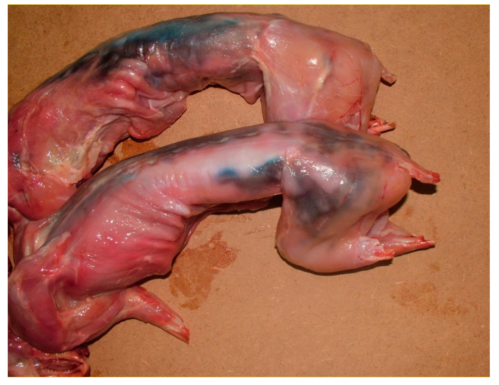
2. Case History
3. Laboratory Investigations
4. Results
5. Discussion
6. Conclusions
Supplementary Materials
Author Contributions
Funding
Acknowledgments
Conflicts of Interest
References
- Decimo, M.; Morandi, S.; Silvetti, T.; Brasca, M. Characterization of Gram negative psychrotrophic bacteria isolated from Italian bulk tank milk. J. Food Sci. 2014, 79, M2081–M2090. [Google Scholar] [CrossRef]
- De Jonghe, V.; Coorevits, A.; Van Hoorde, K.; Messens, W.; Van Landschoot, A.; De Vos, P.; Heyndrickx, M. Influence of storage conditions on the growth of Pseudomonas species in refrigerated raw milk. Appl. Environ. Microbiol. 2011, 77, 460–470. [Google Scholar] [CrossRef] [Green Version]
- Anzai, Y.; Kim, H.; Park, J.Y.; Wakabayashi, H.; Oyazu, H. Phylogenetic affiliation of the pseudomonas based on 16S rRNA sequences. Int. J. Syst. Evol. Microbiol. 2000, 50, 1563–1589. [Google Scholar] [CrossRef] [PubMed] [Green Version]
- Frapolli, M.; Défago, G.; Moënne-Loccoz, Y. Multilocus sequence analysis of biocontrol fluorescent Pseudomonas spp. producing the antifungal compound 2,4-diacetylphloroglucinol. Environ. Microbiol. 2007, 9, 1939–1955. [Google Scholar] [CrossRef] [PubMed]
- Tümmler, B.; Wiehlmann, L.; Klockgether, J.; Cramer, N. Advances in understanding Pseudomonas. F1000 Prime Rep. 2014, 6, 9. [Google Scholar] [CrossRef] [PubMed]
- Peix, A.; Ramirez-Bahena, M.H.; Velazquez, E. Historical evolution and current status of the taxonomy of genus Pseudomonas. Infect. Genet. Evol. 2009, 9, 1132–1147. [Google Scholar] [CrossRef]
- Caldera, L.; Franzetti, L. Effect of storage temperature on the microbial composition of ready-to-use vegetables. Curr. Microbiol. 2014, 68, 133–139. [Google Scholar] [CrossRef]
- Marchand, S.; Heylen, K.; Messens, W.; Coudijzer, K.; De Vos, P.; Dewettinck, K.; Herman, L.; De Block, J.; Heyndrickx, M. Seasonal influence on heat-resistant proteolytic capacity of Pseudomonas lundensis and Pseudomonas fragi, predominant milk spoilers isolated from Belgian raw milk samples. Environ. Microbiol. 2009, 11, 467–482. [Google Scholar] [CrossRef]
- Drosinos, E.H.; Board, R.G. Microbial and physicochemical attributes of minced lamb: Sources of contamination with pseudomonas. Food Microbiol. 1995, 12, 189–197. [Google Scholar] [CrossRef]
- Altinok, I.; Kayis, S.; Capkin, E. Pseudomonas putida infection in rainbow trout. Aquaculture 2006, 261, 850–855. [Google Scholar] [CrossRef]
- Angelini, N.M.; Seigneur, G.N. Disease of the fins of Rhamdia sapo. Isolation of the etiological agents and experimental infection. Revista Argentina de Microbiologia 1988, 20, 37–48. (In Spanish) [Google Scholar] [PubMed]
- Andreani, N.A.; Martino, M.E.; Fasolato, L.; Carraro, L.; Montemurro, F.; Mioni, R.; Bordin, P.; Cardazzo, B. Tracking the blue: A MLST approach to characterise the Pseudomonas fluorescens group. Food Microbiol. 2014, 39, 116–126. [Google Scholar] [CrossRef] [PubMed]
- Gennari, M.; Dragotto, F. A study of the incidence of different fluorescent Pseudomonas species and biovars in the microflora of fresh and spoiled meat and fish, raw milk, cheese, soil and water. J Appl Microbiol. 1992, 72, 281–288. [Google Scholar] [CrossRef] [PubMed]
- Regulation (EC). N° 853/2004 of the European Parliament and of the Council of 29 April 2004 Laying down Specific Hygiene Rules for Food of Animal Origin. Available online: https://eur-lex.europa.eu/legal-content/EN/TXT/?uri=CELEX%3A32004R0853 (accessed on 4 February 2020).
- Council Regulation (EC). N° 1/2005 of 22 December 2004 on the Protection of Animals during Transport and Related Operations. Available online: https://eur-lex.europa.eu/legal-content/en/ALL/?uri=CELEX:32005R0001 (accessed on 4 February 2020).
- Garrido-Sanz, D.; Arrebola, E.; Martinez-Granero, F.; Garcia-Méndez, S.; Muriel, C.; Blanco-Romero, E.; Martin, M.; Rivilla, R.; Redondo-Nieto, M. Classification of isolates from the Pseudomonas fluorescens complex into phylogenomic groups based in group-specific markers. Front. Microbiol. 2017, 8, 413. [Google Scholar] [CrossRef] [Green Version]
- Lane, D.J. 6S/23S rRNA sequencing. In Nucleic Acid Techniques in Bacterial Systematic; Stackebrandt, E., Goodfellow, M., Eds.; John Wiley and Sons: Chichester, UK, 1991; pp. 115–175. [Google Scholar]
- Huang, X.; Madan, A. CAP3: A DNA sequence assembly program. Genome Res. 1999, 9, 868–877. [Google Scholar] [CrossRef] [Green Version]
- Kumar, S.; Stecher, G.; Knyaz, C.; Tamura, K. MEGA X: Molecular Evolutionary Genetics Analysis across Computing Platforms. Mol. Biol. Evol. 2018, 35, 1547–1549. [Google Scholar] [CrossRef]
- Guindon, S.; Dufayard, J.F.; Lefort, V.; Anisimova, M.; Hordijk, W.; Gascuel, O. New algorithms and methods to estimate maximum-likelihood phylogenies: Assessing the performance of PhyML 3.0. Syst. Biol. 2010, 59, 307–321. [Google Scholar] [CrossRef] [Green Version]
- Posada, D.; Crandall, K.A. MODELTEST: Testing the model of DNA substitution. Bioinformatics 1998, 14, 817–818. [Google Scholar] [CrossRef] [PubMed] [Green Version]
- Bogdanova, T.; Flores Rodas, E.M.; Greco, S.; Tolli, R.; Bilei, S. Indagine microbiologica su campioni di mozzarella in occasione dell’allerta Mozzarella blu. In Proceedings of the XII Congresso Nazionale, S.I.Di.L.V., Genova, Italy, 27–29 October 2010; pp. 48–149. [Google Scholar]
- Palleroni, N.J.; Genus, I. Pseudomonas Migula 1894, 237AL (Nom. Cons., Opin. 5 of the Jud. Comm. 1952, 121). In Bergey’s Manual of Systematic Bacteriology, 2nd ed.; Brenner, D.J., Krieg, N.R., Staley, J.T., Garrity, G.M., Eds.; Springer: New York, NY, USA, 2005; Volume 2, Part B; pp. 328–379. [Google Scholar]
- Yamamoto, S.; Kasai, H.; Arnold, D.L.; Jackson, R.W.; Vivian, A.; Harayama, S. Phylogeny of the genus Pseudomonas: Intrageneric structure reconstructed from the nucleotide sequences of gyrB and rpoD genes. Microbiology 2000, 146, 2385–2394. [Google Scholar] [CrossRef] [Green Version]
- Martin, N.H.; Murphy, S.C.; Ralyea, R.D.; Wiedmann, M.; Boor, K.J. When cheese gets the blues: Pseudomonas fluorescens as the causative agent of cheese spoilage. J. Dairy Sci. 2011, 94, 3176–3183. [Google Scholar] [CrossRef]
- Nogarol, C.; Acutis, P.L.; Bianchi, D.M.; Maurella, C.; Peletto, S.; Gallina, S.; Adriano, D.; Zuccon, F.; Borrello, S.; Caramelli, M.; et al. Molecular characterization of Pseudomonas fluorescens isolates involved in the Italian “blue mozzarella” event. J. Food Prot. 2013, 76, 500–504. [Google Scholar] [CrossRef] [PubMed]
- Evanowski, R.L.; Reichler, S.J.; Kent, D.J.; Martin, N.H.; Boor, K.J.; Wiedmann, M. Pseudomonas azotoformans causes gray discoloration in HTST fluid milk. J. Dairy Sci. 2017, 100, 7906–7909. [Google Scholar] [CrossRef] [PubMed] [Green Version]
- Rossi, C.; Chaves-Lòpez, C.; Serio, A.; Goffredo, E.; Cenci-Goga, B.T.; Paparella, A. Influence of incubation conditions on biofilm formation by Pseudomonas fluorescens isolated from dairy products and dairy manufacturing plants. Ital. J. Food Saf. 2016, 5, 3. [Google Scholar] [CrossRef] [PubMed]
- Rossi, C.; Serio, A.; Chaves-Lòpez, C.; Annibali, F.; Auricchio, B.; Goffredo, E.; Cenci-Goga, B.T.; Lista, F.; Fillo, S.; Paparella, A. Biofilm formation, pigment production and motility in Pseudomonas spp. isolated from dairy industry. Food Control 2018, 86, 241–248. [Google Scholar] [CrossRef]
- Cenci-Goga, B.T.; Karama, M.; Sechi, P.; Iulietto, M.F.; Novelli, S.; Mattei, S. Evolution under different storage conditions of anomalous blue coloration of Mozzarella cheese intentionally contaminated with a pigment-producing strain of Pseudomonas fluorescens. J. Dairy Sci. 2014, 97, 6708–6718. [Google Scholar] [CrossRef] [PubMed] [Green Version]
- Garcia-Lopez, I.; Otero, A.; Garcia-Lopez, M.-L.; Santos, J.A. Molecular and phenotypic characterization of non motile Gram-negative bacteria associated with spoilage of freshwater fish. J. Appl. Microbiol. 2004, 96, 878–886. [Google Scholar] [CrossRef]
- Tryfinopoulou, P.; Tsakalidon, E.; Nychas, G.J. Characterization of Pseudomonas spp. associated with spoilage of Gilt-head sea bream stored under various conditions. Appl. Environ. Microbiol. 2002, 68, 65–72. [Google Scholar] [CrossRef] [Green Version]
- Commission Regulation (EC). N° 2073/2005 of 15 November 2005 on Microbiological Criteria for Foodstuffs. Available online: https://eur-lex.europa.eu/legal-content/EN/ALL/?uri=CELEX:32005R2073 (accessed on 4 February 2020).
- Regulation (EC). N° 178/2002 of the European Parliament and of the Council of 28 January 2002 Laying down the General Principles and Requirements of Food Law, Establishing the European Food Safety Authority and Laying down Procedures in Matters of Food Safety. Available online: https://eur-lex.europa.eu/legal-content/IT/TXT/?uri=CELEX:32002R0178 (accessed on 4 February 2020).


© 2020 by the authors. Licensee MDPI, Basel, Switzerland. This article is an open access article distributed under the terms and conditions of the Creative Commons Attribution (CC BY) license (http://creativecommons.org/licenses/by/4.0/).
Share and Cite
Circella, E.; Schiavone, A.; Barrasso, R.; Camarda, A.; Pugliese, N.; Bozzo, G. Pseudomonas azotoformans Belonging to Pseudomonas fluorescens Group as Causative Agent of Blue Coloration in Carcasses of Slaughterhouse Rabbits. Animals 2020, 10, 256. https://0-doi-org.brum.beds.ac.uk/10.3390/ani10020256
Circella E, Schiavone A, Barrasso R, Camarda A, Pugliese N, Bozzo G. Pseudomonas azotoformans Belonging to Pseudomonas fluorescens Group as Causative Agent of Blue Coloration in Carcasses of Slaughterhouse Rabbits. Animals. 2020; 10(2):256. https://0-doi-org.brum.beds.ac.uk/10.3390/ani10020256
Chicago/Turabian StyleCircella, Elena, Antonella Schiavone, Roberta Barrasso, Antonio Camarda, Nicola Pugliese, and Giancarlo Bozzo. 2020. "Pseudomonas azotoformans Belonging to Pseudomonas fluorescens Group as Causative Agent of Blue Coloration in Carcasses of Slaughterhouse Rabbits" Animals 10, no. 2: 256. https://0-doi-org.brum.beds.ac.uk/10.3390/ani10020256