Article

Antibacterial and Hemolytic Activity of Crotalus triseriatus and Crotalus ravus Venom

by
Adrian Zaragoza-Bastida
1,
Saudy Consepcion Flores-Aguilar
2,
Liliana Mireya Aguilar-Castro
2,
Ana Lizet Morales-Ubaldo
1,
Benjamín Valladares-Carranza
3,
Lenin Rangel-López
1,
Agustín Olmedo-Juárez
4,
Carla E. Rosenfeld-Miranda
5 and
Nallely Rivero-Pérez
1,*
1
Universidad Autónoma del Estado de Hidalgo, Área Académica de Medicina Veterinaria y Zootecnia, Instituto de Ciencias Agropecuarias, Rancho Universitario Av. Universidad km 1, EX-Hda de Aquetzalpa, Tulancingo, Hidalgo 43600, Mexico
2
Universidad Autónoma del Estado de Hidalgo, Área Académica de Biología, Instituto de Ciencias Básicas e Ingeniería. Carretera Pachuca-Tulancingo S/N Int. 22 Colonia Carboneras, Mineral de la Reforma, Hidalgo 42180, Mexico
3
Facultad de Medicina Veterinaria y Zootecnia Universidad Autónoma del Estado de México, El Cerrillo Piedras Blancas, Toluca 50295, Mexico
4
Centro Nacional de Investigación Disciplinaria en Salud Animal e Inocuidad (CENID SAI-INIFAP), Carretera Federal Cuernavaca-Cuautla No. 8534 / Col. Progreso, Jiutepec 62550, Morelos, Mexico
5
Facultad de Ciencias Veterinarias, Universidad Austral de Chile, Isla Teja s/n, Casilla 567, Valdivia, Chile
*
Author to whom correspondence should be addressed.
Submission received: 14 January 2020 / Revised: 7 February 2020 / Accepted: 7 February 2020 / Published: 11 February 2020
(This article belongs to the Special Issue Veterinary Microbiology & Parasitology)

Abstract

:

Simple Summary

Rattlesnakes (Crotalus ravus and Crotalus triseriatus) have some compounds that resemble polypeptides and proteins in their venoms which can be used in therapeutic treatment as antibacterial compounds. The aim of the present study is to evaluate the antibacterial and hemolytic activity of two rattlesnake venoms. The results of the present study indicate that the evaluated venoms have bactericidal activity against Pseudomonas aeruginosa, an important bacterium that affects animals and humans, thereby providing a new and efficient treatment alternative against this pathogenic bacterium.

Abstract

Rattlesnakes have venoms with a complex toxin mixture comprised of polypeptides and proteins. Previous studies have shown that some of these polypeptides are of high value for the development of new medical treatments. The aim of the present study is to evaluate, in vitro, the antibacterial and hemolytic activity of Crotalus triseriatus and Crotalus ravus venoms. A direct field search was conducted to obtain Crotalus triseriatus and Crotalus ravus venom samples. These were evaluated to determine their antibacterial activity against Escherichia coli, Staphylococcus aureus and Pseudomonas aeruginosa through the techniques of Minimum Inhibitory Concentration (MIC) and Minimum Bactericidal Concentration (MBC). Hemolytic activity was also determined. Antibacterial activity was determined for treatments (Crotalus triseriatus 2) CT2 and (Crotalus ravus 3) CR3, obtaining a Minimum Inhibitory Concentration of 50 µg/mL and a Minimum Bactericidal Concentration of 100 µg/mL against Pseudomonas aeruginosa. CT1 (Crotalus triseriatus 1), CT2, and CR3 presented hemolytic activity; on the other hand, Crotalus ravus 4 (CR4) did not show hemolytic activity. The results of the present study indicate for the first time that Crotalus triseriatus and Crotalus ravus venoms contain some bioactive compounds with bactericidal activity against Pseudomonas aeruginosa which could be used as alternative treatment in diseases caused by this pathogenic bacterium.

Graphical Abstract

1. Introduction

Rattlesnakes are a species widely distributed through Mexico, occupying practically the whole territory. There exists a great variety of these species, among them, are Crotalus triseriatus, distributed in the States of Veracruz, Puebla, Tlaxcala, México, Morelos, and Michoacán and Crotalus ravus, which occupies the States of Morelos, México, Puebla, Tlaxcala, Guerrero, Oaxaca, and Hidalgo. These species are primarily recognized for their characteristic hemotoxic venoms [1,2,3].
Crotalid venoms are comprised mainly of enzymes that cause severe local inflammation, necrosis, hemorrhagic syndromes, and neurological manifestations. These responses would typically help rapid prey subjugation or capture, as well as serve as a defense mechanism [4].
Animal venoms, including that of snakes, are complex mixtures of bioactive compounds that contain large amounts of proteins, peptides, and small molecules that can be considered for use in a wide range of medical applications [5,6].
There are several examples in the development of treatments derived from snake venom compounds. One of the most widely known is Capoten®, a hypotensive agent, used for the treatment of congestive heart failure, diabetic nephropathy, and heart attacks. Another known example is Viprinex®, developed to treat acute strokes [7,8].
Aside from their qualities as potential therapeutic agents, venoms are currently considered as possible sources of molecules with antibacterial activity [9]. This, in fact, has a great impact on public health especially due to the increase of antibacterial resistant bacteria.
In 2017 the World Health Organization (WHO) compiled a list of antibiotic-resistant priority pathogens, among which, were the Gram-negative bacteria Escherichia coli and Pseudomonas aeruginosa bacteria resistant to carbapenems, and Staphylococcus aureus resistant to methicillin and vancomycin [10]. Due to the increased antibiotic resistance found in these pathogens, the aim of the present study was to evaluate, in vitro, the antibacterial and hemolytic response of Crotalus triseriatus and Crotalus ravus venoms on bacteria of public health importance.

2. Material and Methods

2.1. Field Sampling

Two field outings were carried out per month during each of the months of August, September, October and November 2018 in the state of Hidalgo, Mexico; covering the municipalities of Acatlán, Almoloya, Cuautepec de Hinojosa, Mineral del Chico, Mineral del Monte, Santiago Tulantepec, Singuilucan, Tula de Allende and Zacualtipán.
A direct search was conducted according to the methodology described by McDiarmid et al. in 2012. The rattlesnakes were trapped in accordance with the official norms for wildlife protection (NOM-059-SEMARNAT-2010) established by the government of Mexico and with a scientific collecting permit issued by General Directorate of Wildlife of the Secretariat of Environment and Natural Resources of Mexico (Office N° SGPA/SGVS/003613/18) [11,12].

2.2. Obtaining Venom Samples

Four samples were collected in the field, two of them belonging to the species Crotalus triseriatus (CT1 and CT2) and the remaining from the species Crotalus ravus (CR3 and CR4). A record of each individual was noted.
Once the samples were obtained, they were subjected to a lyophilization process and kept at −70 °C until further evaluation.

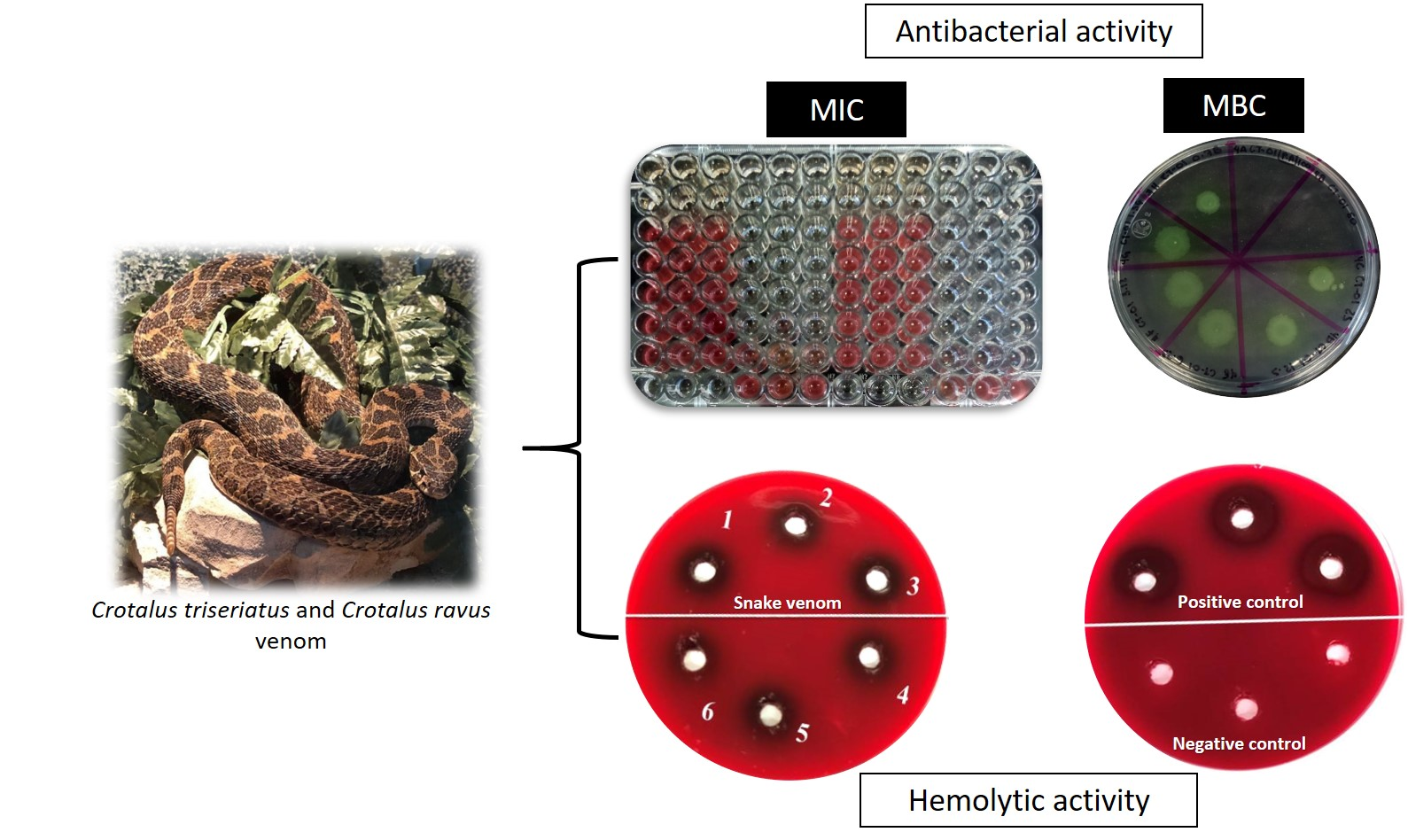
2.3. Antibacterial Activity

The venom’s antibacterial activity was determined through the Minimum Inhibitory Concentration (MIC) and the Minimum Bactericidal Concentration (MBC) procedures, in accordance with the CLSI guidelines and with the standards published by Olmedo-Juárez et al., in 2019 and by Morales-Ubaldo et al., in 2020 [13,14,15].
Escherichia coli ATCC35218, Pseudomonas aeruginosa ATCC9027, and Staphylococcus aureus ATCC6538 strains were used to perform the evaluation. These samples were the same which were reactivated from cryopreservation in Müller–Hinton agar (BD Bioxon, Heidelberg, Germany) through simple strain technique to obtain isolated colonies. A Gram staining was performed to corroborate their morphology.
Once the purity was confirmed, one colony of each strain was inoculated in nutritive broth (BD Bioxon), and incubated under constant agitation at 70 rpm for 24 h at 37 °C. The bacterial cell suspension was adjusted to a 0.5 McFarland (Remel, R20421, Kansas, U.S.A.) standard (approximately 1.5 × 10⁶ Colony Forming Units (CFU) per mL).

2.3.1. Minimal Inhibitory Concentration (MIC)

Micro-dilution was used to determine the MIC, evaluating different venom concentrations (100, 50, 25, 12.5, 6.25, 3.12, 1.56, 0.78 µg/mL).
In a sterile 96- well plate, 100 µL of each venom concentrations were added along with 10 µL of bacterial cell suspension previously adjusted to a 0.5 McFarland standard. The plates were incubated at 37 °C for 24 h at 70 rpm. Kanamycin (AppliChem 4K10421, Darmstadt, Germany) was used as a positive control (128 to 1 µg/mL) and nutritive broth as the negative control. Treatments were evaluated by triplicate.
After incubation 20 μL of a 0.04% (w/v) p-iodonitrotetrazolium (Sigma-Aldrich I8377, Missouri, U.S.A.) solution was added into each well and incubated for 30 min. The MIC was determined by the concentration at which the solution turned to a pinkish color.

2.3.2. Minimal Bactericidal Concentration (MBC)

After incubation and previous addition of p-iodonitrotetrazolium, 5 μL from each well was inoculated in Müller–Hinton agar (BD Bioxon) and incubated at 37 °C for 24 h. The MBC was considered as the lowest concentration where no visible growth of the bacteria was observed on the plates.

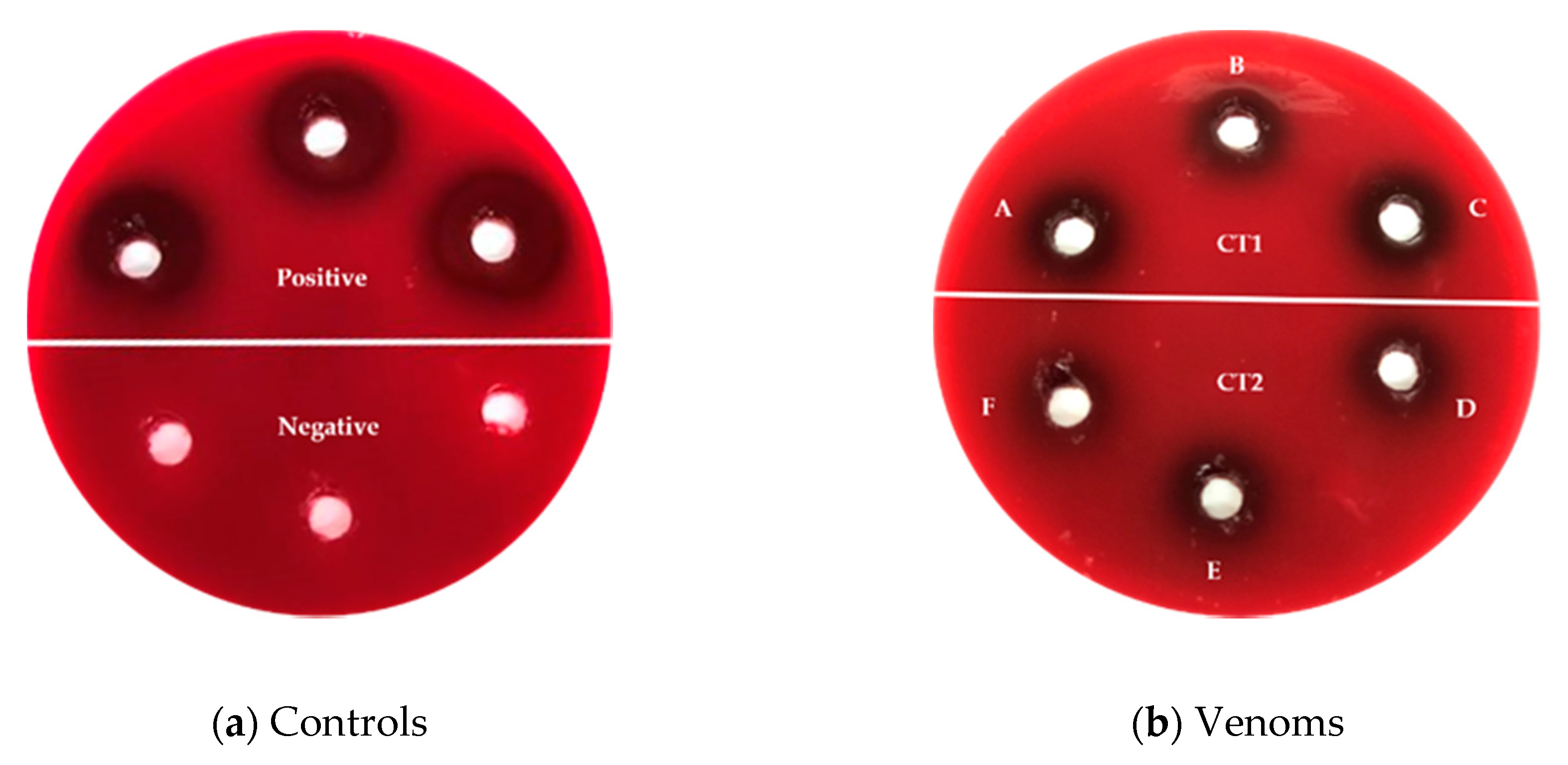
2.4. Indirect Hemolytic Activity

In accordance with the protocols described by Pirela et al., in 2006 with modifications, the venom’s indirect hemolytic activity was evaluated [16]. A donor donkey blood sample was collected. The blood sample was stored in 10 mL sodium citrate (3.2%) tubes (DB Vacutainer) and in 3 mL EDTA (10.8 mg) tubes (BD Vacutainer).
Blood agar was used (Merck©, Darmstadt, Germany). To obtain plates with 8% blood concentration, 250 mL of agar base was prepared, and 20 mL of blood was added.
One hundred micrograms (100 µg) of each treatment were weighed out (lyophilized venom) and reconstituted in 1 mL of nutritive broth (BD Bioxon). Dilutions were made (100, 50, 25, 12.5, 6.25, 3.12 µg/mL) from this concentrated solution for further evaluation.
Four wells were made (6 mm diameter) on the plate’s surface. Twenty micrograms (20 µL) were added of each concentrate to be evaluated. Treatments were performed by triplicate. Tween 80 at 100% (Sigma-Aldrich) and nutritive broth (BD Bioxon) were used as positive and negative controls, respectively. Plates were incubated for 24 h at 37 °C. Once the incubation period elapsed, hemolysis halos were measured (mm).

2.5. Statistical Analysis.

Obtained data were analyzed using two-way variance analysis (ANOVA) and a means comparison by Tukey at a significance level of 0.05% through Minitab 18 statistical package [17].

3. Results and Discussion

3.1. Individuals Data

A record of each individual was made with the following information: length, weight, age, and gender (Table 1). The characteristics of the rattlesnakes in the study coincided with those reported by Campbell and Lamar in 2004 [1], as seen in Figure 1.

3.2. Antibacterial Activity

A MIC of 50 µg/mL and an MBC of 100 µg /mL were determined as effective for treatments CT2 and CR3 over P. aeruginosa (Table 2, Figure 2). Nevertheless, antibacterial activity was not detected for E. coli and S. aureus.
It was determined that the antibacterial response seen in treatments CT2 and CR3 were bactericidal, since the relation between MIC and MBC is less than 4, in accordance with González-Alamilla et al., in 2019 [18].
Boda et al., in 2019 evaluated the antibacterial activity of eleven crude venoms from different snake species including Crotalus atrox and Crotalus polystictus against Staphylococcus aureus, Escherichia coli and Pseudomonas aeruginosa among others, at varied concentrations of 500 to 1.95 µg/mL, determining a MIC and MBC of 125 and 500 µg/mL against S. aureus for Crotalus atrox and Crotalus polystictus, respectively. In the present study, antibacterial activity was not found for S. aureus and E. coli but was determined for Pseudomonas aeruginosa, obtaining a MIC of 50 and a MBC of 100 µg/mL for C. triseriatus and C. ravus (CT2 and CR3). According to Boda et al., 2019, the antibacterial activity of venoms from viperid species is probably due to their content of proteins with proteolytic activity [19].
Samy et al., in 2014, evaluated CaTx-II a toxin isolated from Crotalus adamanteus venom, determining a MIC of 7.8 µg/mL for S. aureus and 62.5 to 125 µg/mL for P. aeruginosa. Oguiura et al., in 2011, evaluated crotamine, a myotoxin from Crotalus durissus venom against different bacteria strains which included E. coli, S. aureus, and P. aeruginosa. They report a MIC of 100 µg/mL for E. coli and >200 µg/mL for the other two [20,21], a contrast with the results obtained in our present study since the antibacterial activity was not determined for E. coli or S. aureus. Since it was determined that a MIC of 50 µg/mL from C. triseriatus and C. ravus (CT2 and CR3) occurred in crude venom, the activity could be attributed to the presence of these bioactive compounds in the venom of the individuals used for this evaluation since both compounds were isolated from snake venom of the same genus (Crotalus).
Although the aim of the study did not include identifying the venom’s active mechanism, it has been reported that phospholipase A2 (CaTx-II) interacts with lipopolysaccharide (LPS), particularly with lipid A, a Gram-negative bacteria component, causing membrane permeabilization. Crotamine also has effects over some bacteria through membrane permeabilization, so it could be suggested that CT2 and CR3 treatments antibacterial activity is related to this mechanism [21,22].
In this respect, the efficiency of these compounds, specially phospholipase A2 against antibiotic-resistant bacteria, holds promise for biotechnological applications, in this case, new medical treatment alternatives, however, it should be understood there are different antibacterial activity mechanisms from venom-based drugs [23,24].
In accordance with WHO, P. aeruginosa actually is in the critical priority group of the list of antibiotic-resistant pathogens. WHO has been expressing its interest by promoting the research and development of new antibiotics for this bacterium [10]. These results obtained herein show that CT2 and CR3 treatments demonstrated bactericidal activity against this pathogen showing its importance, since rattlesnake venoms or compounds thereof could be used to develop effective therapeutic agents to treat infections caused by P. aeruginosa.

3.3. Hemolytic Activity

With respect to the hemolysis produced, the generated halos showed significant statistical differences between them (p < 0.05) (Table 3). It was observed that CT1, CT2, and CR3 showed the highest hemolytic potential and there were no statistically significant differences between them at 100 µg/mL concentration compared with the other treatments (Figure 3).
Macías-Rodríguez et al., in 2014 [25] evaluated the hemolytic activity of Crotalus molossus venom (C. molossus molossus and C. molossus nigrescens) at a 50 µg/mL concentration. In the present study, hemolytic halos were measured over different periods of time, (1, 2, 3, and 14 h). The results obtained showed that at 14 h halos generated measured 19.2 ± 1.5 and 17.00 ± 1.2 mm for C. molossus molossus and C. m. molossus nigrescens, respectively, whereas at the same concentration over a longer period of time (24 h) with the venoms of C. triseriatus and C. ravus generated smaller halos 15.00 ± 0.0 (CT1), 12.33 ± 0.58 (CT2), 16.67 ± 0.58 (CR3), and 0.00 ± 0.00 (CR4), showing the hemolytic potential of these species is lower.
On the other hand, Pirela et al., in 2006, determined that the indirect hemolytic dose of Crotalus durissus cumanensis venom to produce a 20 mm hemolytic halo was 379.51 ± 67.67 µg of venom [16]. In a similar study, Dos Santos et al. in 1993 obtained a dose of approximately 310 µg for the white venom and 350 µg for the yellow venom of Crotalus durissus ruruima to produce hemolytic halos of 20 mm [26]. With respect to Crotalus triseriatus and Crotalus ravus venoms, an average of 18.67 ± 1.53 mm was obtained at 100 µg/mL concentration of venom. Although there are no equivalent values in the measurements of hemolytic halos, the venom of C.triseriatus and C. ravus have close values in the measure of their halos in comparison with the other studies and in a lower venom concentration.
In accordance with Macías-Rodríguez et al., in 2014 [27], during the fall months, there exists a high proteomic concentration in rattlesnake venom. C. ravus and C. triseriatus were sampled in September and November, respectively, months which correspond to the autumn, while the individuals sampled by Pirela et al. in 2006 [16] were sampled in May, June, and July, months that have been shown to have decreased protein concentration. On the other hand, in 2010 Chippaux et al., [6] reported that the species C. durissus durissus and C. durissus terrificus have myotoxic and neurotoxic venoms compared to other species of the genus Crotalus, which mostly have hemotoxic and histologic venoms [28]. Therefore, due to this, in C. triseriatus and C. ravus, the highest concentration evaluated in this study (100 µg/mL) was enough to produce halos with measurements similar to those of the aforementioned study.
Treatment CR4, characterized by its transparent color, did not show hemolytic activity. This variation in color has been observed in other viperids. In the study carried out by Macías-Rodríguez et al., in 2014 [25], C. molossus presented a yellowish venom which turned out to be more hemolytic than Crotalus tigris venom, which was transparent in appearance, similar to C. ravus (CR4). Galán et al., in 2004 [29], reported that yellowish venoms have greater toxicity compared to white venoms. Lourenço et al., in 2013 [30], reported that the yellow coloration of the venom is due to the presence of crotamine, a myotoxin from rattlesnakes.
Snake venom complexity produces a source of bioactive molecules with different activities. The results obtained in this study confirm rattlesnake’s crude venom contains compounds that could be used as therapeutic models, in this case, molecules with antibacterial activity. Although the venom cannot be used directly due to its high toxicity, some of its compounds will serve as prototypes for the development of new drugs.

4. Conclusions

Until today, there are no studies reporting on the antibacterial and hemolytic activity of the venoms of C. triseriatus and C. ravus. The results of the present study indicate that both rattlesnakes produce venoms rich in bioactive compounds with a bactericidal effect against Pseudomonas aeruginosa. These compounds could also serve as new antimicrobial drugs for the treatment of diseases caused by this bacterium; however, the isolation, identification, and evaluation of these molecules is necessary since it could present hemolytic activity.

Author Contributions

Conceptualization and Methodology, N.R.-P. and A.Z.-B.; Validation and Formal analysis, A.Z.-B., A.L.M.-U. and L.M.A.-C.; Investigation and Resources, N.R.-P., A.Z.-B. and L.R.-L.; Data curation, S.C.F.-A., C.E.R.-M. and A.O.-J.; Writing—original draft preparation, S.C.F.-A., A.L.M.-U and N.R.-P.; Writing—review and editing, A.Z.-B., B.V.-C., A.O.-J. and L.R.-L.; Supervision, N.R.-P., C.E.R.-M. and A.Z.-B.; Project administration, N.R.-P. and A.Z.-B.; Funding acquisition, N.R.-P., A.Z.-B. and L.M.A.-C. All authors have read and agreed to the published version of the manuscript.

Funding

This research did not receive any specific grant from funding agencies in the public, commercial, or not-for-profit sectors.

Acknowledgments

The authors would like to acknowledge to the Universidad Autónoma del Estado de Hidalgo (UAEH) by for the support provided for carrying out the study in its facilities.

Conflicts of Interest

The authors declare no conflicts of interest.

References

  1. Campbell, J.A.; Lamar, W.W.; Brodie, E.D. The Venomous Reptiles of the Western Hemisphere; Cornell University Press: Ithaca, NY, USA, 2004; p. 528. [Google Scholar]
  2. Mata-Silva, V.; Johnson, J.D.; Wilson, L.D.; García-Padilla, E. The herpetofauna of Oaxaca, Mexico: Composition, physiographic distribution, and conservation status. Mesoam. Herpetol. 2015, 2, 6–62. [Google Scholar]
  3. Palacios-Aguilar, R.; Flores-Villela, O. An updated checklist of the herpetofauna from Guerrero, Mexico. Zootaxa 2018, 4422, 1–24. [Google Scholar] [CrossRef] [PubMed]
  4. Chippaux, J. Snakebite in Africa: Current situation and urgent needs. In Handbook of Venoms and Toxins of Reptiles; CRC Press Inc.: London, UK, 2009; pp. 453–473. [Google Scholar]
  5. Vetter, I.; Davis, J.L.; Rash, L.D.; Anangi, R.; Mobli, M.; Alewood, P.F.; Lewis, R.J.; King, G.F. Venomics: A new paradigm for natural products-based drug discovery. Amino Acids 2011, 40, 15–28. [Google Scholar] [CrossRef] [PubMed]
  6. Prashanth, J.R.; Brust, A.; Jin, A.H.; Alewood, P.F.; Dutertre, S.; Lewis, R.J. Cone snail venomics: From novel biology to novel therapeutics. Future Med. Chem. 2014, 6, 1659–1675. [Google Scholar] [CrossRef]
  7. Fox, J.W.; Serrano, S.M. Approaching the golden age of natural product pharmaceuticals from venom libraries: An overview of toxins and toxin-derivatives currently involved in therapeutic or diagnostic applications. Curr. Pharm. Des. 2007, 13, 2927–2934. [Google Scholar] [CrossRef] [PubMed] [Green Version]
  8. Beeton, C. Targets and Therapeutic Properties. In Handbook of Biologically Active Peptides, 2nd ed.; Kastin, A.J., Ed.; Academic Press: Cambridge, MA, USA, 2013; pp. 473–482. [Google Scholar]
  9. Vargas-Muñoz, L.J.; Estrada-Gomez, S.; Vásquez-Escobar, J. Toxinas de venenos de serpientes y escorpiones, una fuente natural de moléculas con actividad antimicrobiana. Curare 2015, 2. [Google Scholar] [CrossRef]
  10. World Health Organization. WHO Publishes List of Bacteria for Which New Antibiotics are Urgently Needed. 2017. Available online: https://www.who.int/news-room/detail/27-02-2017-who-publishes-list-of-bacteria-for-which-new-antibiotics-are-urgently-needed (accessed on 15 December 2019).
  11. McDiarmid, R.; Foster, M.; Guyer, C.; Gibbons, W.; Chernoff, N. Reptile Biodiversity: Standard Methods for Inventory and Monitoring; University of California Press: Berkely, CA, USA, 2012; p. 412. [Google Scholar]
  12. SEMARNAT: Secretaría del Medio Ambiente y Recursos Naturales. México: Gobierno de México. 2016. Licencia de Colecta Científica con Propósitos de Enseñanza en Materia de vida Silvestre. Available online: https://www.gob.mx/tramites/ficha/licencia-de-colecta-cientifica-con-propositos-de-ensenanza-en-materia-de-vidasilvestre/SEMARNAT442 (accessed on 15 December 2019).
  13. Clinical and Laboratory Standards Institute. Methods for Dilution Antimicrobial Susceptibility Tests for Bacteria that Grow Aerobically (Approved Standards); CLSI document M7-A5; CLSI: Wayne, PA, USA, 2012. [Google Scholar]
  14. Olmedo-Juárez, A.; Briones-Robles, T.I.; Zaragoza-Bastida, A.; Zamilpa, A.; Ojeda-Ramírez, D.; Mendoza-de Gives, P.; Olivares-Pérez, J.; Rivero-Perez, N. Antibacterial activity of compounds isolated from Caesalpinia coriaria (Jacq) Willd against important bacteria in public health. Microb. Pathog. 2019, 136, 103660. [Google Scholar] [CrossRef] [PubMed]
  15. Morales-Ubaldo, A.; Hernández-Alvarado, J.; Valladares-Carranza, B.; Velázquez-Ordoñez, V.; Delgadillo-Ruiz, L.; Rosenfeld-Miranada, C.; Rivero-Pérez, N.; Zaragoza-Bastida, A. Actividad antibacteriana del extracto hidroalcohólico Croton draco sobre bacterias de importancia sanitaria. Abanico Vet. 2019, 10, 10. [Google Scholar] [CrossRef]
  16. Pirela De las Salas, R.d.C.; López-Jonsthon, J.C.; Hernández Rangel, J.L. Caracterización toxinológica del veneno total de la serpiente de cascabel Crotalus durissus cumanensis (viperidae), presente en la localidad de Porshoure, Guajira Venezolana. Rev. Cient. 2006, 16, 232–238. [Google Scholar]
  17. Minitab 18 Statistical Software 2010; Minitab, Inc.: State College, PA, USA, 2010; Available online: www.minitab.com (accessed on 15 November 2019).
  18. González-Alamilla, E.N.; Gonzalez-Cortazar, M.; Valladares-Carranza, B.; Rivas-Jacobo, M.A.; Herrera-Corredor, C.A.; Ojeda-Ramírez, D.; Zaragoza-Bastida, A.; Rivero-Perez, N. Chemical Constituents of Salix babylonica L. and Their Antibacterial Activity Against Gram-Positive and Gram-Negative Animal Bacteria. Molecules 2019, 24, 2992. [Google Scholar] [CrossRef] [Green Version]
  19. Boda, F.A.; Mare, A.; Szabó, Z.I.; Berta, L.; Curticapean, A.; Dogaru, M.; Man, A. Antibacterial activity of selected snake venoms on pathogenic bacterial strains. Rev. Rom. Med. Lab. 2019, 27, 305. [Google Scholar] [CrossRef] [Green Version]
  20. Samy, R.P.; Kandasamy, M.; Gopalakrishnakone, P.; Stiles, B.G.; Rowan, E.G.; Becker, D.; Shanmugam, M.K.; Sethi, G.; Chow, V.T. Wound healing activity and mechanisms of action of an antibacterial protein from the venom of the eastern diamondback rattlesnake (Crotalus adamanteus). PLoS ONE 2014, 9, e80199-e. [Google Scholar] [CrossRef] [PubMed] [Green Version]
  21. Oguiura, N.; Boni-Mitake, M.; Affonso, R.; Zhang, G. In vitro antibacterial and hemolytic activities of crotamine, a small basic myotoxin from rattlesnake Crotalus durissus. J. Antibiot. 2011, 64, 327–331. [Google Scholar] [CrossRef] [PubMed]
  22. Santamaría, C.; Larios, S.; Angulo, Y.; Pizarro-Cerda, J.; Gorvel, J.P.; Moreno, E.; Lomonte, B. Antimicrobial activity of myotoxic phospholipases A2 from crotalid snake venoms and synthetic peptide variants derived from their C-terminal region. Toxicon 2005, 45, 807–815. [Google Scholar] [CrossRef] [PubMed]
  23. Canhas, I.; Heneine, L.G.; Fraga, T.; Assis, D.; Borges, M.; Chartone-Souza, E.; Amaral Nascimento, A.M. Antibacterial activity of different types of snake venom from the Viperidae family against Staphylococcus aureus. Acta Sci. Biol. Sci. 2017, 39, 309. [Google Scholar] [CrossRef] [Green Version]
  24. Bocian, A.; Hus, K. Antibacterial properties of snake venom components. Chem. Pap. 2019. [Google Scholar] [CrossRef] [Green Version]
  25. Macias-Rodríguez, E.; Martínez-Martínez, A.; Gatica-Colima, A.; Bojórquez-Rangel, G.; Plenge-Tellechea, L. Análisis comparativo de la actividad hemolítica entre las subespecies Crotalus molossus y Crotalus molossus nigrescens. Revista Bio Ciencias 2014, 2, 302–312. [Google Scholar]
  26. Dos Santos, M.C.; Ferreira, L.C.L.; Da Silva, W.D.; Furtado, M.d.F.D. Caracterizacion de las actividades biologicas de los venenos ‘amarillo’ y ‘blanco’ de Crotalus durissus ruruima comparados con el veneno de Crotalus durissus terrificus. Poder neutralizante de los antivenenos frente a los venenos de Crotalus durissus ruruim. Toxicon 1993, 31, 1459–1469. [Google Scholar]
  27. Macias-Rodríguez, E.; Díaz-Cárdenas, C.O.; Gatica-Colima, A.; Plenge, L. Variación estacional del contenido proteico y actividad de la PLA2 del veneno de Crotalus molossus molossus entre especímenes de origen silvestre y en cautiverio. Acta Univ. 2014, 24, 38–47. Available online: http://www.redalyc.org/articulo.oa?id=41630112002 (accessed on 15 December 2019).
  28. De Roodt, A.R.; Estévez-Ramírez, J.; Paniagua-Solís, J.F.; Litwin, S.; Carvajal-Saucedo, A.; Dolab, J.A.; Robles–Ortiz, L.E.; Alagón, A. Toxicidad de venenos de serpientes de importancia médica en México. Gac. Méd. Méx. 2005, 141, 13–21. [Google Scholar]
  29. Galán, J.A.; Sánchez, E.E.; Rodríguez-Acosta, A.; Pérez, J.C. Neutralization of venoms from two Southern Pacific Rattlesnakes (Crotalus helleri) with commercial antivenoms and endothermic animal sera. Toxicon 2004, 43, 791–799. [Google Scholar] [CrossRef] [PubMed]
  30. Lourenço, A.; Zorzella-Creste, C.F.; Curtolo de Barros, L.; Delazari-dos Santos, L.; Pimenta, D.C.; Barraviera, B.; Ferreira, R.S., Jr. Individual venom profiling of Crotalus durissus terrificus specimens from a geographically limited region: Crotamine assessment and captivity evaluation on the biological activities. Toxicon 2013, 69, 75–81. [Google Scholar] [CrossRef] [PubMed]
Figure 1. Captured species (a) Crotalus triseriatus and (b) Crotalus ravus.
Animals 10 00281 g001
Figure 2. Antibacterial activity of rattlesnake’s venoms against P. aeruginosa: (a) columns 1–3, CT1 from 100 at 0.78 µg/mL, columns 4–6, CR4 from 100 at 0.78 µg/mL, columns 7–9, CT2 from 100 at 0.78 µg/mL, columns 10–12, CR3 from 100 at 0.78 µg/mL. The MIC value is read at the minimal concentration in which the color changes to pink; (b) Plate with P. aeruginosa + CT2 in Müller–Hinton agar; A CT2 to 100 µg/mL, B CT2 to 50 µg/mL, C CT2 to 25 µg/mL, D CT2 to 12.5 µg/mL, E CT2 to 6.25 µg/mL, F CT2 to 12.5 µg/mL, G CT2 to 6.25 µg/mL, H CT2 to 0.78 µg/mL. The MBC is read to the lowest concentration where no visible growth of the bacteria.
Animals 10 00281 g002
Figure 3. Indirect hemolytic activity of the rattlesnake’s venoms: (a) hemolytic activity of controls, positive Tween 80, negative nutritive broth; (b) hemolytic activity of venoms A, B, C 100 µg/mL of CT1; D, E, F 100 µg/mL of CT2.
Animals 10 00281 g003
Table 1. Individual data of trapped rattlesnakes in Hidalgo State.
SpeciesSpecies CharacteristicsIndividual IdentificationGenderAgeLength (cm)Weight (g)
Crotalus triseriatusTriangular head
8–10 rattles
Postocular strip
CT1MaleAdult37210
CT2MaleAdult25175
Crotalus ravusTriangular head
Thin rattle
Symmetric scales in head
CR3MaleAdult25180
CR4MaleAdult25175
Table 2. Minimal Inhibitory Concentration (MIC) and Minimal Bactericidal Concentration (MBC) of Crotalus triseriatus and Crotalus ravus venoms.
Evaluated BacteriaEvaluated Treatments µg/mL (MIC/MBC)Controls (MIC/MBC)
CT1CT2CR3CR4Nutritive BrothKanamycin (µg/mL)
E. coli-----2/4
P. aeruginosa-50 a/100 A50 a/100 A--16 b/64 B
S. aureus-----1/4
CT1 Crotalus triseriatus 1, CT2 Crotalus triseriatus 2, CR3 Crotalus ravus 3, CR4 Crotalus ravus 4 a,b Different small letters indicate significant statistical differences between MIC (p < 0.05) A,B Different capital letters indicate significant statistical differences between MBC (p < 0.05)
Table 3. Hemolysis halos generated by C. triseriatus and C. ravus venoms.
Concentration (µg/mL)Evaluated Treatments
CT1CT2CR3CR4Nutritive BrothTween 80
10018.67 ± 1.53 a,A,*17.00 ± 1.00 a,A18.67 ± 1.15 a,A,*0.0 b0.0020.33 ± 0.58 *
5015.00 ± 0.0 b,B12.33 ± 0.58 c,B16.67 ± 0.58 a,B0.0 d
2513.67 ± 0.58 a,B,C13.67 ± 1.15 a,B13.33 ± 0.58 a,C0.0 b
12.512.00 ± 1.00 a,C10.00 ± 0.00 a,b,C10.33 ± 0.58 b,D0.0 c
6.258.67 ± 0.58 a,D8.33 ± 0.58 a,C7.33 ± 0.58 a,E0.0 b
3.120.00 ± 0.00 a,E0.00 ±0.00 a,D0.00 ± 0.00 a,F0.0 a
a,b,c Different letters indicate significant statistical differences between treatments (p < 0.05). A,B,C Different letters indicate significant statistical differences between concentrations (p < 0.05). * No statistical differences between treatments (p > 0.05). CT1 Crotalus triseriatus 1, CT2 Crotalus triseriatus 2, CR3 Crotalus ravus 3, and CR4 Crotalus ravus 4.

