
Inhibition of Candida albicans and Mixed Salivary Bacterial Biofilms on Antimicrobial Loaded Phosphated Poly(methyl methacrylate)

 and
and 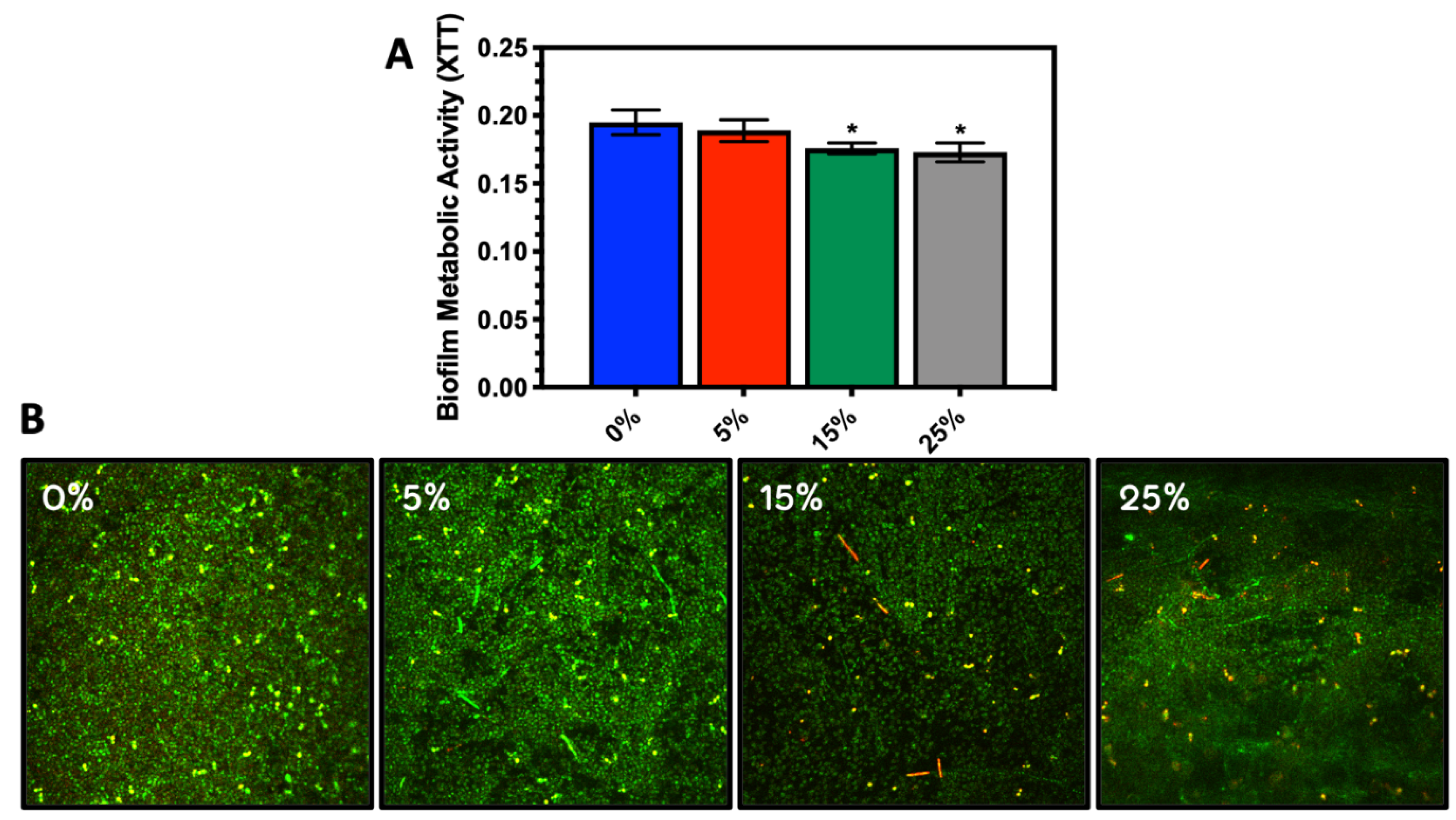
Abstract
:1. Introduction
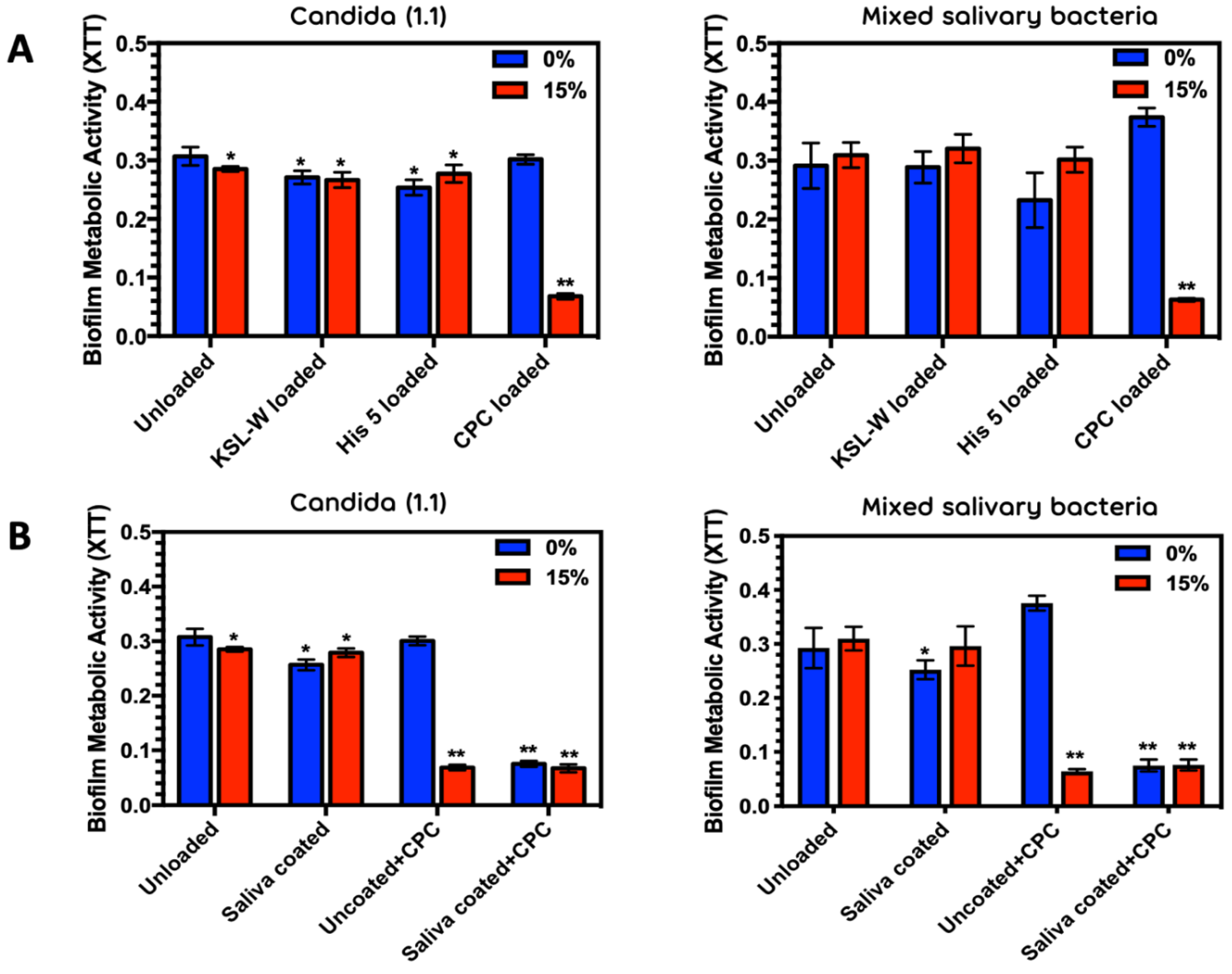
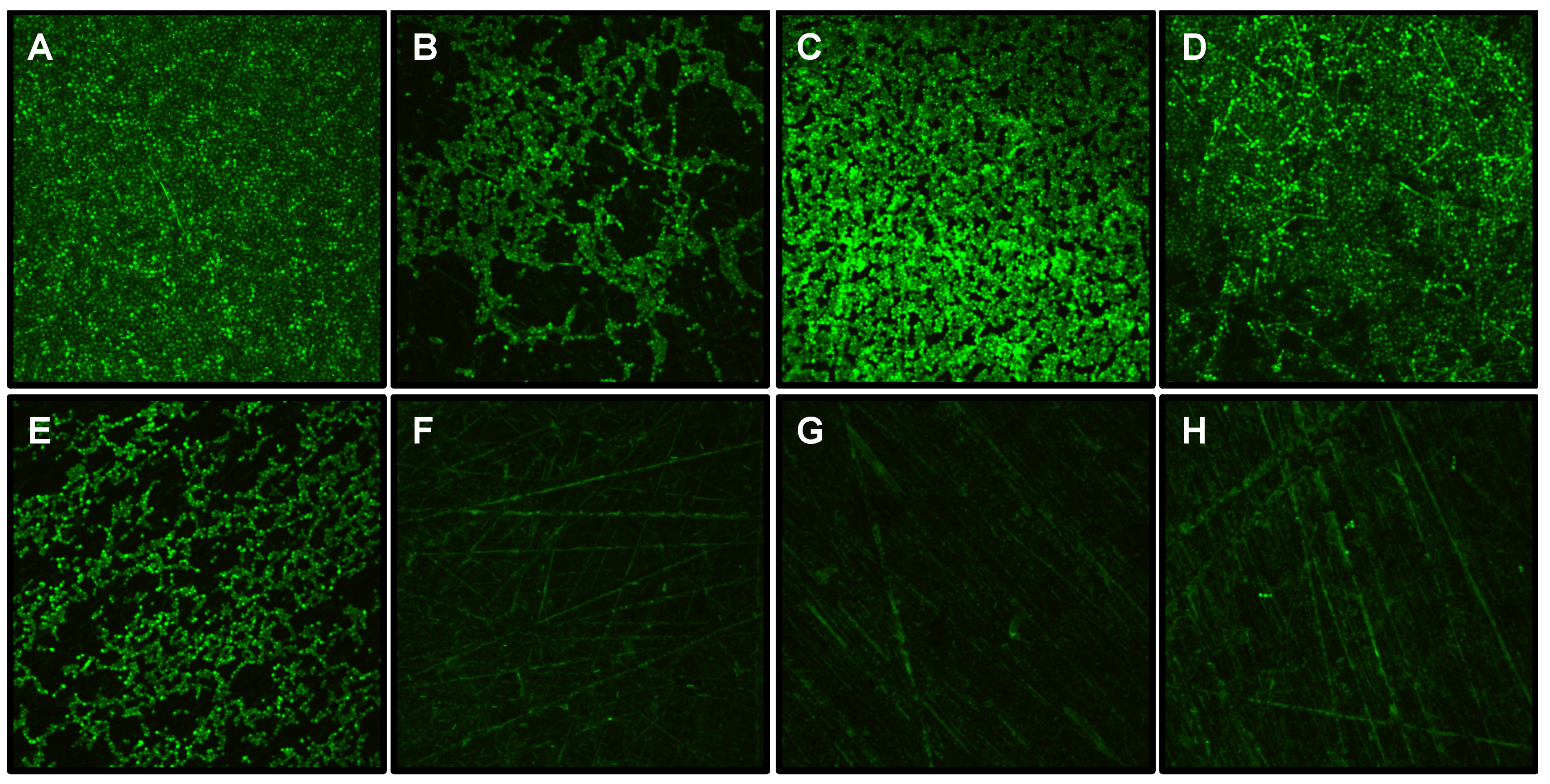
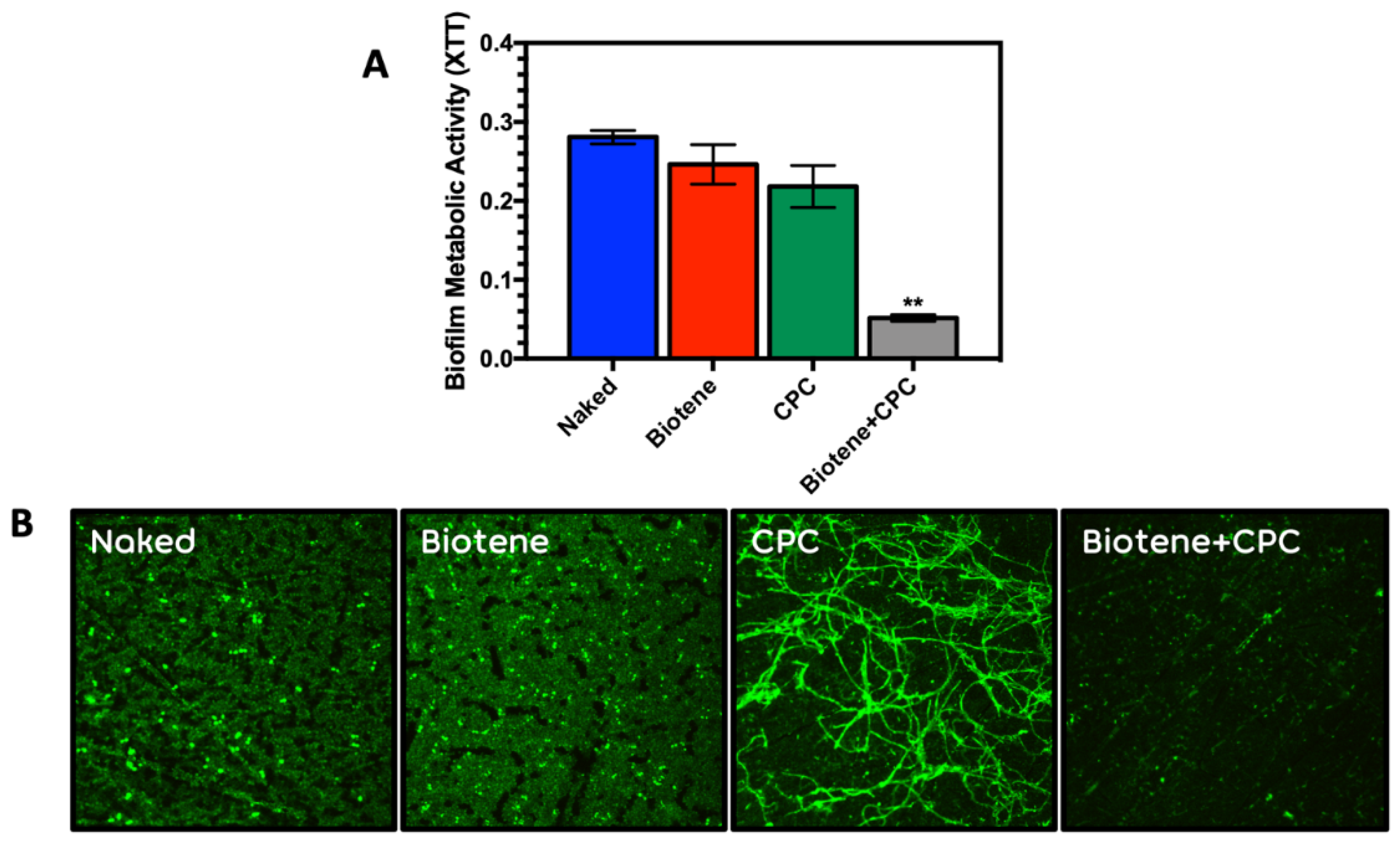
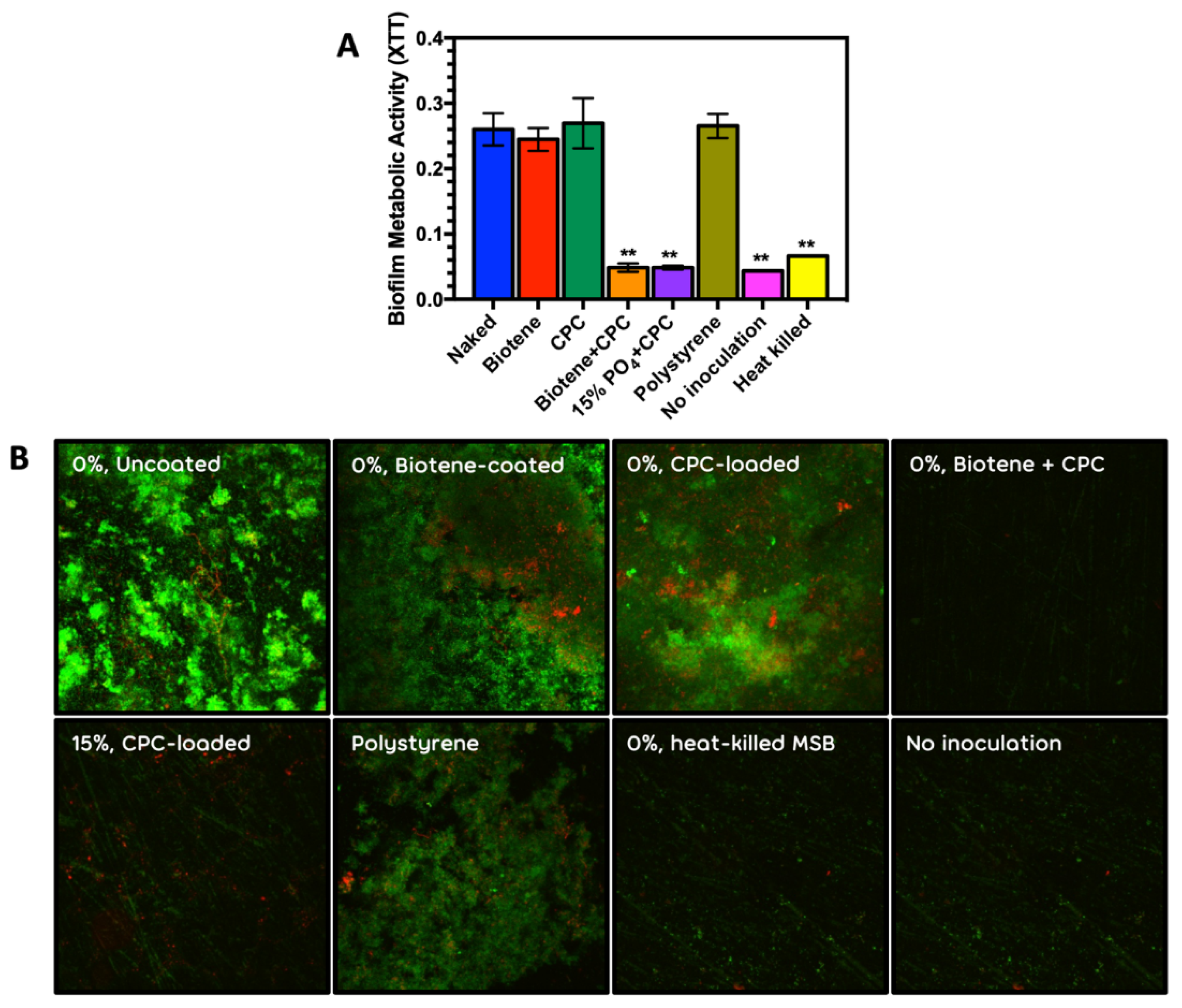
2. Results
3. Discussion
4. Experimental Procedure
4.1. Polymer Synthesis and Fabrication of Phosphated PMMA Disks
4.2. Candida and Mixed Salivary Bacterial Adhesion and Biofilm Metabolism on Phosphated PMMA Surfaces
4.3. Microbicidal Activity of Antimicrobials on Oral Microorganisms
4.4. Image Analysis
5. Conclusions
Supplementary Materials
Author Contributions
Funding
Institutional Review Board Statement
Informed Consent Statement
Conflicts of Interest
References
- Raad, I. Intravascular-catheter-related infections. Lancet 1998, 351, 893–898. [Google Scholar] [CrossRef]
- Kojic, E.M.; Darouiche, R.O. Candida infections of medical devices. Clin. Microbiol. Rev. 2004, 17, 255–267. [Google Scholar] [CrossRef] [PubMed] [Green Version]
- Khatoon, Z.; McTiernan, C.D.; Suuronen, E.J.; Mah, T.-F.; Alarcon, E.I. Bacterial biofilm formation on implantable devices and approaches to its treatment and prevention. Heliyon 2018, 4, e01067. [Google Scholar] [CrossRef] [PubMed] [Green Version]
- Budtz-Jörgensen, E. Clinical aspects of Candida infection in denture wearers. J. Am. Dent. Assoc. (1939) 1978, 96, 474–479. [Google Scholar] [CrossRef] [PubMed]
- Uyen, H.; Van der Mei, H.; Weerkamp, A.; Busscher, H. Zeta potential and the adhesion of oral streptococci to polymethylmethacrylate. Biomater. Artif. Cells Artif. Organs 1989, 17, 385–391. [Google Scholar] [CrossRef] [PubMed]
- Tsutsumi-Arai, C.; Takakusaki, K.; Arai, Y.; Terada-Ito, C.; Takebe, Y.; Imamura, T.; Ide, S.; Tatehara, S.; Tokuyama-Toda, R.; Wakabayashi, N. Grapefruit seed extract effectively inhibits the Candida albicans biofilms development on polymethyl methacrylate denture-base resin. PLoS ONE 2019, 14, e0217496. [Google Scholar] [CrossRef] [PubMed] [Green Version]
- Budtz-Jorgensen, E.; Theilade, E.; Theilade, J. Quantitative relationship between yeasts and bacteria in denture-induced stomatitis. Eur. J. Oral Sci. 1983, 91, 134–142. [Google Scholar] [CrossRef] [PubMed]
- Budtz-Jorgensen, E.; Theilade, E. Regional variations in viable bacterial and yeast counts of 1-week-old denture plaque in denture-induced stomatitis. Eur. J. Oral Sci. 1983, 91, 288–295. [Google Scholar] [CrossRef] [PubMed]
- Moskona, D.; Kaplan, I. Oral lesions in elderly denture wearers. Clin. Prev. Dent. 1992, 14, 11–14. [Google Scholar]
- Kulak-Ozkan, Y.; Kazazoglu, E.; Arikan, A. Oral hygiene habits, denture cleanliness, presence of yeasts and stomatitis in elderly people. J. Oral Rehabil. 2002, 29, 300–304. [Google Scholar] [CrossRef]
- Ramage, G.; Tomsett, K.; Wickes, B.L.; López-Ribot, J.L.; Redding, S.W. Denture stomatitis: A role for Candida biofilms. Oral Surg. Oral Med. Oral Pathol. Oral Radiol. Endod. 2004, 98, 53–59. [Google Scholar] [CrossRef]
- Samaranayake, Y.; Cheung, B.; Parahitiyawa, N.; Seneviratne, C.; Yau, J.; Yeung, K.; Samaranayake, L. Synergistic activity of lysozyme and antifungal agents against Candida albicans biofilms on denture acrylic surfaces. Arch. Oral Biol. 2009, 54, 115–126. [Google Scholar] [CrossRef]
- Reichart, P.A. Oral mucosal lesions in a representative cross-sectional study of aging Germans. Community Dent. Oral Epidemiol. 2000, 28, 390–398. [Google Scholar] [CrossRef]
- Budtz-Jörgensen, E.; Mojon, P.; Rentsch, A.; Deslauriers, N. Effects of an oral health program on the occurrence of oral candidosis in a long-term care facility. Community Dent. Oral Epidemiol. 2000, 28, 141–149. [Google Scholar] [CrossRef] [PubMed]
- Yoneyama, T.; Yoshida, M.; Ohrui, T.; Mukaiyama, H.; Okamoto, H.; Hoshiba, K.; Ihara, S.; Yanagisawa, S.; Ariumi, S.; Morita, T. Oral care reduces pneumonia in older patients in nursing homes. J. Am. Geriatr. Soc. 2002, 50, 430–433. [Google Scholar] [CrossRef]
- Edgerton, M.; Raj, P.; Levine, M. Surface-modified poly(methyl methacrylate) enhances adsorption and retains anticandidal activities of salivary histatin 5. J. Biomed. Mater. Res. Part A 1995, 29, 1277–1286. [Google Scholar] [CrossRef] [PubMed]
- Park, S.E.; Periathamby, A.R.; Loza, J.C. Effect of surface-charged poly(methyl methacrylate) on the adhesion of Candida albicans. J. Prosthodont. 2003, 12, 249–254. [Google Scholar] [CrossRef]
- Chandra, J.; Patel, J.D.; Li, J.; Zhou, G.; Mukherjee, P.K.; McCormick, T.S.; Anderson, J.M.; Ghannoum, M.A. Modification of surface properties of biomaterials influences the ability of Candida albicans to form biofilms. Appl. Environ. Microbiol. 2005, 71, 8795–8801. [Google Scholar] [CrossRef] [Green Version]
- Yoshinari, M.; Kato, T.; Matsuzaka, K.; Hayakawa, T.; Inoue, T.; Oda, Y.; Okuda, K.; Shimono, M. Adsorption behavior of antimicrobial peptide histatin 5 on PMMA. J. Biomed. Mater. Res. Part B Appl. Biomater. 2006, 77, 47–54. [Google Scholar] [CrossRef]
- Carlson, R.P.; Taffs, R.; Davison, W.M.; Stewart, P.S. Anti-biofilm properties of chitosan-coated surfaces. J. Biomater. Sci. Polym. Ed. 2008, 19, 1035–1046. [Google Scholar] [CrossRef]
- Pusateri, C.R.; Monaco, E.A.; Edgerton, M. Sensitivity of Candida albicans biofilm cells grown on denture acrylic to antifungal proteins and chlorhexidine. Arch. Oral Biol. 2009, 54, 588–594. [Google Scholar] [CrossRef] [PubMed] [Green Version]
- Zamperini, C.A.; Machado, A.L.; Vergani, C.E.; Pavarina, A.C.; Giampaolo, E.T.; da Cruz, N.C. Adherence in vitro of Candida albicans to plasma treated acrylic resin. Effect of plasma parameters, surface roughness and salivary pellicle. Arch. Oral Biol. 2010, 55, 763–770. [Google Scholar] [CrossRef] [PubMed]
- Edgerton, M.; Levine, M.J. Characterization of acquired denture pellicle from healthy and stomatitis patients. J. Prosthet. Dent. 1992, 68, 683–691. [Google Scholar] [CrossRef]
- Edgerton, M.; Scannapieco, F.; Reddy, M.; Levine, M. Human submandibular-sublingual saliva promotes adhesion of Candida albicans to polymethylmethacrylate. Infect. Immun. 1993, 61, 2644–2652. [Google Scholar] [CrossRef] [PubMed] [Green Version]
- Klotz, S.; Drutz, D.; Zajic, J. Factors governing adherence of Candida species to plastic surfaces. Infect. Immun. 1985, 50, 97–101. [Google Scholar] [CrossRef]
- Klotz, S. The contribution of electrostatic forces to the process of adherence of Candida albicans yeast cells to substrates. FEMS Microbiol. Lett. 1994, 120, 257–262. [Google Scholar] [CrossRef]
- Park, S.E.; Blissett, R.; Susarla, S.M.; Weber, H.P. Candida albicans Adherence to Surface-Modified Denture Resin Surfaces. J. Prosthodont. 2008, 17, 365–369. [Google Scholar] [CrossRef]
- van Vugt, T.; Arts, C.; Geurts, J. Antibiotic-loaded polymethylmethacrylate beads and spacers in treatment of orthopaedic infections and the role of biofilm formation. Front. Microbiol. 2019, 10, 1626. [Google Scholar] [CrossRef]
- Gad, M.M.; Abualsaud, R. Behavior of PMMA Denture Base Materials Containing Titanium Dioxide Nanoparticles: A Literature Review. Int. J. Biomater. 2019, 2019. [Google Scholar] [CrossRef]
- Gad, M.M.; Al-Thobity, A.M.; Rahoma, A.; Abualsaud, R.; Al-Harbi, F.A.; Akhtar, S. Reinforcement of PMMA Denture Base Material with a Mixture of ZrO2 Nanoparticles and Glass Fibers. Int. J. Dent. 2019, 2019. [Google Scholar] [CrossRef] [Green Version]
- Naji, S.A.; Kashi, T.S.J.; Pourhajibagher, M.; Behroozibakhsh, M.; Masaeli, R.; Bahador, A. Evaluation of Antimicrobial Properties of Conventional Poly(Methyl Methacrylate) Denture Base Resin Materials Containing Hydrothermally Synthesised Anatase TiO2 Nanotubes against Cariogenic Bacteria and Candida albicans. Iran. J. Pharm. Res. IJPR 2018, 17, 161. [Google Scholar]
- Li, L.; Finnegan, M.; Özkan, S.; Kim, Y.; Lillehoj, P.; Ho, C.; Lux, R.; Mito, R.; Loewy, Z.; Shi, W. In vitro study of biofilm formation and effectiveness of antimicrobial treatment on various dental material surfaces. Mol. Oral Microbiol. 2010, 25, 384–390. [Google Scholar] [CrossRef] [PubMed]
- Zoccolotti, J.; Tasso, C.; Arbeláez, M.; Malavolta, I.; Pereira, E.; Esteves, C.; Jorge, J. Properties of an acrylic resin after immersion in antiseptic soaps: Low-cost, easy-access procedure for the prevention of denture stomatitis. PLoS ONE 2018, 30, e0203187. [Google Scholar] [CrossRef] [PubMed]
- Moffa, E.B.; Mussi, M.; Xiao, Y.; Garrido, S.S.; Machado, M.A.; Giampaolo, E.T.; Siqueira, W.L. Histatin 5 inhibits adhesion of C. albicans to reconstructed human oral epithelium. Front. Microbiol. 2015, 6, 885. [Google Scholar] [CrossRef] [Green Version]
- Monteiro, D.; Silva, S.; Negri, M.; Gorup, L.; de Camargo, E.; Oliveira, R.; Barbosa, D.; Henriques, M. Antifungal activity of silver nanoparticles in combination with nystatin and chlorhexidine digluconate against Candida albicans and Candida glabrata biofilms. Mycoses 2013, 56, 672–680. [Google Scholar] [CrossRef]
- Araujo, H.; Arias, L.; Caldeirão, A.; Assumpção, L.; Morceli, M.; de Souza Neto, F.; de Camargo, E.; Oliveira, S.; Pessan, J.; Monteiro, D. Novel Colloidal Nanocarrier of Cetylpyridinium Chloride: Antifungal Activities on Candida Species and Cytotoxic Potential on Murine Fibroblasts. J. Fungi 2020, 12, 218. [Google Scholar] [CrossRef]
- Cao, Z.; Sun, X.; Yeh, C.-K.; Sun, Y. Rechargeable infection-responsive antifungal denture materials. J. Dent. Res. 2010, 89, 1517–1521. [Google Scholar] [CrossRef] [Green Version]
- Dhir, G.; Berzins, D.; Dhuru, V.; Periathamby, A.; Dentino, A. Physical properties of denture base resins potentially resistant to Candida adhesion. J. Prosthodont. 2007, 16, 465–472. [Google Scholar] [CrossRef] [Green Version]
- Theberge, S.; Semlali, A.; Alamri, A.; Leung, K.P.; Rouabhia, M.C. albicans growth, transition, biofilm formation, and gene expression modulation by antimicrobial decapeptide KSL-W. BMC Microbiol. 2013, 13, 1–14. [Google Scholar] [CrossRef] [Green Version]
- Concannon, S.P.; Crowe, T.D.; Abercrombie, J.J.; Molina, C.M.; Hou, P.; Sukumaran, D.K.; Raj, P.A.; Leung, K.P. Susceptibility of oral bacteria to an antimicrobial decapeptide. J. Med. Microbiol. 2003, 52, 1083–1093. [Google Scholar] [CrossRef] [PubMed]
- Helmerhorst, E.; Hodgson, R.; van’t Hof, W.; Veerman, E.; Allison, C.; Nieuw Amerongen, A. The effects of histatin-derived basic antimicrobial peptides on oral biofilms. J. Dent. Res. 1999, 78, 1245–1250. [Google Scholar] [CrossRef]
- Mao, X.; Auer, D.; Buchalla, W.; Hiller, K.; Maisch, T.; Hellwig, E.; Al-Ahmad, A.; Cieplik, F. Cetylpyridinium Chloride: Mechanism of Action, Antimicrobial Efficacy in Biofilms, and Potential Risks of Resistance. Antimicrob. Agents Chemother. 2020, 64, e00576-00520. [Google Scholar] [CrossRef] [PubMed]
- Vukosavljevic, D.; Custodio, W.; Del Bel Cury, A.; Siqueira, W. The effect of histatin 5, adsorbed on PMMA and hydroxyapatite, on Candida albicans colonization. Yeast 2012, 29, 459–466. [Google Scholar] [CrossRef] [PubMed]
- Oppenheim, F.; Salih, E.; Siqueira, W.; Zhang, W.; Helmerhorst, E. Salivary Proteome and Its Genetic Polymorphisms. Ann. N. Y. Acad. Sci. 2007, 1098, 22–50. [Google Scholar] [CrossRef] [PubMed]
- Schmalz, G.; Cieplik, F. Biofilms on Restorative Materials. Monogr. Oral Sci. 2021, 29, 155–194. [Google Scholar] [CrossRef] [PubMed]


Publisher’s Note: MDPI stays neutral with regard to jurisdictional claims in published maps and institutional affiliations. |
© 2021 by the authors. Licensee MDPI, Basel, Switzerland. This article is an open access article distributed under the terms and conditions of the Creative Commons Attribution (CC BY) license (https://creativecommons.org/licenses/by/4.0/).
Share and Cite
Dentino, A.R.; Lee, D.; Dentino, K.; Guentsch, A.; Tahriri, M. Inhibition of Candida albicans and Mixed Salivary Bacterial Biofilms on Antimicrobial Loaded Phosphated Poly(methyl methacrylate). Antibiotics 2021, 10, 427. https://0-doi-org.brum.beds.ac.uk/10.3390/antibiotics10040427
Dentino AR, Lee D, Dentino K, Guentsch A, Tahriri M. Inhibition of Candida albicans and Mixed Salivary Bacterial Biofilms on Antimicrobial Loaded Phosphated Poly(methyl methacrylate). Antibiotics. 2021; 10(4):427. https://0-doi-org.brum.beds.ac.uk/10.3390/antibiotics10040427
Chicago/Turabian StyleDentino, Andrew R., DongHwa Lee, Kelley Dentino, Arndt Guentsch, and Mohammadreza Tahriri. 2021. "Inhibition of Candida albicans and Mixed Salivary Bacterial Biofilms on Antimicrobial Loaded Phosphated Poly(methyl methacrylate)" Antibiotics 10, no. 4: 427. https://0-doi-org.brum.beds.ac.uk/10.3390/antibiotics10040427