1. Introduction
Human existence is essentially dependent on the action of microorganisms, as they play fundamental roles in the fixation of nitrogen, production of vitamins, photosynthesis, and decomposition of organic matter [
1,
2,
3]. Nonetheless, a shift of the delicate balance between the immune system and microorganisms in favor of the latter could cause severe immunodeficiencies [
2,
4]. Accounting for millions of deaths each year worldwide, infectious diseases, which are caused by pathogenic bacteria, viruses, fungi, or parasites and can be directly or indirectly transmitted through air, water, food, or living vectors, have become a challenging threat to public health and a top priority area for health policy [
2,
5,
6,
7].
While ancient times witnessed the deaths of more than half of born individuals before reaching sexual maturity [
8], the discovery of antimicrobial drugs has greatly impacted the global health system by significantly reducing morbidity and mortality associated with infectious diseases [
9]. However, microorganisms have acquired or developed numerous resistance mechanisms over time against all commercially available antimicrobial drugs [
9,
10]. The main causes involved in the development of such mechanisms include genetic modifications of microorganisms, gratuitous over-prescription of antibiotics and broad-spectrum antibiotics, easy access to over-the-counter antibiotics, and contaminations due to antimicrobial drug manufacturing [
11]. As antimicrobial resistance has led to ineffectiveness of currently existing drugs, a worldwide catastrophic issue for the public system, there is an urgent necessity to identify novel alternative strategies [
5,
7,
9,
11,
12,
13,
14,
15].
One of the most intensively studied alternative involves the use of natural compounds, either alone or in combination with conventional antimicrobial drugs to enhance their activity [
7,
13,
16]. Studies have demonstrated the efficiency of various natural compounds, including plant-derived (e.g., essential oils (EOs), polyphenols), animal-derived (e.g., lactoferrin, lysozyme, lactoperoxidase), algal extracts, and microbial metabolites, which act through a series of mechanisms, such as microbial cell membrane rupture, nucleic acid process impairment, proton motive force decay, or adenosine triphosphate depletion [
16,
17,
18].
Increasing scientific interest has focused on EOs, which are secondary plant metabolites consisting of complex mixtures of odoriferous and volatile organic components produced by different plant parts, including flowers, peels, seeds, leaves, buds, twigs, roots, or fruits [
19,
20,
21,
22]. Since early times, EOs have been widely used in numerous medical applications, including cosmetics, dermatology, aromatherapy, and self-care medical products, or for food preservation purposes [
19,
21]. Currently, they are widely found in commercial applications, such as medicines, and there are many clinical trials performed [
23]. There are many research studies demonstrating their biological benefits, including antioxidant, anti-inflammatory, anti-cancer, antimicrobial (antibacterial, antiviral, antifungal, and antiparasitic), analgesic, sedative, and wound healing [
19,
20,
21]. With regard to the current antimicrobial resistance crisis, EOs have proven their efficiency by targeting microbial cell walls or membranes, cellular respiration processes, or quorum sensing mechanisms [
19,
24]. Nevertheless, despite their tremendous potential, EOs have a series of limitations associated with their increased hydrophobicity, volatility, lipophilicity, and oxidation susceptibility and decreased solubility and stability [
21,
25]. Therefore, in order to inhibit the development of microbial pathogens, their combination with nanotechnology-based approaches is necessary [
21]. In this manner, nanocarriers could protect them against thermal and photodegradation, while controlling their release and increasing their solubility, bioavailability, bioaccesibility, and concentration at the target site [
18,
25,
26,
27,
28]. Moreover, since nanoparticles have shown a high potential towards inactivating various microbial species through intrinsic antimicrobial features, as they are capable of microbial membrane and wall permeation and disruption, reactive oxygen species generation, and intracellular component damaging, systems comprising nanoparticles functionalized with EOs could offer synergistic effects [
14,
29,
30,
31]. Specifically, magnetite nanoparticles are well-known for their antimicrobial properties, both for their intrinsic properties that can be potentiated by hyperthermia effects and as antimicrobial drug carriers [
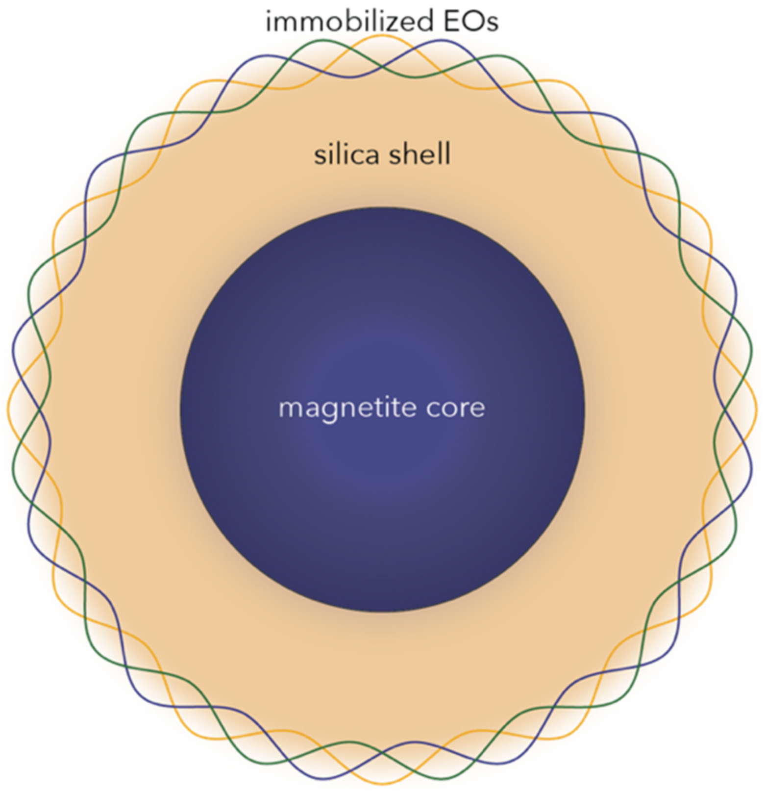
32]. Furthermore, owing to its high surface reactivity due to the high amount of hydroxyl groups onto the surface, silica was added in order to enhance the immobilization of EOs within the systems (
Figure 1).
In this context, the current study involves synthesizing composite core–shell nanoparticles comprising magnetite cores and silica layers obtained through two different methods, namely co-precipitation and the microwave-assisted hydrothermal method. The co-precipitation method was selected as one of the most commonly used techniques for the synthesis of magnetite nanoparticles due to its convenience, low cost, and possibility of large-scale production. However, as the obtained nanoparticles are generally characterized by decreased crystallinity and have a high tendency for agglomeration and oxidation, the microwave-assisted hydrothermal method was applied in order to overcome such limitations. In this manner, this method provides the advantages of synthesizing nanoparticles with a narrow size and shape distribution, due to homogenous high temperature and pressure conditions [
21,
33]. After the synthesis process, the nanosystems were functionalized with three different EOs, namely thyme, rosemary, and basil.
2. Results and Discussion
The Fe
3O
4@SiO
2 nanosystems were obtained through two different methods, namely co-precipitation (CP) and the microwave-assisted hydrothermal technique using the Synthwave (SW) equipment, and functionalized with three types of EOs, i.e., thyme, rosemary, and basil. The eight samples obtained and their associated labels are summarized in
Table 1.
On the one hand, the present study investigated the influence of the synthesis method on the outcome properties of the Fe
3O
4@SiO
2 in terms of mineral phase formation, crystallinity, morphology, size, surface charge, magnetic properties, and thermal stability. On the other hand, after synthesis, three different EOs, namely thyme, rosemary, and basil, were immobilized onto the surface of the nanosystems, and their antimicrobial properties were assessed through in vitro assays. In contrast to other research studies focused on the synthesis of Fe
3O
4@SiO
2 core–shell nanoparticles [
34,
35,
36], this study did not involve the use of surfactants or porogen agents.
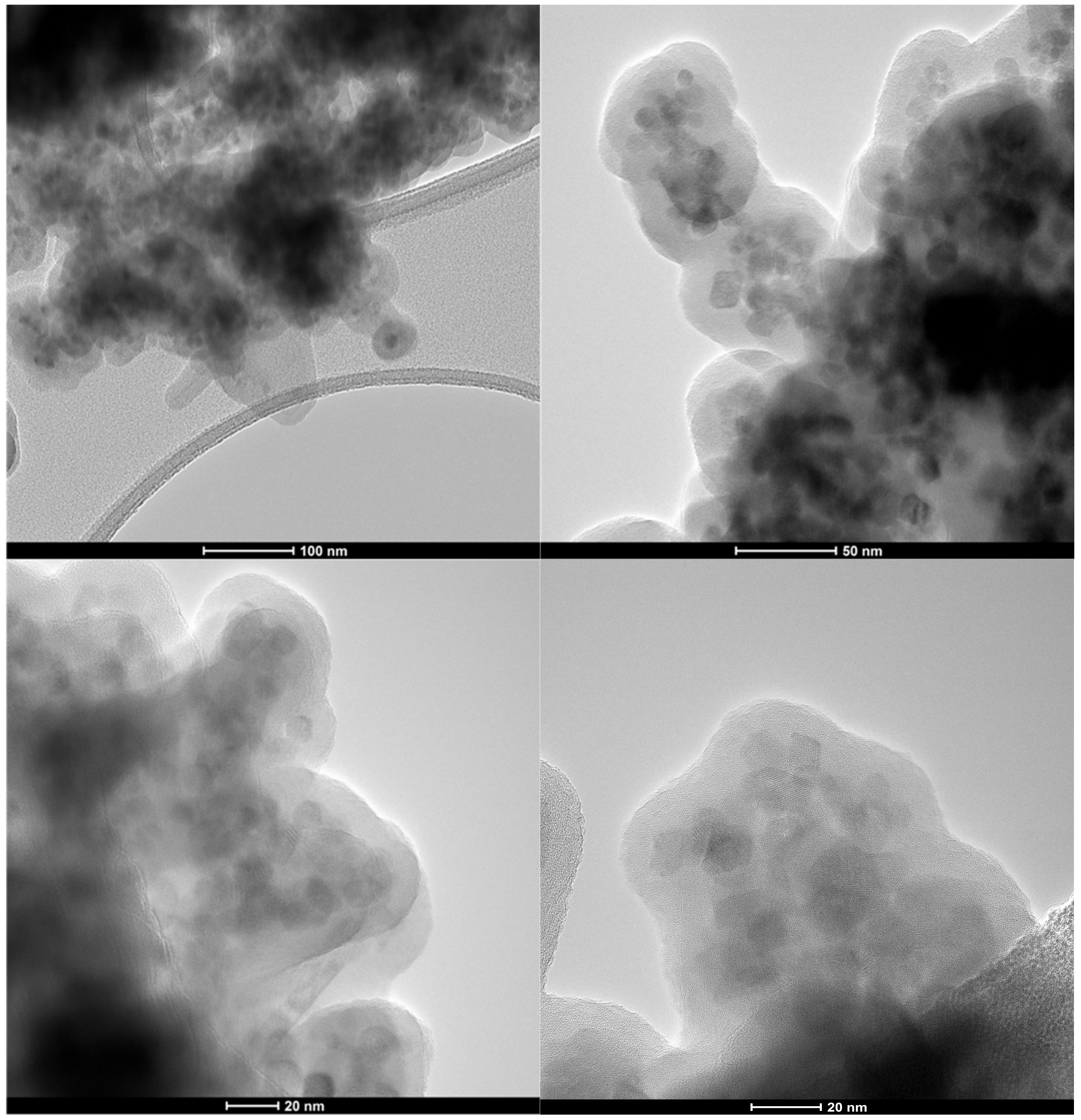
The core–shell structure of the composite nanosystems was demonstrated through Bright Field Transmission Electron Microscopy (BF-TEM) images (
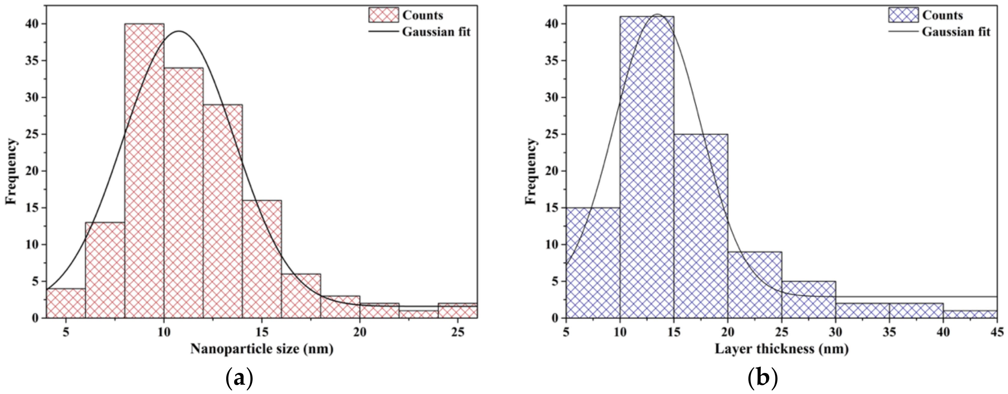
Figure 2), which show clusters of 1 to 10 nanoparticles surrounded by a thick layer of silica. Subsequently, TEM images were used to determine the core nanoparticle size and the shell layer thickness. Specifically, 150 cores and 100 shell regions were measured using the ImageJ software and based on the obtained information, corresponding size distributions were created and fitted using the Gaussian curve fit available in the Origin software (
Figure 3). Considering the FWHM of the fit, the average magnetite nanoparticle size was 6.7 nm, while the average thickness of the layer, 9.5 nm. Additionally, the nanosystems were characterized by an increased size uniformity, which is in accordance to similar nanosystems obtained through the microemulsion technique [
37]. Since the primary advantage of the microemulsion synthesis is represented by the precise control of nanoparticle size through the adjustment of water, oil, and surfactant present within the system, these results prove the efficiency of the present study to overcome current limitations associated with co-precipitation and hydrothermal techniques [
33,
38].
Moreover, TEM images demonstrate the spherical shape of the magnetite nanoparticles. In this context, previous studies reported a quasi-cubic shape of the nanoparticles, which was maintained after coating with silica, which was associated with increased chemical stability provided by the silica shell [
39]. Therefore, it could be safe to assume that the present Fe
3O
4@SiO
2 core–shell nanoparticles are characterized by a prolonged shelf-life due to the presence of the SiO
2 component.
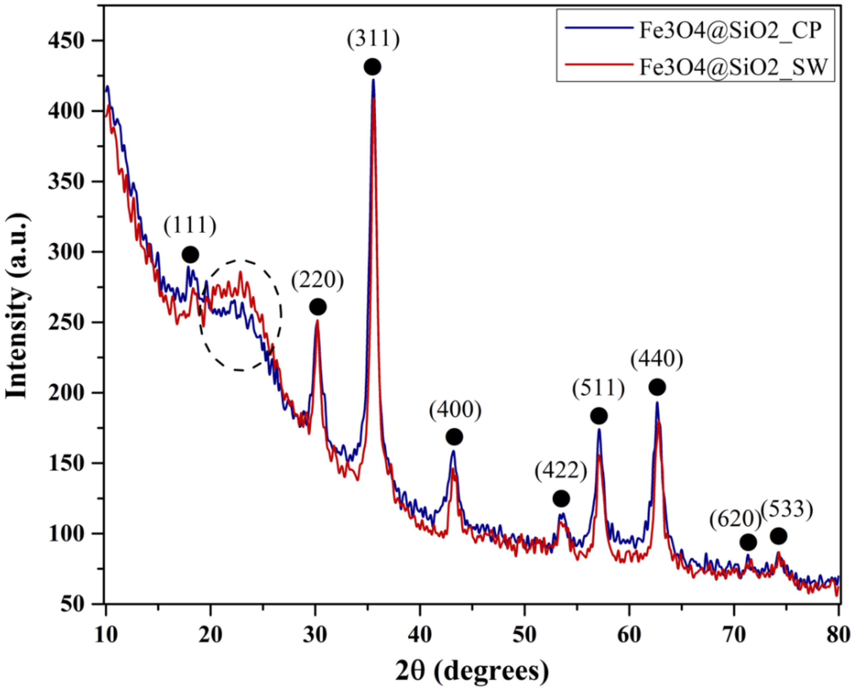
The crystallinity and mineral phase formation of the Fe
3O
4@SiO
2 nanoparticles were assessed through X-ray Diffraction (XRD) (
Figure 4). Both diffractograms show the formation of a single crystalline phase, demonstrated through the diffraction peaks characteristic for magnetite (Fe
3O
4) in the Fd-3m cubic crystal system and the associated Miller indices (according to the PDF 00-065-0731 [
40]). Additionally, the presence of the amorphous halo at the 2θ angles of 20–25° can be attributed to the silica layer within the nanosystems [
41,
42].
Furthermore, by comparing the two samples, it can be said that the microwave-assisted hydrothermal method is associated with a higher percentage of silica within the sample, as the diffraction halo within the Fe3O4@SiO2_SW sample has a higher intensity. Consequently, the crystallinity of this sample is reduced, as the intensity of the diffraction peaks for sample Fe3O4@SiO2_CP is slightly increased.
The crystallite size for both samples was calculated using the Debye–Scherrer equation:
where FWHM is the full width at the half peak height, K is the Scherrer constant that varies between 0.89 and 0.94, λ is the X-ray wavelength, D is the crystallite size, and θ is the diffraction angle [
43,
44].
In this manner, the average crystallite size calculated as the mean value between all peaks is presented in
Table 2. As expected, the crystallite size for the nanoparticles obtained through the hydrothermal method is higher due to the crystal growth under increased pressure and temperature conditions. Moreover, the crystallite size of the nanosystems is considerably lower than other results available in the scientific literature [
45].
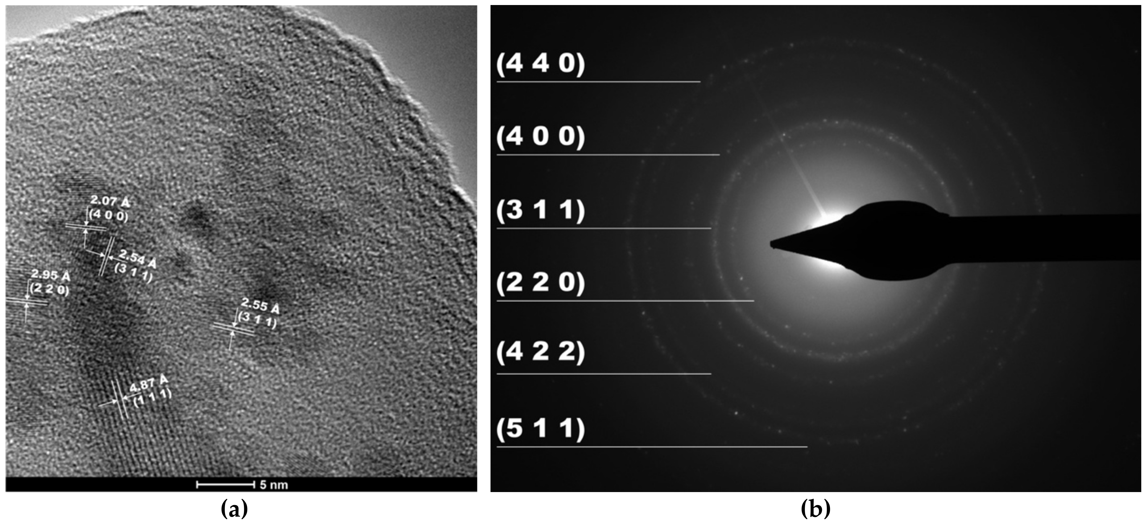
High Resolution-TEM (HR-TEM) and Selected Area Electron Diffraction (SAED) confirmed the formation of magnetite nanoparticles as a single crystalline phase (
Figure 5). As it can be observed, the identified Miller indices measured from the SAED rings matched the ones within the XRD diffractograms. Moreover, the presence of amorphous silica leads to the formation of the halo, which can be seen between the central spot and the first diffraction ring.
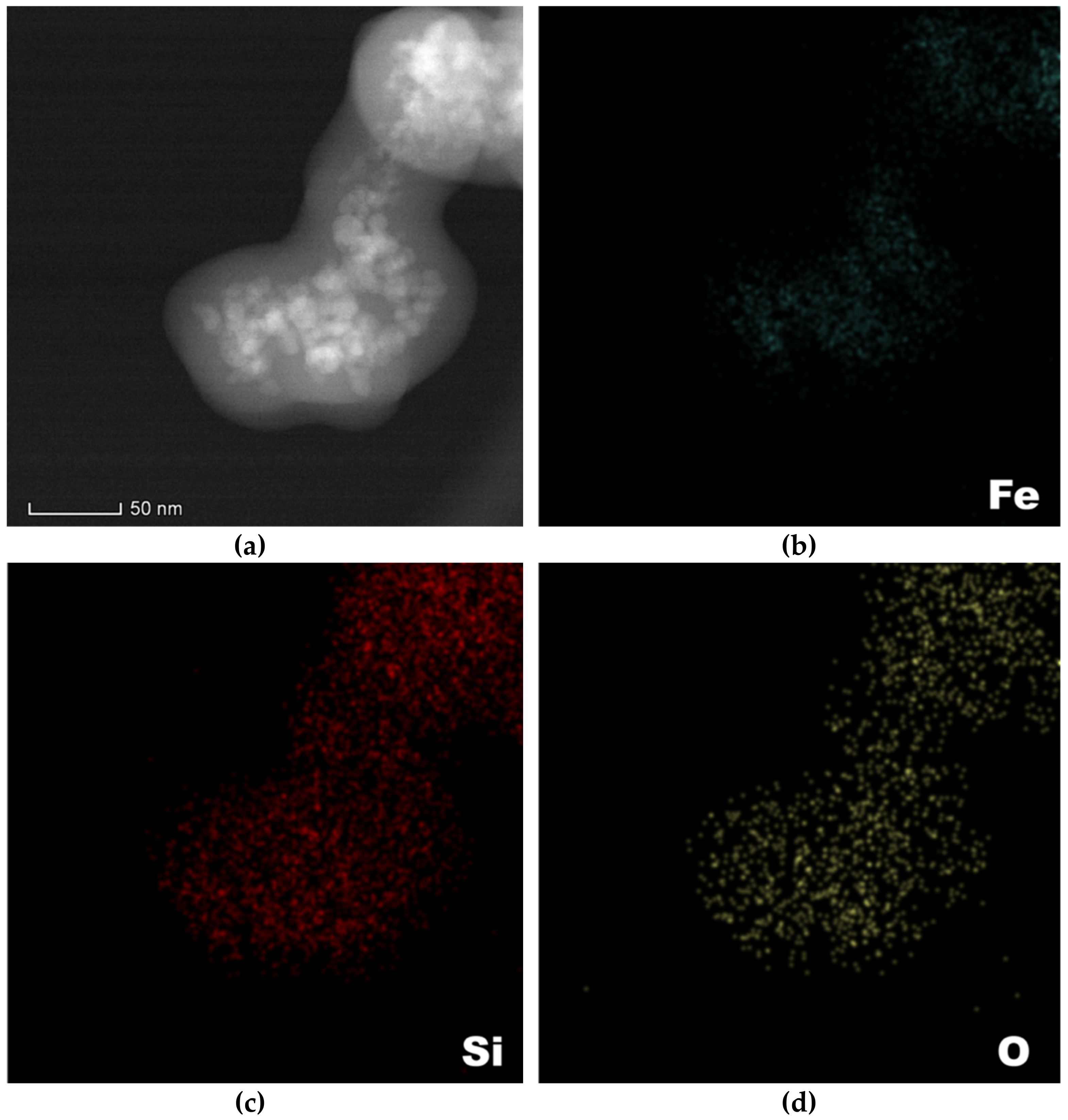
Subsequently, the Fe
3O
4@SiO
2 core–shell nanosystems were subjected to elemental mapping in order to determine the nature of the present elements and to further confirm the formation of the iron oxide core and the silica shell. Thus,
Figure 6 depicts the identified elements and their distribution within the composite nanosystems. Specifically, it can be seen that Fe is exclusively found within the magnetic cores, while Si is found throughout the system, as components of the silica layer. Therefore, both the nature of the constituents and the core–shell structure of the nanosystems were confirmed.
The Gas Chromatography-Mass Spectrometry (GC-MS) chromatograms revealed the presence of a series of volatile compounds characteristic for the EOs that were used for the experiment. The compounds identified within the EO-functionalized nanosystems and their retention time are summarized in
Table 3. The chromatogram profiles for each EO and EO-functionalized core–shell nanoparticles and the mass spectra for each of the major compounds identified within the EOs can be found in the
Supplementary Materials (Figures S4–S17). Additionally, the compounds identified within EOs and their retention time have also been added to the
Supplementary Materials (Table S1).
The identified compounds are consistent with previously published studies investigating the compositional concentration of thyme, rosemary, and basil EOs [
46,
47,
48,
49]. It can be seen that the major compound identified within the thyme EO-functionalized samples is thymol and p-cymen-7-ol. Thymol is well-known for its antimicrobial activities and low MIC values. The peak present within the CP sample is considerably higher, which could be attributed to a higher concentration and, consequently, to an increased antimicrobial activity. The rosemary EO-functionalized samples presented peaks for eucalyptol and (+)-2-bornanone as major compounds. For the basil EO-functionalized systems, the major compounds identified are trans-linalyl formate and estragole.
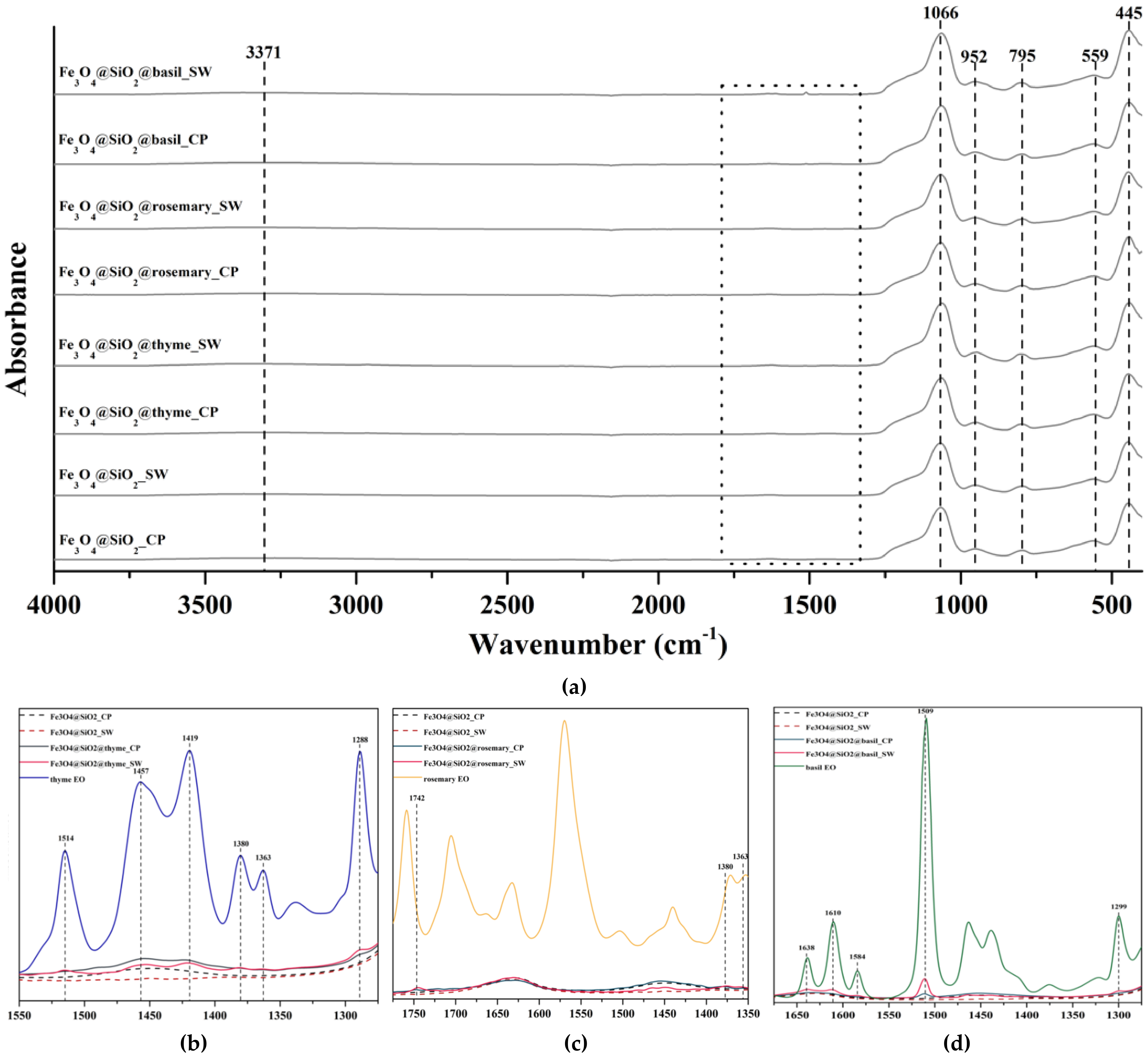
Fourier Transform Infrared (FT-IR) Spectroscopy was used for the assessment of the functional groups present within the synthesized samples. In this context,
Figure 7a presents the FT-IR spectra registered for all eight samples, namely for the simple Fe
3O
4@SiO
2_CP and Fe
3O
4@SiO
2_SW samples, and for the thyme, rosemary, or basil EO-functionalized Fe
3O
4@SiO
2_CP and Fe
3O
4@SiO
2_SW samples. There were five absorption bands at 445, 559, 795, 952, and 1066 cm
−1 registered within all eight samples. The sharp bands at 445 and 1066 cm
−1 correspond to the Si–O–Si or O–Si–O bending mode, while the band at 795 cm
−1 is assigned to the Si–O–Si symmetric stretch. The Fe–O stretching mode characteristic for Fe
3O
4 is shown at 559 cm
−1 [
42,
50]. The absorption peak at 952 cm
−1 is attributed to the Fe-O-Si stretching vibration, thus demonstrating the formation of the silica layers onto the iron oxide core [
51]. The wide absorption band at 3371 cm
−1 present in all samples corresponds to the O-H stretching mode [
42,
50].
The square marked in the FT-IR spectra represents the wavenumber region where the presence of EOs can be observed. Specifically,
Figure 7b–d presents the absorption peaks found in the functionalized samples that are characteristic to the EO that was used. While all samples were successfully functionalized, the absorption bands within the samples synthesized through the hydrothermal method have higher intensities, which could be related to higher functionalization yields. The wavenumbers for each absorption band and the associated bonds [
52,
53,
54] are summarized in
Table 4.
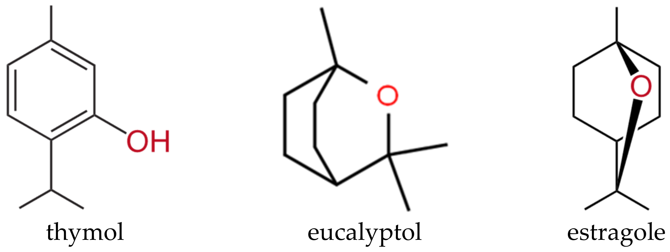
Considering the chemical structures of the main components found within thyme, rosemary, and basil EOs, the absorption peaks registered demonstrate immobilization of the EOs onto the core–shell nanoparticles. Specifically, considering thymol, eucalyptol, and estragole as the primary compounds of thyme, rosemary, and basil EOs, respectively, their characteristic reactive groups, i.e., O-H (phenol), O-H (alcohol), and -C-O, were found on the FT-IR spectra. As the mentioned compounds are well-known for their antimicrobial activity, it could be safe to assume that the antimicrobial activity of the EOs-functionalized core–shell nanoparticles should be ensured. However, since the rosemary EO-functionalized samples lack key absorption peaks specific to rosemary EO, such as C-O and C=O specific for camphor, and the peaks present have significantly low intensities, it could be concluded that the loading efficiency was reduced in this case. For better visualization, the chemical structures of thymol, eucalyptol, and estragole were represented in
Figure 8.
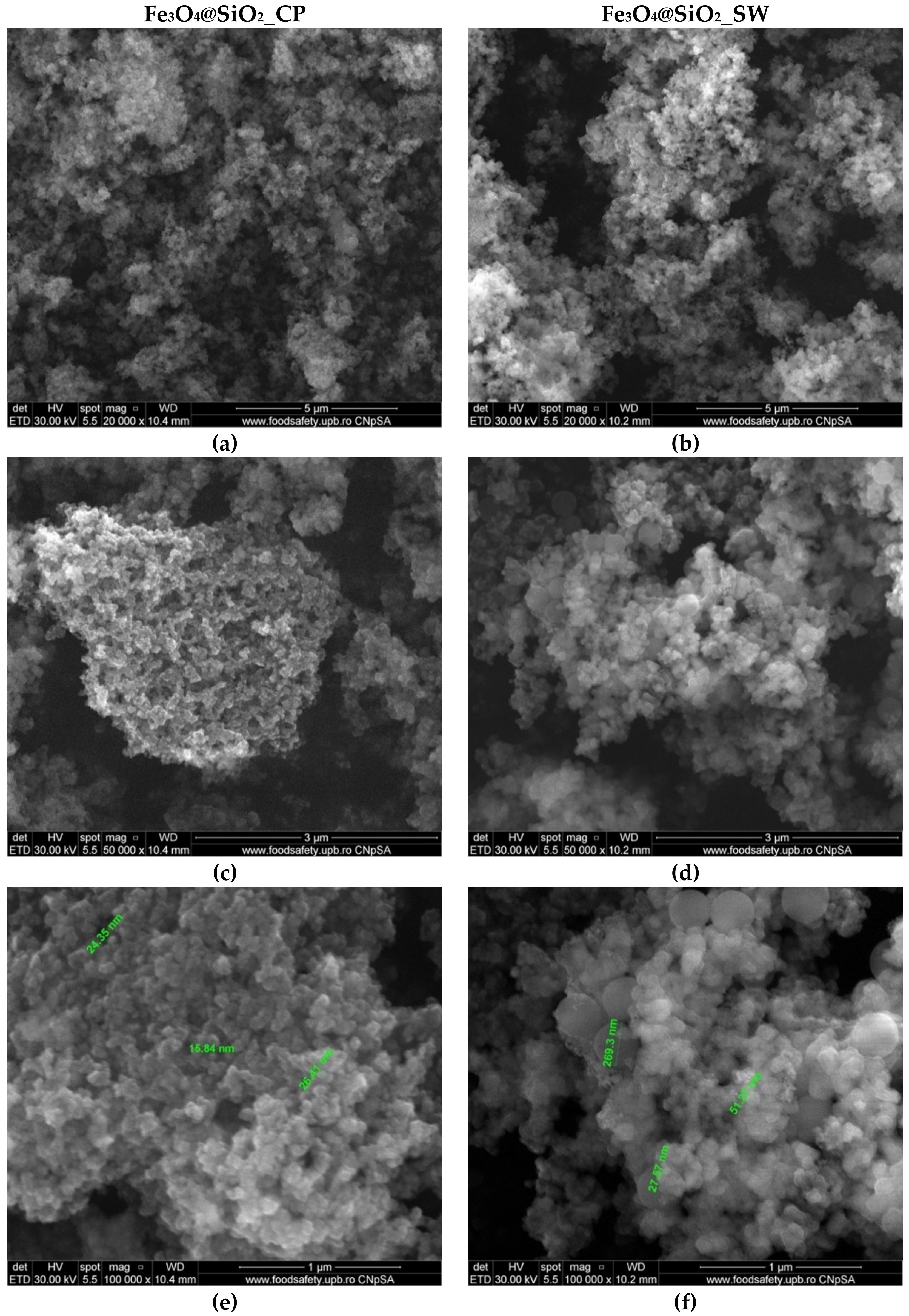
Scanning Electron Microscopy (SEM) analysis allowed for visualization of the composite nanosystems morphology (
Figure 9). In this context, both Fe
3O
4@SiO
2_CP and Fe
3O
4@SiO
2_SW samples exhibited a quasispherical shape with a considerable tendency for agglomeration. It can be seen that the composite systems are characterized by nanoscaled dimensions. Larger sizes were registered for the nanoparticles obtained through the hydrothermal method, thus confirming the previous results. Moreover, there are several nanoparticle aggregates with dimensions in the range of 200 nm corresponding to the Fe
3O
4@SiO
2_SW sample. By contrast to other studies investigating the formation of Fe
3O
4@SiO
2 core–shell nanoparticles which report considerably larger core sizes and shell thicknesses [
45,
55,
56], these results demonstrate the formation of nanoscaled core–shell systems with high uniformity and reduced sizes.
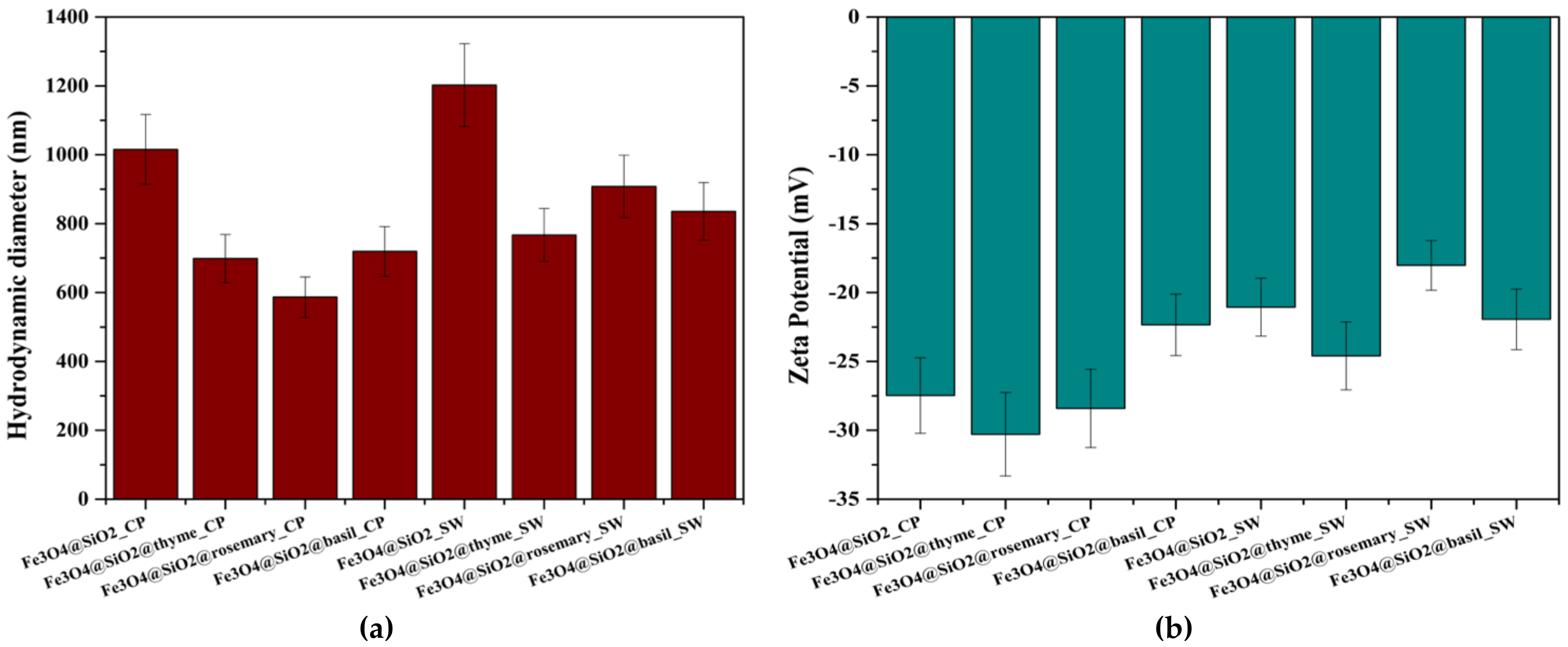
The hydrodynamic diameter and zeta potential of the core–shell nanoparticles were assessed both before and after functionalization with EOs. Five measurements were performed on each sample, and the mean values were calculated accordingly (
Table 5).
Figure 10 presents a visualization of the mean hydrodynamic diameter and zeta potential values registered for the eight samples. The highest hydrodynamic diameter values recorded are associated with the simple Fe
3O
4@SiO
2 samples. Since the hydrodynamic diameter is defined as the diameter of the hypothetical solid sphere formed through the attachment of solvent molecules onto the surface of the core nanoparticles [
57,
58], this can be explained by the high number of interactions between the surface hydroxyl groups characteristic to silica and water molecules. Therefore, since EOs are attached to the composite nanosystems through binding to the reactive groups present onto the surface, interaction with the solvent will be limited. Consequently, the hydrodynamic diameter will be reduced. Moreover, the previous results are confirmed by the higher values registered for the nanosystems synthesized through the hydrothermal method. The size distribution for each sample and the associated correlation graph can be found in the
Supplementary Materials (Figures S1–S3).
The zeta potential is generally considered a reference of the stability of the nanosystems dispersed into a solvent, as it reflects the amount of surface charges. Specifically, at values close to 0 (−25 mV to 25 mV), nanoparticles have a tendency to form aggregates due to attractive forces present at the surface. Therefore, zeta potential values lower than −25 mV and higher than 25 mV are associated with stable dispersions [
59,
60,
61]. As it can be seen in
Figure 4, the lowest zeta potential values were recorded for samples Fe
3O
4@SiO
2@thyme_CP and Fe
3O
4@SiO
2@rosemary_CP, which had the smallest hydrodynamic diameters. By contrast, the values for the nanosystems obtained through the hydrothermal technique were in the range of −24–−18 mV, demonstrating a higher agglomeration tendency due to higher surface reactivity specific to the synthesis method.
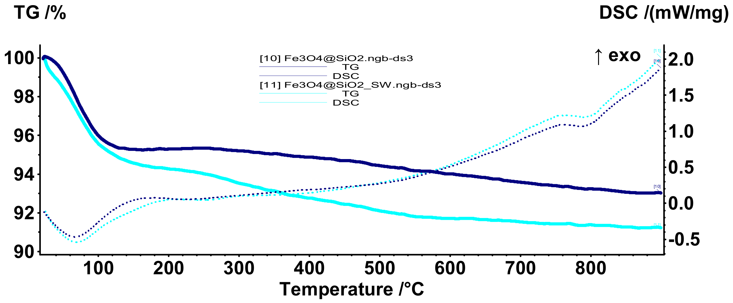
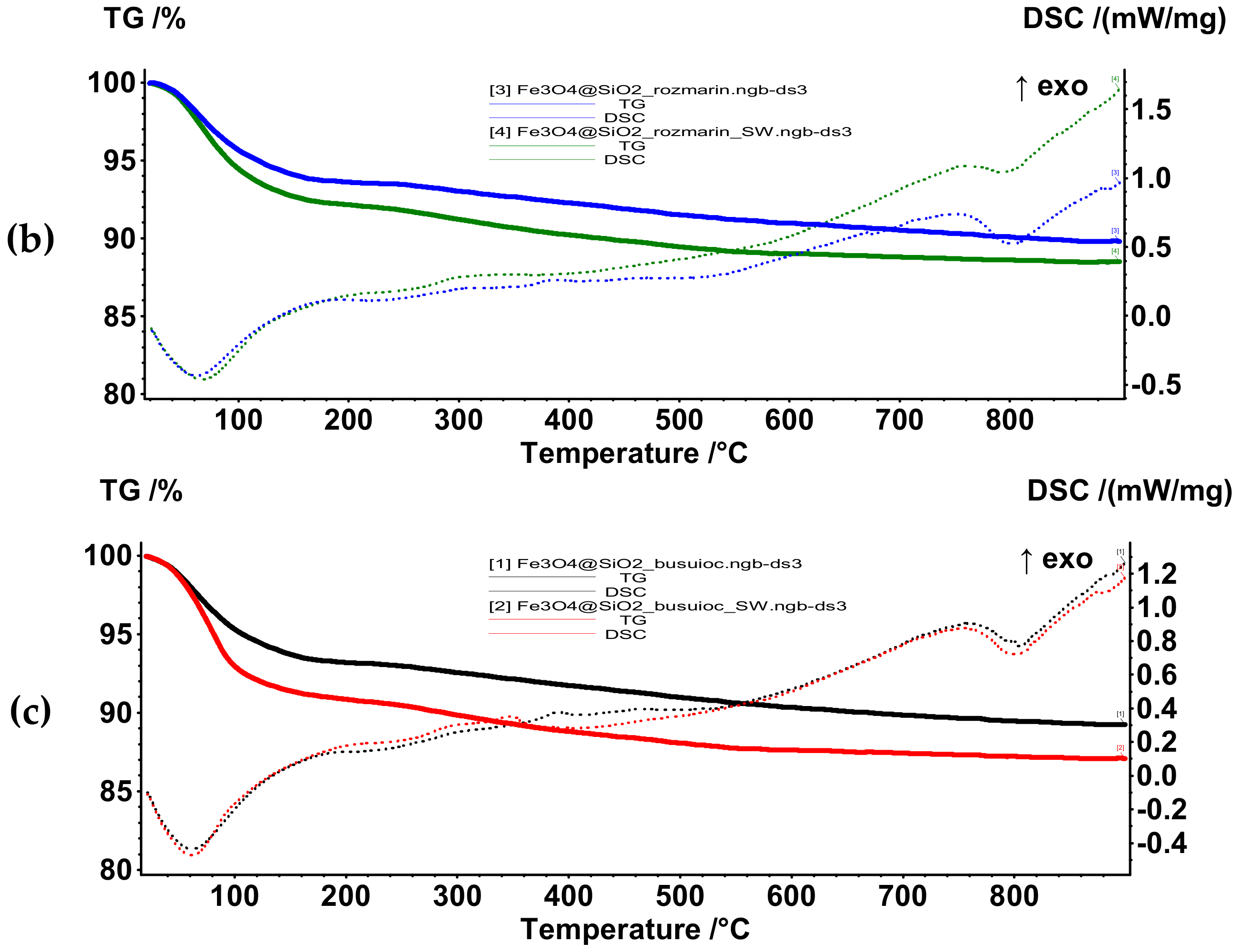
The thermal behavior of the core–shell nanoparticles was assessed through Thermogravimetry and Differential Scanning Calorimetry (TG-DSC). In this context,
Figure 11 presents the TG-DSC curves for the Fe
3O
4@SiO
2_CP and Fe
3O
4@SiO
2_SW nanosystems. In the temperature range of 20–180 °C, there is a mass loss of 4.69% and 5.77% accompanied by an endothermic effect with a minimum at 66.4 °C and 66.1 °C, respectively. The higher mass loss for the system obtained through the hydrothermal method could be associated with a higher amount of water molecules absorbed and adsorbed within the nanoparticles. In this manner, the higher affinity for water adsorption could be attributed to the higher hydrodynamic diameter previously reported. Moreover, the absence of the exothermic effects at ~300 °C and ~600 °C that are generally attributed to the transformation of magnetite into maghemite and subsequently of maghemite to hematite demonstrate that the iron oxide core is protected from thermal oxidation [
62,
63]. This hypothesis was also confirmed after the analysis, as the residual powder maintained its magnetic behavior.
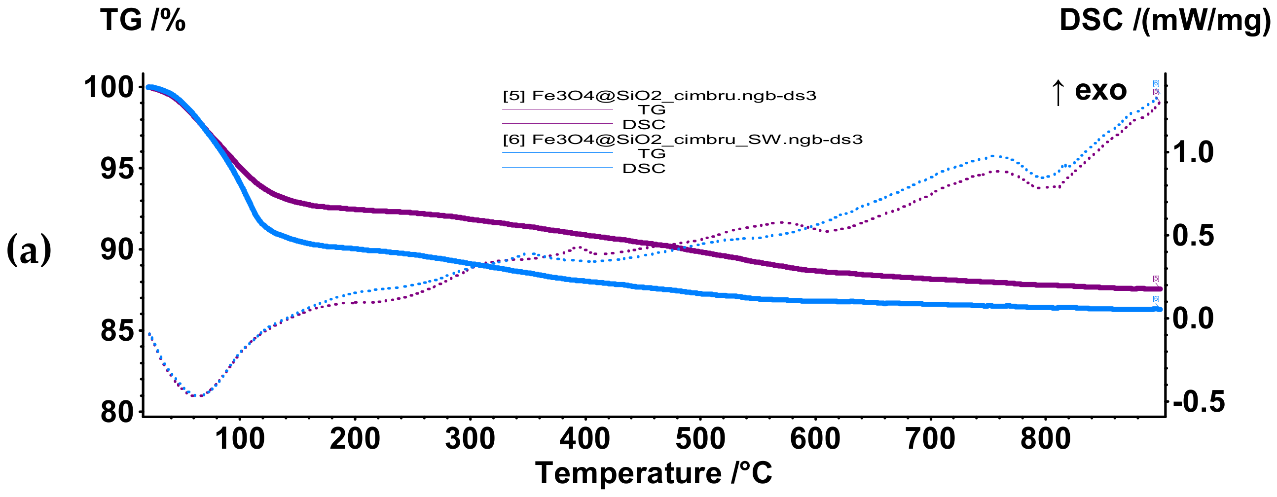
Subsequently, samples Fe
3O
4@SiO
2@thyme_CP, Fe
3O
4@SiO
2@rosemary_CP, Fe
3O
4@SiO
2@basil_CP, Fe
3O
4@SiO
2@thyme_SW, Fe
3O
4@SiO
2@rosemary_SW, and Fe
3O
4@SiO
2@basil_SW were subjected to the same thermal treatment in order to assess the loading efficiency of each sample (
Figure 12). All samples were characterized by an initial mass loss in the temperature range 20–180 °C through an endothermic process with a minimum at 60–70 °C due to the elimination of volatile compounds present within the EOs and residual solvent molecules. The second mass loss was registered between 180–500 °C associated with an exothermic effect with the maximum at 300–400 °C, indicating the oxidation of less volatile compounds present within the EOs and the elimination of hydroxyl groups from the surface of the nanoparticles. The final mass loss occurs between 500–900 °C.
Table 6 summarizes the mass losses mentioned for each sample and the associated thermal effects.
As it can be seen in
Table 6, the mass losses associated with the nanosystems obtained through the hydrothermal method are considerably higher in the temperature interval 20–180 °C. As this interval is attributed to the elimination of most of the EO compounds, it could be stated that these nanosystems are characterized by a higher loading capacity possibly due to a higher surface reactivity, as shown by the zeta potential measurements. Moreover, the estimated loading efficiency for each system, calculated as the difference between the mass loss associated with the EO-functionalized core–shell nanoparticles and the mass loss associated with the unfunctionalized core–shell nanoparticles, confirms this hypothesis. Additionally, the highest loading efficiency is attributed to thyme, followed by basil and, subsequently, rosemary. This trend is respected in both types of synthesis methods. These results are consistent with the information obtained from the FT-IR spectra.
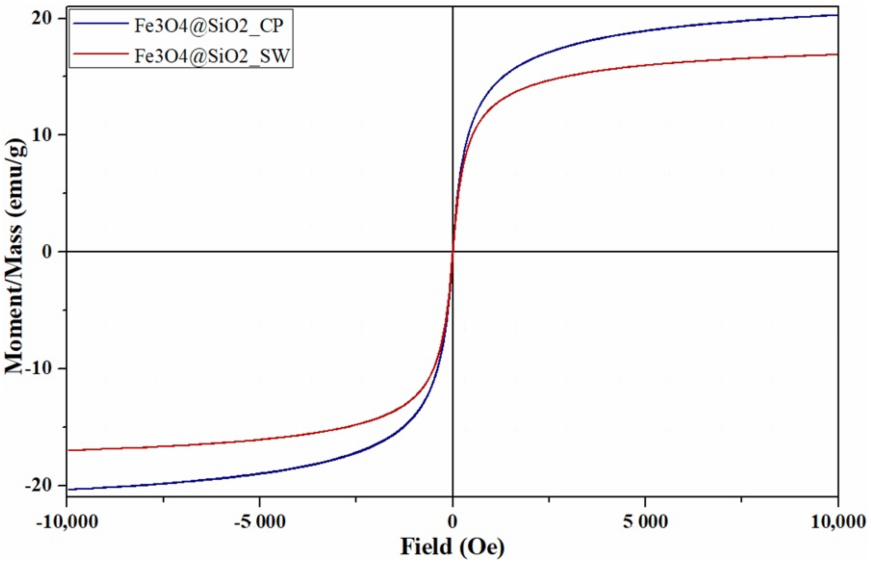
The magnetic properties of the Fe
3O
4@SiO
2 core–shell nanoparticles were measured through the Vibrating Sample Magnetometry analysis (
Figure 13). The superparamagnetic behavior of the nanoparticles is demonstrated by the S-shaped hysteresis curve of magnetization versus applied magnetic field with a width of zero [
64,
65,
66]. Such behavior further allows for developing drug delivery applications, as the drug-carrying nanoparticles are magnetized under an external magnetic field and lose their magnetization once the magnetic field is removed [
67,
68,
69]. Furthermore, the values associated with the saturation magnetization (M
s), remanence magnetization (M
r), and coercivity field (H
c) of the core–shell nanoparticles are shown in
Table 7.
Considering the silica shell thickness of 9.5 nm incorporating 3 magnetic cores of about 6.7 nm, as given by TEM results, and the density for silica and magnetite of 2.65 g/cm
3 and 5.18 g/cm
3 [
21], respectively, it can be deduced that the percentage of magnetite mass within the composite nanosystem is about 31%. Therefore, since the M
s of the core–shell nanosystems is 20.32 emu/g and 16.95 emu/g, the M
s of the magnetic core would be approximately 65 emu/g and 55 emu/g, respectively. Moreover, besides normalizing the magnetization by sample mass, its decrease could also be explained by the diamagnetic behavior of the silica shell [
41,
70]. While the magnitude of the demagnetizing field generated in the opposite direction of the applied external field is generally small for diamagnetic materials [
71], it cannot be neglected in this case, as a high field (10,000 Oe) is applied [
41,
72].
Furthermore, low M
r and H
c values are specific to small nanoparticles with superparamagnetic behavior [
73]. In this context, as previous results proved larger sizes for the nanosystems obtained through the hydrothermal method, the higher M
r and H
c values for the Fe
3O
4@SiO
2_SW were expected.
In this manner, the potential of the core–shell nanosystems for the use in hyperthermia-associated controlled release of bioactive substances was confirmed.
In the context of antimicrobial studies, the absence of interferences due to initial bacterial and fungal contamination of the samples was confirmed, as the nanoparticles seeded at the concentration of 1 mg/mL and incubated for 14 days at 37 °C and 28 °C, respectively, showed no signs of bacterial strains or yeast development.
Subsequently, the concentrations of 1, 2, and 4 mg/mL and 0.1, 0.2, and 0.4 µL/mL of Fe3O4@SiO2_CP, Fe3O4@SiO2@thyme_CP, Fe3O4@SiO2@rosemary_CP, Fe3O4@SiO2@basil_CP, Fe3O4@SiO2_SW, Fe3O4@SiO2@thyme_SW, Fe3O4@SiO2@rosemary_SW, and Fe3O4@SiO2@basil_SW and of thyme, rosemary, and basil EOs, respectively, were tested against Staphylococcus aureus ATCC 25923 (2 × 10⁶ UFC/mL test), Pseudomonas aeruginosa ATCC 27853 (4 × 10⁵ UFC/mL test), Escherichia coli ATCC 25922 (3.6 × 10⁶ UFC/mL test), and Candida albicans ATCC 10231 (1 × 10⁵ UFC/mL test).
The results are summarized in
Table 8. Briefly, the concentrations of 1 mg/mL and 0.1 µL/mL could not inhibit any of the microbial cultures. The concentration of 4 mg/mL of Fe
3O
4@SiO
2@thyme_CP and Fe
3O
4@SiO
2@thyme_SW samples inhibited the development of
Staphylococcus aureus,
Escherichia coli, and
Candida albicans cultures. However, the concentration of 0.4 µL/mL only inhibited the development of
Staphylococcus aureus. In this manner, the contribution of the Fe
3O
4@SiO
2 substrate to the antimicrobial activity of the nanosystem, but not alone or in combination with the other two types of EOs, was demonstrated. Additionally, immobilizing the EOs onto the surface of the nanosystems prevents their volatilization, thus ensuring the antimicrobial activity of their components.
Pseudomonas aeruginosa culture development was not inhibited by these concentrations. The concentration of 2 mg/mL of samples Fe
3O
4@SiO
2@thyme_CP and Fe
3O
4@SiO
2@thyme_SW, but not 0.2 µL/mL of thyme EO, inhibited the development of
Staphylococcus aureus. Thus, the minimum inhibitory concentration (MIC) value against the
Staphylococcus aureus strain of the two nanoparticle samples was established at 2 mg/mL and 0.4 µL/mL for the thyme EO. Furthermore, only sample Fe
3O
4@SiO
2@thyme_CP inhibited the development of
Escherichia coli and
Candida albicans cultures. The MIC value against the two microbial species was determined at 2 mg/mL for sample Fe
3O
4@SiO
2@thyme_CP and 4 mg/mL for sample Fe
3O
4@SiO
2@thyme_SW.
This outcome is in accordance with previous results, as GC-MS, DSC-TG, and FT-IR analyses indicated a considerably higher loading efficiency for the thyme EO-functionalized core–shell nanoparticles. Furthermore, previous studies investigating the antimicrobial efficiency of various EOs and their major compounds reported a significantly higher activity for eucalyptol, the major rosemary EO compound, compared to thymol, the major thyme EO compound [
74]. Other results report considerably high MIC values for estragole, the major compound of basil EO [
75]. Additionally, the higher antibacterial activity of thymol against Gram positive than Gram negative bacterial strains is well-known and reported in the literature [
76,
77,
78], which explains the higher antibacterial activity against
Staphylococcus aureus. Precise MIC values for each of the three compounds, as reported in the literature, are summarized in
Table 9.
Moreover, the increased antimicrobial activity of the nanoparticles synthesized through the hydrothermal method could be related to the smaller Fe
3O
4 nanoparticle size, which is generally associated with a higher capacity to penetrate and disrupt microbial cell walls [
9]. Furthermore, since there was a higher amount of silica within these samples, the intrinsic antimicrobial activity of the Fe
3O
4 nanoparticles could be reduced due to the limited contact between the cores and the bacterial cells.


 ,
, 


