Article

Microbiological and Molecular Features Associated with Persistent and Relapsing Staphylococcus aureus Prosthetic Joint Infection

by
Irene Muñoz-Gallego
1,†,
María Ángeles Meléndez-Carmona
1,*,†,
Jaime Lora-Tamayo
2,3,
Carlos Garrido-Allepuz
4,
Fernando Chaves
1,
Virginia Sebastián
4 and
Esther Viedma
1
1
Department of Clinical Microbiology, Hospital Universitario 12 de Octubre, Instituto de Investigación Biomédica “i + 12” Hospital 12 de Octubre, 28041 Madrid, Spain
2
Department of Internal Medicine, Hospital Universitario 12 de Octubre, Instituto de Investigación Biomédica “i + 12” Hospital 12 de Octubre, 28041 Madrid, Spain
3
CIBER de Enfermedades Infecciosas, Instituto de Salud Carlos III, 28222 Madrid, Spain
4
Helix BioS. Bioinformatics Solutions, 28049 Madrid, Spain
*
Author to whom correspondence should be addressed.
These authors contributed equally to this work.
Submission received: 20 July 2022 / Revised: 7 August 2022 / Accepted: 11 August 2022 / Published: 18 August 2022

Abstract

:
Background: Persistent and relapsing prosthetic joint infection (PJI) due to Staphylococcus aureus presents a clinical challenge. This study aimed to provide an extensive description of phenotypic and genomic changes that could be related to persistence or relapse. Methods: Initial and second S. aureus isolates from 6 cases of persistent and relapsing PJI, along with clinical isolates from 8 cases, with favorable outcome were included. All isolates were studied by phenotypic and genotypic approaches. Results: Recurrent S. aureus isolates exhibited a significant increase in adhesive capacity, invasion and persistence compared to resolved isolates. No association was found for the presence or absence of certain genes with the persistence or relapse of PJI. All sequential isolates showed identical sequence type (ST). Resistance gene loss during the infection and a great diversity of variants in different virulence genes between the pair of strains, mainly in genes encoding adhesins such as fnbA, were observed. Conclusions: S. aureus-caused relapse and persistence PJI is associated with bacterial phenotypical and genotypical adaptation. The main paths of adaptation were persistence in the intracellular compartment, and the loss of antibiotic resistance genes and variant acquisition, especially in genes encoding adhesins.

1. Introduction

Prosthetic joint infections (PJIs) are described as difficult to treat [1]. Although the incidence of PJI is not very high, it is still a devastating complication for patients, often requiring surgical intervention and long-term antibiotics, leading to impaired joint function [2]. Debridement, antibiotics and implant retention (DAIR) may be attempted in selected acute cases to minimize the morbidity associated with prosthesis removal and loss of bone stock [1,3]. Unfortunately, however, a large percentage of patients with an acute episode of Staphylococcus aureus PJI treated with DAIR eventually develop recurrent or persistent infection [4].
S. aureus has been identified as one of the main pathogens responsible for PJI [5]. It is a versatile microorganism with a wide genetic background and virulence factors that play an important role in the pathogenesis of infection and its evolution to persistent or recurrent forms [6,7]. The diversity of virulence factors allows S. aureus to adhere to and invade tissue, form biofilm and evade the immune system [7]. The causes of persistent infection are not well established, but possibilities include a limited antibiotic effect, suboptimal surgical management, host immune response or underlying diseases predisposed to recurrence. However, these could also be related to the intrinsic characteristics of the microorganism [8]. In this respect, previous studies have linked certain intrinsic characteristics of S. aureus to the persistence or chronicity of the infection, which include biofilm formation in bacterial communities [9,10], persistence in osteoblasts [11], increased adhesion capacity [12], accessory gene regulator (agr) dysfunction [13] and the formation of small colony variants (SCVs) [14].
Previous studies have focused on analyzing the clinical characteristics of the Staphylococcal–PJI evolution [4,15], but few have delved into the study of the microbiological characteristics and molecular factors that influence PJI development [16,17,18]. We hypothesized that some clinical isolates of S. aureus exhibit certain phenotypic and genotypic characteristics that eventually favor persistent or relapsing PJIs. We also investigated potential adaptive mechanisms in recurrent S. aureus isolates that allow certain infections to become chronic despite optimal antimicrobial and surgical management.

2. Material and Methods

2.1. Setting and Patients

Our group recently conducted a prospective, multicenter, observational study of 88 cases of PJI caused by S. aureus [18]. Most patients were treated with DAIR (debridement < 21 days after symptom onset) and received antibiotic treatment according to the antimicrobial susceptibility profile. Despite this, in six patients, infection persisted or relapsed with the original S. aureus strain. We included the initial (recurrent) isolates of the patients with recurrent staphylococcal infection and a second S. aureus isolate recovered during antimicrobial treatment in three cases (persistent isolates), and after antimicrobial treatment in another three cases (relapsed isolates). A random selection of clinical isolates from eight patients treated with DAIR who had a favorable clinical course with no evidence of recurrent S. aureus infection (resolved isolates) were also included (Table 1).

2.2. Antimicrobial Susceptibility Testing

Antimicrobial susceptibility testing was performed using the MicroScan WalkAway System (Siemens, West Sacramento, CA, USA). Minimum inhibitory concentrations (MICs) were interpreted according to EUCAST criteria (Version 7.1, 2017. http://www.eucast.org, access on 10 March 2017).

2.3. Characterization of Hemolytic Activity

Alpha-hemolysin (Hla) activity was analyzed and quantified by a rabbit erythrocyte lysis assay in 96-well plates and by visual inspection and assessment of zones cleared of round bacterial colonies [19]. The activity of the agr operon in gamma hemolysin production was measured and categorized as negative, weak, or strong [20]. Beta hemolysin production was also determined [21].

2.4. Biofilm Formation

Biofilm formation was assessed in triplicate by the 0.7% crystal violet method on microtiter plates using 33% glacial acetic acid as the decolorizing solution [18]. Absorbance was measured at 595 nm. The results were interpreted in accordance with Stepanovic [22].

2.5. Fibronectin Adhesion Assay

To measure the adhesion of S. aureus to fibronectin, 96-well tissue plates were prepared as described by Seidl et al. [23]. Bound staphylococci were detected by crystal violet staining at 620 nm using an enzyme-linked immunosorbent assay (ELISA) plate reader.

2.6. Cytotoxicity Assay on MG63 Osteoblasts

As described previously, the cytotoxicity assay was performed on an MG 63 (CRL-1427) human osteoblast cell line [11] using the Cytotoxicity Detection Kit Plus LDH (Roche Applied Science, Indianapolis, IN, USA). The percentage of cell lysis was calculated relative to the value for the maximum LDH release control (cells lysed with a 1% solution of Triton X-100), following the manufacturer’s instructions.

2.7. Adhesion, Invasion and Persistence Intracellular Assay

The MG 63 human osteoblast cell line (CRL-1427) (LGC standard, Manassas, VA, USA) was cultured and maintained in Dulbecco’s modified eagle medium (DMEM) supplemented with 10% fetal calf serum and 100 µg/mL gentamicin [24]. Cells were infected with stationary phase bacterial suspensions at a multiplicity of infection of 100, equivalent to 107 CFU/mL. Inoculum CFU levels were confirmed during each experiment by serial dilution and plating. The first 24-well cell plate was incubated at 37 °C in 5% CO2 for 2 h (adhesion). In the next two 24-well culture plates, the medium was replaced with DMEM + 10 µg/mL lysostaphin to lyse all extracellular or adherent staphylococci, and the plates were returned to the incubator for a further 3 h (invasion) and 48 h (persistence). At each of these time points, cells were lysed in 1 mL of sterile water for 10 min; serial dilutions were made; and suspensions were plated to calculate the bacterial CFU. Each isolate was tested in triplicate.

2.8. Whole Genome Sequencing

Whole genome sequencing of all S. aureus isolates was performed using Nextera (Illumina, San Diego, CA, USA). Libraries were sequenced in a single run on the Illumina NextSeq instrument (150 bp paired-end reads) to generate a coverage of ∼100×. Quality analysis was performed with FastQC (version 0.11.8) (https://www.bioinformatics.babraham.ac.uk/projects/fastqc/, access on 13 June 2018). The resulting reads were assembled with SPAdes (version 3.9) [25] and their quality was evaluated with Quality Assessment Tool for Genome Assemblies (QUAST), version 5.0.2 [26]. Genome annotation was carried out with Prokka (version 1.13.3) [27]. Multilocus sequence typing (MLST) was analyzed by the Center for Genomic Epidemiology (CGE) website (MLST, version 2.0) [28].
Antibiotic resistance genes were obtained by Mykrobe (version 6.1) [29]. The molecular epidemiology analysis was completed (SCCmec and spa type) with SCCmecFinder (version 1.2) and spaTyper (version 1.0) [30]. A pan-genomic analysis of the isolates was performed using Roary (version 3.11.2) [31].
Alignment was carried out according to the MLST obtained, using a reference genome belonging to the same or closest CC of the genome under study (Supplementary Table S1) [28,29]. Variant calling was performed with Snippy (https://github.com/tseemann/snippy accessed on 18 August 2020) [32] (version 4.4.0), and visualized with IGV (Integrative Genomics Viewer) [33]. The phylogenetic relationship was inferred from maximum likelihood with 1000 replicates, using IQ-TREE (version 1.6.11), after previous deletion of the recombinant regions by Gubbins (version 2.4.1), and was visualized using iTOL (version 5.5) [34,35,36]. Variant analysis was performed with breseq (version 0.33.2) [37]; the variant-calling criteria were adjusted to minimum mapping quality 30, consensus minimum variant coverage 30×, and consensus minimum total coverage 40×. Hypothetical proteins, pseudogenes, intergenic regions and variants in mobile genetic elements (phages) were excluded.
Sequence files were deposited at GenBank under BioProject PRJNA774351 and accession numbers JAJHKM000000000-JAJHNN000000000.

2.9. Statistical Analysis

All statistical analysis was performed using GraphPad Prism 8.2.1 software (San Diego, CA, USA). Two-way analysis of variance (ANOVA) was used to compare differences between multiple groups, and the unpaired Mann–Whitney test to compare differences between two samples: p values < 0.05 were considered to be significant.
The association between the presence or absence of genes and clinical variables (pan-microbial GWAS) was analyzed with Scoary software (version 1.6.16) [38]. The association between the presence or absence of genes and the clinical evolution of persistent and relapsing PJI was studied by adjusting the p value using the Benjamini–Hochberg method (FDR, False Discovery Rate) according to the following criteria: p < 0.10 and FDR < 0.05, p < 0.05 and FDR < 0.10 and, finally, p < 0.05 and FDR < 0.05.

3. Results

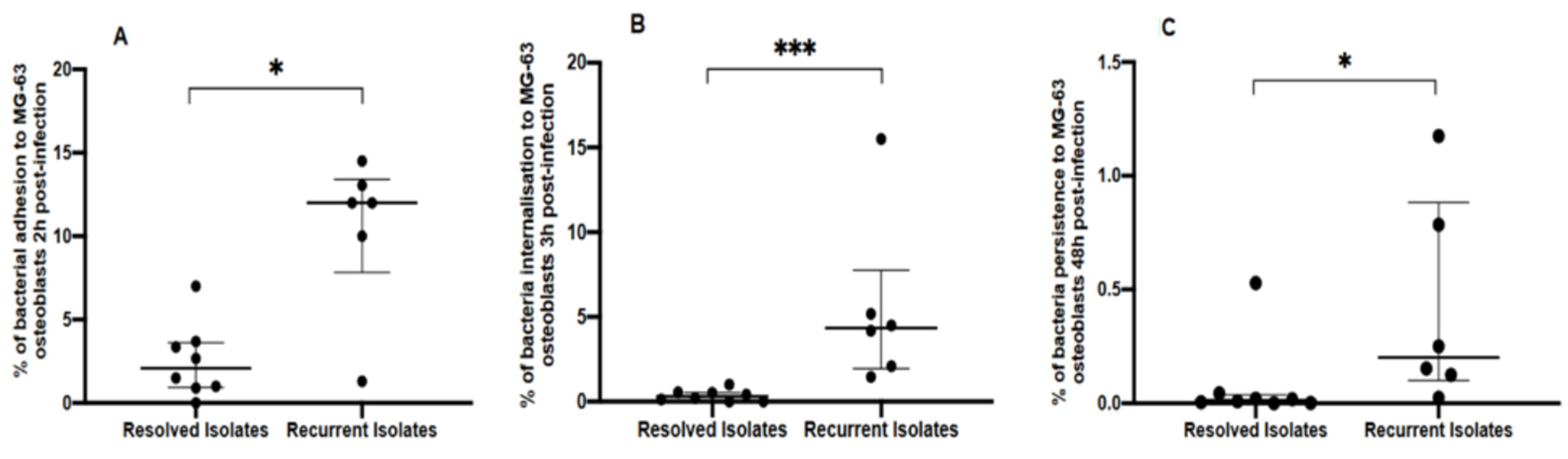
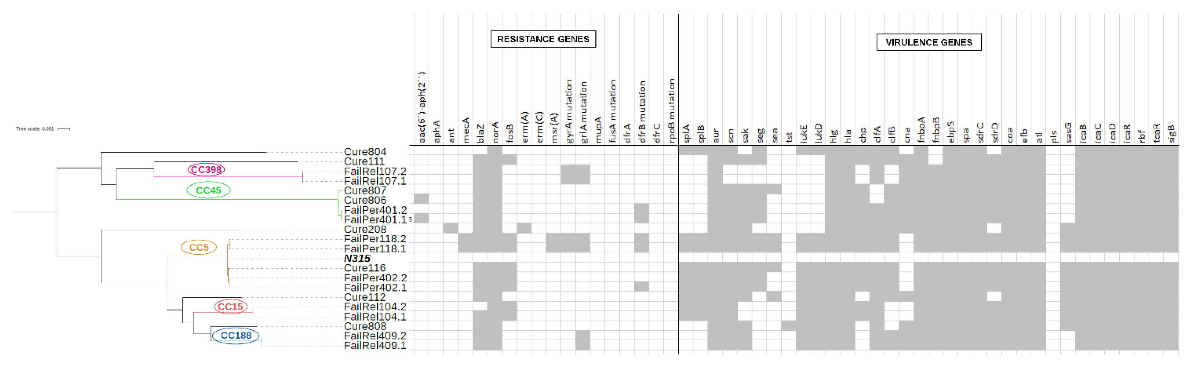
3.1. Genotypic and Phenotypic Comparisons of Isolates from Resolved and Recurrent PJIs

Isolates leading to recurrent PJI showed increased levels of resistance to all antimicrobials tested (Table 1). One isolate (FailPer118) was methicillin-resistant and harbored SCCmec IVc. Three recurrent isolates (FailRel107, FailRel409 and FailPer118) showed resistance to fluoroquinolones, while all isolates in the resolved group were susceptible. FailRel409 isolates were resistant to ciprofloxacin but susceptible to levofloxacin and harbored the norA gene, a mutation in the grlA gene but not the gyrA gene. FailRel107 isolates were resistant to clindamycin but susceptible to erythromycin. Overall, the genotypic and phenotypic antimicrobial resistance correlation was high, with the exceptions of resistance of fosfomycin and cotrimoxazole antibiotics (Table 2).
We analyzed the intracellular activity of the clinical isolates and found that recurrent S. aureus isolates exhibited a significant increase in capacity for adhesion (12.0%, (IQR: 7.8–13.4%) vs. 2.1%, (IQR: 0.9–3.6%), p = 0.01), invasion (4.3%, (IQR: 1.9–7.8%) vs. 0.3%, (IQR:0.03–0.5%), p = 0.001) and persistence (0.2%, (IQR:0.1–0.9%)) vs. 0.01% (IQR: 0.004–0.002%), p = 0.01) compared to those of the resolved PJIs (Figure 1). Hemolytic activity was similar in both groups of isolates (Table 1). On sheep blood agar, all isolates displayed a hemolysis pattern characteristic of Hla-induced beta-hemolysis although by rabbit erythrocyte lysis assay, three resolved isolates and one recurrent isolate did not hemolyze the erythrocytes (Supplementary Figure S1A). β-hemolysin with a phage insertion harboring immune evasion genes was detected in all strains except FailRel107, and δ-hemolysin activity was observed in five recurrent isolates (83.3%) and six isolates (75%) recovered from cured patients. Similarly, under conditions of stasis, all S. aureus isolates formed biofilm, but it was weak in most and no significant differences between the two groups of isolates was observed (Table 1 and Supplementary Figure S1B). Adhesion to fibronectin in recurrent and resolved isolates was also similar: 0.2 (95% CI: 0.17–0.19) vs. 0.15 (95% CI: 0.13–0.18, p = 0.3) (Supplementary Figure S1C) as was cytotoxic activity against MG63 osteoblasts: 49.2% (IQR: 38.9–51.7%) vs. 44%, (IQR: 34.7–51.1%) (Supplementary Figure S1D).
Figure 2 shows the phylogenetic tree of all isolates causing persistent, relapsing, and resolved PJIs together with the main resistance and virulence genes. The isolates were grouped according to CC and MLST similarity, forming two clearly differentiated clusters. Supplementary Table S2 shows the virulence genes of all strains causing PJI according to clinical evolution. All isolates harbored genes encoding microbial surface components recognizing adhesive matrix molecules (MSCRAMMs) and genes involved in biofilm formation. The genes fnbpA and fnbpB were identified in 100 and 85.7% of isolates, respectively, while cna was present in only 64.3%. In microbial pan-GWAS, no association was found between the presence or absence of genes and persistent or/relapsing PJI versus resolved PJI. Similarly, no significant variants were identified between strains causing persistent or relapsing PJI and those leading to a favorable outcome.

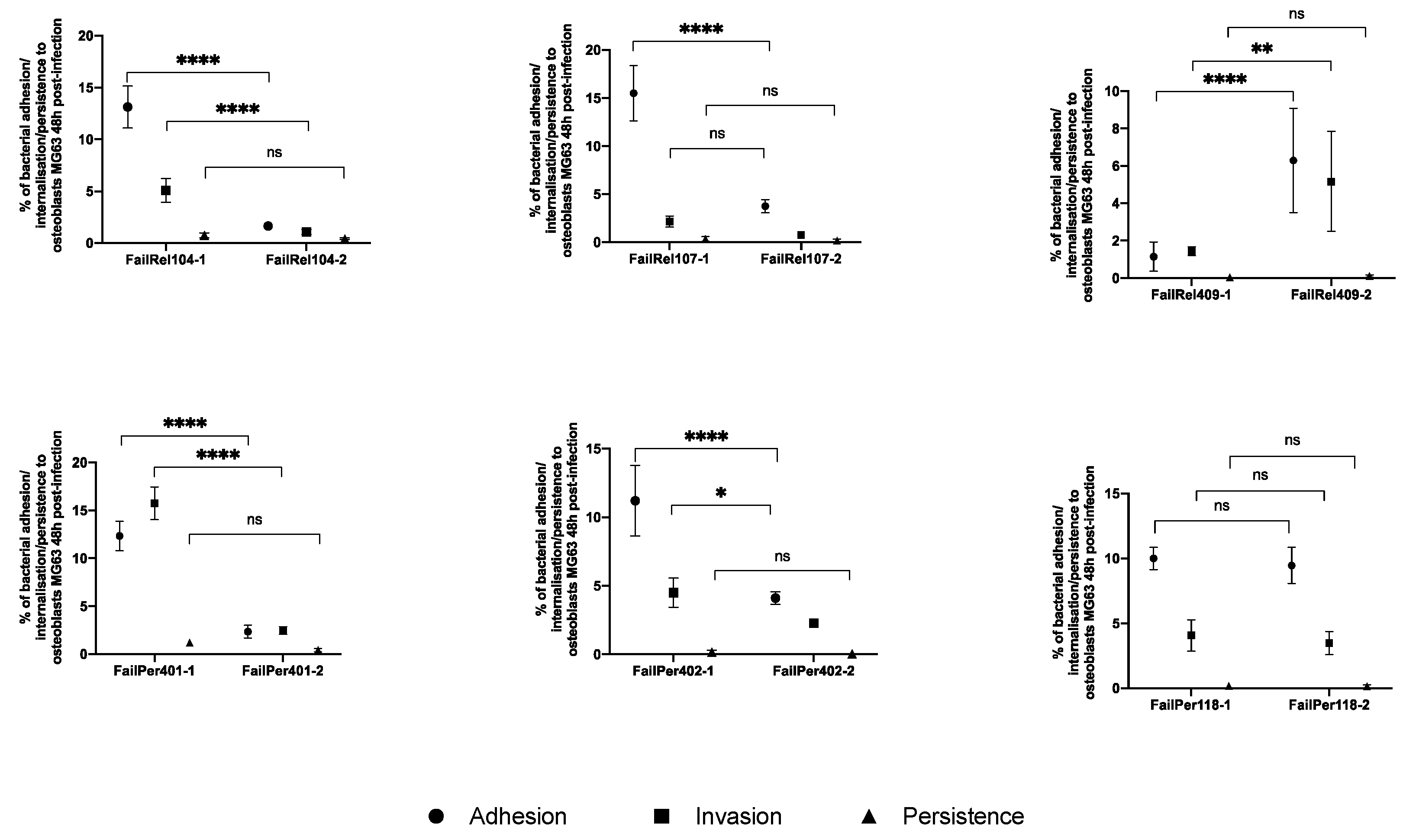
3.2. Phenotypic and Genotypic Changes during Persistent and Relapsing S. aureus PJIs

Sequential isolates from six recurrent PJIs were available for phenotypic and genotypic studies. In the relapsing cases, the second isolate was obtained 20, 55 and 217 days after the end of antimicrobial treatment. All sequential isolates showed an identical sequence type (ST) (Supplementary Table S1). All pairs of isolates showed identical antimicrobial resistance patterns except for persistent case 401, which harbored the aminoglycoside resistance gene aac(6′)-aph(2″), implying that FailPer401.1 was resistant to gentamicin, while FailPer401.2 was susceptible. Furthermore, unlike FailPer402.2, FailPer402.1 exhibited the H31N mutation in the dfrB gene, which confers resistance to trimethoprim although both strains were susceptible to cotrimoxazole. Finally, unlike FailRel104.2, FailRel104.1 harbored the blaZ gene, but we were unable to obtain a definitive resistance phenotype for FailRel104.2 because of the SCV phenotype (Figure 2).
Analyses of cell adhesion, invasion, and persistence in the sequential isolates revealed no significant differences between initial and second isolates obtained from the same patient (Supplementary Figure S2). However, we observed certain changes in five pairs of isolates (Figure 3). In four pairs (FailRel104, FailRel107, FailPer401 and FailPer402), the initial isolate showed a greater capacity for adhesion and invasion than the second one (p < 0.05), while in just one relapse case (FailRel409), the second isolate showed a greater capacity for adhesion and internalization (adhesion: 1.1 ± 0.8% vs. 6.3 ± 2.8%; p < 0.0001 and invasion 1.5 ± 0.2% vs. 5.2 ± 2.7%; p = 0.001).
In the same way, all sequential pairs of isolates showed the same agr functionality and hemolytic pattern, except for the FailRel104.2 relapse isolate, which showed a loss of hla activity and conversion to the SCV phenotype. Surprisingly, we did not observe an increased capacity for biofilm formation in the second isolates (Supplementary Figure S3), nor did we observe any difference in cytotoxic effect or fibronectin-binding capacity (Supplementary Figures S4 and S5).
With respect to the presence or absence of virulence genes, we found no differences between the initial and second isolates but observed a wide diversity of variants in different virulence genes between strain pairs in relapsing and persistent PJIs (Table 3 and Tables S1 and S2). In the FailRel104 and FailRel409 cases, the same non-synonymous SNP was found in the fnbA gene in the initial and relapsing strain: Q819H (reference MSSA476 (BX571857.1)). Nevertheless, no differential variants were found between all first and second isolates in the persistent and relapsing cases.

4. Discussion

Our study provides an extensive description of the adaptation of S. aureus strains during the clinical evolution of persistent and relapsing PJIs and a comprehensive analysis of possible phenotypic and genomics variations of all strains isolated in the same PJI. The availability of sequential isolates offers the opportunity to gain insight into adaptation during S. aureus infection. Other authors were limited by a small number of selected cases and so provided only a limited understanding of the general pattern of S. aureus evolution during a PJI [17].
A large panel of phenotypic experiments was used to investigate possible pathogenic traits associated with the S. aureus isolates that cause recurrent PJI. We found no evidence to support changes in bacterial virulence due to agr dysfunction, increased SCV and biofilm formation, adhesion to fibronectin protein or decreased alpha-hemolysin and LDH production. However, we identified one phenotypic feature associated with recurrent but not resolved PJI isolates: an increased capacity for adhesion, invasion and persistence in osteoblasts, which allows S. aureus to hide in a protected zone of infection and then re-emerge over time. These results could be important from a clinical point of view since they could indicate that there are isolates with greater potential to cause recurrence, either because infection persists despite treatment or because infection recurs after having been considered cured. After a thorough genetic analysis, we discovered a wide variety of clonal groups and virulence factors in S. aureus causing both PJI groups but no genetic molecular signature related to recurrence. The only potential factor that appeared to be associated with PJI recurrence was the higher frequency of quinolone resistance in the recurrent group [39].
Next, we investigated possible phenotypic and genotypic traits of sequential isolates recovered from the same patient at different time points of infection. In one case, the loss of antibiotic resistance genes was observed and antibiotic susceptibility increased. This result contrasted with a recent study that showed increased antibiotic resistance in persistent S. aureus bacteremia [40]. Interestingly, the FailRel107 strain showed an infrequent pattern of antibiotic susceptibility, with phenotypic susceptibility to erythromycin and phenotypic resistance to clindamycin. This could be caused by the lnu gene, which appears to be related to animal clonal lineage CC398 [41]. In addition, this strain did not present the phage carrying the immune system evasion genes, which allows the expression of β-hemolysin [21]. Therefore, it showed turbid halos of hemolysis on blood agar. Phenotypic analysis did not reveal any trait associated with the adaptation of S. aureus to persistence or recurrence of infection. In this context, we did not observe any differences between sequential isolates concerning cytotoxicity, biofilm formation or intracellular persistence. This contrasted with a previous study in which recurrent isolates were less cytotoxic, formed more biofilm and persisted longer in the intracellular compartment. However, that study included a small number of strains, which may be problematic for drawing general conclusions [17].
With respect to the role of MSCRAMMs in the pathogenesis of S. aureus in implant-associated infections [42], in all cases except one (FailPer402), significant variants in adhesin-encoding genes were produced between the initial and second strains although we did not find the same variants in all or most of the strains causing persistent or relapsing cases. Despite the fact that one study on S. aureus PJI did not find any association between polymorphisms in fibronectin-binding protein (FnBP) and PJI [43], other studies suggested that antigenic variation in FnBPA could contribute to S. aureus evasion of host immune response [44,45]. In connection with this, FailRel104 and FailRel409 showed variants in different MSCRAMMS (bbp, sdrD, clfA, fnbA and fnbB) and we found the same non-synonymous SNP in the fnbA gene between the initial and second strain in both isolates (Q819N). In the intracellular assays, however, the two isolates behaved differently: the initial FailRel104 and the second FailRel409 showed greater adhesion and invasion than the corresponding paired isolate in the sequence.
A non-synonymous SNP was found in the fmt gene of the FailRel104 strain, which has been associated with decreased production of extracellular virulence factors and decreased pathogenesis, which may contribute to increased bacterial survival [46] and persistence. Similarly, a deletion in DNA-3-methyladenine glycosylase, which catalyzes the removal of chemical DNA base lesions, was also observed in FailPer118 cases [47]. This variant could play a role in bacterial adaptation to stressful environments. It is likely that there are reservoirs of latent microorganisms in recurrent infections that are not eliminated by antibiotic treatment or by the host immune system. Under certain conditions, these microorganisms could reactivate and cause recurrence of infection. However, despite the diversity of variants found, we did not observe any in the agr operon, the main global regulator linked to the ability to downregulate pro-inflammatory virulence factors and increase the expression of factors promoting persistence.
The involvement of S. aureus SCVs in chronic prosthetic joint infections has been reported [48]. In the course of chronic osteomyelitis, after host cell invasion, S. aureus strains have been shown to develop adaptive mechanisms that decrease cytotoxicity and increase the percentage of SCV formation that persist in osteoblasts [16]. In relapsing case 104 (the initial strain was wild phenotype; the second one an SCV), mutations in exotoxins were observed. It has been reported that the SCV phenotype in S. aureus is favored by disrupting the electron transport chain with the consequent reduction in ATP production, which inhibits agr growth and functionality. Under these conditions, there is a reduction in hemolysin production and an increase in FnBP expression, providing SCV mutants with a high capacity for invasion into new host cells [44].
Our study did have some limitations. First, when analyzing the differences between strains, many variants were found in intergenic regions, hypothetical proteins and synonymous SNPs, so we do not know their possible involvement in these infections. Second, many variants were identified between the strains of cases 104 and 409, probably because the reference strain used for analysis (MSSA476) did not belong to the same CC. Third, we did not carry out a transcriptomic analysis of the genome, which may provide more accurate information about the molecular factors of S. aureus involved in the persistence and recurrence of PJI.
In our study, the phenotypic assays showed that the recurrent isolates had greater capacity for adhesion, invasion, and persistence in osteoblasts than those that caused resolved PJI. Conversely, the behavior between pairs of PJI isolates was very heterogeneous, and we were therefore unable to identify any phenotypic pattern associated with persistence or recurrence.

5. Conclusions

Our results showed that S. aureus is capable of modifying its genotype during the clinical course of PJI by adapting to recur or persist, especially by the loss of antibiotic resistance genes and the acquisition of variants, mainly in genes encoding adhesins. However, there was no convergence at the genetic level, indicating that adaptation pathways may be episode specific. This, coupled with other key aspects of the pathogenic complexity of PJI, such as the impact of host immunity on the evolution of S. aureus, could lead to the progression of infection to a cure or chronicity.

Supplementary Materials

The following are available online at https://0-www-mdpi-com.brum.beds.ac.uk/article/10.3390/antibiotics11081119/s1, Figure S1: Cytotoxicity activity, alpha-haemolysin, biofilm formation and fibronectin binding capacity by S. aureus strains included in our study, Figure S2: Comparisons in the adhesion, invasion and persistence capacities between initial and second S. aureus isolates. Figure S3: Ability to form biofilm of initial- and second S. aureus isolates retrieved from the same patient at the initial time of diagnosis and at the time of persistence or relapsed. Figure S4: Cytotoxic effect after infection between the different pairs of S. aureus grouped into relapse- or persistent- in human osteoblasts. Figure S5: Abilities of S. aureus strains to bind to fibronectin. Table S1: Molecular epidemiology of S. aureus strains causing PJI and reference strains used for each strain and each bioinformatic analysis. Table S2: Virulence genes of all the strains causing the 14 prosthetic joint infections included in this study.

Author Contributions

I.M.-G., M.Á.M.-C., J.L.-T. and F.C. conceived and designed the study. I.M.-G., E.V., C.G.-A. and V.S. performed the bioinformatic analyses. I.M.-G. and M.Á.M.-C. wrote the first draft of the manuscript supervised by F.C., J.L.-T. and E.V. All authors have read and agreed to the published version of the manuscript.

Funding

This work was supported by the Plan Nacional I + D + I (PI18/01623 and PI19/00571), Instituto de Salud Carlos III, Ministerio de Economía y Competitividad. MA.M was supported by a “Río Hortega” research contract (CM20/00146, Instituto de Salud Carlos III).

Institutional Review Board Statement

Not applicable.

Informed Consent Statement

Not applicable.

Data Availability Statement

The data presented in this study are available in supplementary material.

Acknowledgments

We thank the staff of the Hospital Universitario 12 de Octubre for their dedication. The preliminary results of this study were reported in part at the European Congress of Clinical Microbiology and Infectious Diseases (ECCMID), 2021 (Final Programme 2021; Abstract 4148, p 243).

Conflicts of Interest

The authors declare no conflict of interest.

References

  1. Zimmerli, W.; Trampuz, A.; Ochsner, P.E. Prosthetic-joint infections. N. Engl. J. Med. 2004, 351, 1645–1654. [Google Scholar] [CrossRef] [PubMed]
  2. Osmon, D.R.; Berbari, E.F.; Berendt, A.R.; Lew, D.; Zimmerli, W.; Steckelberg, J.M.; Rao, N.; Hanssen, A.; Wilson, W.R. Diagnosis and management of prosthetic joint infection: Clinical practice guidelines by the infectious diseases Society of America. Clin. Infect. Dis. 2013, 56, e1–e25. [Google Scholar] [CrossRef] [PubMed]
  3. El Helou, O.C.; Berbari, E.F.; Lahr, B.D.; Eckel-Passow, J.E.; Razonable, R.R.; Sia, I.G.; Virk, A.; Walker, R.C.; Steckelberg, J.M.; Wilson, W.R.; et al. Efficacy and safety of rifampin containing regimen for staphylococcal prosthetic joint infections treated with debridement and retention. Eur. J. Clin. Microbiol. Infect. Dis. 2010, 29, 961–967. [Google Scholar] [CrossRef] [PubMed]
  4. Lora-Tamayo, J.; Murillo, O.; Iribarren, J.A.; Soriano, A.; Sánchez-Somolinos, M.; Baraia-Etxaburu, J.M.; Rico, A.; Palomino, J.; Rodríguez-Pardo, D.; Horcajada, J.P.; et al. A large multicenter study of methicillin-susceptible and methicillin-resistant staphylococcus aureus prosthetic joint infections managed with implant retention. Clin. Infect. Dis. 2013, 56, 182–194. [Google Scholar] [CrossRef]
  5. Benito, N.; Franco, M.; Ribera, A.; Soriano, A.; Rodriguez-Pardo, D.; Sorlí, L.; Fresco, G.; Fernández-Sampedro, M.; Dolores del Toro, M.; Guío, L.; et al. Time trends in the aetiology of prosthetic joint infections: A multicentre cohort study. Clin. Microbiol. Infect. 2016, 22, 732.e1–732.e8. [Google Scholar] [CrossRef]
  6. Planet, P.J.; Narechania, A.; Chen, L.; Mathema, B.; Boundy, S.; Archer, G.; Kreiswirth, B. Architecture of a Species: Phylogenomics of Staphylococcus aureus. Trends Microbiol. 2017, 25, 153–166. [Google Scholar] [CrossRef]
  7. Montanaro, L.; Speziale, P.; Campoccia, D.; Ravaioli, S.; Cangini, I.; Pietrocola, G.; Giannini, S.; Arciola, C.R. Scenery of Staphylococcus implant infections in orthopedics. Future Microbiol. 2011, 6, 1329–1349. [Google Scholar] [CrossRef]
  8. Alamanda, V.K.; Springer, B.D. Perioperative and Modifiable Risk Factors for Periprosthetic Joint Infections (PJI) and Recommended Guidelines. Curr. Rev. Musculoskelet. Med. 2018, 11, 325–331. [Google Scholar] [CrossRef]
  9. Costerton, W.; Veeh, R.; Shirtliff, M.; Pasmore, M.; Post, C.; Ehrlich, G. The application of biofilm science to the study and control of chronic bacterial infections. J. Clin. Investig. 2003, 112, 1466–1477. [Google Scholar] [CrossRef]
  10. Paharik, A.E.; Horswill, A.R. The Staphylococcal Biofilm: Adhesins, Regulation, and Host Response. Microbiol. Spectr. 2016, 4, VMBF-0022–2015. [Google Scholar] [CrossRef]
  11. Valour, F.; Trouillet-Assant, S.; Riffard, N.; Tasse, J.; Flammier, S.; Rasigade, J.P.; Chidiac, C.; Vandenesch, F.; Ferry, T.; Laurent, F. Antimicrobial activity against intraosteoblastic Staphylococcus aureus. Antimicrob. Agents Chemother. 2015, 59, 2029–2036. [Google Scholar] [CrossRef] [PubMed]
  12. Foster, T.J.; Höök, M. Surface protein adhesins of Staphylococcus aureus. Trends Microbiol. 1998, 6, 484–488. [Google Scholar] [CrossRef]
  13. Post, V.; Wahl, P.; Uçkay, I.; Ochsner, P.; Zimmerli, W.; Corvec, S.; Loiez, C.; Richards, R.G.; Moriarty, T.F. Phenotypic and genotypic characterisation of Staphylococcus aureus causing musculoskeletal infections. Int. J. Med. Microbiol. 2014, 304, 565–576. [Google Scholar] [CrossRef]
  14. Tuchscherr, L.; Heitmann, V.; Hussain, M.; Viemann, D.; Roth, J.; Von Eiff, C.; Peters, G.; Becker, K.; Löffler, B. Staphylococcus aureus small-colony variants are adapted phenotypes for intracellular persistence. J. Infect. Dis. 2010, 202, 1031–1040. [Google Scholar] [CrossRef] [PubMed]
  15. Lora-Tamayo, J.; Euba, G.; Cobo, J.; Horcajada, J.P.; Soriano, A.; Sandoval, E.; Pigrau, C.; Benito, N.; Falgueras, L.; Palomino, J.; et al. Short- versus long-duration levofloxacin plus rifampicin for acute staphylococcal prosthetic joint infection managed with implant retention: A randomised clinical trial. Int. J. Antimicrob. Agents 2016, 48, 310–316. [Google Scholar] [CrossRef]
  16. Kalinka, J.; Hachmeister, M.; Geraci, J.; Sordelli, D.; Hansen, U.; Niemann, S.; Oetermann, S.; Peters, G.; Löffler, B.; Tuchscherr, L. Staphylococcus aureus isolates from chronic osteomyelitis are characterized by high host cell invasion and intracellular adaptation, but still induce inflammation. Int. J. Med. Microbiol. 2014, 304, 1038–1049. [Google Scholar] [CrossRef] [PubMed]
  17. Trouillet-Assant, S.; Lelièvre, L.; Martins-Simões, P.; Gonzaga, L.; Tasse, J.; Valour, F.; Rasigade, J.-P.; Vandenesch, F.; Guedes, R.L.M.; de Vasconcelos, A.T.R.; et al. Adaptive processes of Staphylococcus aureus isolates during the progression from acute to chronic bone and joint infections in patients. Cell Microbiol. 2016, 18, 1405–1414. [Google Scholar] [CrossRef] [PubMed]
  18. Muñoz-Gallego, I.; Viedma, E.; Esteban, J.; Mancheño-Losa, M.; García-Cañete, J.; Blanco-García, A.; Rico, A.; García-Perea, A.; Garbajosa, P.R.; Escudero-Sánchez, R.; et al. Genotypic and phenotypic characteristics of Staphylococcus aureus prosthetic joint infections: Insight on pathogenesis and prognosis of a multicentre prospective cohort. Open Forum Infect. Dis. 2020, 7, ofaa344. [Google Scholar] [CrossRef]
  19. Stulik, L.; Malafa, S.; Hudcova, J.; Rouha, H.; Henics, B.Z.; Craven, D.E.; Sonnevend, A.M.; Nagy, E. α-hemolysin activity of methicillin-susceptible Staphylococcus aureus predicts ventilator-associated pneumonia. Am. J. Respir. Crit. Care Med. 2014, 190, 1139–1148. [Google Scholar] [CrossRef]
  20. Traber, K.E.; Lee, E.; Benon, S.; Corrigan, R.; Cantera, M.; Shopsin, B.; Novick, R.P. agr function in clinical Staphylococcus aureus isolates. Microbiology 2008, 154, 2265–2274. [Google Scholar] [CrossRef]
  21. Salgado-Pabón, W.; Herrera, A.; Vu, B.G.; Stach, C.S.; Merriman, J.A.; Spaulding, A.R.; Schlievert, P.M. Staphylococcus aureus β-toxin production is common in strains with the β-toxin gene inactivated by bacteriophage. J. Infect. Dis. 2014, 210, 784–792. [Google Scholar] [CrossRef]
  22. Stepanović, S.; Vuković, D.; Hola, V.; Di Bonaventura, G.; Djukić, S.; Ćirković, I.; Ruzicka, F. Quantification of biofilm in microtiter plates: Overview of testing conditions and practical recommendations for assessment of biofilm production by staphylococci. Apmis 2007, 115, 891–899. [Google Scholar] [CrossRef] [PubMed]
  23. Seidl, K.; Bayer, A.S.; Fowler, V.G.; McKinnell, J.A.; Hady, W.A.; Sakoulas, G.; Yeaman, M.R.; Xiong, Y.Q. Combinatorial phenotypic signatures distinguish persistent from resolving methicillin-resistant Staphylococcus aureus bacteremia isolates. Antimicrob. Agents Chemother. 2011, 55, 575–582. [Google Scholar] [CrossRef]
  24. Richards, R.L.; Haigh, R.D.; Pascoe, B.; Sheppard, S.K.; Price, F.; Jenkins, D.; Rajakumar, K.; Morrissey, J.A. Persistent Staphylococcus aureus isolates from two independent cases of bacteremia display increased bacterial fitness and novel immune evasion phenotypes. Infect. Immun. 2015, 83, 3311–3324. [Google Scholar] [CrossRef] [PubMed]
  25. Nurk, S.; Bankevich, A.; Antipov, D.; Gurevich, A.A.; Korobeynikov, A.; Lapidus, A.; Prjibelski, A.D.; Pyshkin, A.; Sirotkin, A.; Sirotkin, Y.; et al. Assembling single-cell genomes and mini-metagenomes from chimeric MDA products. J. Comput. Biol. 2013, 20, 714–737. [Google Scholar] [CrossRef] [PubMed]
  26. Mikheenko, A.; Prjibelski, A.; Saveliev, V.; Antipov, D.; Gurevich, A. Versatile genome assembly evaluation with QUAST-LG. Bioinformatics 2018, 34, i142–i150. [Google Scholar] [CrossRef]
  27. Seemann, T. Prokka: Rapid prokaryotic genome annotation. Bioinformatics 2014, 30, 2068–2069. [Google Scholar] [CrossRef] [PubMed]
  28. Larsen, M.V.; Cosentino, S.; Rasmussen, S.; Friis, C.; Hasman, H.; Marvig, R.L.; Jelsbak, L.; Sicheritz-Pontéen, T.; Ussery, D.W.; Aarestrup, F.M.; et al. Multilocus sequence typing of total-genome-sequenced bacteria. J. Clin. Microbiol. 2012, 50, 1355–1361. [Google Scholar] [CrossRef]
  29. Bradley, P.; Gordon, N.C.; Walker, T.M.; Dunn, L.; Heys, S.; Huang, B.; Earle, S.; Pankhurst, L.J.; Anson, L.; de Cesare, M.; et al. Rapid antibiotic-resistance predictions from genome sequence data for Staphylococcus aureus and Mycobacterium tuberculosis. Nat. Commun. 2015, 6, 10063. [Google Scholar] [CrossRef]
  30. Bartels, M.D.; Petersen, A.; Worning, P.; Nielsen, J.B.; Larner-Svensson, H.; Johansen, H.K.; Andersen, L.P.; Jarløv, J.O.; Boye, K.; Larsen, A.R.; et al. Comparing whole-genome sequencing with sanger sequencing for spa typing of methicillin-resistant staphylococcus aureus. J. Clin. Microbiol. 2014, 52, 4305–4308. [Google Scholar] [CrossRef]
  31. Page, A.J.; Cummins, C.A.; Hunt, M.; Wong, V.K.; Reuter, S.; Holden, M.T.G.; Fookes, M.; Falush, D.; Keane, J.A.; Parkhill, J. Roary: Rapid large-scale prokaryote pan genome analysis. Bioinformatics 2015, 31, 3691–3693. [Google Scholar] [CrossRef] [PubMed]
  32. Petkau, A.; Mabon, P.; Sieffert, C.; Knox, N.C.; Cabral, J.; Iskander, M.; Iskander, M.; Weedmark, K.; Zaheer, R.; Katz, L.S.; et al. SNVPhyl: A single nucleotide variant phylogenomics pipeline for microbial genomic epidemiology. Microb. Genom. 2017, 3, e000116. [Google Scholar] [CrossRef]
  33. Robinson, J.T.; Thorvaldsdóttir, H.; Winckler, W.; Guttman, M.; Lander, E.S.; Getz, G.; Mesirov, J.P. Integrative genomics viewer. Nat. Biotechnol. 2011, 29, 24–26. [Google Scholar] [CrossRef] [PubMed]
  34. Nguyen, L.T.; Schmidt, H.A.; Von Haeseler, A.; Minh, B.Q. IQ-TREE: A fast and effective stochastic algorithm for estimating maximum-likelihood phylogenies. Mol. Biol. Evol. 2015, 32, 268–274. [Google Scholar] [CrossRef] [PubMed]
  35. Croucher, N.J.; Page, A.J.; Connor, T.R.; Delaney, A.J.; Keane, J.A.; Bentley, S.D.; Parkhill, J.; Harris, S.R. Rapid phylogenetic analysis of large samples of recombinant bacterial whole genome sequences using Gubbins. Nucleic Acids Res. 2015, 43, e15. [Google Scholar] [CrossRef]
  36. Letunic, I.; Bork, P. Interactive Tree Of Life (iTOL): An online tool for phylogenetic tree display and annotation. Bioinformatics 2007, 23, 127–128. [Google Scholar] [CrossRef]
  37. Deatherage, D.E.; Barrick, J.E. Identification of mutations in laboratory-evolved microbes from next-generation sequencing data using breseq. Methods Mol. Biol. 2014, 1151, 165–188. [Google Scholar] [CrossRef]
  38. Brynildsrud, O.; Bohlin, J.; Scheffer, L.; Eldholm, V. Rapid scoring of genes in microbial pan-genome-wide association studies with Scoary. Genome Biol. 2016, 17, 238. [Google Scholar] [CrossRef]
  39. Wildeman, P.; Tevell, S.; Eriksson, C.; Lagos, A.C.; Söderquist, B.; Stenmark, B. Genomic characterization and outcome of prosthetic joint infections caused by Staphylococcus aureus. Sci. Rep. 2020, 10, 5938. [Google Scholar] [CrossRef]
  40. Giulieri, S.G.; Baines, S.L.; Guerillot, R.; Seemann, T.; da Silva, A.G.; Schultz, M.; Massey, R.C.; Holmes, N.E.; Stinear, T.P.; Howden, B.P.; et al. Genomic exploration of within-host microevolution reveals a distinctive molecular signature of persistent staphylococcus aureus bacteraemia. BioRxiv 2018, 10, 273904. [Google Scholar] [CrossRef]
  41. Lozano, C.; Aspiroz, C.; Sáenz, Y.; Ruiz-García, M.; Royo-García, G.; Gómez-Sanz, E.; Ruiz-Larrea, F.; Zarazaga, M.; Torres, C. Genetic environment and location of the lnu(A) and lnu(B) genes in methicillin-resistant Staphylococcus aureus and other staphylococci of animal and human origin. J. Antimicrob. Chemother. 2012, 67, 2804–2808. [Google Scholar] [CrossRef] [PubMed]
  42. Ma, D.; Brothers, K.M.; Maher, P.L.; Phillips, N.J.; Simonetti, D.; William Pasculle, A.; Richardson, A.R.; Cooper, V.S.; Urish, K.L. Staphylococcus aureus genotype variation among and within periprosthetic joint infections. J. Orthop. Res. 2021, 40, 420–428. [Google Scholar] [CrossRef] [PubMed]
  43. Eichenberger, E.M.; Thaden, J.T.; Sharma-Kuinkel, B.; Park, L.P.; Rude, T.H.; Ruffin, F.; Hos, N.J.; Seifert, H.; Rieg, S.; Kern, W.V.; et al. Polymorphisms in fibronectin binding proteins A and B among staphylococcus aureus bloodstream isolates are not associated with arthroplasty infection. PLoS ONE 2015, 10, e0141436. [Google Scholar] [CrossRef] [PubMed]
  44. Löffler, B.; Tuchscherr, L.; Niemann, S.; Peters, G. Staphylococcus aureus persistence in non-professional phagocytes. Int. J. Med. Microbiol. 2014, 304, 170–176. [Google Scholar] [CrossRef]
  45. Loughman, A.; Sweeney, T.; Keane, F.M.; Pietrocola, G.; Speziale, P.; Foster, T.J. Sequence diversity in the A domain of Staphylococcus aureus fibronectin-binding protein A. BMC Microbiol. 2008, 8, 74. [Google Scholar] [CrossRef]
  46. Lewandowski, T.; Huang, J.; Fan, F.; Rogers, S.; Gentry, D.; Holland, R.; DeMarsh, P.; Aubart, K.; Zalacain, M. Staphylococcus aureus formyl-methionyl transferase mutants demonstrate reduced virulence factor production and pathogenicity. Antimicrob. Agents Chemother. 2013, 57, 2929–2936. [Google Scholar] [CrossRef]
  47. Zhu, X.; Yan, X.; Carter, L.G.; Liu, H.; Graham, S.; Coote, P.J.; Naismith, J. A model for 3-methyladenine recognition by 3-methyladenine DNA glycosylase i (TAG) from Staphylococcus aureus. Acta Crystallogr. Sect. F Struct. Biol. Cryst. Commun. 2012, 68, 610–615. [Google Scholar] [CrossRef]
  48. Loss, G.; Simões, P.M.; Valour, F.; Cortês, M.F.; Gonzaga, L.; Bergot, M.; Trouillet-Assant, S.; Josse, J.; Diot, A.; Ricci, E.; et al. Staphylococcus aureus Small Colony Variants (SCVs): News from a Chronic Prosthetic Joint Infection. Front. Cell Infect. Microbiol. 2019, 9, 363. [Google Scholar] [CrossRef]
Figure 1. Adhesion (A), invasion (B) and persistence intracellular assay (C) on MG63 osteoblasts of PJI S. aureus isolates. All results are expressed as the percentage of the initial strain (≈5 × 10⁶). The horizontal bars denote the median with the interquartile range derived from three independent experiments in triplicate for each clinical isolate. Statistical analysis were performed by using the Mann–Whitney U-test (* p < 0.05; *** p < 0.001).
Antibiotics 11 01119 g001
Figure 2. Phylogenetic tree of all the strains causing persistent, relapsing and resolved prosthetic joint infections included in this study, along with the main resistance and virulence genes. Grey boxes indicate the presence of that gene and the white ones its absence.
Antibiotics 11 01119 g002
Figure 3. Comparison of the adhesion, invasion and persistence capacities between initial and second isolates of persistent-and relapsing-PJI cases recovered from the same patient. Results represent means ± standard deviation of experiments conducted in triplicate. The statistical analyses were performed by using two-way ANOVA (* p < 0.05; ** p < 0.01; **** p < 0.0001). ns: non-significant (i.e., p ≥ 0.05).
Antibiotics 11 01119 g003
Table 1. Clinical and microbiological characteristics of recurrent (persistence and relapse) and resolved prosthetic joint infection cases included in this study 1.
Strain CodeSex, Age (years)Infection locationType of Infection 2Duration of Antibiotherapy (days) 3Outcome 4Antimicrobial ResistanceHemolytic Activity BiofilmST 5
α-Hemolysin (hla)β-Hemolysin
(hlb)
δ-Hemolysin
(agr)
FailRel104.1F, 63KneeEPI60-PENPositiveNegativeStrongWeak15
FailRel104.2-RelapseNegativeNegativeStrongWeak15
FailRel107.1F, 86KneeAHI66-PEN, CL, GEN, CIP, LEVPositivePositiveNegativeWeak398
FailRel107.2-RelapsePEN, CL, GEN CIP, LEVPositivePositiveNegativeWeak398
FailRel409.1F, 87HipEPI37-PEN, CIPPositiveNegativeWeakWeak188
FailRel409.2-RelapsePEN, CIPPositiveNegativeWeak Moderate188
FailPer401.1F, 65KneeEPI144-PEN, GENPositiveNegativeStrongWeak45
FailPer401.2-PersistencePENPositiveNegativeStrongWeak45
FailPer402.1M, 81KneeAHI78-PENPositiveNegativeWeakWeak5
FailPer402.2-PersistencePENPositiveNegativeWeakWeak5
FailPer118.1F, 78HipEPI25-PEN, MET, ER, CIP, LEVPositiveNegativeStrongModerate125
FailPer118.2-PersistencePEN, MET, ER, CIP, LEVPositiveNegativeStrongModerate125
Cure116F, 62KneeEPI79ResolvedPENPositiveNegativeWeakWeak5
Cure804F, 84KneeAHI89ResolvedPositiveNegativeWeakWeak10
Cure806M, 37KneeEPI81ResolvedPEN, GENPositiveNegativeStrongWeak45
Cure111M, 69KneeEPI82ResolvedPENPositiveNegativeStrongWeak30
Cure112F, 83KneeAHI90ResolvedPENPositiveNegativeNegativeWeak6
Cure208M, 53HipEPI93ResolvedPEN, ER, CLPositiveNegativeNegativeStrong509
Cure807F, 63KneeEPI57ResolvedPENPositiveNegativeStrongWeak45
Cure808F, 72KneeEPI100ResolvedPENPositiveNegativeStrongWeak852
1 All patients had an acute PJI and were managed with the DAIR strategy (debridement <21 days after onset of symptoms). 2 Type of infection; EPI: Early Postoperative Infections; AHI: Acute Haematogenous Infections. 3 The duration of antimicrobial therapy among cases of persisting infection was considered from debridement until antimicrobial withdrawal or until salvage surgical treatment. 4 Persistence: the second S. aureus strain was re-isolated in the course of antibiotic treatment recovered from the same patient. Relapse: the second S. aureus strain was re-isolated after the end of the antibiotic treatment. For strains (FailRel104.2, FailRel107.2 and FailRel409.2) the time to relapse was 217, 55 and 20 days, respectively. Abbreviations: AHI, Acute Haematogenous Infections; CIP, ciprofloxacin; CL, clindamycin; EPI, Early Postoperative Infection; ER, erythromycin; F, female; GEN, gentamicin; LEV, levofloxacin; M, male; MET, methicillin; PEN, penicillin; 5 ST, sequence type. † We could not obtain definitive phenotype resistance for FailRel104.2 because of its small colony variants (SCV) phenotype. ‡ The Cure804 isolate was susceptible to all antibiotics tested.
Table 2. Correlation of phenotypic and genotypic antimicrobial resistance of 14 PJI isolates a.
Antibiotic TypePhenotypic Resistance
N (%)
Genotypic Resistance
N (%)
BetalactamsPenicillin
Methicillin
13 (92.9)
1 (7.1)
blaZ
mecA
13 (92.9)
1 (7.1)
AminoglycosidesGentamicin3 (21.4)aac(6′)-aph(2″)
aphA
ant
2 (14.3)
0 (0.0)
1 (7.1)
FluoroquinolonesCiprofloxacin
Levofloxacin
3 (21.4)
2 (14.3)
norA
gryA mutation (S8AL)
grlA mutation (S80F)
14 (100)
2 (14.3)
3 (21.4)
Macrolides, lincosamides, streptogramin BErythromycin
Clindamycin
2 (14.3)2 (14.3)erm(A)
erm(C)
msr(A)
1 (7.1)
0 (0.0)
1 (7.1)
TrimethoprimCotrimoxazole0 (0.0)dfrA
dfrB mutation
(2 H31N, 1 F99Y)
dfrC
0 (0.0)
3 (21.4)

0 (0.0)
FosfomycinFosfomycin0 (0.0)fosB5 (35.7)
MupirocinMupirocin0 (0.0)mupA0 (0.0)
Fusidic acidFusidic acid0 (0.0)fusA mutation0 (0.0)
RifampinRifampin0 (0.0)rpoB mutation0 (0.0)
a In relapsing or persistent cases, it has only been included the first strain for this analysis.
Table 3. Variants between relapsing or persistent isolates from the same patient.
IsolatesNucleotide PositionInitial IsolateRelapsing/
Persistent Isolate
AnnotationType of MutationDescription (Gene)
FailRel10482110ACG57GSYNlipoprotein
126756 Δ8 bp83–90/741 ntDELGntR family regulatory protein
241537–243250 4 MBS, 3 NSYN, 5 SYNstaphylocoagulase precursor
428158–431375 6 MBS, 11 NSYN, 4 SYNexotoxin
437491–437497 3 NSYNexotoxin5
442703, 442706 SYN, NSYNexotoxin
446421–448928 5 NSYN, 6 SYN, 1 MBSlipoprotein
479985, 479991 2 SYNglutamate synthase, small subunit
560689 Δ3 bp343–345/1401 ntDELcysteinyl-tRNA synthetase
598737TAA364ASYNserine-aspartate repeat-containing protein D (sdrD)
604977CTD947DSYNbone sialoprotein-binding protein
842802, 842812 MBS, SYNclumping factor A (clfA)
946908–947417 3 SYN, 1 DEL, 1 MBStransport system extracellular binding lipoprotein
1059255 +TGTTGGTTTCGACGGTGT1279/3753 ntINSbifunctional autolysin precursor
1388164, 1388182 MBS, SYNtopoisomerase IV subunit A
1883013CTA234TNSYNserine protease
2088541–2088547 3SYNautoinducer sensor protein
2275393–2275829 2 DEL, 4 NSYN, 1 SYN, 1 MBShyaluronate lyase precursor 2
2464027 Δ4 bp126–129/780 ntDELextracellular solute-binding lipoprotein
2538425–2538596 6 SYN, 1 NSYN, 4MBSlipoprotein
2557255–2558646 2 SYN, 2MBSfibronectin-binding protein B precursor (fnbB)
2560605–2562933 5 NSYN, 7 SYN, 2 MBS, 2 DELfibronectin-binding protein A precursor (fnbA)
FailRel107915183AGG139DNSYNlpxtg-motif cell wall anchor domain
1642042TCY18YSYNintegrase
1699959ACT582TSYNpenicillin-binding Protein dimerisation domain family
FailRel40981598–83027 6 NSYN, 6 SYN, 3 MBSlipoprotein
98820GAG298GSYNimmunoglobulin G binding protein A precursor
243016CTN532NSYNstaphylocoagulase precursor
431559, 431562 2 NSYNexotoxin
598737ATA364ASYNserine-aspartate repeat-containing protein D (sdrD)
843649GTV867VSYNclumping factor A (clfA)
1059255+TGTTGGTTTCGACGGTGT 1279/3753 ntINSbifunctional autolysin precursor
1148233 +68 bp931/939 ntINSribonuclease HIII
1622493, 1622511 DEL, NSYNDNA primase
2088515 3 bp→CTC610–612/1293 ntMBSautoinducer sensor protein
2496948–2497602 7 SYN, 1 NSYN, 7MBSprotein flp
2538455, 2539355 SYN, MBS, NSYNexported protein (lipoprotein)
2560599–2562279 5 SYN, 3 NSYNfibronectin-binding protein A precursor (fnbA)
2567893–2567901 2 SYN, 2 NSYNMerR family regulatory protein
2700008, 2700011 2 SYNfibrinogen and keratin-10 binding surface anchored protein
FailPer40119218 +G874/1968 ntINSDHHA1 domain protein
103426TAI503FNSYNN-acetyl-ornithine/N-acetyl-lysine deacetylase family protein
199157 Δ131 bp730-860/960 ntDELcation diffusion facilitator transporter family protein
1197044ACK309QNSYNmethionyl-tRNA formyltransferase (fmt)
1598596 Δ8 bp695-702/1020 ntDELmaltose operon transcriptional repressor-like protein
2042765 Δ107 bp1167-1273/1539 ntDELsodium/proline symporter (putP)
2215085ATN169INSYNATP synthase F1, gamma subunit (atpG)
2397306 +T193/348 ntINStranscriptional regulator family protein
FailPer402No mutations were found in the strains belonging to the persistent PJI 402.
FailPer1181696986(AATG)3→4 11/561 ntDELDNA-3-methyladenine glycosidase
Abbreviations: bp, base pair; DEL, deletion; INS, insertion; MBS, multiple-base substitution; NSYN, non-synonymous SNP; nt, nucleotide; SYN, synonymous SNP. Variants in a single protein were grouped and, therefore, no annotation was shown in those variants.
Publisher’s Note: MDPI stays neutral with regard to jurisdictional claims in published maps and institutional affiliations.

