Review

Ubisol-Q10, a Nanomicellar and Water-Dispersible Formulation of Coenzyme-Q10 as a Potential Treatment for Alzheimer’s and Parkinson’s Disease

1
Department of Chemistry and Biochemistry, University of Windsor, 401 Sunset Avenue, Windsor, ON N9B 3P4, Canada
2
Human Health Therapeutics Centre (HHT), National Research Council Canada, 1200 Montreal Road, Ottawa, ON K1A 0R6, Canada
3
Department of Biochemistry, Microbiology and Immunology, University of Ottawa, 451 Smyth Road, Ottawa, ON K1H 8M5, Canada
4
Researcher Emeritus, Human Health Therapeutics Centre, National Research Council Canada, 1200 Montreal Road, Ottawa, ON K1A 0R6, Canada
5
Department of Psychology, University of Windsor, 401 Sunset Avenue, Windsor, ON N9B 3P4, Canada
*
Authors to whom correspondence should be addressed.
Submission received: 18 April 2021 / Accepted: 7 May 2021 / Published: 11 May 2021
(This article belongs to the Special Issue Health Effects of Coenzyme Q10)

Abstract

:
The world continues a desperate search for therapies that could bring hope and relief to millions suffering from progressive neurodegenerative diseases such as Alzheimer’s (AD) and Parkinson’s (PD). With oxidative stress thought to be a core stressor, interests have long been focused on applying redox therapies including coenzyme-Q10. Therapeutic use has failed to show efficacy in human clinical trials due to poor bioavailability of this lipophilic compound. A nanomicellar, water-dispersible formulation of coenzyme-Q10, Ubisol-Q10, has been developed by combining coenzyme-Q10 with an amphiphilic, self-emulsifying molecule of polyoxyethanyl α-tocopheryl sebacate (derivatized vitamin E). This discovery made possible, for the first time, a proper assessment of the true therapeutic value of coenzyme-Q10. Micromolar concentrations of Ubisol-Q10 show unprecedented neuroprotection against neurotoxin exposure in in vitro and in vivo models of neurodegeneration and was extremely effective when delivered either prior to, at the time of, and most significantly, post-neurotoxin exposure. These findings indicate a possible way forward for clinical development due to effective doses well within Federal Drug Administration guidelines. Ubisol-Q10 is a potent mobilizer of astroglia, antioxidant, senescence preventer, autophagy activator, anti-inflammatory, and mitochondrial stabilizer. Here we summarize the work with oil-soluble coenzyme-Q10, its limitations, and focus mainly on efficacy of water-soluble coenzyme-Q10 in neurodegeneration.

1. Introduction

Various mechanisms have been implicated in the progression of neurodegenerative processes. These include excessive oxidative stress, mitochondrial dysfunction, autophagy deficiencies, protein aggregation and misfolding, inflammation, excitotoxicity, cell death pathways, and loss of trophic support. However, none of these mechanisms have been proven to be a primary cause of neurodegenerative disorders [1,2,3]. This is likely because these pathogenic pathways are engaged at different stages of disease progression and are often a secondary manifestation of the disease process. Accordingly, no single drug and/or therapeutic approach to mitigate the neurodegeneration derived from various preclinical studies has been successful so far, but the needs are urgent and critically important.
It has long been recognized that CoQ10, a natural lipid soluble antioxidant and enzyme cofactor, possesses many molecular features that could play a role in neuroprotection. CoQ10 is an essential constituent of cell membranes where it controls cellular redox states as a critical cofactor of cellular oxidoreductases. Its function is essential for cellular homeostasis and viability. It can act as either a two electron or one electron carrier during the transition from fully oxidized (ubiquinone) to fully reduced (ubiquinol) forms. It is critical not only for mitochondrial electron transport chains and cellular energy production, but also for the function of many cellular oxidoreductases [4]. It is also a powerful antioxidant and free radical scavenger [5,6]. Although it is produced by cells, its content is low, decreasing with age or disease. The efforts to increase the cellular content through supplementation are mostly unsuccessful due to its lipophilic nature. Within the scientific communities, very significant efforts have been made to solubilize and increase the absorption/bioavailability of supplemented CoQ10. Multiple methods have been developed and tested and these include various approaches to emulsify and/or disperse CoQ10 and more recently methods to achieve water solubility which are extensively reviewed [7,8,9,10]. Most of these formulations have been shown to have an increased cellular absorption/bioavailability; however, none is suitable for parenteral delivery due to drawbacks of drug emulsions such as rate of dispensation, degree of emulsification, particle size, or drug precipitation from the formulation upon dispersion.

2. Standard Oil-Soluble Formulation of Coenzyme-Q10 in Neurodegenerative Diseases

A significant amount of work has been done with oil-soluble coenzyme-Q10 and many excellent reviews have been written on the subject focusing on general human health [11,12] and neurodegenerative diseases [13,14,15,16] such as Parkinson’s disease [17,18,19]. Oil-soluble coenzyme-Q10 has been studied as a potential therapeutic for many neurodegenerative diseases including Parkinson’s disease, Alzheimer’s disease, Huntington’s disease, amyotrophic lateral sclerosis, among others [20]. The next section will briefly discuss previous results testing oil-soluble CoQ10 on these various neurodegenerative diseases.

2.1. Parkinson’s Disease

Mitochondrial dysfunction and excessive reactive oxygen species (ROS) have been shown to be major factors in the development of Parkinson’s disease. Initially, Beal et al. used a 1-methyl-4-phenyl-1,2,3,6-tetrahydropyridine (MPTP)-induced mouse model of PD and demonstrated the mitigation of MPTP-induced loss of striatal dopamine and dopaminergic axons in mice treated with 200 mg/kg/day CoQ10 in their diet (Table 1) [21]. Further investigation by this group using a formulation of CoQ10 with surfactant (Tishcon CoQ10) demonstrated significant neuroprotection at doses of 1600 mg/kg/day in MPTP mice [22]. Administration of normal oil-soluble CoQ10 at 1600 mg/kg/day through diet significantly reduced the loss of dopaminergic neurons in the substantia nigra pars compacta (SNpc) while preventing the formation of α-synuclein aggregates in dopaminergic neurons [22]. Combining CoQ10 (1% of diet) with creatine (2% of diet), shown to protect against excitotoxicity and β-amyloid toxicity in vitro [23], researchers demonstrated additive neuroprotective effects against MPTP neurotoxicity in mice [24]. This combination was able to reduce lipid peroxidation, the accumulation of α-synuclein in SNpc neurons, and dopaminergic neuron loss in vivo. Examining various antioxidants on a Drosophila model of Parkinson’s disease, Faust et al. found no neuroprotection following CoQ10 supplementation at very high doses of 100 mg/mL in their drinking water (Table 1) [25]. This group obtained approval for a clinical trial using the Tishcon CoQ10 formulation; however, the maximum dose given to the patient as approved by the FDA was far lower than that reported in the in vivo work. Therefore, it is not surprising that this formulation did not show any significant effects in patients [26]. A formulation with bioavailability and efficacy at FDA-approved doses should be evaluated and advanced to a clinical study.
In an attempt to improve the bioavailability of CoQ10, Park et al. used direct intrastriatal delivery of oil-soluble CoQ10 [27]. Using 6-hydroxydopamine (6-OHDA)-induced Parkinson’s rats, they compared the progression of cell death in the substantia nigra (SN) region of the brain between the rats receiving intrastriatal CoQ10 and rats receiving oral administration of CoQ10 at significantly higher doses. Results indicated increased tyrosine hydroxylase expression in the striatum and SN as well as a reduction in TNF-α, a pro-inflammatory cytokine, following intrastriatal delivery of CoQ10 [27]. This demonstrates the targeting and amelioration of two critical Parkinsonian pathologies, those being the loss of tyrosine hydroxylase positive neurons in the SN and neuroinflammation. These results indicated considerable neuroprotection at doses significantly lower than what is seen with oral delivery of oil-soluble CoQ10 (Table 1). Furthermore, this study helps demonstrate the importance of increasing the bioavailability of CoQ10 as it can result in significantly improved neuroprotection. Although it gave good results, intrastriatal injections are not a conceivable way to treat patients.

2.2. Alzheimer’s Disease

Oxidative stress and mitochondrial dysfunction are also critical causative features of Alzheimer’s disease development [28,29]. Researchers have investigated the therapeutic properties of oil-soluble coenzyme-Q10 both in vitro and in vivo. MC65 cells are a human neuroblastoma cell line expressing residues of the amyloid precursor protein (APP), commonly cleaved by secretases to form amyloid-β plaques in AD patients [30,31,32]. Woodworth et al. used hydrophobic CoQ10 solubilized in ethanol in MC65 cells and demonstrated the neuroprotective effects with a dose of 6.25 µM (around 5 mg/mL, a relatively high dose) against neurotoxicity and oxidative stress (Table 1) [30]. Another research group examined the effects of 10 µM CoQ10 (dissolved in acetone) against Aβ- and zinc-induced mitochondrial dysfunction in M17 human neuroblastoma cells (Table 1). They demonstrated CoQ10′s ability to restore zinc-mediated cellular dysfunction and provide neuroprotection against the associated mitochondrial dysfunction [33]. It was also shown that CoQ10 can stabilize the membrane of neuronal cells following Aβ-induced alterations in membrane potential [33]. Furthermore, in vitro application of CoQ10 has reported its ability to prevent the accumulation of the characteristic Aβ aggregates and thus inhibit its toxicity [34].
Interestingly, in vivo studies with oral supplementation of 10 g/kg diet of CoQ10 have shown antioxidative effects in the brains of wild-type mice; however, the levels of mitochondrial CoQ10 were not increased in their brains casting doubt about the blood-brain-barrier permeability and brain bioavailability (Table 1) [30]. Furthermore, using a Tg19959 transgenic mouse model of AD, Dumont et al. showed that oral CoQ10 supplementation reversed AD pathologies through the reduction of oxidative stress and Aβ42 levels, while improving cognitive performance (Table 1) [35]. Overall, researchers have shown the neuroprotective efficacy of CoQ10 and its ability to target multiple pathologies associated with Alzheimer’s disease. Unfortunately, the daily CoQ10 intake required to show this efficacy is very high and cannot be translated to human patients. If an improved formulation were created that can increase bioavailability particularly in the brain, it could permit the use of more biologically relevant doses experimentally and clinically to treat AD.

2.3. Huntington’s Disease

Huntington’s disease (HD) occurs due to neurodegeneration in the striatum [36] with mitochondrial dysfunction and increased oxidative stress playing crucial roles in its pathogenesis [37,38]. Furthermore, the misfolding and aggregation of the Huntington (HTT) protein indicates that a supplement which can improve mitochondrial function and eliminate oxidative stress while activating autophagy may be beneficial in the treatment of HD [38]. Specifically, Matthews et al. found that oral supplementation of 200 mg/kg/day CoQ10 was effective in reducing 3-NP associated neurotoxicity in rat models of HD (Table 1) [39]. In transgenic mouse models of HD, 400 mg/kg/day CoQ10 supplementation was shown to significantly increase survival while influencing disease progression as well (Table 1) [40]. CoQ10 intake led to improved motor performance, mitigated the formation of striatal neuron intranuclear inclusions, delayed reductions in body weight, and slowed neuronal and gross brain atrophy in the striatum [40]. Combining a diet of 0.2% CoQ10 and minocycline, Stack et al. demonstrated improved survival, behavioral performance, and improved biochemical pathologies including brain atrophy, neuronal atrophy, and HTT aggregation compared to either treatment alone in R6/2 transgenic mice (Table 1) [41]. Examining weight loss, a common feature of HD patients and R6/2 transgenic mice [42], CoQ10 alone demonstrated drastic improvements, even compared to the combined treatment [41]. It could be that HD-associated weight loss occurs as a result of energy metabolism defects which are being ameliorated by CoQ10 [41]. Other combinatorial studies examined high oral doses of CoQ10 in combination with creatine on a 3-NP rat model of HD and R6/2 transgenic mice (Table 1) [24]. Additive neuroprotective effects were observed whereby striatal lesion volume decreased, glutathione homeostasis was maintained, and oxidative damage was attenuated following 3-NP administration. The expected increase in motor performance and survival were demonstrated in the R6/2 transgenic mice [24]. Unfortunately, the dosing used in this experiment is extremely high and unrealistic for humans corresponding to approximately 1 kg of CoQ10 per day for a 62 kg individual (the average weight worldwide). As a model of the more common middle-age onset form of HD, Hickey et al. used CAG140 knock-in mice to better demonstrate the progressive nature of the disorder. Feeding the mice with 0.2% or 0.6% CoQ10 in their diet (Table 1), they observed improvements in behavior deficits without blocking huntingtin protein aggregation in the striatum [43]. If another formulation were able to more readily cross the blood-brain-barrier increasing bioavailability in the brain, this could aid in the clearance of these protein aggregates. Interestingly, they found more benefits using the lower dose of 0.2% compared to 0.6%. Furthermore, 0.2% CoQ10 led to deleterious effects in WT mice whereby open field rearing and activity and rotarod performance were reduced; however, the absence of these deleterious effects in the mutant mice along with the outstanding safety profile of CoQ10 in humans may render this result insignificant [43]. Overall, the antioxidant and mitochondrial stabilizing properties of CoQ10 make it an excellent candidate to treat the progression of Huntington’s disease if clinically relevant doses can be achieved.

2.4. Amyotrophic Lateral Sclerosis

Amyotrophic Lateral Sclerosis (ALS) is another neurodegenerative disease characterized by progressive muscle weakness and atrophy, as well as the loss of motor neurons [44]. Mutations in superoxide dismutase (SOD1) commonly associated with ALS implicate the role of oxidative stress in its pathogenesis [45]. As a result, coenzyme-Q10 with its antioxidant and neuroprotective abilities may serve as a potential therapeutic to halt the progression of ALS. Using transgenic rat models of ALS, oral supplementation of 200 mg/kg/day of CoQ10 (Table 1) led to a subsequent increase in coenzyme-Q9 and coenzyme-Q10 in the cerebral tissues and increases in mitochondrial CoQ10 concentrations [39]. In a transgenic mouse model of familial ALS containing a point mutation in the SOD1 gene, known to lead to motor neuron degeneration in ALS patients [46], mitochondrial dysfunction of motor neurons has been observed [47]. However, oral supplementation of CoQ10 resulted in antioxidant effects and preserved mitochondrial function in SOD1-mutated transgenic mice resulting in an overall increase in their lifespan [39]. Unfortunately, similar studies on SOD1G93A mice demonstrated no effect on disease progression following oral supplementation of 200 mg/kg/day CoQ10 (Table 1) [44]. As endogenous CoQ10 levels are significantly increased in these ALS mice in an attempt toward a protective antioxidant state, it could be that the supplementation of exogenous CoQ10 has little to no additional effects on CoQ10 levels in the brain. However, if the bioavailability of CoQ10 is improved, it could allow for important neuroprotection in the Central Nervous System (CNS) of ALS models and patients providing a potential therapeutic treatment.

2.5. Other Neurodegenerative Diseases

Various other neurodegenerative diseases present similar hallmarks to the aforementioned conditions including oxidative stress and mitochondrial dysfunction, ultimately resulting in neuronal death. Frontotemporal dementia is characterized by progressive atrophy of the frontal and temporal lobes as well as neurofibrillary tangles (NFT) [48,49]. Using P301S mice containing a mutation in the tau protein, resulting in NFT generation and tau hyperphosphorylation, Elipenahli et al. demonstrated improved survival and behavior without significantly affecting tau hyperphosphorylation levels following supplementation of CoQ10 comprising 0.5% of the mouse diet (Table 1) [50].
Machado-Joseph Disease (MJD) or spinocerebellar ataxia type 3 (SCA3) is a neurodegenerative disease caused by Cytosine-Adenine-Guanine (CAG) triplet repeat expansions which result in an expanded polyglutamine tract in the ataxin-3 (ATX3) protein [51]. This results in protein misfolding, dysfunction and aggregation resulting in neuronal cell death [51,52]. Using PC12 cells transfected with expanded ATX3 as a model of MJD, treatment with 10µM CoQ10 was shown to improve cell viability, reduce the percentage of apoptotic cells, and prevent ATX3 protein aggregation ultimately ameliorating MJD-like pathologies (Table 1).
Multiple System Atrophy (MSA) is another neurodegenerative disease which is characterized by autonomic failure with combinations of parkinsonism, cerebellar ataxia, and pyramidal dysfunction [53]. Patients with MSA often display oligodendrocytes containing α-synuclein aggregates and neurons containing apoptotic proteins [54,55]. Furthermore, decreased levels of CoQ10 have been observed in the cerebellum of MSA patients [56]. As a result, Nakamoto et al. obtained induced pluripotent stem cells (iPSCs) from MSA patients and differentiated them into neurons. These cells were then supplemented with 25µM CoQ10 (Table 1) and showed improved mitochondrial oxidative metabolism and reduced amounts of apoptosis [53].

3. Water-Soluble Coenzyme-Q10

Various water-soluble formulations of coenzyme-Q10 have been developed in the hopes of increasing bioavailability for the treatment of human diseases. Prosek et al. water solubilized coeznyme-Q10 through the encapsulation of CoQ10 in the lipophilic cavity of β-cyclodextrin and demonstrated increased bioavailability in beagle dogs [64]. Micellar aqueous CoQ10 was generated by combining oil-soluble coenzyme-Q10 with polyethylene glycol monostearate (stPEG) and demonstrated higher blood circulation in mice compared to oil-soluble formulations [65]. Supplementation of another water-soluble form of coenzyme-Q10 named Qter in H9c2 cardiomyocytes or T67 astrocytoma cells resulted in elevated mitochondrial and cellular ubiquinone content compared to oil-soluble CoQ10 supplementation while further enhancing mitochondrial activity and reducing oxidative stress [66]. Using a water-soluble formulation on murine hippocampal HT22 cells, rotenone induced toxicity was severely depleted whereby water-soluble coenzyme-Q10 reduced oxidative stress, stabilized mitochondria, and prevented apoptosis-inducing factor (AIF) translocation and subsequent cell death [67]. Cui et al. developed their own water-soluble formulation from a glycyrrhizic-carnitine mixed layer CoQ10 micelle based on acyltransferases and tested it against oil-soluble coenzyme-Q10 in a rat model of chronic tacrolimus nephropathy. Water-soluble CoQ10 showed elevated blood stream levels compared to the oil-soluble formulation while reducing oxidative stress and apoptosis levels [68]. Water-soluble formulations have also been tested clinically and shown promise in trials for fibromyalgia where it reduced symptoms including pain and fatigue [69] and in patients suffering from presbycusis where significant improvements were observed compared to groups receiving vitamin E or a placebo [70]. Although these formulations of water-soluble coenzyme-Q10 have been tested on various in vitro and in vivo models as well as in clinical trials in certain cases, most works done on neurodegenerative diseases have used the Ubisol-Q10 formulation of water-soluble CoQ10 described in the next section.

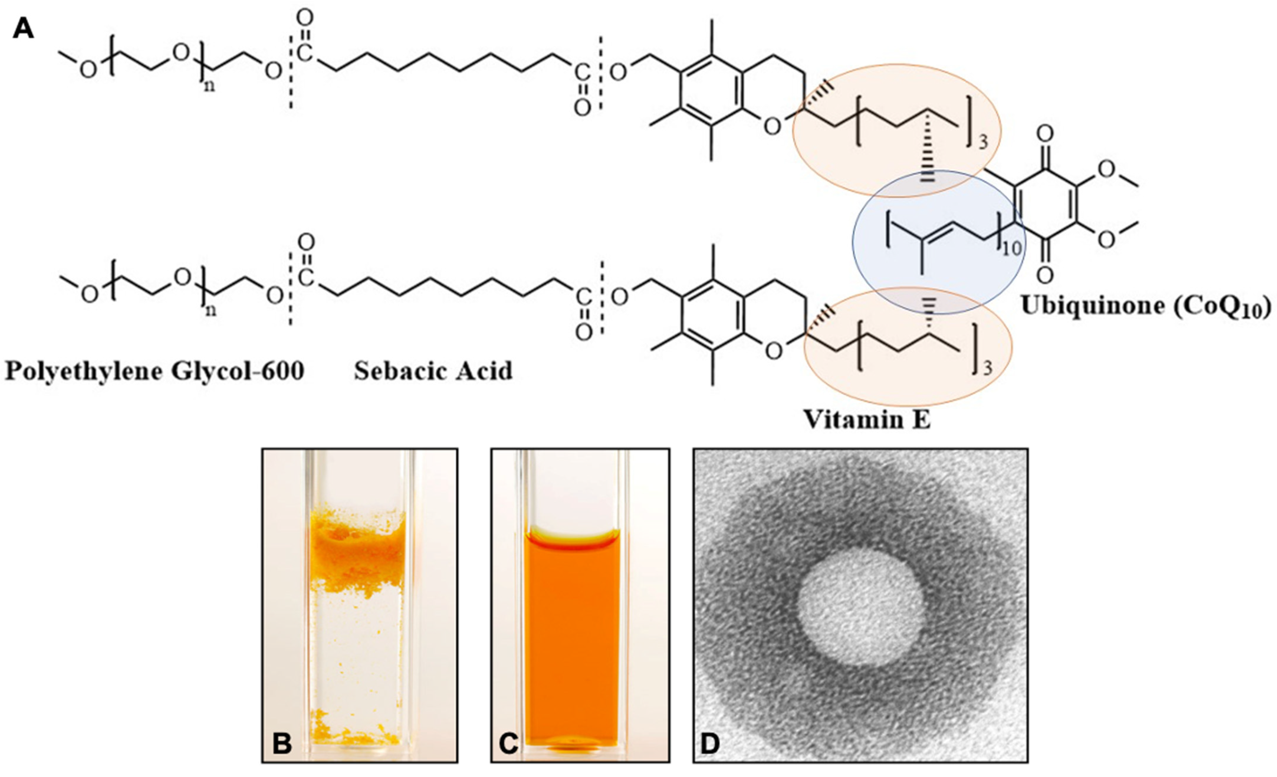
4. Ubisol-Q10 Formulation and Its Properties

Micellization is a well-known process in the art of drug delivery and is applied to many lipid soluble compounds, i.e., vitamins (vitamin E, D3, K) and many novel drugs [71,72], as they become more readily absorbed by the body. The water-soluble products have enhanced bioavailability, stability, efficacy, and safety [7,8,9,10,73]. Recently Wang and Hekimi (2020) reported a simple two component nanomicellar formation of CoQ10 with an antifungal drug caspofungin. The researchers discovered that the synthetic drug caspofungin can form stable micelles and delivers better bioavailability of CoQ10 [74]. In their formulations, the antifungal pharmaceutical drug is combined with CoQ10 at a ratio of 0.225 µM:10 or 20 µM and forms wide size distribution micelles with an average size of ~40 nm. Our formulation is simple, it delivers simultaneously two bioactive compounds, vitamin E and CoQ10, of well-known biological activities, at a molar ratio of 2 moles vitamin E per 1 mol CoQ10.
The National Research Council of Canada researchers have created Ubisol-Q10, a water-dispersible CoQ10 formulation allowing an easy and quantitative delivery of CoQ10. This discovery made possible, for the first time, a proper assessment of its true therapeutic value and potential mechanisms of action. In the pursuit to improve solubility and uptake of CoQ10, the researchers have taken lessons from mechanisms of bodily absorption of lipids and physiological roles of Coenzyme Q10. In the human body, the absorption and uptake of CoQ10 involves secretion into the small intestine with pancreatic enzymes and bile, which facilitates emulsification and micelle formation, typically required for absorption of lipophilic substances. Furthermore, in cellular membranes, CoQ10 interacts with vitamin E (α-tocopherol) and participates in its regeneration to facilitate free radical scavenging efficacy [73]. It has been discovered that α-tocopherol can be transformed into an amphiphilic self-emulsifying molecule of PTS (polyoxyethanyl α-tocopheryl sebacate). PTS can be synthesized by joining polyoxyethanyl-600 and α-tocopherol to decanedioic (sebacic) acid. The PTS molecule was created using well-known and generally regarded as safe (GRAS) components and it was granted GRAS status by the USA FDA. To achieve a water-dispersion, researchers have subjected CoQ10 to the micellization procedure and created Ubisol-Q10, a water-dispersible nanomicellar formulation of CoQ10, which actually consists of 2 components: CoQ10 and polyoxyethanyl-α-tocopheryl sebacate (PTS), combined at a ratio 1:2 mol/mol (Figure 1). The PTS molecule is an amphiphile, possessing both hydrophilic (PEG-600) and lipophilic (α-tocopherol) properties, separated by an aliphatic spacer sebacic (decanedioic) acid, and has self-emulsifying properties [75,76,77,78]. PTS is a waxy semisolid substance and similarly to coenzyme-Q10, has a low melting temperature (close to 50 °C); hence both substances could be co-melted to combine and form stable uniform nanomicelles when dispersed in water. When combined with water, the formulation can be heated to the boiling point of water and hence sterilized, and it is ready for parenteral delivery. In these studies, it has been tested mainly as a drinking tonic, and this form of application was sufficient to block the ongoing neurodegeneration in animal models, but it also could be easily injected (for example in stroke) [79]. A transmission electron microscopy analysis revealed that a single PTS-CoQ10 micelle measures 22 ± 7 nm in diameter. This Ubisol-Q10 formulation remained as a stable clear solution for ≥2 years, even at room temperature, and rendered CoQ10 fully dispersed in water and easily absorbed by the body as demonstrated by the elevated plasma and brain contents of CoQ10 within 1 h of Ubisol-Q10 ingestion. Thus, a new potent formulation has been created named Ubisol-Q10, a water-dispersible coenzyme-Q10, allowing easy and quantitative delivery of CoQ10 [60,61,62,63,79,80,81].
Upon systematic delivery, the PTS is converted back to α-tocopherol, CoQ10 is released and both compounds, vitamin E and ubiquinone are readily absorbed and find their way into the brain where they evoke powerful neuroprotection. They do not accumulate in tissues, but they provide neuroprotection for as long as they are continuously provided [60,61,62,63,79,81]. This seems to be a perfect solution for the treatment of ongoing neurodegeneration and will help millions of patients for whom there is little or no hope now.
In this review, we summarized up to date research results pertaining to the mechanism of action and efficacy of neuroprotection by Ubisol-Q10 in both in vitro and in vivo studies.

5. Ubisol-Q10 Protects Differentiated Neuronal Cells from Oxidative Stress and Excitotoxicity

CoQ10 (oil-soluble formulation) could not be properly tested in cell culture models, as it floats on top of aqueous media (Figure 1). The application of lipofection for internalization of CoQ10 in differentiated neuronal cell culture leads to a degeneration of neuronal extensions and neuronal cell death. CoQ10 is partially soluble in ethanol, but the addition of ethanol to cell media, particularly in larger volumes to ensure delivery of correct concentration of CoQ10, can denature the cellular components leading to solvent induced cell death. The bioavailability and water solubility of Ubisol-Q10 (Figure 1) allowed us to perform successful experimentation on neuronal cell cultures and examine the effects of direct oxidative stress (H2O2 treatment), hypoglycemia, hypoxia and glutamate excitotoxicity, as well as paraquat (PQ) neurotoxicity [82,83,84]. The results show that Ubisol-Q10 can significantly reduce the oxidative stress burden, contribute to the maintenance of the cellular Adenosine Triphosphate (ATP) pool, stabilize the mitochondrial membrane potential, and ultimately protect differentiated neurons. Typically, the exposure of differentiated human teratocarcinoma (NT2) and neuroblastoma (SH-SY5Y) cells to hydrogen peroxide leads to excessive cellular ROS generation and apoptotic cell death. However, pre-treatment of the neuronal cells with Ubisol-Q10 inhibited ROS production, stabilized the mitochondrial membrane, and prevented apoptosis [82].
Researchers have also investigated the cytotoxic effects of glutamate on cell cultures consisting of NT2/N neurons and NT2/A astrocytes derived from human NT2/D1 cells. In these experiments the cultures were exposed to 0.5 mM glutamate and 6 h of hypoxia. Neuronal cell death occurred during subsequent periods of reoxygenation [83]. Significantly, when these cells were pretreated for 72 h with Ubisol-Q10 before glutamate/hypoxia exposure, there was a decrease in ROS, increase in ATP production, and neuronal cells were protected, suggesting the incorporation of CoQ10 into cellular sub-compartments created an environment that could neutralize the effects of these neurotoxic treatments [83].
Furthermore, McCarthy et al. demonstrated that paraquat, an herbicide and mitochondrial toxin, caused increases in oxidative stress, and induced apoptosis in differentiated neuroblastoma cells [84]. The authors observed that treatment with paraquat led to increased ROS generation, depolarization of the inner mitochondrial membrane, and cell death. Importantly, pre-treatment with Ubisol-Q10 provided near complete protection against paraquat toxicity through inhibition of ROS generation and preventing the collapse of the mitochondrial membrane potential in these differentiated neuroblastoma cells [84]. These findings are very important as exposure to paraquat has been shown to be corelated with an increased risk of Parkinson’s disease.

6. Ubisol-Q10 Inhibits Oxidative Stress and Premature Senescence while Inducing Autophagy in Human Fibroblasts Obtained from AD Patients

It has been well established that mutations in the gene coding for presenilin-1 (PS-1) are a risk factor that predispose for development of Alzheimer’s disease. Although human neuron models of PD exist using various toxic insults, obtaining neurons from AD patients with the PS-1 mutation is not possible. Although not neurons, skin cells/fibroblast are very easy to obtain from AD patients who have the PS-1 mutation. In principle, these fibroblasts have the same PS-1 mutation as a neuron would from the same patient and at the biochemical level, the PS-1 mutation should have the same effect on fibroblasts similar to neurons. PS-1 mutated fibroblasts (PSAF) obtained from Alzheimer’s disease patients serve as a useful model for analyzing the induction of senescence and autophagy, as well as studying the relationship between them which is not well understood. A strong link between PS-1 and functional autophagy has been demonstrated with autophagy deficiencies observed in AD-related PS-1 mutations [85]. The PS-1 mutation results in constant endogenous ROS generation and leads to the inhibition of autophagy in these cells. Cellular stressors in PSAF such as endogenous ROS result in the overexpression of cell cycle arrest proteins p21, p53, and p16Ink4A [57]. Furthermore, retinoblastoma (Rb), a protein involved in cell cycle regulation shows higher expression levels in PSAF compared to normal human fibroblasts (NHF). These characteristics lead to the induction of stress-induced premature senescence (SIPS) in PSAF. Interestingly, growing PSAF in the presence of Ubisol-Q10 led to the potent inhibition of oxidative stress while lowering cell cycle arrest protein expression levels and preventing the onset of SIPS. Differential gene expression profiling demonstrated the down-regulation of autophagy-related genes in PSAF compared to NHF [59]. However, Ubisol-Q10 treated PSAF demonstrated gene expression profiles with an up-regulation of autophagy-related genes in a manner comparable to NHF. In particular, autophagy-related genes beclin-1 and JNK1, which are down-regulated in PSAF compared to NHF, were shown to be drastically up-regulated in PSAF following Ubisol-Q10 treatment at both the gene and protein level. Autophagy is known to be an important cellular process through which defective/damaged organelles and proteins can be broken down and recycled. The accumulation of these dysfunctional proteins and organelles due to an autophagy deficiency could be leading to the onset of premature senescence in PSAF. As a result, it was suggested that the induction of autophagy was essential for the inhibition of premature senescence. Indeed, the inhibition of autophagy in Ubisol-Q10 treated PSAF resulted in the return of the SIPS phenotype, indicating the importance of autophagy induction in Ubisol-Q10′s neuroprotective effect. Overall, the various deficiencies associated with the PS-1 mutation in fibroblasts including increased oxidative stress, autophagy inhibition, and premature senescence were overcome following Ubisol-Q10 treatment. Another study used NHF and transformed human embryonic kidney (HEK293) cells challenging them with apoptosis-inducing external oxidative stress [86]. Water-soluble CoQ10 was able to quench the external ROS allowing the cells to maintain their mitochondrial membrane potential under otherwise lethal doses of H2O2. The pro-apoptotic activity of Bax was inhibited by Ubisol-Q10 treatment through the potent quenching of ROS and the reduction of cytochrome-C release triggered by Bax insertion into the mitochondrial membrane. This study indicates that Ubisol-Q10 supplementation can stabilize mitochondria while inhibiting oxidative stress-induced apoptosis in neuronal cells, further supporting its role as a neuroprotective agent.
More recently, the same mechanism of autophagy induction observed in PSAF was observed in PQ exposed rats [87]. Beclin-1 protein levels were elevated in the brains of PQ exposed rats given Ubisol-Q10 supplemented water compared to rats given plain drinking water. Furthermore, levels of oxidative stress as indicated by lipid peroxidation product 4-hydroxynonenal (4-HNE) were reduced in PQ exposed rats given Ubisol-Q10 further reinforcing the potent antioxidant properties of Ubisol-Q10. Microglia activation and astroglia activation was also observed in the brains of PQ exposed rats indicating anti-inflammatory properties of Ubisol-Q10.
Results from in vitro studies provided compelling evidence that Ubisol-Q10 acted as a potent inhibitor of neuronal cell death caused by excessive oxidative stress, excitotoxicity, or mitochondrial toxins. Its ability to prevent premature senescence in presenilin mutated human fibroblasts and resumption of autophagy is a very significant discovery.
The results from the in vitro studies summarized above revealed the involvement of broad-spectrum cellular processes in the observed neuroprotection. CoQ10, a lipid soluble electron carrier, can act as either antioxidant or prooxidant during the reversible transition from fully oxidized (ubiquinone) to fully reduced (ubiquinol) form (Figure 2A). Due to its lipophilic nature CoQ10 conversion takes place mainly in the milieu of cellular membranes, where it supports the functionality of multiple enzymatic complexes requiring electron transfer reactions; hence, it participates in a multitude of cellular pro-survival metabolic processes (Figure 2B). Clearly, to achieve the optimal responses of these many enzymatic complexes, a sustained supplementation in the range of nano to micromolar is sufficient to achieve this. Therefore, its presence is crucial not only for mitochondrial electron transport chain and cellular energy production, but also for the function of many cellular oxidoreductases [4].
Here, the investigators were faced with a question as to whether Ubisol-Q10 with such a broad spectrum of neuroprotective efficacy in vitro could potentially halt the progression of neurodegenerative diseases by protecting neurons in the brain in animal models of PD and AD neurodegenerative diseases. The outcomes of such studies are summarized in the section below.

7. Ubisol-Q10 Prevents Neuronal Loss and Halts the Progression of Neurodegeneration in Rodent Models of PD

Parkinson’s disease is the second most common neurodegenerative disease characterized by the loss of dopaminergic (DA) neurons in the substantia nigra pars compacta (SNpc) region of the brain. Physical symptoms of PD such as bradykinesia, rigidity, postural instability, and resting tremors become obvious when approximately 60–70% of DA neurons in the SNpc are lost [88]. The exact cause of PD development is not known, but a correlation between high exposure to the environmental toxin paraquat and increased risk of PD has been shown [89,90]. Paraquat is known to inhibit complex I of the ETC in mitochondria. Indeed, several chemicals including MPTP (1-methyl-4-phenyl-1,2,3,6-tetrahydropyridine), paraquat, and rotenone that inactivate complex I of the oxidative phosphorylation pathway do induce degeneration of dopaminergic neurons in rodents. It is generally hypothesized that dysfunction of mitochondria, increased ROS burden, and the accumulation of dysfunctional proteins are at the center of PD pathophysiology. Therefore, CoQ10 with a critical role in cellular energy production and antioxidant properties could be an excellent therapeutic for PD. Accordingly, studies have shown CoQ10 is effective in preventing cell death caused by toxins such as PQ, but only at very high doses of an oil-soluble formulation of CoQ10 [91]. This could be due to poor bioavailability in the brain. A water-soluble formulation of CoQ10, Ubisol-Q10, which is already shown to be extremely effective in protecting neuronal cells in culture against oxidative stress, glutamate excitotoxicity and paraquat toxicity, could be the better solution.
Furthermore, the investigators have demonstrated that Ubisol-Q10 was efficiently absorbed into the blood and then into tissues including the brain when administered orally at low doses to mice or rats [62,63]. Feeding of Ubisol-Q10 supplemented drinking water to rats prevented Paraquat-induced loss of dopaminergic neurons and motor deficit as a prophylactic and intervention treatment [60]. Furthermore, in an MPTP-induced mouse model of PD, in which MPTP had already initiated neurodegeneration, orally administered Ubisol-Q10 blocked the neuronal death pathway allowing the DA neurons to survive as long as the supplementation was continued (for at least 8 weeks post-MPTP treatment) suggesting a potential therapeutic efficacy for PD patients [63]. The neuroprotective Ubisol-Q10 treatment triggered an astrocytic activation response indicating the pro-survival role of these cells [63]. The therapeutic neuroprotective effect of Ubisol-Q10 was also demonstrated in a Paraquat-induced chronic model of PD in rats [62]. Feeding Ubisol-Q10 supplemented water to rats post-Paraquat injection halted the progression of neurodegeneration and ameliorated motor dysfunction [62]. Most importantly, in both models, the effective dose of Ubisol-Q10 was only 6 mg/kg/day, far lower than that of the oil-soluble formulation (400–1600 mg/kg/day) [22]. Ubisol-Q10 is shown to be safe and has been given GRAS status by the FDA. A genetic susceptibility model of PD in mice was also used and it was found that Ubisol-Q10 protects against MPTP-induced neurodegeneration and motor dysfunction in DJ-1 deficient mice [61]. Thus, Ubisol-Q10 might be a treatment prospect for people genetically predisposed to PD as well as with sporadic PD. Currently there is no therapeutic available to stop the progressive loss of neurons in PD patients; only symptomatic relief is obtained by levodopa, but prolonged use leads to drug induced dyskinesia, which severely affects the patient’s quality of life [92]. Ubisol-Q10 could be a safe nutraceutical therapeutic that has the potential to halt the progression of PD and is a safe material that can be taken over long periods of time.

8. Ubisol-Q10 Halts Progression of AD Pathology and Symptoms by Inhibition of Oxidative Stress, Inflammation, and Activation of Autophagy

Hippocampal neurodegeneration is a key pathological feature of AD, resulting in severe cognitive impairments [93]. A functional hippocampus is crucial for the formation of long-term memories and the performance of spatial memory tasks [94,95,96]. As a result, any damage to the hippocampal region of the brain, whether it be directly through lesions [97,98,99,100,101] or indirectly through AD development [102,103], drastically influences long-term memory and spatial working memory. This vindicates the usage of behavioral testing of long-term and spatial memory on transgenic rodent AD models to assess potential therapeutic effects. For example, double transgenic mice containing mutations in both the PS-1 and APP genes serve as a useful AD model for examining therapeutic effects in vivo. Despite oil-soluble coenzyme-Q10 showing neuroprotection in neurodegenerative rodent models, it failed in clinical trials for Parkinson’s disease, Huntington’s disease, and amyotrophic lateral sclerosis [22,26,104]. Due to its poor bioavailability, its effective dose in rodent models was 200–1600 mg/kg/day, well above FDA-approved limits in humans for clinical trials. Thus, the lower doses administered during clinical trials likely resulted in the lack of neuroprotection observed. However, water-soluble coenzyme-Q10, Ubisol-Q10, demonstrates very promising results in vivo at doses of just 6 mg/kg/day.
Muthukumaran et al., examined long-term memory in double transgenic AD mice through observing the degree of dishabituation in a Y-maze [58]. Untreated transgenic mice showed a greater spontaneous recovery of a previously habituated exploratory response following a long rest from exposure to the Y-maze compared to wild-type mice. However, oral Ubisol-Q10 supplementation in double transgenic mice reduced the spontaneous recovery compared to the untreated group indicating improved memory processes following treatment. They also conducted novel location / novel object recognition (NL/NOR) testing where they found untreated transgenic mice explored a moved familiar object less than Ubisol-Q10 treated transgenic and wild-type mice indicating decreased memory of the object’s spatial features due to hippocampal neurodegeneration [58].
Confirmation of neuronal preservation was seen through NeuN immunohistochemical staining which qualitatively demonstrated higher neuron counts in Ubisol-Q10 treated transgenic brains compared to untreated mice. Biochemically, Ubisol-Q10 reduced the circulating amyloid-beta and amyloid-beta plaque levels in the hippocampus characteristically observed in AD patients [58]. Due to autophagy’s role in clearing damaged/defective proteins and organelles, it was questioned as to whether autophagy activation could be important for the clearing of these amyloid-beta plaques in transgenic AD mice. Indeed, it was shown that autophagy is inhibited in the brains of the double transgenic mice but is activated following Ubisol-Q10 treatment [59]. Furthermore, pro-survival astrocytes were activated in the brains of double transgenic mice following Ubisol-Q10 supplementation while reactive microglial clumping around amyloid-beta plaques was drastically reduced in a manner similar to the wild-type mice. The NLRP3 (nucleotide-binding and oligomerization domain (NOD)-like receptor family, pyrin domain containing 3) inflammasome pathway is activated in immune and glial cells in response to sterile triggers such as dying neurons, cytokines and chemokines, mitochondrial ROS, lysosomal disruption, extracellular glutamate, K+ efflux and Ca2+ influx. Upon sensing cellular stress, NLRP3 recruits an adaptor protein (ASC) that activates caspase-1 leading to the maturation and secretion of pro-inflammatory cytokines, IL-1β and IL-18 [105]. Oral administration of 300 mg/day CoQ10 (soft gel capsules, Pharma Nord) for 40 days in patients suffering from fibromyalgia (n = 20) reduced serum levels of IL-1β and IL-18 [106]. In addition, Peng et. al., have recently shown that intraperitoneal injections of idebenone (an artificial derivative of CoQ10) at 100 mg/kg in an animal model of ischemic-reperfusion injury attenuated cerebral inflammatory responses by dampening microglial NLRP3 inflammasome activity [107]. Overall, Ubisol-Q10 treatment was successful in reversing many AD hallmarks including spatial memory impairments, amyloid-beta plaque formation, neuronal loss in the hippocampus, pro-neuroinflammatory microglial activation, neuroprotective astroglia inhibition, and autophagy deficiencies. As current treatments such as cholinesterase inhibitors and N-methyl-D-Aspartate (NMDA) antagonists strictly provide symptomatic relief, Ubisol-Q10 given as a simple oral supplement could serve as a potential therapeutic to halt the progression of AD.

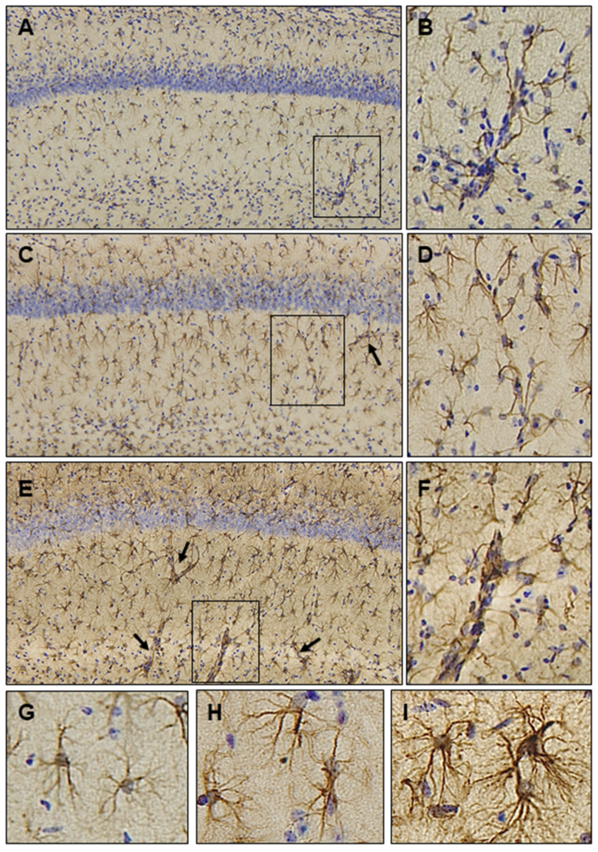
9. Astrocytic Responses in the Brains of Ubisol-Q10 Treated Animals

As elucidated above, preclinical evidence for the extraordinary efficacy of Ubisol-Q10 in protecting neurons and halting the ongoing neurodegenerative processes in the cellular and rodent models of AD and PD has been provided. Astrocytes are the most abundant type of glial cells in the brain that play a pivotal role in regulating almost all homeostatic functions, including maintenance and regulation of the extracellular microenvironment, blood-brain barrier integrity, cerebral blood flow, antioxidant and trophic factor support, uptake and recycling of neurotransmitters as well as detoxification of ROS [108,109,110]. Accordingly, histopathological studies on the brain tissues were carried out and astrocytic responses to Ubisol-Q10 supplementation was examined. Microscopic examination revealed a robust increase in astrocytic GFAP expression in the brains of MPTP-injected animals receiving Ubisol-Q10 (Figure 3). Interestingly, astrocyte activation was seen in all the examined brain regions, including hippocampus, cortex, substantia nigra and striatum, not limited only to the diseased brain region. Similar changes in astrocytic cells were also seen in rats receiving Ubisol-Q10 and challenged with paraquat.
Under higher magnification, astrocytic processes appeared longer and extensively ramified, extending GFAP-positive hypertrophic processes and contacting neighboring astrocytes (Figure 3E,I), possibly establishing an astrocytic syncytial network. Many studies have highlighted the significance of this highly specialized intercellular machinery in facilitating communication through proteins called gap junctions [111]. These communication channels allow the direct passage of ions (Ca2+, Na+ and K+), small molecules (glucose, water, metabolites, second messengers and neurotransmitters) and growth factors to support neuronal function [112]. The astrocyte syncytium is also thought to be involved in the detoxification of excess extracellular glutamate, protecting neurons from excitotoxic damage [113]. Furthermore, astrocytes engage in an intensive bidirectional crosstalk with neurons to regulate synaptic activity. Recently, a plethora of evidence has emerged supporting the role of astrocytes as significant modulators of neuronal circuits, networks, and synaptic neurotransmission. Indeed, astrocytes wrap around neuronal synapses forming the ‘tripartite synapse’ comprising of pre- and post-synaptic neurons and the astrocyte [114]. Astrocytes use intracellular Ca2+ transients as their encoding language to communicate complex signals to neurons in a highly synchronized bursting behavior via neuronal networks and regulate synaptic transmission. Using co-cultures of astrocytes and neurons that recapitulates neuronal network synchronization and connectivity, it has been shown that astrocytes are essential for the development of synchronized activity [115]. Furthermore, the spike and burst rates were reduced in matured networks of glutamatergic neuron-astrocyte co-cultures as compared to monolayer cultures [116]. Similarly, numerous studies using human induced pluripotent stem cells (iPSC)-derived neurons have demonstrated an increased maturation of network functionality and synchronization in the presence of astrocytes [117,118]. Together, these results demonstrate that astrocytes play an important role in neuronal firing synchronicity and synaptic coordination. It has been hypothesized that selective loss of neurons during disease progression results in disintegration of neuronal networks and abnormal synchronization. Supplementation with Ubisol-Q10 could help in neuronal firing synchronicity and synaptic coordination leading to stabilization of neural activities.
Astrocytes are also known to establish a bidirectional communication with the Blood-Brain-Barrier (BBB) which plays an important role in the maintenance of CNS homeostasis and neuronal activity [119]. An increase in GFAP-expressing astrocytic endfeet was seen extending into the cerebral blood vessels in animals receiving Ubisol-Q10 as compared to toxin-treated or control animals (Figure 3B,D,F). It is plausible that an increased neurovascular and neurometabolic coupling [120] was established between astrocytes and brain endothelial cells resulting in increased cerebral blood flow and preservation of BBB function [110]. It is noteworthy that astrocytes rely on mitochondrial oxidative phosphorylation for ATP production and Ubisol-Q10 potentially enhanced their ability to synthesize ATP and maintain neuronal bioenergetics. Indeed, astrocytes act as gatekeepers and protect neurons from oxidative and nitrosative stress [121]. Taken together, studies support the notion that astrocytes could be active players in the neuroprotection observed in these experimental models.
Mounting evidence indicates that activated astrocytes may initiate pathological processes by secreting neurotoxic factors such as ROS and pro-inflammatory cytokines (IL-1β, IL-6 and TNF-α) that likely contribute to neuroinflammation and neurodegeneration [122]. Exposure of mixed neuron-astrocyte cultures derived from human NT2/D1 cells to toxic levels of glutamate resulted in oxidative stress and neurotoxicity which was suppressed by pre-treatment with Ubisol-Q10 [83]. In addition, cortical neurons co-cultured with senescent astrocytes showed increased susceptibility to glutamate toxicity, possibly due to decreased efficiency to detoxify glutamate [123]. Oil-soluble CoQ10 was also shown to protect cultured murine astrocytes from ROS-induced (Ultraviolet-B radiation) damage by targeting mitochondrial function and oxidative stress [124]. An increase in markers of spinal oxidative stress, neuroinflammation (astrocyte and microglial activation) and hyperalgesia was seen in sickle mice, which were remarkably decreased with administration (by gavage) of oil-soluble CoQ10 at 45 mg/kg daily for 4 weeks [125]. Thus, maintenance of active astrocyte-neuron communication is important for neuronal survival and function. CoQ10 functions as a scavenger of ROS and an important antioxidant by activating antioxidant enzymes through Nrf2 signaling and protecting neuronal cells from oxidative stress.
Increasing evidence suggests that CoQ10 can also function as an anti-inflammatory molecule by modulating nuclear factor kappaB (NFĸB)-dependent gene expression. Consequently, treatment of human and murine macrophage and monocytic cell lines with lipopolysaccharide induced secretion of various cytokines and chemokines which were significantly reduced in the presence of CoQ10 [126,127]. Similarly, exposure of rat pheochromocytoma PC-12 cells to β-amyloid (25–35) induced cell death and increased the production of pro-inflammatory lipid mediators, cyclooxygenase-2 (COX-2) and prostaglandin E2 (PGE2), respectively. Pre-incubation of these cultures with CoQ10 decreased COX-2 and PGE2 levels, mainly by blocking nuclear translocation of NFĸB and inhibiting pro-inflammatory gene expression [128]. CoQ10 can act as an antioxidant and an anti-inflammatory molecule to protect astrocytes from oxidative damage and neuroinflammation therefore enhancing their ability to protect neurons, in turn, leading to amelioration of pathological symptoms.

10. Ubisol-Q10 Effectiveness in Contrast to Other Coenzyme-Q10 Formulations

The reasons for failed clinical trials with oral supplementation of oil-soluble CoQ10 formulations are unclear, particularly since the bioactive component of tested formulations is coenzyme-Q10, the same molecule used in Ubisol-Q10. However, Ubisol-Q10 also contains PTS, a prodrug form of vitamin E. It should be noted that no clinical trials have reported the therapeutic efficacy of α-tocopherol alone [129]. Taking all published data into consideration, one explanation that comes to mind is the difference in quantities of supplied CoQ10. In some of the clinical trials mentioned above, extremely large doses of CoQ10 were used, and yet no benefits were seen from such applications. Interestingly, micromolar concentrations of Ubisol-Q10 have shown remarkable neuroprotection both in vitro and in vivo, possibly by improving cellular metabolism (Figure 4) (Table 1) [57,58,59,60,61,62,63]. In each of these in vivo studies, drinking water was supplemented with 50 µg/mL CoQ10 and 100 µmol/mL of PTS (of which 30% is converted to vitamin E) or the equivalent of 6 mg/kg body weight. This was sufficient to penetrate the brain as demonstrated by a remarkable neuroprotection [57,58,59,60,61,62,63] and the engagement of astroglia within the brain parenchyma (Figure 3). To achieve such effects, this supplementation must be sufficient to mobilize cellular metabolism and cyto-protection. As depicted in Figure 4, these metabolic outcomes must be a result of multiple cellular enzymatic complexes that use the conversion of ubiquinone to ubiquinol and are localized to various specialized subcellular compartments. Clearly, micromolar concentrations of Ubisol-Q10 are sufficient to achieve this high efficacy.
The neuroprotective ability of Ubisol-Q10 has been demonstrated through both in vitro and in vivo studies [57,58,59,60,61,62,63]. All the data indicates just how effective it could be as a therapeutic treatment if biologically relevant doses were available for patients. A presented model based on these results and the literature on CoQ10 is depicting potential mechanisms of the neuroprotective functions of Ubisol-Q10 (Figure 4).

11. Conclusions and Future Prospects

It is now well established that CoQ10 has a great therapeutic potential for neurodegenerative diseases. However, due to poor bioavailability, an approach was taken to use very high doses of oil-soluble CoQ10 (400–1600 mg/kg/day which translates to 28–114 g/day for a 70 kg patient) in rodent models of neurodegenerative diseases. Clinical trial results did not show any efficacy as a maximum of only 2.4 g/day was used in the study [22,26]. On the other hand, water-soluble Ubisol-Q10 with an effective dose of 6 mg/kg/day (translating to 420 mg/day for a 70 kg patient, well within the recommended range of CoQ10 dosing by the FDA), which was efficient in halting the progression of neurodegeneration in both rodent models of PD and AD, would be an optimal candidate for clinical studies. It is important to note that oral administration of Ubisol-Q10, a GRAS approved material led to reduced oxidative stress, activation of autophagy, activation of pro-survival astroglia, and inhibition of cell death in addition to the amelioration of behavioral deficits in rodent models of AD and PD. Neurodegenerative diseases such as AD and PD are progressive debilitating diseases spanning over long periods of time. Any therapeutic intervention must be non-toxic and without any side-effects as a long-term treatment. Ubisol-Q10, a safe nutraceutical material with great preventative and therapeutic potential could be the ideal candidate for development as an AD and PD treatment.
Ubisol-Q10 can be easily produced; although it requires commercial manufacturing of PTS, its synthesis is not complicated, and formulation production is very simple (refs patent and Sikorska et al.). Both compounds of Ubisol-Q10 are readily absorbed in the body and find their way into the brain within 1 h of oral delivery where they participate in evoking powerful neuroprotection. They do not accumulate in tissues but are present for as long as they are continuously supplied. This seems to be a perfect solution for the treatment of ongoing neurodegeneration, and it will help millions of individuals for whom there is currently no hope. It should be clearly highlighted that the lack of beneficial results from the studies using a huge amount of CoQ10 suggest that an incorrect approach was taken in clinical trials and the studies should be repeated using less and not more of this bioactive compound. It has been demonstrated that use of micromolar concentrations of Ubisol-Q10 seems to be sufficient to successfully interfere with neurodegeneration. As we understand that these claims need to be further validated through more preclinical and clinical studies, and to our knowledge there is no other CoQ10 formulation suitable for parenteral delivery of CoQ10, it is our hope to find enough open-mindedness in the medical and pharmaceutical industries willing to test this further, particularly since currently there are no effective treatments to halt the progression of neurodegenerative diseases.

Author Contributions

Conceptualization, S.P., J.K.S., D.W., C.V., M.S., J.C.: investigation, data curation; S.P., M.S., J.K.S. and D.W., C.V.: writing—original draft preparation; D.W., C.V., M.S., J.C. and S.P.: writing—review and editing; M.S., J.K.S. and D.W., C.V.: visualization, supervision; S.P.: project administration. All authors have read and agreed to the published version of the manuscript.

Funding

This review was supported by the Natural Sciences and Engineering Research Council.

Acknowledgments

We dedicate this review in memory of Joseph Szecsei who was a great warrior and supporter in the research on neurodegenerative diseases.

Conflicts of Interest

The authors declare no conflict of interest.

References

  1. Jellinger, K.A. Dementia diagnosis. Am. J. Geriatr. Psychiatry 2010, 18, 92. [Google Scholar] [CrossRef] [PubMed]
  2. Gitler, A.D.; Lehmann, R. Modeling Human Disease. Science 2012, 337, 269. [Google Scholar] [CrossRef] [Green Version]
  3. Guo, L.; Wu, C. Mitochondrial dynamics links PINCH-1 signaling to proline metabolic reprogramming and tumor growth. Cell Stress 2020, 5, 23–25. [Google Scholar] [CrossRef]
  4. Scarlett, D.-J.G.; Herst, P.M.; Berridge, M.V. Multiple proteins with single activities or a single protein with multiple activities: The conundrum of cell surface NADH oxidoreductases. Biochim. Biophys. Acta 2005, 1708, 108–119. [Google Scholar] [CrossRef] [Green Version]
  5. Forsmark-Andrée, P.; Dallner, G.; Ernster, L. Endogenous ubiquinol prevents protein modification accompanying lipid peroxidation in beef heart submitochondrial particles. Free Radic. Biol. Med. 1995, 19, 749–757. [Google Scholar] [CrossRef]
  6. Frei, B.; Kim, M.C.; Ames, B.N. Ubiquinol-10 is an effective lipid-soluble antioxidant at physiological concentrations. Proc. Natl. Acad. Sci. USA 1990, 87, 4879–4883. [Google Scholar] [CrossRef] [Green Version]
  7. Balakrishnan, P.; Lee, B.-J.; Oh, D.H.; Kim, J.O.; Lee, Y.-I.; Kim, D.-D.; Jee, J.-P.; Lee, Y.-B.; Woo, J.S.; Yong, C.S.; et al. Enhanced oral bioavailability of Coenzyme Q10 by self-emulsifying drug delivery systems. Int. J. Pharm. 2009, 374, 66–72. [Google Scholar] [CrossRef]
  8. Beg, S.; Javed, S.; Kohli, K. Bioavailability enhancement of coenzyme Q10: An extensive review of patents. Recent Patents Drug Deliv. Formul. 2010, 4, 245–255. [Google Scholar] [CrossRef]
  9. Masotta, N.E.; Martinefski, M.R.; Lucangioli, S.; Rojas, A.M.; Tripodi, V.P. High-dose coenzyme Q10-loaded oleogels for oral therapeutic supplementation. Int. J. Pharm. 2019, 556, 9–20. [Google Scholar] [CrossRef]
  10. Pastor-Maldonado, C.J.; Suárez-Rivero, J.M.; Povea-Cabello, S.; Álvarez-Córdoba, M.; Villalón-García, I.; Munuera-Cabeza, M.; Suárez-Carrillo, A.; Talaverón-Rey, M.; Sánchez-Alcázar, J.A. Coenzyme Q10: Novel Formulations and Medical Trends. Int. J. Mol. Sci. 2020, 21, 8432. [Google Scholar] [CrossRef]
  11. Saha, S.P.; Whayne, T.F., Jr. Coenzyme Q-10 in Human Health: Supporting Evidence? South. Med. J. 2016, 109, 17–21. [Google Scholar] [CrossRef] [PubMed]
  12. Muztaba, M.; Khan, M.I.; Khalid, M.; Tarique, M.; Akhtar, J.; Ahmad, M. CoQ10: A miraculous and clinically vital coenzyme for normal body functions, prevention and treatment of diseases. J. Pharm. Sci. Res. 2018, 10, 976–982. [Google Scholar]
  13. Manzar, H.; Abdulhussein, D.; Yap, T.E.; Cordeiro, M.F. Francesca Cellular consequences of coenzyme Q10 deficiency in neurodegeneration of the retina and brain. Int. J. Mol. Sci. 2020, 21, 9299. [Google Scholar] [CrossRef]
  14. Yang, X.; Zhang, Y.; Xu, H.; Luo, X.; Yu, J.; Liu, J.; Chuen-Chung, R. Neuroprotection of Coenzyme Q10 in Neurodegenerative Diseases. Curr. Top. Med. Chem. 2015, 16, 858–866. [Google Scholar] [CrossRef]
  15. Chang, K.-H.; Cheng, M.-L.; Chiang, M.-C.; Chen, C.-M. Lipophilic antioxidants in neurodegenerative diseases. Clin. Chim. Acta 2018, 485, 79–87. [Google Scholar] [CrossRef] [PubMed]
  16. Pelegay, E.C.; Puzzo, F.; Yilmazer, A.; Cagin, U. Targeting Mitochondrial Defects to Increase Longevity in Animal Models of Neurodegenerative Diseases. Single Mol. Single Cell Seq. 2019, 1134, 89–110. [Google Scholar] [CrossRef]
  17. Oertel, W.; Schulz, J.B. Current and experimental treatments of Parkinson disease: A guide for neuroscientists. J. Neurochem. 2016, 139, 325–337. [Google Scholar] [CrossRef]
  18. Negida, A.; Menshawy, A.; El Ashal, G.; Elfouly, Y.; Hani, Y.; Hegazy, Y.; El Ghonimy, S.; Fouda, S.; Rashad, Y. Coenzyme Q10 for Patients with Parkinson’;s Disease: A Systematic Review and Meta-Analysis. CNS Neurol. Disord. Drug Targets 2016, 15, 45–53. [Google Scholar] [CrossRef] [PubMed]
  19. Zhu, Z.-G.; Sun, M.-X.; Zhang, W.-L.; Wang, W.-W.; Jin, Y.-M.; Xie, C.-L. The efficacy and safety of coenzyme Q10 in Parkinson’s disease: A meta-analysis of randomized controlled trials. Neurol. Sci. 2016, 38, 215–224. [Google Scholar] [CrossRef]
  20. Ernster, L.; Dallner, G. Biochemical, physiological and medical aspects of ubiquinone function. Biochim. Biophys. Acta (BBA)—Mol. Basis Dis. 1995, 1271, 195–204. [Google Scholar] [CrossRef] [Green Version]
  21. Beal, M.F.; Matthews, R.T.; Tieleman, A.; Shults, C.W. Coenzyme Q10 attenuates the 1-methyl-4-phenyl-1,2,3,6-tetrahydropyridine (MPTP) induced loss of striatal dopamine and dopaminergic axons in aged mice. Brain Res. 1998, 783, 109–114. [Google Scholar] [CrossRef]
  22. Cleren, C.; Yang, L.; Lorenzo, B.; Calingasan, N.Y.; Schomer, A.; Sireci, A.; Wille, E.J.; Beal, M.F. Therapeutic effects of coenzyme Q10 (CoQ10) and reduced CoQ10 in the MPTP model of Parkinsonism. J. Neurochem. 2008, 104, 1613–1621. [Google Scholar] [CrossRef]
  23. Beal, M.F. Neuroprotective effects of creatine. Amino Acids 2011, 40, 1305–1313. [Google Scholar] [CrossRef] [PubMed]
  24. Yang, L.; Calingasan, N.Y.; Wille, E.J.; Cormier, K.; Smith, K.; Ferrante, R.J.; Flint Beal, M. Combination therapy with coenzyme Q10 and creatine produces additive neuroprotective effects in models of Parkinson’s and Huntington’s diseases. J. Neurochem. 2009, 109, 1427–1439. [Google Scholar] [CrossRef] [Green Version]
  25. Faust, K.; Gehrke, S.; Yang, Y.; Yang, L.; Beal, M.F.; Lu, B. Neuroprotective effects of compounds with antioxidant and anti-inflammatory properties in a Drosophila model of Parkinson’s disease. BMC Neurosci. 2009, 10, 109. [Google Scholar] [CrossRef] [Green Version]
  26. Beal, M.F.; Oakes, D.; Shoulson, I.; Henchcliffe, C.; Galpern, W.R.; Haas, R.; Juncos, J.L.; Nutt, J.G.; Voss, T.S.; Ravina, B.; et al. A Randomized Clinical Trial of High-Dosage Coenzyme Q10 in Early Parkinson Disease. JAMA Neurol. 2014, 71, 543–552. [Google Scholar] [CrossRef]
  27. Park, H.W.; Park, C.G.; Park, M.; Lee, S.H.; Park, H.R.; Lim, J.; Paek, S.H.; Bin Choy, Y. Intrastriatal administration of coenzyme Q10 enhances neuroprotection in a Parkinson’s disease rat model. Sci. Rep. 2020, 10, 9572. [Google Scholar] [CrossRef]
  28. Gabuzda, D.; Busciglio, J.; Chen, L.B.; Matsudaira, P.; Yankner, B.A. Inhibition of energy metabolism alters the processing of amyloid precursor protein and induces a potentially amyloidogenic derivative. J. Biol. Chem. 1994, 269, 13623–13628. [Google Scholar] [CrossRef]
  29. Frederikse, P.H.; Garland, D.; Zigler, J.S.; Piatigorsky, J. Oxidative Stress Increases Production of β-Amyloid Precursor Protein and β-Amyloid (Aβ) in Mammalian Lenses, and Aβ Has Toxic Effects on Lens Epithelial Cells (∗). J. Biol. Chem. 1996, 271, 10169–10174. [Google Scholar] [CrossRef] [PubMed] [Green Version]
  30. Wadsworth, T.L.; Bishop, J.A.; Pappu, A.S.; Woltjer, R.L.; Quinn, J.F. Evaluation of Coenzyme Q as an Antioxidant Strategy for Alzheimer’s Disease. J. Alzheimers Dis. 2008, 14, 225–234. [Google Scholar] [CrossRef]
  31. Kang, J.; Lemaire, H.-G.; Unterbeck, A.; Salbaum, J.M.; Masters, C.L.; Grzeschik, K.H.; Multhaup, G.; Beyreuther, K.; Müller-Hill, B. The precursor of Alzheimer’s disease amyloid A4 protein resembles a cell-surface receptor. Nature 1987, 325, 733–736. [Google Scholar] [CrossRef]
  32. Nunan, J.; Small, D.H. Regulation of APP cleavage by α-, β- and γ-secretases. FEBS Lett. 2000, 483, 6–10. [Google Scholar] [CrossRef] [Green Version]
  33. Sadli, N.; Barrow, C.J.; McGee, S.; Suphioglu, C. Effect of DHA and CoenzymeQ10 Against Aβ- and Zinc-Induced Mitochondrial Dysfunction in Human Neuronal Cells. CPB 2013, 32, 243–252. [Google Scholar]
  34. Ono, K.; Hasegawa, K.; Naiki, H.; Yamada, M. Preformed β-amyloid fibrils are destabilized by coenzyme Q10 in vitro. Biochem. Biophys. Res. Commun. 2005, 330, 111–116. [Google Scholar] [CrossRef]
  35. Dumont, M.; Kipiani, K.; Yu, F.; Wille, E.; Katz, M.; Calingasan, N.Y.; Gouras, G.K.; Lin, M.T.; Beal, M.F. Coenzyme Q10 Decreases Amyloid Pathology and Improves Behavior in a Transgenic Mouse Model of Alzheimer’s Disease. J. Alzheimer’s Dis. 2011, 27, 211–223. [Google Scholar] [CrossRef]
  36. Beal, M.F. Mitochondrial dysfunction in neurodegenerative diseases. Biochim. Biophys. Acta (BBA)—Bioenerg. 1998, 1366, 211–223. [Google Scholar] [CrossRef] [Green Version]
  37. MacDonald, M.E.; Ambrose, C.M.; Duyao, M.P.; Myers, R.H.; Lin, C.; Srinidhi, L.; Barnes, G.; Taylor, S.A.; James, M.; Groot, N.; et al. A novel gene containing a trinucleotide repeat that is expanded and unstable on Huntington’s disease chromosomes. Cell 1993, 72, 971–983. [Google Scholar] [CrossRef]
  38. Tsunemi, T.; Ashe, T.D.; Morrison, B.E.; Soriano, K.R.; Au, J.; Roque, R.A.; Lazarowski, E.R.; Damian, V.A.; Masliah, E.; La Spada, A.R. PGC-1α rescues Huntington’s disease proteotoxicity by preventing oxidative stress and promoting TFEB function. Sci. Transl. Med. 2012, 4, ra97–ra142. [Google Scholar] [CrossRef] [Green Version]
  39. Matthews, R.T.; Yang, L.; Browne, S.; Baik, M.; Beal, M.F. Coenzyme Q10 administration increases brain mitochondrial concentrations and exerts neuroprotective effects. Proc. Natl. Acad. Sci. USA 1998, 95, 8892–8897. [Google Scholar] [CrossRef] [Green Version]
  40. Ferrante, R.J.; Andreassen, O.A.; Dedeoglu, A.; Ferrante, K.L.; Jenkins, B.G.; Hersch, S.M.; Beal, M.F. Therapeutic Effects of Coenzyme Q10 and Remacemide in Transgenic Mouse Models of Huntington’s Disease. J. Neurosci. 2002, 22, 1592–1599. [Google Scholar] [CrossRef] [PubMed] [Green Version]
  41. Stack, E.C.; Smith, K.M.; Ryu, H.; Cormier, K.; Chen, M.; Hagerty, S.W.; Del Signore, S.J.; Cudkowicz, M.E.; Friedlander, R.M.; Ferrante, R.J. Combination therapy using minocycline and coenzyme Q10 in R6/2 transgenic Huntington’s disease mice. Biochim. Biophys. Acta 2006, 1762, 373–380. [Google Scholar] [CrossRef] [Green Version]
  42. Stack, E.C.; Kubilus, J.K.; Smith, K.; Cormier, K.; Del Signore, S.J.; Guelin, E.; Ryu, H.; Hersch, S.M.; Ferrante, R.J. Chronology of behavioral symptoms and neuropathological sequela in R6/2 Huntington’s disease transgenic mice. J. Comp. Neurol. 2005, 490, 354–370. [Google Scholar] [CrossRef]
  43. Hickey, M.A.; Zhu, C.; Medvedeva, V.; Franich, N.R.; Levine, M.S.; Chesselet, M.-F. Evidence for behavioral benefits of early dietary supplementation with CoEnzymeQ10 in a slowly progressing mouse model of Huntington’s disease. Mol. Cell Neurosci. 2012, 49, 149–157. [Google Scholar] [CrossRef] [Green Version]
  44. Lucchetti, J.; Marino, M.; Papa, S.; Tortarolo, M.; Guiso, G.; Pozzi, S.; Bonetto, V.; Caccia, S.; Beghi, E.; Bendotti, C.; et al. A Mouse Model of Familial ALS Has Increased CNS Levels of Endogenous Ubiquinol9/10 and Does Not Benefit from Exogenous Administration of Ubiquinol10. PLoS ONE 2013, 8, e69540. [Google Scholar] [CrossRef] [Green Version]
  45. Rosen, D.R.; Siddique, T.; Patterson, D.; Figlewicz, D.A.; Sapp, P.; Hentati, A.; Donaldson, D.; Goto, J.; O’Regan, J.P.; Deng, H.X.; et al. Mutations in Cu/Zn superoxide dismutase gene are associated with familial amyotrophic lateral sclerosis. Nature 1993, 362, 59–62. [Google Scholar] [CrossRef]
  46. Gurney, M.E.; Pu, H.; Chiu, A.Y.; Dal Canto, M.C.; Polchow, C.Y.; Alexander, D.D.; Caliendo, J.; Hentati, A.; Kwon, Y.W.; Deng, H.X.; et al. Motor neuron degeneration in mice that express a human Cu, Zn superoxide dismutase mutation. Science 1994, 264, 1772–1775. [Google Scholar] [CrossRef]
  47. Wong, P.C.; Pardo, C.A.; Borchelt, D.R.; Lee, M.K.; Copeland, N.G.; Jenkins, N.A.; Sisodia, S.S.; Cleveland, D.W.; Price, D.L. An adverse property of a familial ALS-linked SOD1 mutation causes motor neuron disease characterized by vacuolar degeneration of mitochondria. Neuron 1995, 14, 1105–1116. [Google Scholar] [CrossRef] [Green Version]
  48. Warren, J.D.; Rohrer, J.D.; Rossor, M.N. Frontotemporal dementia. BMJ 2013, 347, f4827. [Google Scholar] [CrossRef] [Green Version]
  49. Yoshiyama, Y.; Higuchi, M.; Zhang, B.; Huang, S.-M.; Iwata, N.; Saido, T.C.; Maeda, J.; Suhara, T.; Trojanowski, J.Q.; Lee, V.M.-Y. Synapse Loss and Microglial Activation Precede Tangles in a P301S Tauopathy Mouse Model. Neuron 2007, 53, 337–351. [Google Scholar] [CrossRef] [Green Version]
  50. Elipenahli, C.; Stack, C.; Jainuddin, S.; Gerges, M.; Yang, L.; Starkov, A.; Beal, M.F.; Dumont, M. Behavioral Improvement after Chronic Administration of Coenzyme Q10 in P301S Transgenic Mice. J. Alzheimer’s Dis. 2012, 28, 173–182. [Google Scholar] [CrossRef]
  51. Lopes-Ramos, C.; Pereira, T.; Dogini, D.; Gilioli, R.; Lopes-Cendes, I. Lithium carbonate and coenzyme Q10 reduce cell death in a cell model of Machado-Joseph disease. Braz. J. Med. Biol. Res. 2016, 49, e5805. [Google Scholar] [CrossRef] [PubMed] [Green Version]
  52. Kawaguchi, Y.; Okamoto, T.; Taniwaki, M.; Aizawa, M.; Inoue, M.; Katayama, S.; Kawakami, H.; Nakamura, S.; Nishimura, M.; Akiguchi, I.; et al. CAG expansions in a novel gene for Machado-Joseph disease at chromosome 14q32.1. Nat. Genet. 1994, 8, 221–228. [Google Scholar] [CrossRef]
  53. Nakamoto, F.K.; Okamoto, S.; Mitsui, J.; Sone, T.; Ishikawa, M.; Yamamoto, Y.; Kanegae, Y.; Nakatake, Y.; Imaizumi, K.; Ishiura, H.; et al. The pathogenesis linked to coenzyme Q10 insufficiency in iPSC-derived neurons from patients with multiple-system atrophy. Sci. Rep. 2018, 8, 14215. [Google Scholar] [CrossRef]
  54. Wakabayashi, K.; Yoshimoto, M.; Tsuji, S.; Takahashi, H. α-Synuclein immunoreactivity in glial cytoplasmic inclusions in multiple system atrophy. Neurosci. Lett. 1998, 249, 180–182. [Google Scholar] [CrossRef]
  55. Burn, D.J.; Jaros, E. Multiple system atrophy: Cellular and molecular pathology. Mol. Pathol. 2001, 54, 419–426. [Google Scholar]
  56. Schottlaender, L.V.; Bettencourt, C.; Kiely, A.P.; Chalasani, A.; Neergheen, V.; Holton, J.L.; Hargreaves, I.; Houlden, H. Coenzyme Q10 Levels Are Decreased in the Cerebellum of Multiple-System Atrophy Patients. PLoS ONE 2016, 11, e0149557. [Google Scholar] [CrossRef] [Green Version]
  57. Ma, D.; Stokes, K.; Mahngar, K.; Domazet-Damjanov, D.; Sikorska, M.; Pandey, S. Inhibition of stress induced premature senescence in presenilin-1 mutated cells with water soluble Coenzyme Q10. Mitochondrion 2014, 17, 106–115. [Google Scholar] [CrossRef] [PubMed]
  58. Muthukumaran, K.; Kanwar, A.; Vegh, C.; Marginean, A.; Elliott, A.; Guilbeault, N.; Badour, A.; Sikorska, M.; Cohen, J.; Pandey, S. Ubisol-Q10 (a Nanomicellar Water-Soluble Formulation of CoQ10) Treatment Inhibits Alzheimer-Type Behavioral and Pathological Symptoms in a Double Transgenic Mouse (TgAPEswe, PSEN1dE9) Model of Alzheimer’s Disease. J. Alzheimers Dis. 2018, 61, 221–236. [Google Scholar] [CrossRef]
  59. Vegh, C.; Pupulin, S.; Wear, D.; Culmone, L.; Huggard, R.; Ma, D.; Pandey, S. Resumption of Autophagy by Ubisol-Q10 in Presenilin-1 Mutated Fibroblasts and Transgenic AD Mice: Implications for Inhibition of Senescence and Neuroprotection. Oxid. Med. Cell. Longev. 2019, 2019, 7404815. [Google Scholar] [CrossRef] [Green Version]
  60. Somayajulu-Niţu, M.; Sandhu, J.K.; Cohen, J.; Sikorska, M.; Sridhar, T.S.; Matei, A.; Borowy-Borowski, H.; Pandey, S. Paraquat induces oxidative stress, neuronal loss in substantia nigra region and parkinsonism in adult rats: Neuroprotection and amelioration of symptoms by water-soluble formulation of coenzyme Q10. BMC Neurosci. 2009, 10, 88. [Google Scholar] [CrossRef] [Green Version]
  61. Muthukumaran, K.; Smith, J.; Jasra, H.; Sikorska, M.; Sandhu, J.K.; Cohen, J.; Lopatin, D.; Pandey, S. Genetic susceptibility model of Parkinson’s disease resulting from exposure of DJ-1 deficient mice to MPTP: Evaluation of neuroprotection by Ubisol-Q10. J. Park. Dis. 2014, 4, 523–530. [Google Scholar] [CrossRef] [Green Version]
  62. Muthukumaran, K.; Leahy, S.; Harrison, K.; Sikorska, M.; Sandhu, J.K.; Cohen, J.; Keshan, C.; Lopatin, D.; Miller, H.; Borowy-Borowski, H.; et al. Orally delivered water soluble Coenzyme Q10 (Ubisol-Q10) blocks on-going neurodegeneration in rats exposed to paraquat: Potential for therapeutic application in Parkinson’s disease. BMC Neurosci. 2014, 15, 21. [Google Scholar] [CrossRef] [Green Version]
  63. Sikorska, M.; Lanthier, P.; Miller, H.; Beyers, M.; Sodja, C.; Zurakowski, B.; Gangaraju, S.; Pandey, S.; Sandhu, J.K. Nanomicellar formulation of coenzyme Q10 (Ubisol-Q10) effectively blocks ongoing neurodegeneration in the mouse 1-methyl-4-phenyl-1,2,3,6-tetrahydropyridine model: Potential use as an adjuvant treatment in Parkinson’s disease. Neurobiol. Aging 2014, 35, 2329–2346. [Google Scholar] [CrossRef] [Green Version]
  64. Prosek, M.; Butinar, J.; Lukanc, B.; Fir, M.M.; Milivojevic, L.; Krizman, M.; Smidovnik, A. Bioavailability of water-soluble CoQ10 in beagle dogs. J. Pharm. Biomed. Anal. 2008, 47, 918–922. [Google Scholar] [CrossRef] [PubMed]
  65. Banshoya, K.; Nakamura, T.; Tanaka, T.; Kaneo, Y. Coenzyme Q10-Polyethylene Glycol Monostearate Nanoparticles: An Injectable Water-Soluble Formulation. Antioxidants 2020, 9, 86. [Google Scholar] [CrossRef] [Green Version]
  66. Bergamini, C.; Moruzzi, N.; Sblendido, A.; Lenaz, G.; Fato, R. A Water Soluble CoQ10 Formulation Improves Intracellular Distribution and Promotes Mitochondrial Respiration in Cultured Cells. PLoS ONE 2012, 7, e33712. [Google Scholar] [CrossRef] [Green Version]
  67. Li, H.; Chen, G.; Ma, W.; Li, P.-A.A. Water-Soluble Coenzyme Q10 Inhibits Nuclear Translocation of Apoptosis Inducing Factor and Cell Death Caused by Mitochondrial Complex I Inhibition. Int. J. Mol. Sci. 2014, 15, 13388–13400. [Google Scholar] [CrossRef] [Green Version]
  68. Cui, S.; Luo, K.; Quan, Y.; Lim, S.W.; Shin, Y.J.; Lee, K.E.; Kim, H.L.; Ko, E.J.; Kim, J.H.; Chung, S.J.; et al. Water-soluble coenzyme Q10 provides better protection than lipid-soluble coenzyme Q10 in a rat model of chronic tacrolimus nephropathy. Korean J. Intern. Med. 2021, 12. [Google Scholar] [CrossRef]
  69. Di Pierro, F.; Rossi, A.; Consensi, A.; Giacomelli, C.; Bazzichi, L. Role for a water-soluble form of CoQ10 in female subjects affected by fibromyalgia. A preliminary study. Clin. Exp. Rheumatol. 2017, 35 (Suppl. 105), S20–S27. [Google Scholar]
  70. Guastini, L.; Mora, R.; Dellepiane, M.; Santomauro, V.; Giorgio, M.; Salami, A. Water-soluble coenzyme Q10 formulation in presbycusis: Long-term effects. Acta Oto-Laryngol. 2011, 131, 512–517. [Google Scholar] [CrossRef] [PubMed]
  71. Tiwari, G.; Tiwari, R.; Bannerjee, S.K.; Bhati, L.; Pandey, S.; Pandey, P.; Sriwastawa, B. Drug delivery systems: An updated review. Int. J. Pharm. Investig. 2012, 2, 2–11. [Google Scholar] [CrossRef] [PubMed] [Green Version]
  72. Li, M.; Lan, J.; Li, X.; Xin, M.; Wang, H.; Zhang, F.; Lu, X.; Zhuang, Z.; Wu, X. Novel ultra-small micelles based on ginsenoside Rb1: A potential nanoplatform for ocular drug delivery. Drug Deliv. 2019, 26, 481–489. [Google Scholar] [CrossRef] [Green Version]
  73. Mantle, D.; Dybring, A. Bioavailability of Coenzyme Q10: An Overview of the Absorption Process and Subsequent Metabolism. Antioxidants 2020, 9, 386. [Google Scholar] [CrossRef]
  74. Wang, Y.; Hekimi, S. Micellization of coenzyme Q by the fungicide caspofungin allows for safe intravenous administration to reach extreme supraphysiological concentrations. Redox Biol. 2020, 36, 101680. [Google Scholar] [CrossRef]
  75. Borowy-Borowski, H.; Sikorska, M.; Walker, P.R. Water-Soluble Composition of Bioactive Lipophilic Compounds. U.S. Patent 6045826A, 4 April 2000. [Google Scholar]
  76. Borowy-Borowski, H.; Sikorska, M.; Walker, P.R. Water-Soluble Compositions of Bioactive Lipophilic Compounds. U.S. Patent 6191172B1, 20 February 2001. [Google Scholar]
  77. Borowy-Borowski, H.; Sikorska, M.; Walker, P.R. Delivery Compositions of Bioactive Compounds. U.S. Patent 6632443, 14 October 2003. [Google Scholar]
  78. Borowy-Borowski, H.; Sikorska, M.; Walker, P.R. Water-Soluble Compositions of Bioactive Lipophilic Compounds. U.S. Patent 7645816, 12 January 2010. [Google Scholar]
  79. Sikorska, M.; Borowy-Borowski, H.; Zurakowski, B.; Walker, P.R. Derivatised alpha-tocopherol as a CoQ10 carrier in a novel water-soluble formulation. Biofactors 2003, 18, 173–183. [Google Scholar] [CrossRef]
  80. Graves, S.; Sikorska, M.; Borowy-Borowski, H.; Ho, R.J.; Bui, T.; Woodhouse, C. Analysis of coenzyme Q10 content in human plasma and other biological samples. Methods Mol. Biol. 1998, 108, 353–365. [Google Scholar]
  81. Borowy-Borowski, H.; Sodja, C.; Docherty, J.; Walker, P.R.; Sikorska, M. Unique technology for solubilization and delivery of highly lipophilic bioactive molecules. J. Drug Target. 2004, 12, 415–424. [Google Scholar] [CrossRef]
  82. Somayajulu, M.; McCarthy, S.; Hung, M.; Sikorska, M.; Borowy-Borowski, H.; Pandey, S. Role of mitochondria in neuronal cell death induced by oxidative stress; neuroprotection by coenzyme Q10. Neurobiol. Dis. 2005, 18, 618–627. [Google Scholar] [CrossRef] [PubMed]
  83. Sandhu, J.K.; Pandey, S.; Ribecco-Lutkiewicz, M.; Monette, R.; Borowy-Borowski, H.; Walker, P.R.; Sikorska, M. Molecular mechanisms of glutamate neurotoxicity in mixed cultures of NT2-derived neurons and astrocytes: Protective effects of coenzyme Q10. J. Neurosci. Res. 2003, 72, 691–703. [Google Scholar] [CrossRef]
  84. McCarthy, S.; Somayajulu, M.; Sikorska, M.; Borowy-Borowski, H.; Pandey, S. Paraquat induces oxidative stress and neuronal cell death; neuroprotection by water-soluble Coenzyme Q10. Toxicol. Appl. Pharmacol. 2004, 201, 21–31. [Google Scholar] [CrossRef]
  85. Lee, J.H.; Yu, W.H.; Kumar, A.; Lee, S.; Mohan, P.S.; Peterhoff, C.M.; Wolfe, D.M.; Martinez-Vicente, M.; Massey, A.C.; Sovak, G.; et al. Lysosomal proteolysis and autophagy require presenilin 1 and are disrupted by Alzheimer’s-related PS1 mutations. Cell 2010, 141, 1146–1158. [Google Scholar] [CrossRef] [Green Version]
  86. Naderi, J.; Somayajulu-Niţu, M.; Mukerji, A.; Sharda, P.; Sikorska, M.; Borowy-Borowski, H.; Antonsson, B.; Pandey, S. Water-soluble formulation of Coenzyme Q10 inhibits Bax-induced destabilization of mitochondria in mammalian cells. Apoptosis 2006, 11, 1359–1369. [Google Scholar] [CrossRef]
  87. Vegh, C.; Wear, D.; Okaj, I.; Huggard, R.; Culmone, L.; Eren, S.; Cohen, J.; Rishi, A.; Pandey, S. Combined Ubisol-Q10 and Ashwagandha Root Extract Target Multiple Biochemical Mechanisms and Halt Neurodegeneration in a Paraquat Induced Rat Model of Parkinson’s Disease. Antioxidants 2021, 10, 563. [Google Scholar] [CrossRef]
  88. Berry, C.; La Vecchia, C.; Nicotera, P. Paraquat and Parkinson’s disease. Cell Death Differ. 2010, 17, 1115–1125. [Google Scholar] [CrossRef] [Green Version]
  89. Liou, H.H.; Tsai, M.C.; Chen, C.J.; Jeng, J.S.; Chang, Y.C.; Chen, S.Y.; Chen, R.C. Environmental risk factors and Parkinson’s disease: A case–control study in Taiwan. Neurology 1997, 48, 1583–1588. [Google Scholar] [CrossRef]
  90. Gatto, N.M.; Cockburn, M.; Bronstein, J.; Manthripragada, A.D.; Ritz, M. Well-water consumption and Parkinson’s Disease in rural California. Environ. Health Perspect. 2009, 117, 1912–1918. [Google Scholar] [CrossRef] [Green Version]
  91. Spindler, M.; Beal, M.F.; Henchcliffe, C. Coenzyme Q10 effects in neurodegenerative disease. Neuropsychiatr. Dis. Treat. 2009, 5, 597–610. [Google Scholar]
  92. Olanow, C.W.; Lees, A.; Obeso, J. Levodopa therapy for Parkinson’s disease: Challenges and future prospects.le first published online. Mov. Disord. 2008, 23, S495–S496. [Google Scholar] [CrossRef]
  93. Braak, H.; Braak, E. Neuropathological stageing of Alzheimer-related changes. Acta Neuropathol. 1991, 82, 239–259. [Google Scholar] [CrossRef]
  94. Galani, R.; Weiss, I.; Cassel, J.-C.; Kelche, C. Spatial memory, habituation, and reactions to spatial and nonspatial changes in rats with selective lesions of the hippocampus, the entorhinal cortex or the subiculum. Behav. Brain Res. 1998, 96, 1–12. [Google Scholar] [CrossRef]
  95. Platel, A.; Porsolt, R.D. Habituation of exploratory activity in mice: A screening test for memory enhancing drugs. Psychopharmacology 1982, 78, 346–352. [Google Scholar] [CrossRef] [PubMed]
  96. Wright, J.W.; Murphy, E.S.; Elijah, I.E.; Holtfreter, K.L.; Davis, C.J.; Olson, M.L.; Muhunthan, K.; Harding, J.W. Influence of hippocampectomy on habituation, exploratory behavior, and spatial memory in rats. Brain Res. 2004, 1023, 1–14. [Google Scholar] [CrossRef]
  97. Aggleton, J.; Hunt, P.; Rawlins, J. The effects of hippocampal lesions upon spatial and non-spatial tests of working memory. Behav. Brain Res. 1986, 19, 133–146. [Google Scholar] [CrossRef]
  98. Broadbent, N.J.; Squire, L.R.; Clark, R.E. Spatial memory, recognition memory, and the hippocampus. Proc. Natl. Acad. Sci. USA 2004, 101, 14515–14520. [Google Scholar] [CrossRef] [Green Version]
  99. Forwood, S.; Winters, B.; Bussey, T. Hippocampal lesions that abolish spatial maze performance spare object recognition memory at delays of up to 48 hours. Hippocampus 2004, 15, 347–355. [Google Scholar] [CrossRef]
  100. Save, E.; Poucet, B.; Foreman, N.; Buhot, M.C. Object exploration and reactions to spatial and nonspatial changes in hooded rats following damage to parietal cortex or hippocampal formation. Behav. Neurosci. 1992, 106, 447–456. [Google Scholar] [CrossRef] [PubMed]
  101. Stupien, G.; Florian, C.; Roullet, P. Involvement of the hippocampal CA3-region in acquisition and in memory consolidation of spatial but not in object information in mice. Neurobiol. Learn. Mem. 2003, 80, 32–41. [Google Scholar] [CrossRef]
  102. Billings, L.M.; Oddo, S.; Green, K.N.; McGaugh, J.L.; LaFerla, F.M. Intraneuronal Abeta causes the onset of early Alzheimer’s disease-related cognitive deficits in transgenic mice. Neuron 2005, 45, 675–688. [Google Scholar] [CrossRef] [Green Version]
  103. Holcomb, L.; Gordon, M.N.; McGowan, E.; Yu, X.; Benkovic, S.; Jantzen, P.; Wright, K.; Saad, I.; Mueller, R.; Morgan, D.; et al. Accelerated Alzheimer-type phenotype in transgenic mice carrying both mutant amyloid precursor protein and presenilin 1 transgenes. Nat. Med. 1998, 4, 97–100. [Google Scholar] [CrossRef]
  104. Huntington Study Group. A randomized, placebo-controlled trial of coenzyme Q10 and remacemide in Huntington’s disease. Neurology 2001, 57, 397–404. [Google Scholar]
  105. Broz, P.; Dixit, V.M. Inflammasomes: Mechanism of assembly, regulation and signalling. Nat. Rev. Immunol. 2016, 16, 407–420. [Google Scholar] [CrossRef]
  106. Cordero, M.D.; Alcocer-Gómez, E.; Culic, O.; Carrión, A.M.; De Miguel, M.; Díaz-Parrado, E.; Pérez-Villegas, E.M.; Bullón, P.; Battino, M.; Sánchez-Alcazar, J.A. NLRP3 inflammasome is activated in fibromyalgia: The effect of coenzyme Q10. Antioxid. Redox Sign. 2014, 20, 1169–1180. [Google Scholar] [CrossRef] [Green Version]
  107. Peng, J.; Wang, H.; Gong, Z.; Li, X.; He, L.; Shen, Q.; Pan, J.; Peng, Y. Idebenone attenuates cerebral inflammatory injury in ischemia and reperfusion via dampening NLRP3 inflammasome activity. Mol. Immunol. 2020, 123, 74–87. [Google Scholar] [CrossRef]
  108. Banker, G.A. Trophic interactions between astroglial cells and hippocampal neurons in culture. Science 1980, 209, 809–810. [Google Scholar] [CrossRef] [Green Version]
  109. Rothstein, J.D.; Dykes-Hoberg, M.; Pardo, C.A.; Bristol, L.A.; Jin, L.; Kuncl, R.W.; Kanai, Y.; Hediger, M.A.; Wang, Y.; Schielke, J.P.; et al. Knockout of Glutamate Transporters Reveals a Major Role for Astroglial Transport in Excitotoxicity and Clearance of Glutamate. Neuron 1996, 16, 675–686. [Google Scholar] [CrossRef] [Green Version]
  110. Koehler, R.C.; Roman, R.J.; Harder, D.R. Astrocytes and the regulation of cerebral blood flow. Trends Neurosci. 2009, 32, 160–169. [Google Scholar] [CrossRef]
  111. Yeager, M.; Harris, A.L. Gap junction channel structure in the early 21st century: Facts and fantasies. Curr. Opin. Cell Biol. 2007, 19, 521–528. [Google Scholar] [CrossRef] [Green Version]
  112. Nedergaard, M.; Ransom, B.; Goldman, S.A. New roles for astrocytes: Redefining the functional architecture of the brain. Trends Neurosci. 2003, 26, 523–530. [Google Scholar] [CrossRef]
  113. Volterra, A.; Meldolesi, J. Astrocytes, from brain glue to communication elements: The revolution continues. Nat. Rev. Neurosci. 2005, 6, 626–640. [Google Scholar] [CrossRef] [PubMed]
  114. Araque, A.; Parpura, V.; Sanzgiri, R.P.; Haydon, P.G. Tripartite synapses: Glia, the unacknowledged partner. Trends Neurosci. 1999, 22, 208–215. [Google Scholar] [CrossRef]
  115. Guevara, G.; Sanchez, K.R.; Sun, J.; Harrington, M.A.; Temburni, M.; Biol., Delaware State Univ., Dover, DE. Molecular Mechanisms of Astrocyte-Neuron Interactions in the Development of Synchronized Activity in Neuronal Networks. Program No. 652.29. 2018 Neuroscience Meeting Planner. San Diego, CA: Society for Neuroscience. 2018. Available online: https://www.abstractsonline.com/pp8/#!/4649/presentation/23270 (accessed on 18 April 2021).
  116. Tukker, A.M.; Wijnolts, F.M.J.; de Groot, A.; Westerink, R.H.S. Human iPSC-derived neuronal models for in vitro neurotoxicity assessment. Neurotoxicology 2018, 67, 215–225. [Google Scholar] [CrossRef]
  117. Kuijlaars, J.; Oyelami, T.; Diels, A.; Rohrbacher, J.; Versweyveld, S.; Meneghello, G.; Tuefferd, M.; Verstraelen, P.; Detrez, J.R.; Verschuuren, M.; et al. Sustained synchronized neuronal network activity in a human astrocyte co-culture system. Sci. Rep. 2016, 6, 36529. [Google Scholar] [CrossRef] [Green Version]
  118. Paavilainen, T.; Pelkonen, A.; Mäkinen, M.E.-L.; Peltola, M.; Huhtala, H.; Fayuk, D.; Narkilahti, S. Effect of prolonged differentiation on functional maturation of human pluripotent stem cell-derived neuronal cultures. Stem Cell Res. 2018, 27, 151–161. [Google Scholar] [CrossRef]
  119. Abbott, N.J. Astrocyte-endothelial interactions and blood-brain barrier permeability. J. Anat. 2002, 200, 629–638. [Google Scholar] [CrossRef] [PubMed]
  120. Petzold, G.C.; Murthy, V.N. Role of astrocytes in neurovascular coupling. Neuron 2011, 71, 782–797. [Google Scholar] [CrossRef] [Green Version]
  121. Rosenberg, P.A.; Aizenman, E. Hundred-fold increase in neuronal vulnerability to glutamate toxicity in astrocyte-poor cultures of rat cerebral cortex. Neurosci. Lett. 1989, 103, 162–168. [Google Scholar] [CrossRef]
  122. Colombo, E.; Farina, C. Astrocytes: Key Regulators of Neuroinflammation. Trends Immunol. 2016, 37, 608–620. [Google Scholar] [CrossRef]
  123. Limbad, C.; Oron, T.R.; Alimirah, F.; Davalos, A.R.; Tracy, T.E.; Gan, L.; Desprez, P.-Y.; Campisi, J. Astrocyte senescence promotes glutamate toxicity in cortical neurons. PLoS ONE 2020, 15, e0227887. [Google Scholar] [CrossRef]
  124. Jing, L.; He, M.-T.; Chang, Y.; Mehta, S.L.; He, Q.-P.; Zhang, J.-Z.; Li, P.A. Coenzyme Q10 protects astrocytes from ROS-induced damage through inhibition of mitochondria-mediated cell death pathway. Int. J. Biol. Sci. 2015, 11, 59–66. [Google Scholar] [CrossRef] [PubMed] [Green Version]
  125. Valverde, Y.; Benson, B.; Gupta, M.; Gupta, K. Spinal glial activation and oxidative stress are alleviated by treatment with curcumin or coenzyme Q in sickle mice. Haematologica 2016, 101, e44–e47. [Google Scholar] [CrossRef] [PubMed] [Green Version]
  126. Schmelzer, C.; Lorenz, G.; Lindner, I.; Rimbach, G.; Niklowitz, P.; Menke, T.; Döring, F. Effects of Coenzyme Q10 on TNF-alpha secretion in human and murine monocytic cell lines. Biofactors 2007, 31, 35–41. [Google Scholar] [CrossRef] [PubMed]
  127. Schmelzer, C.; Lorenz, G.; Rimbach, G.; Döring, F. In Vitro Effects of the Reduced Form of Coenzyme Q(10) on Secretion Levels of TNF-alpha and Chemokines in Response to LPS in the Human Monocytic Cell Line THP-1. J. Clin. Biochem. Nutr. 2009, 44, 62–66. [Google Scholar] [CrossRef] [Green Version]
  128. Li, L.; Xu, D.; Lin, J.; Zhang, D.; Wang, G.; Sui, L.; Ding, H.; Du, J. Coenzyme Q10 attenuated β-amyloid25-35-induced inflammatory responses in PC12 cells through regulation of the NF-κB signaling pathway. Brain Res. Bull. 2017, 131, 192–198. [Google Scholar] [CrossRef] [PubMed]
  129. Sano, M.; Ernesto, C.; Thomas, R.G.; Klauber, M.R.; Schafer, K.; Grundman, M.; Woodbury, P.; Growdon, J.; Cotman, C.W.; Pfeiffer, E.; et al. A controlled trial of selegiline, alpha-tocopherol, or both as treatment for Alzheimer’s disease. The Alzheimer’s Disease Cooperative Study. N. Engl. J. Med. 1997, 336, 1216–1222. [Google Scholar] [CrossRef] [PubMed] [Green Version]
Figure 1. Ubisol-Q10 formulation. (A) chemical composition of the formulation consisting of 2 molecules of PTS per 1 molecule of CoQ10; (B) a direct suspension of 100 mg CoQ10 in water; (C) the same quantities of CoQ10 formulated with PTS as Ubisol-Q10 (clear and transparent solution of Ubisol-Q10 stable for years); (D) Transmission electron micrograph of a single Ubisol-Q10 nanomicelle measuring on average 22 ± 7 nm in diameter.
Antioxidants 10 00764 g001
Figure 2. Chemical structure and biological functions of CoQ10. (A) Enzymatic conversion of coenzyme-Q10 from oxidized Ubiquinone (UQ) to reduced Ubiquinol (UQH2) form that is crucial for enzymatic activities of multiple cellular oxidoreductases (depicted in (B)). (B) Schematic representation of cellular and subcellular organelles indicating enzymatic complexes using CoQ10 conversion for their activities (A).
Antioxidants 10 00764 g002
Figure 3. Astrocytic responses to Ubisol-Q10 supplementation in the MPTP model of Parkinson’s disease. Formalin fixed free-floating sections were subjected to immunohistochemistry using an antibody raised against glial fibrillary acidic protein (GFAP), a widely used marker of astrocytes. Astrocytes were identified by a brown precipitate at the site of antigen-antibody reaction. Nuclei were counterstained blue with hematoxylin. Shown are photomicrographs at the level of hippocampus from control (A), MPTP-injected (C) and MPTP-injected mice receiving Ubisol-Q10 (E); Magnification = 20×. Arrows in panels A, C and E depict cerebral microvasculature with increased GFAP staining forming the perivascular astrocyte endfeet. Boxed areas are represented at a higher magnification—control (B), MPTP-injected (D) and MPTP-injected mice receiving Ubisol-Q10 (F). High magnification image of activated astrocyte morphology with increased GFAP staining is shown in MPTP-injected (H) and MPTP-injected mice receiving Ubisol-Q10 (I) as compared to controls (G).
Antioxidants 10 00764 g003
Figure 4. Schematic illustrating the neuroprotective potential of Ubisol-Q10. Ubiquinone (UQ) is converted to Ubiquinol (UQH2), which is involved in key cellular functions. Neuroprotection could be mediated through modulation of several astrocytic and microglial pathways, for instance reduction of oxidative stress by decreasing reactive oxygen and nitrogen species (RNOS), lipid mediators, glutamate excitotoxicity and improving mitochondrial function. Activation of autophagy by increasing beclin-1 levels and up-regulating mTORC1, JNK and cathepsin-B activity. Decreasing cellular senescence by reducing growth arrest and secretion of pro-inflammatory mediators and increasing secretion of neurotrophic growth factors. Increasing anti-inflammatory effects by decreasing inflammatory mediators such as IL-6, TNF-α and IL-1β. These effects result in restoring cellular homeostasis, leading to increased neuron-glia coupling and neuronal survival.
Antioxidants 10 00764 g004
Table 1. Summary of recent research progress made with various formulations of coenzyme-Q10.
Neurodegenerative
Disease
ModelEffective
Dose
Mode of AdministrationMajor OutcomesReference
Oil-Soluble CoQ10Alzheimer’s DiseaseIn-Vivo (Mice)10 g/kg dietOral- Protection against neurotoxicity & oxidative stress
- Mitochondrial stabilization
- Reduced Aβ plaques
- Improved cognitive performance
Wadsworth 2008 [30]
In-Vitro6.25 µMMedia SupplementationWadsworth 2008 [30]
In-Vivo (Mice)0.4% or 2.4%OralDumont 2011 [35]
In-Vitro10 µMMedia SupplementationSadli 2013 [33]
Amyotrophic Lateral Sclerosis (ALS)In-Vivo (Rats & Mice)200 mg/kg/ dayOral- Anti-oxidative effects
- Preserved mitochondrial function
- Increased lifespan
Matthews 1998 [39]
In-Vivo (Mice)200 mg/kg/day (no effect)Oral (Gavage)Lucchetti 2013 [44]
Frontotemporal DementiaIn-Vivo (Mice)0.5% of DietOral- Improved behaviour & survivalElipenahli 2012 [50]
Huntington’s DiseaseIn-Vivo (Rats)200 mg/kg/dayOral- Improved motor performance & survival
- Delayed weight loss
- Prevented striatal neuron intranuclear inclusion formation
- Slowed striatal neuron atrophy
- Reduced HTT aggregate formation
- Reduced oxidative damage
Matthews 1998 [39]
In-Vivo (Mice)400 mg/kg/dayOralFerrante 2002 [40]
In-Vivo (Mice)0.2% of DietOralStack 2006 [41]
In-Vivo (Mice, Rats)1600–2000 mg/kg/dayOralYang 2009 [24]
In-Vivo (Mice)0.2% of DietOralHickey 2012 [43]
Machado-Joseph DiseaseIn-Vitro10 µM, 30 µM, 90 µMMedia Supplementation- Improved cell viability & reduced apoptosis
- Prevented ATX3 protein aggregation
Lopes-Ramos 2016 [51]
Multiple-System AtrophyIn-Vitro25 µMMedia Supplementation- Improved oxidative metabolism
- Reduced apoptosis
Nakamoto 2018 [53]
Parkinson’s DiseaseIn-Vivo (Mice)200 mg/kg/dayOral- Dopaminergic neurons saved in striatum and SNpc
- Clearance of α-synuclein aggregates
- Limited oxidative damage
- Reduced pro-inflammatory cytokines
Beal 1998 [21]
In-Vivo (Mice)200–1600 mg/kg/dayOralCleren 2008 [22]
In-Vivo (Mice)1% of DietOralYang 2009 [24]
In-Vivo (Drosophila)100 mg/mL (no effect)OralFaust 2009 [25]
In-Vivo (Rats)25 µg/mLIntrastriatal InjectionPark 2020 [27]
Ubisol-Q10Alzheimer’s DiseaseIn-Vitro50 µg/mLMedia Supplementation- Inhibited oxidative stress
- Upregulated autophagy
- Maintained MMP
- Reduced cell cycle arrest protein expression
- Prevented SIPS onset
- Inhibited apoptosis
- Improved memory
- Reduced hippocampal neurodegeneration
- Cleared Aβ plaques
- Increased astrocyte activity
- Reduced microglial activity
Ma 2014 [57]
In-Vivo (Mice)6 mg/kg/dayOralMuthukumaran 2018 [58]
In-Vivo (Mice)50 µg/mLOralVegh 2019 [59]
In-Vitro50 µg/mLMedia SupplementationVegh 2019 [59]
Parkinson’s DiseaseIn-Vivo (Rats)50 µg/mLOral- Reduced oxidative stress
- Maintained ATP generation
- Stabilized mitochondrial membrane
- Prevented loss of dopaminergic neurons
- Activated pro
-survival astrocytes
- Ameliorated motor dysfunction
Somayajulu-Nitu 2009 [60]
In-Vivo (Mice)6 mg/kg/dayOralMuthukumaran 2014 [61]
In-Vivo (Rats)6 mg/kg/dayOralMuthukumaran 2014 [62]
In-Vivo (Mice)3 mg/kg/dayOralSikorska 2014 [63]
Publisher’s Note: MDPI stays neutral with regard to jurisdictional claims in published maps and institutional affiliations.

