Review

Erythropoietin as a Neuroprotective Drug for Newborn Infants: Ten Years after the First Use

by
Serafina Perrone
1,*,†,
Chiara Lembo
2,†,
Federica Gironi
2,
Chiara Petrolini
1,
Tiziana Catalucci
2,
Giulia Corbo
2,
Giuseppe Buonocore
2,
Eloisa Gitto
3 and
Susanna Maria Roberta Esposito
1
1
Department of Medicine and Surgery, University of Parma, 43126 Parma, Italy
2
Department of Molecular and Developmental Medicine, University of Siena, 53100 Siena, Italy
3
Department of Human Pathology in Adult and Developmental Age “Gaetano Barresi”, University of Messina, 98125 Messina, Italy
*
Author to whom correspondence should be addressed.
These authors contributed equally to this work and share the first authorship.
Submission received: 14 February 2022 / Revised: 19 March 2022 / Accepted: 24 March 2022 / Published: 28 March 2022

Abstract

:
Protective strategies against perinatal brain injury represent a major challenge for modern neonatology. Erythropoietin (Epo) enhances endogenous mechanisms of repair and angiogenesis. In order to analyse the newest evidence on the role of Epo in prematurity, hypoxic ischemic encephalopathy (HIE) and perinatal stroke, a critical review using 2020 PRISMA statement guidelines was conducted. This review uncovered 26 clinical trials examining the use of Epo for prematurity and brain injury-related outcomes. The effects of Epo on prematurity were analysed in 16 clinical trials. Erythropoietin was provided until 32–35 weeks of corrected postnatal age with a dosage between 500–3000 UI/kg/dose. Eight trials reported the Epo effects on HIE term newborn infants: Erythropoietin was administered in the first weeks of life, at different multiple doses between 250–2500 UI/kg/dose, as either an adjuvant therapy with hypothermia or a substitute for hypothermia. Two trials investigated Epo effects in perinatal stroke. Erythropoietin was administered at a dose of 1000 IU/kg for three days. No beneficial effect in improving morbidity was observed after Epo administration in perinatal stroke. A positive effect on neurodevelopmental outcome seems to occur when Epo is used as an adjuvant therapy with hypothermia in the HIE newborns. Administration of Epo in preterm infants still presents inconsistencies with regard to neurodevelopmental outcome. Clinical trials show significant differences mainly in target population and intervention scheme. The identification of specific markers and their temporal expression at different time of recovery after hypoxia-ischemia in neonates might be implemented to optimize the therapeutic scheme after hypoxic-ischemic injury in the developing brain. Additional studies on tailored regimes, accounting for the risk stratification of brain damage in newborns, are required.

1. Introduction—Description of the Condition

What is Known?
  • Neonates with moderate or severe hypoxic-ischemic encephalopathy treated with therapeutic hypothermia still experience devastating complications.
  • Improvement in the quality of preterm neonatal care has drastically reduced mortality, but morbidity is still high in preterm babies with developmental delay, cerebral palsy, hearing and vision problems.
  • Strokes which occur early during the brain development still represent a challenge in terms of learning causality and optimizing the outcomes
  • Preclinical studies with optimal animal models and pharmacokinetic- pharmacodynamic modelling have demonstrated neuroprotective effects of erythropoietin
What is New?
  • Erythropoietin shows promise in conjunction with therapeutic hypothermia, in neonatal hypoxic-ischemic encephalopathy
  • Studies on very preterm newborns treated with Epo highlighted an improved white matter development
  • There is a need for optimal dose-ranging studies and tailored regimes, accounting for patients’ risk of brain damage

1.1. Neurological Impairment in Neonatal Population

Hypoxic ischemic encephalopathy (HIE) is the most common cause of neonatal brain injury in developed countries, followed by the damage linked to prematurity and neonatal strokes [1]. In the last decades, developments in neonatal care have led to a substantial increase in survival, which at the same time has led to an increase of a broad spectrum of non-fatal neurological complications [1]. In this context, recent research has focused on the development of neuroprotective strategies in order to limit brain damage and improve the quality of life [2]. Erythropoietin (Epo) has been proven to be a potential neuroprotective agent [2]. It has been mostly studied in preterm infants as it was primarily administered as a treatment for anaemia of prematurity, but new trials also have investigated its role in HIE and neonatal stroke [2].

1.2. Pathogenesis of Brain Damage

1.2.1. Preterm Infants and Brain Damage

According to the World Health Organization, prematurity is defined as birth before 37 completed weeks of gestation [3]. The prevalence of preterm birth is estimated at 11.1% globally, decreasing to 5% in some European countries and reaching 18% in certain African countries [4]. Low income-countries present higher rates of prematurity and account for the 60% of the world’s preterm births [4]. However, some higher income countries have shown an increase in preterm birth rates in the last years, as a result of the lower threshold of fetal viability and a reduction in stillbirth rate [4,5,6].
Despite the increase in survival, the majority of premature infants still present an increased morbidity with a wide array of complications both in the short and long-term [7,8]. The major short term complications of prematurity are represented by respiratory diseases, such as bronchopulmonary dysplasia and respiratory distress syndrome, necrotizing enterocolitis, feeding problems, sepsis and neurosensory defects [9]. Short term neurological complications include periventricular leukomalacia, seizures, intraventricular haemorrhage, cerebral palsy and HIE [9,10]. In the long term, premature babies have shown an increased rate of hospitalization and different grades of neurodevelopmental impairment. Severe deficits include cerebral palsy, significant visual and hearing loss and poor intellectual quotient, usually detected within the first two years of age. Mild to moderate deficits refer to behavioural problems, learning impairments and social integration, more often highlighted during school-age [7,11,12,13]. As far as neuropathology is concerned, the complexity of brain abnormalities in premature infants is described as encephalopathy of prematurity [14]. The main feature of encephalopathy of prematurity is characterized by diffuse white matter injury, which is strongly related to neurodevelopmental impairment [15]. In particular, findings in preterm infants brains have shown diffuse microscopic punctate lesions, a decrease in white matter volume and thinner white matter tracts [16,17]. These alterations are caused by defects in myelination, which is essential for brain connectivity as it grants a rapid transmission of the action potential and protects axons [18]. The myelination process starts at 34 weeks of gestation and proceeds during the first years of life until 30 years of age [19,20,21]. The main role in the myelination process is played by oligodendrocytes, specific type of neuroglia cells which migrate in specific areas of the developing brain during the third trimester of pregnancy [22]. Thus, myelinization appears to be particularly vulnerable in the last months of pregnancy [23,24]. Consequently, perinatal damage and premature birth interfere with oligodendrocytes physiological development leading to preterm brain injury [23,24]. Furthermore, brain damage in premature babies is also represented by grey matter injury, in particular in the cerebral and cerebellar cortex, thalamus, hippocampus and basal ganglia [25]. In the last few it represents the most frequent years, increasing evidence has highlighted the cerebellar involvement in encephalopathy of prematurity [26]. The cerebellum presents a rapid increase in volume and complexity during the last weeks of gestation, hence a premature arrest of its development may lead to adverse neurodevelopmental outcomes [27,28].

1.2.2. Hypoxic Ischemic Encephalopathy

A significant part of the investigations about neuroprotective effects of Epo has focused on HIE, as it represents the most frequent cause of brain injury in term newborns [1]. The incidence is estimated approximately 2 per 1000 live births in developed countries and 26 per 1000 live births in low-resource countries [29]. In this context, the discovery of therapeutic hypothermia (TH) has determined an essential change in the prognosis, contributing to a huge decrease of morbidity after the ischemic event [30,31]. Nevertheless, this approach is reserved to term neonates and has only a partial effectiveness, because 45% of patients still die or have neurodevelopmental disability despite the treatment [32]. In addition, the use of TH remains significantly lower in developing countries compared to high-resource countries [33]. Therefore, new adjuvant therapies have been proposed with the aim to determine neuroprotective effects alone or combined with TH [34]. A better understanding of HIE pathophysiology has helped to introduce different types and timings of treatments [35]. In the pathogenesis of HIE injury, four phases can be recognized [35]. The primary phase is characterized by the acute hypoxic ischemia which causes a primary energy failure, with consequent decrease of ATP, increase of lactate levels and systemic acidosis [36,37]. This process generates a dysfunction of the Na/K ATPase, with a consequent increase of intracellular Na which leads to intracellular oedema and cell lysis [36,37]. Moreover, under a condition of lack of substrates for respiration and oxidative phosphorylation, the ATPase can act in the opposite direction by consuming ATP to maintain the transmembrane mitochondrial potential, causing additional acidification of the cytoplasm [38,39].
This mechanism initiates the cascade of excitotoxic damage [38,39]. The subsequent latent phase lasts from 1 to 6–24 h and is characterized by a transitory increase in oxygenation, with a partial recovery of the energy metabolism [40,41]. This period offers an initial therapeutic window to limit further neuronal damage [34]. In fact, therapeutic hypothermia is recommended in this time frame, typically before 6 h from birth [42]. Subsequently, a secondary energy failure occurs. In this phase the damage is perpetrated by the mechanisms of excitotoxicity, oxidative stress, mitochondrial damage, inflammation and cell death by necrosis and apoptosis [37]. Finally, the tertiary phase lasts for months to years after the hypoxic insult [43]. This is characterized by a persistent damage leading to reactive gliosis, persistent inflammation and epigenetic changes, coexisting with a component of reparation and regeneration [43].

1.2.3. Perinatal and Neonatal Stroke

Neonatal stroke is defined as a cerebrovascular event occurring in the first 28 days of life, while perinatal stroke also includes the fetal period after 20 weeks of gestation [44]. Perinatal stroke has an estimated incidence of 1:1600–1:2300 live births [45]. The stroke can be both an arterial or venous process, with a haemorrhagic, ischemic or mixed component [45,46]. Acute symptomatic perinatal strokes account for the 50% of perinatal strokes [47]. The neonatal arterial ischemic stroke is the most frequent, followed by cerebral sinovenous thrombosis and neonatal haemorrhagic stroke [47]. The most common clinical presentation in the first weeks of life is represented by seizures [48,49]. Some perinatal strokes, such as an arterial presumed perinatal ischemic stroke, periventricular venous infarction and presumed neonatal haemorrhagic stroke, have a delayed presentation after the 28th day of life [47]. Long term morbidities of perinatal strokes are represented by epilepsy, cerebral palsy, congenital hemiplegia and neurodevelopmental impairment, including intellectual disability, language retardation and behavioural problems from infancy to later life [50,51,52]. Studies on animal models have described two phases of the damage caused by perinatal stroke: the first one is represented by the acute injury which determines a core of rapid cell-death; the second phase is characterized by the perpetration of the damage in the peri-infarct region during the following days or weeks [36]. The peri-infarct region is particularly vulnerable and can be potentially preserved by the activation of endogenous mechanisms, still not entirely understood [53]. These may include neoangiogenesis which allows neuronal progenitor migration in the peri-ischemic area [54].

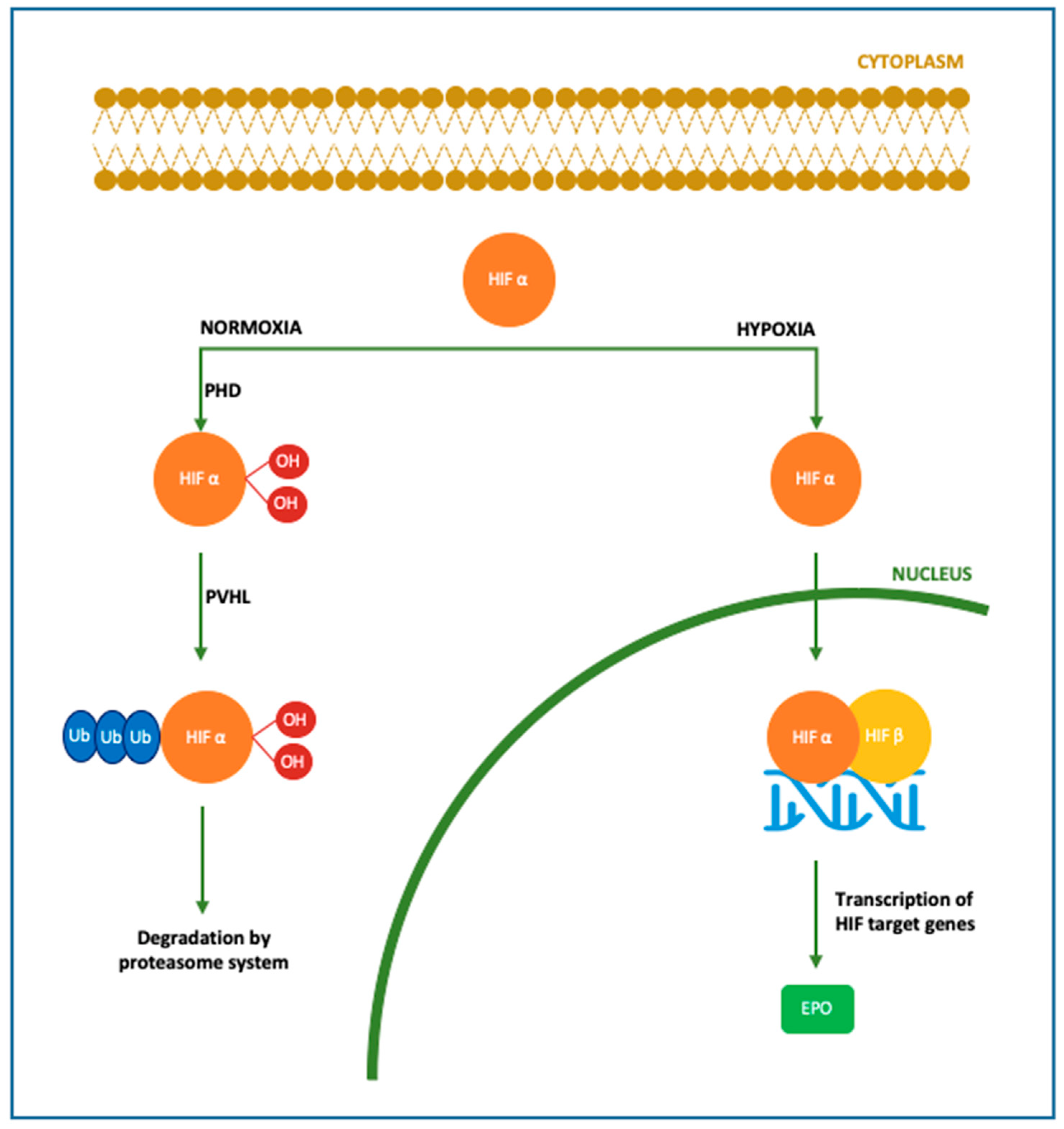
1.3. Erythropoietin (Epo) Actions and Neuroprotective Effects

Erythropoietin is a 30.4 kDa glycoprotein and pleiotropic cytokine, first described by Carnot in 1906 and isolated by Goldwasser and Kung in 1971, successfully produced for clinical use [55]. During fetal life Epo is produced by the liver, while after birth the production progressively takes place in the peritubular cells of the kidney [56]. Erythropoietin is primarily known for the haematological functions. After an ischemic insult lasting at least 30 min, the transcription of the Hypoxia Inducible Transcription Factor induced by hypoxia determines an increase of Epo hormone production in the kidney [57,58]. Binding of Epo to receptors on erythroid progenitor cells causes an increase in red blood cell mass. The enhanced oxygen-carrying capacity of the blood suppress the further expression of Epo completing the feedback loop [59].
The endogenous levels of Epo have been reported to increase in the brain for four hours after hypoxic exposure [60]. Hypoxia induce hypoxia-inducible factor-1, which determines the expression of growth factors such as the vascular endothelial growth factor (VEGF) and Epo [61]. The immediate effect of Epo is represented by an augmented expression of haemoglobin which improves the oxygen consumption and storage in the hypoxic tissue [59]. Therefore, high serum or amniotic fluid concentrations of Epo may suggest chronic hypoxia in newborns [62]. However, the lack of brain perfusion and oxygenation which occurs in brief periods, but still severe enough to cause brain injury, may not trigger endogenous Epo production [63]. Erythropoietin exerts intracellular protective effects after the ischemia-reperfusion damage, such as decreasing apoptosis, oxidative stress and Blood Brain Barrier (BBB) injury [64]. Furthermore, Epo has been proved to be able to stimulate angiogenesis, neurogenesis and neuronal plasticity after the ischemic damage [65,66].
Pleiotropic functions of Epo concerning neuroprotection have been studied in the last few decades. The first evidence for the extra-hematopoietic properties of Epo were published between 1993 and 2000 [67,68]. Normally, only a small percentage of circulating Epo is able to pass the BBB, binding the Epo receptor (EpoR) located on the capillary vessels [69]. However, it has been demonstrated that Epo is also produced in the developing brain, where it acts as a neuroprotective agent and growing factor [70,71,72]. In particular, Epo is primarily produced by astrocytes, followed by oligodendrocytes, endothelial cells, neurons and microglia, induced by hypoxia through the Hypoxia Inducible Transcription Factor pathway [70,71,72]. Figure 1. Both in animal models and in newborns, Epo has shown anti-apoptotic [73,74,75], antioxidant [76,77,78] and anti-inflammatory properties [79,80,81,82,83]. These actions can be exerted directly or mediated by a specific receptor named EpoR, which has been isolated in glial cells, neurons and brain endothelial cells in the hippocampus, cortex internal capsule and midbrain regions [70]. Molecular mechanisms following Epo treatment are shown in Figure 2. When not binding to its ligand, EpoR activates pathways leading to cell death. On the contrary, when Epo is bound to EpoR, cellular survival is promoted [84,85]. In addition, the role of Epo has also been described in promoting angiogenesis [74,86,87], oligodendrogenesis and neurogenesis [88,89,90,91]. In most of clinical studies, Epo is given in the form of recombinant human erythropoietin (rhEpo), which was the first purified and cloned [92]. Considering the difficulties in obtaining a sufficient quantity of Epo from the blood serum of living humans, rhEpo is produced in higher quantities from cultured mammalian hamster ovary cells and is highly purified [92,93]. At least nine isoforms of rhEpo can be identified, among which are epoetin alfa/alpha/beta, darbepoetin (Darbe) and carbamylated Epo [94]. Darbe is an analog of Epo with an additional sialic acid, able to confer a three-fold longer serum half-life compared with epoetin alfa, the most used isoform [95]. Epo and Darbe are termed as erythropoiesis-stimulating agents (ESAs) [96].
The aim of this review was to analyse the most recent evidence about the role of Epo as a neuroprotective strategy in the most common causes of neonatal neurological injury such as prematurity, HIE and perinatal stroke.

2. Materials and Methods

2.1. Research Strategy for Study Identification

A critical review was conducted using the 2020 PRISMA statement guidelines (www.prisma-statement.org, 15 December 2021), from February 2021 to May 2021, using the following databases: PubMed (2000–2021), Cochrane Trials Library CENTRAL (2000–2021). Additional ongoing studies were sought on the US National Institute of Health online registry for clinical trials (www.clinicaltrials.gov, 15 December 2021). The research strategy was conducted as follows: “erythropoietin” [All Fields] AND “neuroprotection” [All Fields] AND “newborns” [All Fields]. The results were combined with cross-referencing of previous reviews.

2.2. Eligibility Criteria and Study Selection

Clinical trials exploring the potential role of Epo or Darbe for neuroprotection in newborns were selected. Study population included human preterms, full-term newborns suffering from HIE or affected by/at risk of neonatal stroke.
After removing duplicates, studies were screened by title and abstract by three investigators independently. The following inclusion criteria were used: (1) human newborns as the study population, (2) use of Epo or Darbe as the only administered drug, (3) trials that reported effects of Epo or Darbe on neuroprotection as primary or secondary outcome.
Exclusion criteria were: (1) human adults or animal models as study population, (2) different outcomes from neuroprotection, (3) trials with Epo or Darbe given in association with other drugs, (4) not available full-text studies, (5) reviews, meta-analysis or journal editorials. The specific usage of Epo or Darbe was reported in each study represented on the Tables 1 and 2.

2.3. Data Extraction of the Studies

Data from the studies were collected using the “PICO” methodology (population, intervention, control and outcome).

3. Results

A total of 278 records were identified. After adjusting for duplicates and inclusion criteria, a total of 37 trials were eligible for our study. Eleven studies were excluded after full-text assessment: nine studies were protocols for developing or ongoing studies without preliminary data; one study had outcomes which were irrelevant for the aim of the review. Finally, one paper was a pilot study whose findings were updated later in a trial already included in our review. Twenty-six trials were finally analysed: 16 trials about Epo effects in preterm population, 8 studies investigating the role of Epo in HIE and 2 studies concerning Epo administration in patients with stroke or at risk of it Figure 3.

3.1. Erythropoietin Administration in Preterm Infants

Sixteen studies investigated the neuroprotective role of Epo in preterm infants, for a total number of 2923 preterm babies with a gestational age at birth between 24–32 weeks or birth weight 500–1500 g. Two trials enrolled a group of term controls [98,99]. Table 1 reports target population, intervention, outcomes and findings.
Out of 16, seven studies described Epo intravenous (iv) administration in the first 2–3 days of life, with a dosage between 500–3000 UI/kg/dose [103,105,106,108,109,110,111]. Three studies evaluated the cumulative dose of Epo, 8574 U/kg [102] or 6300 UI/kg [107] or 3750 U/kg [101], given either iv or subcutaneous (sc) in the first 6-9 weeks of life. Four studies evaluated sc Epo at a dose of 400 UI/kg administered three times per week until 35 weeks of corrected postnatal age [98,99,100,104]. A single study evaluated a combination of Epo given iv and sc. Firstly the iv administration was given at a dose of 1000 UI/kg every 48 h for a total of six doses, subsequently sc doses of 400 UI/kg were provided for three times per week until 32 weeks of corrected postnatal age [112]. In eight trials Epo treatment was compared to placebo [100,103,105,106,108,109,111,113], while three protocols evaluated also the administration of Darbe, at a dose of 10 mcg/kg once a week until 35 weeks of corrected postnatal age [98,99,104].
A single study evaluated mortality rate and major adverse events as a primary outcome [108]. Safety of early high-dose of Epo administration was demonstrated, with no excess in mortality [108]. Furthermore, no differences in the incidence of IVH, ventricular dilatation, cystic and non-cystic PVL, sepsis, necrotizing enterocolitis, persistent ductus arteriosus ligation, bronchopulmonary dysplasia were reported [108]. In particular, no differences were reported in the incidence of ROP and infantile hemangioma in Epo treated babies compared to controls, as possible adverse effects of Epo administration [114,115]. An increased erythropoiesis was described as a result of the megakaryocytes shift towards the erythroid lineage, although hemocytometric values remained within the normal range [108].
Neurodevelopmental outcomes were evaluated in the first two years of life in seven studies [100,101,104,105,107,109,112]. Four studies considered a longer follow-up until 3–4 years [98,103], 5 years [113] or even 13 years [102]. In addition to standard neurological examination, anthropometric measures and the evaluation of vision and hearing abilities were analysed [98,100,101,102,103,104,105,107,109,112,113]. Neurodevelopmental outcomes were assessed through Hamburg-Wechsler intelligence tests for children—Third Edition (HAWIK-III—Tewes et al., 1999) score [102], Bayley scales of infant and development—Second Edition (BSID-II—Bayley, 1993) or Third Edition (BSID-III—Bayley, 2005) [100,101,103,104,109,112], Griffiths’ Mental Developmental Scales (Huntley, 1996) [107], Behavior Assessment System for Children—Second Edition (BASC-II—Reynolds et al., 2004) [98], Gross Motor Function Classification System (GMFCS—Palisano et al., 1997–2007) [112,113] and Mental Processing Composite of the Kaufman Assessment Battery for Children—First Edition (KABC—I, Rozenblatt, 2011) [113]. Five studies reported a beneficial effect of Epo treatment in at least one of the neurodevelopment scales used for the analysis. In Epo treated patients, an increased score at Motor Development Index (MDI) [100,101] or BSID [103,104] or HAWIK-III IQ score [102] was reported compared to controls. Two studies highlighted a dose-response correlation between Epo doses and higher MDI scores [100,101]. The longest-term—until 10–13 years—follow up study reported better anthropometric measures and sensorineural functions when newborns were treated with Epo [102]. In addition, significant cognitive improvements and OP scores (evaluation of Object Permanence, included in BSID III) were associated with Epo and Darbe administration [104]. Positive cognitive results were also reported in a subgroup of ELBW affected by IVH using the HAWIK-III IQ score [102]. On the other hand, four studies didn’t show any difference in neurodevelopmental outcomes in Epo treated patients compared to controls, assessed through Griffiths’ [107], MDI, Psychomotor Development Index (PDI) [109] and GMFCS [112] at 2 years of age, or GMFCS and KABC at 5 years of age [113].
Four studies performed brain MRI to evaluate the outcomes of brain injury. The MRI analyses highlighted a reduced structural brain damage in the Epo treated group [105]. In addition, an improved white matter development assessed was observed using DTI and TBSS [106] and a weak but widespread effect in the overall structural connectivity network after Epo treatment, measured on DTI [111]. Higher FA values at the posterior limb of the internal capsule, the splenium of the corpus callosum, frontal white matter, and occipital white matter were found in Epo treated group compared to controls [110].
On the other hand, the spectroscopy analysis showed no difference for any brain metabolite level neither in premature nor in ESAs treated babies [99]. There was no significant difference in FA values at the parietal white matter, thalamus, lenticular nucleus, and caudate nucleus between Epo treated and control group [110].

3.2. Erythropoieti Administration in HIE

Eight studies investigated the neuroprotective role of Epo in newborns with HIE, for a total number of 533 patients. In most of these studies, patients enrolled had a gestational age ≥ 37 weeks [116,117,118,119,120], one study considered patients ≥ 40 weeks [121] and two studies ≥ 36 weeks [122,123]. All newborns were affected by HIE according to Sarnat score [124]. Table 2 reports target population, intervention and outcome.
Erythropoietin was administered in the first weeks of life, at different doses between 200–2500 U/kg/dose. Concerning the administration scheme, in three studies Epo was given every two days for a total of 12, 7, 6 or 5 doses [116,118,119,120]. In three studies Epo was given once a day for the first three days of life [117,122,123]. Two more doses at day 5 and 7 were administered in two of these protocols [122,123]. In one study Epo was given as a single administration [121].
All the studies evaluated Epo safety, reporting mortality rate and/or adverse events and/or the incidence of disability. None of them reported an increase in mortality or disability rate in patients treated with Epo. Erythropoietin was well tolerated, without allergic reactions, venous thrombosis or alterations of electrolytes, liver and renal functions [116]. Erythropoietin was not related to higher incidence of hypotension, hepatic dysfunction, prolonged coagulation, necrotizing enterocolitis, hypertension, polycythemia or thrombosis [119]. As a consequence of Epo hematopoietic effect, patients treated with Epo had higher levels of haemoglobin and erythrocytes compared to controls [116].
In two studies Epo therapy was associated with TH in comparison with TH alone [122,123]. The effect of different doses of Epo administered with TH was also described [118]. Only one study enrolled healthy controls [121]. Study protocols comparing Epo with placebo or only supportive therapy, reported a lower death rate [117,119,121] and disability index, evaluated with GMFCS, visual and hearing assessment [119] or BSID at 18 months [117]. Moreover, Epo alone decreased brain injury at MRI analysis performed at 10–14 days of life [119].
Compared to TH alone, The Epo effect was controversial. In fact, in one study Epo treatment compared to TH alone in moderate HIE showed a lower mortality and disability rate, together with higher scores at 18 months BSID-II [116].
Yet, another study described a weaker effect of Epo treatment on mortality rate and disability compared to TH alone [121]. The combined treatment of Epo and TH was found to be safe, not worsening brain damage at 4–13 days MRI nor affecting disability evaluated with GMFCS and neurodevelopment assessed through BSID-II or III at 22 months [118]. Erythropoietin associated with TH determined a lower mortality rate [122], a lower brain injury score at MRI performed between 4–7 days of age [122,123] and a better neurodevelopmental outcome with higher score of AIMS and WIDEA at 12 months when compared to TH alone [122]. Furthermore, the combined Epo and TH group improved neurodevelopmental outcomes assessed with Warner Initial Developmental Evaluation (WIDEA-Msall, 2006) and Alberta Infant Motor Scale (AIMS, Piper, 1994) at 12 months and had a smaller volume of acute brain injury assessed with MRI at ≤7 days of life [123].
Erythropoietin treatment in neonates with moderate and severe HIE showed better physical and mental developmental indexes at 6 months of age compared to controls [120].
No differences in terms of mortality in Epo treatment compared to phenobarbital administration were found and both therapies determined a reduced death rate compared to controls [117]. The antioxidant effects of the treatments were evaluated through the dosage of antioxidant enzymes Superoxide Dismutase and Glutathione Peroxidase, Total Serum Antioxidant Status and Malondialdehyde, as markers of free radical damage [117]. Plasma levels of Superoxide Dismutase and Glutathione Peroxidase were lower in Epo and Phenobarbital groups, while Total Serum Antioxidant Status levels were higher, especially in Epo compared with Phenobarbital group. However, Malondialdehyde did not show any significant difference in the groups [117]. A lower mortality rate in Epo or phenobarbital treatment was reported and a better cognitive and motor function assessed through BSID-II at 18 months of age was also described [117].
A single study evaluated the effect of TH or Epo or supportive therapy on serum brain-derived neurotrophic factor and neuron-specific enolase concentration, compared to healthy controls [121], reporting an increased brain-derived neurotrophic factor and decreased neuron-specific enolase level in treated groups on day 5 compared to day 1 [121].

3.3. Erythropoietin Administration in Neonatal Stroke

Two studies investigated the effects of Epo administration in children with neonatal stroke [125] or at risk of stroke in the neonatal period in a population of neonates scheduled for cardiac surgery with hypothermic cardiopulmonary bypass [126]. A total of 80 full-term newborns were enrolled. Table 3 reports target population, intervention and outcome.
Erythropoietin was administered intravenously in both trials, at a dose of 1000 UI/kg, except for twenty-six patients who received 500 UI/kg [126]. In Benders’ study Epo was administered for three days straight after radiological diagnosis of idiopathic perinatal arterial ischemic stroke [125]. In Andropoulos’ study, Epo was given in three doses at 1000 UI/kg or 500 UI/kg pre and post-surgery [126].
In both trials, the safety profile of Epo administration was registered and effects of Epo were evaluated with brain MRI exams and neurodevelopmental assessment. None of them highlighted a significant benefit on neuroprotection in Epo treated children vs. placebo groups [125,126].
Both studies analysed the safety profile of EPO administration and no significant differences in adverse events or death rates were detected in Epo or placebo groups. In particular, in both studies vital signs and biochemistry blood parameters remained within the normal range. Andropoulos registered six cases of postoperative dural sinovenous thrombosis, equally distributed in Epo and control group [126].
In Benders et al. study, brain MRI was performed after clinical and ultrasound suspicion of perinatal arterial ischemic stroke to confirm diagnosis and then repeated at 3 months of age [125]. In the other study, MRI imaging was executed as preoperative assessment and then as a control 7–10 day after cardiac surgery [126]. The brain MRI evaluation of both studies showed comparable results of neuroimaging performed at 7–10 days [126] and 3 months [125] in the Epo treated group and the controls. In particular, no difference on intracranial haemorrhage, thrombosis, infarction or white matter injury occurrence was described in Epo treated population [126]. Even in Benders’ follow-up study, any significant discrepancy was highlighted at the age of 3 months in the percentage of the stroke tissue that had dissolved [125] and no difference in the total brain volume was found in Epo treated and controls [125].
Neurodevelopmental outcome was assessed, respectively at 3 months follow up with Griffiths’ scale [125] and at one year follow up with BSID-III [126]. None of the two trials found any statistically difference between Epo treated and placebo groups.

4. Discussion

4.1. Erythropoieti in Preterm Infants

Erythropoietin has been administered to preterm infants since 1990 for haematological purposes. In fact, Epo has been extensively used for the prevention of anemia of prematurity, but the discovery of the potential role as a neuroprotective agent suggested new purposes for its administration [127,128,129].
In the last few decades the survival of preterm infants has dramatically grown, yet this population is still exposed to a great incidence of neurodevelopmental impairment [7,8]. Trials conducted in animal models have shown neuroprotective effects of Epo, thus clinical studies investigating Epo’s new potential benefits in premature babies have followed [130,131,132,133,134]. In particular, it has been described that Epo administration in preterm is able to protect neurons and oligodendrocytes from apoptosis, prevent inflammation and promote neurogenesis and angiogenesis [135,136]. Preterm infants benefit by a prolonged treatment during the period of oligodendrocyte maturation, between 24 and 32 weeks [2,130,137], in which Epo exerts a neuroprotective function. Evidence in newborns and animal models suggest neuroprotective effects at doses >200 U/kg [138]. Cumulative doses have been associated with more beneficial effects compared to a single-dose administration [139].
The dosage of EPO ranged from 250 to 3000 UI/kg in all studies. Darbe alfa was administered at 10 mcg/Kg sc [98,99,104].
The population target was represented of extremely and very preterm babies (gestational age below 32 weeks or BW < 1500 gr). Only three clinical trials focused on the role of Epo in extremely preterm infants (gestational age below 28 weeks or 1000 gr) [102,103,112].
Erythropoietin administration was proved to be safe in all preterm infants, not affecting mortality rate and major adverse effects occurrence in the short and long term [103,108,112]. These data confirmed previous analyses on animal models [139,140,141].
Out of 16, 10 trials performed clinical neurodevelopmental outcome evaluated mainly in the first years of life. No uniform results were reported.
In addition, neurodevelopmental outcomes were not assessed in all trials in which neuroimaging was performed. It has been well recognized that a correlation between white matter injuries and neurodevelopmental outcome, in terms of increased incidence of cerebral palsy, motor and cognitive delay exists [15,16,142,143]. In addition, values of fractional anisotropy of the corpus callosum assessed using TBSS have been reported to directly correlate with motor and cognitive outcomes [144]. Thus, neuroimaging alterations have been related with poorer neurodevelopmental outcomes [95,98,99,106,145]. Studies on Epo treated very preterm newborns highlighted an improved white matter development and a weak but widespread effect in the overall structural connectivity network after Epo treatment [110,111]. Yet, no difference for any brain metabolite has been reported in very preterm treated group [99].

4.2. Erythropoietin in HIE

After several years of studies conducted in animal models, in 2009 the first trial of Epo administration as adjuvant therapy with TH in newborns affected by HIE was published. The study underlined Epo favourable effects, paving the way to further studies [116]. Since then Epo showed beneficial effects during the secondary energy failure and the tertiary phases of HIE [146].
The therapeutic scheme is influenced by the type of damage [2]. In HIE term newborns high Epo doses are more effective when the injury has not been established yet [2,130,137]. Evidence from animal models have shown that Epo should be administered at high doses within 6 h after the onset of brain injury to reach a substantial neuroprotective effect [147]. In this context, Epo may influence the mechanisms of cerebral flow restoration, neovascularization and neuroregeneration, limiting the ischemic damage [148].
Our analysis revealed that Epo administration was safe and well tolerated [116], confirming previous findings in animal models [132,139,141].
The passage through the BBB is an essential issue to be considered for any neuroprotective drug given systemically. Erythropoietin presents a molecular weight too high to be carried across the BBB through the lipid mediated transport [116]. Transport of Epo through the BBB is mediated by a specific receptor-facilitated process [69] as well as a time, dose and peak serum concentration-dependent mechanism [149,150]. Furthermore, the alteration of BBB permeability caused by asphyxia determines in patient with HIE an easier passage from serum to cerebrospinal fluid [151]. In the studies analysed, Epo administration is followed by a higher concentration of Epo in the cerebrospinal fluid compared to HIE controls, thus confirming the increased levels are due to the contribution of Epo exogenous administration [116].
Administration of Epo has shown some advantages compared to TH treatment. Treatment with Epo implies easier technical performance, with less side effects [116,121]. Moreover, Epo treatment is effective if administered within 48 h of life, differently from TH which must be initiated within the first 6 h of life [116]. Thus, Epo treatment has been proposed as both an alternative therapy or combined treatment with TH [116,121].
In all the protocols comparing Epo vs. placebo or Epo associated with TH vs. supportive therapy, Epo treatment determined a lower death rate and improved neurologic outcomes [117,119,120,121,122,123]. Valera et al. treated 15 HIE neonates with EPO for two weeks, starting within three hours from birth along with TH. At 18 months, there was 80% survival with no neurodevelopmental disability. Unfortunately, there was no control group for comparison [152].
Rogers et al. recruited 24 newborns with HIE and administered Epo 24 h after HIE combined with standard TH. The authors found that significant neurodevelopmental disability occurred in only 12.5% of infants with moderate to severe MRI changes who received Epo plus TH, compared to 70–80% significant disability or death in infants treated with TH alone. However, these results were not statistically significant as the number of infants was too small [118]. The Phase II clinical trial on newborns with HIE reported better 12-months motor outcomes for treatment with Epo plus TH compared to TH alone [123]. A randomized case-control study by Wu et al. [122] evaluating 24 newborns treated with Epo and TH for moderate/severe HIE, showed a significantly reduced brain injury on MRI at 5 days-of-age and better 12-months motor outcomes compared to the 26 infants who received TH as a standard routine measure for HIE. The Epo treatment was also associated with an antioxidant effect and an increase of neuroprotective factors [117,121]. The study by Wang et al. clearly demonstrated the protection from the damage resulting from asphyxia through inhibiting apoptosis, reducing oxygen free radicals and enhancing antioxidant capacity. In this research trial, 34 newborns received Epo and Vitamin C plus TH compared to 34 newborns receiving conventional treatment plus ascorbic acid. There were lower oxidative stress index levels and higher antioxidant enzymes in both groups after treatment, with better results in the group treated with Epo. Moreover, lower pro-apoptotic molecules and higher anti-apoptotic molecules were found in both groups after treatment, with better results again in the group treated with Epo [153].
In addition, MRI analysis confirmed the beneficial effect of Epo in the reduction of brain injury, both if administered alone [119] or combined with TH [123]. Despite all these data, the optimal dosage and administration regimens for the treatment of HIE in newborns still needs to be defined [116,118,121].

4.3. Erythropoietin in Neonatal Stroke

At the diagnosis of neonatal stroke, the damage has already been established and the reparation mechanisms are not fully known, thus nowadays acute therapies for perinatal strokes are still not available [154]. Currently the management of the disease is mainly supportive, based on the treatment of complications, such as antiepileptic therapy, and on the improvement of neurologic chronic sequelae through rehabilitation [155]. Erythropoietin has become one of the most remarkable neuroprotective strategy since Sakanaka et al. reported for the first time the role of Epo as a neuroprotective agent in the ischemic brain damage [156]. Recent researches are focused on new approaches including the administration of adjuvant neuroprotective therapies, with the aim to enhance endogenous mechanisms of repair and angiogenesis [157,158].
In our review, the safety profile of Epo administration was also confirmed in the studies on neonatal stroke. This is consistent with previous data available from animal models and preterm and HIE-suffering newborns, which showed a good tolerance of treatment with Epo in those populations [128,147,159,160].
The brain MRI evaluation in both studies didn’t find a beneficial effect of Epo treatment on stroke volume [125,126]. Furthermore, Epo treatment was not associated with an overall difference in neurodevelopmental outcome, at 3 months and one year follow up respectively with Griffiths’ scale and BSID-III [125,126].
These data are in contrast with most of the newborn stroke animal models in which several neuroprotective effects and reduction of stroke volume induced by Epo are described [61,130,161,162]. Recently, a delayed Epo therapy administered to mice model with middle brain artery occlusion stroke reported to uphold brain volume and to improve behavioural and sensorimotor functions, compared with placebo-treated animals [61].
The discrepancy between the experimental animal model and clinical trials may be due to the small neonatal study population, as the trials are both phase I and II studies. Furthermore, Andropoulos and colleagues found that six enrolled newborns presented microdeletions at the chromosome 22q11.2 region, a condition known to be linked to a variable degree on neurodevelopmental impairment in a syndromic condition [163].
Another limiting factor is the therapeutic window, as the beneficial effect is limited to specific time after injury [164]. One interventional study used Epo after stroke diagnosis, which is usually done 24–48 h after cerebral vascular accident. The use of specific biomarkers that will increase within the first hours of life in hypoxic-ischemic injury may help in the early diagnosis of stroke, allowing a prompt identification of neonates who may qualify for neuroprotection.
Finally, the variability in period and tests used to assess the neurodevelopmental outcome may represent a weakness and suggests that scores should be interpreted with caution. Since the determinants of cognitive outcomes at school age are multifactorial, the predictive value of tests administered at age < 24 months might never approach 100%.

5. Conclusions

Brain injury still represents an open challenge in the neonatal setting. Therapeutic hypothermia has become the standard therapy in term newborns suffering from perinatal asphyxia. Since 2009, Erythropoietin (Epo) has been identified as a pharmacological compound playing an important role in preserving the developing brain. Clinical trials reveal that Epo can be employed as one of the therapeutic treatments of perinatal asphyxia either as an adjuvant therapy with hypothermia or a substitute for hypothermia in HIE. No beneficial effect in improving morbidity was observed after Epo administration in perinatal stroke.
When administered in preterm infants, some clinical studies report beneficial effect on neurodevelopmental outcome, others do not show efficacy.
An early diagnosis and a shorter latency time to treatment are essential for successful outcomes both in preterm and term newborns suffering from hypoxic-ischemic injury. Further studies focused on tailored regimens accounting for risk stratification of brain damage are needed. Moreover, the identification of specific markers and their temporal expression at different time of recovery after hypoxic-ischemia may be implemented to optimize the Epo therapeutic scheme in the developing brain.

Author Contributions

Conceptualization, S.P., C.L., G.B.; writing—original draft preparation, C.L., F.G., C.P., T.C., G.C.; writing—review and editing, C.L., S.P., F.G., T.C., G.B.; visualization and supervision, S.P., C.L, G.B., E.G., S.M.R.E. All authors have read and agreed to the published version of the manuscript.

Funding

This research received no external funding.

Conflicts of Interest

The authors declare no conflict of interest.

Abbreviations

1H-MRSProton Magnetic Resonance Spectroscopy
AIMSAlberta Infant Motor Scale for motor functions
BASC2Behavior Assessment System for Children—2nd edition
BBBblood brain barrier
BSID-II or IIIBayley Scales of Infant Development-II or III edition, composed by three scores—MDI, PDI and Infant Behavior Record
BWbirth weight
Darbedarbepoetin
DTIDiffusion Tensor Imaging
ELBWExtremely Low Birth Weight
EpoErythropoietin
EpoRErythropoietin receptors
GMFCSGross Motor Function Classification System
HAWIK-IIIHamburg-Wechsler Intelligence Test for Children-III edition
HIEHypoxic Ischemic Encephalopathy
IQIntelligence Quotient
Ivintravenous
IVHIntraventricular Haemorrhage
KABCKaufman Assessment Battery for Children
MDIMotor Development Index
MRIMagnetic Resonance Imaging
NMSNeuromuscular Function Scale
OPObject of Permanence
PDIPsychomotor Development Index
Scsubcutaneous
TBSSTract Based Spatial Statistics
THTherapeutic Hypothermia
WIDEAWarner Initial Developmental Evaluation

References

  1. Hamilton, B.E.; Hoyert, D.L.; Martin, J.A.; Strobino, D.M.; Guyer, B. Annual summary of vital statistics: 2010–2011. Pediatrics 2013, 131, 548–558. [Google Scholar] [CrossRef] [PubMed] [Green Version]
  2. Tataranno, M.L.; Perrone, S.; Longini, M.; Buonocore, G. New Antioxidant Drugs for Neonatal Brain Injury. Oxidative Med. Cell. Longev. 2015, 2015, 108251. [Google Scholar] [CrossRef] [PubMed] [Green Version]
  3. WHO: Recommended Definitions, Terminology and Format for Statistical Tables Related to The Perinatal Period And Use of A New Certificate For Cause of Perinatal Deaths. Acta Obstet. Gynecol. Scand. 1977, 56, 247–253. [CrossRef]
  4. Blencowe, H.; Cousens, S.; Oestergaard, M.Z.; Chou, D.; Moller, A.-B.; Narwal, R.; Adler, A.; Vera Garcia, C.; Rohde, S.; Say, L.; et al. National, regional, and worldwide estimates of preterm birth rates in the year 2010 with time trends since 1990 for selected countries: A systematic analysis and implications. Lancet 2012, 379, 2162–2172. [Google Scholar] [CrossRef] [Green Version]
  5. Morisaki, N.; Ganchimeg, T.; Vogel, J.P.; Zeitlin, J.; Cecatti, J.G.; Souza, J.P.; Pileggi Castro, C.; Torloni, M.R.; Ota, E.; Mori, R.; et al. Impact of stillbirths on international comparisons of preterm birth rates: A secondary analysis of the WHO multi-country survey of Maternal and Newborn Health. BJOG 2017, 124, 1346–1354. [Google Scholar] [CrossRef] [PubMed] [Green Version]
  6. Bonet, M.; Cuttini, M.; Piedvache, A.; Boyle, E.M.; Jarreau, P.H.; Kollée, L.; Maier, R.F.; Milligan, D.; Van Reempts, P.; Weber, T.; et al. Changes in management policies for extremely preterm births and neonatal outcomes from 2003 to 2012: Two population-based studies in ten European regions. BJOG 2017, 124, 1595–1604. [Google Scholar] [CrossRef] [PubMed] [Green Version]
  7. Latal, B. Prediction of neurodevelopmental outcome after preterm birth. Pediatr. Neurol. 2009, 40, 413–419. [Google Scholar] [CrossRef] [PubMed]
  8. Saigal, S.; Doyle, L. An overview of mortality and sequelae of preterm birth from infancy to adulthood. Lancet 2008, 371, 261–269. [Google Scholar] [CrossRef]
  9. de Araújo, B.F.; Zatti, H.; Madi, J.M.; Coelho, M.B.; Olmi, F.B.; Canabarro, C.T. Analysis of neonatal morbidity and mortality in late-preterm newborn infants. J. Pediatr. 2012, 88, 259–266. [Google Scholar] [CrossRef] [Green Version]
  10. Platt, M.J. Outcomes in preterm infants. Public Health 2014, 128, 399–403. [Google Scholar] [CrossRef]
  11. Orchinik, L.J.; Taylor, H.G.; Espy, K.A.; Minich, N.; Klein, N.; Sheffield, T.; Hack, M. Cognitive outcomes for extremely preterm/extremely low birth weight children in kindergarten. J. Int. Neuropsychol. Soc. 2011, 17, 1067–1079. [Google Scholar] [CrossRef] [Green Version]
  12. Moreira, R.S.; Magalhães, L.C.; Alves, C.R.L. Effect of preterm birth on motor development, behavior, and school performance of school-age children: A systematic review. J. Pediatr. 2014, 90, 119–134. [Google Scholar] [CrossRef] [Green Version]
  13. Johnson, S.; Evans, T.A.; Draper, E.S.; Field, D.J.; Manktelow, B.N.; Marlow, N.; Matthews, R.; Petrou, S.; Seaton, S.E.; Smith, L.K.; et al. Neurodevelopmental outcomes following late and moderate prematurity: A population-based cohort study. Arch. Dis. Child. Fetal Neonatal Ed. 2015, 100, F301–F308. [Google Scholar] [CrossRef] [Green Version]
  14. Kinney, H.C. The encephalopathy of prematurity: One pediatric neuropathologist’s perspective. Semin. Pediatr. Neurol. 2009, 16, 179–190. [Google Scholar] [CrossRef] [Green Version]
  15. Woodward, L.; Anderson, P.; Austin, N.; Howard, K.; Inder, T. Neonatal MRI to predict neurodevelopmental outcomes in preterm infants. N. Engl. J. Med. 2006, 355, 685–694. [Google Scholar] [CrossRef] [Green Version]
  16. Rutherford, M.; Supramaniam, V.; Ederies, A.; Chew, A.; Bassi, L.; Groppo, M.; Anjari, M.; Counsell, S.; Ramenghi, L. Magnetic resonance imaging of white matter diseases of prematurity. Neuroradiology 2010, 52, 505–521. [Google Scholar] [CrossRef]
  17. Alexandrou, G.; Mårtensson, G.; Skiöld, B.; Blennow, M.; Adén, U.; Vollmer, B. White matter microstructure is influenced by extremely preterm birth and neonatal respiratory factors. Acta Paediatr. 2014, 103, 48–56. [Google Scholar] [CrossRef]
  18. Jakovcevski, I.; Zecevic, N. Sequence of oligodendrocyte development in the human fetal telencephalon. Glia 2005, 49, 480–491. [Google Scholar] [CrossRef]
  19. Batalle, D.; O’Muircheartaigh, J.; Makropoulos, A.; Kelly, C.J.; Dimitrova, R.; Hughes, E.J.; Hajnal, J.V.; Zhang, H.; Alexander, D.C.; Edwards, A.D.; et al. Different patterns of cortical maturation before and after 38 weeks gestational age demonstrated by diffusion MRI in vivo. Neuroimage 2019, 185, 764–775. [Google Scholar] [CrossRef]
  20. Deoni, S.C.L.; Dean, D.C., 3rd; Remer, J.; Dirks, H.; O’Muircheartaigh, J. Cortical maturation and myelination in healthy toddlers and young children. Neuroimage 2015, 115, 147–161. [Google Scholar] [CrossRef] [Green Version]
  21. Ouyang, M.; Dubois, J.; Yu, Q.; Mukherjee, P.; Huang, H. Delineation of early brain development from fetuses to infants with diffusion MRI and beyond. Neuroimage 2019, 185, 836–850. [Google Scholar] [CrossRef]
  22. van Tilborg, E.; de Theije, C.G.M.; van Hal, M.; Wagenaar, N.; de Vries, L.S.; Benders, M.J.; Rowitch, D.H.; Nijboer, C.H. Origin and dynamics of oligodendrocytes in the developing brain: Implications for perinatal white matter injury. Glia 2018, 66, 221–238. [Google Scholar] [CrossRef]
  23. Volpe, J.J.; Kinney, H.C.; Jensen, F.E.; Rosenberg, P.A. The developing oligodendrocyte: Key cellular target in brain injury in the premature infant. Int. J. Dev. Neurosci. 2011, 29, 423–440. [Google Scholar] [CrossRef] [Green Version]
  24. Back, S.A.; Gan, X.; Li, Y.; Rosenberg, P.A.; Volpe, J.J. Maturation-dependent vulnerability of oligodendrocytes to oxidative stress-induced death caused by glutathione depletion. J. Neurosci. 1998, 18, 6241–6253. [Google Scholar] [CrossRef]
  25. Keunen, K.; Kersbergen, K.J.; Groenendaal, F.; Isgum, I.; de Vries, L.S.; Benders, M.J.N.L. Brain tissue volumes in preterm infants: Prematurity, perinatal risk factors and neurodevelopmental outcome: A systematic review. J. Matern. Fetal Neonatal Med. 2012, 25 (Suppl. 1), 89–100. [Google Scholar] [CrossRef]
  26. Van Kooij, B.J.M.; Benders, M.J.N.L.; Anbeek, P.; Van Haastert, I.C.; De Vries, L.S.; Groenendaal, F. Cerebellar volume and proton magnetic resonance spectroscopy at term, and neurodevelopment at 2 years of age in preterm infants. Dev. Med. Child Neurol. 2012, 54, 260–266. [Google Scholar] [CrossRef]
  27. Volpe, J.J. Cerebellum of the premature infant: Rapidly developing, vulnerable, clinically important. J. Child Neurol. 2009, 24, 1085–1104. [Google Scholar] [CrossRef] [Green Version]
  28. Pieterman, K.; White, T.J.; van den Bosch, G.E.; Niessen, W.J.; Reiss, I.K.M.; Tibboel, D.; Hoebeek, F.E.; Dudink, J. Cerebellar Growth Impairment Characterizes School-Aged Children Born Preterm without Perinatal Brain Lesions. AJNR Am. J. Neuroradiol. 2018, 39, 956–962. [Google Scholar] [CrossRef]
  29. Kurinczuk, J.J.; White-Koning, M.; Badawi, N. Epidemiology of neonatal encephalopathy and hypoxic-ischaemic encephalopathy. Early Hum. Dev. 2010, 86, 329–338. [Google Scholar] [CrossRef]
  30. Shankaran, S.; Laptook, A.; Ehrenkranz, R.; Tyson, J.; McDonald, S.; Donovan, E.; Fanaroff, A.; Poole, W.; Wright, L.; Higgins, R.; et al. Whole-Body Hypothermia for Neonates with Hypoxic–Ischemic Encephalopathy. N. Engl. J. Med. 2005, 353, 1574–1584. [Google Scholar] [CrossRef]
  31. Thoresen, M.; Tooley, J.; Liu, X.; Jary, S.; Fleming, P.; Luyt, K.; Jain, A.; Cairns, P.; Harding, D.; Sabir, H. Time Is Brain: Starting Therapeutic Hypothermia within Three Hours after Birth Improves Motor Outcome in Asphyxiated Newborns. Neonatology 2013, 104, 228–233. [Google Scholar] [CrossRef] [PubMed]
  32. Azzopardi, D.V.; Strohm, B.; Edwards, A.D.; Dyet, L.; Halliday, H.L.; Juszczak, E.; Kapellou, O.; Levene, M.; Marlow, N.; Porter, E.; et al. Moderate hypothermia to treat perinatal asphyxial encephalopathy. N. Engl. J. Med. 2009, 361, 1349–1358. [Google Scholar] [CrossRef] [PubMed] [Green Version]
  33. Kali, G.T.J.; Martinez-Biarge, M.; Van Zyl, J.; Smith, J.; Rutherford, M. Management of therapeutic hypothermia for neonatal hypoxic ischaemic encephalopathy in a tertiary centre in South Africa. Arch. Dis. Child. Fetal Neonatal Ed. 2015, 100, F519–F523. [Google Scholar] [CrossRef] [PubMed]
  34. van Bel, F.; Groenendaal, F. Drugs for neuroprotection after birth asphyxia: Pharmacologic adjuncts to hypothermia. Semin. Perinatol. 2016, 40, 152–159. [Google Scholar] [CrossRef] [PubMed] [Green Version]
  35. Cánovas-Ahedo, M.; Alonso-Alconada, D. Combined therapy in neonatal hypoxic-ischaemic encephalopathy. An. Pediatr. 2019, 91, 59-e1. [Google Scholar] [CrossRef]
  36. Ferriero, D.M. Neonatal brain injury. N. Engl. J. Med. 2004, 351, 1985–1995. [Google Scholar] [CrossRef]
  37. Albrecht, M.; Zitta, K.; Groenendaal, F.; van Bel, F.; Peeters-Scholte, C. Neuroprotective strategies following perinatal hypoxia-ischemia: Taking aim at NOS. Free Radic. Biol. Med. 2019, 142, 123–131. [Google Scholar] [CrossRef]
  38. Hilton, G.D.; Nunez, J.L.; Bambrick, L.; Thompson, S.M.; McCarthy, M.M. Glutamate-mediated excitotoxicity in neonatal hippocampal neurons is mediated by mGluR-induced release of Ca++ from intracellular stores and is prevented by estradiol. Eur. J. Neurosci. 2006, 24, 3008–3016. [Google Scholar] [CrossRef] [Green Version]
  39. Szydlowska, K.; Tymianski, M. Calcium, ischemia and excitotoxicity. Cell Calcium 2010, 47, 122–129. [Google Scholar] [CrossRef]
  40. Iwata, O.; Iwata, S.; Bainbridge, A.; De Vita, E.; Matsuishi, T.; Cady, E.B.; Robertson, N.J. Supra- and sub-baseline phosphocreatine recovery in developing brain after transient hypoxia-ischaemia: Relation to baseline energetics, insult severity and outcome. Brain 2008, 131, 2220–2226. [Google Scholar] [CrossRef] [Green Version]
  41. Hassell, K.J.; Ezzati, M.; Alonso-alconada, D.; Hausenloy, D.J.; Robertson, N.J. New horizons for newborn brain protection: Enhancing endogenous neuroprotection. Arch. Dis. Child Fetal Neonatal 2015, 100, 541–552. [Google Scholar] [CrossRef]
  42. Perlman, J.M.; Wyllie, J.; Kattwinkel, J.; Atkins, D.L.; Chameides, L.; Goldsmith, J.P.; Guinsburg, R.; Hazinski, M.F.; Morley, C.; Richmond, S.; et al. Part 11: Neonatal resuscitation: 2010 International Consensus on Cardiopulmonary Resuscitation and Emergency Cardiovascular Care Science With Treatment Recommendations. Circulation 2010, 122, S516–S538. [Google Scholar] [CrossRef] [Green Version]
  43. Fleiss, B.; Gressens, P. Tertiary mechanisms of brain damage: A new hope for treatment of cerebral palsy? Lancet Neurol. 2012, 11, 556–566. [Google Scholar] [CrossRef]
  44. Raju, T.N.K.; Nelson, K.B.; Ferriero, D.; Lynch, J.K. Ischemic perinatal stroke: Summary of a workshop sponsored by the National Institute of Child Health and Human Development and the National Institute of Neurological Disorders and Stroke. Pediatrics 2007, 120, 609–616. [Google Scholar] [CrossRef] [Green Version]
  45. Nelson, K.B.; Lynch, J.K. Stroke in newborn infants. Lancet Neurol. 2004, 3, 150–158. [Google Scholar] [CrossRef] [Green Version]
  46. Laugesaar, R.; Kolk, A.; Tomberg, T.; Metsvaht, T.; Lintrop, M.; Varendi, H.; Talvik, T. Acutely and retrospectively diagnosed perinatal stroke: A population-based study. Stroke 2007, 38, 2234–2240. [Google Scholar] [CrossRef] [Green Version]
  47. Lee, J.; Croen, L.A.; Backstrand, K.H.; Yoshida, C.K.; Henning, L.H.; Lindan, C.; Ferriero, D.M.; Fullerton, H.J.; Barkovich, A.J.; Wu, Y.W. Maternal and infant characteristics associated with perinatal arterial stroke in the infant. JAMA 2005, 293, 723–729. [Google Scholar] [CrossRef]
  48. Kirton, A.; Deveber, G. Life after perinatal stroke. Stroke 2013, 44, 3265–3271. [Google Scholar] [CrossRef] [Green Version]
  49. Bernson-Leung, M.E.; Rivkin, M.J. Stroke in Neonates and Children. Pediatr. Rev. 2016, 37, 463–477. [Google Scholar] [CrossRef] [Green Version]
  50. Kirton, A. Advancing non-invasive neuromodulation clinical trials in children: Lessons from perinatal stroke. Eur. J. Paediatr. Neurol. 2017, 21, 75–103. [Google Scholar] [CrossRef]
  51. Wusthoff, C.J.; Kessler, S.K.; Vossough, A.; Ichord, R.; Zelonis, S.; Halperin, A.; Gordon, D.; Vargas, G.; Licht, D.J.; Smith, S.E. Risk of later seizure after perinatal arterial ischemic stroke: A prospective cohort study. Pediatrics 2011, 127, e1550–e1557. [Google Scholar] [CrossRef] [Green Version]
  52. Wanigasinghe, J.; Reid, S.M.; Mackay, M.T.; Reddihough, D.S.; Harvey, A.S.; Freeman, J.L. Epilepsy in hemiplegic cerebral palsy due to perinatal arterial ischaemic stroke. Dev. Med. Child Neurol. 2010, 52, 1021–1027. [Google Scholar] [CrossRef]
  53. Arai, K.; Jin, G.; Navaratna, D.; Lo, E.H. Brain angiogenesis in developmental and pathological processes: Neurovascular injury and angiogenic recovery after stroke. FEBS J 2009, 276, 4644–4652. [Google Scholar] [CrossRef] [Green Version]
  54. Ohab, J.J.; Fleming, S.; Blesch, A.; Carmichael, S.T. A neurovascular niche for neurogenesis after stroke. J. Neurosci. 2006, 26, 13007–13016. [Google Scholar] [CrossRef] [Green Version]
  55. Goldwasser, E.; Kung, C. Purification of erythropoietin. Proc. Natl. Acad. Sci. USA 1971, 68, 697–698. [Google Scholar] [CrossRef] [Green Version]
  56. Juul, S.; Pet, G. Erythropoietin and Neonatal Neuroprotection. Clin. Perinatol. 2015, 42, 469–481. [Google Scholar] [CrossRef] [Green Version]
  57. Ivan, M.; Kondo, K.; Yang, H.; Kim, W.; Valiando, J.; Ohh, M.; Salic, A.; Asara, J.M.; Lane, W.S.; Kaelin, W.G.J. HIFalpha targeted for VHL-mediated destruction by proline hydroxylation: Implications for O2 sensing. Science 2001, 292, 464–468. [Google Scholar] [CrossRef]
  58. Jaakkola, P.; Mole, D.R.; Tian, Y.M.; Wilson, M.I.; Gielbert, J.; Gaskell, S.J.; von Kriegsheim, A.; Hebestreit, H.F.; Mukherji, M.; Schofield, C.J.; et al. Targeting of HIF-alpha to the von Hippel-Lindau ubiquitylation complex by O2-regulated prolyl hydroxylation. Science 2001, 292, 468–472. [Google Scholar] [CrossRef]
  59. Fisher, J.W. Erythropoietin: Physiology and pharmacology update. Exp. Biol. Med. 2003, 228, 1–14. [Google Scholar] [CrossRef]
  60. Chikuma, M.; Masuda, S.; Kobayashi, T.; Nagao, M.; Sasaki, R. Tissue-specific regulation of erythropoietin production in the murine kidney, brain, and uterus. Am. J. Physiol. Endocrinol. Metab. 2000, 279, E1242–E1248. [Google Scholar] [CrossRef]
  61. Larpthaveesarp, A.; Georgevits, M.; Ferriero, D.M.; Gonzalez, F.F. Delayed Erythropoietin Therapy Improves Histological and Behavioral Outcomes after Transient Neonatal Stroke. Neurobiol. Dis. 2016, 57–63. [Google Scholar] [CrossRef] [PubMed] [Green Version]
  62. Teramo, K.A.; Widness, J.A. Increased fetal plasma and amniotic fluid erythropoietin concentrations: Markers of intrauterine hypoxia. Neonatology 2009, 95, 105–116. [Google Scholar] [CrossRef] [PubMed]
  63. Traudt, C.M.; McPherson, R.J.; Bauer, L.A.; Richards, T.L.; Burbacher, T.M.; McAdams, R.M.; Juul, S.E. Concurrent erythropoietin and hypothermia treatment improve outcomes in a term nonhuman primate model of perinatal asphyxia. Dev. Neurosci. 2013, 35, 491–503. [Google Scholar] [CrossRef] [PubMed] [Green Version]
  64. Zhang, S.-J.; Luo, Y.-M.; Wang, R.-L. The effects of erythropoietin on neurogenesis after ischemic stroke. J. Integr. Neurosci. 2020, 19, 561–570. [Google Scholar] [CrossRef] [PubMed]
  65. Xiong, T.; Qu, Y.; Mu, D.; Ferriero, D. Erythropoietin for neonatal brain injury: Opportunity and challenge. Int. J. Dev. Neurosci. Off. J. Int. Soc. Dev. Neurosci. 2011, 29, 583–591. [Google Scholar] [CrossRef]
  66. Sargin, D.; El-Kordi, A.; Agarwal, A.; Müller, M.; Wojcik, S.M.; Hassouna, I.; Sperling, S.; Nave, K.-A.; Ehrenreich, H. Expression of constitutively active erythropoietin receptor in pyramidal neurons of cortex and hippocampus boosts higher cognitive functions in mice. BMC Biol. 2011, 9, 27. [Google Scholar] [CrossRef] [Green Version]
  67. Li, Y.; Juul, S.E.; Morris-Wiman, J.A.; Calhoun, D.A.; Christensen, R.D. Erythropoietin receptors are expressed in the central nervous system of mid-trimester human fetuses. Pediatr. Res. 1996, 40, 376–380. [Google Scholar] [CrossRef] [Green Version]
  68. Morishita, E.; Masuda, S.; Nagao, M.; Yasuda, Y.; Sasaki, R. Erythropoietin receptor is expressed in rat hippocampal and cerebral cortical neurons, and erythropoietin prevents in vitro glutamate-induced neuronal death. Neuroscience 1997, 76, 105–116. [Google Scholar] [CrossRef]
  69. Brines, M.L.; Ghezzi, P.; Keenan, S.; Agnello, D.; De Lanerolle, N.C.; Cerami, C.; Itri, L.M.; Cerami, A. Erythropoietin crosses the blood—Brain barrier to protect against experimental brain injury. Proc. Natl. Acad. Sci. USA 2000, 97, 10526–10531. [Google Scholar] [CrossRef] [Green Version]
  70. Juul, S.; Anderson, D.; Li, Y.; Christensen, R. Erythropoietin and erythropoietin receptor in the developing human central nervous system. Pediatr. Res. 1998, 43, 40–49. [Google Scholar] [CrossRef] [Green Version]
  71. Ott, C.; Martens, H.; Hassouna, I.; Oliveira, B.; Erck, C.; Zafeiriou, M.-P.; Peteri, U.-K.; Hesse, D.; Gerhart, S.; Altas, B.; et al. Widespread Expression of Erythropoietin Receptor in Brain and Its Induction by Injury. Mol. Med. 2015, 21, 803–815. [Google Scholar] [CrossRef]
  72. Masuda, S.; Okano, M.; Yamagishi, K.; Nagao, M.; Uedas, M. A Novel Site of Erythropoietin Production. Oxygen-dependent production in cultured rat astrocytes. J. Biol. Chem. 1994, 269, 19488–19493. [Google Scholar] [CrossRef]
  73. Sirén, A.L.; Fratelli, M.; Brines, M.; Goemans, C.; Casagrande, S.; Lewczuk, P.; Keenan, S.; Gleiter, C.; Pasquali, C.; Capobianco, A.; et al. Erythropoietin prevents neuronal apoptosis after cerebral ischemia and metabolic stress. Proc. Natl. Acad. Sci. USA 2001, 98, 4044–4049. [Google Scholar] [CrossRef] [Green Version]
  74. Ghezzi, P.; Brines, M. Erythropoietin as an antiapoptotic, tissue-protective cytokine. Cell Death Differ. 2004, 11 (Suppl. 1), S37–S44. [Google Scholar] [CrossRef]
  75. Merelli, A.; Czornyj, L.; Lazarowski, A. Erythropoietin as a new therapeutic opportunity in brain inflammation and neurodegenerative diseases. Int. J. Neurosci. 2015, 125, 793–797. [Google Scholar] [CrossRef]
  76. Bailey, D.M.; Lundby, C.; Berg, R.M.G.; Taudorf, S.; Rahmouni, H.; Gutowski, M.; Mulholland, C.W.; Sullivan, J.L.; Swenson, E.R.; McEneny, J.; et al. On the antioxidant properties of erythropoietin and its association with the oxidative-nitrosative stress response to hypoxia in humans. Acta Physiol. 2014, 212, 175–187. [Google Scholar] [CrossRef]
  77. Genc, S.; Akhisaroglu, M.; Kuralay, F.; Genc, K. Erythropoietin restores glutathione peroxidase activity in 1-methyl-4-phenyl-1,2,5,6-tetrahydropyridine-induced neurotoxicity in C57BL mice and stimulates murine astroglial glutathione peroxidase production in vitro. Neurosci. Lett. 2002, 321, 73–76. [Google Scholar] [CrossRef]
  78. Akisu, M.; Tuzun, S.; Arslanoglu, S.; Yalaz, M.; Kultursay, N. Effect of recombinant human erythropoietin administration on lipid peroxidation and antioxidant enzyme(s) activities in preterm infants. Acta Med. Okayama 2001, 55, 357–362. [Google Scholar] [CrossRef]
  79. Zhou, Z.-W.; Li, F.; Zheng, Z.-T.; Li, Y.-D.; Chen, T.-H.; Gao, W.-W.; Chen, J.-L.; Zhang, J.-N. Erythropoietin regulates immune/inflammatory reaction and improves neurological function outcomes in traumatic brain injury. Brain Behav. 2017, 7, e00827. [Google Scholar] [CrossRef]
  80. Wei, S.; Luo, C.; Yu, S.; Gao, J.; Liu, C.; Wei, Z.; Zhang, Z.; Wei, L.; Yi, B. Erythropoietin ameliorates early brain injury after subarachnoid haemorrhage by modulating microglia polarization via the EPOR/JAK2-STAT3 pathway. Exp. Cell Res. 2017, 361, 342–352. [Google Scholar] [CrossRef]
  81. Agnello, D.; Bigini, P.; Villa, P.; Mennini, T.; Cerami, A.; Brines, M.L.; Ghezzi, P. Erythropoietin exerts an anti-inflammatory effect on the CNS in a model of experimental autoimmune encephalomyelitis. Brain Res. 2002, 952, 128–134. [Google Scholar] [CrossRef]
  82. Genc, K.; Genc, S.; Baskin, H.; Semin, I. Erythropoietin decreases cytotoxicity and nitric oxide formation induced by inflammatory stimuli in rat oligodendrocytes. Physiol. Res. 2006, 55, 33–38. [Google Scholar] [CrossRef]
  83. Robinson, S.; Winer, J.L.; Berkner, J.; Chan, L.A.S.; Denson, J.L.; Maxwell, J.R.; Yang, Y.; Sillerud, L.O.; Tasker, R.C.; Meehan, W.P., 3rd; et al. Imaging and serum biomarkers reflecting the functional efficacy of extended erythropoietin treatment in rats following infantile traumatic brain injury. J. Neurosurg. Pediatr. 2016, 17, 739–755. [Google Scholar] [CrossRef] [PubMed] [Green Version]
  84. Jantzie, L.; Miller, R.; Robinson, S. Erythropoietin Signaling Promotes Oligodendrocyte Development Following Prenatal Systemic Hypoxic-Ischemic Brain Injury. Pediatr. Res. 2013, 74, 658–667. [Google Scholar] [CrossRef] [Green Version]
  85. Jantzie, L.L.; Corbett, C.J.; Firl, D.J.; Robinson, S. Postnatal Erythropoietin Mitigates Impaired Cerebral Cortical Development Following Subplate Loss from Prenatal Hypoxia—Ischemia. Cereb Cortex 2015, 25, 2683–2695. [Google Scholar] [CrossRef] [Green Version]
  86. Li, Y.; Lu, Z.; Keogh, C.; Yu, S.; Wei, L. Erythropoietin-induced neurovascular protection, angiogenesis, and cerebral blood flow restoration after focal ischemia in mice. J. Cereb. Blood Flow Metab. 2007, 27, 1043–1054. [Google Scholar] [CrossRef] [Green Version]
  87. Cantarelli, C.; Angeletti, A.; Cravedi, P. Erythropoietin, a multifaceted protein with innate and adaptive immune modulatory activity. Am. J. Transpl. 2019, 19, 2407–2414. [Google Scholar] [CrossRef] [PubMed]
  88. Mazur, M.; Miller, R.H.; Robinson, S. Postnatal erythropoietin treatment mitigates neural cell loss after systemic prenatal hypoxic-ischemic injury. J. Neurosurg. Pediatr. 2010, 6, 206–221. [Google Scholar] [CrossRef] [PubMed] [Green Version]
  89. Knabe, W.; Knerlich, F.; Washausen, S.; Kietzmann, T.; Sirén, A.L.; Brunnett, G.; Kuhn, H.J.; Ehrenreich, H. Expression patterns of erythropoietin and its receptor in the developing midbrain. Anat. Embryol. 2004, 207, 503–512. [Google Scholar] [CrossRef] [PubMed]
  90. Wang, L.; Zhang, Z.; Wang, Y.; Zhang, R.; Chopp, M. Treatment of stroke with erythropoietin enhances neurogenesis and angiogenesis and improves neurological function in rats. Stroke 2004, 35, 1732–1737. [Google Scholar] [CrossRef] [PubMed] [Green Version]
  91. Park, M.H.; Lee, S.M.; Lee, J.W.; Son, D.J.; Moon, D.C.; Yoon, D.Y.; Hong, J.T. ERK-mediated production of neurotrophic factors by astrocytes promotes neuronal stem cell differentiation by erythropoietin. Biochem. Biophys. Res. Commun. 2006, 339, 1021–1028. [Google Scholar] [CrossRef]
  92. Ng, T.; Marx, G.; Littlewood, T.; Macdougall, I. Recombinant erythropoietin in clinical practice. Postgrad. Med. J. 2003, 79, 367–376. [Google Scholar] [CrossRef] [Green Version]
  93. Inoue, N.; Takeuchi, M.; Ohashi, H.; Suzuki, T. The production of recombinant human erythropoietin. Biotechnol. Annu. Rev. 1995, 1, 297–313. [Google Scholar] [CrossRef]
  94. Sirén, A.-L.; Fasshauer, T.; Bartels, C.; Ehrenreich, H. Therapeutic potential of erythropoietin and its structural or functional variants in the nervous system. Neurotherapeutics 2009, 6, 108–127. [Google Scholar] [CrossRef] [Green Version]
  95. Smith, R.E.J.; Jaiyesimi, I.A.; Meza, L.A.; Tchekmedyian, N.S.; Chan, D.; Griffith, H.; Brosman, S.; Bukowski, R.; Murdoch, M.; Rarick, M.; et al. Novel erythropoiesis stimulating protein (NESP) for the treatment of anaemia of chronic disease associated with cancer. Br. J. Cancer 2001, 84 (Suppl. 1), 24–30. [Google Scholar] [CrossRef] [Green Version]
  96. Maxwell, J.R.; Ohls, R.K. Update on Erythropoiesis-Stimulating Agents Administered to Neonates for Neuroprotection. Neoreviews 2019, 20, e622–e635. [Google Scholar] [CrossRef]
  97. Lee, J.W.; Ko, J.; Ju, C.; Eltzschig, H.K. Hypoxia signaling in human diseases and therapeutic targets. Exp. Mol. Med. 2019, 51, 1–13. [Google Scholar] [CrossRef]
  98. Lowe, J.R.; Rieger, R.E.; Moss, N.C.; Yeo, R.A.; Winter, S.; Patel, S.; Phillips, J.; Campbell, R.; Baker, S.; Gonzales, S.; et al. Impact of Erythropoiesis-Stimulating Agents on Behavioral Measures in Children Born Preterm. J. Pediatr. 2017, 184, 75–80.e1. [Google Scholar] [CrossRef]
  99. Gasparovic, C.; Caprihan, A.; Yeo, R.; Phillips, J.; Lowe, J.; Campbell, R.; Ohls, R. The long-term effect of erythropoiesis stimulating agents given to preterm infants: A proton magnetic resonance spectroscopy study on neurometabolites in early childhood. Pediatr. Radiol. 2018, 48, 374–382. [Google Scholar] [CrossRef]
  100. Bierer, R.; Peceny, M.; Hartenberger, C.; Ohls, R. Erythropoietin concentrations and neurodevelopmental outcome in preterm infants. Pediatrics 2006, 118, e635–e640. [Google Scholar] [CrossRef] [Green Version]
  101. Brown, A.M.S.; Eichorst, D. Higher Cumulative Doses of Erythropoietin and Developmental Outcomes in Preterm Infants. Pediatrics 2009, 124, e681–e687. [Google Scholar] [CrossRef]
  102. Neubauer, A.; Voss, W.; Wachtendorf, M.; Jungmann, T. Erythropoietin Improves Neurodevelopmental Outcome of Extremely Preterm Infants. Ann. Neurol. 2010, 67, 657–666. [Google Scholar] [CrossRef]
  103. McAdams, R.; McPherson, R.; Mayock, D.; Juul, S. Outcomes of extremely low birth weight infants given early high- dose erythropoietin. J. Perinatol. 2013, 33, 226–230. [Google Scholar] [CrossRef] [Green Version]
  104. Ohls, R.; Kamath-Rayne, B.; Christensen, R.; Wiedmeier, S.; Rosenberg, A.; Fuller, J.; Lacy, C.; Roohi, M.; Lambert, D.; Burnett, J.; et al. Cognitive Outcomes of Preterm Infants Randomized to Darbepoetin, Erythropoietin, or Placebo. Pediatrics 2014, 133, 1023–1030. [Google Scholar] [CrossRef] [Green Version]
  105. Leuchter, R.; Gui, L.; Poncet, A.; Hagmann, C.; Lodygensky, G.; Martin, E.; Koller, B.; Darqué, A.; Bucher, H.; Hüppi, P. Association Between Early Administration of High-Dose Erythropoietin in Preterm Infants and Brain MRI Abnormality at Term-Equivalent Age. JAMA 2014, 312, 817–824. [Google Scholar] [CrossRef]
  106. O’Gorman, R.; Bucher, H.; Held, U.; Koller, B.; Hüppi, P.; Hagmann, C.; Swiss EPO Neuroprotection Trial Group. Tract-based spatial statistics to assess the neuroprotective effect of early erythropoietin on white matter development in preterm infants. Brain 2015, 138, 388–397. [Google Scholar] [CrossRef] [Green Version]
  107. Luciano, R.; Fracchiolla, A.; Ricci, D.; Cota, F.; Andrea, V.D.; Gallini, F.; Papacci, P.; Mercuri, E.; Romagnoli, C. Are high cumulative doses of erythropoietin neuroprotective in preterm infants ? A two year follow-up report. Ital. J. Pediatr. 2015, 1–6. [Google Scholar] [CrossRef] [Green Version]
  108. Fauchère, J.; Koller, B.; Tschopp, A.; Dame, C.; Ruegger, C.; Bucher, H.; Group, S.E.N.T. Safety of Early High-Dose Recombinant Erythropoietin for Neuroprotection in Very Preterm Infants. J. Pediatr. 2015, 167, 52–57. [Google Scholar] [CrossRef]
  109. Natalucci, G.; Latal, B.; Koller, B.; Rüegger, C.; Sick, B.; Held, L.; Bucher, H.; Fauchère, J.; Group, S.E.N.T. Effect of Early Prophylactic High-Dose Recombinant Human Erythropoietin in Very Preterm Infants on Neurodevelopmental Outcome at 2 Years: A Randomized Clinical Trial. JAMA 2016, 315, 2079–2085. [Google Scholar] [CrossRef] [Green Version]
  110. Yang, S.-S.; Xu, F.-L.; Cheng, H.-Q.; Xu, H.-R.; Yang, L.; Xing, J.-Y.; Cheng, L. [Effect of early application of recombinant human erythropoietin on white matter development in preterm infants]. Zhongguo Dang Dai Er Ke Za Zhi 2018, 20, 346–351. [Google Scholar] [CrossRef]
  111. Jakab, A.; Ruegger, C.; Bucher, H.U.; Malek, M.; Huppi, P.S.; Tuura, R.; Hagmann, C. Network based statistics reveals trophic and neuroprotective effect of early high dose erythropoetin on brain connectivity in very preterm infants. NeuroImage Clin. 2019, 22. [Google Scholar] [CrossRef] [PubMed]
  112. Juul, S.; Comstock, B.; Wadhawan, R.; Mayock, D.; Courtney, S.; Robinson, T.; Ahmad, K.; Bendel-Stenzel, E.; Baserga, M.; LaGamma, E.; et al. A Randomized Trial of Erythropoietin for Neuroprotection in Preterm Infants. N. Engl. J. Med. 2020, 382, 233–243. [Google Scholar] [CrossRef] [PubMed]
  113. Natalucci, G.; Latal, B.; Koller, B.; Rüegger, C.; Sick, B.; Held, L.; Fauchère, J.; Group, S.E.N.T. Neurodevelopmental Outcomes at Age 5 Years After Prophylactic Early High-Dose Recombinant Human Erythropoietin for Neuroprotection in Very Preterm Infants. JAMA 2020, 324, 2324–2327. [Google Scholar] [CrossRef] [PubMed]
  114. Suk, K.; Dunbar, J.; Liu, A.; Daher, N.; Leng, C.; Leng, J.; Lim, P.; Weller, S.; Fayard, E. Human recombinant erythropoietin and the incidence of retinopathy of prematurity: A multiple regression model. J. AAPOS 2008, 12, 233–238. [Google Scholar] [CrossRef]
  115. Doege, C.; Pritsch, M.; Frühwald, M.; Bauer, J. An association between infantile haemangiomas and erythropoietin treatment in preterm infants. Arch. Dis. Child Fetal Neonatal 2012, 97, F45-9. [Google Scholar] [CrossRef]
  116. Zhu, C.; Kang, W.; Xu, F.; Cheng, X.; Zhang, Z.; Jia, L.; Ji, L.; Guo, X.; Simbruner, G.; Blomgren, K.; et al. Erythropoietin Improved Neurologic Outcomes in Newborns With Hypoxic-Ischemic Encephalopathy. Pediatrics 2009, 124, 218–226. [Google Scholar] [CrossRef]
  117. Avasiloaiei, A.; Dimitriu, C.; Moscalu, M.; Paduraru, L.; Stamatin, M. High-dose phenobarbital or erythropoietin for the treatment of perinatal asphyxia in term newborns. Pediatr. Int. 2013, 55, 589–593. [Google Scholar] [CrossRef]
  118. Rogers, E.E.; Bonifacio, S.L.; Glass, H.C.; Juul, S.E.; Chang, T.; Mayock, D.E.; Durand, D.J.; Song, D.; Barkovich, A.J.; Ballard, R.A.; et al. Pediatric Neurology Erythropoietin and Hypothermia for Hypoxic-Ischemic Encephalopathy. Pediatr. Neurol. 2014, 51, 657–662. [Google Scholar] [CrossRef] [Green Version]
  119. Malla, R.R.; Asimi, R.; Teli, M.A.; Shaheen, F.; Bhat, M.A. Erythropoietin monotherapy in perinatal asphyxia with moderate to severe encephalopathy: A randomized placebo-controlled trial. J. Perinatol. 2017, 37, 596–601. [Google Scholar] [CrossRef]
  120. Wang, Y.-J.; Pan, K.-L.; Zhao, X.-L.; Qiang, H.; Cheng, S.-Q. Therapeutic effects of erythropoietin on hypoxic-ischemic encephalopathy in neonates. Zhongguo Dang Dai Er Ke Za Zhi 2011, 13, 855–858. [Google Scholar]
  121. El Shimi, M.; Awad, H.; Hassanein, S.; Gad, G.; Imam, S.; Shaaban, H.; El Maraghy, M. Single dose recombinant erythropoietin versus moderate hypothermia for neonatal hypoxic ischemic encephalopathy in low resource settings. J. Matern. Fetal Neonatal Med. 2014, 27, 1295–1300. [Google Scholar] [CrossRef]
  122. Wu, Y.; Mathur, A.; Chang, T.; McKinstry, R.; Mulkey, S.; Mayock, D.; Van Meurs, K.; Rogers, E.; Gonzalez, F.; Comstock, B.; et al. High-dose Erythropoietin and Hypothermia for Hypoxic-Ischemic Encephalopathy: A Phase II Trial. Pediatrics 2016, 137, e20160191. [Google Scholar] [CrossRef] [Green Version]
  123. Mulkey, S.B.; Ramakrishnaiah, R.H.; Mckinstry, R.C.; Chang, T.; Mathur, A.M.; Mayock, D.E.; Van Meurs, K.P.; Schaefer, G.B.; Luo, C.; Bai, S.; et al. Erythropoietin and Brain Magnetic Resonance Imaging Findings in Hypoxic-Ischemic Encephalopathy: Volume of Acute Brain Injury and 1-Year Neurodevelopmental Outcome. J. Pediatr. 2017, 3–6. [Google Scholar] [CrossRef]
  124. Sarnat, H.B.; Sarnat, M.S. Neonatal encephalopathy following fetal distress. A clinical and electroencephalographic study. Arch. Neurol. 1976, 33, 696–705. [Google Scholar] [CrossRef]
  125. Benders, M.J.; Van Der Aa, N.E.; Roks, M.; Van Straaten, H.L.; Isgum, I.; Viergever, M.A.; Groenendaal, F.; De Vries, L.S.; Bel, F. Van Feasibility and Safety of Erythropoietin for Neuroprotection after Perinatal Arterial Ischemic Stroke. J. Pediatr. 2014, 164, 481–486.e2. [Google Scholar] [CrossRef]
  126. Andropoulos, D.; Brady, K.; Easley, R.; Dickerson, H.; Voigt, R.; Shekerdemian, L.; Meador, M.; Eisenman, C.; Hunter, J.; Turcich, M.; et al. Erythropoietin Neuroprotection in Neonatal Cardiac Surgery: A Phase I/II Safety and Efficacy Trial. J. Thorac. Cardiovasc. Surg. 2013, 146, 124–131. [Google Scholar] [CrossRef] [Green Version]
  127. Shannon, K.M.; Mentzer, W.C.; Abels, R.I.; Freeman, P.; Newton, N.; Thompson, D.; Sniderman, S.; Ballard, R.; Phibbs, R.H. Recombinant human erythropoietin in the anemia of prematurity: Results of a placebo-controlled pilot study. J. Pediatr. 1991, 118, 949–955. [Google Scholar] [CrossRef]
  128. Maier, R.F.; Obladen, M.; Scigalla, P.; Linderkamp, O.; Duc, G.; Hieronimi, G.; Halliday, H.L.; Versmold, H.T.; Moriette, G.; Jorch, G. The effect of epoetin beta (recombinant human erythropoietin) on the need for transfusion in very-low-birth-weight infants. European Multicentre Erythropoietin Study Group. N. Engl. J. Med. 1994, 330, 1173–1178. [Google Scholar] [CrossRef]
  129. Ahmad, K.; Bennett, M.; Juul, S.; Ohls, R.; Clark, R.; Tolia, V. Utilization of Erythropoietin within the United States Neonatal Intensive Care Units from 2008 to 2017. Am. J. Perinatol. 2021, 38, 734–740. [Google Scholar] [CrossRef]
  130. Iwai, M.; Stetler, R.; Xing, J.; Hu, X.; Gao, Y.; Zhang, W.; Chen, J.; Cao, G. Enhanced Oligodendrogenesis and Recovery of Neurological Function by Erythropoietin following Neonatal Hypoxic/Ischemic Brain Injury. Stroke 2010, 41, 1032–1037. [Google Scholar] [CrossRef]
  131. Iwai, M.; Cao, G.; Yin, W.; Stetler, R.A.; Liu, J.; Chen, J. Erythropoietin promotes neuronal replacement through revascularization and neurogenesis after neonatal hypoxia/ischemia in rats. Stroke 2007, 38, 2795–2803. [Google Scholar] [CrossRef] [PubMed] [Green Version]
  132. Keller, M.; Yang, J.; Griesmaier, E.; Gorna, A.; Sarkozy, G.; Urbanek, M.; Gressens, P.; Simbruner, G. Erythropoietin is neuroprotective against NMDA-receptor-mediated excitotoxic brain injury in newborn mice. Neurobiol. Dis. 2006, 24, 357–366. [Google Scholar] [CrossRef] [PubMed]
  133. Rees, S.; Hale, N.; De Matteo, R.; Cardamone, L.; Tolcos, M.; Loeliger, M.; Mackintosh, A.; Shields, A.; Probyn, M.; Greenwood, D.; et al. Erythropoietin is neuroprotective in a preterm ovine model of endotoxin-induced brain injury. J. Neuropathol. Exp. Neurol. 2010, 69, 306–319. [Google Scholar] [CrossRef] [PubMed]
  134. Zhu, L.; Huang, L.; Wen, Q.; Wang, T.; Qiao, L.; Jiang, L. Recombinant human erythropoietin offers neuroprotection through inducing endogenous erythropoietin receptor and neuroglobin in a neonatal rat model of periventricular white matter damage. Neurosci. Lett. 2017, 650, 12–17. [Google Scholar] [CrossRef]
  135. Shingo, T.; Sorokan, S.T.; Shimazaki, T.; Weiss, S. Erythropoietin regulates the in vitro and in vivo production of neuronal progenitors by mammalian forebrain neural stem cells. J. Neurosci. 2001, 21, 9733–9743. [Google Scholar] [CrossRef] [Green Version]
  136. Juul, S. Neuroprotective role of erythropoietin in neonates. J. Matern. Neonatal Med. 2012, 25 (Suppl. 4), 105–107. [Google Scholar] [CrossRef]
  137. Reitmeir, R.; Kilic, E.; Kilic, U.; Bacigaluppi, M.; ElAli, A.; Salani, G.; Pluchino, S.; Gassmann, M.; Hermann, D. Post-acute delivery of erythropoietin induces stroke recovery by promoting perilesional tissue remodelling and contralesional pyramidal tract plasticity. Brain 2011, 134 Pt 1, 84–99. [Google Scholar] [CrossRef] [Green Version]
  138. Newton, N.; Leonard, C.; Piecuch, R.; Phibbs, R. Neurodevelopmental outcome of prematurely born children treated with recombinant human erythropoietin in infancy. J. Perinatol. 1999, 19, 403–406. [Google Scholar] [CrossRef] [Green Version]
  139. Kellert, B.A.; Mcpherson, R.J.; Juul, S.E. A Comparison of High-Dose Recombinant Erythropoietin Treatment Regimens in Brain-Injured Neonatal Rats. Pediatr. Res. 2007, 61, 451–455. [Google Scholar] [CrossRef] [Green Version]
  140. Lee, H.S.; Song, J.; Min, K.; Choi, Y.-S.; Kim, S.-M.; Cho, S.-R.; Kim, M. Short-term effects of erythropoietin on neurodevelopment in infants with cerebral palsy: A pilot study. Brain Dev. 2014, 36, 764–769. [Google Scholar] [CrossRef]
  141. McPherson, R.J.; Demers, E.J.; Juul, S.E. Safety of high-dose recombinant erythropoietin in a neonatal rat model. Neonatology 2007, 91, 36–43. [Google Scholar] [CrossRef]
  142. de Bruïne, F.; van den Berg-Huysmans, A.; Leijser, L.; Rijken, M.; Steggerda, S.; van der Grond, J.; van Wezel-Meijler, G. Clinical implications of MR imaging findings in the white matter in very preterm infants: A 2-year follow-up study. Radiology 2011, 261, 899–906. [Google Scholar] [CrossRef] [Green Version]
  143. Ment, L.; Hirtz, D.; Hüppi, P. Imaging biomarkers of outcome in the developing preterm brain. Lancet Neurol. 2009, 8, 1042–1055. [Google Scholar] [CrossRef]
  144. van Kooij, B.; de Vries, L.; Ball, G.; van Haastert, I.; Benders, M.; Groenendaal, F.; Counsell, S. Neonatal tract-based spatial statistics findings and outcome in preterm infants. Am. J. Neuroradiol. 2012, 33, 188–194. [Google Scholar] [CrossRef] [Green Version]
  145. Porter, E.; Counsell, S.; Edwards, A.; Allsop, J.; Azzopardi, D. Tract-based spatial statistics of magnetic resonance images to assess disease and treatment effects in perinatal asphyxial encephalopathy. Pediatr. Res. 2010, 68, 205–209. [Google Scholar] [CrossRef] [Green Version]
  146. Solevåg, A.L.; Schmölzer, G.M.; Cheung, P. Novel interventions to reduce oxidative-stress related brain injury in neonatal asphyxia. Free Radic. Biol. Med. 2019, 142, 113–122. [Google Scholar] [CrossRef]
  147. Fauchère, J.; Dame, C.; Vonthein, R.; Koller, B.; Arri, S.; Wolf, M.; Bucher, H. An approach to using recombinant erythropoietin for neuroprotection in very preterm infants. Pediatrics 2008, 122, 375–382. [Google Scholar] [CrossRef]
  148. Lv, H.-Y.; Wu, S.-J.; Wang, Q.-L.; Yang, L.-H.; Ren, P.-S.; Qiao, B.-J.; Wang, Z.-Y.; Li, J.-H.; Gu, X.-L.; Li, L.-X. Effect of erythropoietin combined with hypothermia on serum tau protein levels and neurodevelopmental outcome in neonates with hypoxic-ischemic encephalopathy. Neural Regen. Res. 2017, 12, 1655–1663. [Google Scholar] [CrossRef]
  149. Xenocostas, A.; Cheung, W.K.; Farrell, F.; Zakszewski, C.; Kelley, M.; Lutynski, A.; Crump, M.; Lipton, J.H.; Kiss, T.L.; Lau, C.Y.; et al. The pharmacokinetics of erythropoietin in the cerebrospinal fluid after intravenous administration of recombinant human erythropoietin. Eur. J. Clin. Pharmacol. 2005, 61, 189–195. [Google Scholar] [CrossRef]
  150. Juul, S.E.; McPherson, R.J.; Farrell, F.X.; Jolliffe, L.; Ness, D.J.; Gleason, C.A. Erytropoietin concentrations in cerebrospinal fluid of nonhuman primates and fetal sheep following high-dose recombinant erythropoietin. Biol. Neonate 2004, 85, 138–144. [Google Scholar] [CrossRef]
  151. Statler, P.A.; McPherson, R.J.; Bauer, L.A.; Kellert, B.A.; Juul, S.E. Pharmacokinetics of high-dose recombinant erythropoietin in plasma and brain of neonatal rats. Pediatr. Res. 2007, 61, 671–675. [Google Scholar] [CrossRef] [Green Version]
  152. Valera, I.; Vázquez, M.; González, M.; Jaraba, M.; Benítez, M.; Moraño, C.; Laso, E.; Cabañas, J.; Quiles, M. Erythropoietin with hypothermia improves outcomes in neonatal hypoxic ischemic encephalopathy. J. Clin. Neonatol. 2015, 4, 244–249. [Google Scholar] [CrossRef]
  153. Wang, S. Effect of mild hypothermia combined with VitC and EPO therapy on target organ damage in children with neonatal asphyxia. J. Hainan Med. Univ. 2017, 23, 117–120. [Google Scholar]
  154. Monagle, P.; Chan, A.K.C.; Goldenberg, N.A.; Ichord, R.N.; Journeycake, J.M.; Nowak-Göttl, U.; Vesely, S.K. Antithrombotic therapy in neonates and children: Antithrombotic Therapy and Prevention of Thrombosis, 9th ed: American College of Chest Physicians Evidence-Based Clinical Practice Guidelines. Chest 2012, 141, e737S–e801S. [Google Scholar] [CrossRef] [Green Version]
  155. Hebert, D.; Lindsay, M.P.; McIntyre, A.; Kirton, A.; Rumney, P.G.; Bagg, S.; Bayley, M.; Dowlatshahi, D.; Dukelow, S.; Garnhum, M.; et al. Canadian stroke best practice recommendations: Stroke rehabilitation practice guidelines, update 2015. Int. J. Stroke Off. J. Int. Stroke Soc. 2016, 11, 459–484. [Google Scholar] [CrossRef] [Green Version]
  156. Sakanaka, M.; Wen, T.C.; Matsuda, S.; Masuda, S.; Morishita, E.; Nagao, M.; Sasaki, R. In vivo evidence that erythropoietin protects neurons from ischemic damage. Proc. Natl. Acad. Sci. USA 1998, 95, 4635–4640. [Google Scholar] [CrossRef] [Green Version]
  157. Wagenaar, N.; de Theije, C.G.M.; de Vries, L.S.; Groenendaal, F.; Benders, M.J.N.L.; Nijboer, C.H.A. Promoting neuroregeneration after perinatal arterial ischemic stroke: Neurotrophic factors and mesenchymal stem cells. Pediatr. Res. 2018, 83, 372–384. [Google Scholar] [CrossRef]
  158. Kim, E.S.; Ahn, S.Y.; Im, G.H.; Sung, D.K.; Park, Y.R.; Choi, S.H.; Choi, S.J.; Chang, Y.S.; Oh, W.; Lee, J.H.; et al. Human umbilical cord blood-derived mesenchymal stem cell transplantation attenuates severe brain injury by permanent middle cerebral artery occlusion in newborn rats. Pediatr. Res. 2012, 72, 277–284. [Google Scholar] [CrossRef] [Green Version]
  159. Elmahdy, H.; El-Mashad, A.; El-Bahrawy, H.; El-Gohary, T.; El-Barbary, A.; Aly, H. Human Recombinant Erythropoietin in Asphyxia Neonatorum: Pilot Trial. Pediatrics 2010, 125, e1135–e1142. [Google Scholar] [CrossRef] [Green Version]
  160. Wu, Y.; Bauer, L.; Ballard, R.; Ferriero, D.; Glidden, D.; Mayock, D.; Chang, T.; Durand, D.; Song, D.; Bonifacio, S.; et al. Erythropoietin for Neuroprotection in Neonatal Encephalopathy: Safety and Pharmacokinetics. Pediatrics 2012, 130, 683–691. [Google Scholar] [CrossRef] [Green Version]
  161. Gonzalez, F.; McQuillen, P.; Mu, D.; Chang, Y.; Wendland, M.; Vexler, Z.; Ferriero, D. Erythropoietin Enhances Long-Term Neuroprotection and Neurogenesis in Neonatal Stroke. Neuroscience 2007, 29, 321–330. [Google Scholar] [CrossRef] [PubMed]
  162. Keogh, C.; Yu, S.; Wei, L. The Effect of Recombinant Human Erythropoietin on Neurovasculature Repair after Focal Ischemic Stroke in Neonatal Rats. J. Pharmacol. Exp. Ther. 2007, 322, 521–528. [Google Scholar] [CrossRef] [PubMed]
  163. Atallah, J.; Joffe, A.; Robertson, C.; Leonard, N.; Blakley, P.; Nettel-Aguirre, A.; Sauve, R.; Ross, D.; Rebeyka, I.; Western Canadian Complex Pediatric Therapies Project Follow-Up Group. Two-year general and neurodevelopmental outcome after neonatal complex cardiac surgery in patients with deletion 22q11.2: A comparative study. J. Thorac. Cardiovasc. Surg. 2007, 134, 772–779. [Google Scholar] [CrossRef] [PubMed] [Green Version]
  164. Fleiss, B.; Gressens, P. Neuroprotection of the preterm brain. Handb. Clin. Neurol. 2019, 162, 315–328. [Google Scholar] [CrossRef] [PubMed]
Figure 1. Molecular mechanism of Epo production. In the brain Epo production is upregulated by oxygen levels mainly in astrocytes, followed by oligodendrocytes, endothelial cells and microglia. In case of normoxia, cytoplasmic HIFα is hydroxylated and polyubiquitinated by PHD and pVHL, respectively. In this form, HIF is degraded by the proteasome system. In case of hypoxia, HIFα is dehydroxylated and deubiquinated, thus able to translocate into the nucleus and bind HIF β, inducing the transcription of its target genes among which Epo gene [70,71,72,97]. HIF: hypoxia-inducible factor; PHD: prolyl-4-hydroxylases; PVHL: von Hippel–Lindau protein.
Antioxidants 11 00652 g001
Figure 2. Molecular mechanisms of Epo action. (A) After hypoxia, the production of HIF determines an augmented synthesis of Epo which binds to its transmembrane receptor EpoR. The cytoplasmic tail of the Epo-EpoR complex phosphorylates JAK2, which activates a complex cascade of signalling pathways. The activation of PI3K/AKT pathway leads to the increase of eNOS activity in endothelial cells, determining augmented levels of NO and cerebral vasodilation. PI3K/AKT, together with NF-kb, STAT-5 and RAS pathways, reduces inflammation, oxidation and apoptosis acting both at nuclear and intramitochondrial levels. Moreover, MAPK and PKC mediated mechanisms promote neurogenesis and angiogenesis in damaged tissues. Neurogenesis, especially oligodendrogenesis, facilitates the process of remyelination and axonal repair, while angiogenesis permits flow restoration and migration of neuronal progenitors in the ischemic areas. The erythropoietic effect of Epo-EpoR binding is an additional antioxidant mechanism, increasing iron utilization resulting in a lower iron oxidant potential. JAK-2: Janus-tyrosine-kinase-2; PI3K: phosphoinositide 3-kinase; AKT: v-akt murine thymoma viral oncogene homolog; eNOS: endothelial nitric oxide synthase; NO: nitric oxide; NF-kb: nuclear factor kappa-light-chain-enhancer of activated B cells; STAT-5: signal transducer and activator of transcription 5; RAS: rat sarcoma; MAPK: mitogen-activated protein kinase; PKC: protein kinase C. (B) Mechanisms promoted and inhibited following Epo treatment. Epo has shown anti-apoptotic [73,74,75], antioxidant [76,77,78] and anti-inflammatory properties [79,80,81,82,83]. Epo promotes angiogenesis [74,86,87], oligodendrogenesis, neuronal progenitors migration in the ischemic areas (Wang et al., 2004; Ohab et al., 2006; Li et al., 2007) and neurogenesis [88,89,90,91].
Antioxidants 11 00652 g002
Figure 3. Study selection diagram.
Antioxidants 11 00652 g003
Table 1. Erythropoietin (Epo) administration in preterm infants.
First Author (Year)Target PopulationInterventionOutcomes FindingsRef.
Bierer (2006)GA: ≤32 weeks
BW 401–1000 g
N = 12
Epo 400 U/kg 3 times/week, from the 4th day of life to 35th postmenstrual week
(n = 6) (n = 6 placebo)
Evaluation at 18–22 months
  • Anthropometric measurements
  • BSID II
  • Neurologic examination
  • Incidence of neurodevelopmental impairment
  • No difference
  • No difference in PDI, higher MDI score in patients with higher Epo concentrations
  • No difference
  • No difference
[100]
Brown (2009)GA: ≤30 weeks
BW < 1500 g
N = 82
Epo 3 times/weeks for 6 weeks, mean cumulative dose 3750 U/kg (range 250–400 U/kg/dose) firstly iv then sc.Evaluation at 22 months
  • PDI and MDI scores of BSID II.
  • Relationship between Epo doses and PDI and MDI scores of BSID II.
[101]
Neubauer (2010)BW ≤ 1000 g
N = 146
Epo at a cumulative 8574 U/kg administered over 68 days sc or iv
(n = 89)
(n = 57 no treatment)
Evaluation at 10–13 years
  • Neurodevelopmental/sensorineural outcome
  • Cognitive outcome defined by IQ, determined with HAWIK-III;
  • Anthropometric measurements.
  • Better neurodevelopmental outcome in Epo treated group, lower incidence of major impairment. No difference in sensorineural outcome.
  • Higher IQ scores at 10–13 years in Epo treated patients.
  • Better values of head circumference in Epo treated group, no effect on weight and height.
[102]
McAdams (2013)BW ≤ 1000 g
N = 35
Epo 500 U/kg, 1000 U/kg or 2500 U/kg once a day for the first 3 days of life (n = 17)
(n = 18 no treatment)
Evaluation at 4–36 months
  • Anthropometric outcomes
  • Neurodevelopmental outcome through BSID II or III
  • Hearing and vision status.
  • No difference
  • Modest improvement in cognitive and motor scores in Epo group
  • No difference
[103]
Ohls
(2014)
BW 500–1250 g
N = 80
Darbe alfa 10 mcg/kg once a week sc (n = 27);
or Epo 400 U/kg, 3 times a week sc,
until 35 weeks of postnatal corrected age
(n = 29)
(n = 24 placebo)
Evaluation at 18–22 months:
  • Language and social-emotional scores alone
  • Evaluation of OP and early working memory at BSID II.
  • Epo and Darbe treated groups had higher cognitive scores compared to placebo group at BSID II.
  • Treated groups had higher OP scores, with a positive trend in language skills. Darbe group better OP scores than Epo group. No differences were found for other outcomes.
[104]
Leuchter
(2014)
GA 26–31 weeks
N = 165
Epo 3000 IU/kg at <3 h, 12–18 h and 36–42 h after birth
(n = 77)
(n = 88 placebo)
Evaluation at term equivalent age
  • MRI alterations
  • Reduced structural brain damage at MRI acquired at term-equivalent age in Epo treated group
[105]
O’Gorman
(2015)
GA 26–31 weeks
N = 58
Epo 3000 IU/kg at <3 h, 12–18 h and 36–42 h after birth (n = 24)
(n = 34 placebo)
Evaluation at term equivalent age
  • MRI alterations (white matter development, using DTI and TBSS)
  • improved white matter development in Epo treated group assessed through DTI and TBSS.
[106]
Luciano
(2015)
GA ≤ 30 weeks
N = 104
Epo at a median cumulative dose of 6300 UI/Kg (6337 ± 2434 UI/Kg) for 6.9 ± 2.4 weeks, starting at age of 4 days
(n = 59)
(n = 49 not treated)
Evaluation at 24 months
  • neurodevelopmental quotient through Griffiths’ Mental Developmental Scales
  • No improvement in neurodevelopmental outcome
[107]
Fauchère
(2015)
GA 26–31 weeks
N = 443
Epo 3000 IU/kg at <3 h, 12–18 h and 36–42 h after birth (n = 229)
(n = 114 placebo)
Evaluation between 7–10 days of age
  • Mortality rate
  • Incidence of major adverse neonatal complications
  • No difference
  • No difference
[108]
Natalucci
(2016)
GA 26–31 weeks
N = 450
Epo 3000 IU/kg at <3 h, 12–18 h and 36–42 h after birth (n = 225)
(n = 225 placebo)
Evaluation at 2 years of corrected age
  • anthropometric parameters
  • MDI, PDI score, incidence of cerebral palsy, severe hearing and visual impairment
  • No difference
  • No difference
[109]
Lowe
(2017)
BW 500–1250 g
N = 71
Darbe alfa 10 mcg/kg once a week sc or Epo 400 U/kg, 3 times a week sc,
or placebo
until 35 weeks of postnatal corrected age
(n = 35)
(n = 14 placebo preterm; n = 22 full term controls)
Evaluation at 3.5–4 years
  • behavioural outcomes at the four components of BASC-2: adaptive skills, behaviour symptoms, externalizing problems, internalizing problems
  • Beneficial role of EPO and Darbe on behavior in preterm infants with low socioeconomic status. The finding was independent from the effect on cognition
[98]
Gasparovic
(2018)
BW 500–1250 g
N = 52
Darbe alfa 10 mcg/kg once a week sc or Epo 400 U/kg, 3 times a week sc, until 35 weeks of postnatal corrected age
(n = 15)
(n = 15 placebo preterm)
(n = 22 term controls)
Evaluation 4 and 6 years of age
  • 1H-MRS spectra from the anterior cingulate (grey matter) and frontal lobe white matter, assessing combined N-acetylaspartate and N-acetylaspartylglutamate (tNAA), myo-inositol, choline compounds (Cho), combined creatine and phosphocreatine, and combined glutamate and glutamine
  • No significant differences for any investigated metabolite level; Significant age-related increases in white-matter tNAA and Cho, increased grey-matter tNAA.
[99]
Yang
(2018)
GA < 32 weeks,
BW < 1500 g
N = 81
Epo 500 U/kg dose within 72 h from birth every 48 h for 2 weeks
(n = 42)
(n = 39 term controls)
Evaluation at term equivalent age:
  • MRI alterations (white matter development, using fractional anisotopy (FA) of DTI.
  • Higher FA values at the posterior limb of the internal capsule, the splenium of the corpus callosum, frontal white matter, and occipital white matter in treated group. No significant difference in FA values at the parietal white matter, thalamus, lenticular nucleus, and caudate nucleus between the two groups.
[110]
Jakab (2019)GA 26–31 weeks
N = 58
Epo 3000 IU/kg at <3 h, 12–18 h and 36–42 h after birth (n = 24)
(n = 34 placebo)
Evaluation at term equivalent age:
  • MRI alterations (overall structural brain connectivity, using DTI)
  • Weak effect of Epo treatment on the overall structural brain connectivity. Increase of local structural connectivity strengths in Epo treated infants, not limited to lobes of the brain.
[111]
Juul
(2020)
GA 24+0–27+6 weeks
N = 741
Epo iv 1000 U/kg every 48 h for a total of six doses, followed by sc maintenance of 400 U/kg 3/week up to 32 weeks of postmenstrual age
(n = 376)
(n = 365 placebo)
Evaluation at 22–26 months
  • death or severe neurodevelopmental impairment;
  • death or moderate-to-severe neurodevelopmental impairment;
  • incidence of adverse events and common complications of prematurity
  • No difference
  • No difference
  • No difference
[112]
Natalucci
(2020)
GA 26–31 weeks
N = 345
Epo 3000 IU/kg at <3 h, 12–18 h and 36–42 h after birth (n = 177)
(n = 168 placebo)
Evaluation at at 5 years of age
  • Neurodevelopmental outcome assessed through the evaluation of general intelligence, using the KABC
  • somatic growth, occurrence of cerebral palsy through GMFCS and severe hearing and visual problems
  • No difference
  • No difference
[113]
Table 2. Erythropoieti (Epo) administration in HIE.
First Author (Year)Target Population InterventionOutcomes FindingsRef.
Zhu
(2009)
GA > 37 weeks
BW > 2500 g moderate/severe HIE
N = 153
Epo at 300 U/kg every two days for 2 weeks (n = 45) or 500 U/kg every two days for 2 weeks (n = 28)
Hypotermia (n = 84)
Evaluation at 18 months
  • Epo administration impact on mortality
  • BSID-II
  • Minor rates of death
  • Minor rate of disabilities and delayed IQ in newborns with moderate HIE
[116]
Wang
(2011)
GA > 37 weeks
moderate or severe HIE N = 70
Epo 200 U/kg/dose 3 times weekly for 2–4 weeks n = 35 (n = 22 moderate and n = 13 severe)
Controls n = 35 (n = 24 moderate and n = 11 severe)
Evaluation at 28 days and 3, 6 months:
  • Neonatal Behavioural Neurological Assessment (NBNA) at age of 28 days.
  • The infant development test of Child Development Centre of China (including PDI and MDI) at ages of 3 months and 6 months
  • Better neonatal behavioural neurological assessment at 28 days of age in Epo group
  • Higher PDI and MDI scores at 3 and 6 months
[120]
Avasiloaiei (2013)GA ≥ 37 weeks;
perinatal asphyxia
N = 67
Epo 1000 U/kg per days for the first three days after birth (n = 22)
Phenobarbital a 40 mg/kg single dose (n = 22)
Supportive therapy (n = 23)
No hypothermia was available
Evaluation at 3-6-9-12 months
  • mortality rate
  • neurodevelopment outcome with BSID-II.
  • Lower mortality rate
  • Better cognitive and motor function in phenobarbital group at 3 and 6 months of follow up, no statistical difference at 12 months
[117]
El Shimi
(2014)
GA ≥ 40 weeks
HIEN = 45
Epo 1500 U/kg at day 1 of life (n = 10)
Moderate hypothermia (n = 10)
Supportive therapy (n = 10)
(n = 15 controls)
Evaluation at 3 months:
  • safety and efficacy of single dose
  • mortality rates,
  • MRI findings and neuromuscular function
  • No major side effects in newborns treated with Epo.
  • Hypotermia group had less mortality rate than Epo group and supportive care
  • Tendency to better scores in MRI scores and neuromuscular function scales at 3 months of follow up in hypothermia group compared to single-dose Epo group
[121]
Rogers
(2014)
GA ≥ 37 weeks
HIE
N = 24
Epo 250 U/kg (n = 3) or 500 U/kg (n = 6) or 1000 (n = 7) or 2500 U/kg (n = 8) every 48 h for 6 times, starting from day 1 of lifeEvaluation at 22 months
  • BSID-II/III
  • No worsening on neurodevelopmental outcomes in Epo + hypothermia treated patients with HIE
[118]
Wu
(2016)
GA ≥ 36 weeks;
moderate or severe HIEN = 50
Hypothermia and placebo (n = 26) at day 1, 2, 3, 5 and 7 of life
hypothermia and Epo (n = 24) 1000 U/kg at day 1, 2, 3, 5 and 7 of life
Evaluation at 12 months
  • behavioural and neurodevelopmental outcome with WIDEA and AIMS scales at 12 months
  • neonatal death rates
  • brain injury at 3 and 7 days MRI
  • EPO group high scores at WIDEA and AIMS and assessments
  • No differences on neonatal death rates
  • EPO group minor signs of brain injury at MRI
[122]
Malla
(2017)
GA ≥ 37 weeks;
moderate or severe
HIE
Epo 500 IU/kg on alternate days for a total of five doses with first dose < 6 h of age (n = 50)
2 mL of normal saline solution on alternate days for a total of five doses with first dose < 6 h of age (n = 50)
No hypothermia was given
Evaluation at 10–14 days and 19 months
  • death rates
  • moderate/severe disabilities (including cerebral palsy, cortical visual and hearing impairments)
  • brain injury at 10th to 14th day MRI
  • Epo group less rates of death,
  • Epo group: less rates of death moderate/severe disabilities, cerebral palsy and need of anticonvulsivants treatment at mean 19 months of assessment.
  • Epo group: less brains abnormalities at MRI
[119]
Mulkey
(2017)
GA ≥ 36 weeks;
moderate or severe HIEN = 24
Hypothermia and placebo (n = 24) at day 1, 2, 3, 5 and 7 of life
Hypothermia and Epo
(n = 20)
1000 U/kg at day 1, 2, 3, 5 and 7 of life (n = 11 received 3 doses, n = 8 received 4 doses, n = 1 received 5 doses before MRI assessment)
O: evaluation at ≤7 days and 12 months
  • brain injury at ≤7 days MRI
  • neurodevelopment outcome with WIDEA and AIMS
  • Epo group: less brain damage volume at MRI
  • Higher brain damage volume in placebo group correlated with worse neurodevelopmental outcome
[123]
Table 3. Results of Erythropoietin (Epo) administration in neonatal stroke.
First Author (Year)Target Population InterventionOutcomesFindingsRef.
Andropoulos (2013)GA > 37 weeks,
diagnosis of cardiac abnormalities scheduled for hypothermic cardiopulmonary bypass (CPB) for more than 60 minutes
N = 42
Epo at 1000 U/kg over 60 min 12–24 h preoperatively; immediately after CPB and 24 h after dose 2
Or
Epo 500 IU/ kg preoperatively and at days 1 and 3 post-surgery every two days for 2 weeks
(n = 20 placebo)
Evaluation pre and post surgery and 22 months:
  • Safety of Epo administration
  • Evaluation of MRI brain injury pre- and post-surgery
  • Neurodevelopmental outcome with BSID-III.
  • No major side effects in treated newborns
  • No difference in pre and post-surgery brain MRI findings
  • No difference in neurodevelopmental outcome
[126]
Benders
(2014)
Neonates with a MRI confirmed perinatal arterial ischemic stroke
N = 20
Epo 1000 U/kg immediately after MRI diagnosis and at 24 and 48 h after the first dose
(n = 10)
(n = 10 full term controls)
Evaluation at 3, 12 and 24 months
  • Safety of Epo administration
  • brain injury findings at three months MRI follow up
  • neurodevelopmental outcome with Griffith’s scale at 12–24 months
  • No major side effects in treated newborns
  • No beneficial effect of Epo on stroke volume at 3 months MRI
  • No difference in motor and cognitive development assessments.
[125]
Publisher’s Note: MDPI stays neutral with regard to jurisdictional claims in published maps and institutional affiliations.

