Article

Antioxidant and Anti-Inflammatory Activities of the Crude Extracts of Moringa oleifera from Kenya and Their Correlations with Flavonoids

by
Yong-Bing Xu
1,2,
Gui-Lin Chen
1,3 and
Ming-Quan Guo
1,3,*
1
Key Laboratory of Plant Germplasm Enhancement and Specialty Agriculture, Wuhan Botanical Garden, Chinese Academy of Sciences, Wuhan 430074, China
2
Graduate University of Chinese Academy of Sciences, Beijing 100049, China
3
Sino-African Joint Research Center, Chinese Academy of Sciences, Wuhan 430074, China
*
Author to whom correspondence should be addressed.
Submission received: 25 June 2019 / Revised: 1 August 2019 / Accepted: 6 August 2019 / Published: 9 August 2019
(This article belongs to the Special Issue Phenolic Profiling and Antioxidant Capacity in Plants)

Abstract

:
Moringa oleifera Lam. (M. oleifera) is commonly distributed and utilized in tropical and sub-tropical areas. There has been a large number of reports on the antioxidant and anti-inflammatory activity of its leaves, but only a few about its seeds and roots. Hence, in this work we aimed to systematically compare the antioxidant and anti-inflammatory activities of the ethanol crude extracts of leaves, seeds, and roots of M. oleifera from Kenya, and further correlate the differential activities with the chemical constituents from these three parts. The antioxidant activities were measured by using three different assays (DPPH (2,2-diphenyl-1-picrylhydrazyl), ABTS (2,2′-azinobis-(3-ethylbenzthiazoline-6-sulfonic acid) and FRAP (Ferric-Reducing Antioxidant Power), respectively). Results showed that the leaf extracts displayed the highest DPPH radical scavenging and FRAP total reducing power activities with IC50 values of 1.02 ± 0.13 mg/mL and 0.99 ± 0.06 mM Fe2+/g, respectively; the leaf and root extracts exhibited potential ABTS radical scavenging activities with the IC50 values of 1.36 ± 0.02 and 1.24 ± 0.03 mg/mL. Meanwhile, the leaf and seed extracts (11.1–100 µg/mL) also exerted obvious anti-inflammatory activities, as indicated by the inhibition of NO production. To further reveal correlations between these differential activities with the chemical constituents in the three organs, the total flavonoids content (TFC) of the three different extracts were evaluated, and the TFC of leaves, seeds and roots were found to be 192.36 ± 2.96, 5.89 ± 0.65 and 106.79 ± 2.12 mg rutin equivalent (RE)/g, respectively. These findings indicated the important impacts of the total flavonoid contents on antioxidant and anti-inflammatory activities. Additionally, we further determined the phytochemical profiles of M. oleifera by HPLC-UV/ESI-MS/MS, and identified most of the chemical constituents of leaves as flavonoids. In summary, the leaves of M. oleifera are a better potential natural source of antioxidants and anti-inflammatory agents, and very promising for development into the health promoting dietary supplements.

1. Introduction

Moringa oleifera Lam. (M. oleifera) is widely distributed and utilized in tropical and sub-tropical regions of the world, and is mainly native to India and Africa. It is often referred to as drumstick tree or horseradish tree, and also called “miracle tree”, or “natural gift”, or “mother’s best friend”, due to the high nutrients in leaves with protein, minerals, and β-carotene [1]. The M. oleifera leaves can be eaten in multiple ways, and can maintain nutrient levels for a long time when stored as dried powder. Therefore, some reginal and international relief organizations are putting a great deal of emphasis on developing the leaves of M. oleifera as a nutritional supplement in some Africa countries [2].
Traditionally, in some countries like India, Pakistan and Uganda, M. oleifera has long been widely used to relieve various diseases, such as diabetes, obesity, hysteria, scurvy, and even tumors [3,4]. It is reported that the M. oleifera contains many phytoconstituents such as flavonoids [5], alkaloids [6], saponins [7], saccharides [8], glucosinolates [9], tannins [10], phenolic acids [11], and nitrile glycosides [12], etc. These complex natural phytochemicals contribute to its numerous pharmacological activities. For example, M. oleifera leaves usually display good anti-inflammatory [5], anti-cancer [13], antioxidant [14], antibacterial [15], hepatoprotective [16], cardioprotective [6], anti-hypertensive [17], hypolipidemic [18], hypoglycemic [19] activities, and so on; the seeds show distinct antimicrobial [20], antidiabetic [21], anti-inflammatory [22] activities, etc.; the roots could also possess some biological activities, such as anti-inflammatory [23], anti-cancer [24], antiulcer [25], antifertility [26], antiurolithiatic [27] activity and the like. Hence, M. oleifera has been widely studied in recent years because of its enormous potential as a source of healthy food of medicinal value.
There has been a large amount of reports about the antioxidant and anti-inflammatory activities of M. oleifera leaves, but only a few referred to other parts of M. oleifera. For example, Ndhlala et al. [28] had determined the antioxidant activities of leaf extracts of thirteen M. oleifera cultivars from Thailand, South Africa and United States of America, etc. Siddhuraju et al. [29] had examined the antioxidant activities of M. oleifera leaves grown in three different agroclimatic regions (India, Nicaragua and Niger, respectively). Coppin et al. [5] had compared the anti-inflammatory activities of three samples of M. oleifera leaves from sub-Sahara Africa. In this respect, those previous reports showed the M. oleifera leaves are rich in flavonoids and phenolic compounds with high antioxidant and anti-inflammatory activities, whereas there is no such characterization of M. oleifera collected in Kenya, let alone a systematic comparison of its phytochemicals of three different organs (leaves, seeds and roots) as well as the correlation to their different biological activities.
Hence, the objective of this work was to systematically compare the antioxidant and anti-inflammatory activities of three different organs of M. oleifera from Kenya, and to correlate them with the chemical constituents of the organ itself. To this end, this study firstly measured the antioxidant activities of the three organs (leaves, seeds and roots) of M. oleifera and their total flavonoids content, and further determined their anti-inflammatory activities, and then finally evaluated their phytochemical profiles by LC-MS. We expect that the systematic comparison of three different organs of M. oleifera from Kenya could provide some new findings to explain or explore its traditional use or new development, and meanwhile provide valuable information on M. oleifera as a natural source of antioxidants and anti-inflammatory agents to promote their applications for functional food or medicinal values in the near future.

2. Materials and Methods

2.1. Plant Materials

The three different fresh parts (young leaves, seeds, and roots) of M. oleifera (6 years old) were collected from a farm in Machakos County (Kenya) with rich black soil and plenty of water in June of 2018. These fresh plant materials were dried in the sun, and pulverized by a disintegrator, then packed in sealed polyethylene bags and stored in a fridge at 4 °C until use. The specimens were authenticated by a botanist (Prof. Guangwan Hu) of Wuhan Botanical Garden of Chinese Academy of Sciences, and were stored in the herbarium of the Key Laboratory of Plant Germplasm Enhancement and Specialty Agriculture with the voucher specimen numbers (No. 2018001-2018003).

2.2. Chemicals and Reagents

Rutin was purchased from J&K Scientific Ltd. (Beijing, China), and aspirin (ASP), chloramphenicol, streptomycin, and lipopolysaccharide (LPS) were purchased from Shanghai Aladdin Bio-Chem Technology Co., LTD (Shanghai, China). The fetal bovine serum (FBS) and Dulbecco’s Modified Eagle Medium (DMEM) were obtained from Gibco (Beijing, China). LC-MS grade water was prepared with EPED (Nanjing EPED Technology Development Co., Ltd. Nanjing, China). The acetonitrile and formic acid of HPLC grade, 2,2-diphenyl-1-picrylhydrazyl (DPPH), 2,2′-azinobis-(3-ethylbenzthiazoline-6-sulfonic acid) (ABTS), 1,3,5-tri(2-pyridyl)-2.4.6-triazine (TPTZ), and 6-hydroxy-2,5,7,8-tetramethylchroman-2-carboxylic acid (Trolox) were supplied by Sigma-Aldrich Corp. (Shanghai, China). Millipore membranes (0.22 μm) were provided by Tianjin Jinteng Experiment Equipment Co., Ltd. (Tianjin, China). All other chemicals used in this study were of analytical grade, and obtained from Sinopharm Chemical Reagent Co., Ltd. (Shanghai, China), such as ascorbic acid, methanol, sodium hydroxide (NaOH) and so on.

2.3. Preparation of Sample Extraction

Firstly, 5 g dried powders of three different parts of M. oleifera (leaves, seeds, and roots) were immersed in 40 mL 90% ethanol in a 50 mL centrifuge tube for 3 h, and then extracted in an ultrasonic bath (200 W, 40 KHz) for 30 min. Next, the extracts were centrifuged for 5 min at 5000 rpm, and then the extraction process was repeated twice more as described above. After the extraction, all the supernatants were collected and concentrated to dry residues at 45 °C using a rotary evaporator under vacuum. The dried residues were kept at 4 °C until used for further tests. For further LC-MS analysis, each dried sample was dissolved in the methanol solution, and then filtered with a 0.22 μm membrane before injection into the LC-MS system.

2.4. Antioxidant Assays of M. oleifera

2.4.1. DPPH Assay

The DPPH free radical scavenging activities of the crude extracts from three different organs of M. oleifera were evaluated according to a previous method of Zhu et al. [30] with some modifications. Firstly, 10 μL of the adequate diluted samples (dissolved in methanol) or the positive control solutions (Trolox and Ascorbic acid, 31.25–1000 μM) were mixed with 190 μL of methanol solution of DPPH (0.1 mM) in a 96-well plate. After 30 min of reaction at room temperature in darkness, the absorbance of the reaction mixture was recorded at 517 nm with a multifunctional microplate reader (Tecan Infinite M200 PRO, TECAN, Männedorf, Switzerland). Meanwhile, methanol was used as a blank control in this assay, and all the extracts and controls were tested in triplicate. The percentage of inhibition of DPPH free radical was estimated and computed using the following equation:
DPPH-free radical scavenging effect (%) = [(ODC − ODS)/ODC] × 100,
where ODC is the absorbance value of the blank control, ODS is the absorbance value of the tested sample or positive control, and the IC50 value was acquired when the DPPH free radicals were inhibited by 50%.

2.4.2. ABTS Assay

The ABTS radical scavenging assay of the three different parts extracts of M. oleifera was conducted using a slightly modified method reported by Zhu et al. [30]. The working ABTS radical cation (ABTS+) solution was prepared by mixing ABTS solution (7 mM in H2O) and potassium persulfate (4.9 mM in H2O) in equal quantities, and allowed them to react at room temperature in darkness for twelve to sixteen hours. For this assay, the ABTS+ solution was appropriately diluted with methanol to acquire an absorbance value of 0.700 ± 0.100 at 734 nm. Then, the mixture solution consisting of 190 μL of ABTS+ solution and 10 μL of appropriately diluted samples, or Vitamin C solution (31.25 to 1000 μM), was incubated in a 96-well plate for 30 min in darkness before reading its absorbance at 734 nm. Methanol was used as blank and Vitamin C as positive control. All of the extracts and controls were tested in triplicate. The IC50 value of ABTS assay was acquired at different concentrations; the calculation formula was identical to the DPPH radical scavenging method described in Section 2.4.1.

2.4.3. FRAP Assay

The FRAP assay was done in accordance to the previous method of Benzie and Strain [31]. Firstly, the working stock solution (Fe3+–TPTZ solution) was composed of 20 mM FeCl3·6H2O solution, 10 mM TPTZ solution in 40 mM HCl and 300 mM acetate buffer (3.1 g C2H3NaO2·3H2O and 16 mL C2H4O2, pH 3.6) at a ratio of 1:1:10 (v/v/v), and then stored at 37 °C before use. Thereafter, 10 μL of appropriately diluted samples were added to 30 μL of ultrapure water followed by 260 μL of the fresh Fe3+–TPTZ stock solutions in a 96-well plate. After 10 min for incubation at 37 °C, the absorbance of the mixture was acquired at 593 nm by triplicate tests with a multifunctional microplate reader. The calibration curve was established by using FeSO4·7H2O (62.5, 125, 250, 500, 1000, and 2000 μM) as the standard, and the FRAP activity was expressed as mM Fe2+/g of sample.

2.5. Determinations of Total Flavonoids Content (TFC)

The TFC in the dried extracts of three organs of M. oleifera were measured by using the colorimetric method by Zou et al. [32] with minor modifications; rutin was used as the standard. Firstly, 60 μL of adequate diluted sample solution, 360 μL of methanol and 20 μL of a 5% NaNO2 solution were mixed in a 1.5 mL EP tube and shaken gently for 6 min. Next, 40 μL of 10% AlCl3·6H2O was added to stand for another 6 min, and followed by the 120 μL of 4% NaOH solution. After 15 min, the absorbance of reaction mixtures was read at 510 nm with an UV/VIS spectrophotometer (UV-1100, MAPADA, Shanghai, China). Methanol was used as the blank. The TFC of each sample was expressed as mg of rutin equivalents per gram of sample (mg RE/g sample). For each sample, this assay was repeated three times.

2.6. Anti-Inflammatory Activities of M. oleifera

The murine macrophage RAW264.7 cells were obtained from the American Type Culture Collection (ATCC), and cultured at 37 °C in the DMEM medium supplemented with the 10% FBS and 1% penicillin-streptomycin in a humidified incubator containing 5% CO2. The nitrite production was measured according to the Griess reagent protocol [33]. Briefly, the cells were transferred into the 96-well plate (4–6 × 10⁴ cells in each well) and incubated for 24 h. Then, those cells were cultured with serial dilution (11.11, 33.33, 100 μg/mL) of leaves and seeds extracts of M. oleifera for 2 h. Next, those cells were added with the 10 ng/mL of LPS for another 24 h. Then, 100 μL of the culture medium were mixed with the equal volume of the Griess reagent, and after 15 min of reaction, the mixture absorbance was read at 540 nm with a microplate reader. Aspirin (10.0 μg/mL) was used as the positive control.

2.7. HPLC-UV/ESI-MS/MS Analysis

A TSQ Quantum Access MAX mass spectrometer coupled with a Thermo Accela 600 series HPLC system (Thermo Fisher Scientific, San Jose, CA, USA) was employed for the LC-MS analysis, which was equipped with both UV detector and ESI source. The separation of M. oleifera extracts was acquired with a Waters Sunfire RP-C18 column (4.6 × 250 mm, 5 μm) at 25 °C. 0.1% (v/v) formic acid in ultrapure water (mobile phase A) and acetonitrile (mobile phase B) were used as mobile phases. The HPLC gradient condition was optimized as follows: 8%-30% B in 0–30 min and 30%–95% B in 30–40 min for extracts of leaves; 5%–95% B in 0–40 min for extracts of seeds; and 5%–30% B in 0–20 min and 30%–95% B in 20–30 min for that of roots; respectively. All the injection volume of samples was 10 μL, the flow rate was selected at 0.8 mL/min. The ESI-MS conditions were set as following: mass range (m/z) was from 150 to 1500, spray voltage was 3 kV, capillary temperature was 350 °C, vaporizer temperature was 300 °C, sheath gas (N2) was 40 psi, auxiliary gas (N2) was 10 psi, and the negative scan mode was applied. Full scan and the data-dependent mode were selected to obtain the mass spectra data. All compounds in the extracts of M. oleifera were tentatively identified by comparing the retention time, parent ion, and mass fragments in references or available standards.

2.8. Statistical Analysis

In this work, all tests were done in triplicate and the data were represented as the mean ± standard deviation (SD). The statistical software SPSS 17.0 (IBM Corp., New York, NY, USA) was applied for statistical analysis.

3. Results and Discussion

3.1. Antioxidant Activity of M. oleifera Leaves, Seeds and Roots

Because of the complexity of natural phytochemicals and their different modes of action, it is inaccurate to assess the overall antioxidant potential only by a single method. Therefore, in this work we used three different methods (DPPH, ABTS, and FRAP assay, respectively) to assess and compare the antioxidant potential of three organs of M. oleifera [34]. The results are shown in Table 1. It was interesting to find that the leaves, seeds and roots of M. oleifera had the IC50 values of 1.02 ± 0.13, > 64, 3.33 ± 0.11 mg/mL with DPPH radical scavenging activities, whereas the ABTS values were 1.36 ± 0.02, 40.35 ± 1.47 and 1.24 ± 0.03 mg/mL, and the FRAP values were 0.99 ± 0.06, 0.02 ± 0.00 and 0.20 ± 0.01 mM Fe2+/g, respectively. The results of DPPH and FRAP methods were indicated that the antioxidant activities of M. oleifera leaves were the highest, the antioxidant activities of seeds were the lowest, and the roots were in-between. Meanwhile, the roots of M. oleifera had the highest potential antioxidant activities in ABTS assay. Part of these results was similar with those reported by Atawodi et al. [35], but with different activities due to different species, different organs, or different geographic regions. In their study, the descending order of antioxidant activities was root barks > leaves > stem of M. oleifera in Nigeria. However, they did not test the seeds.

3.2. Total Flavonoids Content (TFC)

The TFC of the dried extracts of M. oleifera leaves, seeds and roots was listed in Table 1. As shown in Table 1, the TFC of leaves, seeds and roots were 192.36 ± 2.96, 5.89 ± 0.65 and 106.79 ± 2.12 mg rutin equivalent (RE)/g, respectively. It was obvious to see that the descending order of the TFC of plant parts was: leaf > root > seed (32.66:18.13:1). As known from Table 1, the antioxidant activity of leaves is the highest, followed by the roots and the seeds. These indicated the positive impacts of the total flavonoid contents on antioxidant activities. In a previous study, the descending order of the TFC of the ethanol extracts of M. oleifera in Nigeria was: root > bark > leaf > flower > seed [35]. Compared with this study, the seeds from both Kenya and Nigeria had the lowest TFC, while the TFC of leaves was higher than roots in Kenya and the opposite occurred in Nigeria. The differences may be caused by the differences in climate and soil compositions of the different growing areas [36,37].

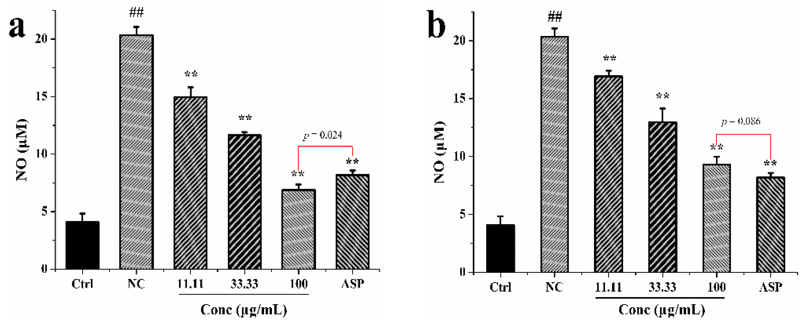
3.3. Anti-Inflammatory Activity

The anti-inflammatory activity was evaluated by determining the production of the nitric oxide (NO) in RAW 264.7 macrophage cells. As shown in Figure 1, the NO in the leaf and seed extracts treated groups was significantly lower than that of the NC group in a dose-dependent manner, within the concentration range investigated (11.11, 33.33, and 100 μg/mL). The leaf extracts at the concentration 100 μg/mL, gave lower NO production than the positive control aspirin (p = 0.024). In contrast, the seed extracts at the same concentration resulted in higher NO production than aspirin, but without significant difference (p = 0.086). Thus, it appeared that the leaf extracts of M. oleifera had a better anti-inflammatory potential than the seeds.

3.4. Analysis of M. oleifera Leaves, Seeds and Roots by HPLC-UV/ESI-MS/MS

Although the TFCs of three different parts of M. oleifera were determined and compared in our study, the phytochemical profiles or flavonoids profiles have not yet been revealed or compared so far. To further explore and explain the chemical differences in these three organs, LC-MS/MS analysis was conducted. As a result, the base peak chromatograms (BPC) of the extracts of M. oleifera leaves, seeds and roots with the LC-MS analysis were shown in Figure 2, where the peaks were numbered orderly according to their retention time. As per the analysis, a total of 17 compounds in three organs of the M. oleifera were identified or tentatively characterized in the ESI negative ion mode after comparing with the data from the references or purified standards. Table 2 summarized mass spectra data of all the compounds in extracts, which included saccharides, nitrile glycosides, organic acids, phenolic acids, phenylpropanoids, glucosinolates and flavonoids, and their chemical structures were presented in Figure 3. As shown in Figure 2 and Table 2, there were two shared constituents of peak 3 and peak 8 identified in all the three organs of M. oleifera. However, there were obvious differences in the metabolite profiles of the extracts from leaves, seeds and roots of M. oleifera, which could provide a good clue to explain their different antioxidant power, and the main phytochemicals responsible for it. It is also found the M. oleifera leaves are rich in flavonoids which may contribute largely to their better antioxidant and anti-inflammatory activities, while the seeds are not. In this sense, the present study for the first time conducted the phytochemical comparisons of three different organs of M. oleifera (leaves, seeds and roots) which could have a good correlation to their distinct antioxidant or anti-inflammatory activities, as shown in Table 1 and Figure 1.

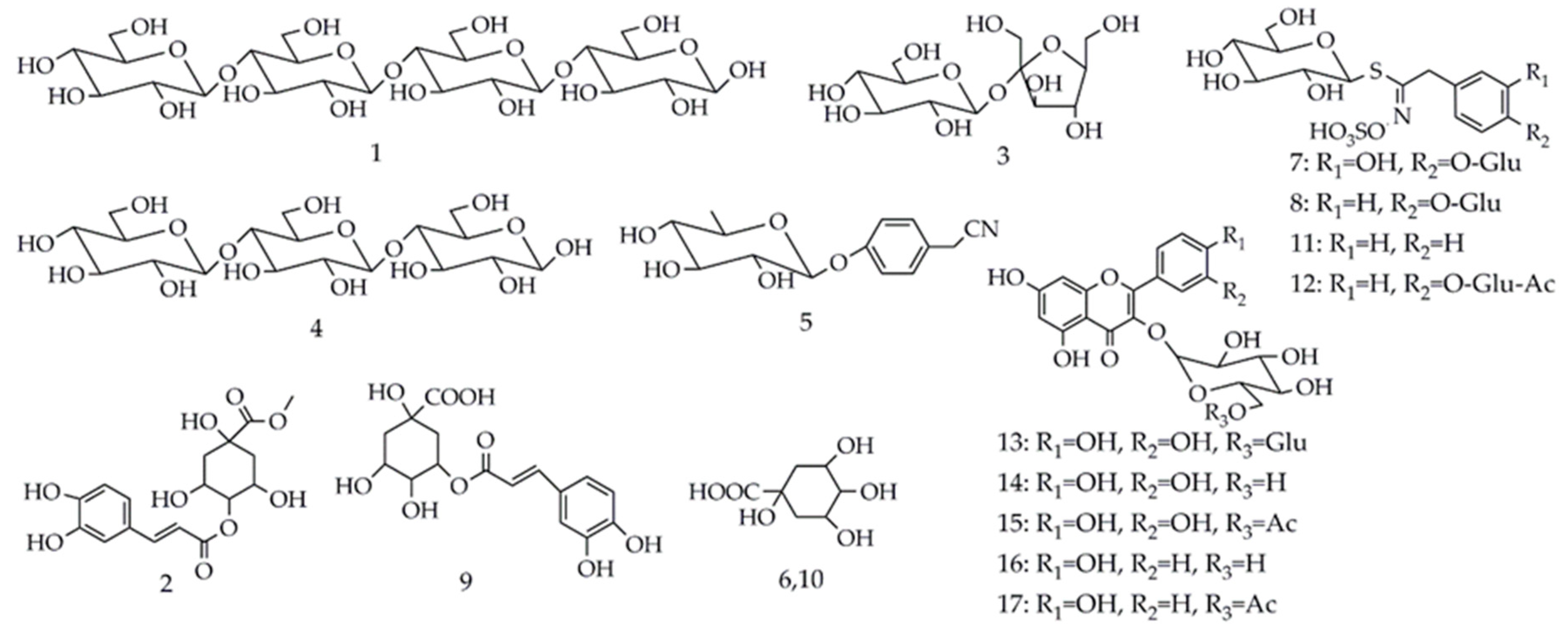
3.4.1. Saccharides and Nitrile Glycosides

As shown in Table 2, peak 1 had a negative molecular ion [M−H] at m/z 665 and peak 4 had a molecular ion [M−H] at m/z 503; according to the report by Peng et al. [38], they could be proposed as cellotetraose and cellotriose. Owing to the same product ions at m/z 179, 119, 113, 101, and 89, the molecular ion [M−H] for peak 3 at m/z 341 was identified as sucrose by comparing their mass spectra data with the previous literature [39]. Peak 5 showed the molecular ion [M−H] at m/z 278. Its main product ions at m/z 188 and 158 correspond to the [M−H−90] and [M−H−120], which are the characteristic diagnostic ions of glycone [40]. Besides, the fragment ion at m/z 116 was generated by the loss of the hexose moiety ([M−H−162]). Hence, peak 5 was temporarily proposed as niazirin [41].

3.4.2. Organic Acids and Phenylpropanoids

Both peaks 6 and 10 exhibited the same molecular ion [M−H] at m/z 191 with the same fragment ions at m/z 173, 127 and 85, suggesting that they were structural isomers. In addition, due to the product ions at m/z 173 ([M−H−18], loss of a H2O) and at m/z 127 ([M−H−64], loss of a H2O and HCOOH), peak 6 and 10 were identified as quinic acid isomer 1 and 2, respectively [42]. Peak 2 showed the molecular ion [M−H] at m/z 367 and its product ions at m/z 277 ([M−H−90]) and 205 ([M−H−162]), indicating the presence of a glycoside for peak 2. Thus, peak 2 was tentatively identified as methyl 4-caffeoylquinate [41].

3.4.3. Glucosinolates

By referring to the previously published report [9], four glucosinolates (peak 7, 8, 11, 12) were identified as 3-hydroxy-4-(α-l-rhamnopyranosyloxy) benzyl glucosinolate (peak 7), glucomoringin (peak 8), glucotropaeolin (peak 11) and acetyl-4-(α-l-rhamnopyranosyloxy) benzyl glucosinolate (peak 12), respectively. All of them shared common characteristic fragments, m/z 97 and 259. The specific characteristic ion at m/z 97 was assigned as the [SO4H] ion, and the major ion peak at m/z 259 ([SO4H+162]) was deduced as being sulfated glucose moiety.

3.4.4. Phenolic Acids and Flavonoids

Peak 9 was tentatively identified as 3-caffeoylquinic acid according to the MS/MS data reported by Clifford et al. [43]. In this study, 5 peaks (peak 13, 14, 15, 16 and 17) were identified as flavonoids. For peak 13, compared with the mass spectra data of rutin standard under the same condition of LC-MS, it was definitely identified as rutin. Peak 14 and 15 showed common product ions at m/z 301 and 300 due to the quercetin core, and m/z 463 corresponded to quercetin glycoside. Similarly, peak 16 and 17 shared common product ions at m/z 285 and 284 due to the kaempferol core, and m/z 447 corresponded to kaempferol glycoside. By comparing with the mass spectra data in previously reported literature, peak 14–17 were tentatively identified as quercetin 3-O-glucoside, quercetin-acetyl-glycoside, kaempferol 3-O-glucoside and kaempferol-acetyl-glycoside [2], respectively. In this study, there were no flavonoids identified in root of M. oleifera, but in the TFC assay, the TFC of root of M. oleifera was 106.79 ± 2.12 mg rutin equivalent (RE)/g, and the reason for this may be due to the presence of ortho-diphenolic hydroxyl analogues, such as quinic acid in the roots, since the ortho-diphenolic hydroxyl compounds can coordinate with aluminum ions, and thus influence the determination of TFC.

4. Conclusions

In this study, the antioxidant assays with DPPH, ABTS and FRAP, anti-inflammatory activity tests by means of the effects on the NO production, and phytochemical analysis with HPLC-UV/ESI-MS/MS were conducted to compare and correlate the antioxidant and anti-inflammatory activities with chemical compositions among the leaves, seeds and roots of M. oleifera in Kenya. To the best of our knowledge, this is the first study aiming to reveal the correlations between the phytochemical profiles and antioxidant and anti-inflammatory activities of three different organs of M. oleifera from Kenya. As a result, we revealed that leaves of M. oleifera are rich in flavonoids and phenolic acids, which explained the best potential antioxidant and anti-inflammatory activities among three organs. This current study revealed the positive correlation between the flavonoids content in three different organs of M. oleifera with their antioxidant and anti-inflammatory activities, which suggested that the leaves from M. oleifera could be a more potential natural source of antioxidants and anti-inflammatory source than that of other organs, and is very promising for development into health promoting dietary supplements now and in the near future.

Author Contributions

M.-Q.G. conceived of, designed and supervised the study. Y.-B.X. performed the experiments, analyzed the data and wrote the manuscript. G.-L.C. participated in the chemical assays and revised the manuscript for submission. All authors reviewed and approved the final manuscript.

Funding

This work was partly supported by the Major Project for Special Technology Innovation of Hubei Province (Grant No. 2017AHB054 to M. Guo). The funder played no roles in the study design, data collection and analysis, and decision to publish.

Conflicts of Interest

The authors declare no conflict of interest.

References

  1. Leone, A.; Spada, A.; Battezzati, A.; Schiraldi, A.; Aristil, J.; Bertoli, S. Cultivation, genetic, ethnopharmacology, phytochemistry and pharmacology of Moringa oleifera leaves: An overview. Int. J. Mol. Sci. 2015, 16, 12791–12835. [Google Scholar] [CrossRef] [PubMed]
  2. Rodríguez-Pérez, C.; Quirantes-Piné, R.; Fernández-Gutiérrez, A.; Carretero, A.S. Optimization of extraction method to obtain a phenolic compounds-rich extract from Moringa oleifera Lam leaves. Ind. Crop. Prod. 2015, 66, 246–254. [Google Scholar] [CrossRef]
  3. Gupta, S.; Jain, R.; Kachhwaha, S.; Kothari, S. Nutritional and medicinal applications of Moringa oleifera Lam.—Review of current status and future possibilities. J. Herb. Med. 2018, 11, 1–11. [Google Scholar] [CrossRef]
  4. Kasolo, J.N.; Bimenya, G.S.; Ojok, L.; Ochieng, J.; Ogwal-Okeng, J.W. Phytochemicals and uses of Moringa oleifera leaves in Ugandan rural communities. J. Med. Plants Res. 2010, 4, 753–757. [Google Scholar]
  5. Coppin, J.P.; Xu, Y.; Chen, H.; Pan, M.H.; Ho, C.T.; Juliani, R.; Simon, J.E.; Wu, Q. Determination of flavonoids by LC/MS and anti-inflammatory activity in Moringa oleifera. J. Funct. Foods 2013, 5, 1892–1899. [Google Scholar] [CrossRef]
  6. Panda, S.; Kar, A.; Sharma, P.; Sharma, A. Cardioprotective potential of N,α-l-rhamnopyranosyl vincosamide, an indole alkaloid, isolated from the leaves of Moringa oleifera in isoproterenol induced cardiotoxic rats: In vivo and in vitro studies. Bioorganic Med. Chem. Lett. 2013, 23, 959–962. [Google Scholar] [CrossRef] [PubMed]
  7. Mathur, M.; Yadav, S.; Katariya, P.K.; Kamal, R. In vitro propagation and biosynthesis of steroidal sapogenins from various morphogenetic stages of Moringa oleifera Lam., and their antioxidant potential. Acta Physiol. Plant 2014, 36, 1749–1762. [Google Scholar] [CrossRef]
  8. Roy, S.K.; Chandra, K.; Ghosh, K.; Mondal, S.; Maiti, D.; Ojha, A.K.; Das, D.; Mondal, S.; Chakraborty, I.; Islam, S.S. Structural investigation of a heteropolysaccharide isolated from the pods (fruits) of Moringa oleifera (Sajina). Carbohydr. Res. 2007, 342, 2380–2389. [Google Scholar] [CrossRef] [PubMed]
  9. Maldini, M.; Maksoud, S.A.; Natella, F.; Montoro, P.; Petretto, G.L.; Foddai, M.; De Nicola, G.R.; Chessa, M.; Pintore, G.A.M. Moringa oleifera: Study of phenolics and glucosinolates by mass spectrometry. J. Mass Spectrom. 2014, 49, 900–910. [Google Scholar] [CrossRef] [PubMed]
  10. Bhatta, R.; Saravanan, M.; Baruah, L.; Sampath, K.T. Nutrient content, in vitro ruminal fermentation characteristics and methane reduction potential of tropical tannin-containing leaves. J. Sci. Food Agric. 2012, 92, 2929–2935. [Google Scholar] [CrossRef] [PubMed]
  11. Coz-Bolaños, X.; Campos-Vega, R.; Reynoso-Camacho, R.; Ramos-Gómez, M.; Loarca-Piña, G.F.; Guzmán-Maldonado, S.H. Moringa infusion (Moringa oleifera) rich in phenolic compounds and high antioxidant capacity attenuate nitric oxide pro-inflammatory mediator in vitro. Ind. Crops Prod. 2018, 118, 95–101. [Google Scholar] [CrossRef]
  12. Sahakitpichan, P.; Mahidol, C.; Disadee, W.; Ruchirawat, S.; Kanchanapoom, T. Unusual glycosides of pyrrole alkaloid and 4′-hydroxyphenylethanamide from leaves of Moringa oleifera. Phytochemistry 2011, 72, 791–795. [Google Scholar] [CrossRef] [PubMed]
  13. Jung, I.L. Soluble extract from Moringa oleifera leaves with a new anticancer activity. PLoS ONE 2014, 9, e95492. [Google Scholar] [CrossRef] [PubMed]
  14. Verma, A.R.; Vijayakumar, M.; Mathela, C.S.; Rao, C.V. In vitro and in vivo antioxidant properties of different fractions of Moringa oleifera leaves. Food Chem. Toxicol. 2009, 47, 2196–2201. [Google Scholar] [CrossRef] [PubMed]
  15. Peixoto, J.R.O.; Silva, G.C.; Costa, R.A.; Fontenelle, J.R.L.D.S.; Vieira, G.H.F.; Filho, A.A.F.; Vieira, R.H.S.D.F. In vitro antibacterial effect of aqueous and ethanolic Moringa leaf extracts. Asian Pac. J. Trop. Med. 2011, 4, 201–204. [Google Scholar] [CrossRef]
  16. Atta, A.H.; Nasr, S.M.; Almaweri, A.H.; Sedky, D.; Mohamed, A.M.; Desouky, H.M.; Shalaby, M.A. Phytochemical, antioxidant and hepatoprotective effects of different fractions of Moringa oleifera leaves methanol extract against liver injury in animal model. Asian Pac. J. Trop. Med. 2018, 11, 423–429. [Google Scholar]
  17. Dangi, S.Y.; Jolly, C.I.; Narayanan, S. Antihypertensive activity of the total alkaloids from the leaves of Moringa oleifera. Pharm. Biol. 2008, 40, 144–148. [Google Scholar] [CrossRef]
  18. Helmy, S.A.; Morsy, N.F.; Elaby, S.M.; Ghaly, M.A. Hypolipidemic effect of Moringa oleifera Lam leaf powder and its extract in diet-induced hypercholesterolemic rats. J. Med. Food 2017, 20, 755–762. [Google Scholar] [CrossRef] [PubMed]
  19. Jaiswal, D.; Rai, P.K.; Kumar, A.; Mehta, S.; Watal, G. Effect of Moringa oleifera Lam. leaves aqueous extract therapy on hyperglycemic rats. J. Ethnopharmacol. 2009, 123, 392–396. [Google Scholar] [CrossRef] [PubMed]
  20. Singh, R.G.; Negi, P.S.; Radha, C. Phenolic composition, antioxidant and antimicrobial activities of free and bound phenolic extracts of Moringa oleifera seed flour. J. Funct. Foods 2013, 5, 1883–1891. [Google Scholar] [CrossRef]
  21. Al-Malki, A.L.; El Rabey, H.A. The antidiabetic effect of low doses of Moringa oleifera Lam. seeds on streptozotocin induced diabetes and diabetic nephropathy in male rats. BioMed Res. Int. 2015, 2015, 1–13. [Google Scholar] [CrossRef] [PubMed]
  22. Araújo, L.C.C.; Aguiar, J.S.; Napoleao, T.H.; Mota, F.V.B.; Barros, A.L.S.; Moura, M.C.; Coriolano, M.C.; Coelho, L.C.B.B.; Silva, T.G.; Paiva, P.M.G. Evaluation of cytotoxic and anti-Inflammatory activities of extracts and lectins from Moringa oleifera seeds. PLoS ONE 2013, 8, e81973. [Google Scholar] [CrossRef] [PubMed]
  23. Cui, C.; Chen, S.; Wang, X.; Yuan, G.; Jiang, F.; Chen, X.; Wang, L. Characterization of Moringa oleifera roots polysaccharide MRP-1 with anti-inflammatory effect. Int. J. Biol. Macromol. 2019, 132, 844–851. [Google Scholar] [CrossRef] [PubMed]
  24. Ghosh, A.; Bhattacharya, R.; Pradhan, C.; Chaudhuri, K.; Mukhopadhyay, A.; Bose, C.K. Antiproliferative effect of Moringa oleifera root extract on ovarian carcinoma: An in vitro study. Ann. Oncol. 2016, 27, 318. [Google Scholar] [CrossRef]
  25. Choudhary, M.K.; Bodakhe, S.H.; Gupta, S.K. Assessment of the antiulcer potential of Moringa oleifera root-bark extract in rats. J. Acupunct. Meridian Stud. 2013, 6, 214–220. [Google Scholar] [CrossRef]
  26. Shukla, S.; Mathur, R.; Prakash, A.O. Antifertility profile of the aqueous extract of Moringa oleifera roots. J. Ethnopharmacol. 1988, 22, 51–62. [Google Scholar] [CrossRef]
  27. Karadi, R.V.; Gadge, N.B.; Alagawadi, K.R.; Savadi, R.V. Effect of Moringa oleifera Lam. root-wood on ethylene glycol induced urolithiasis in rats. J. Ethnopharmacol. 2006, 105, 306–311. [Google Scholar] [CrossRef]
  28. Ndhlala, A.R.; Mulaudzi, R.; Ncube, B.; Abdelgadir, H.A.; du Plooy, C.P.; Van Staden, J. Antioxidant, antimicrobial and phytochemical variations in thirteen Moringa oleifera Lam. cultivars. Molecules 2014, 19, 10480–10494. [Google Scholar] [CrossRef]
  29. Siddhuraju, P.; Becker, K. Antioxidant properties of various solvent extracts of total phenolic constituents from three different agroclimatic origins of drumstick tree (Moringa oleifera Lam.) leaves. J. Agric. Food Chem. 2003, 51, 2144–2155. [Google Scholar] [CrossRef]
  30. Zhu, M.-Z.; Wu, W.; Jiao, L.L.; Yang, P.F.; Guo, M.Q. Analysis of flavonoids in lotus (Nelumbo nucifera) leaves and their antioxidant activity using macroporous resin chromatography coupled with LC-MS/MS and antioxidant biochemical assays. Molecules 2015, 20, 10553–10565. [Google Scholar] [CrossRef]
  31. Benzie, I.F.; Strain, J. The ferric reducing ability of plasma (FRAP) as a measure of “antioxidant power”: The FRAP assay. Anal. Biochem. 1996, 239, 70–76. [Google Scholar] [CrossRef] [PubMed]
  32. Zou, Y.; Chang, S.K.C.; Gu, Y.; Qian, S.Y. Antioxidant activity and phenolic compositions of lentil (Lens culinaris var. Morton) extract and its fractions. J. Agric. Food Chem. 2011, 59, 2268–2276. [Google Scholar] [CrossRef] [PubMed]
  33. Huang, C.; Li, W.; Zhang, Q.; Chen, L.; Chen, W.; Zhang, H.; Ni, Y. Anti-inflammatory activities of Guang-Pheretima extract in lipopolysaccharide-stimulated RAW 264.7 murine macrophages. BMC Complement. Altern. Med. 2018, 18, 46. [Google Scholar] [CrossRef] [PubMed]
  34. Zhu, M.; Liu, T.; Zhang, C.; Guo, M. Flavonoids of lotus (Nelumbo nucifera) seed embryos and their antioxidant potential. J. Food Sci. 2017, 82, 1834–1841. [Google Scholar] [CrossRef] [PubMed]
  35. Atawodi, S.E.; Atawodi, J.C.; Idakwo, G.A.; Pfundstein, B.; Haubner, R.; Wurtele, G.; Bartsch, H.; Owen, R.W. Evaluation of the polyphenol content and antioxidant properties of methanol extracts of the leaves, stem, and root barks of Moringa oleifera Lam. J. Med. Food 2010, 13, 710–716. [Google Scholar] [CrossRef] [PubMed]
  36. Nwidu, L.L.; Elmorsy, E.; Aprioku, J.S.; Siminialayi, I.; Carter, W.G. In vitro anti-cholinesterase and antioxidant activity of extracts of Moringa oleifera plants from Rivers State, Niger Delta, Nigeria. Medicines (Basel) 2018, 5, 71. [Google Scholar] [CrossRef] [PubMed]
  37. Iqbal, S.; Bhanger, M. Effect of season and production location on antioxidant activity of Moringa oleifera leaves grown in Pakistan. J. Food Compos. Anal. 2006, 19, 544–551. [Google Scholar] [CrossRef]
  38. Peng, H.; Lin, L.; Liu, Y.H.; Ruan, R.S.; Liu, C.M. Negative ion mode ESI-MS analysis of cellooligosaccharides. Food Sci. 2009, 30, 147–149. [Google Scholar]
  39. Jaja-Chimedza, A.; Graf, B.L.; Simmler, C.; Kim, Y.; Kühn, P.; Pauli, G.F.; Raskin, I. Biochemical characterization and anti-inflammatory properties of an isothiocyanate-enriched moringa (Moringa oleifera) seed extract. PLoS ONE 2017, 12, e0182658. [Google Scholar] [CrossRef] [PubMed]
  40. Chen, G.; Li, X.; Saleri, F.; Guo, M. Analysis of flavonoids in Rhamnus davurica and its antiproliferative activities. Molecules 2016, 21, 1275. [Google Scholar] [CrossRef] [PubMed]
  41. Li, F.H.; Wang, H.Q.; Su, X.M.; Li, C.K.; Li, B.M.; Chen, R.Y.; Kang, J. Constituents isolated from n-butanol extract of leaves of Moringa oleifera. China J. Chin. Mater. Med. 2018, 43, 114–118. [Google Scholar]
  42. Matei, M.F.; Jaiswal, R.; Bassil, B.S.; Kortz, U.; Kuhnert, N.; Deshpande, S. Synthesis, structure, and tandem mass spectrometric characterization of the diastereomers of quinic acid. J. Agric. Food Chem. 2016, 64, 7298–7306. [Google Scholar]
  43. Clifford, M.N.; Knight, S.; Kuhnert, N. Discriminating between the six isomers of dicaffeoylquinic acid by LC-MSn. J. Agric. Food Chem. 2005, 53, 3821–3832. [Google Scholar] [CrossRef] [PubMed]
Figure 1. Anti-inflammatory activities of leaves (a) and seeds (b) of M. oleifera on NO production in RAW264.7 cells. ** p < 0.01, compared with the NC (negative control) group.
Antioxidants 08 00296 g001
Figure 2. The base peak chromatogram (BPC) of M. oleifera leaves (A), seeds (B) and roots (C) extracts by LC-MS analysis in the negative ion mode.
Antioxidants 08 00296 g002
Figure 3. The molecular structures of all detected compounds in extracts of leaves, seeds and roots of M. oleifera. Glu: Glucoside; Ac: Acetyl.
Antioxidants 08 00296 g003
Table 1. Total flavonoids content and their corresponding antioxidant activity of M. oleifera.
SampleIC50 of DPPH * (mg/mL)IC50 of ABTS * (mg/mL)FRAP * (mM Fe2+/g)TFC * (mg RE/g)
Leaves1.87 ± 0.03 c1.36 ± 0.02 b0.99 ± 0.06 a192.36 ± 2.96 a
Seeds>64 a40.35 ± 1.47 a0.02 ± 0.00 c5.89 ± 0.65 c
Roots3.33 ± 0.11 b1.24 ± 0.03 b0.20 ± 0.01 b106.79 ± 2.12 b
Trolox0.10 ± 0.01 dNT *NTNT
Ascorbic acid0.05 ± 0.00 e0.11 ± 0.01 bNTNT
* All of the data were expressed as means ± standard deviation; DPPH: 2,2-diphenyl-1-picrylhydrazyl; ABTS: 2,2′-azinobis-(3-ethylbenzthiazoline-6-sulfonic acid); FRAP: Ferric-Reducing Antioxidant Power; TFC: Total flavonoids content; Half maximal inhibitory concentration (IC50) value was acquired when DPPH and ABTS radicals were inhibited by 50%; The FRAP value was represented as mM Fe2+/g of sample; Mean values with different letters (a–e) within a row were significantly different at a level of p < 0.05 by DMRT (Duncan’s multiple range test); NT: Not tested.
Table 2. Compounds identified from leaves, seeds and roots of M. oleifera by LC-MS/MS.
Peak No. aRT b (min)[M−H]Molecule FormulaMS2 (m/z)IdentificationPart cReference
Saccharides
12.83665C24H42O21485, 383, 341, 179CellotetraoseS[38]
32.88341C12H22O11179, 161, 131, 119, 113, 101, 89SucroseL, S, R[39]
42.90503C18H32O16383, 323, 281, 221, 179, 119, 89CellotrioseS, R[38]
Phenylpropanoids
22.87367C17H20O9277, 205, 187, 157, 113Methyl 4-caffeoylquinateR[41]
Nitrile Glycosides
52.95278C14H17NO5212, 188, 158, 116, 101NiazirinL[41]
Organic acids
63.20191C7H12O6173, 127, 111, 85Quinic acid isomer 1R[42]
1010.55191C7H12O6173, 127, 87, 85Quinic acid isomer 2L[42]
Glucosinolates
77.94586C20H29NO15S2440, 390, 344, 259, 198, 164, 973-Hydroxy-4-(α-l-rhamnopyranosyloxy) benzyl glucosinolateS[9]
88.44570C20H29NO14S2490, 328, 275, 259, 241, 97, 96GlucomoringinL, S, R[9]
1116.90408C14H19NO9S2259, 241, 215, 212, 195, 166, 97GlucotropaeolinR[9]
1220.20612C22H31NO15S2370, 275, 259, 241, 106, 97Acetyl-4-(α-l-rhamnopyranosyloxy) benzyl glucosinolateL[9]
Phenolic acids
910.44353C16H18O9191, 179, 1353-Caffeoylquinic acidL[43]
Flavonoids
1322.03609C27H30O16301, 300, 271RutinLstandard
1423.65463C21H20O12301, 300, 271Quercetin 3-O-glucosideL[2]
1526.47505C23H22O13301, 300, 271Quercetin-acetyl-glycosideL[2]
1626.61447C21H20O11285, 284, 255Kaempferol 3-O-glucosideL[2]
1729.75489C23H22O12285, 284, 255Kaempferol-acetyl-glycosideL[2]
a Peak number was ordered according to the retention time; b RT: retention time; c Part including L: leaf, S: seed, R: root.

