Review

Ulvan, a Polysaccharide from Macroalga Ulva sp.: A Review of Chemistry, Biological Activities and Potential for Food and Biomedical Applications

by
Faradila Ratu Cindana Mo’o
1,
Gofarana Wilar
2,
Hari Prasad Devkota
3 and
Nasrul Wathoni
1,4,*
1
Department of Pharmaceutics and Pharmaceutical Technology, Faculty of Pharmacy, Universitas Padjadjaran, Sumedang 45363, Indonesia
2
Department of Pharmacology and Clinical Pharmacy, Faculty of Pharmacy, Universitas Padjadjaran, Sumedang 45363, Indonesia
3
Graduate School of Pharmaceutical Sciences, Kumamoto University, 5-1 Oe-honmachi, Kumamoto 862-0973, Japan
4
West Java Development Institute, Universitas Padjadjaran, Sumedang 45363, Indonesia
*
Author to whom correspondence should be addressed.
Submission received: 7 July 2020 / Revised: 3 August 2020 / Accepted: 5 August 2020 / Published: 7 August 2020
(This article belongs to the Special Issue New Trends on Marine Biomaterials)

Abstract

:
The species of green macroalga belonging to the genus Ulva (family: Ulvaceae) are utilized in various fields, from food supplements to biomedical applications. Ulvan, a polysaccharide obtained from various Ulva species, has shown various biological activities, including antioxidant, anti-inflammatory, anticancer, antibacterial, and antiviral activities. To obtain the polysaccharide ulvan that can be utilized in various fields, it is necessary to understand the critical points that affect its physicochemical nature, the extraction procedures, and the mechanism of action for biological activities. This article discusses the physicochemical properties, extraction, isolation and characterization procedures and benefits in food and biomedical applications of ulvan. In conclusion, ulvan from Ulva sp. has the potential to be used as a therapeutic agent and also as an additional ingredient in the development of tissue engineering procedures.

Graphical Abstract

1. Introduction

The species of green macroalga belonging to genus Ulva (family: Ulvaceae) are utilized in various fields, from food supplements to biomedical applications. This marine plant is often developed as a food source of natural fiber, as an additional ingredient in the production of biomass fuels, and as a pharmaceutical supplement. Ulvan is a major polysaccharide that is present in Ulva sp. [1] Ulvan is reported to comprise from 9% to 36% of the dry weight biomass of the cells of Ulva sp. [2]. Lahaye et al. reported that the ulvan yield ranges from 8% to 29% of the dry weight of algae based on the extraction and purification methods [3]. It is built on two main repeating disaccharides, type A ulvanobiuronic acid 3-sulfate (A3S) and type B ulvanobiuronic acid 3-sulfate (B3S) [2,3,4,5,6,7,8]. Ulvan is widely used as a food additive and several studies have examined its therapeutic benefits [7,8,9]. New developments will aim to utilize ulvan from various Ulva species as an active ingredient or as an additive to deliver active ingredients in therapeutic preparations [10]. Several studies have indicated that ulvan has optimal physicochemical properties and active ingredients that can serve as therapeutic biological agents. There is a growing interest in research related to the extraction, purification, characterization, and food and biomedical application of ulvan polysaccharides. A Scopus search (www.scopus.com, accessed on 19 July 2020) with the term “Ulvan” resulted in 269 publications and most of them were published after 2010 (Figure 1). However, to obtain ulvan as a raw material, various procedures are needed for the extraction and physicochemical characterization. It is also essential to explore the potential of utilization of ulvan in the food and biomedical industry. Thus, the main aim of this article is to critically review the available scientific information related to the extraction, isolation, purification methods, characterization, pharmacokinetics, and biological activities of ulvan from Ulva species with particular focus on Ulva lactuca. It also explores the future potential of ulvan for food and biomedical applications.

2. Methodology

This review comprises literature obtained from Scopus, PubMed, and Google Scholar using the keywords “ulvan extraction from Ulva”, “ulvan isolation from Ulva”, “characterization ulvan from Ulva”, “cytotoxicity of ulvan from Ulva”, “antioxidant activity of ulvan”, “anti-inflammatory activity of ulvan from Ulva”, “anticancer activity of ulvan from Ulva”, etc.

3. Traditional Uses of Ulvan from Ulva sp.

In line with some beneficial flora, seaweed has been widely analyzed by the scientific community for the benefits of promoting their health. With increasing interest in nutraceuticals and functional foods, seaweed is progressively being considered, where they provide many functions ranging from simple nutrition improvement to complex physiological mechanisms. U. lactuca as green seaweed has been widely used as a food ingredient and nutraceutical agent because it contains high polysaccharides and natural fiber ingredients [11].
Ulva sp. as a nutraceutical agent can be utilized as food that shows beneficial physiological functions, improves well-being, and reduces the possibility of suffering from certain diseases, such as, inflammatory disorders [12], cancer [13], antibacterial [14], and virus infections [15,16]; and also act as an immunomodulating [17] and hypolipidemic agent [2,18]. It is widely believed that these functions are preventive rather than curative. Functional or nutraceutical foods are often obtained from traditional sources that are rich in bioactive agents such as Ulva sp. which are known for their health-promoting actions. Besides, new types of functional products originating from seaweed have recently been developed and studied extensively. Sometimes, these types of products are taken as whole foods or as dietary supplements, and most are sold as pills or tablets. It is recommended that seaweed contains many biologically active components, which can be used as therapeutic agents in food supplements [19]. Ulva sp. has become an ingredient that can be used to overcome daily life problems. For example, the use of U. linza as food and biomedical additives [20]. In other studies, U. intestinalis and U. rigida are widely used as therapeutic agents for anti-bacterial and anti-viral drugs [15].

4. Chemical Aspects of Ulvan from Ulva sp.

4.1. Chemistry and Physicochemical Properties
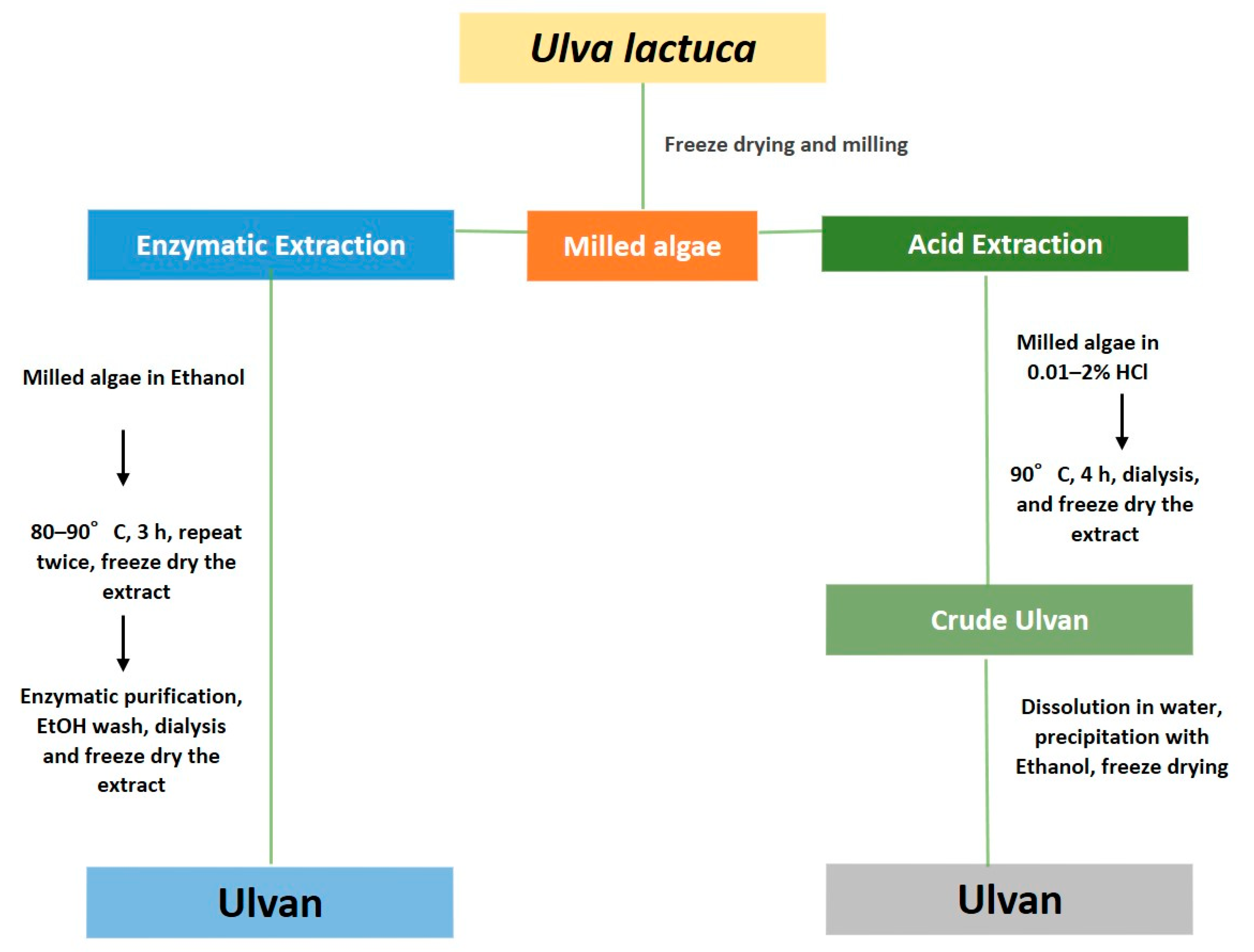
Ulvan mainly consists of cellulose, xyloglucan, and glucuroran with other various types of sugars. It has a low content of mannose, galactose, and arabinose [10,21]. Ulvan is mainly built on a repetitive sequence of disaccharides that consist of rhamnose sulfate, uronic acid, iduronic acid, or xylose. The two main repeating disaccharides are aldobiuronic acids, which are designated as type A ulvanobiuronic acid 3-sulfate (A3S) and type B ulvanobiuronic acid 3-sulfate (B3S) (Figure 2) [2,3,22]. In addition to the polysaccharide content, there are also other contents such as protein around 1–3% and fatty acids less than 1% [10,22,23]. The physicochemical properties of the active components contained in ulvan—hydration, polarity, swelling, and viscosity—are related to stability and physiological effects. These properties will depend on the extraction process.
Ulvans from U. pertusa and U. rigida show a multimodal distribution by gel permeation chromatography. In addition, the major fraction of ulvans are distributed as a single polydispersed population on gel permeation chromatography. The polymolecularity was due to linear xyloglucan structure of ulvans [24,25]. Ulvan extracted from U. lactuca contains polysaccharides that can be used as natural fibers or matrix; they can also be modified for other purposes, including as part of a composite [26]. Ulvan is also used as a substitute for Avicel, a polymer that contains cellulose. Ulvan has the same characteristics as Avicel in terms of thermal stability (degradation at 360–365 °C) [21]. Ulvan has a soluble fiber content of around 15.8% and an insoluble fiber content of 24.2%; the total fiber content is around 36.6–44.6% [27].
In a study conducted by Yaich et al., ulvan extracted using alcohol at pH 1–2 and a drastic temperature (80–90 °C) produced 20.37–23.60% uronic acid and 20.09–29.12% sugars. A reduction in molecular weight was influenced by a more acidic pH and altered extraction time [22,28,29]. The process of dissolving the fiber is influenced by pH: At pH 3, the insoluble fiber content is around 5.5%, while at pH 7, the insoluble fiber content is 7.5%. The pH also affects the zeta potential: the higher the extraction pH, the lower the zeta potential produced (with a range of −15 to −35) [10]. Ulvan has a low viscosity [22].
When analyzing the distribution of macromolecules using high performance size exclusion chromatography (HPSEC), there are two main distributions: population A, with a molecular weight of 7.21 × 10² kDa; and population B, with a molecular weight of 2.25 × 10² kDa. Some studies stated that the differences in molecular weight was due to selection of the extraction method and parameters [22,30]. Based on several parameters, ulvan content is markedly degraded at 360 °C [29].
Ulvan extracted has a moisture content > 90% [21]. Another study stated that a film with U. lactuca polymer is stable between −50 to 250 °C [10]. Yields resulting from acid extraction decrease when constant pH and temperature are used. Yields will increase with increasing extraction pH at a constant temperature [22]. The polysaccharides in ulvan have a strong ligand bond, a factor that may provide a therapeutic effect [31].
Ulvan can be employed in various fields, one of which is pharmaceutical formulation. Ulvan can be used as a polymer because of its high polysaccharide content [29]. In formulating ulvan to be used as a polymer in pharmaceutical products, the extraction procedures, pH, temperature, and choice of solvent(s) must be carefully considered to produce a good quality product that can serve an active ingredient.

4.2. Extraction

In general, there are two types of extraction: acid and enzyme. The quantity and quality of extracted ulvan are influenced by the extraction procedure and the selection of the solvent used for extraction. The extraction procedure has several parameters such as pH, temperature, and mechanical movements that determine the amount of obtained extract and metal content [32]. Acid extraction causes almost complete desulfation of isolated ulvan while purifying enzymatic methods maintain a significant SO3 substituent level [33]. Several Ulva sp. has been studied for their extraction methods in the last five years. For instance, to increase the purity of the ulvan, U. ohnoi is extracted at 90 °C with a pH of 2.2 [34]. To increase the purity of the ulvan from U. ohnoi, salt removal can be done by the ultrafiltration method [35]. In addition, ulvan from U. fasciata is extracted twice using distiled water and then doing the oxidation process for 72 h [36]. To improve the quantity of ulvan, extraction of ulvan from U. pertusa is carried out with hot water followed by alcohol precipitation [37]. The latest method is extraction with the one freeze-dried method [14]. In the extraction of ulvan from U. prolifera by enzyme method, to catalyze the initial depolymerization step of ulvan, ulvan lyase can be obtained from fermentation of Catenovulum sp. [38].
The choice of extraction method generally depends on the physicochemical properties of ulvan, which can interact with components of the cell wall when it comes in contact with a solvent [39,40]. The many active components contained in ulvan that can be obtained through the extraction process are polysaccharides (starch, cellulose, xyloglucan, and glucoronan); other components such as protein, lipids, minerals, and secondary metabolites can be obtained by co-extraction. This review specifically describes ulvan extraction from U. lactuca that provides a high yield, high selectivity, and low degradation [2].
Extraction with pure distilled water is the most widely used method [41,42]. This procedure provides ulvan that is glucuronic acid, glucose, arabinose, and xylose, with an average yield of 25–40% [22,33,43]. Another extraction method involves adding 0.05–1% hydrochloric acid (pH 1.5 to 2), with an average total yield of 15–45% [10,22,33]. Enzyme extraction utilizes onozula, pectinase macerozyme, α-amylase, and proteinase K, which are suspended in the buffer. The yield in enzyme extraction is 15–47% [10,33,44]. This yield has the potential to be improved because there has been no optimization. In addition to the three widely used methods, other methods include multilevel extraction, using water followed by NaCO3 and NaOH solvents, with a 3.14–6.50% yield. There is also a sequential extraction method that involves acidic solutions and ammonium oxalate with an average ulvan yield [45].
Various methods can be employed to extract ulvan depending on the active component that will be analyzed. Variations in concentration, pH, and temperature are important parameters to achieve the desired results. One study compared hot water, NaCO3, and NaOH and reported that the highest yield is obtained from hot water extraction [46]. The combination of extraction temperature, solvent pH, and extraction duration influences the yield and quality of extraction (e.g., purity and molecular integrity). The higher extraction temperatures allow the greater ulvan dissolution and the lower pH allows the higher ulvan selectivity. In addition, an increase in extraction duration will increase the yield of ulvan. However, critical care must be taken to protect the integrity of the ulvan structure. For example, stability may be decreased at high temperature, low pH, and longer extraction time. The time duration and the lower pH cause a significant depolymerization of ulvan. Therefore, based on the available literature data, the extraction conditions for a high extraction yield, a high selectivity, and a low degradation are at temperature 80–90 °C, pH 2–4.5, and 1–3 h duration. Figure 3 provides an outline of ulvan extraction from Ulva lactuca. It should be noted that the highest yield is obtained in the third stage—after purification.

4.3. Isolation, Purification, and Characterization

After the stages of the extraction process are carried out, to obtain crude ulvan and pure ulvan, isolation and purification steps are needed. In the isolation of the ulvan from the U. pertusa, the separation was carried out using a column chromatography equipped with a QFF column. The polysaccharides were eluted with a linear gradient of 0–2 M NaCl at 2.0 mL/min [16]. There is also isolation using ion column chromatography followed by a depolymerization process on the U. claratha [47]. In the purification step, fractionation process is carried out such as to obtain ulvan from U. lactuca and U. compressa [33]. The isolation of ulvan from U. ohnoi using the biorefinery method has been reported in other studies [48]. Ulvan isolation from U. lactuca is carried out to separate the desired active components after extraction using a variety of suitable solvents and considering its physicochemical properties.
Ulvan isolation separates the polysaccharide components, including uronic acid, sulfated molecules, and sugar components [49,50]. The isolation process can be carried out by dissolving the extract in water, centrifuging at 8000 rpm, and then separating on a diethylaminoethyl (DEAE)-Sepharose fast flow column (5 × 2 cm). It can then be fractionated using sulfuric acid: The higher the concentration of sulfuric acid used, the higher the sulfate content obtained [27]. One study utilized acetone for sulfate fractionation; the sulfate concentrations were 1.07–11.17% [51]. In another study, the sulfate content was 2.21%, in which there was a high content of free sulfate (54%) and total sugar content (15%) [52]. Isolation of sulfated polysaccharides is carried out with NaOH at 90 °C; there was 17.6% sulfate isolation [53]. The next process is to purify the active components in the multicomponent ulvan into a single component. This review will focus on the purification method and the number of active components obtained [2].
The isolation process will vary depending on a laboratory/factory capabilities and availability or raw material and resources. Extraction yields a crude extract that is purified using a vacuum to remove residual solvents [31]. Another study isolated the ulvan component using 2 N HCl solvent reflux in methanol for 2 h, followed by separating the solvent using vacuum chromatography, then purified the extract using preparative thin layer chromatography (TLC) [13]. Isolation can also be done with dimethyl sulfoxide (DMSO), while other studies have isolated bacteria from algae with the loop inoculation method [54,55]. Saccharide components, such as glucuronic acid oligosaccharides, have been isolated using acid precipitation after hot water extraction with sodium oxalate [55].
In practice, an optimal strategy for extraction, isolation, and purification procedures should consider the available resources so that the process can be selective and effective. It can also concentrate on evaporation, purification using dialysis, and isolation with precipitation. In resource-rich situations, selective acid extraction is ideal, although less selective extraction processes can be tolerated, with concentration and diafiltration with ultrafiltration. Purification can then be carried out using anion exchange chromatography, and lyophilization or spray drying to isolate pure ulvan.
Characterization determines the polysaccharide structure obtained from the extraction, purification, and isolation processes. As mentioned above, the ulvan polysaccharides have various monosaccharides, namely rhamnose, arabinose, xylose, mannose, glucose, and galactose [56].
Before advanced ulvan characterization, thermogravimetric analysis (TGA) is used to determine the percent of product loss due to extraction [57]. TGA can also reveal the effect of the extraction temperature on the quality of ulvan obtained. For the first stage of ulvan extraction, temperatures below 160 °C are associated with initial mass loss due to moisture volatilization [22]. Analysis of the structural content in ulvan is mostly done by a turbidimetric assay. Specific structural analyses employ chromatographic methods, such as high-performance liquid chromatography, coupled with conductivity or Fourier transform infrared (FTIR) spectroscopy [58,59]. FTIR spectra of dry cellulose fractions show peaks in the range of 400–600 cm−1 at room temperature [53,57]. There are absorption bands at 3412, 1645, 1251, 1080, and 852 cm−1 caused by hydroxyl groups, uronic acid, SO stretching (sulfate) vibrations, CO hand COC glycosidic linkages, and COS sulfate flexural vibrations in the axial and/or equatorial position (in C-2 or C-3 rhamnose), respectively [58,60].
Some monosaccharide contents have been analyzed using chromatographic techniques, namely gas chromatography (GC), high performance liquid chromatography (HPLC), and liquid chromatography (LC) [21,61]. Other studies have employed high performance anion exchange chromatography (HPAEC) and HPLC to determine the number of monosaccharides contained in ulvan [41,62]. HPAEC determines the carbohydrate composition and purity of glucose. Most of the carbohydrate content of ulvan is dominated by glucose and xylose [57]. HPAEC can selectively measure those monosaccharides and determine the quantity of acid and sugars present in ulvan without further derivatization [63]. GC has been used to measure volatile compounds contained in ulvan, such as alditol [41]. Bond structures, link positions, and substitution patterns may be analyzed with a two-dimensional nuclear magnetic resonance (2D NMR) instrument [41,57,64]. Signals at 65 and 63 ppm correspond to the carbon C-6 in crystals and amorphous regions in cellulose. Strong signals at 76 and 73 ppm correspond to the carbons C-2, C-3, and C-5 [57]. In addition to the uronic acid component, 2D NMR can analyze hydrolyzed saccharide fragments. Analysis of sulfate groups can be seen from the resonance of H and C atoms [22,55].
Molecular weight were analyzed by using size exclusion chromatography (SEC), which can measure the molecular weight of ulvan. SEC is also used to measure particle size distribution; it is performed during the final stages of endoglucanase purification. An SEC is equipped with a refractive index and ultraviolet (UV) detector [21,65]. X-ray diffraction can be used to determine the crystalline structure of the obtained ulvan. It typically displays four characteristic peaks of cellulose at 14.6°, 16.5°, 22.9° and 34.7° [66,67].
The important point of characterizing extracted ulvan is to pay close attention to the equipment components and the time required for analysis. For fast ulvan content analysis, a colorimetric assay approach can be performed to determine the sugar content, such as uronic acid, rhamnose, and xylose, as well as the protein content [41]. This colorimetric approach can also be used to quantitatively determine the sugar content [68]. To analyze the sulfate content, a turbidimetric assay method can be performed. The combination of two methods, namely chemometric assay and spectral analysis such as IR, NIR, and NMR, is very effective to quickly and accurately measure single-component polysaccharides [69].
The biological activity of ulvan can be identified from characterization of spectral analysis such as IR and NMR, which describe the structural and physicochemical properties of ulvan. Based on the published literature, sugar content analysis using colorimetric analysis, which provides fast and accurate results, can be combined with spectral analysis.

5. Current Food Applications, Available Formulation Products, and Future Potential as Food Ingredient

Ulvan polysaccharides in recent decades have been widely developed in the fields of food, pharmaceutical, and biomedical applications. The saccharide content that dominates in ulvan can be developed with promising prospects. Ulvan from U. fasciata was recently developed into an edible film from SRC. The SRC film was purified through exchange anion and gel permeation chromatography [70].
Recently, ulvan from U. lactuca was successfully investigated as a stabilizer and emulsifier in colloidal formulations to maintain functional agents for food and cosmetic applications based on their edible nature with amphiphilic properties [71]. In addition, ulvan can be used as a symbiotic yogurt or as prebiotics [72].
Ulvan can be used as polymer agents in pharmaceutical formulations. Polymers can be used as carriers, excipients in pharmaceutical formulations. It is known that the ulvan fiber polymer from U. lactuca is formulated in a film preparation with a plasticizer combination. Physicochemical properties and molecular characteristics, showing the existence of typical properties and behavior of self-aggregation molecules that are not easily found in other natural polymers. In the study of Guidara [10], proving the feasibility of ulvan is advantageous to be used as a film layer forming system, with a large scale suitable for various packaging applications. Besides, all films produced have demonstrated solubility, barrier, optical, and good mechanical properties, which are very important for biomedical, food, and packaging products [10,29].

6. Biomedical Applications

Ulvan has a variety of biological benefits that has the potential to be used as therapies for human health. Ulvan contributes a prominent amount of energy when consumed. Indeed, ulvan from Ulva sp. can be used as a source of metabolism in humans.
Ulvan can be utilized in the field of biomedical application; it is based on the benefits of ulvan therapy that have been sufficiently studied, as described in Table 1. The strong antioxidant activity in ulvan is able to make it a therapeutic agent [10,22,28,70] and in subsequent studies, the benefits of ulvan were investigated to have anti-inflammatory, anti-cancer, immunomodulatory, anti-hyperlipidemic, anti-bacterial, and anti-virus activities. Ulvan, which is composed most of polysaccharides, has the potential to be developed in biomedical formulations as polymers in hydrogel formulations [29]; this characteristic of ulvan polysaccharides is similar to that of alginate polymers [73]. Ulvan is also used in the field of tissue engineering for cartilage combined with poly (D-lactic acid) (PDLLA)[74].

6.1. Antioxidant Activity

Antioxidants can be obtained from animals and plants, including U. lactuca. This alga has an anti-radical capacity by inhibiting lipid peroxidation and increasing the activity of antioxidant enzymes. Studies have reported that the strong antioxidant activity of ulvan correlates to the degree substitution of sulfate groups along the polymeric backbone [106].
In 2019 and 2020, several sources of ulvan from Ulva sp were found to have an antioxidant effect; they include U. lactuca, U. australis, U. rigida, and U. ohnoi [48,107,108]. The antioxidant capacity of ulvan has been measured using various in vitro systems such as 2,2-diphenyl-2-picryl hydrazine (DPPH) radical scavenging, superoxide scavenging, ferric reducing antioxidant power (FRAP), hydroxyl radical scavenging, and lipid peroxidation inhibition assays. Sulfate content and molecular weight have been reported to play an important role in the antioxidant effects of ulvan from Ulva sp. [68,75,76,79,109].
The DPPH assay is the fastest method to measure antioxidant ability, compared to other methods that are also commonly used, namely reducing power and superoxide anion radical scavenging activity [76,102,107,108,110,111]. Notably, using more than one method to assess antioxidant activities increases the accuracy of the results.
Several studies have compared methods to determine which is more sensitive. Among DPPH, oxygen radical absorbance capacity (ORAC), FRAP, and β-carotene linoleic acid bleaching methods, ORAC was most effective for observing changes in peroxide formation [77,78]. The antioxidant power of ulvan has been compared with other molecules, such as BHA, BHT, and tocopherol. While the percent of ulvan inhibition (54.9%) was lower than BHA (73.20%) and BHT (69.40%), the differences were not statistically significant [112]. Based on research conducted for antioxidant testing, it is known that ulvan from Ulva sp has antioxidative ability, as evidenced by the comparison of several methods described above. Products from experimental animals have also been used to assess the antioxidant activity of ulvan, including erythrocyte-based and 2,2-azobis(2-amidinopropane)dihydrochloride (AAPH) assays [80,81]. Ulvan reduces ROS production by AAPH and attenuates lipid peroxidation, evaluated by thiobarbituric acid reactive substances (TBARS), according to erythrocyte-based assays [80]. Ulvan’s antioxidant mechanism involves sulfate and low-molecular-weight polysaccharides [31,83,113]. The latter inhibit cholinesterases and have neuroprotective potential [4]. Components of oligosaccharides can increase glutathione peroxidase (GSH), superoxide dismutase (SOD), catalase (CAT), telomerase, and total antioxidants and decrease the level of malondialdehyde [27,114]. For ulvan, the IC50 for radical activity is 623.58 µg/mL and the IC50 for superoxide anion scavenging is 785.48 µg/mL. Other components in U. lactuca with antioxidant activity are pigments (chlorophyll, carotenoids), essential oils, and low-molecular-weight polysaccharides [76].
The extraction procedure also affects the antioxidant effect produced by ulvan. This is related to the polarity of the solvent used to extract components that have antioxidant activity. The percentage of inhibition produced from methanol extract is higher than the percent inhibition of the water extract [115]. Furthermore, enzymatic extraction provides higher percent inhibition compared with acid extraction [22].
Besides in vitro antioxidant tests, animals can be exposed to radicals, such as thiacloprid, and then treated with an extract [5,6]. In hypercholesterolemia mice, ulvan reduces oxidative stress by increasing the activity of antioxidant enzymes (110% for CAT, 77% for GPx, and 23% for SOD) and the level of nonenzymatic antioxidants (GSH). These changes limit the consequences of damaging macromolecules by minimizing lipid peroxidation and protein oxidation [6]. Ulvan administration to mice exposed to extreme stress prevented abnormal lipid metabolism, modulated liver antioxidant defense systems, and reduced lipid peroxidation [116,117].

6.2. Anti-Inflammatory Activity

Inflammation is caused by the release of chemicals from tissues and cells that migrate throughout the organism. Associated inflammatory receptors are prostaglandin (PG), leukotrienes (LT5), histamine, bradykinin, platelet-activating factor (PAF), and interleukin-1 [118]. Ulvan has anti-inflammatory activity that has been tested in vitro in several animal species, including mice injected with radicals such as D-Gal (500 mg/kg) [82,83,84] and a mouse ear edema model of topical inflammation [85]. Vero cells have been employed to identify the anti-inflammatory effects caused by infection [98].
The ulvan anti-inflammatory mechanism is related to granuloma in the liver. Administration of sulfated polysaccharides for 24 weeks significantly reduces liver inflammation and necrosis and induces apoptosis. Sulfate polysaccharides increase the volume of liver lobules through mild sinusoid dilatation. The biological effects alter the response, stimulate the immune system via splenocytes and macrophages, and induce the release of anti-inflammatory cytokines that can suppress the growth of cancer cells [83]. Ulvan has the potential to reduce the chronic symptoms of inflammation [119]. Indeed, sulfated polysaccharide effectively reduces pro-inflammatory cytokine production in primary glial cells infected with various Japanese encephalitis virus (JEV) strains [98]. Ulvan can also reduce interferon-gamma (IFN-γ), tumor necrosis factor alpha (TNF-α), and interleukin 6 (IL-6), and elevates brain-derived neurotrophic factor (BDNF) and choline acetyltransferase (ChAT) levels as part of hippocampal neuronal protection [13,84]. Mice induced with D-Gal exhibit abnormal liver structure, characterized by hepatocellular necrosis and inflammation. Ulvan (100 mg/kg) attenuates this damage: The mice show partial recovery from tissue damage [82]. Ulvan exhibits vascular anti-inflammatory effects, with bradykinin as the primary target because it does not reduce histamine and serotonin-induced foot edema. Therefore, ulvan acts an on bradykinin pathway in antinociceptive and anti-inflammatory responses [52].

6.3. Cytotoxic and Anticancer Activity

New biomaterials with economic and environmental benefits are required. In this context, toxicity must be determined and considered when determining stability and effectiveness. Given that ulvan may be used as a supplement, nutraceutical, and therapeutic agent, it must be tested to determine potential cytotoxicity and therapeutic dose range that can be used. Cytotoxicity testing is carried out in vitro with cells or in vivo using experimental animals [89,103]. Recently, the anticancer effect of ulvan has been developed from U. lactuca, U. australis, U. rigida, and U. ohnoi [60,107,120]. The National Institutes of Health (USA) has developed standard methods for evaluating potential cytotoxic effects of compounds or extracts using human cancer cell lines [87]. These cell lines include HepG2 (hepatocellular carcinoma), MCF7 (human breast cancer), and HeLa (cervical cancer) [87]. Other toxicity tests use rat lung cells (L929) [89], mammalian L6 cells [65,90,121]. HaCaT keratinocytes, and 3T3 fibroblasts [80]. The most widely used method is the 3-(4,5-dimethylthiazol-2-yl)-5-(3-carboxymethoxyphenyl)-2-((4-sulfophenyl)-2H-tetrazolium (MTT) assay [80,122,123].
The results obtained from HepG2, MCF7, and HeLa cells have shown decreased cell viability directly proportional to the increased ulvan concentration [87]. Human L929 cells were still metabolically active and did not show a decrease in viability after 72 h exposure to ulvan [104]. Studies usually examine cellular metabolic activity using the MTT assay to determine cell number and assess proliferation by quantifying double-stranded DNA (dsDNA) and total protein [105]. Ulvan extracts have been found to be safe for mammalian L6 cells as a control because they do not promote cytotoxicity (half maximal inhibitory concentration [IC50] less than 90 mg/mL) even at the highest tested concentration. Ulvan did not show toxicity at 10,000 mg/mL in 3T3 cells [80]. Based on the description of the cytotoxic test that has been described, ulvan from U. lactuca decreases viability in cancer cells and does not show a decrease in viability in healthy cells.
Cancer occurs due to the abnormal growth of cells and body tissues. Cancer is initiated by various endogenous and exogenous factors that often cause oxidative damage to DNA, a process that produces mutations that disrupt the normal regulation of cell proliferation, differentiation, and apoptosis pathways [124]. Several studies have tested the anticancer activity [68,76], specifically for anti-breast cancer [13,60,87,88], anti-colon [59,88], and anti-cervical cancer activity [87], by investigating ulvan in toxicity and cell viability.
In vitro, ulvan has been tested on several cancer cell lines: HepG2 [31,60,87,88] Caco-2 (human colon cancer) [88], LS174 (human colon cancer), A549 (human lung carcinoma), Fem-x (malignant melanoma) K562 (chronic myelogenous leukemia) [76], HEp-2 (laryngeal epidermoid carcinoma), NCI-H292 (human lung mucoepidermoid carcinoma) [68], and HeLa (cervical cancer) [87]. It has also been tested in rats injected with carcinogens such as diethylnitrosamine (DENA, 200 mg/kg, intraperitoneally) [83] and 7,12-dimethylbenz[a]anthracene (DMBA) [13].
Testing the anticancer activity of ulvan has had different outcomes. Ulvan may be a chemopreventive agent for liver cancer. Sulfated polysaccharides in ulvan can inhibit hepatocellular carcinoma proliferation and induce apoptosis. Sulfated polysaccharides protect against DNEA-induced liver damage by reducing oxidative stress [83]. They also promote improvement in mice subjected to DMBA treatment by augmenting apoptosis, suppressing oxidative stress and inflammation, and enhancing the antioxidant defense system [13]. Ulvan showed weaker cytotoxic activity against cells A459 and LS174 (IC50 > 200 mg/mL), while it was more effective in preventing moderate cytotoxic effects in Fem-x and K562 cells (IC50 74.73 and 82.24 mg/mL, respectively) [76]. Ulvan prepared in nanoparticle albumin has antiproliferative potential in both MCF7 and HepG2 cells because it increased caspase-8 and caspase-9 levels; these changes indicate an induction of apoptosis [60]. Ulvan showed anticancer activity against MCF-7and HCT-116 cells with IC50 ranges from 21 to 99 μg/mL [88]; this seems to be related to sulfated polysaccharides that have strong ligand bonds [31]. Ulvan showed significant cytotoxic activity against hepatocellular carcinoma (IC50 29.67 ± 2.87 g/mL), human breast cancer (IC50 25.09 ± 1.36 g/mL), and cervical cancer (IC50 36.33 ± 3.84 g/mL) [87] Low-molecular-weight polysaccharides (<5000 Da)—usually oligosaccharides—can inhibit Caco-2 cell proliferation or differentiation program [60].

6.4. Antibacterial Activity

An antibacterial compound may be used to control the growth of harmful bacteria. This action aims to prevent the spread of disease and infection, eradicate the infected host, and prevent decay and destruction of a material by bacteria. Bacterial resistance to pharmaceutical drugs is increasing; hence, there is the urgent need to screen new drugs from natural resources. Seaweed from the marine ecosystem is an important source of bioactive compounds and one of the main subjects for screening of various pharmaceutical drugs [124,125]. Several plants have antibacterial activity, one of which is U. lactuca.
Several studies have examined the antibacterial potential of ulvan, with a range of results, including high, moderate, and weak potential. Ulvan has been tested for its antibacterial against a wide range of toxic bacteria (Table 1) [76,77,85,92,93,94,95,96,97]. Ulvan has shown a moderate percentage of inhibition against Staphylococcus sp. Streptococcus sp., and Bordetella sp. and a weak percent inhibition against Bacillus subtilis, Proteus sp., Salmonella sp., and Bacillus cereus, among others.
Endophytic bacteria isolated from U. lactuca, identified as strains of B. subtilis, are active against Staphylococcus aureus (1.6 μg/mL) and Enterococcus faecalis (0.2 μg/mL). In vivo testing of the strain Enterobacter cloacae is active against S. aureus and Klebsiella pneumonia at 0.4 μg/mL [97]. Several factors influence antibacterial activity, such as the extraction procedure, the solvent used, and the physiological conditions of U. lactuca itself. The antibacterial effect is also influenced by the harvest period; an antibacterial effect is particularly prominent when the plant enters the third harvest [93].
Antibacterial testing of ulvan and U. lactuca has produced variable results, with weak to moderate inhibition in some strains of bacteria. This variability may depend on internal factors such as the content of metabolites and endophytic bacteria present in the ulvan and external factors, namely the extraction procedure and harvest time.

6.5. Antiviral Activity

Viruses are obligate intracellular parasites; they require host cells for replication. Antiviral drugs can inhibit viral entry or exit or inhibit viral processes in cells. If the inhibitor is non-selective, host cell function can be disrupted and toxicity can occur. Ulvan has been tested against several viruses (Table 1) [98,99,100,101].
Further mechanical research has revealed that the sulfated polysaccharides from ulvan can block the absorption of the virus and therefore make the virus unable to enter the cell. Sulfated polysaccharides also effectively reduce pro-inflammatory cytokine production caused by JEV mixes that primarily infected glia cells. In animal studies, JEV-infected mice appear to have neurobehavioral abnormalities on day 5 and die on day 7 post-infection. However, treatment with sulfated polysaccharides can delay the onset of paralytic hind limbs and thus prevent mice from dying [98]. Ulvan mainly consists of heteroglycuronan, which has antiviral effects against influenza A/PR/8/34 (H1N1) [99]. In the polysaccharide delivery system, U. lactuca is made up as comprising monogalactosyldiacylglycerol (MGDG) tubular immunostimulating complexes (TI-complexes) because they are considered to be effective as lipid matrix for protein subunits in antigens [100,101].
Ulvan in some studies is considered to have antiviral activity because of its metabolic content in ulcers and polysaccharides. U. lactuca is considered to be a good lipid matrix in the transmission of anti-genes [98]. However, the mechanism of U. lactuca as an antiviral source is yet to be clarified.

6.6. Tissue Engineering

Research into the engineering of marine ulvan or marine polysaccharides or materials sourced from marine plants has been developed due to the nature of marine polysaccharides, namely biocompatibility, biodegradability, non-toxicity, low cost, and abundance. Most marine polysaccharides are derived from natural sources, such as fucoidan, alginate, carrageenan, agarose, porphyry, ulvan, ulcers, chitin, chitosan, and chitooligosaccharides [125,126,127].
U. lactuca represents a source of three-dimensional porous structure based on ulvan, which might be applied as a medical device, especially for tissue engineering applications [105]. Ulvan-based hydrogels can be used in cartilage tissue engineering [128]. Ulvan also contains polysaccharides that are similar to alginates, so it can be used as a hydrogel polymer [73]. Ulvan extracted from U. lactuca and chitosan extracted from Loligo forbesii squid-pen produced 98% carboxymethylated ulvan (CMU) and 87% carboxymethyl chitosan. These compounds are not cytotoxic and might represent suitable materials for in vitro and in vivo evaluation as tissue candidate [104]. Ulvan, combined with poly(D-lactic acid) (PDLLA), may be used to produce new scaffolding for bone tissue engineering applications [74].

6.7. Immunomodulating Activity

The immune system is a form of body defense in humans that is used to protect themselves from invading agents. Modulation of the immune system plays an essential role in health management and the mechanism of disease in humans. The importance of the immune system comes from the need to eliminate and modulate pathogenic and nonpathogenic microorganisms that can inhibit the body’s ability to maintain homeostasis [129]. Immune system enhancing agents can be obtained from natural materials such as seaweed. Ulva sp. has effectiveness as immunomodulating and the active ingredient is ulvan. Over the last five years, various Ulva species have been studied for effectiveness as immunomodulators, one of which is Ulva intestinalis. U. intestinalis has biochemical characteristics and immunomodulator activity. It is known from research that U. intestinalis shows immunomodulatory activity by stimulating the production of pro-inflammatory cytokines, including nitric oxide (NO), tumor necrosis factor-α (TNF-α), and interleukin-1β (IL-1β) in J774A macrophage cells [95]. Other studies confirm that ulvan from U. ohnoi has an immunomodulatory effect, which was tested in vitro. The in vitro immunomodulatory effect of the ulvan fraction was quantified by measuring the ability of the molecular level to mediate inflammation released from murine macrophages RAW 264.7 stimulated by LPS. All ulvan fractions showed no toxicity to RAW 264.7 cells at concentrations below 100 μg/mL for more than 48 h. Interleukin-10 and prostaglandin E2 exhibit anti-inflammatory effects by higher molecular weight ulvan fractions at 100 μg/mL [94]. Water-soluble sulfated polysaccharides extracted from U. intestinalis and fractionated using DEAE Sepharose’s fast flow column to identify molecular properties and stimulating activity of macrophage cells [130]. There are also effects of U. ohnoi on nutraceuticals and aquaculture, which have tested the immunomodulating effect on Senegalese sole (Solea senegalensis). The results of this study provide new evidence about the role of ulvan as a bioactive compound with immunomodulatory activity in Senegalese sole (Solea senegalensis). This is the first published study evaluating transcriptomic responses from Senegal that were injected with ulvan [14].

6.8. Antihyperlipidemic Activity

Hyperlipidemia is a disease with elevated blood lipid levels, including total cholesterol (TC), total glyceride (TG), and low-density lipoprotein cholesterol (LDL-C). In recent decades, polysaccharides are considered to have an antihyperlipidemic effect [127]. The antihyperlipidemic activity of one type of Ulva sp, namely U. pertusa, was investigated in vivo using rats to obtain a significant reduction in TC, TG, and LDL-C, and a significant increase in HDL-C; this is related to the presence of phosphorylation in the ulvan fraction [96]. Other in vivo studies tested the antihyperlipidemic activity of U. pertusa in mice induced by malondialdehyde (MDA), superoxide dismutase (SOD), catalase (CAT), and glutathione peroxidase (GSH-Px) in the liver. The results obtained are those that have the highest uronic acid content, highest sulfuric content, and low molecular weight have more potent activity [17]. U. australis is traditionally used as a food ingredient in Korea and to treat several diseases, such as hyperlipidemia and urinary tract diseases [107].

6.9. Anticoagulant Activity

Anticoagulants are the cornerstone of treatment for various thrombotic and thromboembolic disorders. General indications for anticoagulation include atrial fibrillation [131]. The risk of thrombosis continues to increase due to the increased incidence of higher chronic disease burden and old age. In some studies, ulvan showed anticoagulant activity. U. fasciata was studied using activated partial thromboplastin time (APTT) assay. These results indicate that the anticoagulant analysis of polycarboxyl ulvans depends on the high carboxyl and sulfate content with low molecular weight. In addition, it depends on the flexible conformation of the structure [36]. U. lactuca also has anticoagulant activity, which was analyzed in vitro using the APTT, prothrombin time (PT), and thrombin time (TT) and in vivo methods to reduce the weight of thrombus in mice possibly by association with anti-inhibiting anti factor Xa and IIa. These results provide strong evidence about the anticoagulant potential of sulfated polysaccharides isolated from U. lactuca [51].

7. Pharmacokinetic Studies

Molecules with pharmacological activity or as nutrients administered orally must be released from the drug delivery system, or as food must be absorbed from gastrointestinal mucosal epithelium and reach the target cell or tissue after entering the systemic circulation, and finally be excreted from the body intact or in metabolite form. Ulvan polysaccharides from U. lactuca are characterized by β (1→4) relationships and are not digestible by humans. Ulvan forms, known as dietary fiber, is digested through the small intestine without being metabolized and partially fermented by colon bacteria into short-chain fatty acids (SCFA). Ulvan is proven to be beneficial to humans because of its immunostimulatory effects and its ability to change human intestinal microbiomes or microbiota. Some are also found to have lower glycemic activity than other carbohydrate-rich vegetables. Ulvan belongs to the class of soluble dietary fiber. Because of the high intrinsic viscosity in water media, they can slow down the digestive process, reduce the bioavailability of minerals and other important nutrients by chelating, and can also increase the amount of Bifidobacteria and Lactobacillus in the cecum and large intestine, respectively. A comprehensive review of published data reveals that ulvan is not degraded in the human digestive system but is selectively absorbed in certain organs and tissues with no clear signs of toxicity in normal cells. The literature has no data about absorption, distribution, metabolism, and excretion, but reports of cytotoxicity studies are available. Several active carbohydrate enzymes have been identified that can effectively hydrolyze or reduce ulvan to oligomers [132,133,134].

8. Studies Related to Toxicity

As a condition for the development of food and supplements as well as the use as biomedical applications, ulvan must have low toxicity without special warning. A toxicity test needs to be carried out, taken into consideration in determining stability and effectiveness. Cytotoxicity testing was carried out in vitro on cells or in vivo directly on experimental animals [89], [103]. Toxicity tests are using rat lung cells (L929) [89] mammalian L6 cells [65,90,121], HaCaT keratinocytes, and 3T3 fibroblasts [80]. The most widely used method is MTT assay using 3-(4,5-dimethylthiazol-2-yl)-5-(3-carboxymethoxyphenyl)-2-((4-sulfophenyl)-2H-tetrazolium [80,122,123]. Cellular metabolic activity is usually assessed through MTS and cell number and proliferation tests by DNA and total protein quantification [105].
In a human cell test in L929 cells, after 72 h of contact, the ulvan was still metabolically active and did not show a decrease in viability [104]. Ulvan extraction results from Ulva sp. were found to be safe for mammalian L6 cells as a control because they did not have cytotoxicity (IC50 less than 90 mg/mL)l even at the highest level of concentration, the ulvan did not show toxicity at concentrations (10.000 mg/mL) in 3T3 cells [80]. Ulvan from U. ohnoi was tested on liver cells and it was found that ulvan has non-toxic properties [14].

9. Conclusions and Future Perspectives

Ulvan, a polysaccharide from green seaweeds of genus Ulva, is a natural fiber used as a food ingredient and has many benefits, as reported in several studies. The pharmacological activities of ulvan—antioxidant, anti-inflammatory, antibacterial, anticancer, antiviral, and cytotoxic—have been tested in vitro and in vivo. Ulvan can be used in the field of pharmaceutical formulation as a polymer for smart ulvan film and bone tissue engineering. Various research articles have reported that extracting and isolating ulvan requires several stages, methods, and approaches. Research on the early stages of ulvan extraction from U. lactuca has focused on how the solubility and structural properties of ulvan affect its function. In addition, the quantity and quality of ulvan depends on the extraction and isolation processes. The initial stage is polysaccharide extraction; the solvent, procedure temperature, pH, and duration greatly influence the yield and quality of the extract (i.e., purity and molecular integrity). Higher extraction temperatures allow more significant ulvan dissolution and lower pH to increase ulvan selectivity, and an increase in extraction duration can increase the yield. However, care must be taken to protect the integrity of the ulvan structure. For example, its stability can be decreased at a high temperature or with a combination of a high temperature, low pH, and long extraction duration. The duration causes significant depolymerization. However, a low pH significantly affects depolymerization. Overall, the literature data indicate the following conditions to obtain high yield and selectivity and low degradation: 80–90 °C, pH 2–4.5, and a 1–3 h duration.
Ulvan had shown various biological activities such as antioxidant, anti-inflammatory, anticancer, and immunomodulating activity, among others. Ulvan has also been studied as a promising source for tissue engineering such as the use of ulvan-based hydrogels in cartilage tissue engineering. Research on the biological activities and other health benefits of ulvan is still relatively limited, specially about the detailed mechanism of action using animal models, which should be research priority in future studies. Similarly, detailed clinical studies are also necessary regarding pharmacokinetics and safety-related studies along with the health beneficial activities.

Author Contributions

All authors conceived the idea, performed literature review, data collection, interpretation and manuscript preparation. All authors have read and agreed to the published version of the manuscript.

Funding

Ministry of Research and Higher Education, Republic of Indonesia, Penelitian Dasar Unggulan Perguruan grant (grant number 1827/UN6.3.1/LT/2020).

Acknowledgments

The authors thank the Minister of Research and Higher Education, Republic of Indonesia for the Penelitian Dasar Unggulan Perguruan grant (grant number 1827/UN6.3.1/LT/2020).

Conflicts of Interest

The authors declare no conflict of interest.

References

  1. Morelli, A.; Puppi, D.; Chiellini, F. Perspectives on Biomedical Applications of Ulvan; Elsevier Inc.: Amsterdam, The Netherlands, 2017; ISBN 9780128098172. [Google Scholar]
  2. Kidgell, J.T.; Magnusson, M.; de Nys, R.; Glasson, C.R.K. Ulvan: A systematic review of extraction, composition and function. Algal Res. 2019, 39, 101422. [Google Scholar] [CrossRef]
  3. Lahaye, M.; Robic, A. Structure and function properties of Ulvan, a polysaccharide from green seaweeds. Biomacromolecules 2007, 8, 1765–1774. [Google Scholar] [CrossRef]
  4. Olasehinde, T.A.; Mabinya, L.V.; Olaniran, A.O.; Okoh, A.I. Chemical characterization of sulfated polysaccharides from Gracilaria gracilis and Ulva lactuca and their radical scavenging, metal chelating, and cholinesterase inhibitory activities. Int. J. Food Prop. 2019, 22, 100–110. [Google Scholar] [CrossRef] [Green Version]
  5. Kammoun, I.; Sellem, I.; Ben Saad, H.; Boudawara, T.; Nasri, M.; Gharsallah, N.; Mallouli, L.; Amara, I. Ben Potential benefits of polysaccharides derived from marine alga Ulva lactuca against hepatotoxicity and nephrotoxicity induced by thiacloprid, an insecticide pollutant. Environ. Toxicol. 2019, 34, 1165–1176. [Google Scholar] [CrossRef] [PubMed]
  6. Kammoun, I.; Bkhairia, I.; Ben Abdallah, F.; Jaballi, I.; Ktari, N.; Boudawara, O.; Nasri, M.; Gharsallah, N.; Hakim, A.; Ben Amara, I. Potential protective effects of polysaccharide extracted from Ulva lactuca against male reprotoxicity induced by thiacloprid. Arch. Physiol. Biochem. 2017, 123, 334–343. [Google Scholar] [CrossRef] [PubMed]
  7. Chiellini, F.; Morelli, A. Ulvan: A Versatile Platform of Biomaterials from Renewable Resources. Biomater. Phys. Chem. 2011. [Google Scholar] [CrossRef] [Green Version]
  8. Lakshmi, D.S.; Sankaranarayanan, S.; Gajaria, T.K.; Li, G.; Kujawski, W.; Kujawa, J.; Navia, R. A short review on the valorization of green seaweeds and ulvan: Feedstock for chemicals and biomaterials. Biomolecules 2020, 10, 991. [Google Scholar] [CrossRef]
  9. Alves, A.; Sousa, R.A.; Reis, R.L. A practical perspective on ulvan extracted from green algae. J. Appl. Phycol. 2013, 25, 407–424. [Google Scholar] [CrossRef] [Green Version]
  10. Guidara, M.; Yaich, H.; Richel, A.; Blecker, C.; Boufi, S.; Attia, H.; Garna, H. Effects of extraction procedures and plasticizer concentration on the optical, thermal, structural and antioxidant properties of novel ulvan films. Int. J. Biol. Macromol. 2019, 135, 647–658. [Google Scholar] [CrossRef]
  11. Madhusudan, C.; Manoj, S.; Rahul, K.; Rishi, C.M. Seaweeds: A diet with nutritional, medicinal and industrial value. Res. J. Med. Plant 2011, 5, 153–157. [Google Scholar] [CrossRef] [Green Version]
  12. Fournière, M.; Latire, T.; Lang, M.; Terme, N.; Bourgougnon, N.; Bedoux, G. Production of active poly- and oligosaccharidic fractions from Ulva sp. by combining enzyme-assisted extraction (EAE) and depolymerization. Metabolites 2019, 9, 182. [Google Scholar] [CrossRef] [PubMed] [Green Version]
  13. Abd-Ellatef, G.E.F.; Ahmed, O.M.; Abdel-Reheim, E.S.; Abdel-Hamid, A.H.Z. Ulva lactuca polysaccharides prevent Wistar rat breast carcinogenesis through the augmentation of apoptosis, enhancement of antioxidant defense system, and suppression of inflammation. Breast Cancer Targets Ther. 2017, 9, 67–83. [Google Scholar]
  14. Ponce, M.; Zuasti, E.; Anguís, V.; Fernández-Díaz, C. Effects of the sulfated polysaccharide ulvan from Ulva ohnoi on the modulation of the immune response in Senegalese sole (Solea senegalensis). Fish Shellfish. Immunol. 2020, 100, 27–40. [Google Scholar] [CrossRef] [PubMed]
  15. Klongklaew, N.; Praiboon, J.; Tamtin, M.; Srisapoome, P. Antibacterial and antiviral activities of local Thai green macroalgae crude extracts in pacific white shrimp (Litopenaeus vannamei). Mar. Drugs 2020, 18, 140. [Google Scholar] [CrossRef] [PubMed] [Green Version]
  16. Chi, Y.; Zhang, M.; Wang, X.; Fu, X.; Guan, H.; Wang, P. Ulvan lyase assisted structural characterization of ulvan from Ulva pertusa and its antiviral activity against vesicular stomatitis virus. Int. J. Biol. Macromol. 2020, 157, 75–82. [Google Scholar] [CrossRef]
  17. Wang, L.; Wang, X.; Wu, H.; Liu, R. Overview on Biological Activities and Molecular Characteristics of Sulfated Polysaccharides from Marine Green Algae in Recent Years. Marine Drugs 2014, 12, 4984–5020. [Google Scholar] [CrossRef]
  18. Jiang, N.; Li, B.; Wang, X.; Xu, X.; Liu, X.; Li, W.; Chang, X.; Li, H.; Qi, H. The antioxidant and antihyperlipidemic activities of phosphorylated polysaccharide from Ulva pertusa. Int. J. Biol. Macromol. 2020, 145, 1059–1065. [Google Scholar] [CrossRef]
  19. Carvalho, A.F.U.; Portela, M.C.C.; Sousa, M.B.; Martins, F.S.; Rocha, F.C.; Farias, D.F.; Feitosa, J.P.A. Physiological and physico-chemical characterization of dietary fibre from the green seaweed Ulva fasciata Delile. Braz. J. Biol. 2009, 69, 969–977. [Google Scholar] [CrossRef] [Green Version]
  20. Sari-Chmayssem, N.; Taha, S.; Mawlawi, H.; Guégan, J.P.; Jeftić, J.; Benvegnu, T. Extracted ulvans from green algae Ulva linza of Lebanese origin and amphiphilic derivatives: Evaluation of their physico-chemical and rheological properties. J. Appl. Phycol. 2019, 31, 1931–1946. [Google Scholar] [CrossRef]
  21. Jmel, M.A.; Anders, N.; Ben Messaoud, G.; Marzouki, M.N.; Spiess, A.; Smaali, I. The stranded macroalga Ulva lactuca as a new alternative source of cellulose: Extraction, physicochemical and rheological characterization. J. Clean. Prod. 2019, 234, 1421–1427. [Google Scholar] [CrossRef]
  22. Yaich, H.; Amira, A.B.; Abbes, F.; Bouaziz, M.; Besbes, S.; Richel, A.; Blecker, C.; Attia, H.; Garna, H. Effect of extraction procedures on structural, thermal and antioxidant properties of ulvan from Ulva lactuca collected in Monastir coast. Int. J. Biol. Macromol. 2017, 105, 1430–1439. [Google Scholar] [CrossRef] [PubMed]
  23. Postma, P.R.; Cerezo-Chinarro, O.; Akkerman, R.J.; Olivieri, G.; Wijffels, R.H.; Brandenburg, W.A.; Eppink, M.H.M. Biorefinery of the macroalgae Ulva lactuca: Extraction of proteins and carbohydrates by mild disintegration. J. Appl. Phycol. 2018, 30, 1281–1293. [Google Scholar] [CrossRef] [PubMed] [Green Version]
  24. Kikionis, S.; Ioannou, E.; Toskas, G.; Roussis, V. Electrospun biocomposite nanofibers of ulvan/PCL and ulvan/PEO. J. Appl. Polym. Sci. 2015, 132, 42153. [Google Scholar] [CrossRef]
  25. Le, B.; Golokhvast, K.S.; Yang, S.H.; Sun, S. Optimization of microwave-assisted extraction of polysaccharides from Ulva pertusa and evaluation of their antioxidant activity. Antioxidants 2019, 8, 129. [Google Scholar] [CrossRef] [Green Version]
  26. Dumbrava, A.; Berger, D.; Matei, C.; Radu, M.D.; Gheorghe, E. Characterization and applications of a new composite material obtained by green synthesis, through deposition of zinc oxide onto calcium carbonate precipitated in green seaweeds extract. Ceram. Int. 2018, 44, 4931–4936. [Google Scholar] [CrossRef]
  27. Li, W.; Jiang, N.; Li, B.; Wan, M.; Chang, X.; Liu, H.; Zhang, L.; Yin, S.; Qi, H.; Liu, S. Antioxidant activity of purified ulvan in hyperlipidemic mice. Int. J. Biol. Macromol. 2018, 113, 971–975. [Google Scholar] [CrossRef]
  28. Yaich, H.; Garna, H.; Besbes, S.; Barthélemy, J.P.; Paquot, M.; Blecker, C.; Attia, H. Impact of extraction procedures on the chemical, rheological and textural properties of ulvan from Ulva lactuca of Tunisia coast. Food Hydrocoll. 2014, 40, 53–63. [Google Scholar] [CrossRef]
  29. Guidara, M.; Yaich, H.; Benelhadj, S.; Adjouman, Y.D.; Richel, A.; Blecker, C.; Sindic, M.; Boufi, S.; Attia, H.; Garna, H. Smart ulvan films responsive to stimuli of plasticizer and extraction condition in physico-chemical, optical, barrier and mechanical properties. Int. J. Biol. Macromol. 2020, 150, 714–726. [Google Scholar] [CrossRef]
  30. Yaich, H.; Garna, H.; Bchir, B.; Besbes, S.; Paquot, M.; Richel, A.; Blecker, C.; Attia, H. Chemical composition and functional properties of dietary fibre extracted by Englyst and Prosky methods from the alga Ulva lactuca collected in Tunisia. Algal Res. 2015, 9, 65–73. [Google Scholar] [CrossRef]
  31. El Azm, N.A.; Fleita, D.; Rifaat, D.; Mpingirika, E.Z.; Amleh, A.; El-Sayed, M.M.H. Production of bioactive compounds from the sulfated polysaccharides extracts of Ulva lactuca: Post-extraction enzymatic hydrolysis followed by ion-exchange chromatographic fractionation. Molecules 2019, 24, 2132. [Google Scholar] [CrossRef] [Green Version]
  32. Tsubaki, S.; Oono, K.; Hiraoka, M.; Onda, A.; Mitani, T. Microwave-assisted hydrothermal extraction of sulfated polysaccharides from Ulva spp. and Monostroma latissimum. Food Chem. 2016, 210, 311–316. [Google Scholar] [CrossRef] [PubMed]
  33. Wahlström, N.; Nylander, F.; Malmhäll-Bah, E.; Sjövold, K.; Edlund, U.; Westman, G.; Albers, E. Composition and structure of cell wall ulvans recovered from Ulva spp. along the Swedish west coast. Carbohydr. Polym. 2020, 233, 115852. [Google Scholar] [CrossRef] [PubMed]
  34. Glasson, C.R.K.; Donnet, L.; Angell, A.; Vucko, M.J.; Lorbeer, A.J.; Vamvounis, G.; de Nys, R.; Magnusson, M. Multiple response optimisation of the aqueous extraction of high quality ulvan from Ulva ohnoi. Bioresour. Technol. Rep. 2019, 7, 100262. [Google Scholar] [CrossRef]
  35. Magnusson, M.; Glasson, C.R.K.; Vucko, M.J.; Angell, A.; Neoh, T.L.; de Nys, R. Enrichment processes for the production of high-protein feed from the green seaweed Ulva ohnoi. Algal Res. 2019, 41, 101555. [Google Scholar] [CrossRef]
  36. De Carvalho, M.M.; Noseda, M.D.; Dallagnol, J.C.C.; Ferreira, L.G.; Ducatti, D.R.B.; Gonçalves, A.G.; de Freitas, R.A.; Duarte, M.E.R. Conformational analysis of ulvans from Ulva fasciata and their anticoagulant polycarboxylic derivatives. Int. J. Biol. Macromol. 2020, 162, 599–608. [Google Scholar] [CrossRef] [PubMed]
  37. Li, B.; Xu, H.; Wang, X.; Wan, Y.; Jiang, N.; Qi, H.; Liu, X. Antioxidant and antihyperlipidemic activities of high sulfate content purified polysaccharide from Ulva pertusa. Int. J. Biol. Macromol. 2020, 146, 756–762. [Google Scholar] [CrossRef] [PubMed]
  38. Qiao, L.; Yang, X.; Xie, R.; Du, C.; Chi, Y.; Zhang, J.; Wang, P. Efficient production of ulvan lyase from Ulva prolifera by Catenovulum sp. LP based on stage-controlled fermentation strategy. Algal Res. 2020, 46, 101812. [Google Scholar] [CrossRef]
  39. Fabrowska, J.; Ibañez, E.; Łęska, B.; Herrero, M. Supercritical fluid extraction as a tool to valorize underexploited freshwater green algae. Algal Res. 2016, 19, 237–245. [Google Scholar] [CrossRef]
  40. Gajaria, T.K.; Suthar, P.; Baghel, R.S.; Balar, N.B.; Sharnagat, P.; Mantri, V.A.; Reddy, C.R.K. Integration of protein extraction with a stream of byproducts from marine macroalgae: A model forms the basis for marine bioeconomy. Bioresour. Technol. 2017, 243, 867–873. [Google Scholar] [CrossRef]
  41. He, J.; Xu, Y.; Chen, H.; Sun, P. Extraction, structural characterization, and potential antioxidant activity of the polysaccharides from four seaweeds. Int. J. Mol. Sci. 2016, 17, 1988. [Google Scholar] [CrossRef]
  42. Robic, A.; Gaillard, C.; Sassi, J.F.; Leral, Y.; Lahaye, M. Ultrastructure of Ulvan: A polysaccharide from green seaweeds. Biopolymers 2009, 91, 652–664. [Google Scholar] [CrossRef] [PubMed]
  43. Harrysson, H.; Hayes, M.; Eimer, F.; Carlsson, N.G.; Toth, G.B.; Undeland, I. Production of protein extracts from Swedish red, green, and brown seaweeds, Porphyra umbilicalis Kützing, Ulva lactuca Linnaeus, and Saccharina latissima (Linnaeus) J. V. Lamouroux using three different methods. J. Appl. Phycol. 2018, 30, 3565–3580. [Google Scholar] [CrossRef] [Green Version]
  44. Traugott, H.; Zollmann, M.; Cohen, H.; Chemodanov, A.; Liberzon, A.; Golberg, A. Aeration and nitrogen modulated growth rate and chemical composition of green macroalgae Ulva sp. cultured in a photobioreactor. Algal Res. 2020, 47, 101808. [Google Scholar] [CrossRef]
  45. Mhatre, A.; Gore, S.; Mhatre, A.; Trivedi, N.; Sharma, M.; Pandit, R.; Anil, A.; Lali, A. Effect of multiple product extractions on bio-methane potential of marine macrophytic green alga Ulva lactuca. Renew. Energy 2019, 132, 742–751. [Google Scholar] [CrossRef]
  46. Mhatre, A.; Navale, M.; Trivedi, N.; Pandit, R.; Lali, A.M. Pilot scale flat panel photobioreactor system for mass production of Ulva lactuca (Chlorophyta). Bioresour. Technol. 2018, 249, 582–591. [Google Scholar] [CrossRef] [PubMed]
  47. Chi, Y.; Li, H.; Wang, P.; Du, C.; Ye, H.; Zuo, S.; Guan, H.; Wang, P. Structural characterization of ulvan extracted from Ulva clathrata assisted by an ulvan lyase. Carbohydr. Polym. 2020, 229, 115497. [Google Scholar] [CrossRef]
  48. Prabhu, M.S.; Israel, A.; Palatnik, R.R.; Zilberman, D.; Golberg, A. Integrated biorefinery process for sustainable fractionation of Ulva ohnoi (Chlorophyta): Process optimization and revenue analysis. J. Appl. Phycol. 2020. [Google Scholar] [CrossRef]
  49. Sushytskyi, L.; Lukáč, P.; Synytsya, A.; Bleha, R.; Rajsiglová, L.; Capek, P.; Pohl, R.; Vannucci, L.; Čopíková, J.; Kaštánek, P. Immunoactive polysaccharides produced by heterotrophic mutant of green microalga Chlorella vulgaris G11. Carbohydr. Polym. 2020, 246, 116588. [Google Scholar] [CrossRef]
  50. Pilatti, F.K.; Ramlov, F.; Schmidt, E.C.; Costa, C.; de Oliveira, E.R.; Bauer, C.M.; Rocha, M.; Bouzon, Z.L.; Maraschin, M. Metabolomics of Ulva lactuca Linnaeus (Chlorophyta) exposed to oil fuels: Fourier transform infrared spectroscopy and multivariate analysis as tools for metabolic fingerprint. Mar. Pollut. Bull. 2017, 114, 831–836. [Google Scholar] [CrossRef] [Green Version]
  51. Reis, S.E.; Andrade, R.G.C.; Accardo, C.M.; Maia, L.F.; Oliveira, L.F.C.; Nader, H.B.; Aguiar, J.A.K.; Medeiros, V.P. Influence of sulfated polysaccharides from Ulva lactuca L. upon Xa and IIa coagulation factors and on venous blood clot formation. Algal Res. 2020, 45, 101750. [Google Scholar] [CrossRef]
  52. De Araújo, I.W.; Rodrigues, J.A.; Quinderé, A.L.; Silva, J.D.; de Freitas Maciel, G.; Ribeiro, N.A.; Vanderlei, E.D.; Ribeiro, K.A.; Chaves, H.V.; Pereira, K.M.; et al. Analgesic and anti-inflammatory actions on bradykinin route of a polysulfated fraction from alga Ulva lactuca. Int. J. Biol. Macromol. 2016, 92, 820–830. [Google Scholar] [CrossRef] [PubMed]
  53. Tian, H.; Yin, X.; Zeng, Q.; Zhu, L.; Chen, J. Isolation, structure, and surfactant properties of polysaccharides from Ulva lactuca L. from South China Sea. Int. J. Biol. Macromol. 2015, 79, 577–582. [Google Scholar] [CrossRef]
  54. Kientz, B.; Thabard, M.; Cragg, S.M.; Pope, J.; Hellio, C. A new method for removing microflora from macroalgal surfaces: An important step for natural product discovery. Bot. Mar. 2011, 54, 457–469. [Google Scholar] [CrossRef] [Green Version]
  55. Redouan, E.; Cedric, D.; Emmanuel, P.; Mohamed, E.G.; Bernard, C.; Philippe, M.; Cherkaoui, E.M.; Josiane, C. Improved isolation of glucuronan from algae and the production of glucuronic acid oligosaccharides using a glucuronan lyase. Carbohydr. Res. 2009, 344, 1670–1675. [Google Scholar] [CrossRef] [PubMed]
  56. Yaich, H.; Garna, H.; Besbes, S.; Paquot, M.; Blecker, C.; Attia, H. Effect of extraction conditions on the yield and purity of ulvan extracted from Ulva lactuca. Food Hydrocoll. 2013, 31, 375–382. [Google Scholar] [CrossRef]
  57. Wahlström, N.; Edlund, U.; Pavia, H.; Toth, G.; Jaworski, A.; Pell, A.J.; Choong, F.X.; Shirani, H.; Nilsson, K.P.R.; Richter-Dahlfors, A. Cellulose from the green macroalgae Ulva lactuca: Isolation, characterization, optotracing, and production of cellulose nanofibrils. Cellulose 2020, 5, 1–19. [Google Scholar] [CrossRef] [Green Version]
  58. Barcellos, P.G.; Rodrigues, J.A.G.; De Queiroz, I.N.L.; De Araújo, I.W.F.; Benevides, N.M.B.; De Souza Mourão, P.A. Structural and physical-chemical analyses of sulfated polysaccharides from the sea lettuce Ulva lactuca and their effects on thrombin generation. Acta Sci. Biol. Sci. 2018, 40, e34916. [Google Scholar] [CrossRef] [Green Version]
  59. González-Ballesteros, N.; Rodríguez-Argüelles, M.C.; Prado-López, S.; Lastra, M.; Grimaldi, M.; Cavazza, A.; Nasi, L.; Salviati, G.; Bigi, F. Macroalgae to nanoparticles: Study of Ulva lactuca L. role in biosynthesis of gold and silver nanoparticles and of their cytotoxicity on colon cancer cell lines. Mater. Sci. Eng. C 2019, 97, 498–509. [Google Scholar] [CrossRef]
  60. Al-Malki, A.L. In vitro cytotoxicity and pro-apoptotic activity of phycocyanin nanoparticles from Ulva lactuca (Chlorophyta) algae. Saudi J. Biol. Sci. 2020, 27, 894–898. [Google Scholar] [CrossRef]
  61. Nair, D.; Vanuopadath, M.; Nair, B.G.; Pai, J.G.; Nair, S.S. Identification and characterization of a library of surfactins and fengycins from a marine endophytic Bacillus sp. J. Basic Microbiol. 2016, 56, 1159–1172. [Google Scholar] [CrossRef]
  62. Buré, C.; Cacas, J.L.; Mongrand, S.; Schmitter, J.M. Characterization of glycosyl inositol phosphoryl ceramides from plants and fungi by mass spectrometry. Anal. Bioanal. Chem. 2014, 406, 995–1010. [Google Scholar] [CrossRef] [PubMed]
  63. Harshvardhan, K.; Mishra, A.; Jha, B. Purification and characterization of cellulase from a marine Bacillus sp. H1666: A potential agent for single step saccharification of seaweed biomass. J. Mol. Catal. B Enzym. 2013, 93, 51–56. [Google Scholar] [CrossRef]
  64. Moschakis, T.; Biliaderis, C.G. Biopolymer-based coacervates: Structures, functionality and applications in food products. Curr. Opin. Colloid Interface Sci. 2017, 28, 96–109. [Google Scholar] [CrossRef]
  65. Costa, C.; Alves, A.; Pinto, P.R.; Sousa, R.A.; Borges Da Silva, E.A.; Reis, R.L.; Rodrigues, A.E. Characterization of ulvan extracts to assess the effect of different steps in the extraction procedure. Carbohydr. Polym. 2012, 88, 537–546. [Google Scholar] [CrossRef]
  66. Jmel, M.A.; Anders, N.; Ben Yahmed, N.; Marzouki, M.N.; Spiess, A.; Smaali, I. Efficient enzymatic saccharification of macroalgal biomass using a specific thermostable GH 12 endoglucanase from Aspergillus terreus JL1. World J. Microbiol. Biotechnol. 2020, 36, 5. [Google Scholar] [CrossRef] [PubMed]
  67. Ishwarya, R.; Vaseeharan, B.; Kalyani, S.; Banumathi, B.; Govindarajan, M.; Alharbi, N.S.; Kadaikunnan, S.; Al-anbr, M.N.; Khaled, J.M.; Benelli, G. Facile green synthesis of zinc oxide nanoparticles using Ulva lactuca seaweed extract and evaluation of their photocatalytic, antibiofilm and insecticidal activity. J. Photochem. Photobiol. B Biol. 2018, 178, 249–258. [Google Scholar] [CrossRef] [PubMed]
  68. Guedes, É.A.C.; da Silva, T.G.; Aguiar, J.S.; de Barros, L.D.; Pinotti, L.M.; Sant’Ana, A.E.G. Cytotoxic activity of marine algae against cancerous cells. Braz. J. Pharmacogn. 2013, 23, 668–673. [Google Scholar] [CrossRef] [Green Version]
  69. Kolsi, R.B.A.; Fakhfakh, J.; Krichen, F.; Jribi, I.; Chiarore, A.; Patti, F.P.; Blecker, C.; Allouche, N.; Belghith, H.; Belghith, K. Structural characterization and functional properties of antihypertensive Cymodocea nodosa sulfated polysaccharide. Carbohydr. Polym. 2016, 151, 511–522. [Google Scholar] [CrossRef]
  70. Ramu Ganesan, A.; Shanmugam, M.; Bhat, R. Producing novel edible films from semi refined carrageenan (SRC) and ulvan polysaccharides for potential food applications. Int. J. Biol. Macromol. 2018, 112, 1164–1170. [Google Scholar] [CrossRef]
  71. Morelli, A.; Massironi, A.; Puppi, D.; Creti, D.; Domingo Martinez, E.; Bonistalli, C.; Fabroni, C.; Morgenni, F.; Chiellini, F. Development of ulvan-based emulsions containing flavour and fragrances for food and cosmetic applications. Flavour Fragr. J. 2019, 34, 411–425. [Google Scholar] [CrossRef]
  72. Shalaby, M.S.; Amin, H.H. Potential Using of Ulvan Polysaccharide from Ulva lactuca as a Prebiotic in Synbiotic Yogurt Production. J. Probiotics Health 2019, 7, 1–9. [Google Scholar] [CrossRef] [Green Version]
  73. Wathoni, N.; Yuniarsih, N.; Cahyanto, A.; Muhctaridi, M. A-Mangostin Hydrogel Film Based Chitosan-Alginate for Recurrent Aphthous Stomatitis. Appl. Sci. 2019, 9, 5235. [Google Scholar] [CrossRef] [Green Version]
  74. Alves, A.; Duarte, A.R.C.; Mano, J.F.; Sousa, R.A.; Reis, R.L. PDLLA enriched with ulvan particles as a novel 3D porous scaffold targeted for bone engineering. J. Supercrit. Fluids 2012, 65, 32–38. [Google Scholar] [CrossRef]
  75. Kellogg, J.; Lila, M.A. Chemical and in vitro assessment of Alaskan coastal vegetation antioxidant capacity. J. Agric. Food Chem. 2013, 61, 11025–11032. [Google Scholar] [CrossRef]
  76. Kosanić, M.; Ranković, B.; Stanojković, T. Biological activities of two macroalgae from Adriatic coast of Montenegro. Saudi J. Biol. Sci. 2015, 22, 390–397. [Google Scholar] [CrossRef] [Green Version]
  77. Boisvert, C.; Beaulieu, L.; Bonnet, C.; Pelletier, É. Assessment of the antioxidant and antibacterial activities of three species of edible seaweeds. J. Food Biochem. 2015, 39, 377–387. [Google Scholar] [CrossRef]
  78. Bondu, S.; Bonnet, C.; Gaubert, J.; Deslandes, É.; Turgeon, S.L.; Beaulieu, L. Bioassay-guided fractionation approach for determination of protein precursors of proteolytic bioactive metabolites from macroalgae. J. Appl. Phycol. 2015, 27, 2059–2074. [Google Scholar] [CrossRef]
  79. Raja, R.; Hemaiswarya, S.; Arunkumar, K.; Carvalho, I.S. Antioxidant activity and lipid profile of three seaweeds of Faro, Portugal. Rev. Bras. Bot. 2016, 39, 9–17. [Google Scholar] [CrossRef]
  80. Botta, A.; Martínez, V.; Mitjans, M.; Balboa, E.; Conde, E.; Vinardell, M.P. Erythrocytes and cell line-based assays to evaluate the cytoprotective activity of antioxidant components obtained from natural sources. Toxicol. In Vitro 2014, 28, 120–124. [Google Scholar] [CrossRef] [Green Version]
  81. Hassan, S.; El-Twab, S.A.; Hetta, M.; Mahmoud, B. Improvement of lipid profile and antioxidant of hypercholesterolemic albino rats by polysaccharides extracted from the green alga Ulva lactuca Linnaeus. Saudi J. Biol. Sci. 2011, 18, 333–340. [Google Scholar] [CrossRef] [Green Version]
  82. Sathivel, A.; Raghavendran, H.R.B.; Srinivasan, P.; Devaki, T. Anti-peroxidative and anti-hyperlipidemic nature of Ulva lactuca crude polysaccharide on d-Galactosamine induced hepatitis in rats. Food Chem. Toxicol. 2008, 46, 3262–3267. [Google Scholar] [CrossRef] [PubMed]
  83. Hussein, U.K.; Mahmoud, H.M.; Farrag, A.G.; Bishayee, A. Chemoprevention of Diethylnitrosamine-Initiated and Phenobarbital-Promoted Hepatocarcinogenesis in Rats by Sulfated Polysaccharides and Aqueous Extract of Ulva lactuca. Integr. Cancer Ther. 2015, 14, 525–545. [Google Scholar] [CrossRef] [PubMed]
  84. Liu, X.Y.; Liu, D.; Lin, G.P.; Wu, Y.J.; Gao, L.Y.; Ai, C.; Huang, Y.-F.; Wang, M.F.; El-Seedi, H.R.; Chen, X.H.; et al. Anti-ageing and antioxidant effects of sulfate oligosaccharides from green algae Ulva lactuca and Enteromorpha prolifera in SAMP8 mice. Int. J. Biol. Macromol. 2019, 139, 342–351. [Google Scholar] [CrossRef] [PubMed]
  85. Awad, N.E. Biologically active steroid from the green alga Ulva lactuca. Phyther. Res. 2000, 14, 641–643. [Google Scholar] [CrossRef]
  86. Lin, H.C.; Tsai, W.S.; Chiu, T.H. Antioxidant properties of seven cultivated and natural edible seaweed extracts from Taiwan. J. Aquat. Food Prod. Technol. 2012, 21, 248–264. [Google Scholar] [CrossRef]
  87. Thanh, T.T.T.; Quach, T.M.T.; Nguyen, T.N.; Vu Luong, D.; Bui, M.L.; Tran, T.T. Van Structure and cytotoxic activity of ulvan extracted from green seaweed Ulva lactuca. Int. J. Biol. Macromol. 2016, 93, 695–702. [Google Scholar] [CrossRef]
  88. Arsianti, A.A.; Fadilah, F.; Fatmawaty, Y.; Wibisono, L.K.; Kusmardi, S.; Azizah, N.N.; Putrianingsih, R.; Murniasih, T.; Rasyid, A.; Pangestuti, R. Phytochemical composition and anticancer activity of seaweeds Ulva lactuca and Eucheuma cottonii against breast MCF-7 and colon HCT-116 cells. Asian J. Pharm. Clin. Res. 2016, 9, 115–119. [Google Scholar] [CrossRef]
  89. Alves, A.; Sousa, R.A.; Reis, R.L. In vitro cytotoxicity assessment of ulvan, a polysaccharide extracted from green algae. Phyther. Res. 2013, 27, 1143–1148. [Google Scholar] [CrossRef]
  90. Vonthron-Sénécheau, C.; Kaiser, M.; Devambez, I.; Vastel, A.; Mussio, I.; Rusig, A.M. Antiprotozoal activities of organic extracts from french marine seaweeds. Mar. Drugs 2011, 9, 922–933. [Google Scholar] [CrossRef] [Green Version]
  91. El Amrani Zerrifi, S.; Tazart, Z.; El Khalloufi, F.; Oudra, B.; Campos, A.; Vasconcelos, V. Potential control of toxic cyanobacteria blooms with Moroccan seaweed extracts. Environ. Sci. Pollut. Res. 2019, 26, 15218–15228. [Google Scholar] [CrossRef]
  92. Anjali, K.P.; Sangeetha, B.M.; Devi, G.; Raghunathan, R.; Dutta, S. Bioprospecting of seaweeds (Ulva lactuca and Stoechospermum marginatum): The compound characterization and functional applications in medicine—A comparative study. J. Photochem. Photobiol. B Biol. 2019, 200, 111622. [Google Scholar] [CrossRef] [PubMed]
  93. Deveau, A.M.; Miller-Hope, Z.; Lloyd, E.; Williams, B.S.; Bolduc, C.; Meader, J.M.; Weiss, F.; Burkholder, K.M. Antimicrobial activity of extracts from macroalgae Ulva lactuca against clinically important Staphylococci is impacted by lunar phase of macroalgae harvest. Lett. Appl. Microbiol. 2016, 62, 363–371. [Google Scholar] [CrossRef] [PubMed] [Green Version]
  94. Tan, S.P.; O’Sullivan, L.; Prieto, M.L.; Gardiner, G.E.; Lawlor, P.G.; Leonard, F.; Duggan, P.; McLoughlin, P.; Hughes, H. Extraction and bioautographic-guided separation of antibacterial compounds from Ulva lactuca. J. Appl. Phycol. 2012, 24, 513–523. [Google Scholar] [CrossRef]
  95. Sasikala, C.; Geetha Ramani, D. Comparative study on antimicrobial activity of seaweeds. Asian J. Pharm. Clin. Res. 2017, 10, 384–386. [Google Scholar]
  96. Mishra, A.K. Sargassum, Gracilaria and Ulva Exhibit Positive Antimicrobial Activity against Human Pathogens. OALib 2018, 5, 1–11. [Google Scholar] [CrossRef]
  97. Habbu, P.; Warad, V.; Shastri, R.; Savant, C.; Madagundi, S.; Kekare, P. In vitro and in vivo antimicrobial activity of Ulva lactuca Linn. (green algae) associated endophytic bacterial Strains. J. Appl. Pharm. Sci. 2016, 6, 138–146. [Google Scholar] [CrossRef] [Green Version]
  98. Chiu, Y.H.; Chan, Y.L.; Li, T.L.; Wu, C.J. Inhibition of Japanese Encephalitis Virus Infection by the Sulfated Polysaccharide Extracts from Ulva lactuca. Mar. Biotechnol. 2012, 14, 468–478. [Google Scholar] [CrossRef]
  99. Jiao, G.; Yu, G.; Wang, W.; Zhao, X.; Zhang, J.; Ewart, S.H. Properties of polysaccharides in several seaweeds from Atlantic Canada and their potential anti-influenza viral activities. J. Ocean Univ. China 2012, 11, 205–212. [Google Scholar] [CrossRef] [Green Version]
  100. Sanina, N.; Davydova, L.; Chopenko, N.; Kostetsky, E.; Shnyrov, V. Modulation of immunogenicity and conformation of HA1 subunit of influenza a virus H1/N1 hemagglutinin in tubular immunostimulating complexes. Int. J. Mol. Sci. 2017, 18, 1895. [Google Scholar] [CrossRef] [Green Version]
  101. Chopenko, N.; Mazeika, A.; Davydova, L.; Stenkova, A.; Leonova, G.; Kostetsky, E.; Sanina, N. Effectivity of nanovaccine against tick-borne encephalitis. J. Phys. Conf. Ser. 2018, 1092, 012020. [Google Scholar] [CrossRef]
  102. Kidgell, J.T.; Glasson, C.R.K.; Magnusson, M.; Vamvounis, G.; Sims, I.M.; Carnachan, S.M.; Hinkley, S.F.R.; Lopata, A.L.; de Nys, R.; Taki, A.C. The molecular weight of ulvan affects the in vitro inflammatory response of a murine macrophage. Int. J. Biol. Macromol. 2020, 150, 839–848. [Google Scholar] [CrossRef] [PubMed]
  103. Peasura, N.; Laohakunjit, N.; Kerdchoechuen, O.; Vongsawasdi, P.; Chao, L.K. Assessment of biochemical and immunomodulatory activity of sulphated polysaccharides from Ulva intestinalis. Int. J. Biol. Macromol. 2016, 91, 269–277. [Google Scholar] [CrossRef] [PubMed]
  104. Barros, A.A.A.; Alves, A.; Nunes, C.; Coimbra, M.A.; Pires, R.A.; Reis, R.L. Carboxymethylation of ulvan and chitosan and their use as polymeric components of bone cements. Acta Biomater. 2013, 9, 9086–9097. [Google Scholar] [CrossRef] [PubMed]
  105. Alves, A.; Sousa, R.A.; Reis, R.L. Processing of degradable ulvan 3D porous structures for biomedical applications. J. Biomed. Mater. Res. Part A 2013, 101, 998–1006. [Google Scholar] [CrossRef]
  106. Massironi, A.; Morelli, A.; Grassi, L.; Puppi, D.; Braccini, S.; Maisetta, G.; Esin, S.; Batoni, G.; Della Pina, C.; Chiellini, F. Ulvan as novel reducing and stabilizing agent from renewable algal biomass: Application to green synthesis of silver nanoparticles. Carbohydr. Polym. 2019, 203, 310–321. [Google Scholar] [CrossRef]
  107. Trentin, R.; Custódio, L.; Rodrigues, M.J.; Moschin, E.; Sciuto, K.; da Silva, J.P.; Moro, I. Exploring Ulva australis Areschoug for possible biotechnological applications: In vitro antioxidant and enzymatic inhibitory properties, and fatty acids contents. Algal Res. 2020, 50, 101980. [Google Scholar] [CrossRef]
  108. Fernandes, H.; Salgado, J.M.; Martins, N.; Peres, H.; Oliva-Teles, A.; Belo, I. Sequential bioprocessing of Ulva rigida to produce lignocellulolytic enzymes and to improve its nutritional value as aquaculture feed. Bioresour. Technol. 2019, 281, 277–285. [Google Scholar] [CrossRef] [Green Version]
  109. Del Olmo, A.; Picon, A.; Nuñez, M. The microbiota of eight species of dehydrated edible seaweeds from North West Spain. Food Microbiol. 2018, 70, 224–231. [Google Scholar] [CrossRef]
  110. Otero, P.; Quintana, S.E.; Reglero, G.; Fornari, T.; García-Risco, M.R. Pressurized Liquid Extraction (PLE) as an innovative green technology for the effective enrichment of galician algae extracts with high quality fatty acids and antimicrobial and antioxidant properties. Mar. Drugs 2018, 16, 156. [Google Scholar] [CrossRef] [Green Version]
  111. Fumanal, M.; Di Zeo, D.E.; Anguís, V.; Fernández-Diaz, C.; Alarcón, F.J.; Piñera, R.; Albaladejo-Riad, N.; Esteban, M.A.; Moriñigo, M.A.; Balebona, M.C. Inclusion of dietary Ulva ohnoi 5% modulates Solea senegalensis immune response during Photobacterium damselae subsp. piscicida infection. Fish Shellfish Immunol. 2020, 100, 186–197. [Google Scholar] [CrossRef]
  112. Abd El-Baky, H.H.; El-Baz, F.K.; El-Baroty, G.S. Natural preservative ingredient from marine alga Ulva lactuca L. Int. J. Food Sci. Technol. 2009, 44, 1688–1695. [Google Scholar] [CrossRef]
  113. Tariq, A.; Athar, M.; Ara, J.; Sultana, V.; Ehteshamul-Haque, S.; Ahmad, M. Biochemical evaluation of antioxidant activity in extracts and polysaccharide fractions of seaweeds. Glob. J. Environ. Sci. Manag. 2015, 1, 47–62. [Google Scholar]
  114. Wathoni, N.; Yuan Shan, C.; Yi Shan, W.; Rostinawati, T.; Indradi, R.B.; Pratiwi, R.; Muchtaridi, M. Characterization and antioxidant activity of pectin from Indonesian mangosteen (Garcinia mangostana L.) rind. Heliyon 2019, 5, e02299. [Google Scholar] [CrossRef] [PubMed]
  115. Al-Amoudi, O.A.; Mutawie, H.H.; Patel, A.V.; Blunden, G. Chemical composition and antioxidant activities of Jeddah corniche algae, Saudi Arabia. Saudi J. Biol. Sci. 2009, 16, 23–29. [Google Scholar] [CrossRef] [Green Version]
  116. Wang, R.; Paul, V.J.; Luesch, H. Seaweed extracts and unsaturated fatty acid constituents from the green alga Ulva lactuca as activators of the cytoprotective Nrf2-ARE pathway. Free Radic. Biol. Med. 2013, 57, 141–153. [Google Scholar] [CrossRef] [Green Version]
  117. Nunes, N.; Ferraz, S.; Valente, S.; Barreto, M.C.; Pinheiro de Carvalho, M.A.A. Biochemical composition, nutritional value, and antioxidant properties of seven seaweed species from the Madeira Archipelago. J. Appl. Phycol. 2017, 29, 2427–2437. [Google Scholar] [CrossRef]
  118. Vane, J.; Botting, R. Inflammation and the mechanism of action of anti-inflammatory drugs. FASEB J. 1987, 1, 89–96. [Google Scholar] [CrossRef]
  119. Zhu, C.; Zhang, S.; Song, C.; Zhang, Y.; Ling, Q.; Hoffmann, P.R.; Li, J.; Chen, T.; Zheng, W.; Huang, Z. Selenium nanoparticles decorated with Ulva lactuca polysaccharide potentially attenuate colitis by inhibiting NF-ΚB mediated hyper inflammation. J. Nanobiotechnol. 2017, 15, 1–15. [Google Scholar] [CrossRef] [Green Version]
  120. Sanjivkumar, M.; Chandran, M.N.; Suganya, A.M.; Immanuel, G. Investigation on bio-properties and in-vivo antioxidant potential of carrageenans against alloxan induced oxidative stress in Wistar albino rats. Int. J. Biol. Macromol. 2020, 151, 650–662. [Google Scholar] [CrossRef]
  121. Spavieri, J.; Allmendinger, A.; Kaiser, M.; Itoe, M.A.; Blunden, G.; Mota, M.M.; Tasdemir, D. Assessment of dual life stage antiplasmodial activity of British seaweeds. Mar. Drugs 2013, 11, 4019–4034. [Google Scholar] [CrossRef] [Green Version]
  122. Estevinho, B.N.; Rocha, F.; Santos, L.; Alves, A. Microencapsulation with chitosan by spray drying for industry applications—A review. Trends Food Sci. Technol. 2013, 31, 138–155. [Google Scholar] [CrossRef]
  123. Van Tran, T.T.; Huy, B.T.; Truong, H.B.; Bui, M.L.; Thanh, T.T.T.; Dao, D.Q. Structure analysis of sulfated polysaccharides extracted from green seaweed Ulva lactuca: Experimental and density functional theory studies. Mon. Chem. Chem. Mon. 2018, 149, 197–205. [Google Scholar] [CrossRef]
  124. Valko, M.; Izakovic, M.; Mazur, M.; Rhodes, C.J.; Telser, J. Role of oxygen radicals in DNA damage and cancer incidence. Mol. Cell. Biochem. 2004, 266, 37–56. [Google Scholar] [CrossRef] [PubMed]
  125. Mahady, G. Medicinal Plants for the Prevention and Treatment of Bacterial Infections. Curr. Pharm. Des. 2005, 11, 2405–2427. [Google Scholar] [CrossRef]
  126. Manivasagan, P.; Oh, J. Marine polysaccharide-based nanomaterials as a novel source of nanobiotechnological applications. Int. J. Biol. Macromol. 2016, 82, 315–327. [Google Scholar] [CrossRef]
  127. Venkatesan, J.; Lowe, B.; Anil, S.; Manivasagan, P.; Kheraif, A.A.A.; Kang, K.H.; Kim, S.K. Seaweed polysaccharides and their potential biomedical applications. Starch Staerke 2015, 67, 381–390. [Google Scholar] [CrossRef]
  128. Popa, E.G.; Reis, R.L.; Gomes, M.E. Seaweed polysaccharide-based hydrogels used for the regeneration of articular cartilage. Crit. Rev. Biotechnol. 2015, 35, 410–424. [Google Scholar] [CrossRef] [Green Version]
  129. Nader, N.D. Immunomodulation Mechanisms in Disease and in the Surgical Patient. Immunol. Investig. 2017, 46, 765–768. [Google Scholar] [CrossRef] [Green Version]
  130. Tabarsa, M.; You, S.G.; Dabaghian, E.H.; Surayot, U. Water-soluble polysaccharides from Ulva intestinalis: Molecular properties, structural elucidation and immunomodulatory activities. J. Food Drug Anal. 2018, 26, 599–608. [Google Scholar] [CrossRef]
  131. Tomaselli, G.F.; Mahaffey, K.W.; Cuker, A.; Dobesh, P.P.; Doherty, J.U.; Eikelboom, J.W.; Florido, R.; Hucker, W.; Mehran, R.; Messé, S.R.; et al. 2017 ACC Expert Consensus Decision Pathway on Management of Bleeding in Patients on Oral Anticoagulants. J. Am. Coll. Cardiol. 2017, 70, 3042–3067. [Google Scholar] [CrossRef]
  132. Taboada, C.; Millán, R.; Míguez, I. Composition, nutritional aspects and effect on serum parameters of marine algae Ulva rigida. J. Sci. Food Agric. 2010, 90, 445–449. [Google Scholar] [PubMed]
  133. De Jesus Raposo, M.F.; De Morais, A.M.M.B.; De Morais, R.M.S.C. Emergent sources of prebiotics: Seaweeds and microalgae. Mar. Drugs 2016, 14, 27. [Google Scholar] [CrossRef] [PubMed]
  134. Hemsworth, G.R.; Déjean, G.; Davies, G.J.; Brumer, H. Learning from microbial strategies for polysaccharide degradation. Biochem. Soc. Trans. 2016, 44, 94–108. [Google Scholar] [CrossRef] [PubMed]
Figure 1. Number of publications on Ulvan from 2010. (Source: www.scopus.com, keyword: Ulvan, accessed on 19 July 2020).
Applsci 10 05488 g001
Figure 2. Structures of two main repeating disaccharides, type A ulvanobiuronic acid 3-sulfate (A3S) having repeating β-D-glucuronic acid (1→4)-α-L-rhamnose-3-sulphate units, and type B ulvanobiuronic acid 3-sulfate (B3S) having repeating α-L-iduronic acid (1→4)-α-L-rhamnose-3-sulphate units.
Applsci 10 05488 g002
Figure 3. Schematic flowchart of the ulvan extraction method from Ulva lactuca.
Applsci 10 05488 g003
Table 1. Test objects used to determine the biological activities of ulvan.
Biomedical ApplicationsTest ObjectTest TypeRef
Antioxidant2,2-Diphenyl-1-picrylhydrazyl (DPPH) radical scavengingin vitro[68,75,76]
Oxygen radical absorbance capacity (ORAC)in vitro[77,78]
Ferric reducing antioxidant power assays; β-carotene linoleic acid bleaching methodsin vitro[75,79]
Erythrocyte-based assaysin vitro[80,81]
AAPH assaysin vitro
Nitric oxidein vitro[75]
Cytoprotection against H2O2-induced damage in yeast cells and zebrafish embryosin vitro[76]
Anti-inflammatoryMice injected with radicals such as D-Gal (500 mg/kg)in vivo[82,83,84]
Topical mice ear edema testin vivo[85]
Use of Vero cells (anti-inflammatory effect due to infection)in vitro[86]
AnticancerHepG2 cellsin vitro[31,60,87]
MCF-7 cellsin vitro[31,60,88]
Non-cancerous baby hamster kidney (BHK) cellsin vitro[60]
Caco-2 cells (human colon cancer)in vitro[88]
LS174 cells (human colon carcinoma)in vitro[76]
A549 cells (human lung carcinoma)in vitro
Fem-x cells (malignant melanoma)in vitro
K562 cells (chronic myelogenous leukemia)in vitro
NCI-H292 cells (human lung mucoepidermoid carcinoma)in vitro[68]
HeLa cellsin vitro[87]
L929 cells (mouse lung)in vitro[89]
Mammalian L6 cellsin vivo[90]
Keratinocytesin vivo[80]
3T3 fibroblasts
AntibacterialMicrocystis aeruginosa (cyanobacterium)in vitro[91]
Escherichia coli, followed by Klebsiella pneumonia, and Salmonella typhiin vivo[92]
Methicillin-resistant Staphylococcus aureus (MRSA)in vitro[93]
Micrococcus luteusin vitro[77]
Brochothrix thermosphactain vitro
E. coliin vitro
Bacillus subtilisin vitro[94]
Streptococcus lactisin vitro[95]
Pseudomonas putidain vitro[96]
Mycobacterium tuberculosis H37 RV strainin vitro[97]
AntiviralJapanese encephalitis virusin vivo[98]
Influenza virusin vivo[99]
HA1 subunit of influenza A H1/N1virusin vivo[100]
the matrix of the encephalitis antigenin vitro[101]
Immunomodulatingmakrofag murine RAW264.7in vitro[102]
Senegalese solein vivo[14]
sel makrofag J774A.1in vitro[103]
AntihyperlipidemicMice induced malondialdehyde (MDA), superoxide dismutase (SOD), catalase (CAT), and glutathione peroxidase (GSH-Px)in vitro[18,37]
AnticoagulantActivated partial thromboplastin time (APTT), prothrombin time (PT), and thrombin time (TT)in vitro[51]
Micein vivo[36]
Tissue engineeringulvan can be used in cartilage tissue engineeringin vitro and in vivo[74,104,105]

