Article

Macrophage Accumulation and Angiogenesis in Epicardial Adipose Tissue in Cardiac Patients with or without Chronic Heart Failure

1
Department of Pathology, Institute of Cardiovascular Diseases, 50, Carol I Avenue, 700503 Iasi, Romania
2
Department of Morpho-Functional Sciences I, University of Medicine and Pharmacy, 16, Universitatii Street, 700115 Iasi, Romania
3
Department of Morpho-Functional Sciences II, University of Medicine and Pharmacy, 16, Universitatii Street, 70011 Iasi, Romania
4
Department of Surgery, University of Medicine and Pharmacy, 16, Universitatii Street, 700115 Iasi, Romania
5
Department of Oral and Maxillofacial Surgery, University of Medicine and Pharmacy, 16, Universitatii Street, 700115 Iasi, Romania
6
Department of Internal Medicine, University of Medicine and Pharmacy, 16, Universitatii Street, 700115 Iasi, Romania
7
Department of Cardiology, Institute of Cardiovascular Diseases, 50, Carol I Avenue, 700503 Iasi, Romania
8
Department of Mother and Child Medicine, University of Medicine and Pharmacy, 16, Universitatii Street, 700115 Iasi, Romania
*
Authors to whom correspondence should be addressed.
Submission received: 10 July 2020 / Revised: 15 August 2020 / Accepted: 19 August 2020 / Published: 25 August 2020
(This article belongs to the Section Applied Biosciences and Bioengineering)

Abstract

:

Featured Application

This study, performed on surgical cardiac biopsies, is important because it reveals that the epicardial adipose tissue (EAT) thickness measurement, routinely performed by imaging cardiac diagnosis, cannot be easily interpreted, as the EAT thickness is subject to changes made by inflammation and angiogenesis.

Abstract

Routinely measuring epicardial fat had become a novel tool for cardiovascular risk stratification. Structural changes in epicardial adipose tissue (EAT), including fat thickness, inflammation, and angiogenesis, have been described in coronary artery disease (CAD) patients. We proposed to measure EAT thickness and characterize inflammatory infiltrate and angiogenesis in epicardial adipose tissue in CAD patients with and without chronic heart failure (CHF), established by cardiac dysfunction on echocardiography (left ventricular ejection fraction, LVEF ≤ 50%) and symptoms of heart failure (New York Heart Association (NYHA) functional class II or III).The study included 15 patients with CAD (demonstrated by coronary angiography),, who underwent right atrial appendages (RAA) excision during coronary artery bypass graft (CABG). The study was performed by histopathological, immunohistochemical (IHC), and morphometrical analysis. EAT thickness was assessed by using morphometry applied on routine histological stains. Inflammatory cell infiltration and angiogenesis were investigated immunohistochemically by using antibodies against CD68 and CD34 markers. Diminished EAT thickness in the CAD patients with CHF was associated with increased macrophage infiltration and reduced angiogenesis of the EAT as compared to CAD patients without CHF. In conclusion, the present study on epicardial fat samples of the RAA suggested that high expression of CD68 appeared to be associated with severe deterioration of heart function in CAD patients who underwent myocardial revascularization consisting of CABG.

1. Introduction

The epicardium is the inner serous layer of the pericardium located between the mesothelial cell layer and myocardiumNormally, EAT is localized on the free wall of the right ventricle, on the left ventricular apex, around the atria, and around the two appendages [1,2].
Histologically, epicardial fat is predominantly composed of adipocytes, nerves, inflammatory, and stromovascular cells [2,3]. The expansion of epicardial adipocytes is associated with the upregulation of pro-inflammatory factors and inflammatory cell infiltration including T and B cells and macrophages [4].
Extensive evidence shows that EAT thickness is closely correlated with coronary artery disease (CAD) [5], atrial fibrillation [6], and heart failure [7], in particular. Ding J et al. [8] found that the size of fat depots around the heart is an independent risk predictor for cardiovascular dysfunction in CAD patients. According to Mahabadi A et al. [9], EAT thickness is increased in patients suffering from CAD, including those with thoracic obesity [10,11]. Fitzgibbons T et al. [8] found that increased EAT thickness was the strongest determinant of left ventricular (LV) mass, meanwhile, recent studies have shown that patients with heart failure (HF) have a reduction in EAT volume compared with normal controls [11,12].
Other authors [13,14] appreciated that the extension of inflammation and angiogenesis in fat tissue may be important markers of “abnormal” adipose tissues. They consider that the macrophages number infiltrating fat tissue increases in obesity-related cardiovascular disease and angiogenesis is involved in adipose tissue remodeling and expansion.
Our study is focused on the role of EAT as a biomarker of CAD. We obtained EAT biopsies from CAD patients undergoing cardiac surgery. We proposed to evaluate the relationship between some cardiovascular (CV) risk factors (diabetes, obesity, and others) and morphological EAT features represented by histological evidence of EAT thickness, inflammation, and angiogenesis, in patients diagnosed with CAD with or without concomitant CHF.

2. Materials and Methods

2.1. Patients

The study included 15 patients with CAD (demonstrated by coronary angiography), 9 males, and 6 females, aged between 50 to 66 years old, who underwent right atrial appendages (RAA) excision during coronary artery bypass graft (CABG) surgery in 2017. Patients with other associated CV pathologies, inflammatory disease, connective tissue disease, active malignancy, and thyroid disease were excluded. The initial group was divided into two smaller groups: with or without concomitant chronic heart failure (CHF), established by cardiac dysfunction on echocardiography (left ventricular ejection fraction ≤50%) and symptoms of heart failure (NYHA functional class II or III).
All patients gave their consent to participate in the study before cardiopulmonary bypass. The study was approved by the Ethics Committee of the Institute of Cardiovascular Diseases (Iasi, Romania, no 974/17.02.2016).

2.2. Study Procedures

Body mass index (BMI) was determined as the weight (kilograms) divided by the square of the height (meters) and expressed in units of kg/m2. Obesity was defined as BMI > 30kg/m2. The arterial hypertension was established using medical-record review combined with the need of antihypertensive drugs; the presence of dyslipidemia was confirmed by the clinical medical history, by current treatment with hypolipidemic drugs, or by of plasma low-density lipoprotein (LDL)-cholesterol ≥115 mg/dL or plasma triglycerides ≥150 mg/dL, the diagnosis of diabetes mellitus was confirmed by clinical history, or by fasting plasma glucose ≥126 mg/dL; smoking status was defined as a combination of present smoking or absent if stopped in the last year [15].
The echocardiographic examination included an assessment of the following: LV ejection fraction (LVEF), left ventricular end-diastolic diameter (LVEDD), and left ventricular end-systolic diameter (LVESD).
After CABG surgery, EAT samples of the RAA of the CAD patients with CHF were compared with samples from CAD patients without CHF.

2.3. Risk Factor Analysis

Age, diabetes mellitus, arterial hypertension, history of cigarette smoking, dyslipidemia, as CV risk factors, were assessed.
The associations between the histological EAT hallmarks, including EAT thickness, inflammation, and angiogenesis, and the number of CV risk factors were investigated. Results were expressed as mean values or frequencies.

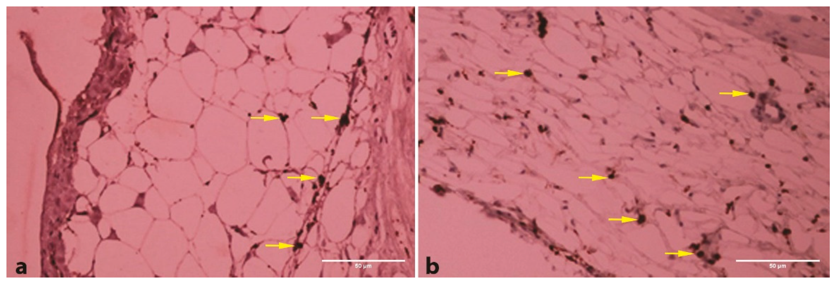
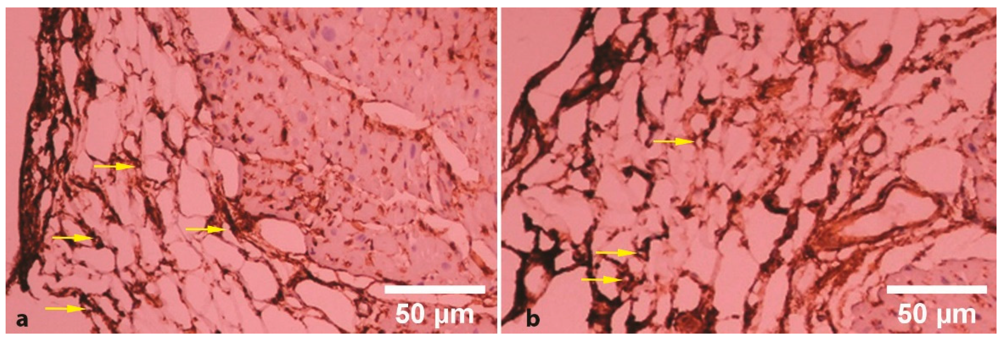
2.4. EAT Histopathological, Immunohistochemical and Morphometrical Analysis

EAT histology assessment was done with an optical microscope (Olympus CX41, Olympus Corporation, Tokyo, Japan). We evaluated the RAA epicardial adipose tissue thickness and area by using usual hematoxylin-eosin staining [16].
The measurements were analyzed with a color image analysis system (Quick PHOTO MICRO version 3.0, Olympus Corporation, Tokyo, Japan) at a magnification of 200×.
EAT thickness was expressed as a mean value of ten EAT thickness from the entire histological section area.
Further, epicardial inflammatory foci and angiogenesis required immunohistochemical (IHC) confirmation.

2.5. EAT Inflammation and Angiogenesis

Immunohistochemistry (IHC) was performed according to standard protocols on formalin-fixed, paraffin-embedded sections. IHC examination focused on inflammation and angiogenesis assessed by using CD68 and CD34 markers (3). The endogenous peroxidase of deparaffinized sections was neutralized by hydrogen peroxide 3% for 10 min. Antigen unmasking was performed by incubating the slides in a pH 9 buffer at 98 °C for 45 min. For immunohistochemical detection, antibodies to CD68 (monoclonal antibodies Clone PG-M1; RTU, DAKO, Glostrup, Denmark, catalog code no. IS609) and antibodies to CD34 (monoclonal antibodies Class II, Clone QBEnd 10, RTU, DAKO, Glostrup, Denmark, catalog code no. IS632) were applied for 30 min followed by the streptavidin-horseradish peroxidase conjugate for 15 min. Peroxidase was developed using the DAB working solution (DAKO, Glostrup, Denmark) and washed in deionized water. Sections were counterstained using hematoxylin Gill II (Sigma, Sigma-Aldrich, Darmstadt, Germany). The negative control was obtained by the replacement of the primary antibody with PBS.
Assessment of immunohistochemistry stained sections was made by the presence of positive brown staining; CD 34, as cell surface glycoprotein, gives brown membranous staining, allowing to determine microvessel density, while CD68, as glycoprotein expressed predominantly on the lysosomal membrane, gives brown cytoplasmic staining.
Morphometry was performed by the assessment of the macrophages and microvessel density. Immunohistochemically, we analyzed five histological fields at 200× magnification for each case (one field corresponded to a manually traced area reported to 100,000 µm2/0.1 mm2). The results were expressed as percentages or mean values of the number of positive cells or vessels related to the studied area.
The semi-quantitative evaluation was made by using a manually-driven evaluation method.

2.6. Statistical Analysis

The statistical analysis was performed using IBM SPSS Statistics 21 Software (IBM SPSS, Chicago, IL, USA). The Mann–Whitney test and Pearson chi-square test were used for parametrical (non-normal distribution due to small size data) and non-parametrical data, respectively. Numerical data were presented as means ± SD and categorical data were presented as percentages. Statistical significance was set at p < 0.05.

3. Results

3.1. CV Risk Factor Analysis

Table 1 shows the demographic, clinical, hemodynamic, and biochemical characteristics of CAD patients. The mean age in CAD cases without HF (58 years) was lower than in CAD cases with HF (63 years). All patients showed known CV risk factors for CAD.
In CAD patients with CHF, the prevalence of male sex, obesity, and dyslipidemia was higher (p < 0.05) as compared to the ones without CHF.

3.2. EAT Histopathological, Immunohistochemical and Morphometrical Analysis

EAT thickness, macrophage infiltration (Figure 1a,b), and capillary network (Figure 2a,b) were identified within RAA specimens.
The EAT thickness (185.8 µm) was non-significantly lower (p = 0.07) in CAD patients with CHF, as compared to the ones without CHF (217.2 µm).
EAT macrophage infiltration was more extensive in CAD patients with CHF than in those without CHF (Figure 1a,b). The mean value of CD68 positive count in CAD patients with CHF (325.78/μm2) was significantly higher (p < 0.001) than in CAD patients without CHF (244.9/µm2). EAT angiogenesis was less extensive in patients with CHF than in those without CHF (Figure 2a,b). The mean value of CD34 positive count in CAD patients with CHF (22.83/μm2) was significantly lower (p < 0.001) than in CAD patients without CHF (65.2/μm2).
The relationship between the accumulative number of risk factors and histological EAT labels is shown in Table 2.

4. Discussion

Identifying the patients with risk of developing cardiac ischemia subsequently to cardiac surgery would result in the prevention of undesired clinical consequences associated with CAD (6). In this paper, we studied the known CV risk factors in association with EAT on RAA samples following open-heart surgery. The commonest risk factors identified in our patients that are likely to be significant predictors of CAD progression were male sex, obesity, and dyslipidemia. Previously, Nagy et al. [17] and Kitagawa et al. [18] identified EAT thickness as a CV risk factor in patients with or without preoperative heart failure, operated with standard CABG technique.
Epicardial fat is visceral fat between the myocardial surface and the visceral layer of the pericardium. Scarce evidence is available on EAT measurement and the association of EAT and the severity of cardiac ischemia in CAD patients who underwent CABG surgery.
Large evidence showed that the severity of CAD was associated with EAT thickness [19,20]. Mookadam et al. [21] revealed that epicardial fat was associated with lower ejection fraction. Other studies have suggested that epicardial tissue could serve as a predictor of CAD, and EAT values >2.4 mm can predict the presence of significant (>50% diameter) CAD [7,22].
In the current study, we noticed a non-significant reduction of EAT thickness in patients with CHF as compared to those without CHF, associated to EAT fibrous tissue remodeling. According to Nagy et al. [17], the EAT amount is an independent CAD risk factor and is related to other CV risk factors. In concordance with previous studies [23,24], we consider that EAT thickness, measured by imaging techniques, may serve as a tool in the follow-up assessment of the CAD patients.
In our previous study [6], we noticed extracellular matrix alterations in EAT of CAD patients due to a great number of collagen deposits. By assessing the adipocyte size in EAT, we noticed that fibrosis could limit the adipocyte expandability [25]. We [3] showed that the adipocyte size in EAT had no significant difference between non-CAD individuals and CAD patients. According to Aitken-Buck et al. [26], these data indicated that the increased EAT thickness in CAD patients may not be due to adipocyte hypertrophy because fibrosis might restrict the extension of EAT by limiting adipocyte hypertrophy. A notable finding of this study was the presence of the EAT inflammatory foci, including macrophages, which may act to stimulate angiogenesis in the adipose tissue. Both Hirata et al. [27] and Matloch et al. [14], consider that epicardial fat is a source of inflammatory cells in CAD.
The particularity of our CABG patient series is that patients with CHF were obese. Our histologic analysis suggests that in obese cardiac patients with CHF, epicardial adipose tissue changes were increased inflammation and decreased thickness as compared to non-obese patients without CHF. Several studies reported that EAT thickness measured post-mortem [28] or clinically by echocardiography, computed tomography (CT), or magnetic resonance imaging (MRI) was lower in patients with CHF [11,12,24]. The diminished epicardial fat could explain the increased inflammation by a reduction in the local secretion of protective anti-inflammatory adipokines [24,29].
The current results, which are consistent with previous studies [1,30], revealed angiogenesis as another morphological hallmark of the EAT in CAD patients. Angiogenesis was of about three times greater in our CAD patients without CHF (of about 65 microvessels/μm2) than in CAD patients with CHF (23 microvessels/μm2) on coronary arteries [3,23,31].
The reduced number of participants was a limitation of our study. Another limitation is that immunohistochemistry can only measure one or two markers per sample, and it may not completely reflect the complex mechanisms involved in epicardial adipose tissue thickening.

5. Conclusions

In conclusion, the present study on epicardial fat samples of the RAA demonstrated that the abundance of CD68-macrophages appeared to be associated with CHF in CAD patients. Also, the individuals referred for CABG, in whom systolic heart dysfunction was confirmed by echocardiography, had decreased angiogenesis. Histologically, the increased EAT thickness in CAD patients was related to the connective tissue repair process.

Author Contributions

Conceptualization, D.B., C.S.; methodology, D.B., C.S., V.M., D.V.T., A.P.V.; investigation, D.B., A.P.V., V.V.C., R.D., B.M.C., R.E.H., R.A.S.; data curation, A.P.V., B.M.C.; writing—original draft preparation, D.B., V.M., D.V.T., V.V.C.; writing—review and editing, V.M., G.G. All authors have read and agreed to the published version of the manuscript.

Funding

This research was funded by “Grigore T. Popa” University of Medicine and Pharmacy Iasi, Romania, through the grants Ideas-Teams contract (29032/28.12.2016 and 30340/28.12.2017).

Conflicts of Interest

The authors declare no conflict of interest.

References

  1. Matloch, Z.; Kotulak, T.; Haluzik, M. The role of epicardial adipose tissue in heart disease. Physiol. Res. 2016, 65, 23–32. [Google Scholar] [CrossRef] [PubMed]
  2. Iacobellis, G.; Barbaro, G. Epicardial adipose tissue feeding and overfeeding the heart. Nutrition 2019, 59, 1–6. [Google Scholar] [CrossRef] [PubMed]
  3. Butcovan, D.; Oboroceanu, T.; Cimpeanu, C.; Mironescu, A.; Haliga, R.; Pinzariu, A.; Lupusoru, R.; Popescu, E.; Mocanu, V. The involvement of Epicardial adiposity and inflammation in Postoperatory atrial Fibrilation-Immunohistochemical qualitative and quantitative assessment. Rev. Chim. (Bucharest) 2017, 68, 8–13. [Google Scholar] [CrossRef]
  4. Ansaldo, A.M.; Montecucco, F.; Sahebkar, A.; Dallegri, F.; Carbone, F. Epicardial adipose tissue and cardiovascular diseases. Int. J. Cardiol. 2019, 278, 254–260. [Google Scholar] [CrossRef] [PubMed]
  5. Bachar, G.N.; Dicker, D.; Kornowski, R.; Atar, E. Epicardial adipose tissue as a predictor of coronary artery disease in asymptomatic subjects. Am. J. Cardiol. 2012, 110, 534–538. [Google Scholar] [CrossRef] [PubMed]
  6. Tinica, G.; Mocanu, V.; Zugun-Eloae, F.; Butcovan, D. Clinical and histological predictive risk factors of atrial fibrillation in patients undergoing open-heart surgery. Exp Ther Med 2015, 10, 2299–2304. [Google Scholar] [CrossRef] [Green Version]
  7. Iacobellis, G.; Ribaudo, M.C.; Zappaterreno, A.; Iannucci, C.V.; Leonetti, F. Relation between epicardial adipose tissue and left ventricular mass. Am J Cardiol 2004, 94, 1084–1087. [Google Scholar] [CrossRef]
  8. Ding, J.; Hsu, F.C.; Harris, T.B.; Liu, Y.; Kritchevsky, S.B.; Szklo, M.; Ouyang, P.; Espeland, M.A.; Lohman, K.K.; Criqui, M.H.; et al. The association of pericardial fat with incident coronary heart disease: the Multi-Ethnic Study of Atherosclerosis (MESA). Am. J. Clin. Nutr. 2009, 90, 499–504. [Google Scholar] [CrossRef]
  9. Mahabadi, A.A.; Massaro, J.M.; Rosito, G.A.; Levy, D.; Murabito, J.M.; Wolf, P.A.; O’Donnell, C.J.; Fox, C.S.; Hoffmann, U. Association of pericardial fat, intrathoracic fat, and visceral abdominal fat with cardiovascular disease burden: the Framingham Heart Study. Eur. Heart J. 2009, 30, 850–856. [Google Scholar] [CrossRef] [Green Version]
  10. Bala, C.; Craciun, A.E.; Hancu, N. Updating the Concept of Metabolically Healthy Obesity. Acta Endocrinol. (Bucharest) 2016, 12, 197–205. [Google Scholar] [CrossRef]
  11. Khawaja, T.; Greer, C.; Chokshi, A.; Chavarria, N.; Thadani, S.; Jones, M.; Schaefle, K.; Bhatia, K.; Collado, J.E.; Shimbo, D.; et al. Epicardial fat volume in patients with left ventricular systolic dysfunction. Am. J. Cardiol. 2011, 108, 397–401. [Google Scholar] [CrossRef] [PubMed]
  12. Doesch, C.; Haghi, D.; Fluchter, S.; Suselbeck, T.; Schoenberg, S.O.; Michaely, H.; Borggrefe, M.; Papavassiliu, T. Epicardial adipose tissue in patients with heart failure. J. Cardiovasc. Magn. Reson. 2010, 12, 40. [Google Scholar] [CrossRef] [PubMed] [Green Version]
  13. Rosito, G.A.; Massaro, J.M.; Hoffmann, U.; Ruberg, F.L.; Mahabadi, A.A.; Vasan, R.S.; O’Donnell, C.J.; Fox, C.S. Pericardial fat, visceral abdominal fat, cardiovascular disease risk factors, and vascular calcification in a community-based sample: the Framingham Heart Study. Circulation 2008, 117, 605–613. [Google Scholar] [CrossRef] [PubMed] [Green Version]
  14. Matloch, Z.; Cinkajzlova, A.; Mraz, M.; Haluzik, M. The Role of Inflammation in Epicardial Adipose Tissue in Heart Diseases. Curr. Pharm. Des. 2018, 24, 297–309. [Google Scholar] [CrossRef]
  15. Parisi, V.; Paolillo, S.; Rengo, G.; Formisano, R.; Petraglia, L.; Grieco, F.; D’Amore, C.; Dellegrottaglie, S.; Marciano, C.; Ferrara, N.; et al. Sleep-disordered breathing and epicardial adipose tissue in patients with heart failure. Nutr. Metab. Cardiovasc. Dis. 2018, 28, 126–132. [Google Scholar] [CrossRef]
  16. Timofte, D.; Mocanu, V.; Zugun Eloae, F.; Hristov, I.; Silivestru Cretu, I.; Aursulesei, V.; Balan, G.G.; Ciuntu, B.M.; Oboroceanu, T.; Tiron, A.; et al. Immunohistochemical Expression of Growth Hormone Secretagogue Receptor (GSH-R) of Adipose Tissue Macrophagesin Obese Bariatric Patients. Rev. Chim. (Bucharest) 2019, 70, 3428–3430. [Google Scholar] [CrossRef]
  17. Nagy, E.; Jermendy, A.L.; Merkely, B.; Maurovich-Horvat, P. Clinical importance of epicardial adipose tissue. Arch. Med. Sci. 2017, 13, 864–874. [Google Scholar] [CrossRef]
  18. Kitagawa, T.; Yamamoto, H.; Sentani, K.; Takahashi, S.; Tsushima, H.; Senoo, A.; Yasui, W.; Sueda, T.; Kihara, Y. Data set for volumetric and pathological findings of epicardial adipose tissue. Data Brief 2015, 5, 337–341. [Google Scholar] [CrossRef] [Green Version]
  19. Eroglu, S.; Sade, L.E.; Yildirir, A.; Bal, U.; Ozbicer, S.; Ozgul, A.S.; Bozbas, H.; Aydinalp, A.; Muderrisoglu, H. Epicardial adipose tissue thickness by echocardiography is a marker for the presence and severity of coronary artery disease. Nutr. Metab. Cardiovasc. Dis. 2009, 19, 211–217. [Google Scholar] [CrossRef]
  20. Mahabadi, A.A.; Berg, M.H.; Lehmann, N.; Kalsch, H.; Bauer, M.; Kara, K.; Dragano, N.; Moebus, S.; Jockel, K.H.; Erbel, R.; et al. Association of epicardial fat with cardiovascular risk factors and incident myocardial infarction in the general population: the Heinz Nixdorf Recall Study. J. Am. Coll. Cardiol. 2013, 61, 1388–1395. [Google Scholar] [CrossRef] [Green Version]
  21. Mookadam, F.; Goel, R.; Alharthi, M.S.; Jiamsripong, P.; Cha, S. Epicardial fat and its association with cardiovascular risk: a cross-sectional observational study. Heart Views 2010, 11, 103–108. [Google Scholar]
  22. Ahn, S.G.; Lim, H.S.; Joe, D.Y.; Kang, S.J.; Choi, B.J.; Choi, S.Y.; Yoon, M.H.; Hwang, G.S.; Tahk, S.J.; Shin, J.H. Relationship of epicardial adipose tissue by echocardiography to coronary artery disease. Heart 2008, 94, e7. [Google Scholar] [CrossRef] [PubMed] [Green Version]
  23. Salazar, J.; Luzardo, E.; Mejias, J.C.; Rojas, J.; Ferreira, A.; Rivas-Rios, J.R.; Bermudez, V. Epicardial Fat: Physiological, Pathological, and Therapeutic Implications. Cardiol. Res. Pract. 2016, 2016, 1291537. [Google Scholar] [CrossRef] [Green Version]
  24. Fitzgibbons, T.P.; Czech, M.P. Epicardial and perivascular adipose tissues and their influence on cardiovascular disease: basic mechanisms and clinical associations. J. Am. Heart Assoc. 2014, 3, e000582. [Google Scholar] [CrossRef] [PubMed] [Green Version]
  25. Jiang, D.S.; Zeng, H.L.; Li, R.; Huo, B.; Su, Y.S.; Fang, J.; Yang, Q.; Liu, L.G.; Hu, M.; Cheng, C.; et al. Aberrant Epicardial Adipose Tissue Extracellular Matrix Remodeling in Patients with Severe Ischemic Cardiomyopathy: Insight from Comparative Quantitative Proteomics. Sci. Rep. 2017, 7, 43787. [Google Scholar] [CrossRef]
  26. Aitken-Buck, H.M.; Babakr, A.A.; Coffey, S.; Jones, P.P.; Tse, R.D.; Lamberts, R.R. Epicardial adipocyte size does not correlate with body mass index. Cardiovasc. Pathol. 2019, 43, 107144. [Google Scholar] [CrossRef] [PubMed]
  27. Hirata, Y.; Kurobe, H.; Akaike, M.; Chikugo, F.; Hori, T.; Bando, Y.; Nishio, C.; Higashida, M.; Nakaya, Y.; Kitagawa, T.; et al. Enhanced inflammation in epicardial fat in patients with coronary artery disease. Int. Heart J. 2011, 52, 139–142. [Google Scholar] [CrossRef] [Green Version]
  28. Schejbal, V. Epicardial fatty tissue of the right ventricle--morphology, morphometry and functional significance. Pneumologie 1989, 43, 490–499. [Google Scholar]
  29. Fosshaug, L.E.; Dahl, C.P.; Risnes, I.; Bohov, P.; Berge, R.K.; Nymo, S.; Geiran, O.; Yndestad, A.; Gullestad, L.; Aukrust, P.; et al. Altered Levels of Fatty Acids and Inflammatory and Metabolic Mediators in Epicardial Adipose Tissue in Patients With Systolic Heart Failure. J. Card. Fail. 2015, 21, 916–923. [Google Scholar] [CrossRef]
  30. Dozio, E.; Briganti, S.; Vianello, E.; Dogliotti, G.; Barassi, A.; Malavazos, A.E.; Ermetici, F.; Morricone, L.; Sigruener, A.; Schmitz, G.; et al. Epicardial adipose tissue inflammation is related to vitamin D deficiency in patients affected by coronary artery disease. Nutr. Metab. Cardiovasc. Dis. 2015, 25, 267–273. [Google Scholar] [CrossRef]
  31. Iacobellis, G.; di Gioia, C.R.; Cotesta, D.; Petramala, L.; Travaglini, C.; De Santis, V.; Vitale, D.; Tritapepe, L.; Letizia, C. Epicardial adipose tissue adiponectin expression is related to intracoronary adiponectin levels. Horm. Metab. Res. 2009, 41, 227–231. [Google Scholar] [CrossRef] [PubMed]
Figure 1. Immunohistochemical EAT analysis of the macrophages (CD68 200×) in CAD patients without CHF (a) and CAD patients with CHF (b).
Applsci 10 05871 g001
Figure 2. Immunohistochemical EAT analysis of the vessels (CD34 200×) in CAD patients without CHF (a) and CAD patients with CHF (b).
Applsci 10 05871 g002
Table 1. Demographic, clinical, hemodynamic, and biochemical characteristics of CAD patients
VariablePatients without CHF
(n = 9)
Patients with CHF
(n = 6)
Age (y)58.3 ± 14.763.4 ± 11.2
Male sex (%)33.3100 *
Weight (kg)78.3 ± 14.588.0 ± 22.1
BMI (kg/m2)27.6 ± 5.632.3 ± 7.1 *
Obesity (%)0100 *
History
Dyslipidemia (%)33.3100
Arterial hypertension (%)66.650
DM type 2 (%)66.650
Smoking (%)33.30
Echocardiography
LVEDD (cm)5.0 ± 0.56.9 ± 1.2 *
LVESD (cm)3.3± 0.65.6 ± 1.3 *
LVEF (%)54.1 ± 3.328.5 ± 7.2 *
Biochemical values
Total cholesterol (mg/dL)212 ± 42206 ± 38 ∗
Triglycerides (mg/dL)161 ± 12175 ±54 *
LDL cholesterol (mg/dL)124 ± 9135 ± 11 *
HDL cholesterol (mg/dL)57 ± 445 ± 3 *
* p-value ≤ 0.05 was considered significant; CAD, coronary artery disease; CHF, chronic heart failure, DM, diabetes mellitus, LVEF, left ventricular ejection fraction, LVEDD, left ventricular end-diastolic diameter, LVESD, left ventricular end-systolic diameter.
Table 2. Mean EAT thickness and IHC markers of macrophages accumulation and angiogenesis in peri-atrial EAT samples from CAD patients.
PatientsEAT thickness
(μm)
CD68
(No/μm2)
CD34
(No/μm2)
Without CHF217.1 ± 21.3244.9 ± 5.565.2 ± 1.7
With CHF185.8 ± 27.9325.7 ± 16.122.7 ± 3.8
p-value0.07<0.001<0.001
p-value <0.05 was considered significant, CAD, coronary artery disease; CHF, chronic heart failure.

