Diarylureas as Antitumor Agents


1
Department of Pharmacy-Drug Sciences, University of Bari Aldo Moro, 70126 Bari, Italy
2
Department of Pharmacy, Health and Nutritional Sciences, University of Calabria, 87036 Arcavacata di Rende, Italy
*
Author to whom correspondence should be addressed.
Appl. Sci. 2021, 11(1), 374; https://0-doi-org.brum.beds.ac.uk/10.3390/app11010374
Submission received: 24 November 2020
/
Revised: 12 December 2020
/
Accepted: 28 December 2020
/
Published: 2 January 2021
(This article belongs to the Special Issue Anticancer Drugs Activity and Underlying Mechanisms)
Abstract
:The diarylurea is a scaffold of great importance in medicinal chemistry as it is present in numerous heterocyclic compounds with antithrombotic, antimalarial, antibacterial, and anti-inflammatory properties. Some diarylureas, serine-threonine kinase or tyrosine kinase inhibitors, were recently reported in literature. The first to come into the market as an anticancer agent was sorafenib, followed by some others. In this review, we survey progress over the past 10 years in the development of new diarylureas as anticancer agents.
1. Introduction
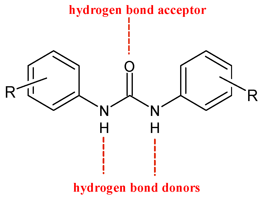
Ureas (R-NHCONH-R’) are known organic compounds that possess biological activities and serve as templates for numerous medicinal chemistry researches [1]. Barbital is a diethylmalonyl urea discovered at the beginning of 1900, used as sleep aid and hypnotic [2]. In the following century, the urea scaffold has represented the pharmacophore the backbone motif for entire classes of therapeutic agents [3]. This review focuses on diarylureas, i.e., ureas substituted with two aromatic moieties also known as bis-aryl ureas. Diarylureas are found in numerous heterocyclic compounds with various biological activities [4], such as antithrombotic, antimalarial, antibacterial, antinflammatory, and anticancer [5,6]. In particular, diarylurea is a prominent pharmacophore in anticancer drugs. This activity is due to its near-perfect binding with certain acceptors. The NH moiety behaves as hydrogen bond donor and the urea oxygen atom acts as acceptor (Figure 1) [7].
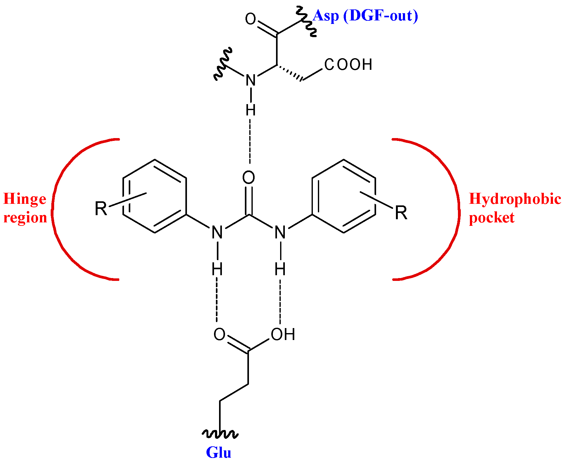
This structure provides urea derivatives endowed with capability of binding several enzymes and receptors [8,9,10]. Moreover, it may link different pharmacophore fragments of new biological active compounds. A urea linker has been used to overcome the poor solubility of some phenyl N-mustards [11]. In this way, the authors obtained water soluble N-mustards, some of which showing high anticancer activity against various human tumor xenograft models and were able to introduce cross-linking within the DNA double strand. Diarylurea-based compounds present strong inhibitory activity against kinases, including RAF kinases [12], platelet derived growth factor receptor (PDGF) [13], vascular endothelial growth factor receptor 2 (VEGFR-2) [14], receptor tyrosine kinase (RTKs) [15], and Aurora kinases [16]. The diarylurea moiety is, in fact, widespread in type II kinase inhibitors. These compounds circumvent kinases in an inactive state, the so-called DFG-out, and occupy a hydrophobic pocket next to the ATP-binding site. The diarylurea fragment is able to link the hinge-binding moiety with the portion that occupies the hydrophobic pocket that is in the inactive conformation of kinases [17] (Figure 2).
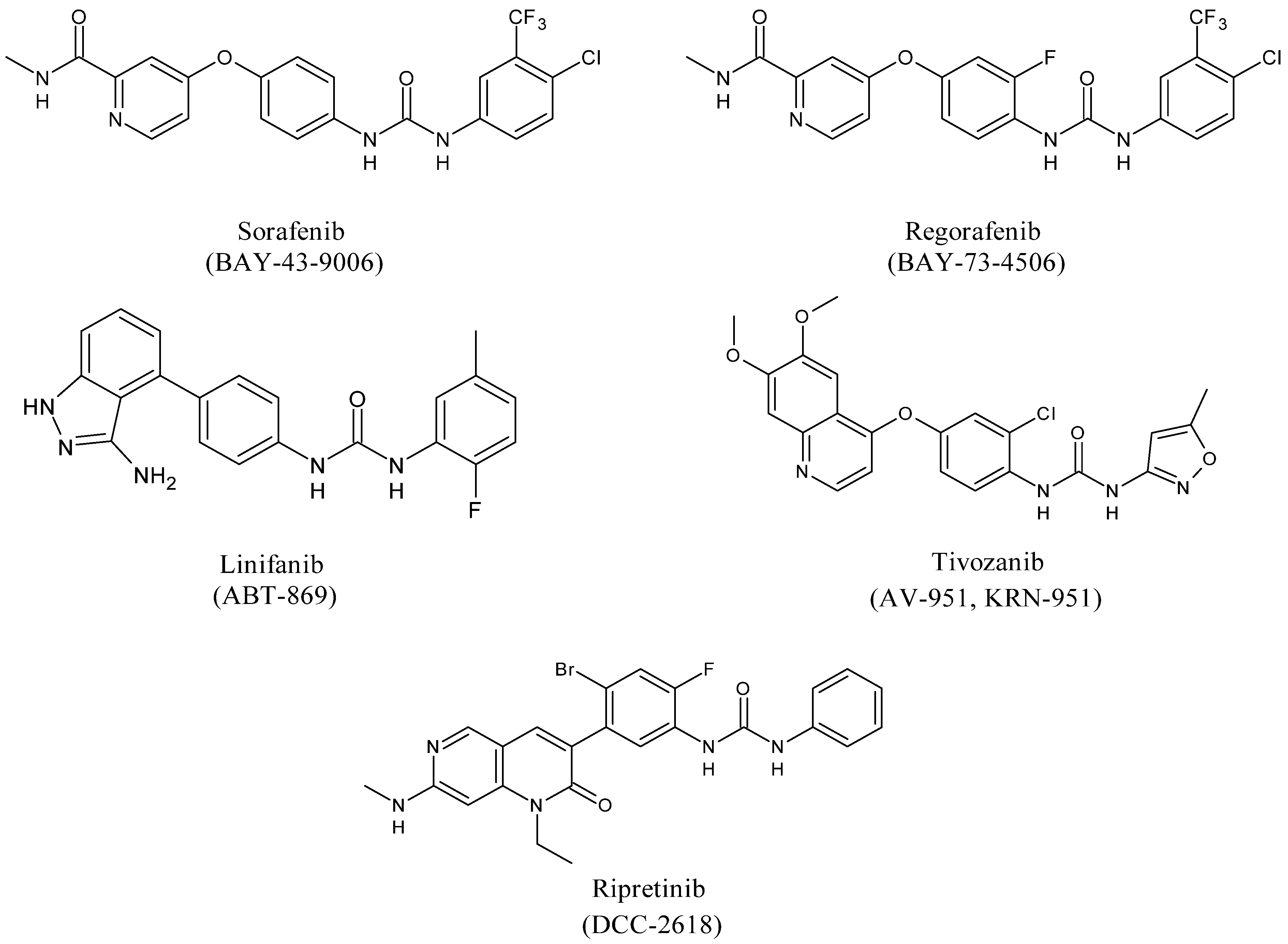
Diarylureas represent the skeleton of the main systemic therapies for several cancers, as advanced, metastatic hepatocellular carcinoma (HCC) [18], advanced renal cell carcinoma (RCC) [19], gastrointestinal stromal tumors (GISTs) [20], metastatic colorectal cancer (mCRC) [21]. Sorafenib is a multi-targeted small molecule tyrosine protein kinase that improves median survival over placebo for unresectable HCC patients [22]. In 2008 it obtained Food and Drug Administration (FDA) and European Medicinal Agency (EMEA) approval for the treatment of RCC and HCC [23]. Basing on sorafenib as the lead compound, several other diarylurea derivatives, such as regorafenib, linifanib, tivozanib, and ripretinib have been synthesized and evaluated as kinase inhibitors. Regorafenib was approved by the FDA in the United States in February 2013 in patients with advanced GISTs for those who had failed on imatinib and sunitinib [24,25]. Linifanib is currently being studied in HCC clinical trials [26]. Unlike the other inhibitors of VEGFR and PDGFR, linifanib seems to be also involved in adipocyte browning, thus being considered for the treatment of obesity [27]. Tivozanib is a diarylurea which is to be considered as third and fourth line therapy in patients with metastatic RCC in phase 3 study [28]. By end of 2019, the Chinese and European regulatory authorities have marketed five small-molecule protein kinase inhibitors (PKIs), including tivozanib [29]. Ripretinib has been suggested as a promising treatment for advanced GISTs [30]. In this paper, these already known drugs bearing a diarylurea skeleton are reviewed, along with new synthetic diarylureas described in the literature as promising agents for the treatment of diverse type of tumors.
2. Diarylureas in Therapy or in Clinical Studies as Anticancer Agents
2.1. Sorafenib
Sorafenib (BAY-43-9006, Nexavar®, Figure 3) is an oral receptor TKI that determines the inhibition of Raf serine/threonine kinases and receptor tyrosine kinases (VEGF 1, 2, 3 and PDGF-β, FMS-like tyrosine kinase-3 (FLT-3), and c-KIT) that are components of signaling pathways controlling tumor growth and angiogenesis [31].
Sorafenib inhibits the kinase activity of C-RAF and B-RAF (wild type and V600E mutant) showing IC50 = 6.22 and 38 nM, respectively. This compound is considered the most important drug in the late stage of injury for advanced stages of HCC [32] which is the second cause of cancer-related mortality all over the world [33]. For more than ten years, sorafenib has been the sole systemic treatment for advanced HCC [34]. However, some advanced HCC patients do not respond to therapy with sorafenib. Thus, combination studies of sorafenib with other drugs have been studied. Combined treatment with interferon-lambda 3 (IFN-λ3) and sorafenib show an effect of synergism in suppressing HCC cancer growth and in the promotion of cell apoptosis in vitro and in vivo [35]. A more recent study demonstrated that combination immunotherapy of sorafenib with atezolizumab, an immune checkpoint inhibitor (ICI) that target the programmed-cell death-1 receptor/ligand (PD-1/PD-L1) pathway, and bevacizumab, an anti-VEGF mAb, is superior to sorafenib alone as the first-line therapy of advanced HCC [36]. Moreover, given that liver function is essential for a correct prognosis, a precise rating for the safe prescription and clinical development of ICI in HCC is required. Recently, the albumin-bilirubin (ALBI) grade was used as an alternative biomarker for the prognosis [37]. The efficacy of sorafenib is limited by several factors as systematic tolerance and the poor solubility in water. Furthermore, the hydrophobicity of sorafenib is responsible of its low bioavailability as it decreases the absorption by the gastrointestinal tract. Nexavar® (Bayer Healthcare Pharmaceuticals–Onyx Pharmaceuticals) is used as tablets containing sorafenib tosylate to slightly improve the solubility. In order to increase the solubility and bioavailability of the drug, sorafenib-loaded lipid-based nanosuspensions were used [38]. The administration of sorafenib to the target cells could ameliorate patient survival and reduce the further proliferation of the tumor [39]. Thus, a drug delivery system for sorafenib has been recently studied in order to help the administration of therapies in malignant cells and raise its clinical efficacy [40]. Recently, the treatment of cancers of the gastrointestinal tract during the COVID-19 pandemic [41] has been studied: coronavirus-adapted institutional recommendations have been formulated. Sorafenib was recommended in the first-line setting only in patients with disease subtype Eastern Cooperative Oncology Group performance status (ECOG PS) 0 or 1 and Child-Pughscore A hepatocellular carcinoma [42]. In spite of its selectivity, sorafenib can determine adverse effects, such as severe respiratory and liver failure, fatigue, stomatitis, hand-foot syndrome, diarrhea, and myelosuppression, thus posing a challenge for oncologists [43]. The therapy with sorafenib might be done ad hoc to increase the therapeutic effects and reducing adverse effects [44]. Sorafenib is also studied for iodine-resistant advanced thyroid carcinoma [45]. Finally, it is important to note that sorafenib is also involved in cytoskeleton alteration that leads to cancer cells death by apoptosis. Wang et al. [46] reported that the treatment of Hep3B and PLC/PRF/5 human hepatoma cells with sorafenib induces a drastic loss of actin fibers and the redistribution of F-actin around the cell nuclei. This effect is due to the regulation of protein kinases and phosphatases that ends with cofilin dephosphorylation, which is an actin-binding factor necessary for the reorganization of actin. Chen et al. [47] demonstrated that the ability of sorafenib in inducing human prostate cancer cell line PC-3 apoptosis, through cytoskeleton destabilization, increased if combined with zinc exposure, suggesting that zinc may sensitize prostate cancer cells to sorafenib treatment. D’Alessandro et al. [48] demonstrated a synergistic effect on HCC cells migration using combined doses of sorafenib and/or vitamin K1 with insulin like growth factor I receptor (IGF1-R) antagonists enhancing the reduction and reorganization of F-actin, probably through the modulation of MAPK cascade.
2.2. Regorafenib
Regorafenib (BAY 73-4506, Stivarga®, Figure 3) is the fluorinated analogue of sorafenib. It is an orally active diphenylurea multikinase inhibitor that targets stromal (PDGFR-β, FGFR-1), angiogenic (VEGFR1-3, TIE-2), and oncogenic receptor tyrosine kinases (c-KIT, RET, and RAF-1) [49]. It is the first multi-targeting kinase inhibitor which was approved by FDA in 2012 for the treatment of mCRC patients in refractory to standard chemotherapy [50]. In addition, regorafenib treatment determined an important amelioration in progression-free survival (PFS) in comparison with placebo in patients with metastatic GISTs after standard treatments; thus, it has also received FDA-approval for this indication since 2013. Then, in 2017, FDA approved regorafenib as a therapy for patients with advanced HCC [51]. Regorafenib showed a significant amelioration of PFS and overall survival (OS) in comparison with placebo. Regorafenib has been described in phase II clinical trials in different tumors, including RCC, soft-tissue sarcoma (STS), second- and third-line treatments for medullary thyroid cancer. However, several post-marketing observational studies, after the treatment of mCRC patients showed extensive data of toxicities (CORRELATE, REBECCA, RECORA, Japanese post-marketing study) [52,53,54]. It was found that adverse reactions due to regorafenib frequently occurred in the initial stages of treatment, mostly in the first cycle [55]. Thus, in patients with mCRC during the first cycle of regorafenib, the use of a dose-escalation strategy treatment was suggested [56]. This strategy was then supported by a multicenter, open-label, phase II study [57]. Recently, during the COVID-19 outbreak, regorafenib has been considered as a therapy for gastrointestinal cancer [42]. A drug-delivery system has been studied for regorafenib, too [40]. The pivotal RESORCE (NCT01774344) phase III trial studied regorafenib therapy in patients with HCC who were tolerant to sorafenib, but who had progressed during sorafenib treatment [58]. Regorafenib has been demonstrated to inhibit glioblastoma multiforme (GBM) growth through PSAT1-mediated autophagy arrest [59], and to have beneficial effect in Alzheimer’s disease (AD) and formation of dendritic spine in vitro and in vivo [60].
2.3. Linifanib
Linifanib (ABT-869, Abbott Laboratories, Abbott Park, IL, USA, Figure 3) is an orally available TKI which targets VEGFR and PDGFR with relevant specificity and low off-target inhibition. Linifanib can also inhibit FLT-3 [61]. It does not show significant activity against representative cytosolic tyrosine and serine/threonine kinases [62]. Linifanib is a colony-stimulating factor-1 receptor (CSF-1R) inhibitor through the inhibition of the phosphorylation of CSF-1R tyrosine kinase in transfected cells [63]. It is used as a therapy for non-small cell lung carcinoma (NSCLC), liver cancer, breast cancer, colorectal cancer [45]. Preclinical and early clinical trials showed interesting activity in various human neoplasms with a satisfactory profile of toxicity. Linifanib competes with ATP in the binding site domain of tyrosine kinase, thus it prevents downstream signaling [64]. Phase II trial studies show that linifanib is useful for the treatment of patients with advanced, refractory colorectal cancer that expresses k-Ras mutations [65]. In an open-label phase II trial linifanib showed interesting clinical activity, as monotherapy, in patients with advanced HCC [66]. Linifanib versus sorafenib was studied in terms of efficacy and tolerability. Linifanib and sorafenib showed similar OS in advanced HCC. Linifanib did not meet predefined superiority and non-inferiority OS boundaries; thus, the study did not reach the primary end point. Secondary end points, time to progression (TTP), and objective response rate (ORR), favored Linifanib; safety results favored sorafenib [67]. Although linifanib is currently examined in HCC clinical trials, it has not yet been studied in preclinical and clinical studies for gastric cancer. 5-Fluorouracil (5-FU) and cisplatin represent the first-line chemotherapy for patients with gastric cancer and the combined use with linifanib inhibits synergistically the viability of some gastric cancer cell lines and led to remarkable suppression of VEGF-induced angiogenesis in vitro and in vivo [68]. Linifanib has demonstrated to be also useful in the treatment of anaplastic thyroid cancer (ATC), that is considered the most aggressive form of thyroid cancer. The synergistic use of linifanib and irinotecan significantly increased the survival of ATC-affected mice. These observations have been made by using an orthotopic in vivo model that better recapitulates features of human tumors than the more simplistic subcutaneous xenograft models, suggesting a potential role of this co-treatment in ATC patient’s treatment [69]. Finally, linifanib was demonstrated to interfere with adipocyte browning. It suppresses STAT3 signaling pathway, thus leading to the enhancement of adipocyte browning and inhibition of adipogenesis. Linifanib’s blocking browning effect was demonstrated as the phosphorylation of STAT3 was reduced by linifanib and the STAT3 activator SD19, as well [27].
2.4. Tivozanib
Tivozanib (AV-951, KRN-951, FOTIVDA®, Figure 3), used as the hydrochloride monohydrate salt, is a bioavailable inhibitor of angiogenesis which targets VEGFR tyrosine kinases with high antitumor activity. It is a VEGF-TKI specific for VEGFR1–3, showing an inhibitor effect at nanomolar concentrations, with IC50 values of 30 nM, 6.5 nM, and 15 nM for VEGFR1, 2 and 3, respectively. The compound is unique in that it is highly specific for VEGFR1–3, and presents minimal residual effects on c-KIT and PDGFR-β [70]. It presents a long half-life, too [71]. It has shown considerable efficacy for the treatment of advanced RCC over the past decade. In August 2017, tivozanib was approved by the EMEA as a first-line therapy for patients with advanced RCC and those who are VEGFR and mTOR pathway inhibitor-naïve following disease progression after previous therapy with cytokines for advanced RCC. Tivozanib was compared with sorafenib in a phase III trial for patients with metastatic RCC. Tivozanib improved PFS, but not OS, and showed a differentiated safety profile, in comparison with sorafenib, as initial targeted therapy for metastatic RCC [72]. Preclinical data and phase III trials of tivozanib in RCC, TIVO-1, and TIVO-3 have been recently summarized. Given the agent’s excellent tolerability profile it is appropriate for those patients with heavily pretreated disease that could exhibit clinical deterioration. Currently, the standard therapy is represented by nivolumab and ipilimumab, followed by cabozantinib. Tivozanib may represent a third-line treatment after failure of these agents [73]. The results of phase III TIVO-3 trial (American Society of Clinical Oncology Virtual Scientific Program, 2020) showed that tivozanib significantly improved PFS, compared with sorafenib, in patients with highly relapsed or refractory metastatic RCC [74,75,76,77]. Tivozanib activity has been also investigated in hepatocellular carcinoma in association with durvalumab [78] and in recurrent, platinum-resistant ovarian cancer, fallopian tube cancer, and primary peritoneal cancer [79,80]. Tivozanib has been studied in phase I and II clinical trials as monotherapy and in combination with other drugs for the treatment of STS [81], glioblastoma [82], breast [83], and colorectal cancers, and other advanced gastrointestinal cancers [84,85].
2.5. Ripretinib
Ripretinib (DCC-2618, QINLOCKTM, Figure 3) is an oral inhibitor of tyrosine kinase that primarily inhibits KIT proto-oncogene receptor tyrosine kinase and platelet-derived growth factor receptor A (PDGFRA) kinase signaling. Ripretinib also inhibits other kinases, such as PDGFRB, TIE2, VEGFR2, and BRAF. It was designed for cancers and myeloproliferative neoplasms, especially GISTs. Ripretinib is a “switch-control” kinase inhibitor that forces the activation loop (or activation “switch”) into the inactive conformation. In preclinical cancer models it has shown efficacy, and preliminary clinical data show that Ripretinib inhibits a broad range of KIT mutants in patients with drug-resistant GISTs [86]. The INVICTUS study demonstrated the efficacy and safety of ripretinib as the fourth-line treatment versus placebo in patients with advanced GISTs [87]. In May 2020, ripretinib received approval from the US FDA for the treatment of patients with advanced GISTs who had received previous treatment with more than two kinase inhibitors [88]. Ripretinib is being evaluated in an ongoing phase III study (INTRIGUE) as a second-line therapy in comparison with sunitinib after progressing on imatinib [89]. Recently, ripretinib is being investigated in clinical trials for systemic mastocytosis (SM) [90], and has been also proposed for the treatment of STS [91].
2.6. Mechanisms of Inhibition of Diarylureas
The proposed inhibitory mechanisms of diarylureas depend on their structure. Garuti et al. [5] reported the crystal structure of V600EB-RAF kinase domains in complex with sorafenib. The pyridyl ring is shown to occupy the ATP adenine-binding pocket and to interact with three amino acids residues. The trifluoromethyl phenyl, that is a lipophilic moiety, fits into a hydrophobic pocket. The urea moiety forms two hydrogen bonds with V600EB-RAF, one with the aspartate, and one with the glutamate residue. Recently, the 2D interaction of the co-crystallized sorafenib inside the active site of B-Raf has been reported [92]. Regorafenib differs from sorafenib only for a fluorine atom, thus its interactions are similar to those of sorafenib. Chen et al. (2017) proposed an alternative mechanism for colorectal cancer for regorafenib. It seems that it interacts with microRNA-21 (miR-21), an oncogenic miRNA which plays a crucial role in resisting programmed cell death in CRC cells. RNA–ligand docking, molecular dynamics simulation showed that regorafenib can directly bind to miR-21 pre-element [93]. Docking studies of linifanib with FLT3 were recently reported, evidencing that 3-amino-indazole interacts with the ATP-bind site [61]. Kajal et al. (2018) has reported the 2D co-crystal-binding conformation of VEGFR2-Tivozanib, in which tivozanib mimics the binding pattern of ATP [94]. Finally, studies on the mechanism of action of ripretinib have been recently reported [86].
3. Other Diarylureas
In this paragraph several studies on other diarylureas are described (Table 1). Babić et al. synthesized several diarylurea derivatives in order to study their cytostatic activity [95]. The compounds were tested on tumor cell lines: HCT 116 (colon carcinoma), SW 620 (colon carcinoma), MCF-7 (breast carcinoma), H460 (lung carcinoma), L1210 (murine leukemia), CEM (human lymphoma), and HeLa (cervix carcinoma). Compounds 1a–e exerted the highest effect (IC50 from 1 to 4.3 μM, with an average of 2.6 ± 1.6 μM) even though with low selectivity for the different tumor cell lines. The compounds were also cytostatic against primary human embryonic lung (HEL) fibroblast cells. Kapuriya et al. [11] studied a series of water-soluble N-mustard-benzene conjugates containing a urea linker. The urea linker was introduced in order to overcome the low solubility of compounds previously studied. The authors studied a series of water-soluble N-mustards, in which the phenyl N-mustard is linked to a benzene ring through a urea linker. In particular, the diarylurea 2 (BO-1055), as the hydrochloride salt, exhibited high in vitro cytotoxicity and therapeutic efficacy against various human tumor cell lines. It was demonstrated to possess potent therapeutic effect against several human solid tumor cell lines, including human breast cancer (MX-1), colon cancer (HCT-116), and prostate cancer (PC3), in xenograft model. The DNA repair capacity of compound BO-1055, named ureidomustin, was then studied. It was proposed for the treatment of tumors with deficient nucleotide excision repair (NER), homologous recombination (HR), and O6-methylguanine-DNA methyltransferase (MGMT) DNA repair genes, or in synergy with other drugs in tumors in which DNA damage response has been repressed [96].BO-1055 was also proposed as a therapeutic agent for Ewing sarcoma and rhabdomyosarcoma given its potency and relative lack of toxicity against normal tissue [97]. It also showed a potent activity against B-cell lymphomas, as mantle cell lymphoma (MCL) and diffuse large B-cell lymphoma (DLBCL) [98].
In a following paper, the same research group indicated that compound 2 has a quite narrow therapeutic window; thus, following a bioisosteric approach, an inversion of the carboxamide functionality was addressed. Compound 3 was superior to compound 2 against colon cancer (HCT-116) and lung cancer (H460) cell lines, and displayed minor toxicity. Cotreatment of compound 3 and 5-fluorouracil suppressed the growth of HCT-116 xenografts. Moreover, compound 3 could induce DNA cross-linking and cell-cycle arrest at the G2/M phase. This compound was selected for early preclinical studies [99]. A series of diarylureas was studied for in vitro antiproliferative activities against HepG2, MGC-803, and A549 cancer cell lines [100]. Compound 4 displayed optimal antiproliferative activity against the three cell lines in comparison with sorafenib and gefitinib. Indeed, it induced A549 cells apoptosis through the cell cycle block at the G0/G1 phase, the increase of intracellular reactive oxygen species, and the reduction of mitochondrial membrane potential. This compound also influenced the Raf/MEK/ERK pathway. A series of diarylurea derivatives was studied for its cytotoxicity in vitro against H-460, HT-29, A549, and MDA-MB-231 cancer cell lines [101]. Some of them showed higher activity than sorafenib (IC50 between 0.089 and 5.46 µM). In particular, compound 5 was the most potent both in cellular (IC50 = 0.15, 0.089, 0.36, and 0.75 µM, respectively) and enzymatic assay (IC50 = 56 nM against EGFR). The antiproliferative activity of diarylureas bearing a 4-anilinoquinazoline group was evaluated via MTT assay against A431 and A549 cells [102]. Three compounds showed high antiproliferative activities and their inhibitory activity against EGFR-TK was evaluated. Compound 6 was a potent EGFR-TK inhibitor. This completely inhibited cancer growth in established nude mouse A549 xenograft model in vivo, at 50 mg/kg. Diarylureas were studied as LIM-kinase (Limk) inhibitors for their therapeutic potential against prostate cancers [103]. Limk is a serine-threonine protein kinase existing in two isoforms, LIM kinase 1 (Limk1) and LIM kinase 2 (Limk2). The inhibition of Limk1 activity in cancer prostate cells and tissues determines reduction of phosphorylated cofilin and cancer cells motility, thus reducing invasiveness of the tumor and evolution to metastasis. The substituted diarylurea 7, at 1 μM, inhibited only Limk1 and STK16 with ≥80% inhibition. The use of Limk inhibitors has been also suggested to target the invasive machinery in GBM [104]. Recently, a diarylurea, N69B, was evaluated for its anticancer activity and its molecular mechanism was investigated [105]. The compound was shown to inhibit proliferation of murine and human cancer cells in vitro, and reduce tumor growth in mouse 4T1 breast tumor model in vivo. Compound N69B significantly increased protein levels of cathepsins, in particular cathepsin D, a lysosomal aspartyl protease with various biological functions. Several diarylureas bearing a coumarine moiety [106,107] have been recently tested for their in vitro antiproliferative activities against the H4IIE and HepG2 cancer cell lines, and has been proposed as a promising lead for further optimization [108]. Compound 8b exhibited a higher inhibition of H4IIE cells compared to sorafenib. 8a also showed a better inhibition against HepG2 cells than sorafenib. In particular, 8b arrested cell cycle at the S phase and induced H4IIE cells apoptosis. A library of diarylureas has been designed and the in vitro antiproliferative activities was studied against HT-29 and A549 cancer cell lines. Compound 9 was the most active against HT-29 cells showing an IC50 value of 3.38 μM, compared to that of sorafenib (IC50 = 17.28 μM). It induced cell cycle arrest at G0/G1 phase, interfered with Raf/MEK/ERK signaling pathway, increased intracellular reactive oxygen species level, and led to HT-29 cells apoptosis [109]. The same research group studied a series of benzo[b]thiophene-diarylureas with potential anticancer effects, too. Compound 10 was the most active (IC50 = 5.91 and 14.64 μM on HT-29 and A549 cells, respectively). It induced apoptosis and cell cycle arrest at the G0/G1 phase on HT-29 cells, too [110]. Several diphenyl indazoles, containing diarylurea moieties, in the low micromolar range, inhibited cell viability of various cancer cell lines including murine metastatic breast cancer 4T1, murine glioma GL261-luc2, human triple negative breast cancer MDA-MB-231, human pancreatic cancer MIAPaCa-2, and human colorectal adenocarcinoma WiDr. The lead candidate 11 significantly reduced the tumor growth in aggressive stage IV breast cancer 4T1 syngraft model in vivo [111]. A series of diarylureas bearing a substituted thiadiazole as one of the two aryl moieties was studied against human chronic myeloid leukemia (CML) cell line K562. The diarylurea 12 exhibited the least cytotoxicity and higher biological activity (IC50 = 0.038 μM). It also displayed good induced-apoptosis effect for human CML cell line K562; its effect seems to happen via a significant reduction of protein phosphorylation of PI3K/AKT signal pathway by human phospho-kinase array analysis [112]. Forchlorfenuron (FCF; N-(2-Chloro-4-pyridyl)-N’-phenylurea) is a small synthetic diarylurea currently used in agriculture as a plant fertilizer that increases fruit size because of its potent cytokinin activity. FCF inhibits proliferation, anchorage-independent growth, migration, and invasion of cancer cell lines in various cancer types, such as prostate, mesothelioma, lung, colon, breast, ovary, and cervix [113]. FCF was also found to be effective in a mouse model, in which tumor growth was inhibited. FCF treatment caused the suppression of HIF-1α and HER2, both of them playing a crucial role in cancer cell survival [114]. Recently, several FCF analogues (UR214-1, UR214-7, and UR214-9) were demonstrated to be more effective in decreasing viability and proliferation in both ovarian and endometrial cancer cell lines, and suppress HER2 expression at a concentration lower than that of FCF. Moreover, FCF and its analogues were found to decrease the expression of human epididymis protein 4 (HE4), which is commonly upregulated in ovarian and endometrial cancers [115]. Diarylurea PQ401 is a small molecule that behaves as an inhibitor of IGF-1R signaling. It is also able to prevent breast cancer cells growth in in vivo mouse models [116]. It has also shown anti-cancer properties in glioma by inducing cellular apoptosis in U87MG cells, thus reducing cell viability and proliferation and attenuating cell mobility in vitro. Moreover, through a mouse xenograft model, PQ401 administration led to the suppression of glioma tumor growth in vivo in mice [117]. Recently, PQ401 potential as a putative chemotherapy drug in osteosarcoma cells has been investigated. PQ401 effectively suppressed osteosarcoma cell growth, migration, and colony formation in vitro, as well as induced apoptosis in vitro. PQ401 inhibited U2OS cell viability almost as effective as cisplatin. PQ401 can significantly cause U2OS cell apoptosis and clonogenesis at the IC50 concentration with the blockade of IGF1-R phosphorylation and related downstream signaling [118]. The diphenyl urea-derivative DUD was designed on the basis of a docking study for the optimization of a natural product, taspine. The anti-metastatic potential of DUD for NSCLC was studied in vitro. DUD inhibited A549 cells migration by reversing EMT via Wnt/β-catenin and PI3K/Akt signaling, thus it has been suggested as a potential therapy for NSCLC treatment [119]. Several fluorinated diarylureas were studied as activators of adenosine monophosphate-activated kinase (AMPK). Compound FND-4b determined the induction of phosphorylated AMPK and the decrease in markers of cell proliferation, as cyclin D1, in all CRC cell lines. Apoptosis was also increased in CRC cells treated with FND-4b [120]. Thidiazuron (TDZ, 1-phenyl-3-(1,2,3-thiadiazol-5-yl) urea) is a synthetic plant hormone which has been widely used as herbicide, pesticide, and as growth regulator in plant tissue culture [121]. Given its cytotoxic effect on HeLa (human cervical carcinoma) cell lines, it has been recently proposed as a potential agent to act against cervical cancer cells. It has also suggested to have a role on apoptosis in cancer cells through DNA damage. Furthermore, the activity of TDZ as anticancer was tested against Hela cells by mitochondrial dysfunction, DNA damage, in silico caspase-3 inhibition, and some gene expression [122]. This observation has been recently confirmed by radiolabeling TDZ with 99mTc. The in silico study supported the ability of 99mTc-TDZ complex to bind caspase-3 protein that is overexpressed in cancers, suggesting that 99mTc-TDZ might be a potential agent for diagnosis of solid tumors, such as the cervix cancer [123].

Table 1.
Structures of compounds described in the literature.
| Structure | Compd | Ref |
|---|---|---|
 | 1a–e | [95] |
 | 2 (BO-1055) | [11] |
 | 3 | [99] |
 | 4 | [100] |
 | 5 | [101] |
 | 6 | [102] |
 | 7 | [103] |
 | N69B | [105] |
 | 8a,b | [106,107,108] |
 | 9 | [109] |
 | 10 | [110] |
 | 11 | [111] |
 | 12 | [112] |
 | Forchlorfenuron (FCF) | [113,114] |
 | UR214-1 UR214-7 UR214-9 | [115] |
 | PQ401 | [116,117,118] |
 | DUD | [119] |
 | FND-4b | [120] |
 | Thidiazuron (TDZ) | [121,122,123] |
4. Summary
Diarylureas are considered a privileged structure in medicinal chemistry, particularly for anticancer drugs. Sorafenib is a neovascular blocker that prevents the formation of new blood vessels, followed by the growth of cancer tissue, through multiple kinase inhibitors that target angiogenesis. It is approved for the treatment of advanced inoperable HCC and advanced RCC. An alternative for the treatment of these tumors under study may be represented by tivozanib. Regorafenib is an effective therapy for patients with advanced GSTIs or mCRC. Linifanib may represent a promising therapeutic agent for human gastric cancer, NSCLC, liver cancer, breast cancer, colorectal cancer. Ripretinib is addressed to GISTs. In this paper, we report an overview of the development and application of these drugs. The current treatment trends in oncology have shifted to immunotherapy combinations with ICI, as anti-PD-L1-directed monoclonal antibodies. Pending improved understanding of HCC, RCC, GSTIs, and mCRC tumorigenesis, it would be very interesting to evaluate the combination of various treatment modalities. Diarylurea combined with a checkpoint inhibitor could be a promising treatment strategy to be deeply investigated in the future. Moreover, this review encompasses the recent advances in scientific literature in the broad area of diarylureas as anticancer agents. The newly synthesized compounds of this class, that are now in phase of study, may represent promising small molecules able to unseat or help the already known existing drugs.
Funding
This research received no external funding.
Conflicts of Interest
The authors declare no conflict of interest.
Abbreviations
| AD | Alzheimer’s disease |
| AMPK | adenosine monophosphate-activated kinase |
| ATC | Anaplastic thyroid cancer |
| CML | chronic myeloid leukemia |
| CSF-1R | colony stimulating factor-1 receptor |
| DLBCL | diffuse large B-cell lymphoma |
| ECOG PS | Eastern Cooperative Oncology Group performance status |
| EMEA | European Medicinal Agency |
| FCF | Forchlorfenuron |
| FDA | Food and Drug Administration |
| FLT-3 | FMS-like tyrosine kinase-3 |
| 5-FU | 5-Fluorouracil |
| GBM | glioblastoma multiform |
| GIST | gastrointestinal stromal tumors |
| HCC | hepatocellular carcinoma |
| HE4 | human epididymis protein 4 |
| HEL | human embryonic lung |
| HR | homologues recombination |
| ICI | Immune checkpoint inhibitors |
| IFN-λ3 | Interferon-lambda 3 |
| IGF1-R | insulin like growth factor I receptor |
| Limk | LIM-kinase |
| MCL | mantle cell lymphoma |
| mCRC | metastatic colorectal cancer |
| MGMT | O6-methylguanine-DNA methyltransferase |
| NER | nucleotide excision repair |
| NSCLC | non-small cell lung carcinoma |
| ORR | objective response rate |
| OS | overall survival |
| PD-1/PD-L1 | programmed-cell death-1 receptor/ligand |
| PDGFR | platelet derived growth factor receptor |
| PFS | progression-free survival |
| PKIs | protein kinase inhibitors |
| RCC | renal cell carcinoma |
| RTKs | receptor tyrosine kinases |
| SM | systemic mastocytosis |
| STS | soft-tissue sarcoma |
| TDZ | Thidiazuron |
| TKIs | tyrosine protein kinases |
| TTP | time to progression |
| VEGFR-2 | vascular endothelial growth factor receptor 2 |
References
- Ghosh, A.K.; Brindisi, M. Urea derivatives in modern drug discovery and medicinal chemistry. J. Med. Chem. 2019, 63, 2751–2788. [Google Scholar] [CrossRef] [PubMed]
- López-Muñoz, F.; Ucha-Udabe, R.; Alamo, C. The history of barbiturates a century after their clinical introduction. Neuropsychiatr. Dis. Treat. 2005, 1, 329–343. [Google Scholar] [PubMed]
- Jagtap, A.D.; Kondekar, N.B.; Sadani, A.A.; Chern, J.W. Ureas: Applications in Drug Design. Curr. Med. Chem. 2017, 24, 622–651. [Google Scholar] [CrossRef] [PubMed]
- Asif, M. Short Notes on Diaryl Ureas Derivatives. J. Adv. Res. BioChem. Pharmacol. 2018, 1, 38–41. [Google Scholar]
- Garuti, L.; Roberti, M.; Bottegoni, G.; Ferraro, M. Diaryl urea: A privileged structure in anticancer agents. Curr. Med. Chem. 2016, 23, 1528–1548. [Google Scholar] [CrossRef]
- Ceramella, J.; Mariconda, A.; Rosano, C.; Iacopetta, D.; Caruso, A.; Longo, P.; Sinicropi, M.S.; Saturnino, C. α–ω Alkenyl-bis-S-guanidine thiourea dihydrobromide affects HeLa cell growth hampering tubulin polymerization. ChemMedChem 2020. [Google Scholar] [CrossRef]
- Wu, Y.C.; Ren, X.Y.; Rao, G.W. Research Progress of Diphenyl Urea Derivatives as Anticancer Agents and Synthetic Methodologies. Mini Rev. Org. Chem. 2019, 16, 617–630. [Google Scholar] [CrossRef]
- Rizza, P.; Pellegrino, M.; Caruso, A.; Iacopetta, D.; Sinicropi, M.S.; Rault, S.; Lancelot, J.C.; El-Kashef, H.; Lesnard, A.; Rochais, C.; et al. 3-(Dipropylamino)-5-hydroxybenzofuro [2, 3-f] quinazolin-1 (2H)-one (DPA-HBFQ-1) plays an inhibitory role on breast cancer cell growth and progression. Eur. J. Med. Chem. 2016, 107, 275–287. [Google Scholar] [CrossRef]
- Saturnino, C.; Barone, I.; Iacopetta, D.; Mariconda, A.; Sinicropi, M.S.; Rosano, C.; Campana, A.; Catalano, S.; Longo, P.; Andò, S. N-heterocyclic carbene complexes of silver and gold as novel tools against breast cancer progression. Fut. Med. Chem. 2016, 8, 2213–2229. [Google Scholar] [CrossRef]
- Iacopetta, D.; Grande, F.; Caruso, A.; Mordocco, R.A.; Plutino, M.R.; Scrivano, L.; Ceramella, J.; Miuà, N.; Saturnino, C.; Puoci, F.; et al. New insights for the use of quercetin analogs in cancer treatment. Fut. Med. Chem. 2017, 9, 2011–2028. [Google Scholar] [CrossRef]
- Kapuriya, N.; Kakadiya, R.; Dong, H.; Kumar, A.; Lee, P.-C.; Zhang, X.; Chou, T.-C.; Lee, T.-C.; Chen, C.-H.; Lam, K.; et al. Design, synthesis, and biological evaluation of novel water-soluble N-mustards as potential anticancer agents. Bioorg. Med. Chem. 2011, 19, 471–485. [Google Scholar] [CrossRef] [PubMed]
- El-Nassan, H.B. Recent progress in the identification of BRAF inhibitors as anticancer agents. Eur. J. Med. Chem. 2014, 72, 170–205. [Google Scholar] [CrossRef] [PubMed]
- Ravez, S.; Barczyk, A.; Six, P.; Cagnon, A.; Garofalo, A.; Goossens, L.; Depreux, P. Inhibition of tumor cell growth and angiogenesis by 7-aminoalkoxy-4-aryloxy-quinazoline ureas, a novel series of multi-tyrosine kinase inhibitors. Eur. J. Med. Chem. 2014, 79, 360–381. [Google Scholar] [CrossRef] [PubMed]
- Wang, C.; Gao, H.; Dong, J.; Zhang, Y.; Su, P.; Shi, Y.; Zhang, J. Biphenyl derivatives incorporating urea unit as novel VEGFR-2 inhibitors: Design, synthesis and biological evaluation. Bioorg. Med. Chem. 2014, 22, 277–284. [Google Scholar] [CrossRef] [PubMed]
- Lu, Y.Y.; Zhao, C.R.; Wang, R.Q.; Li, W.-B.; Qu, X.-J. A novel anticancer diarylurea derivative HL-40 as a multi-kinases inhibitor with good pharmacokinetics in Wistar rats. Biomed. Pharmacother. 2015, 69, 255–259. [Google Scholar] [CrossRef]
- Curtin, M.L.; Frey, R.R.; Heyman, H.R.; Soni, N.B.; Marcotte, P.A.; Pease, L.J.; Glaser, K.B.; Magoc, T.J.; Tapang, P.; Albert, D.H.; et al. Thienopyridine ureas as dual inhibitors of the VEGF and Aurora kinase families. Bioorg. Med. Chem. Lett. 2012, 22, 3208–3212. [Google Scholar] [CrossRef]
- Ceramella, J.; Iacopetta, D.; Barbarossa, A.; Caruso, A.; Fedora, G.; Bonomo, M.G.; Mariconda, A.; Longo, P.; Saturnino, C.; Sinicropi, M.S. Carbazole derivatives as kinase-targeting inhibitors for cancer treatment. Mini Rev. Med. Chem. 2020, 20, 444–465. [Google Scholar] [CrossRef]
- Tella, S.H.; Kommalapati, A.; Mahipal, A. Systemic therapy for advanced hepatocellular carcinoma: Targeted therapies. Chin. Clin. Oncol. 2020. [Google Scholar] [CrossRef]
- Escudier, B.; Worden, F.; Kudo, M. Sorafenib: Key lessons from over 10 years of experience. Exp. Rev. Anticanc. Ther. 2019, 19, 177–189. [Google Scholar] [CrossRef]
- Mazzocca, A.; Napolitano, A.; Silletta, M.; Spalato Ceruso, M.; Santini, D.; Tonini, G.; Vincenzi, B. New frontiers in the medical management of gastrointestinal stromal tumours. Ther. Adv. Med. Oncol. 2019, 11. [Google Scholar] [CrossRef] [Green Version]
- Strumberg, D.; Scheulen, M.E.; Schultheis, B.; Richly, H.; Frost, A.; Büchert, M.; Christensen, O.; Jeffers, M.; Heinig, R.; Boix, O.; et al. Regorafenib (BAY 73-4506) in advanced colorectal cancer: A phase I study. Br. J. Canc. 2012, 106, 1722–1727. [Google Scholar] [CrossRef] [PubMed] [Green Version]
- Llovet, J.M.; Ricci, S.; Mazzaferro, V.; Hilgard, P.; Gane, E.; Blanc, J.F.; De Oliveira, A.C.; Santoro, A.; Raoul, J.-L.; Forner, A.; et al. Sorafenib in advanced hepatocellular carcinoma. N. Engl. J. Med. 2008, 359, 378–390. [Google Scholar] [CrossRef] [PubMed]
- Nagai, H.; Mukozu, T.; Ogino, Y.U.; Matsui, D.; Matsui, T.; Wakui, N.; Momiyama, K.; Igarashi, Y.; Sumino, Y.; Higai, K. Sorafenib and hepatic arterial infusion chemotherapy for advanced hepatocellular carcinoma with portal vein tumor thrombus. Anticancer Res. 2015, 35, 2269–2277. [Google Scholar] [PubMed]
- Sirohi, B.; Philip, D.S.; Shrikhande, S.V. Regorafenib in gastrointestinal stromal tumors. Future Oncol. 2014, 10, 1581–1587. [Google Scholar] [CrossRef] [PubMed]
- Scrivano, L.; Parisi, O.I.; Iacopetta, D.; Ruffo, M.; Ceramella, J.; Sinicropi, M.S.; Puoci, F. Molecularly imprinted hydrogels for sustained release of sunitinib in breast cancer therapy. Pol. Adv. Technol. 2019, 30, 743–748. [Google Scholar] [CrossRef]
- Mossenta, M.; Busato, D.; Baboci, L.; Di Cintio, F.; Toffoli, G.; Dal Bo, M. New insight into therapies targeting angiogenesis in hepatocellular carcinoma. Cancers 2019, 11, 1086. [Google Scholar] [CrossRef] [Green Version]
- Zhao, S.; Chu, Y.; Zhang, Y.; Zhou, Y.; Jiang, Z.; Wang, Z.; Mao, L.; Li, K.; Sun, W.; Li, P.; et al. Linifanib exerts dual anti-obesity effect by regulating adipocyte browning and formation. Life Sci. 2019, 222, 117–124. [Google Scholar] [CrossRef]
- Jacob, A.; Shook, J.; Hutson, T.E. Tivozanib, a highly potent and selective inhibitor of VEGF receptor tyrosine kinases, for the treatment of metastatic renal cell carcinoma. Future Oncol. 2020, 16, 2147–2164. [Google Scholar] [CrossRef]
- Bournez, C.; Carles, F.; Peyrat, G.; Aci-Sèche, S.; Bourg, S.; Meyer, C.; Bonnet, P. Comparative assessment of protein kinase inhibitors in public databases and in PKIDB. Molecules 2020, 25, 3226. [Google Scholar] [CrossRef]
- Nemunaitis, J.; Bauer, S.; Blay, J.Y.; Choucair, K.; Gelderblom, H.; George, S.; Schöffski, P.; von Mehren, M.; Zalcberg, J.; Achour, H.; et al. Intrigue: Phase III study of ripretinib versus sunitinib in advanced gastrointestinal stromal tumor after imatinib. Future Oncol. 2020, 16, 4251–4264. [Google Scholar] [CrossRef] [Green Version]
- Iyer, R.; Fetterly, G.; Lugade, A.; Thanavala, Y. Sorafenib: A clinical and pharmacologic review. Exp. Opin. Pharmacother. 2010, 11, 1943–1955. [Google Scholar] [CrossRef] [PubMed]
- Raoul, J.L.; Kudo, M.; Finn, R.S.; Edeline, J.; Reig, M.; Galle, P.R. Systemic therapy for intermediate and advanced hepatocellular carcinoma: Sorafenib and beyond. Canc. Treat. Rev. 2018, 68, 16–24. [Google Scholar] [CrossRef] [PubMed] [Green Version]
- Qiu, X.; Li, M.; Wu, L.; Xin, Y.; Mu, S.; Li, T.; Song, K. Severe Fatigue is an Important Factor in the Prognosis of Patients with Advanced Hepatocellular Carcinoma Treated with Sorafenib. Cancer Manag. Res. 2020, 12, 7983. [Google Scholar] [CrossRef] [PubMed]
- Rich, N.E.; Yopp, A.C.; Singal, A.G. Medical Management of Hepatocellular Carcinoma. J. Oncol. Pract. 2017, 13, 356–364. [Google Scholar] [CrossRef] [PubMed]
- Yan, Y.; Wang, L.; He, J.; Liu, P.; Lv, X.; Zhang, Y.; Xu, X.; Zhang, L.; Zhang, Y. Synergy with interferon-lambda 3 and sorafenib suppresses hepatocellular carcinoma proliferation. Biomed. Pharmacother. 2017, 88, 395–402. [Google Scholar] [CrossRef] [PubMed]
- Cheng, A.-L.; Qin, S.; Ikeda, M.; Galle, P.; Ducreux, M.; Zhu, A.; Kim, T.-Y.; Kudo, M.; Breder, V.; Merle, P.; et al. LBA3IMbrave150: Efficacy and safety results from a ph III study evaluating atezolizumab (atezo) + bevacizumab (bev) vs sorafenib (Sor) as first treatment (tx) for patients (pts) with unresectable hepatocellular carcinoma (HCC). Ann. Oncol. 2019, 30, ix186–ix187. [Google Scholar] [CrossRef]
- Pinato, D.J.; Kaneko, T.; Saeed, A.; Pressiani, T.; Kaseb, A.; Wang, Y.; Szafron, D.; Jun, T.; Dharmapuri, S.; Naqash, A.R.; et al. Immunotherapy in hepatocellular cancer patients with mild to severe liver dysfunction: Adjunctive role of the ALBI grade. Cancers 2020, 12, 1862. [Google Scholar] [CrossRef]
- Zhang, N.; Zhang, B.; Gong, X.; Wang, T.; Liu, Y.; Yang, S. In vivo biodistribution, biocompatibility, and efficacy of sorafenib-loaded lipid-based nanosuspensions evaluated experimentally in cancer. Int. J. Nanomed. 2016, 11, 2329–2343. [Google Scholar] [CrossRef] [Green Version]
- Guo, Y.; Zhong, T.; Duan, X.-C.; Zhang, S.; Yao, X.; Yin, Y.-F.; Huang, D.; Ren, W.; Zhang, Q.; Zhang, X. Improving anti-tumor activity of sorafenib tosylate by lipid- and polymer-coated nanomatrix. Drug Deliv. 2017, 24, 270–277. [Google Scholar] [CrossRef] [Green Version]
- Srimathi, U.; Nagarajan, V.; Chandiramouli, R. Investigation on graphdiyne nanosheet in adsorption of sorafenib and regorafenib drugs: A DFT approach. J. Mol. Liq. 2019, 277, 776–785. [Google Scholar] [CrossRef]
- Catalano, A. COVID-19: Could Irisin Become the Handyman Myokine of the 21st Century? Coronaviruses 2020, 1, 32–41. [Google Scholar] [CrossRef]
- Pietrantonio, F.; Morano, F.; Niger, M.; Corallo, S.; Antista, M.; Raimondi, A.; Prisciandaro, M.; Pagani, F.; Prinzi, N.; Nichetti, F.; et al. Systemic treatment of patients with gastrointestinal cancers during the COVID-19 outbreak: COVID-19-adapted recommendations of the national cancer institute of Milan. Clin. Colorect. Canc. 2020, 19, 156–164. [Google Scholar] [CrossRef] [PubMed]
- Granito, A.; Marinelli, S.; Negrini, G.; Menetti, S.; Benevento, F.; Bolondi, L. Prognostic significance of adverse events in patients with hepatocellular carcinoma treated with sorafenib. Ther. Adv. Gastroenterol. 2016, 9, 240–249. [Google Scholar] [CrossRef] [PubMed] [Green Version]
- Tovoli, F.; Ielasi, L.; Casadei-Gardini, A.; Granito, A.; Foschi, F.G.; Rovesti, G.; Negrini, G.; Orsi, G.; Renzulli, M.; Piscaglia, F. Management of adverse events with tailored sorafenib dosing prolongs survival of hepatocellular carcinoma patients. J. Hepatol. 2019, 71, 1175–1183. [Google Scholar] [CrossRef] [Green Version]
- Borriello, A.; Caldarelli, I.; Bencivenga, D.; Stampone, E.; Perrotta, S.; Oliva, A.; Della Ragione, F. Tyrosine kinase inhibitors and mesenchymal stromal cells: Effects on self-renewal, commitment and functions. Oncotarget 2017, 8, 5540. [Google Scholar] [CrossRef] [Green Version]
- Wang, Z.; Wang, M.; Carr, B.I. Involvement of receptor tyrosine phosphatase DEP-1 mediated PI3K-cofilin signaling pathway in Sorafenib-induced cytoskeletal rearrangement in hepatoma cells. J. Cell. Physiol. 2010, 224, 559–565. [Google Scholar] [CrossRef]
- Chen, X.; Che, X.; Wang, J.; Chen, F.; Wang, X.; Zhang, Z.; Fan, B.; Yang, D.; Song, X. Zinc sensitizes prostate cancer cells to sorafenib and regulates the expression of Livin. Acta Biochim Biophys Sin. 2013, 45, 353–358. [Google Scholar] [CrossRef] [Green Version]
- D’Alessandro, R.; Refolo, M.G.; Lippolis, C.; Carella, N.; Messa, C.; Cavallini, A.; Carr, B.I. Strong enhancement by IGF1-R antagonists of hepatocellular carcinoma cell migration inhibition by Sorafenib and/or vitamin K1. Cell. Oncol. 2018, 41, 283–296. [Google Scholar] [CrossRef]
- Ettrich, T.J.; Seufferlein, T. Regorafenib. In Small Molecules in Oncology; Springer: Cham, Switzerland; Berlin/Heidelberg, Germany, 2018; pp. 45–56. [Google Scholar]
- Arai, H.; Battaglin, F.; Wang, J.; Lo, J.H.; Soni, S.; Zhang, W.; Lenz, H.J. Molecular insight of regorafenib treatment for colorectal cancer. Canc. Treat. Rev. 2019, 81, 101912. [Google Scholar] [CrossRef]
- Pelosof, L.; Lemery, S.; Casak, S.; Jiang, X.; Rodriguez, L.; Pierre, V.; Bi, Y.; Liu, J.; Zirkelbach, J.F.; Patel, A.; et al. Benefit-risk summary of regorafenib for the treatment of patients with advanced hepatocellular carcinoma that has progressed on sorafenib. Oncologist 2018, 23, 496–500. [Google Scholar] [CrossRef] [Green Version]
- O’Connor, J.M.; Öhler, L.; Scheithauer, W.; Metges, J.P.; Dourthe, L.M.; de Groot, J.W.; Thaler, J.; Yeh, K.H.; Lin, J.K.; Falcone, A.; et al. Real-world dosing of regorafenib in metastatic colorectal cancer (mCRC): Interim analysis from the prospective, observational CORRELATE study. Liver 2017, 260, 52. [Google Scholar] [CrossRef] [Green Version]
- Komatsu, Y.; Muro, K.; Yamaguchi, K.; Satoh, T.; Uetake, H.; Yoshino, T.; Nishida, T.; Takikawa, H.; Kato, T.; Chosa, M.; et al. Safety and efficacy of regorafenib post-marketing surveillance (PMS) in Japanese patients with metastatic colorectal cancer (mCRC). J. Clin. Oncol. 2017, 35, 721. [Google Scholar] [CrossRef]
- Adenis, A.; de la Fouchardiere, C.; Paule, B.; Burtin, P.; Tougeron, D.; Wallet, J.; Dourthe, L.M.; Etienne, P.L.; Mineur, L.; Clisant, S.; et al. Survival, safety, and prognostic factors for outcome with Regorafenib in patients with metastatic colorectal cancer refractory to standard therapies: Results from a multicenter study (REBECCA) nested within a compassionate use program. BMC Cancer 2016, 16, 412. [Google Scholar]
- Grothey, A.; George, S.; Van Cutsem, E.; Blay, J.Y.; Sobrero, A.; Demetri, G.D. Optimizing treatment outcomes with regorafenib: Personalized dosing and other strategies to support patient care. Oncologist 2014, 19, 669–680. [Google Scholar] [CrossRef] [PubMed] [Green Version]
- NCCN Clinical Practice Guidelines in Oncology (NCCN Guidelines). Colon Cancer. Version 4.2018 National Comprehensive Cancer Network. Available online: https://www.nccn.org/professionals/physician_gls/pdf/colon.pdf (accessed on 12 November 2020).
- Bekaii-Saab, T.S.; Ou, F.S.; Ahn, D.H.; Boland, P.M.; Ciombor, K.K.; Heying, E.N.; Dockter, T.J.; Jacobs, N.L.; Pasche, B.C.; Cleary, J.M.; et al. Regorafenib dose-optimisation in patients with refractory metastatic colorectal cancer (ReDOS): A randomised, multicentre, open-label, phase 2 study. Lancet Oncol. 2019, 20, 1070–1082. [Google Scholar] [CrossRef]
- Bruix, J.; Qin, S.; Merle, P.; Granito, A.; Huang, Y.H.; Bodoky, G.; Pracht, M.; Yokosuka, O.; Rosmorduc, O.; Breder, V.; et al. Regorafenib for patients with hepatocellular carcinoma who progressed on sorafenib treatment (RESORCE): A randomised, double-blind, placebo-controlled, phase 3 trial. Lancet 2017, 389, 56–66. [Google Scholar] [CrossRef] [Green Version]
- Jiang, J.; Zhang, L.; Chen, H.; Lei, Y.; Zhang, T.; Wang, Y.; Jin, P.; Lan, J.; Zhou, L.; Huang, Z.; et al. Regorafenib induces lethal autophagy arrest by stabilizing PSAT1 in glioblastoma. Autophagy 2020, 16, 106–122. [Google Scholar] [CrossRef]
- Han, K.M.; Kang, R.J.; Jeon, H.; Lee, H.; Lee, J.S.; Park, H.H.; Jeon, S.G.; Suk, K.; Seo, J.; Hoe, H.S. Regorafenib regulates AD pathology, neuroinflammation, and dendritic spinogenesis in cells and a mouse model of AD. Cells 2020, 9, 1655. [Google Scholar] [CrossRef]
- Shi, Z.H.; Liu, F.T.; Tian, H.Z.; Zhang, Y.M.; Li, N.G.; LU, T. Design, synthesis and structure-activity relationship of diaryl-ureas with novel isoxazol [3,4-b]pyridine-3-amino-structure as multi-target inhibitors against receptor tyrosine kinase. Bioorg. Med. Chem. 2018, 26, 4735–4744. [Google Scholar] [CrossRef]
- Zhou, J.; Goh, B.C.; Albert, D.H.; Chen, C.S. ABT-869, a promising multi-targeted tyrosine kinase inhibitor: From bench to bedside. J. Hematol. Oncol. 2009, 2, 33. [Google Scholar] [CrossRef] [Green Version]
- Guo, J.; Marcotte, P.A.; McCall, J.O.; Dai, Y.; Pease, L.J.; Michaelides, M.R.; Davidsen, S.K.; Glaser, K.B. Inhibition of phosphorylation of the colonystimulating factor-1 receptor (c-Fms) tyrosine kinase in transfected cells by ABT-869 and other tyrosine kinase inhibitors. Mol. Cancer Ther. 2006, 5, 1007–1013. [Google Scholar] [CrossRef] [PubMed] [Green Version]
- Aversa, C.; Leone, F.; Zucchini, G.; Serini, G.; Geuna, E.; Milani, A.; Valdembri, D.; Martinello, R.; Montemurro, F. Linifanib: Current status and future potential in cancer therapy. Exp. Rev. Anticancer Ther. 2015, 15, 677–687. [Google Scholar] [CrossRef] [PubMed]
- Lin, W.H.; Hsu, J.T.A.; Hsieh, S.Y.; Chen, C.T.; Song, J.S.; Yen, S.C.; Hsu, T.; Chen, C.H.; Chou, L.H.; Yang, Y.N.; et al. Discovery of 3-phenyl-1H-5-pyrazolylamine derivatives containing a urea pharmacophore as potent and efficacious inhibitors of FMS-like tyrosine kinase-3 (FLT3). Bioorg. Med. Chem. 2013, 21, 2856–2867. [Google Scholar] [CrossRef] [PubMed]
- Toh, H.C.; Chen, P.J.; Carr, B.I.; Knox, J.J.; Gill, S.; Ansell, P.; McKeegan, E.M.; Dowell, B.; Pedersen, M.; Qin, Q.; et al. Phase 2 trial of linifanib (ABT-869) in patients with unresectable or metastatic hepatocellular carcinoma. Cancer 2013, 119, 380–387. [Google Scholar] [CrossRef]
- Cainap, C.; Qin, S.; Huang, W.T.; Chung, I.J.; Pan, H.; Cheng, Y.; Kudo, M.; Kang, Y.K.; Chen, P.J.; Toh, H.C.; et al. Linifanib versus Sorafenib in patients with advanced hepatocellular carcinoma: Results of a randomized phase III trial. J. Clin. Oncol. 2015, 33, 172–180. [Google Scholar] [CrossRef]
- Chen, J.; Guo, J.; Chen, Z.; Wang, J.; Liu, M.; Pang, X. Linifanib (ABT-869) potentiates the efficacy of chemotherapeutic agents through the suppression of receptor tyrosine kinase-mediated akt/mtor signaling pathways in gastric cancer. Sci. Rep. 2016, 6, 1–11. [Google Scholar] [CrossRef] [Green Version]
- Banchi, M.; Orlandi, P.; Gentile, D. Synergistic activity of linifanib and irinotecan increases the survival of mice bearing orthotopically implanted human anaplastic thyroid cancer. Am. J. Cancer Res. 2020, 10, 2120–2127. [Google Scholar]
- Nakamura, K.; Taguchi, E.; Miura, T.; Yamamoto, A.; Takahashi, K.; Bichat, F.; Guilbaud, N.; Hasegawa, K.; Kubo, K.; Fujiwara, Y.; et al. KRN951, a highly potent inhibitor of vascular endothelial growth factor receptor tyrosine kinases, has antitumor activities and affects functional vascular properties. Cancer Res. 2006, 66, 9134–9142. [Google Scholar] [CrossRef] [Green Version]
- Momeny, M.; Moghaddaskho, F.; Gortany, N.K.; Yousefi, H.; Sabourinejad, Z.; Zarrinrad, G.; Mirshahvaladi, S.; Eyvani, H.; Barghi, F.; Ahmadinia, L. Blockade of vascular endothelial growth factor receptors by tivozanib has potential anti-tumour effects on human glioblastoma cells. Sci. Rep. 2017, 7, 44075. [Google Scholar] [CrossRef]
- Motzer, R.J.; Nosov, D.; Eisen, T.; Bondarenko, I.; Lesovoy, V.; Lipatov, O.; Tomczak, P.; Lyulko, O.; Alyasova, A.; Harza, M.; et al. Tivozanib versus sorafenib as initial targeted therapy for patients with metastatic renal cell carcinoma: Results from a phase III trial. J. Clin. Oncol. 2013, 31, 3791. [Google Scholar] [CrossRef]
- Salgia, N.J.; Zengin, Z.B.; Pal, S.K. Tivozanib in renal cell carcinoma: A new approach to previously treated disease. Ther. Adv. Med. Oncol. 2020, 12. [Google Scholar] [CrossRef] [PubMed]
- Wright, K.M. Final data analysis supports tivozanib as superior treatment for patients with RCC. Oncology (Williston Park, NY) 2020, 34, 257. [Google Scholar]
- Rini, B.I.; Pal, S.K.; Escudier, B.J.; Atkins, M.B.; Hutson, T.E.; Porta, C.; Verzoni, E.; Needle, M.N.; McDermott, D.F. Tivozanib versus sorafenib in patients with advanced renal cell carcinoma (TIVO-3): A phase 3, multicentre, randomised, controlled, open-label study. Lancet Oncol. 2020, 21, 95–104. [Google Scholar] [CrossRef]
- Pal, S.K.; Escudier, B.J.; Atkins, M.B. Final overall survival results from a phase 3 study to compare tivozanib to sorafenib as third-or fourth-line therapy in subjects with metastatic renal cell carcinoma. Eur. Urol. 2020, 78, 783–785. [Google Scholar] [CrossRef]
- Westerman, M.E.; Wood, C.G. Editorial Commentary: Tivozanib versus sorafenib in patients with advanced renal cell carcinoma (TIVO-3): A phase 3, multicentre, randomised, controlled, open-label study. Ann. Transl. Med. 2020, 8, 1037. [Google Scholar] [CrossRef]
- Iyer, R.V.; Li, D.; Dayyani, F.; Needle, M.N.; Abrams, T.A. A phase Ib/II, open-label study of tivozanib in combination with durvalumab in subjects with untreated advanced hepatocellular carcinoma. J. Clin. Oncol. 2020, 38 (Suppl. 16599). [Google Scholar] [CrossRef]
- Swetzig, W.M.; Lurain, J.R.; Berry, E.; Pineda, M.J.; Shahabi, S.; Perry, L.; Neubauer, N.L.; Nieves-Neira, W.; Schink, J.C.; Schiller, A.; et al. Efficacy and safety of tivozanib in recurrent, platinum resistant ovarian, fallopian tube or primary peritoneal cancer. J. Clin. Oncol. 2019, 37 (Suppl. 5538). [Google Scholar] [CrossRef]
- Momeny, M.; Sabourinejad, Z.; Zuzzinrad, G.; Maghaddaskho, F.; Eyvani, H.; Yousefi, H.; Mirshahvaladi, S.; Poursani, E.M.; Barghi, F.; Poursheikhani, A.; et al. Anti-tumour activity of tivozanib, a pan-inhibitor of VEGF receptors, in therapy-resistant ovarian carcinoma cells. Sci. Rep. 2017, 7, 1–13. [Google Scholar] [CrossRef]
- Martin-Liberal, J.; Pérez, E.; García Del Muro, X. Investigational therapies in phase II clinical trials for the treatment of soft tissue sarcoma. Exp. Opin. Invest. Drugs 2019, 28, 39–50. [Google Scholar] [CrossRef]
- Kalpathy-Cramer, J.; Chandra, V.; Da, X.; Ou, Y.; Emblem, K.E.; Muzikansky, A.; Cai, X.; Douw, L.; Evans, J.G.; Dietrich, J.; et al. Phase II study of tivozanib, an oral VEGFR inhibitor, in patients with recurrent glioblastoma. J. Neurooncol. 2017, 131, 603–610. [Google Scholar] [CrossRef]
- Jamil, M.O.; Hathaway, A.; Mehta, A. Tivozanib: Status of development. Curr. Oncol. Rep. 2015, 17, 24. [Google Scholar] [CrossRef] [PubMed]
- Oldenhuis, C.N.; Loos, W.J.; Esteves, B.; van Doorn, L.; Cotreau, M.M.; Strahs, A.L.; den Hollander, M.W.; Gietema, J.A.; de Vries, E.G.E.; Eskens, F.A.L.M. A phase Ib study of the VEGF receptor tyrosine kinase inhibitor tivozanib and modified FOLFOX-6 in patients with advanced gastrointestinal malignancies. Clin. Colorectal Cancer 2015, 14, 18–24.e1. [Google Scholar] [CrossRef] [PubMed]
- Benson, A.B.; Kiss, I.; Bridgewater, J.; Eskens, F.A.; Sasse, C.; Vossen, S.; Chen, J.; Van Sant, C.; Ball, H.A.; Keating, A.; et al. BATON-CRC: A phase II randomized trial comparing tivozanib plus mFOLFOX6 with bevacizumab plus mFOLFOX6 in stage IV metastatic colorectal cancer. Clin. Cancer Res. 2016, 22, 5058–5067. [Google Scholar] [CrossRef] [PubMed] [Green Version]
- Smith, B.D.; Kaufman, M.D.; Lu, W.P.; Gupta, A.; Leary, C.B.; Wise, S.C.; Rutkoski, T.J.; Ahn, Y.M.; Ahn, G.; Bulfer, S.L.; et al. Ripretinib (DCC-2618) is a switch control kinase inhibitor of a broad spectrum of oncogenic and drug-resistant KIT and PDGFRA variants. Cancer Cell 2019, 35, 738–751. [Google Scholar] [CrossRef]
- Blay, J.Y.; Serrano, C.; Heinrich, M.C.; Zalcberg, J.; Bauer, S.; Gelderblom, H.; Schöffski, P.; Jones, R.L.; Attia, S.; D’Amato, G.; et al. Ripretinib in patients with advanced gastrointestinal stromal tumours (INVICTUS): A double-blind, randomised, placebo-controlled, phase 3 trial. Lancet Oncol. 2020, 21, e415. [Google Scholar] [CrossRef]
- US Food and Drug Administration. QINLOCK Prescribing Information. 2020. Available online: https://www.accessdata.fda.gov/drugsatfda_docs/label/2020/213973s000lbl.pdf (accessed on 12 November 2020).
- Khoshnood, A. Gastrointestinal stromal tumor—A review of clinical studies. J. Oncol. Pharm. Pract. 2019, 25, 1473–1485. [Google Scholar] [CrossRef]
- Jawhar, M.; Gotlib, J.; Reiter, A. Tyrosine kinase inhibitors in systemic mastocytosis. In Mastocytosis; Springer: Cham, Switzerland, 2020; pp. 257–265. [Google Scholar]
- Haddox, C.L.; Riedel, R.F. Individualizing systemic therapy for advanced soft tissue sarcomas based on tumor histology and biology. Exp. Rev. Anticancer Ther. 2020, 20, 5–8. [Google Scholar] [CrossRef]
- Thabit, M.G.; Mostafa, A.S.; Selim, K.B.; Elsayed, M.A.; Nasr, M.N. Design, synthesis and molecular modeling of phenyl dihydropyridazinone derivatives as B-Raf inhibitors with anticancer activity. Bioorg. Chem. 2020, 103, 104148. [Google Scholar] [CrossRef]
- Chen, X.; Xie, B.; Cao, L.; Zhu, F.; Zhu, B.; Lv, H.; Fan, X.; Han, L.; Bie, L.; Cao, X.; et al. Direct binding of microRNA-21 pre-element with Regorafenib: An alternative mechanism for anti-colorectal cancer chemotherapy? J. Mol. Graph. Model. 2017, 73, 48–53. [Google Scholar] [CrossRef]
- Kajal, K.; Panda, A.K.; Bhat, J.; Chakraborty, D.; Bose, S.; Bhattacharjee, P.; Sarkar, T.; Chatterjee, S.; Kar, S.K.; Sa, G. Andrographolide binds to ATP-binding pocket of VEGFR2 to impede VEGFA-mediated tumor-angiogenesis. Sci. Rep. 2019, 9, 1–10. [Google Scholar] [CrossRef] [Green Version]
- Babić, Ž.; Crkvenčić, M.; Rajić, Z.; Mikecin, A.M.; Kralj, M.; Balzarini, J.; Petrova, M.; Vanderleyden, J.; Zorc, B. New sorafenib derivatives: Synthesis, antiproliferative activity against tumour cell lines and antimetabolic evaluation. Molecules 2012, 17, 1124–1137. [Google Scholar] [CrossRef] [PubMed] [Green Version]
- Kuo, C.Y.; Chou, W.C.; Wu, C.C.; Wong, T.S.; Kakadiya, R.; Lee, T.C.; Su, T.L.; Wang, H.C. Repairing of N-mustard derivative BO-1055 induced DNA damage requires NER, HR, and MGMT-dependent DNA repair mechanisms. Oncotarget 2015, 6, 25770. [Google Scholar] [CrossRef] [PubMed] [Green Version]
- Ambati, S.R.; Shieh, J.H.; Pera, B.; Lopes, E.C.; Chaudhry, A.; Wong, E.W.; Saxena, A.; Su, T.L.; Moore, M.A. BO-1055, a novel DNA cross-linking agent with remarkable low myelotoxicity shows potent activity in sarcoma models. Oncotarget 2016, 7, 43062. [Google Scholar] [CrossRef] [PubMed] [Green Version]
- Lopes, E.C.; Correa, F.; Peguero, E.; Ambati, S.R.; Shieh, J.H.; Su, T.L.; Moore, M.A.S. Pre-Clinical Evaluation of a Novel DNA Crosslinking Agent, BO-1055 in B-Cell Lymphoma. Blood 2014, 124, 5483. [Google Scholar] [CrossRef]
- Tala, S.D.; Ou, T.H.; Lin, Y.W.; Tala, K.S.; Chao, S.H.; Wu, M.H.; Tsai, T.H.; Kakadiya, R.; Suman, S.; Chen, C.H.; et al. Design and synthesis of potent antitumor water-soluble phenyl N-mustard-benzenealkylamide conjugates via a bioisostere approach. Eur. J. Med. Chem. 2014, 76, 155–169. [Google Scholar] [CrossRef] [PubMed]
- Chen, J.N.; Wang, X.F.; Li, T.; Wu, D.W.; Fu, X.B.; Zhang, G.J.; Shen, X.C.; Wang, H.S. Design, synthesis, and biological evaluation of novel quinazolinyl-diaryl urea derivatives as potential anticancer agents. Eur. J. Med. Chem. 2016, 107, 12–25. [Google Scholar] [CrossRef]
- Jiang, N.; Bu, Y.; Wang, Y.; Nie, M.; Zhang, D.; Zhai, X. Design, synthesis and structure-activity relationships of novel diaryl urea derivatives as potential EGFR inhibitors. Molecules 2016, 21, 1572. [Google Scholar] [CrossRef] [Green Version]
- Zuo, S.J.; Zhang, S.; Mao, S.; Xie, X.X.; Xiao, X.; Xin, M.H.; Xuan, W.; He, Y.Y.; Cao, Y.X.; Zhang, S.Q. Combination of 4-anilinoquinazoline, arylurea and tertiary amine moiety to discover novel anticancer agents. Bioorg. Med. Chem. 2016, 24, 179–190. [Google Scholar] [CrossRef]
- Yin, Y.; Zheng, K.; Eid, N.; Howard, S.; Jeong, J.H.; Yi, F.; Guo, J.; Park, C.M.; Bibian, M.; Wu, W.; et al. Bis-aryl urea derivatives as potent and selective LIM kinase (Limk) inhibitors. J. Med. Chem. 2015, 58, 1846–1861. [Google Scholar] [CrossRef]
- Park, J.B.; Agnihotri, S.; Golbourn, B.; Bertrand, K.C.; Luck, A.; Sabha, N.; Smith, C.A.; Byron, S.; Zadeh, G.; Croul, S.; et al. Transcriptional profiling of GBM invasion genes identifies effective inhibitors of the LIM kinase-Cofilin pathway. Oncotarget 2014, 5, 9382. [Google Scholar] [CrossRef] [Green Version]
- Wu, J.; Huang, Y.; Xie, Q.; Zhang, J.; Zhan, Z. A novel bis-aryl urea compound inhibits tumor proliferation via cathepsin D-associated apoptosis. Anticancer Drugs 2020, 31, 500–506. [Google Scholar] [CrossRef] [PubMed]
- Sinicropi, M.S.; Caruso, A.; Conforti, F.; Marrelli, M.; El Kashef, H.; Lancelot, J.C.; Rault, S.; Statti, G.A.; Menichini, F. Synthesis, inhibition of NO production and antiproliferative activities of some indole derivatives. J. Enzyme Inhib. Med. Chem. 2009, 24, 1148–1153. [Google Scholar] [CrossRef] [PubMed]
- Iacopetta, D.; Catalano, A.; Ceramella, J.; Barbarossa, A.; Carocci, A.; Fazio, A.; La Torre, C.; Caruso, A.; Ponassi, M.; Rosano, C.; et al. Synthesis, anticancer and antioxidant properties of new indole and pyranoindole derivatives. Bioorg. Chem. 2020, 105, 104440. [Google Scholar] [CrossRef] [PubMed]
- Kurt, B.Z.; Kandas, N.O.; Dag, A.; Sonmez, F.; Kucukislamoglu, M. Synthesis and biological evaluation of novel coumarin-chalcone derivatives containing urea moiety as potential anticancer agents. Arab. J. Chem. 2020, 13, 1120–1129. [Google Scholar] [CrossRef]
- Azimian, F.; Hamzeh-Mivehroud, M.; Mojarrad, J.S.; Hemmati, S.; Dastmalchi, S. Synthesis and biological evaluation of diaryl urea derivatives designed as potential anticarcinoma agents using de novo structure-based lead optimization approach. Eur. J. Med. Chem. 2020, 201, 112461. [Google Scholar] [CrossRef] [PubMed]
- Zarei, O.; Azimian, F.; Hamzeh-Mivehroud, M.; Shahbazi Mojarrad, J.; Hemmati, S.; Dastmalchi, S. Design, synthesis, and biological evaluation of novel benzo[b]thiophene-diaryl urea derivatives as potential anticancer agents. Med. Chem. Res. 2020, 1–11. [Google Scholar] [CrossRef]
- Solano, L.N.; Nelson, G.L.; Ronayne, C.T.; Jonnalagadda, S.; Jonnalagadda, S.K.; Kottke, K.; Chitren, R.; Johnson, J.L.; Pandey, M.K.; Jonnalagadda, S.C.; et al. Synthesis, in vitro, and in vivo evaluation of novel N-phenylindazolyl diarylureas as potential anti-cancer agents. Sci. Rep. 2020, 10, 1–10. [Google Scholar] [CrossRef]
- Li, W.; Chu, J.; Fan, T.; Zhang, W.; Yao, M.; Ning, Z.; Wang, M.; Sun, J.; Zhao, X.; Wen, A. Design and synthesis of novel 1-phenyl-3-(5-(pyrimidin-4-ylthio)-1,3,4-thiadiazol-2-yl) urea derivatives with potent anti-CML activity throughout PI3K/AKT signaling pathway. Bioorg. Med. Chem. Lett. 2019, 29, 1831–1835. [Google Scholar] [CrossRef]
- Blum, W.; Henzi, T.; Pecze, L.; Diep, K.L.; Bochet, C.G.; Schwaller, B. The phytohormone forchlorfenuron decreases viability and proliferation of malignant mesothelioma cells in vitro and in vivo. Oncotarget 2019, 10, 6944–6956. [Google Scholar] [CrossRef] [Green Version]
- Marcus, E.A.; Tokhtaeva, E.; Turdikulova, S.; Capri, J.; Whitelegge, J.P.; Scott, D.R.; Sachs, G.; Berditchevski, F.; Vagin, O. Septin oligomerization regulates persistent expression of ErbB2/HER2 in gastric cancer cells. Biochem. J. 2016, 473, 1703–1718. [Google Scholar] [CrossRef] [Green Version]
- Kim, K.K.; Singh, R.K.; Khazan, N.; Kodza, A.; Singh, N.A.; Jones, A.; Sivagnanalingam, U.; Towner, M.; Itamochi, H.; Turner, R.; et al. Development of potent forchlorfenuron analogs and their cytotoxic effect in cancer cell lines. Sci. Rep. 2020, 10, 1–8. [Google Scholar] [CrossRef] [PubMed]
- Gable, K.L.; Maddux, B.A.; Penaranda, C.; Zavodovskaya, M.; Campbell, M.J.; Lobo, M.; Robinson, L.; Schow, S.; Kerner, J.A.; Goldfine, I.D.; et al. Diarylureas are small-molecule inhibitors of insulin-like growth factor I receptor signaling and breast cancer cell growth. Mol. Cancer Ther. 2006, 5, 1079–1086. [Google Scholar] [CrossRef] [PubMed] [Green Version]
- Zhou, X.; Zhao, X.; Li, X.; Ping, G.; Pei, S.; Chen, M.; Wang, Z.; Zhou, W.; Jin, B. PQ401, an IGF-1R inhibitor, induces apoptosis and inhibits growth, proliferation and migration of glioma cells. J. Chemother. 2016, 28, 44–49. [Google Scholar] [CrossRef] [PubMed]
- Qi, B.; Zhang, R.; Sun, R.; Guo, M.; Zhang, M.; Wei, G.; Zhang, L.; Yu, S.; Huang, H. IGF-1R inhibitor PQ401 inhibits osteosarcoma cell proliferation, migration and colony formation. Int. J. Clin. Exp. Pathol. 2019, 12, 1589–1598. [Google Scholar]
- Dai, B.; Fan, M.; Yu, R.; Su, Q.; Wang, B.; Yang, T.; Liu, F.; Zhang, Y. Novel diphenyl urea derivative serves as an inhibitor on human lung cancer cell migration by disrupting EMT via Wnt/β-catenin and PI3K/Akt signaling. Toxicol. In Vitro 2020, 69, 105000. [Google Scholar] [CrossRef]
- Sinner, H.F.; Johnson, J.; Rychahou, P.G.; Watt, D.S.; Zaytseva, Y.Y.; Liu, C.; Evers, B.M. Novel chemotherapeutic agent, FND-4b, activates AMPK and inhibits colorectal cancer cell proliferation. PLoS ONE 2019, 14, e0224253. [Google Scholar] [CrossRef]
- Parmar, V.R.; Jasrai, Y.T. Effect of thidiazuron (TDZ) on in vitro propagation of valuable medicinal plant: Uraria picta (Jacq.) Desv. ex DC. J. Agricult. Res. (03681157) 2015, 53, 513–521. [Google Scholar]
- Enkhtaivan, G.; Kim, D.H.; Pandurangan, M. Cytotoxic effect of TDZ on human cervical cancer cells. J. Photochem. Photobiol. B Biol. 2017, 173, 493–498. [Google Scholar] [CrossRef]
- Shamsel-Din, H.A.; Gizawy, M.A.; Abdelaziz, G. Molecular docking and preliminary bioevaluation of 99m Tc-Thiadiazuron as a novel potential agent for cervical cancer imaging. J. Radioanal. Nucl. Chem. 2020, 326, 1375–1381. [Google Scholar] [CrossRef]
Figure 1.
H-bonds in diarylureas.

Figure 2.
Diarylureas in the type II kinase inhibitor.

Figure 3.
Structures of diarylureas.

Publisher’s Note: MDPI stays neutral with regard to jurisdictional claims in published maps and institutional affiliations. |
© 2021 by the authors. Licensee MDPI, Basel, Switzerland. This article is an open access article distributed under the terms and conditions of the Creative Commons Attribution (CC BY) license (http://creativecommons.org/licenses/by/4.0/).
Share and Cite
MDPI and ACS Style
Catalano, A.; Iacopetta, D.; Sinicropi, M.S.; Franchini, C. Diarylureas as Antitumor Agents. Appl. Sci. 2021, 11, 374. https://0-doi-org.brum.beds.ac.uk/10.3390/app11010374
AMA Style
Catalano A, Iacopetta D, Sinicropi MS, Franchini C. Diarylureas as Antitumor Agents. Applied Sciences. 2021; 11(1):374. https://0-doi-org.brum.beds.ac.uk/10.3390/app11010374
Chicago/Turabian StyleCatalano, Alessia, Domenico Iacopetta, Maria Stefania Sinicropi, and Carlo Franchini. 2021. "Diarylureas as Antitumor Agents" Applied Sciences 11, no. 1: 374. https://0-doi-org.brum.beds.ac.uk/10.3390/app11010374
Note that from the first issue of 2016, this journal uses article numbers instead of page numbers. See further details here.