
Dual-Slope Diffuse Reflectance Instrument for Calibration-Free Broadband Spectroscopy

 , , , , , and
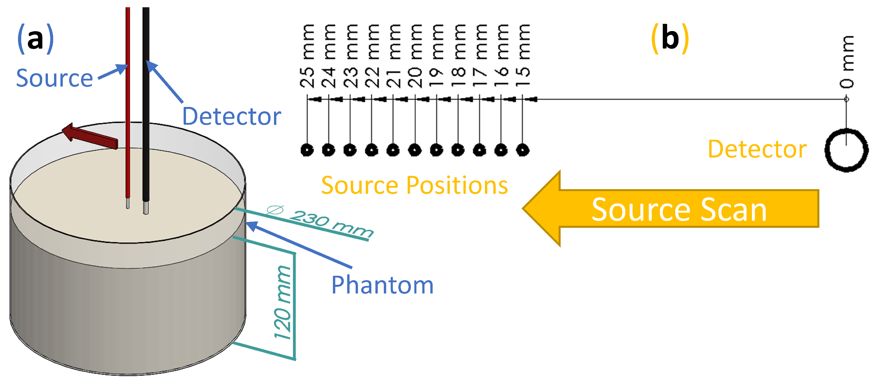
Abstract
:1. Introduction
2. Materials and Methods
2.1. Techniques
2.1.1. Frequency-Domain Near-Infrared Spectroscopy
2.1.2. Broadband Diffuse Reflectance Spectroscopy
2.2. Experiment
2.3. Analysis
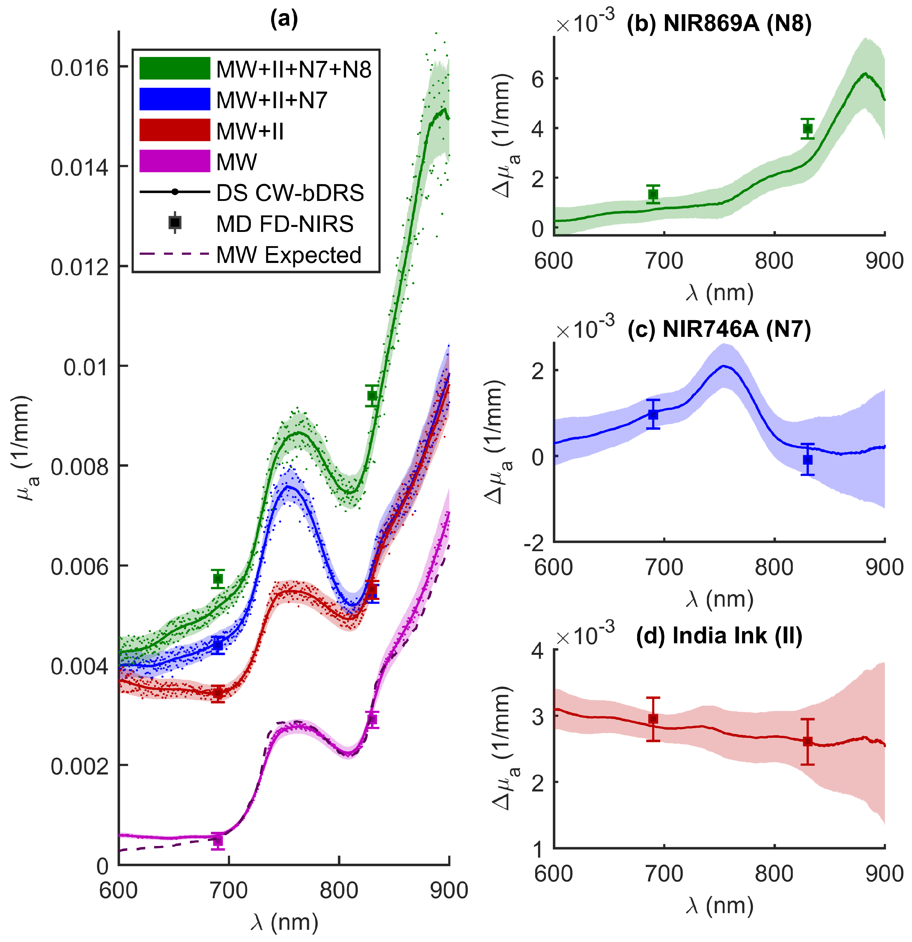
3. Results
4. Discussion
5. Conclusions
Author Contributions
Funding
Data Availability Statement
Conflicts of Interest
References
- Fantini, S.; Sassaroli, A. Frequency-Domain Techniques for Cerebral and Functional Near-Infrared Spectroscopy. Front. Neurosci. 2020, 14, 1–18. [Google Scholar] [CrossRef] [PubMed] [Green Version]
- Bigio, I.J.; Fantini, S. Quantitative Biomedical Optics; Cambridge University Press: Cambridge, UK, 2016. [Google Scholar]
- Scholkmann, F.; Kleiser, S.; Metz, A.J.; Zimmermann, R.; Mata Pavia, J.; Wolf, U.; Wolf, M. A review on continuous wave functional near-infrared spectroscopy and imaging instrumentation and methodology. NeuroImage 2014, 85, 6–27. [Google Scholar] [CrossRef]
- Ozaki, Y.; McClure, W.F.; Christy, A.A. Near-Infrared Spectroscopy in Food Science and Technology; John Wiley & Sons: Hoboken, NJ, USA, 2006. [Google Scholar]
- Johnson, J.B. An overview of near-infrared spectroscopy (NIRS) for the detection of insect pests in stored grains. J. Stored Prod. Res. 2020, 86, 101558. [Google Scholar] [CrossRef]
- Kademi, H.I.; Ulusoy, B.H.; Hecer, C. Applications of miniaturized and portable near infrared spectroscopy (NIRS) for inspection and control of meat and meat products. Food Rev. Int. 2019, 35, 201–220. [Google Scholar] [CrossRef]
- Razuc, M.; Grafia, A.; Gallo, L.; Ramírez-Rigo, M.V.; Romañach, R.J. Near-infrared spectroscopic applications in pharmaceutical particle technology. Drug Dev. Ind. Pharm. 2019, 45, 1565–1589. [Google Scholar] [CrossRef]
- Stranzinger, S.; Markl, D.; Khinast, J.G.; Paudel, A. Review of sensing technologies for measuring powder density variations during pharmaceutical solid dosage form manufacturing. TrAC Trends Anal. Chem. 2020, 116147. [Google Scholar] [CrossRef]
- Trant, P.L.K.; Kristiansen, S.M.; Sindbæk, S.M. Visible near-infrared spectroscopy as an aid for archaeological interpretation. Archaeol. Anthropol. Sci. 2020, 12, 280. [Google Scholar] [CrossRef]
- Tsuchikawa, S.; Kobori, H. A review of recent application of near infrared spectroscopy to wood science and technology. J. Wood Sci. 2015, 61, 213–220. [Google Scholar] [CrossRef] [Green Version]
- Chen, Z.; Gu, A.; Zhang, X.; Zhang, Z. Authentication and inference of seal stamps on Chinese traditional painting by using multivariate classification and near-infrared spectroscopy. Chemom. Intell. Lab. Syst. 2017, 171, 226–233. [Google Scholar] [CrossRef]
- Lange, F.; Tachtsidis, I. Clinical Brain Monitoring with Time Domain NIRS: A Review and Future Perspectives. Appl. Sci. 2019, 9, 1612. [Google Scholar] [CrossRef] [Green Version]
- Agbangla, N.F.; Audiffren, M.; Albinet, C.T. Use of near-infrared spectroscopy in the investigation of brain activation during cognitive aging: A systematic review of an emerging area of research. Ageing Res. Rev. 2017, 38, 52–66. [Google Scholar] [CrossRef] [PubMed]
- Grosenick, D.; Rinneberg, H.; Cubeddu, R.; Taroni, P. Review of optical breast imaging and spectroscopy. J. Biomed. Opt. 2016, 21, 091311. [Google Scholar] [CrossRef] [PubMed] [Green Version]
- Perrey, S.; Ferrari, M. Muscle Oximetry in Sports Science: A Systematic Review. Sport. Med. 2018, 48, 597–616. [Google Scholar] [CrossRef]
- Durduran, T.; Choe, R.; Baker, W.B.; Yodh, A.G. Diffuse optics for tissue monitoring and tomography. Rep. Prog. Phys. 2010, 73, 076701. [Google Scholar] [CrossRef] [Green Version]
- Jacques, S.L.; Pogue, B.W. Tutorial on diffuse light transport. J. Biomed. Opt. 2008, 13, 041302. [Google Scholar] [CrossRef] [Green Version]
- Fishkin, J.B.; Gratton, E. Propagation of photon-density waves in strongly scattering media containing an absorbing semi-infinite plane bounded by a straight edge. J. Opt. Soc. Am. A 1993, 10, 127. [Google Scholar] [CrossRef] [Green Version]
- Blaney, G.; Sassaroli, A.; Pham, T.; Krishnamurthy, N.; Fantini, S. Multi-Distance Frequency-Domain Optical Measurements of Coherent Cerebral Hemodynamics. Photonics 2019, 6, 83. [Google Scholar] [CrossRef] [Green Version]
- Hallacoglu, B.; Sassaroli, A.; Guerrero-Berroa, E.; Schnaider Beeri, M.; Haroutunian, V.; Shaul, M.; Rosenberg, I.H.; Toren, A.; Fantini, S. Absolute measurement of cerebral optical coefficients, hemoglobin concentration and oxygen saturation in old and young adults with near-infrared spectroscopy. J. Biomed. Opt. 2012, 17, 081406. [Google Scholar] [CrossRef] [PubMed] [Green Version]
- Fantini, S.; Hueber, D.; Franceschini, M.A.; Gratton, E.; Rosenfeld, W.; Stubblefield, P.G.; Maulik, D.; Stankovic, M.R. Non-invasive optical monitoring of the newborn piglet brain using continuous-wave and frequency-domain spectroscopy. Phys. Med. Biol. 1999, 44, 1543–1563. [Google Scholar] [CrossRef]
- Fantini, S.; Franceschini, M.A.; Gratton, E. Semi-infinite-geometry boundary problem for light migration in highly scattering media: A frequency-domain study in the diffusion approximation. JOSA B 1994, 11, 2128–2138. [Google Scholar] [CrossRef] [Green Version]
- Hueber, D.M.; Fantini, S.; Cerussi, A.E.; Barbieri, B.B. New optical probe designs for absolute (self-calibrating) NIR tissue hemoglobin measurements. In Optical Tomography and Spectroscopy of Tissue III; International Society for Optics and Photonics: San Jose, CA, USA, 1999; Volume 3597, pp. 618–631. [Google Scholar] [CrossRef]
- Scholkmann, F.; Metz, A.J.; Wolf, M. Measuring tissue hemodynamics and oxygenation by continuous-wave functional near-infrared spectroscopy—How robust are the different calculation methods against movement artifacts? Physiol. Meas. 2014, 35, 717–734. [Google Scholar] [CrossRef] [PubMed]
- Kleiser, S.; Ostojic, D.; Nasseri, N. In vivo precision assessment of a near-infrared spectroscopy-based tissue oximeter (OxyPrem v1.3) in neonates considering systemic hemodynamic fluctuations. J. Biomed. Opt. 2018, 23, 1. [Google Scholar] [CrossRef] [Green Version]
- Blaney, G.; Sassaroli, A.; Pham, T.; Fernandez, C.; Fantini, S. Phase dual-slopes in frequency-domain near-infrared spectroscopy for enhanced sensitivity to brain tissue: First applications to human subjects. J. Biophotonics 2020, 13. [Google Scholar] [CrossRef]
- Sassaroli, A.; Blaney, G.; Fantini, S. Dual-slope method for enhanced depth sensitivity in diffuse optical spectroscopy. J. Opt. Soc. Am. A 2019, 36, 1743. [Google Scholar] [CrossRef]
- Lanka, P.; Segala, A.; Farina, A.; Konugolu Venkata Sekar, S.; Nisoli, E.; Valerio, A.; Taroni, P.; Cubeddu, R.; Pifferi, A. Non-invasive investigation of adipose tissue by time domain diffuse optical spectroscopy. Biomed. Opt. Express 2020, 11, 2779. [Google Scholar] [CrossRef]
- Vasudevan, S.; Forghani, F.; Campbell, C.; Bedford, S.; O’Sullivan, T.D. Method for quantitative broadband diffuse optical spectroscopy of tumor-like inclusions. Appl. Sci. 2020, 10, 1419. [Google Scholar] [CrossRef] [Green Version]
- Ganesan, G.; Warren, R.V.; Leproux, A.; Compton, M.; Cutler, K.; Wittkopp, S.; Tran, G.; O’Sullivan, T.; Malik, S.; Galassetti, P.R.; et al. Diffuse optical spectroscopic imaging of subcutaneous adipose tissue metabolic changes during weight loss. Int. J. Obes. 2016, 40, 1292–1300. [Google Scholar] [CrossRef] [PubMed] [Green Version]
- Konugolu Venkata Sekar, S.; Dalla Mora, A.; Bargigia, I.; Martinenghi, E.; Lindner, C.; Farzam, P.; Pagliazzi, M.; Durduran, T.; Taroni, P.; Pifferi, A.; et al. Broadband (600–1350 nm) Time-Resolved Diffuse Optical Spectrometer for Clinical Use. IEEE J. Sel. Top. Quantum Electron. 2016, 22, 406–414. [Google Scholar] [CrossRef]
- O’Sullivan, T.D.; Cerussi, A.E.; Cuccia, D.J.; Tromberg, B.J. Diffuse optical imaging using spatially and temporally modulated light. J. Biomed. Opt. 2012, 17, 0713111. [Google Scholar] [CrossRef] [Green Version]
- Tachtsidis, I.; Gao, L.; Leung, T.S.; Kohl-Bareis, M.; Cooper, C.E.; Elwell, C.E. A Hybrid Multi-Distance Phase and Broadband Spatially Resolved Spectrometer and Algorithm for Resolving Absolute Concentrations of Chromophores in the Near-Infrared Light Spectrum. In Oxygen Transport to Tissue XXXI, Advances in Experimental Medicine and Biology; Springer: Boston, MA, USA, 2010; pp. 169–175. [Google Scholar] [CrossRef] [Green Version]
- Bevilacqua, F.; Berger, A.J.; Cerussi, A.E.; Jakubowski, D.; Tromberg, B.J. Broadband absorption spectroscopy in turbid media by combined frequency-domain and steady-state methods. Appl. Opt. 2000, 39, 6498. [Google Scholar] [CrossRef] [Green Version]
- Di Ninni, P.; Martelli, F.; Zaccanti, G. The use of India ink in tissue-simulating phantoms. Opt. Express 2010, 18, 26854. [Google Scholar] [CrossRef] [PubMed]
- Martelli, F.; Zaccanti, G. Calibration of scattering and absorption properties of a liquid diffusive medium at NIR wavelengths. CW method. Opt. Express 2007, 15, 486–500. [Google Scholar] [CrossRef] [PubMed]
- Water Soluble NIR Dyes—QCR Solutions Corp. Available online: https://qcrsolutions.com/water-soluble-near-infrared-dyes/ (accessed on 1 June 2020).
- Cugmas, B.; Naglič, P.; Menachery, S.P.M.; Pernuš, F.; Likar, B. Poor optical stability of molecular dyes when used as absorbers in water-based tissue-simulating phantoms. In Design and Quality for Biomedical Technologies XII; Liang, R., Hwang, J., Eds.; SPIE: San Francisco, CA, USA, 2019; Volume 10870, pp. 56–65. [Google Scholar] [CrossRef]
- Contini, D.; Martelli, F.; Zaccanti, G. Photon migration through a turbid slab described by a model based on diffusion approximation. I. Theory. Appl. Opt. 1997, 36, 4587–4599. [Google Scholar] [CrossRef] [PubMed]
- Pope, R.M.; Fry, E.S. Absorption spectrum (380–700 nm) of pure water II Integrating cavity measurements. Appl. Opt. 1997, 36, 8710. [Google Scholar] [CrossRef] [PubMed]
- Kou, L.; Labrie, D.; Chylek, P. Refractive indices of water and ice in the 065- to 25-mum spectral range. Appl. Opt. 1993, 32, 3531. [Google Scholar] [CrossRef] [PubMed] [Green Version]
- van Veen, R.; Sterenborg, H.; Pifferi, A.; Torricelli, A.; Cubeddu, R. Determination of VIS- NIR absorption coefficients of mammalian fat, with time- and spatially resolved diffuse reflectance and transmission spectroscopy. In Biomedical Topical Meeting; OSA: Washington, DC, USA, 2004; p. SF4. [Google Scholar] [CrossRef] [Green Version]


Publisher’s Note: MDPI stays neutral with regard to jurisdictional claims in published maps and institutional affiliations. |
© 2021 by the authors. Licensee MDPI, Basel, Switzerland. This article is an open access article distributed under the terms and conditions of the Creative Commons Attribution (CC BY) license (http://creativecommons.org/licenses/by/4.0/).
Share and Cite
Blaney, G.; Donaldson, R.; Mushtak, S.; Nguyen, H.; Vignale, L.; Fernandez, C.; Pham, T.; Sassaroli, A.; Fantini, S. Dual-Slope Diffuse Reflectance Instrument for Calibration-Free Broadband Spectroscopy. Appl. Sci. 2021, 11, 1757. https://0-doi-org.brum.beds.ac.uk/10.3390/app11041757
Blaney G, Donaldson R, Mushtak S, Nguyen H, Vignale L, Fernandez C, Pham T, Sassaroli A, Fantini S. Dual-Slope Diffuse Reflectance Instrument for Calibration-Free Broadband Spectroscopy. Applied Sciences. 2021; 11(4):1757. https://0-doi-org.brum.beds.ac.uk/10.3390/app11041757
Chicago/Turabian StyleBlaney, Giles, Ryan Donaldson, Samee Mushtak, Han Nguyen, Lydia Vignale, Cristianne Fernandez, Thao Pham, Angelo Sassaroli, and Sergio Fantini. 2021. "Dual-Slope Diffuse Reflectance Instrument for Calibration-Free Broadband Spectroscopy" Applied Sciences 11, no. 4: 1757. https://0-doi-org.brum.beds.ac.uk/10.3390/app11041757