
Mechanical and Electrical Properties of DNA Hydrogel-Based Composites Containing Self-Assembled Three-Dimensional Nanocircuits

 , , and
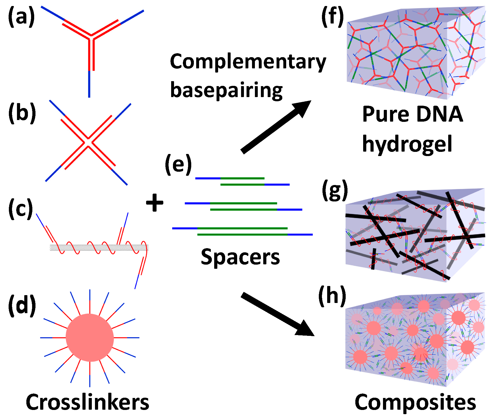
Abstract
:1. Introduction
2. Materials and Methods
3. Results
3.1. Sequence Design of Crosslinker and Spacer Strands for DNA Gel Formations
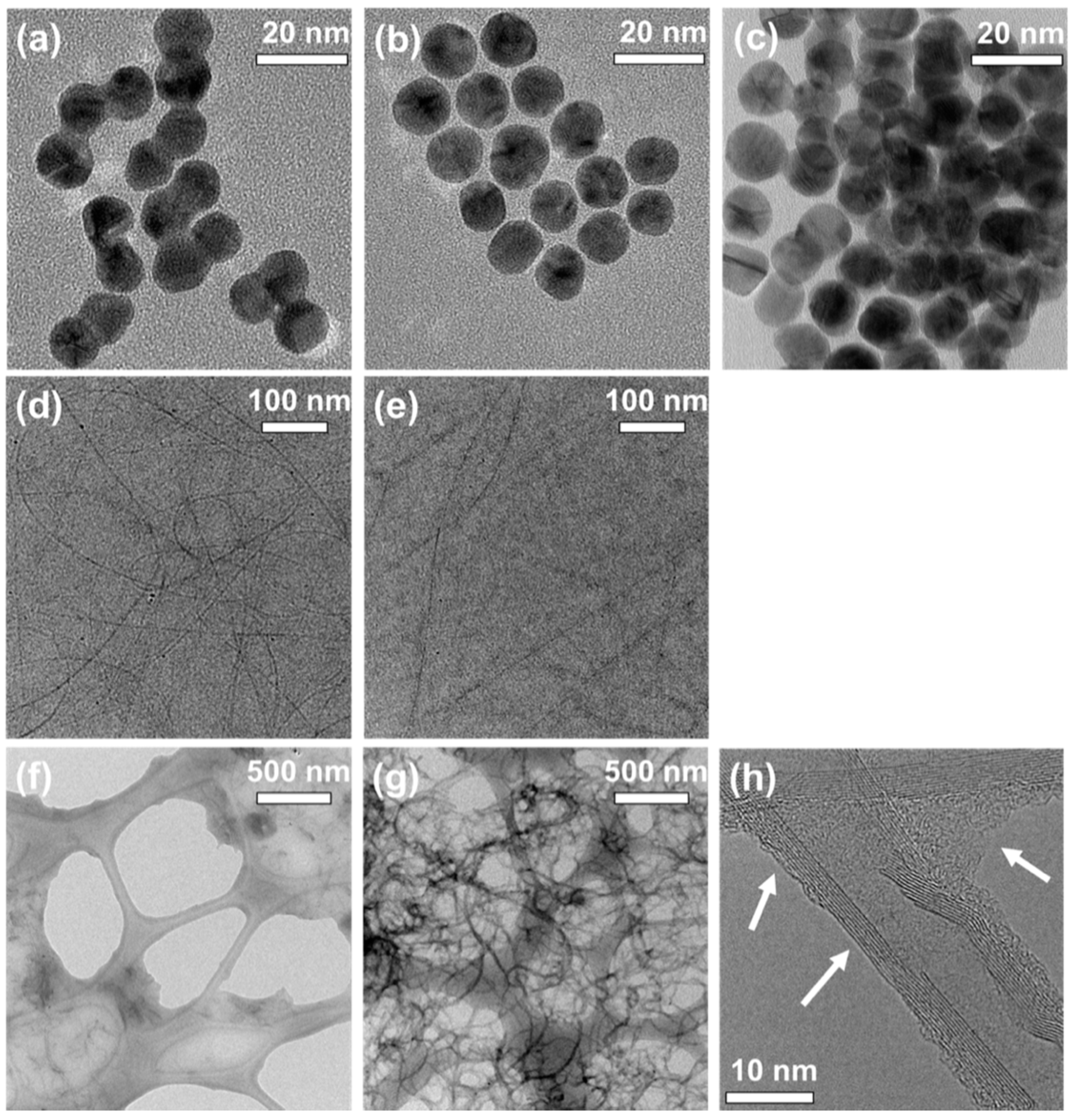
3.2. Characterization of Conjugates and Hydrogels
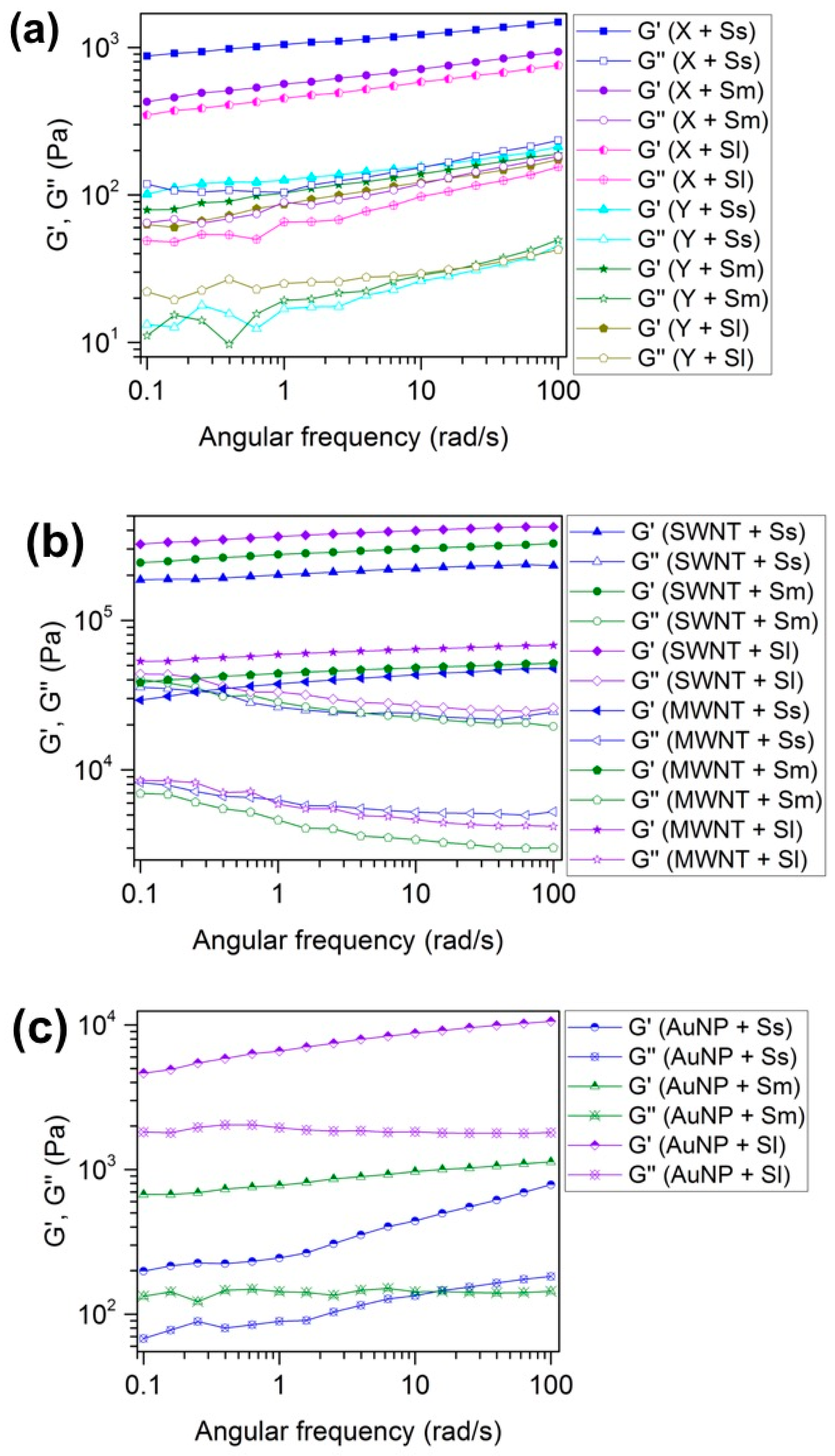
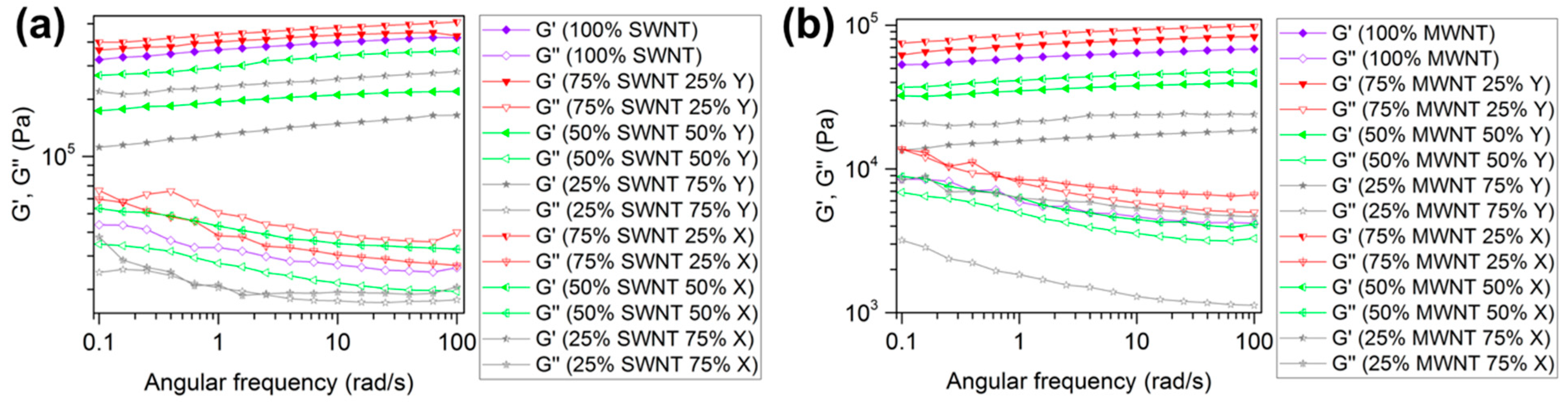
3.3. Rheological Properties of DNA Hydrogels
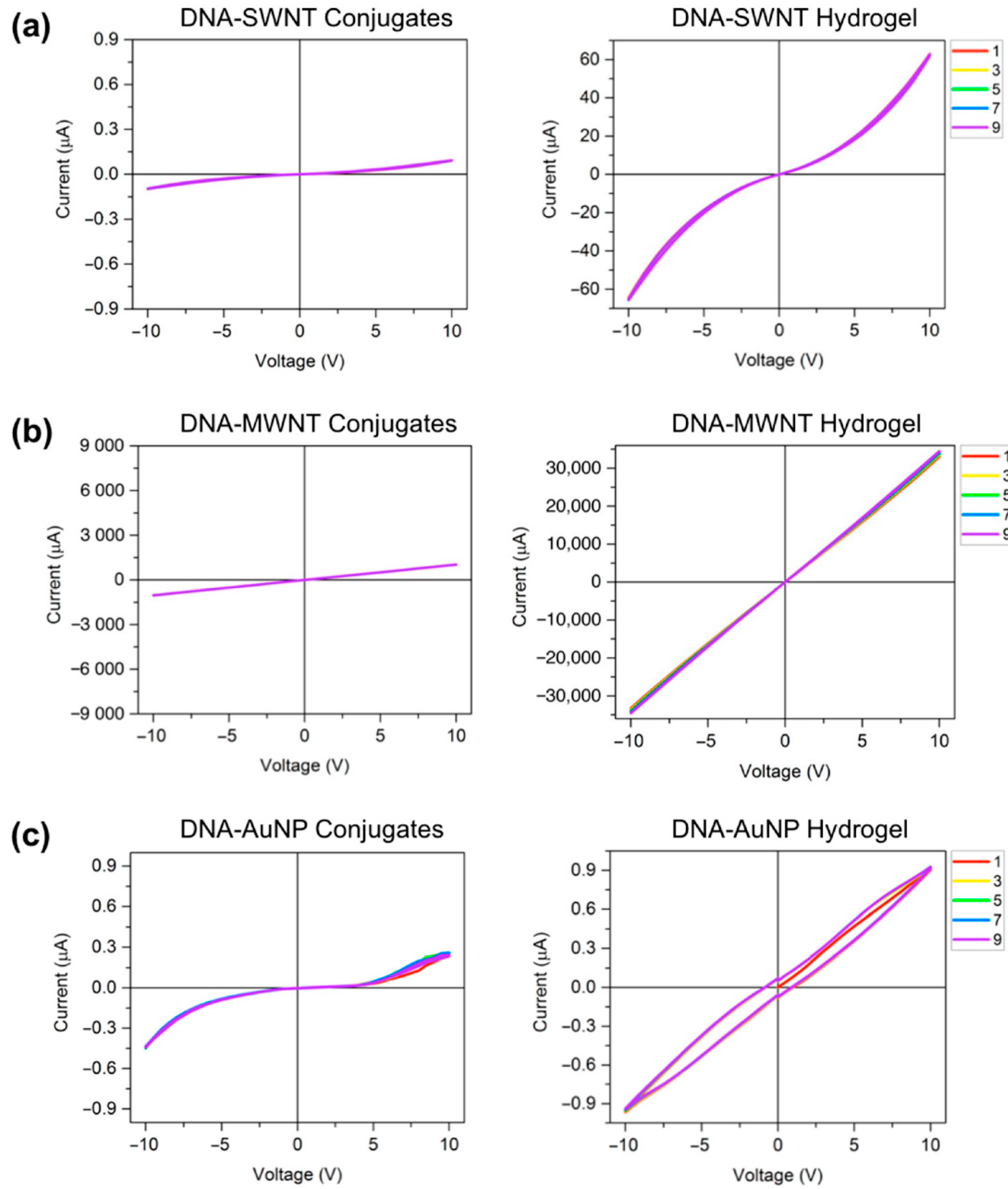
3.4. Electrical Characterization
4. Discussion
4.1. Characterizations
4.2. Rheological Results
4.3. Electrical Studies
5. Conclusions
Supplementary Materials
Author Contributions
Funding
Institutional Review Board Statement
Informed Consent Statement
Acknowledgments
Conflicts of Interest
References
- Winfree, E.; Liu, F.; Wenzler, L.A.; Seeman, N.C. Design and self-assembly of two-dimensional DNA crystals. Nature 1998, 394, 539–544. [Google Scholar] [CrossRef]
- Rothemund, P.W.K. Folding DNA to create nanoscale shapes and patterns. Nature 2006, 440, 297–302. [Google Scholar] [CrossRef] [PubMed] [Green Version]
- Douglas, S.M.; Dietz, H.; Liedl, T.; Högberg, B.; Graf, F.; Shih, W.M. Self-assembly of DNA into nanoscale three-dimensional shapes. Nature 2009, 459, 414–418. [Google Scholar] [CrossRef]
- Marchi, A.N.; Saaem, I.; Vogen, B.N.; Brown, S.; LaBean, T.H. Toward Larger DNA Origami. Nano Lett. 2014, 14, 5740–5747. [Google Scholar] [CrossRef]
- Wagenbauer, K.F.; Sigl, C.; Dietz, H. Gigadalton-scale shape-programmable DNA assemblies. Nature 2017, 552, 78–83. [Google Scholar] [CrossRef]
- Yan, H.; Park, S.H.; Finkelstein, G.; Reif, J.H.; LaBean, T.H. DNA-Templated Self-Assembly of Protein Arrays and Highly Conductive Nanowires. Science 2003, 301, 1882–1884. [Google Scholar] [CrossRef] [PubMed]
- Knudsen, J.; Liu, L.; Kodal, A.; Madsen, M.; Li, Q.; Song, J.; Woehrstein, J.; Wickham, S.; Strauss, M.; Schueder, F.; et al. Routing of individual polymers in designed patterns. Nat. Nanotechnol. 2015, 10, 892–898. [Google Scholar] [CrossRef]
- Krissanaprasit, A.; Madsen, M.; Knudsen, J.B.; Gudnason, D.A.; Surareungchai, W.; Birkedal, V.; Gothelf, K.V. Programmed Switching of Single Polymer Conformation on DNA Origami. ACS Nano 2016, 10, 2243–2250. [Google Scholar] [CrossRef] [PubMed]
- Madsen, M.; Christensen, R.S.; Krissanaprasit, A.; Bakke, M.R.; Riber, C.F.; Nielsen, K.S.; Zelikin, A.N.; Gothelf, K.V. Preparation, Single-Molecule Manipulation, and Energy Transfer Investigation of a Polyfluorene-graft -DNA polymer. Chem. A Eur. J. 2017, 23, 10511–10515. [Google Scholar] [CrossRef]
- Shahbazi, M.-A.; Bauleth-Ramos, T.; Santos, H.A. DNA Hydrogel Assemblies: Bridging Synthesis Principles to Biomedical Applications. Adv. Ther. 2018, 1, 1800042. [Google Scholar] [CrossRef]
- Li, F.; Lyu, D.; Liu, S.; Guo, W. DNA Hydrogels and Microgels for Biosensing and Biomedical Applications. Adv. Mater. 2020, 32, e1806538. [Google Scholar] [CrossRef]
- Lee, J.B.; Peng, S.; Yang, D.; Roh, Y.H.; Funabashi, H.; Park, N.; Rice, E.J.; Chen, L.; Long, R.; Wu, M.; et al. A mechanical metamaterial made from a DNA hydrogel. Nat. Nanotechnol. 2012, 7, 816–820. [Google Scholar] [CrossRef] [PubMed]
- Liu, H.; Cao, T.; Xu, Y.; Dong, Y.; Liu, D. Tuning the Mechanical Properties of a DNA Hydrogel in Three Phases Based on ATP Aptamer. Int. J. Mol. Sci. 2018, 19, 1633. [Google Scholar] [CrossRef] [PubMed] [Green Version]
- Khajouei, S.; Ravan, H.; Ebrahimi, A. DNA hydrogel-empowered biosensing. Adv. Colloid Interface Sci. 2020, 275, 102060. [Google Scholar] [CrossRef] [PubMed]
- Thelu, H.V.P.; Atchimnaidu, S.; Perumal, D.; Harikrishnan, K.S.; Vijayan, S.; Varghese, R. Self-Assembly of an Aptamer-Decorated, DNA—Protein Hybrid Nanogel: A Biocompatible Nanocarrier for Targeted Cancer Therapy. ACS Appl. Bio Mater. 2019, 2, 5227–5234. [Google Scholar] [CrossRef]
- Gačanin, J.; Synatschke, C.V.; Weil, T. Biomedical Applications of DNA-Based Hydrogels. Adv. Funct. Mater. 2020, 30, 1906253. [Google Scholar] [CrossRef] [Green Version]
- Cheng, E.; Xing, Y.; Chen, P.; Yang, Y.; Sun, Y.; Zhou, D.; Xu, L.; Fan, Q.; Liu, D. A pH-Triggered, Fast-Responding DNA Hydrogel. Angew. Chem. Int. Ed. 2009, 48, 7660–7663. [Google Scholar] [CrossRef] [PubMed]
- Li, C.; Faulkner-Jones, A.; Dun, A.R.; Jin, J.; Chen, P.; Xing, Y.; Yang, Z.; Li, Z.; Shu, W.; Liu, D.; et al. Rapid Formation of a Supramolecular Polypeptide-DNA Hydrogel for In Situ Three-Dimensional Multilayer Bioprinting. Angew. Chem. Int. Ed. 2015, 54, 3957–3961. [Google Scholar] [CrossRef]
- Zhou, D.; Ying, L.; Hong, X.; Hall, E.A.; Abell, C.; Klenerman, D. A Compact Functional Quantum Dot—DNA Conjugate: Preparation, Hybridization, and Specific Label-Free DNA Detection. Langmuir 2008, 24, 1659–1664. [Google Scholar] [CrossRef] [Green Version]
- Hurst, S.J.; Lytton-Jean, A.K.R.; Mirkin, C.A. Maximizing DNA Loading on a Range of Gold Nanoparticle Sizes. Anal. Chem. 2006, 78, 8313–8318. [Google Scholar] [CrossRef] [PubMed] [Green Version]
- Li, Z.; Wu, Z.; Li, K. The high dispersion of DNA—multiwalled carbon nanotubes and their properties. Anal. Biochem. 2009, 387, 267–270. [Google Scholar] [CrossRef]
- Hu, Y.; Domínguez, C.M.; Bauer, J.; Weigel, S.; Schipperges, A.; Oelschlaeger, C.; Willenbacher, N.; Keppler, S.; Bastmeyer, M.; Heißler, S.; et al. Carbon-nanotube reinforcement of DNA-silica nanocomposites yields programmable and cell-instructive biocoatings. Nat. Commun. 2019, 10, 1–14. [Google Scholar] [CrossRef] [Green Version]
- Kyrylyuk, A.V.; Hermant, M.C.; Schilling, T.; Klumperman, B.; Koning, C.E.; van der Schoot, P. Controlling electrical percolation in multicomponent carbon nanotube dispersions. Nat. Nanotechnol. 2011, 6, 364–369. [Google Scholar] [CrossRef]
- Vardharajula, S.; Ali, S.Z.; Tiwari, P.M.; Eroğlu, E.; Vig, K.; Dennis, V.A.; Singh, S.R. Functionalized carbon nanotubes: Biomedical applications. Int. J. Nanomed. 2012, 7, 5361–5374. [Google Scholar] [CrossRef] [Green Version]
- Nakashima, N.; Okuzono, S.; Murakami, H.; Nakai, T.; Yoshikawa, K. DNA Dissolves Single-walled Carbon Nanotubes in Water. Chem. Lett. 2003, 32, 456–457. [Google Scholar] [CrossRef]
- Zheng, M.; Jagota, A.; Semke, E.D.; Diner, B.A.; McLean, R.S.; Lustig, S.R.; Richardson, R.E.; Tassi, N.G. DNA-assisted dispersion and separation of carbon nanotubes. Nat. Mater. 2003, 2, 338–342. [Google Scholar] [CrossRef] [PubMed]
- Dovbeshko, G.; Repnytska, O.; Obraztsova, E.; Shtogun, Y. DNA interaction with single-walled carbon nanotubes: A SEIRA study. Chem. Phys. Lett. 2003, 372, 432–437. [Google Scholar] [CrossRef]
- Chen, Y.; Liu, H.; Ye, T.; Kim, A.J.; Mao, C. DNA-Directed Assembly of Single-Wall Carbon Nanotubes. J. Am. Chem. Soc. 2007, 129, 8696–8697. [Google Scholar] [CrossRef] [PubMed]
- Rueckes, T.; Kim, K.; Joselevich, E.; Tseng, G.Y.; Cheung, C.-L.; Lieber, C.M. Carbon Nanotube-Based Nonvolatile Random Access Memory for Molecular Computing. Science 2000, 289, 94–97. [Google Scholar] [CrossRef] [Green Version]
- Kang, J.W.; Lee, J.H.; Lee, H.J.; Hwang, H.J. A study on carbon nanotube bridge as a electromechanical memory device. Phys. E Low Dimens. Syst. Nanostruct. 2005, 27, 332–340. [Google Scholar] [CrossRef]
- Li, D.; Song, S.; Fan, C. Target-Responsive Structural Switching for Nucleic Acid-Based Sensors. Acc. Chem. Res. 2010, 43, 631–641. [Google Scholar] [CrossRef]
- Lin, C.; Liu, Y.; Yan, H. Designer DNA Nanoarchitectures. Biochemistry 2009, 48, 1663–1674. [Google Scholar] [CrossRef] [PubMed] [Green Version]
- Mirkin, C.A.; Letsinger, R.L.; Mucic, R.C.; Storhoff, J.J. A DNA-based method for rationally assembling nanoparticles into macroscopic materials. Nature 1996, 382, 607–609. [Google Scholar] [CrossRef] [PubMed]
- Alivisatos, A.P.; Johnsson, K.P.; Peng, X.; Wilson, T.E.; Loweth, C.J.; Bruchez, M.P.; Schultz, P.G. Organization of ‘nanocrystal molecules’ using DNA. Nature 1996, 382, 609–611. [Google Scholar] [CrossRef] [Green Version]
- Storhoff, J.J.; Elghanian, R.; Mirkin, C.A.; Letsinger, R.L. Sequence-Dependent Stability of DNA-Modified Gold Nanoparticles. Langmuir 2002, 18, 6666–6670. [Google Scholar] [CrossRef]
- Hill, H.D.; Mirkin, C.A. The bio-barcode assay for the detection of protein and nucleic acid targets using DTT-induced ligand exchange. Nat. Protoc. 2006, 1, 324–336. [Google Scholar] [CrossRef] [PubMed]
- Haiss, W.; Thanh, N.T.K.; Aveyard, J.; Fernig, D.G. Determination of Size and Concentration of Gold Nanoparticles from UV—Vis Spectra. Anal. Chem. 2007, 79, 4215–4221. [Google Scholar] [CrossRef] [PubMed]
- Cheng, E.; Li, Y.; Yang, Z.; Deng, Z.; Liu, D. DNA-SWNT hybrid hydrogel. Chem. Commun. 2011, 47, 5545–5547. [Google Scholar] [CrossRef]
- Zhang, X.; Liu, B.; Dave, N.; Servos, M.R.; Liu, J. Instantaneous Attachment of an Ultrahigh Density of Nonthiolated DNA to Gold Nanoparticles and Its Applications. Langmuir 2012, 28, 17053–17060. [Google Scholar] [CrossRef] [Green Version]
- Xing, Y.; Cheng, E.; Yang, Y.; Chen, P.; Zhang, T.; Sun, Y.; Yang, Z.; Liu, D. Self-Assembled DNA Hydrogels with Designable Thermal and Enzymatic Responsiveness. Adv. Mater. 2010, 23, 1117–1121. [Google Scholar] [CrossRef]
- Um, S.H.; Lee, J.B.; Park, N.; Kwon, S.Y.; Umbach, C.C.; Luo, D. Enzyme-catalysed assembly of DNA hydrogel. Nat. Mater. 2006, 5, 797–801. [Google Scholar] [CrossRef] [PubMed]
- Zheng, M.; Jagota, A.; Strano, M.S.; Santos, A.P.; Barone, P.; Chou, S.G.; Diner, B.A.; Dresselhaus, M.S.; McLean, R.S.; Onoa, G.B.; et al. Structure-Based Carbon Nanotube Sorting by Sequence-Dependent DNA Assembly. Science 2003, 302, 1545–1548. [Google Scholar] [CrossRef] [PubMed] [Green Version]
- Lahiji, R.R.; Dolash, B.D.; Bergstrom, D.E.; Reifenberger, R. Oligodeoxyribonucleotide Association with Single-Walled Carbon Nanotubes Studied by SPM. Small 2007, 3, 1912–1920. [Google Scholar] [CrossRef] [PubMed]
- Yang, Q.-H.; Wang, Q.; Gale, N.; Oton, C.J.; Cui, L.; Nandhakumar, I.S.; Zhu, Z.; Tang, Z.; Brown, T.; Loh, W.H. Loosening the DNA wrapping around single-walled carbon nanotubes by increasing the strand length. Nanotechnology 2009, 20, 195603. [Google Scholar] [CrossRef]
- Eguchi, Y.; Kato, T.; Tanaka, T.; Maruyama, T. A DNA–gold nanoparticle hybrid hydrogel network prepared by enzymatic reaction. Chem. Commun. 2017, 53, 5802–5805. [Google Scholar] [CrossRef] [Green Version]
- Li, F.; Tang, J.; Geng, J.; Luo, D.; Yang, D. Polymeric DNA hydrogel: Design, synthesis and applications. Prog. Polym. Sci. 2019, 98, 101163. [Google Scholar] [CrossRef]
- Chambon, F.; Winter, H.H. Linear Viscoelasticity at the Gel Point of a Crosslinking PDMS with Imbalanced Stoichiometry. J. Rheol. 1987, 31, 683–697. [Google Scholar] [CrossRef]
- Cardenas-Vasquez, E.D.; Smith, K.M.; Doolan, T.J.; Hsiao, L.C. Shear-Induced Microstructural Variations in Nanoemulsion-Laden Organohydrogel Fibers. ACS Appl. Polym. Mater. 2019, 2, 594–603. [Google Scholar] [CrossRef]
- Nöll, T.; Schönherr, H.; Wesner, D.; Schopferer, M.; Paululat, T.; Nöll, G. Construction of Three-Dimensional DNA Hydrogels from Linear Building Blocks. Angew. Chem. Int. Ed. 2014, 53, 8328–8332. [Google Scholar] [CrossRef]
- Jiang, H.; Pan, V.; Vivek, S.; Weeks, E.R.; Ke, Y. Programmable DNA Hydrogels Assembled from Multidomain DNA Strands. ChemBioChem 2016, 17, 1156–1162. [Google Scholar] [CrossRef]
- Campbell, J.F.; Tessmer, I.; Thorp, H.H.; Erie, D.A. Atomic Force Microscopy Studies of DNA-Wrapped Carbon Nanotube Structure and Binding to Quantum Dots. J. Am. Chem. Soc. 2008, 130, 10648–10655. [Google Scholar] [CrossRef] [PubMed]
- Navarro, C.G.; de Pablo, P.; Herrero, J.G. Electrical properties of long molecules: Single-walled carbon nanotubes and DNA. Int. J. Nanotechnol. 2005, 2, 90. [Google Scholar] [CrossRef]


Publisher’s Note: MDPI stays neutral with regard to jurisdictional claims in published maps and institutional affiliations. |
© 2021 by the authors. Licensee MDPI, Basel, Switzerland. This article is an open access article distributed under the terms and conditions of the Creative Commons Attribution (CC BY) license (http://creativecommons.org/licenses/by/4.0/).
Share and Cite
Gao, M.; Krissanaprasit, A.; Miles, A.; Hsiao, L.C.; LaBean, T.H. Mechanical and Electrical Properties of DNA Hydrogel-Based Composites Containing Self-Assembled Three-Dimensional Nanocircuits. Appl. Sci. 2021, 11, 2245. https://0-doi-org.brum.beds.ac.uk/10.3390/app11052245
Gao M, Krissanaprasit A, Miles A, Hsiao LC, LaBean TH. Mechanical and Electrical Properties of DNA Hydrogel-Based Composites Containing Self-Assembled Three-Dimensional Nanocircuits. Applied Sciences. 2021; 11(5):2245. https://0-doi-org.brum.beds.ac.uk/10.3390/app11052245
Chicago/Turabian StyleGao, Ming, Abhichart Krissanaprasit, Austin Miles, Lilian C. Hsiao, and Thomas H. LaBean. 2021. "Mechanical and Electrical Properties of DNA Hydrogel-Based Composites Containing Self-Assembled Three-Dimensional Nanocircuits" Applied Sciences 11, no. 5: 2245. https://0-doi-org.brum.beds.ac.uk/10.3390/app11052245