Review

Type I Collagen-Based Devices to Treat Nerve Injuries after Oral Surgery Procedures. A Systematic Review

by
Andrea Roccuzzo
1,2,*,†,
Pedro Molinero-Mourelle
3,4,*,†,
Martina Ferrillo
5,*,
Carlos Cobo-Vázquez
6,7,
Luis Sanchez-Labrador
6,
Antonio Ammendolia
8,
Mario Migliario
9 and
Alessandro de Sire
8
1
Department of Periodontology, School of Dental Medicine, University of Bern, Freiburgstrasse 7, 3010 Bern, Switzerland
2
Department of Oral and Maxillofacial Surgery, Copenhagen University Hospital (Rigshospitalet), 2100 Copenhagen, Denmark
3
Department of Reconstructive Dentistry and Gerodontology, School of Dental Medicine, University of Bern, Freiburgstrasse 7, 3010 Bern, Switzerland
4
Department of Conservative Dentistry and Orofacial Prosthodontics, Faculty of Dentistry, Complutense University of Madrid, Plaza de Ramón y Cajal S/N, 28040 Madrid, Spain
5
Dental School, Department of Surgical Sciences, University of Turin, Via Nizza 230, 10126 Turin, Italy
6
Department of Dental Clinical Specialties, Faculty of Dentistry, Complutense University of Madrid, Plaza de Ramón y Cajal S/N, 28040 Madrid, Spain
7
Department of Dentistry and Stomatology, Gregorio Marañon University General Hospital, Calle Doctor Esquerdo 46, 28007 Madrid, Spain
8
Department of Medical and Surgical Sciences, University of Catanzaro “Magna Graecia”, Viale Europa, 88100 Catanzaro, Italy
9
Department of Translational Medicine, University of Eastern Piedmont, Via Solaroli 17, 10121 Novara, Italy
*
Authors to whom correspondence should be addressed.
These authors share the first author position.
Submission received: 24 March 2021 / Revised: 19 April 2021 / Accepted: 20 April 2021 / Published: 26 April 2021
(This article belongs to the Special Issue Musculoskeletal Rehabilitation)

Abstract

:
The regeneration of nerve injuries after oral surgery procedures is a quite often attempted procedure in dental medicine. Despite several proposed technical approaches, there is still a lack of consensus on which should be considered the gold standard procedure, even-though in the last decades, the use of collagen-based devices allowing a tension-free direct neurorrhaphy has been used. A systematic search of multiple electronic databases and hand searching was conducted to assess the level of evidence behind the use of type I collagen devices to treat nerve injuries after oral surgery procedures. After screening, four articles (one case series and three retrospective studies) including overall 65 patients suffering from inferior alveolar (IAN)/lingual nerve (LN) injury after mandibular wisdom tooth extraction, met the inclusion criteria and could be included. The Oxford Centre for evidence-based medicine (OCEBM) scaling system was used to evaluate the quality of the included studies. Positive clinical results in terms of sensorial improvements were recorded at least 3 months after surgery, even-though the overall level of evidence is low. The use of collagen membranes to enhance nerve regeneration in oral surgery results in promising results. Nevertheless, additional clinical comparative trials with larger sample sizes are needed.

1. Introduction

Mandibular wisdom tooth removal is one of the most frequent oral surgery procedures performed in the clinical practice [1,2]. Nonetheless, this intervention is not free from complications as widely reported in the literature [3,4,5]: in particular, trigeminal injuries might be caused mainly by nerve compression and/or stretching. The incidence is up to 5% for the inferior alveolar nerve (IAN) [3] and ranges from 0.6% to 2% for the lingual nerve (LN) [6]. Recently, a large retrospective university-setting clinical study on 1559 patients, reported sensorial disturbance in 42 cases (2.69%) with only five cases of persistent sensory disturbance (0.32%), and four of these five lesions were in the lingual nerve (0.25%) [7]. Consequently, despite the overall low percentage of complications, oro-facial disturbances cause considerable patients’ morbidity in terms of social disabilities and physical pain negatively affecting the whole quality of life.
More specifically, as summarized by Coulthard and coworkers in 2014 [8], the impaired sensation caused by lesions at the inferior alveolar nerve has been classified as:
  • Paresthesia—An abnormal sensation, whether spontaneous or evoked.
  • Anesthesia—Complete absence of perception of stimuli including touch.
  • Dysesthesia—An unpleasant abnormal sensation, whether spontaneous or evoked.
  • Hyperalgesia—Increased pain from a stimulus that normally provokes pain.
  • Allodynia—Pain due to a stimulus that does not normally provoke pain.
  • Hypoesthesia—Decreased sensitivity to stimulation, excluding the special senses.
  • Hyperesthesia—Increased sensitivity to stimulation, excluding the special senses.
  • The injury to the lingual nerve may also affect taste perception on the same side.
  • Ageusia—Loss of taste perception.
  • Dysgeusia—Altered taste perception.
At the present time, a multitude of different surgical interventions has been proposed to treat nerve injuries at the IAN and LN [9], even-though no consensus has been yet reached [6]. Historically, the use of nerve grafts of autologous origin has been adopted: indeed, in cases of complete nerve injury where a tight approximation of the two nerve edges is not possible, the use of an interpositional graft (i.e., sural, greater auricular or antebrachial nerves) has been propagated mainly with the aim to avoid nerve tension, but to allow physical reconnection of the two injured nerve sections [10]. Nevertheless, the use of autologous nerve grafts often resulted in undesirable post-operative sequalae at the donor site [11].
To overcome these side effects, during the last three decades, the use of hollow devices by means of a technique called stabilization has been proposed [12]. This procedure in intended to enhance nerve repair in cases of defects, which do not allow an intimate contact between the two injured nerve sections by means of a graft material.
Originally proposed with autogenous material by Pogrel and coworkers [13] by means of the use of the saphenous vein to repair IAN and LN injuries, alloplastic hollow conduits of different materials such as polyglycolic acid (PGA) bioabsorbable [14], Gore-Tex [15] and PGA-collagen tubes [16] have been launched to the market. Moreover, the FDA has also authorized the commercialization of type I collagen medical device to treat nerve injuries at IAN and LN, considering that they allow an appropriate nutrient diffusion and retention of representative nerve growth factor [17]. Until now, however, no attempt has been done to summarize the level of evidence behind the clinical use of such devices oral-surgery procedures.
Therefore, the purpose of the present systematic review was to evaluate the current evidence of on the use of type I collagen-based devices for mandibular or lingual nerve injuries regeneration after oral surgery procedures.

2. Materials and Methods

The present systematic review followed the PRISMA-P 2015 statement for reporting systematic reviews (preferred reporting items for systematic reviews and meta-analyses-protocols) [18].

2.1. PICO Question

The following detailed and structured question was developed according to population, intervention, comparison and outcome (PICO):
  • Population (P): Patients suffering nerve injuries at the inferior alveolar/facial nerve after oral surgery procedures.
  • Interventions (I): Microsurgical nerve regeneration approach with type I collagen devices.
  • Comparison (C): Nihil.
  • Outcomes (O): Nerve injuries regeneration assessed with patient’s self-reported scale and/or standardized scales.

2.2. Search Strategy

A comprehensive search of the literature was performed from the inception until 15 March 2021. Multiple electronic databases National Library of Medicine (MEDLINE (PubMed)), Web of Science (WOS) and Cochrane Central Register of Controlled Trials (CENTRAL) and open gray website were screened by means of different combination of medical subject headings (MeSH terms) words. The search strategy was first developed for PubMed and later adapted for the other databases, according to their specific characteristics as controlled vocabulary, wildcards, syntax rules and any other search features. Details of the search strategy are reported in Table 1.

2.3. Inclusion and Exclusion Criteria for Study Selection

Only articles published in English, Spanish and Italian languages were taken into consideration. No restrictions regarding gender, age or publication date were applied. In addition, a manual search of the reference lists of included full text articles and of the journals listed was conducted. Any in vitro, preclinical and animal studies, single case reports, studies reporting on data based on questionnaires and interviews, letters to the editors, PhD thesis, conference proceedings and all studies not meeting the inclusion criteria, were excluded. Details of the inclusion and exclusion criteria are provided in Table 2.

2.4. Inclusion Criteria for Study Selection

Any clinical human study (randomized clinical trials, prospective cohort studies, retrospective studies, case series and cross-sectional studies) with at least 5 patients who underwent microsurgical intervention for the treatment of NAI and/or LN with the use of a type I collagen device, were included in the present review. Patients had to be followed for at least 3 months a year after surgical intervention.

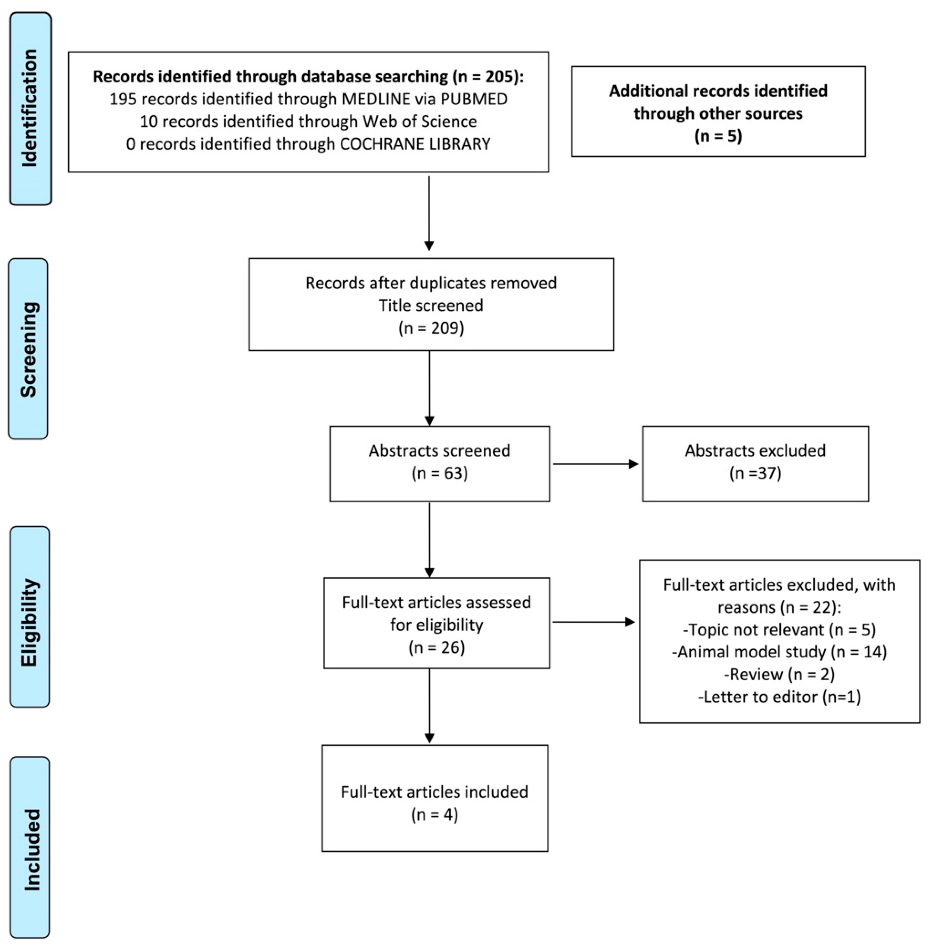
2.5. Study Selection

Retrieved data were screened independently by two of the authors (A.R. and P.M.-M.) using dedicated extraction table sheets prepared during the developing phase of the protocol. Any disagreement was solved through discussion consulting a third experienced reviewer (M.F.). Titles and abstracts identified through the search phase, which did not meet the inclusion criteria or did not provide significant information regarding the investigated technique, were excluded.

2.6. Data Extraction Process

Data extraction from full-text articles was performed in duplicate by two of the authors (A.R. and P.M.-M.) Any disagreement was solved through discussion consulting a third experienced reviewer (M.F.). All retrieved data were then discussed among all authors to reach consensus.
The following parameters, whenever available, were extracted: authors, year of publication, study design and setting, number of patients, oral surgery procedure, type I collagen device applied, adjunctive material, intervention outcomes, follow-up, time interval between injury and repair and adverse effects.

2.7. Quality Assessment of the Level of Evidence

The same two reviewers (A.R. and P.M.-M.) independently evaluated the methodological quality of all included studies using a dedicated quality assessment form according to the Oxford Centre for Evidence-Based Medicine (OCEBM) (https://www.cebm.ox.ac.uk/resources/levels-of-evidence/ocebm-levels-of-evidence) (17 March 2021) from level “1” to level “4”. Moreover, any disagreement was solved through discussion with a third party (M.F.).

2.8. Data Synthesis

The preliminary analysis of the data revealed an overall low level of evidence and a high heterogeneity among studies. However, the authors attempted to report the available data through a qualitative analysis.

3. Results

The systematic screening and selection process is outlined in Figure 1. During the identification process, 210 records were retrieved. Of these, 209 studies were considered after duplicates removal. After titles screening, 63 abstracts were analyzed. The full-text analysis was performed for 26 articles. Finally, 4 studies fulfilled the inclusion criteria and were therefore included. Details of studies’ characteristics are provided in Table 3.

3.1. Study Design, Patients’ Characteristics, Cause and Location of Nerve Injury

The four included studies [19,20,21,22,23] were: one case series and three retrospective studies. Combining the sample-size gathered from the four studies, 65 patients with 64 LN and three IAN injuries were evaluated. All the included patients suffered nerve injuries following third mandibular molar extraction. However, it has to be underlined that, even-though one study [20] included patients who experienced nerve injury after different events (i.e., sagittal split osteotomy, local anesthetic delivery, gunshot wound, second molar extraction, tumor excision and mandible fracture), only patients’ (n = 8) data related to the investigated procedure, were extracted and reported. All the included studies clearly reported on nerve disturbances ranging from no dysesthesia, dysesthesia, numbness with/without pain, complete hypoesthesia, neurotmesis and axonotmesis.

3.2. Time Interval between Injury and Repair and Type I Collagen Membrane Characteristics

The time interval passed between injury and the surgical intervention for the nerve repair was clearly reported in all the studies with a range from 3 to 36 months. Different surgical approaches (i.e., external neurolysis, primary neurorrhaphy and excision with microsurgery) were applied. Intraoperatively, the collagen devices used for the nerve surgery were in three articles, a collagen type I conduit (NeuraGen Integra LifeSciences, Plainsboro, NJ, USA) while in one case, a collagen cuff (Neuroflex; Collagen Matrix, Franklin Lakes, NJ, USA). The follow-up period after surgery ranged from 3 to 30 months.

3.3. Nerve Recovering Outcomes and Overall Assessment of the Quality of Evidence

All the included studies reported perception improvement after surgery. More in detail, Farole and coworkers [19] reported a good improvement in four lesions, four with some recovering and one with no improvement. In the other hand, Baghery and coworkers [20] found complete return of sensation in 90.5% and no or inadequate improvement in 9.5% patients. All patients treated by Erakat and coworkers [21] returned to successful functional sensory recovery. Finally, Wilson and coworkers reported a mean functional sensory recover of S3+ grade in the medical research council (MRC) scale (superficial pain and touch without hyperesthesia/good stimulus localization; static discrimination of 7–15 mm). The nerve recovery assessment was the Pogrel’s classification in one study [14], while three studies used the MRC scale.
All the included studies were categorized as level 4 of evidence according to the OCEBM (i.e., poor quality cohort and case-control studies).

4. Discussion

The aim of the present systematic review was to assess the level of evidence behind the clinical use of type I collagen-based devices to treat nerve injuries after oral surgery procedures. Based on the available literature, only four studies on 65 patients, all of them with a low-level of evidence, fulfilled the inclusion criteria and could be included.
Microsurgical peripheral nerve repair has been extensively investigated especially with respect to hand nerve, wrist and brachial plexus [17]. Historically, autogenous nerve grafts of secondary importance have been used as autografts with good results in term of sensibility regeneration [23,24,25]. However, due the high percentage of post-operative morbidity, loss of function at the donor site and the limited handling related to the grafted nerve sizes [26], alterative treatment options such as the use of nerve conduits have become popular in augmentative nerve grafting [27]. Indeed, the use of a conduits allowed a tension-free direct neurorrhaphy besides avoiding complicated nerve harvesting procedures [28]. The mechanism behind their use is to create a 3-D chamber to promote nerve regeneration and at the same time isolate the injured nerve from the surrounding tissues [29,30]. Among all the various investigated materials (i.e., silicons, PGAs and collagens), the scientific interest on the use of collagen devices have increased due to their characteristics such as maximal biocompatibility, the ability to stimulate different cell types adhesion and to guarantee a long-term cell proliferation [31,32]; nonetheless its use has been proved to be effective mainly in small nerve injury defects (i.e., up to 20 mm) [17]. Despite the level of evidence gathered from the animal model models [32,33], only few clinical studies have tested its reliability in a clinical model with positive promising short-mid-term results in term of functional sensory recovery [34,35]: in particular, Farole et al. [19] (i.e., 8/9 patients) and Erakat et al. [21] (i.e., 22/22 patients) reported significant improvement in terms of functional sensory recovery.
In the literature there is very little evidence of retrospective/prospective studies comparing different grafting materials: on this topic, Wilson and coworkers [23] reported clinical data from a retrospective study on 43 patients treated whether with a collagen conduit (NeuraGen) or with a porcine small intestinal submucosa conduit (AxoGuard) after LN injury. At the latest follow-up examination, no statistically significant differences in terms of achieved functional sensory recovery (FSR) were recorded, which leads to the conclusion that both treatment modalities might be reliable options.
Irrespective to the selected surgical intervention, one aspect that has been proved to be crucial to increase the chances of sensory recovery is the timing: indeed, as reported by Kushnerev and Yates [9], surgery is indicated if neurosensory deficit does not show any improvement 3 months after injury. When evaluating the results obtained from the included studies, despite the wide reported time range (i.e., 2–36 months), all treated patients underwent surgical intervention at least 2 months after injury: therefore, it seems recommended to postpone the decision to surgically treat these injuries after this time lapse.
Due to its promising results in the oro-facial district, the use of type I collagen devices has gained interested in the in orthopedic surgery with a high interest in the rehabilitation field. To date, Lee and coworkers [36] evaluated the effect of wrapping bioabsorbable nerve conduit around primary suture repair on motor nerve regeneration in 40 rats. These latter were randomly allocated into two experimental groups according to the type of repair of the rat sciatic nerve: group I (primary suture repair) and group II (primary suture repair plus bioabsorbable collagen nerve conduit (NeuraGen® 1.5 mm, Integra LifeSciences Corp., Plainsboro, NJ, USA) wrapped around the repair). However, at 12 weeks, no significant differences in the percentage of recovery between the two groups.
On the other hand, Rbia and coworkers [37] have recently demonstrated that digital nerve gap reconstruction with the NeuraGen type I collagen nerve conduit (Integra Life Sciences, Plainsboro, NJ, USA) might be effective in humans in terms of reconstruction of a digital nerve gap < 2.5 cm at a minimum of 12 months of follow-up.
The present systematic review presents some limitations: firstly, even-though a strict search of the literature was performed, it must be reported that literature screening could have been extended to additional databases and performed with no language restrictions. However, the authors consider the chance of missing important data low. In addition, grey literature was not screened. Furthermore, it must be underlined that only four studies could be detected, precluding from any clinical recommendation. One possible explanation of the limited level of evidence is that patients’ enrollment and recruitment for well-design RCTs might be extremely challenging since nerve injuries after oral surgery procedures are quite rare events. Moreover, due to the lack of consensus on the micro-surgical treatment options, a reference standard of care to be used as positive control is difficult to be set.

5. Conclusions

Currently, the use of biodegradable artificial nerve conduits devices made of collagen type I might represent an alternative treatment modality in cases of IAN and LN injuries, even-though with a limited scientific support gathered from a few numbers of included studies. Conventional treatments based on micro-surgical transplantations of autologous grafts are still the most used. Additional clinical randomized clinical trials are needed.

Author Contributions

Conceptualization, A.R. and P.M.-M.; methodology, A.R., P.M.-M., and A.d.S.; data selection, A.R., P.M.-M., M.F.; data extraction, A.R., P.M.-M., M.F.; writing—original draft preparation, A.R., P.M.-M., M.F., and A.d.S.; writing—review and editing, A.R., P.M.-M., M.F., and A.d.S.; visualization, C.C.-V., L.S.-L., A.A., and M.M.; supervision, M.M. and A.d.S.; submission, A.R., P.M.-M., and M.F. All authors have read and agreed to the published version of the manuscript.

Funding

A.R. is the recipient of a three-year scholarship from the Clinical Research Foundation (CFR) for the Promotion of Oral Health, Brienz, Switzerland.

Institutional Review Board Statement

Not applicable.

Informed Consent Statement

Not applicable.

Data Availability Statement

Not applicable.

Conflicts of Interest

The authors declare no conflict of interest.

References

  1. Kipp, D.P.; Goldstein, B.H.; Weiss, W.W., Jr. Dysesthesia after mandibular third molar surgery: A retrospective study and analysis of 1,377 surgical procedures. J. Am. Dent. Assoc. 1980, 100, 185–192. [Google Scholar] [CrossRef] [PubMed]
  2. Glera-Suárez, P.; Soto-Peñaloza, D.; Peñarrocha-Oltra, D.; Peñarrocha-Diago, M. Patient morbidity after impacted third molar extraction with different flap designs. A systematic review and meta-analysis. Med. Oral Patol. Oral Cir. Bucal 2020, 25, e233–e239. [Google Scholar] [CrossRef]
  3. Cheung, L.K.; Leung, Y.Y.; Chow, L.K.; Wong, M.C.; Chan, E.K.; Fok, Y.H. Incidence of neurosensory deficits and recovery after lower third molar surgery: A prospective clinical study of 4338 cases. Int. J. Oral Maxillofac. Surg. 2010, 39, 320–326. [Google Scholar] [CrossRef] [Green Version]
  4. Sukegawa, S.; Yokota, K.; Kanno, T.; Manabe, Y.; Sukegawa-Takahashi, Y.; Masui, M.; Furuki, Y. What are the risk factors for postoperative infections of third molar extraction surgery: A retrospective clinical study? Med. Oral Patol. Oral Cir. Bucal. 2019, 24, e123–e129. [Google Scholar] [CrossRef] [PubMed]
  5. Ali, A.S.; Benton, J.A.; Yates, J.M. Risk of inferior alveolar nerve injury with coronectomy vs surgical extraction of mandibular third molars-A comparison of two techniques and review of the literature. J. Oral Rehabil. 2018, 45, 250–257. [Google Scholar] [CrossRef]
  6. Jerjes, W.; Upile, T.; Nhembe, F.; Gudka, D.; Shah, P.; Abbas, S.; McCarthy, E.; Patel, S.; Mahil, J.; Hopper, C. Experience in third molar surgery: An update. Br. Dent. J. 2010, 209, E1. [Google Scholar] [CrossRef] [PubMed]
  7. Queral-Godoy, E.; Figueiredo, R.; Valmaseda-Castellón, E.; Berini-Aytés, L.; Gay-Escoda, C. Frequency and evolution of lingual nerve lesions following lower third molar extraction. J. Oral Maxillofac. Surg. 2006, 64, 402–407. [Google Scholar] [CrossRef] [PubMed]
  8. Deppe, H.; Mücke, T.; Wagenpfeil, S.; Kesting, M.; Linsenmeyer, E.; Tölle, T. Trigeminal nerve injuries after mandibular oral surgery in a university outpatient setting--a retrospective analysis of 1,559 cases. Clin. Oral Investig. 2015, 19, 149–157. [Google Scholar] [CrossRef]
  9. Coulthard, P.; Bailey, E.; Esposito, M.; Furness, S.; Renton, T.F.; Worthington, H.V. Surgical techniques for the removal of mandibular wisdom teeth. Cochrane Database Syst. Rev. 2014, 7, CD004345. [Google Scholar] [CrossRef] [Green Version]
  10. Kushnerev, E.; Yates, J.M. Evidence-based outcomes following inferior alveolar and lingual nerve injury and repair: A systematic review. J. Oral Rehabil. 2015, 42, 786–802. [Google Scholar] [CrossRef]
  11. Jones, R.H. Microsurgical repair of nerves injured during third molar surgery. Aust. Dent. J. 1992, 37, 253–261. [Google Scholar] [CrossRef] [PubMed]
  12. Rath, E.M. Skeletal muscle autograft for repair of the human inferior alveolar nerve: A case report. J. Oral Maxillofac. Surg. 2002, 60, 330–334. [Google Scholar] [CrossRef] [PubMed]
  13. American Association of Oral and Maxillofacial Surgeons. Parameters and pathways: Clinical practice guidelines for oral and maxillofacial surgery (AAOMS ParPath 01), version 3.0. J. Oral Maxillofac. Surg. 2001, 59 (Suppl. S1). [Google Scholar]
  14. Pogrel, M.A.; Maghen, A. The use of autogenous vein grafts for inferior alveolar and lingual nerve reconstruction. J. Oral. Maxillofac. Surg. 2001, 59, 985–988. [Google Scholar] [CrossRef] [PubMed]
  15. Crawley, W.A.; Dellon, A.L. Inferior alveolar nerve reconstruction with a polyglycolic acid bioabsorbable nerve conduit. Plast. Reconstr. Surg. 1992, 90, 300–302. [Google Scholar] [CrossRef]
  16. Pitta, M.C.; Wolford, L.M.; Mehra, P.; Hopkin, J. Use of Gore-Tex tubing as a conduit for inferior alveolar and lingual nerve repair: Experience with 6 cases. J. Oral Maxillofac. Surg. 2001, 59, 493–497. [Google Scholar] [CrossRef]
  17. Seo, K.; Inada, Y.; Terumitsu, M.; Nakamura, T.; Horiuchi, K.; Inada, I.; Someya, G. One year outcome of damaged lingual nerve repair using a PGA-collagen tube: A case report. J. Oral Maxillofac. Surg. 2008, 66, 1481–1484. [Google Scholar] [CrossRef]
  18. Kehoe, S.; Zhang, X.F.; Boyd, D. FDA approved guidance conduits and wraps for peripheral nerve injury: A review of materials and efficacy. Injury 2012, 43, 553–572. [Google Scholar] [CrossRef]
  19. Moher, D.; Shamseer, L.; Clarke, M.; Ghersi, D.; Liberati, A.; Petticrew, M.; Shekelle, P.; Stewart, L.A.; PRISMA-P Group. Preferred reporting items for systematic review and meta-analysis protocols (PRISMA-P) 2015 statement. Syst. Rev. 2015, 4, 1. [Google Scholar] [CrossRef] [Green Version]
  20. Farole, A.; Jamal, B.T. A bioabsorbable collagen nerve cuff (NeuraGen) for repair of lingual and inferior alveolar nerve injuries: A case series. J. Oral Maxillofac. Surg. 2008, 66, 2058–2062. [Google Scholar] [CrossRef]
  21. Bagheri, S.C.; Meyer, R.A.; Khan, H.A.; Kuhmichel, A.; Steed, M.B. Retrospective review of microsurgical repair of 222 lingual nerve injuries. J. Oral Maxillofac. Surg. 2010, 68, 715–723. [Google Scholar] [CrossRef]
  22. Erakat, M.S.; Chuang, S.K.; Shanti, R.M.; Ziccardi, V.B. Interval between injury and lingual nerve repair as a prognostic factor for success using type I collagen conduit. J. Oral Maxillofac. Surg. 2013, 71, 833–838. [Google Scholar] [CrossRef] [PubMed]
  23. Wilson, M.T.; Chuang, S.K.; Ziccardi, V.B. Lingual Nerve Microsurgery Outcomes Using 2 Different Conduits: A Retrospective Cohort Study. J. Oral Maxillofac. Surg. 2017, 75, 609–615. [Google Scholar] [CrossRef]
  24. Susarla, S.M.; Kaban, L.B.; Donoff, R.B.; Dodson, T.B. Does early repair of lingual nerve injuries improve functional sensory recovery? J. Oral Maxillofac. Surg. 2007, 65, 1070–1076. [Google Scholar] [CrossRef]
  25. Rutner, T.W.; Ziccardi, V.B.; Janal, M.N. Long-term outcome assessment for lingual nerve microsurgery. J. Oral Maxillofac. Surg. 2005, 63, 1145–1149. [Google Scholar] [CrossRef]
  26. Lam, N.P.; Donoff, R.B.; Kaban, L.B.; Dodson, T.B. Patient satisfaction after trigeminal nerve repair. Oral Surg. Oral Med. Oral Pathol. Oral Radiol. Endod. 2003, 95, 538–543. [Google Scholar] [CrossRef]
  27. Gregg, J.M. Studies of traumatic neuralgia in the maxillofacial region: Symptom complexes and response to microsurgery. J. Oral Maxillofac. Surg. 1990, 48, 135–141. [Google Scholar] [CrossRef]
  28. Ichihara, S.; Inada, Y.; Nakamura, T. Artificial nerve tubes and their application for repair of peripheral nerve injury: An update of current concepts. Injury 2008, 39 (Suppl. S4), 29–39. [Google Scholar] [CrossRef] [PubMed]
  29. Ashley, W.W., Jr.; Weatherly, T.; Park, T.S. Collagen nerve guides for surgical repair of brachial plexus birth injury. J. Neurosurg. 2006, 105 (Suppl. S6), 452–456. [Google Scholar] [CrossRef]
  30. Lundborg, G.; Dahlin, L.B.; Danielsen, N. Ulnar nerve repair by the silicone chamber technique. Case report. Scand. J. Plast. Reconstr. Surg. Hand. Surg. 1991, 25, 79–82. [Google Scholar] [CrossRef]
  31. Chen, Y.S.; Hsieh, C.L.; Tsai, C.C.; Chen, T.H.; Cheng, W.C.; Hu, C.L.; Yao, C.H. Peripheral nerve regeneration using silicone rubber chambers filled with collagen, laminin and fibronectin. Biomaterials 2000, 21, 1541–1547. [Google Scholar] [CrossRef]
  32. Konofaos, P.; Ver Halen, J.P. Nerve repair by means of tubulization: Past, present, future. J. Reconstr. Microsurg. 2013, 29, 149–164. [Google Scholar] [CrossRef] [Green Version]
  33. Shin, R.H.; Friedrich, P.F.; Crum, B.A.; Bishop, A.T.; Shin, A.Y. Treatment of a segmental nerve defect in the rat with use of bioabsorbable synthetic nerve conduits: A comparison of commercially available conduits. J. Bone Joint Surg. Am. 2009, 91, 2194–2204. [Google Scholar] [CrossRef]
  34. Thomsen, L.; Bellemere, P.; Loubersac, T.; Gaisne, E.; Poirier, P.; Chaise, F. Treatment by collagen conduit of painful post-traumatic neuromas of the sensitive digital nerve: A retrospective study of 10 cases. Chir. Main. 2010, 29, 255–262. [Google Scholar] [CrossRef] [PubMed]
  35. Wangensteen, K.J.; Kalliainen, L.K. Collagen tube conduits in peripheral nerve repair: A retrospective analysis. Hand 2010, 5, 273–277. [Google Scholar] [CrossRef] [Green Version]
  36. Lee, J.Y.; Parisi, T.J.; Friedrich, P.F.; Bishop, A.T.; Shin, A.Y. Does the addition of a nerve wrap to a motor nerve repair affect motor outcomes? Microsurgery 2014, 34, 562–567. [Google Scholar] [CrossRef] [PubMed]
  37. Rbia, N.; Bulstra, L.F.; Saffari, T.M.; Hovius, S.; Shin, A.Y. Collagen Nerve Conduits and Processed Nerve Allografts for the Reconstruction of Digital Nerve Gaps: A Single-Institution Case Series and Review of the Literature. World Neurosurg. 2019, 127, e1176–e1184. [Google Scholar] [CrossRef] [PubMed]
Figure 1. Flow chart of the search followed the guidance of PRISMA recommendations.
Applsci 11 03927 g001
Table 1. Search Strategy (PubMed, Web of Science (WOS) and Cochrane Central Register of Controlled Trials (CENTRAL).
(“molar, third”[MeSH Terms]) and (“mandibular nerve”[MeSH Terms] or “lingual nerve”[MeSH Terms]) and (“mandibular nerve injuries”[All Fields]) and (“trigeminal nerve”[MeSH Terms]) or (“lingual nerve Injuries”[All Fields]) or (“trigeminal nerve injuries”[MeSH Terms]) and (“nerve regeneration”[MeSH Terms]) or (“nerve”[All Fields] and (“regeneration”[All Fields]) or “nerve regeneration”[All Fields]) and (“collagen”[MeSH Terms]) and (“Humans”[MeSH Terms])
((“molar, third”[MeSH Terms]) and (“mandibular nerve”[MeSH Terms] or “lingual nerve”[MeSH Terms]) and (“mandibular nerve injuries”[All Fields]) and (“trigeminal nerve”[MeSH Terms]) or (“lingual nerve Injuries”[All Fields]) or (“trigeminal nerve injuries”[MeSH Terms]) and (“nerve regeneration”[MeSH Terms]) or (“nerve”[All Fields] and (“regeneration”[All Fields]) or “nerve regeneration”[All Fields]) and (“collagen”[MeSH Terms]) and (“Humans”[MeSH Terms]))
(molar, third) and (mandibular nerve or lingual nerve) and (mandibular nerve injuries) and (trigeminal nerve or lingual nerve Injuries or trigeminal nerve injuries) and (nerve regeneration or nerve regeneration or nerve regeneration) and (collagen) and (Humans)
Table 2. Eligibility criteria applied during the systematic assessment.
Inclusion Criteria
English, Italian, Spanish language.
Time range period: from the inception until 15 March 2021.
Study design: any human clinical studies including at least 5 patients with a follow-up period of at least 3 months.
Studies clearly reporting on the use of type I collagen-based devices to treat nerve injuries after oral surgery procedures.
Exclusion Criteria
Articles published in other languages.
Articles published after 15 March 2021.
In vitro and pre-clinical animal studies.
Review articles.
Studies not specifically addressing the PICO question.
Table 3. Characteristics of the included studies, patients, interventions and outcomes.
Author/YearType of StudyPatients (n)Surgical ProcedureNerve Injury SiteNerve DisturbancesCollagen ConduitTherapyTime interval between Injury and RepairFollow-upOutcomeRecovering Assessment
Farole A. 2008 [19]Case series83rd molar extractions3 IAN
6 LN
3 les. dysesthesia absent
3. les dysesthesia
Collagen type I conduit (NeuraGen Integra LifeSciences, Plainsboro, NJ, USA),External neurolysis, primary neurorrhaphy, and excision of neuroma (if present),3 to 7 months4 pat. at least 12 montths.
4 pat. 30 months.
4 les. Good improvement
4 les. Some recovering
1 les. No improvement
Pogrel classification
Bagheri et al. 2010 [20]Retrospective study83rd molar extraction
(NCM)
8 LNNumbness, Numbness with pain, Pain (NCM)Collagen cuff (Neuroflex; Collagen Matrix, Franklin Lakes, NJ, USA)External decompression and neurolysis6 months to 12 monthsAt least 12 monthsComplete return of sensation (90.5%)/no or inadequate improvement (9.5%) (NCM)MRC scale
Erakat et al. 2013 [21]Retrospective cohort study213rd molar extraction22 LNcomplete hypoesthesiaCollagen type I conduit (NeuraGen Integra LifeSciences, Plainsboro, NJ, USA),External neurolysis 4
External/internal neurolysis 8
Microrepair 10
3 to 9 months12 monthsReturn to successful functional sensory recover in 100% of casesMRC scale
Wilson et al. 2016 [22]Retrospective cohort study283rd molar extraction.28 LN10 neurotmesis
8 axonotmesis
Collagen type I conduit NeuraGen Integra LifeSciences, Plainsboro, NJ, USA),External or
internal neurolysis, neuroma excision and direct primary neurorrhaphy.
2 to 36 months3 to 18 monthsMean functional sensory recover of S3+.grade in the MRC scale (Superficial pain and touch without Hyperesthesia/ good stimulus localization; static discrimination of 7–15 mmTesting responses to hot, cold, wisp, Brush, pinprick/testing 2-point discrimination and fine touch with von Frey fibers and MRC
AGF: autogenous growth factor; GDNF: glial derived neurotrophic factor; NGF: nerve growth factor; IAN: inferior alveolar nerve; LN: lingual nerve; MRC: medical research council; Y: years; Pat: patients; Les: lesion; NCM: not clearly mentioned; NM: non mentioned.
Publisher’s Note: MDPI stays neutral with regard to jurisdictional claims in published maps and institutional affiliations.

