
Bioresorption Control and Biological Response of Magnesium Alloy AZ31 Coated with Poly-β-Hydroxybutyrate

 , ,
, ,
Abstract
:Featured Application
Abstract
1. Introduction
2. Materials and Methods
2.1. Materials
2.2. Preparation of Mg Alloy Disks and Coating Procedures
2.3. Alloy Surface Morphological and Chemico-Physical Characterization
2.4. Electrochemical Analysis
2.5. Static Degradation Tests
2.6. Cell Culture
2.7. Cytotoxicity Assessments
2.8. Hemolysis Assessments
2.9. Nitric Oxide (NO) Assessments
2.10. Total Antioxidant Capacity (T-AOC) Assessments
2.11. Statistical Analysis
3. Results
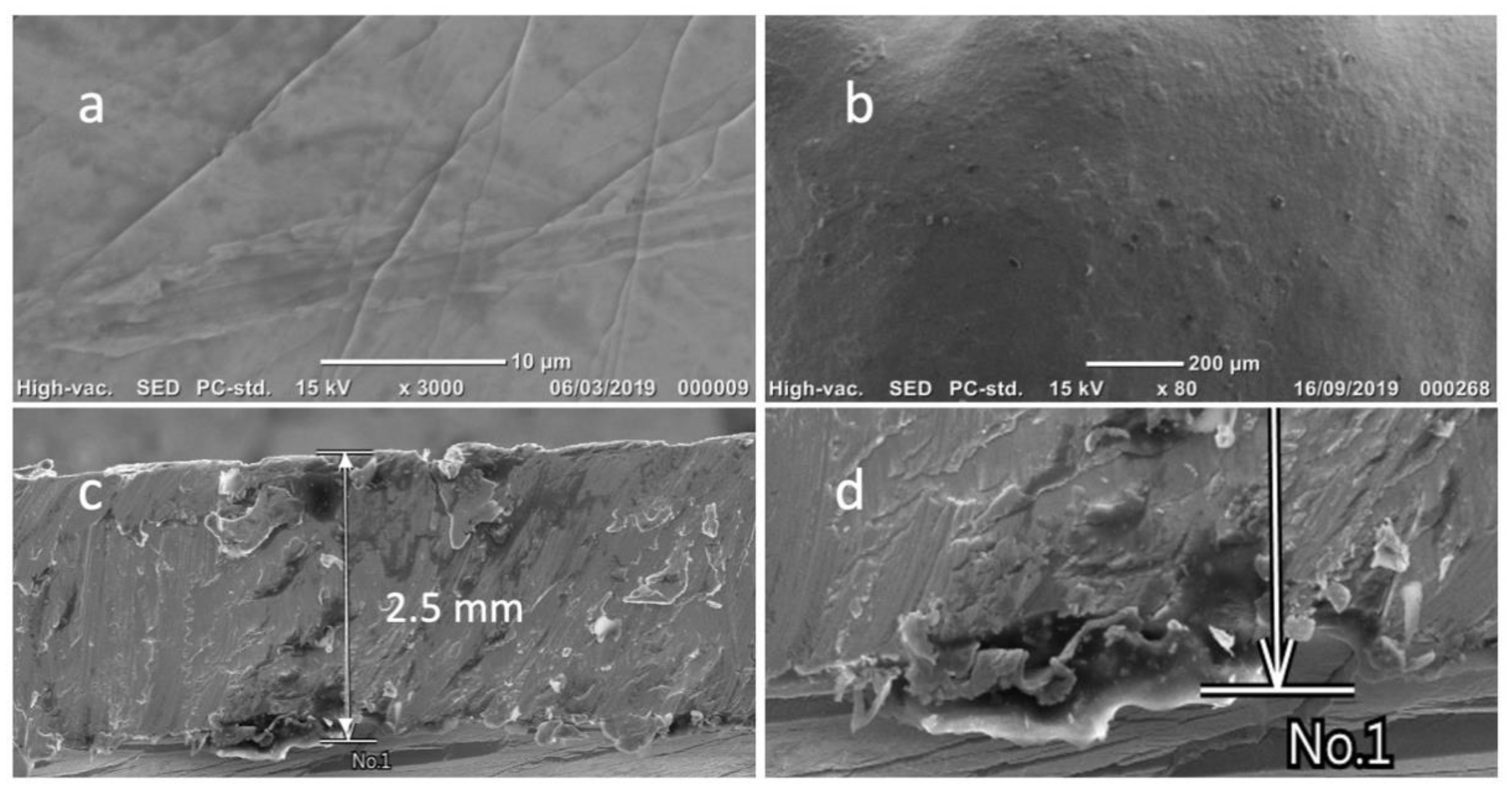
3.1. Morphological and Electrochemical Characterization of the Samples’ Surfaces
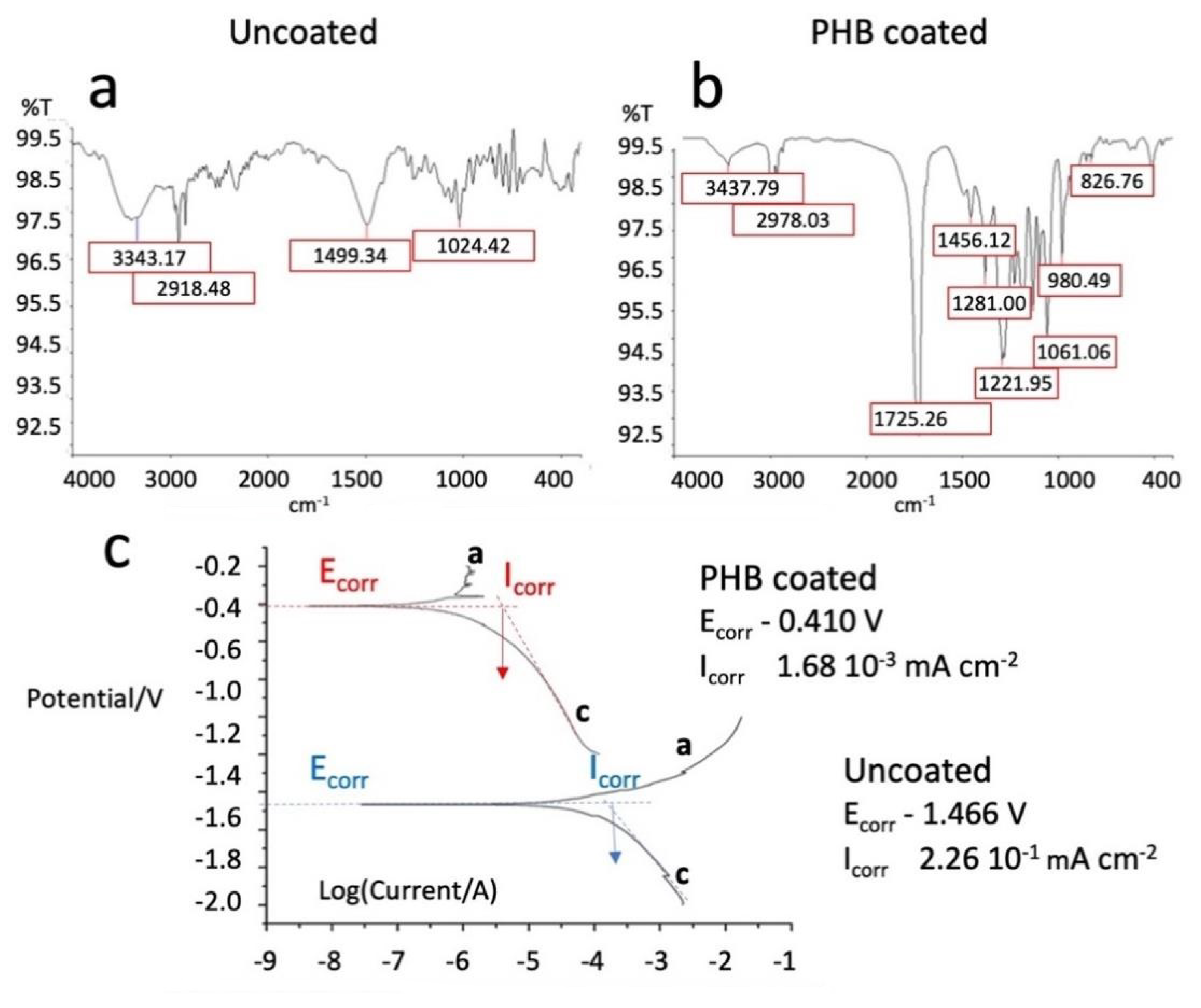
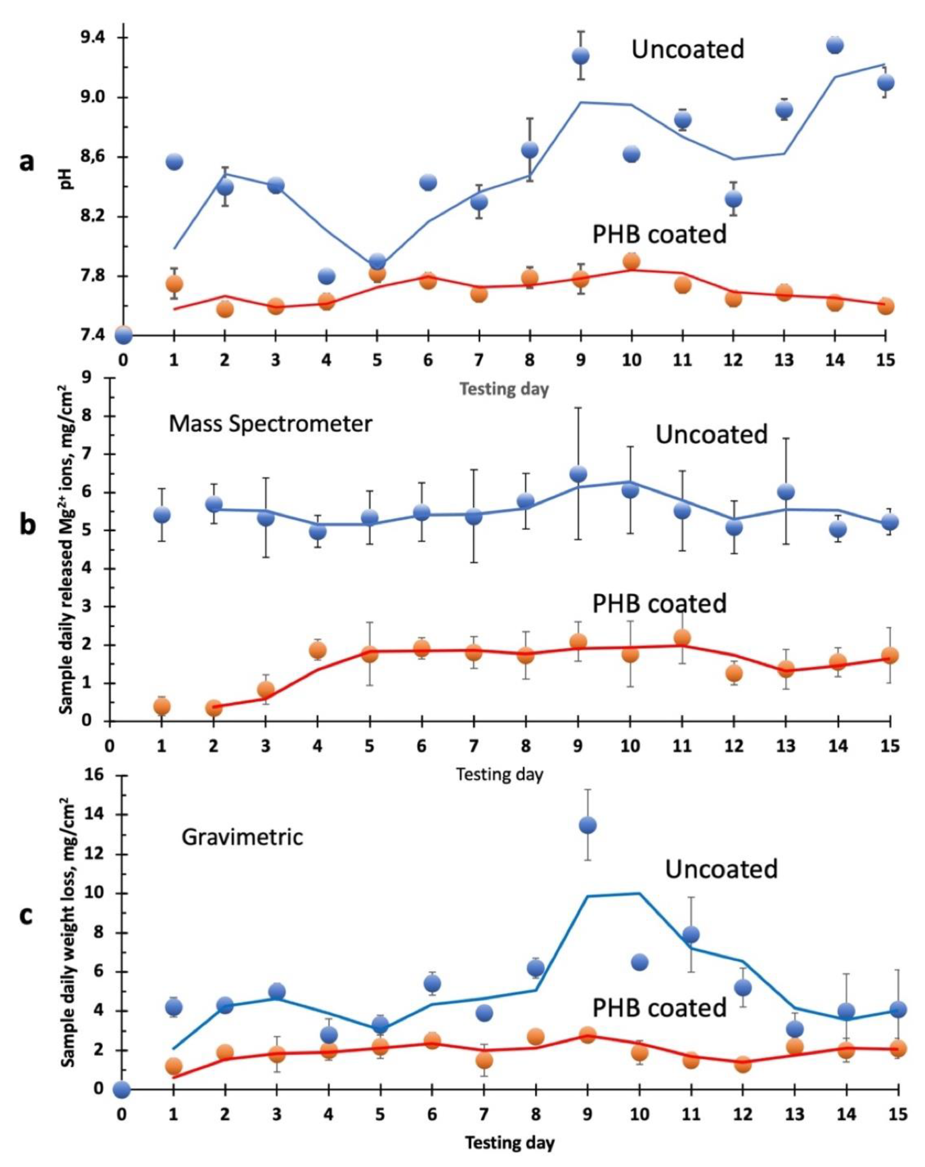
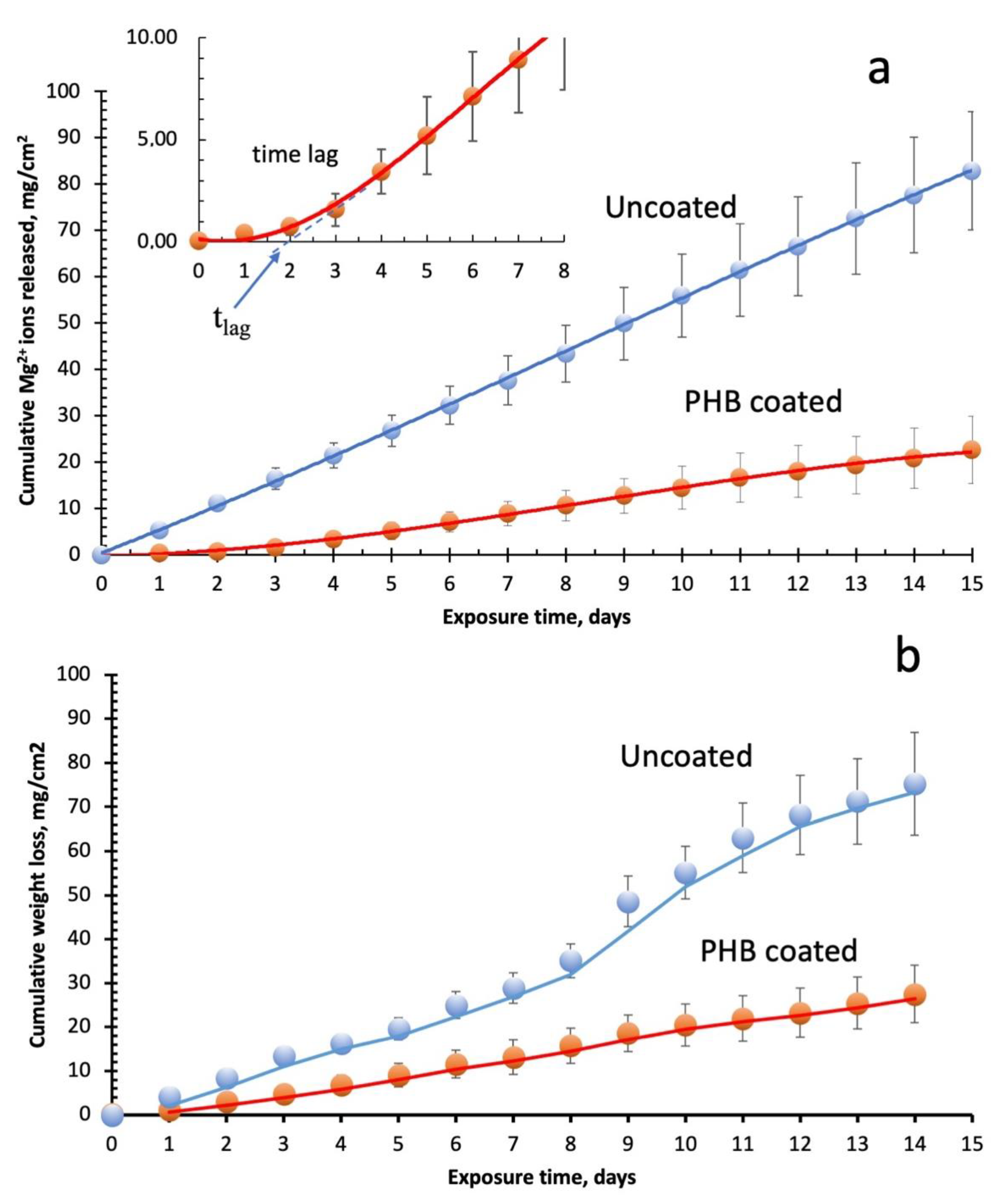
3.2. Wettability, Polarization Curves, and SBF Corrosion Tests
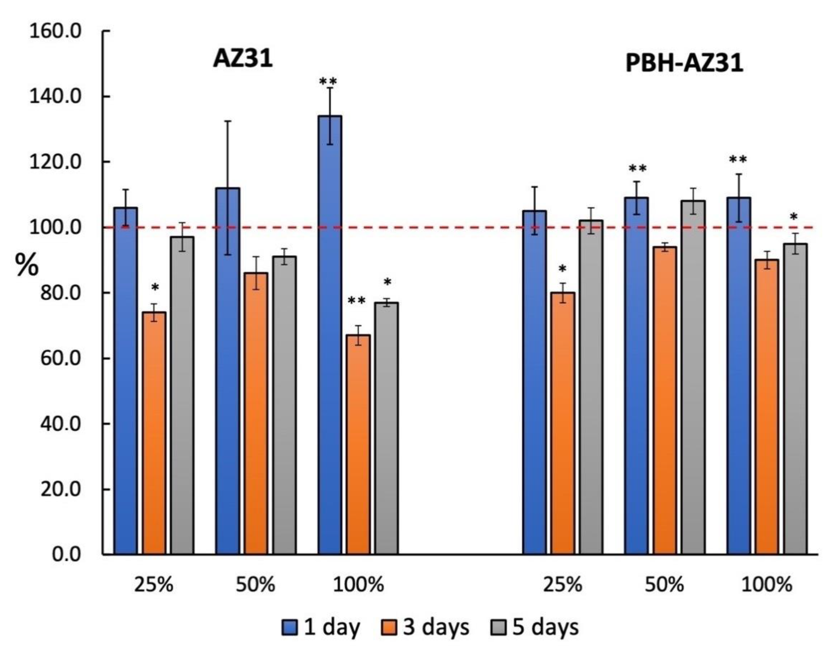
3.3. Cytotoxicity (CCK-8 Assay)
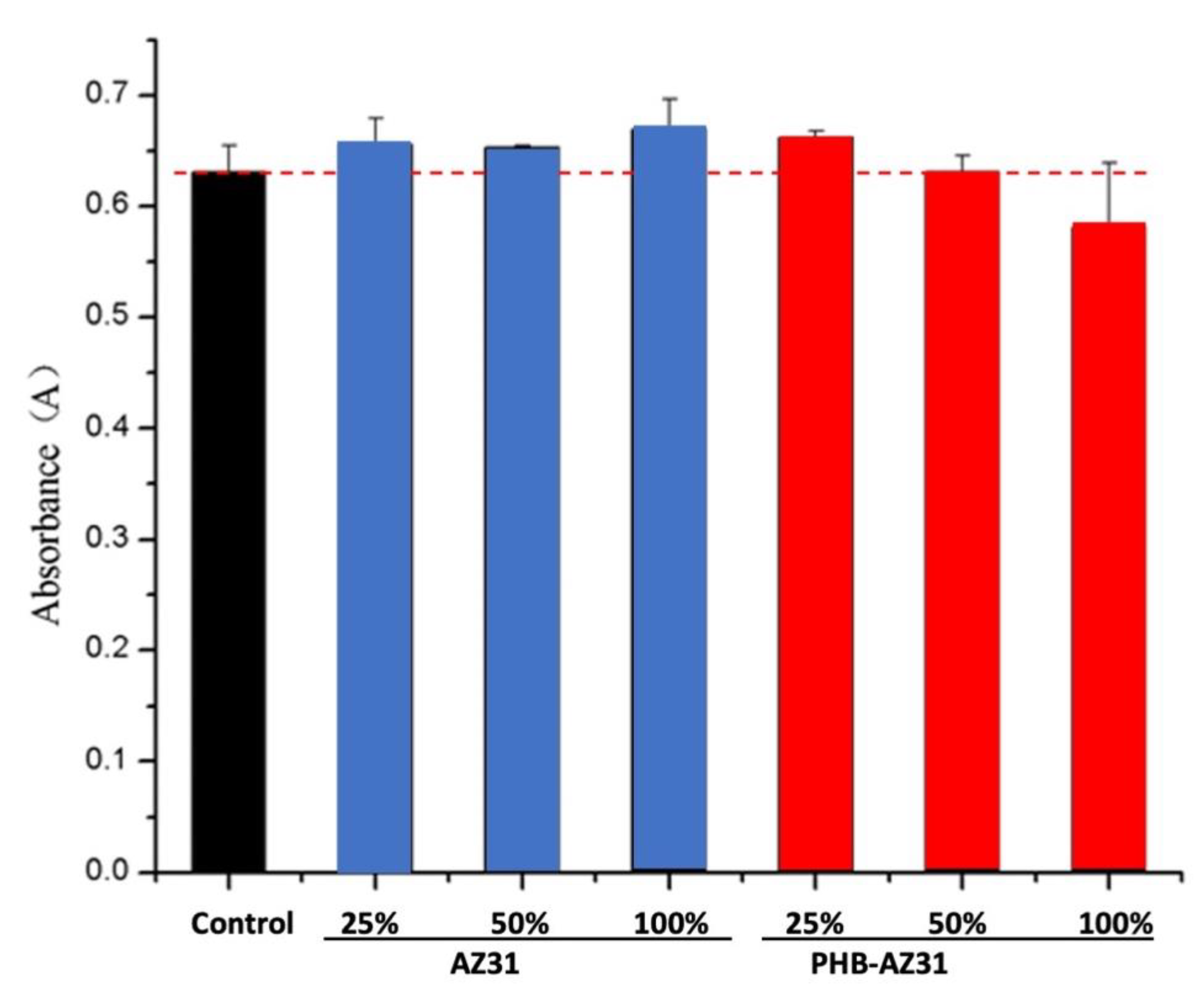
3.4. Hemolysis
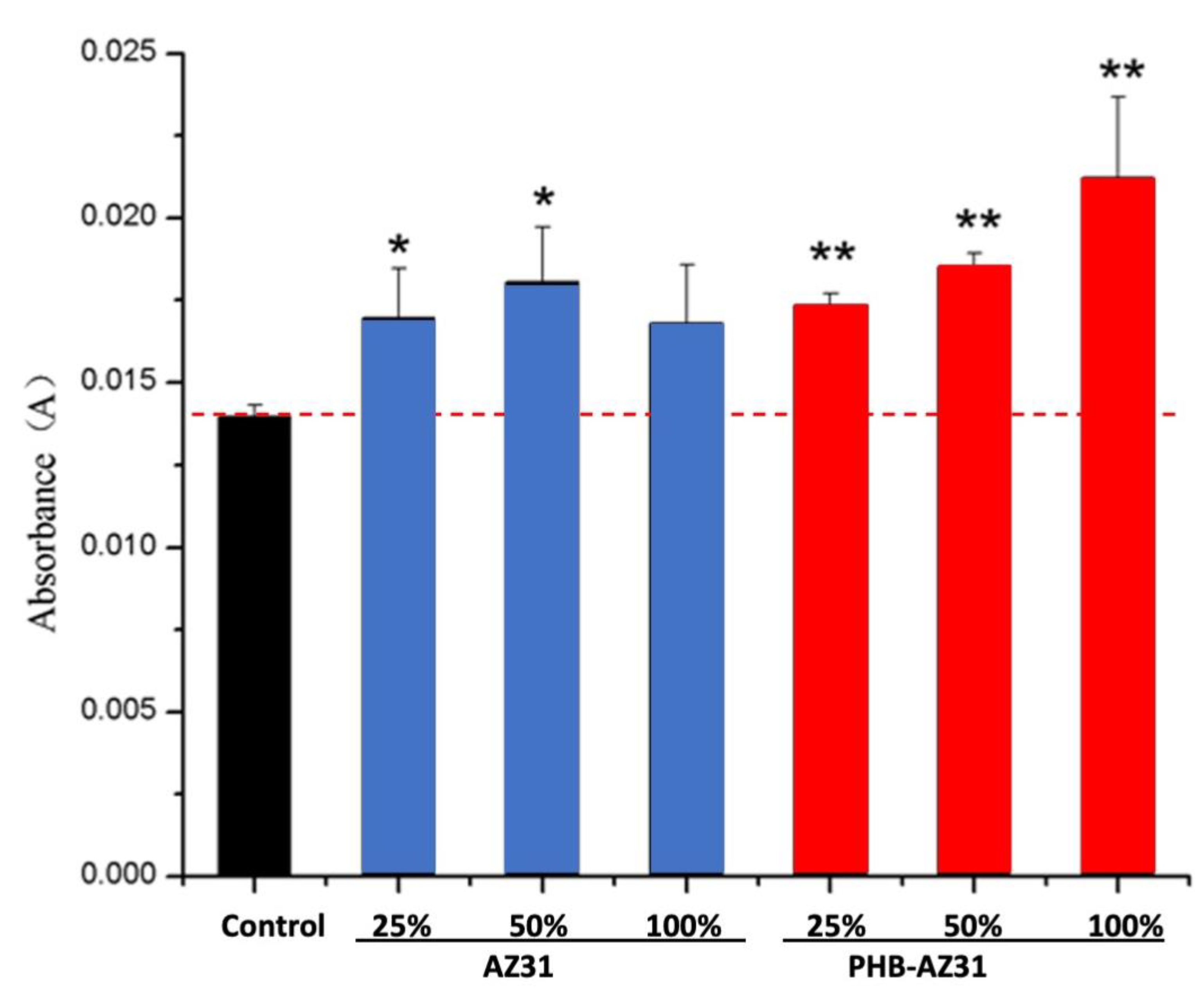
3.5. NO Assay
3.6. T-AOC Assay
4. Discussion
5. Conclusions
Author Contributions
Funding
Institutional Review Board Statement
Conflicts of Interest
References
- Hou, Z.; Yan, W.; Li, T.; Wu, W.; Cui, Y.; Zhang, X.; Chen, Y.-P.; Yin, T.; Qiu, J.; Wang, G. Lactic acid-mediated endothelial to mesenchymal transition through TGF-β1 contributes to in-stent stenosis in poly-L-lactic acid stent. Int. J. Biol. Macromol. 2020, 155, 1589–1598. [Google Scholar] [CrossRef] [PubMed]
- Lin, S.; Ran, X.; Yan, X.; Yan, W.; Wang, Q.; Yin, T.; Zhou, J.G.; Hu, T.; Wang, G. Corrosion behavior and biocompatibility evaluation of a novel zinc-based alloy stent in rabbit carotid artery model. J. Biomed. Mater. Res. Part B Appl. Biomater. 2019, 107, 1814–1823. [Google Scholar] [CrossRef] [PubMed]
- Gao, F.; Hu, Y.; Li, G.; Liu, S.; Quan, L.; Yang, Z.; Wei, Y.; Pan, C. Layer-by-layer deposition of bioactive layers on magnesium alloy stent materials to improve corrosion resistance and biocompatibility. Bioact. Mater. 2020, 5, 611–623. [Google Scholar] [CrossRef]
- Ostrowski, N.J.; Lee, B.; Roy, A.; Ramanathan, M.; Kumta, P.N. Biodegradable poly(lactide-co-glycolide) coatings on magnesium alloys for orthopedic applications. J. Mater. Sci. 2013, 24, 85–96. [Google Scholar] [CrossRef] [PubMed]
- Staiger, M.P.; Pietak, A.M.; Huadmai, J.; Dias, G. Magnesium and its alloys as orthopedic biomaterials: A review. Biomaterials 2006, 27, 1728–1734. [Google Scholar] [CrossRef] [PubMed]
- Hanada, K.; Matsuzaki, K.; Huang, X.; Chino, Y. Fabrication of Mg alloy tubes for biodegradable stent application. Mater. Sci. Eng. C 2013, 33, 4746–4750. [Google Scholar] [CrossRef] [PubMed]
- Salahshoor, M.; Guo, Y. Biodegradable orthopedic magnesium–calcium (MgCa) alloys, processing, and corrosion performance. Materials 2012, 5, 135–155. [Google Scholar] [CrossRef] [Green Version]
- Witte, F.; Ulrich, H.; Rudert, M.; Willbold, E. Biodegradable magnesium scaffolds: Part 1: Appropriate inflammatory response. J. Biomed. Mater. Res. Part A 2007, 81A, 748–756. [Google Scholar] [CrossRef] [PubMed]
- Aversa, R.; Petrescu, R.V.V.; Apicella, A.; Petrescu, F.I. Nano-Diamond Hybrid Materials for Structural Biomedical Application. Am. J. Biochem. Biotechnol. 2017, 13, 34–41. [Google Scholar] [CrossRef]
- Windhagen, H.; Radtke, K.; Weizbauer, A.; Diekmann, J.; Noll, Y.; Kreimeyer, U.; Schavan, R.; Stukenborg-Colsman, C.; Waizy, H. Biodegradable magnesium-based screw clinically equivalent to titanium screw in hallux valgus surgery: Short term results of the first prospective, randomized, controlled clinical pilot study. Biomed. Eng. Online 2013, 12, 1–10. [Google Scholar] [CrossRef] [Green Version]
- Wu, G.; Ibrahim, J.M.; Chu, P.K. Surface design of biodegradable magnesium alloys—A review. Surf. Coat. Technol. 2013, 233, 2–12. [Google Scholar] [CrossRef]
- Chen, S.; Guan, S.; Li, W.; Wang, H.; Chen, J.; Wang, Y.; Wang, H. In vivo degradation and bone response of a composite coating on Mg-Zn-Ca alloy prepared by microarc oxidation and electrochemical deposition. J. Biomed. Mater. Res. Part B Appl. Biomater. 2012, 100, 533–543. [Google Scholar] [CrossRef] [PubMed]
- Sezer, N.; Evis, Z.; Koç, M. Additive manufacturing of biodegradable magnesium implants and scaffolds: Review of the recent advances and research trends. J. Magnes. Alloy 2020, 9, 392–415. [Google Scholar] [CrossRef]
- Aversa, R.; Petrescu, V.; Apicella, A.; Petrescu, I.T. The Basic Elements of Life’s. Am. J. Eng. Appl. Sci. 2016, 9, 1189–1197. [Google Scholar] [CrossRef]
- Aversa, R.; Tamburrino, F.; Petrescu, R.V.V.; Petrescu, F.I.; Artur, M.; Chen, G.; Apicella, A. Biomechanically Inspired Shape Memory Effect Machines Driven by Muscle like Acting NiTi Alloys. Am. J. Appl. Sci. 2016, 13, 1264–1271. [Google Scholar] [CrossRef]
- Jacobs, J.J.; Hallab, N.J.; Skipor, A.K.; Urban, R.M. Metal Degradation Products: A Cause for Concern in Metal-Metal Bearings? Clin. Orthop. Relat. Res. 2003, 417, 139–147. [Google Scholar]
- Granchi, D.; Ciapetti, G.; Stea, S.; Savarino, L.; Filippini, F.; Sudanese, A.; Zinghi, G.; Montanaro, L. Cytokine release in mononuclear cells of patients with Co–Cr hip prosthesis. Biomaterials 1999, 20, 1079–1086. [Google Scholar] [CrossRef]
- Niki, Y.; Matsumoto, H.; Suda, Y.; Otani, T.; Fujikawa, K.; Toyama, Y.; Hisamori, N.; Nozue, A. Metal ions induce bone-resorbing cytokine production through the redox pathway in synoviocytes and bone marrow macrophages. Biomaterials 2003, 24, 1447–1457. [Google Scholar] [CrossRef]
- Waizy, H.; Seitz, J.-M.; Reifenrath, J.; Weizbauer, A.; Bach, F.-W.; Meyer-Lindenberg, A.; Denkena, B.; Windhagen, H. Biodegradable magnesium implants for orthopedic applications. J. Mater. Sci. 2013, 48, 39–50. [Google Scholar] [CrossRef]
- Xu, R.; Yang, X.; Suen, K.W.; Wu, G.; Li, P.; Chu, P.K. Improved corrosion resistance on biodegradable magnesium by zinc and aluminum ion implantation. Appl. Surf. Sci. 2012, 263, 608–612. [Google Scholar] [CrossRef]
- Xin, Y.; Liu, C.; Zhang, X.; Tang, G.; Tian, X.; Chu, P.K. Corrosion behavior of biomedical AZ91 magnesium alloy in simulated body fluids. J. Mater. Res. 2007, 22, 2004–2011. [Google Scholar] [CrossRef] [Green Version]
- Aversa, R.; Petrescu, R.V.V.; Petrescu, F.I.T.; Apicella, A. Biomimetic and evolutionary design driven innovation in sustainable products development. Am. J. Eng. Appl. Sci. 2016, 9, 1027–1036. [Google Scholar] [CrossRef] [Green Version]
- Witte, F.; Fischer, J.; Nellesen, J.; Crostack, H.-A.; Kaese, V.; Pisch, A.; Beckmann, F.; Windhagen, H. In vitro and in vivo corrosion measurements of magnesium alloys. Biomaterials 2006, 27, 1013–1018. [Google Scholar] [CrossRef] [PubMed]
- Li, L.; Gao, J.; Wang, Y. Evaluation of cyto-toxicity and corrosion behavior of alkali-heat-treated magnesium in simulated body fluid. Surf. Coat. Technol. 2004, 185, 92–98. [Google Scholar] [CrossRef]
- Liu, C.; Xin, Y.; Tian, X.; Chu, P.K. Corrosion behavior of AZ91 magnesium alloy treated by plasma immersion ion implantation and deposition in artificial physiological fluids. Thin Solid Film. 2007, 516, 422–427. [Google Scholar] [CrossRef] [Green Version]
- Wang, J.; Tang, J.; Zhang, P.; Li, Y.; Wang, J.; Lai, Y.; Qin, L. Surface modification of magnesium alloys developed for bioabsorbable orthopedic implants: A general review. J. Biomed. Mater. Res. Part B Appl. Biomater. 2012, 100, 1691–1701. [Google Scholar] [CrossRef]
- Raffaella, A.; Petrescu, F.I.T.; Petrescu, R.V.V.; Antonio, A. Biomimetic finite element analysis bone modeling for customized hybrid biological prostheses development. Am. J. Appl. Sci. 2016, 13, 1060–1067. [Google Scholar] [CrossRef]
- Wang, X.; Zeng, X.; Wu, G.; Yao, S.; Lai, Y. Effects of tantalum ion implantation on the corrosion behavior of AZ31 magnesium alloys. J. Alloys Compd. 2007, 437, 87–92. [Google Scholar] [CrossRef]
- Zhou, H.; Chen, F.; Yang, Y.G.; Wan, H.C.; Cai, S. Study on Process of Ion Implantation on AZ31 Magnesium Alloy. Key Eng. Mater. 2008, 373–374, 342–345. [Google Scholar] [CrossRef]
- Orlowski, M.; Ruebben, A. Bioresorbable Metal Stent with Controlled Resorption. U.S. Patent Application No. 12/524,702, 30 January 2008. [Google Scholar]
- Bertsch, T.; Borck, A. Biocorrodible Metallic Implant Having a Coating or Cavity Filling Made of Gelatin. U.S. Patent Application No. 11/850,346, 6 March 2008. [Google Scholar]
- Zharkova, I.I.; Staroverova, O.V.; Voinova, V.V.; Andreeva, N.V.; Shushckevich, A.M.; Sklyanchuk, E.D.; Kuzmicheva, G.M.; Bespalova, A.E.; Akulina, E.A.; Shaitan, K.V.; et al. Biocompatibility of electrospun poly(3-hydroxybutyrate) and its composites scaffolds for tissue engineering. Biomeditsinskaya Khimiya 2014, 60, 553–560. [Google Scholar] [CrossRef] [Green Version]
- Adden, N. Implant of a Biocorrodable Magnesium Alloy and Having a Coating of a Biocorrodable Polyphosphazene. U.S. Patent Application No. 12/192,729, 19 February 2009. [Google Scholar]
- Zhang, X.P.; Zhao, Z.P.; Wu, F.M.; Wang, Y.L.; Wu, J. Corrosion and wear resistance of AZ91D magnesium alloy with and without micro-arc oxidation coating in Hank’s solution. J. Mater. Sci. 2007, 42, 8523–8528. [Google Scholar] [CrossRef]
- Xu, X.; Lu, P.; Guo, M.; Fang, M. Cross-linked gelatin/nanoparticles composite coating on micro-arc oxidation film for corrosion and drug release. Appl. Surf. Sci. 2010, 256, 2367–2371. [Google Scholar] [CrossRef]
- Jo, J.H.; Hong, J.Y.; Shin, K.S.; Kim, H.E.; Koh, Y.H. Enhancing biocompatibility and corrosion resistance of Mg implants via surface treatments. J. Biomater. Appl. 2012, 27, 469–476. [Google Scholar] [CrossRef]
- Salunke, P.; Shanov, V.; Witte, F. High purity biodegradable magnesium coating for implant application. Mater. Sci. Eng. B 2011, 176, 1711–1717. [Google Scholar] [CrossRef]
- Wong, H.M.; Yeung, K.W.K.; Lam, K.O.; Tam, V.; Chu, P.K.; Luk, K.D.K.; Cheung, K.M.C. A biodegradable polymer-based coating to control the performance of magnesium alloy orthopaedic implants. Biomaterials 2010, 31, 2084–2096. [Google Scholar] [CrossRef] [PubMed] [Green Version]
- Aversa, R.; Petrescu, R.; Petrescu, F.; Perrotta, V.; Apicella, D.; Apicella, A. Biomechanically Tunable Nano-Silica/P-HEMA Structural Hydrogels for Bone Scaffolding. Bioengineering 2021, 8, 45. [Google Scholar] [CrossRef] [PubMed]
- Witte, F.; Hort, N.; Vogt, C.; Cohen, S.; Kainer, K.U.; Willumeit, R.; Feyerabend, F. Degradable biomaterials based on magnesium corrosion. Curr. Opin. Solid State Mater. Sci. 2008, 12, 63–72. [Google Scholar] [CrossRef] [Green Version]
- Gray-Munro, J.E.; Seguin, C.; Strong, M. Influence of surface modification on the in vitro corrosion rate of magnesium alloy AZ31. J. Biomed. Mater. Res. Part A 2009, 91, 221–230. [Google Scholar] [CrossRef]
- Raffaella, A.; Petrescu, R.V.V.; Antonio, A.; Petrescu, F.I.T. Physiologic human fluids and swelling behavior of hydrophilic biocompatible hybrid ceramo-polymeric materials. Am. J. Eng. Appl. Sci. 2016, 9, 962–972. [Google Scholar] [CrossRef] [Green Version]
- Gogolewski, S.; Jovanovic, M.; Perren, S.M.; Dillon, J.G.; Hughes, M.K. Tissue response and in vivo degradation of selected polyhydroxyacids: Polylactides (PLA), poly(3-hydroxybutyrate) (PHB), and poly(3-hydroxybutyrate-co-3-hydroxyvalerate) (PHB/VA). J. Biomed. Mater. Res. 1993, 27, 1135–1148. [Google Scholar] [CrossRef]
- Kostopoulos, L.; Karring, T. Augmentation of the rat mandible using guided tissue regeneration. Clin. Oral Implant. Res. 1994, 5, 75–82. [Google Scholar] [CrossRef]
- Shishatskaya, E.I.; Volova, T.G.; Gordeev, S.A.; Puzyr, A.P. Degradation of P(3HB) and P(3HB-co-3HV) in biological media. J. Biomater. Sci. Polym. Ed. 2005, 16, 643–657. [Google Scholar] [CrossRef] [PubMed]
- Li, T.; Qi, K. Microbial Polyesters: The Polymer of the Future. Explor. Nat. 1994, 13, 35–41. [Google Scholar]
- Baptist, J.N. Process for Preparing Poly β-Hyroxybutyric Acid. U.S. Patent Application No. 3044942, 17 July 1960. [Google Scholar]
- Wang, Z.; Yan, J.; Zheng, Q.; Wang, Z. CyclinD1, CDK4, and P21 expression by IEC-6 cells in response to NiTi alloy and polymeric biomaterials. Mater. Sci. Eng. C 2012, 32, 2183–2189. [Google Scholar] [CrossRef]
- Rahmany, M.B.; Van Dyke, M. Biomimetic approaches to modulate cellular adhesion in biomaterials: A review. Acta Biomater. 2013, 9, 5431–5437. [Google Scholar] [CrossRef]
- Hornberger, H.; Virtanen, S.; Boccaccini, A. Biomedical coatings on magnesium alloys—A review. Acta Biomater. 2012, 8, 2442–2455. [Google Scholar] [CrossRef] [PubMed]
- Mueller, P.P.; May, T.; Perz, A.; Hauser, H.; Peuster, M. Control of smooth muscle cell proliferation by ferrous iron. Biomaterials 2006, 27, 2193–2200. [Google Scholar] [CrossRef]
- Walter, R.; Kannan, M.B. In-vitro degradation behaviour of WE54 magnesium alloy in simulated body fluid. Mater. Lett. 2011, 65, 748–750. [Google Scholar] [CrossRef]
- Song, J.; Van Ooij, W.J. Bonding and corrosion protection mechanisms of gamma-APS and BTSE silane films on aluminum substrates. J. Adhes. Sci. Technol. 2003, 17, 2191–2221. [Google Scholar] [CrossRef]
- Jones, D.A.; Amy, P.S. A Thermodynamic Interpretation of Microbiologically Influenced Corrosion. Corrosion 2002, 58, 638–645. [Google Scholar] [CrossRef]
- Song, G.; Atrens, A. Understanding Magnesium Corrosion—A Framework for Improved Alloy Performance. Adv. Eng. Mater. 2003, 5, 837–858. [Google Scholar] [CrossRef]
- Zhu, D.; van Ooij, W.J. Corrosion protection of AA 2024-T3 by bis-[3-(triethoxysilyl)propyl]tetrasulfide in sodium chloride solution.: Part 2: Mechanism for corrosion protection. Corros. Sci. 2003, 45, 2177–2197. [Google Scholar] [CrossRef]
- Liu, X.; Yue, Z.; Romeo, T.; Weber, J.; Scheuermann, T.; Moulton, S.; Wallace, G. Biofunctionalized anti-corrosive silane coatings for magnesium alloys. Acta Biomater. 2013, 9, 8671–8677. [Google Scholar] [CrossRef] [PubMed] [Green Version]
- Jyoti, G.; Keshav, A.; Anandkumar, J. Review on Pervaporation: Theory, Membrane Performance, and Application to Intensification of Esterification Reaction. J. Eng. 2015, 2015, 1–24. [Google Scholar] [CrossRef] [Green Version]
- Ball, I.J.; Huang, S.; Wolf, R.A.; Shimano, J.Y.; Kaner, R.B. Pervaporation studies with polyaniline membranes and blends. J. Membr. Sci. 2000, 174, 161–176. [Google Scholar] [CrossRef]
- Sawada, H.; Takahashi, Y.; Miyata, S.; Kanehashi, S.; Sato, S.; Nagai, K. Gas Transport Properties and Crystalline Structures of Poly(lactic acid) Membranes. Trans. Mater. Res. Soc. Jpn. 2010, 35, 241–246. [Google Scholar] [CrossRef] [Green Version]
- Kawamura, N.; Nakao, Y.; Ishikawa, R.; Tsuchida, D.; Iijima, M. Degradation and Biocompatibility of AZ31 Magnesium Alloy Implants In Vitro and In Vivo: A Micro-Computed Tomography Study in Rats. Materials 2020, 13, 473. [Google Scholar] [CrossRef] [Green Version]
- Leleu, S.; Rives, B.; Causse, N.; Pébère, N. Corrosion rate determination of rare-earth Mg alloys in a Na2SO4 solution by electrochemical measurements and inductive coupled plasma-optical emission spectroscopy. J. Magnes. Alloy 2019, 7, 47–57. [Google Scholar] [CrossRef]
- Hofstetter, J.; Becker, M.; Martinelli, E.; Weinberg, A.M.; Mingler, B.; Kilian, H.; Pogatscher, S.; Uggowitzer, P.J.; Löffler, J.F. High-strength low-alloy (HSLA) Mg-Zn-Ca alloys with excellent biodegradation performance. JOM 2014, 66, 566–572. [Google Scholar] [CrossRef]
- Holweg, P.; Berger, L.; Cihova, M.; Donohue, N.; Clement, B.; Schwarze, U.; Sommer, N.G.; Hohenberger, G.; van den Beucken, J.J.J.P.; Seibert, F.; et al. A lean magnesium–zinc–calcium alloy ZX00 used for bone fracture stabilization in a large growing-animal model. Acta Biomater. 2020, 113, 646–659. [Google Scholar] [CrossRef]
- Rivard, C.H.; Chaput, C.; Rhalmi, S.; Selmani, A. Bio-absorbable synthetic polyesters and tissue regeneration. A study of three-dimensional proliferation of ovine chondrocytes and osteoblasts. Ann. Chir. 1996, 50, 651–658. [Google Scholar]
- Sun, J.; Dai, Z.; Zhao, Y.; Chen, G.Q. In vitro effect of oligo-hydroxyalkanoates on the growth of mouse fibroblast cell line L929. Biomaterials 2007, 28, 3896–3903. [Google Scholar] [CrossRef] [PubMed]
- Wang, Y.; Jiang, X.L.; Yang, S.C.; Lin, X.; He, Y.; Yan, C.; Wu, L.; Chen, G.Q.; Wang, Z.Y.; Wu, Q. MicroRNAs in the regulation of interfacial behaviors of MSCs cultured on microgrooved surface pattern. Biomaterials 2011, 32, 9207–9217. [Google Scholar] [CrossRef] [PubMed]
- Chen, G.-Q.; Zhang, J. Microbial polyhydroxyalkanoates as medical implant biomaterials. Artif. Cells Nanomed. Biotechnol. 2018, 46, 1–18. [Google Scholar] [CrossRef] [PubMed]
- Cheng, G.; Cai, Z.; Wang, L. Biocompatibility and biodegradation of poly(hydroxybutyrate)/poly(ethylene glycol) blend films. J. Mater. Sci. Mater. Electron. 2003, 14, 1073–1078. [Google Scholar] [CrossRef] [PubMed]
- Liu, B.; Zheng, Y. Effects of alloying elements (Mn, Co, Al, W, Sn, B, C and S) on biodegradability and in vitro biocompatibility of pure iron. Acta Biomater. 2011, 7, 1407–1420. [Google Scholar] [CrossRef] [PubMed]
- Ali, S.; Rani, A.M.A.; Baig, Z.; Ahmed, S.W.; Hussain, G.; Subramaniam, K.; Hastuty, S.; Rao, T.V. Biocompatibility and corrosion resistance of metallic biomaterials. Corros. Rev. 2020, 38, 381–402. [Google Scholar] [CrossRef]
- Davide, A.; Raffaella, A.; Marco, T.; Michele, S.; Syed, J.; Massimo, M.; Marco, F.; Antonio, A.; Apicella, A. Direct restoration modalities of fractured central maxillary incisors: A multi-levels validated finite elements analysis with in vivo strain measurements. Dent. Mater. 2015, 31, e289–e305. [Google Scholar] [CrossRef]


| Ions | Plasma, mmol/L | SBF, mmol/L |
|---|---|---|
| Na+ | 142.0 | 142.0 |
| K+ | 5.0 | 5.0 |
| Mg2+ | 1.5 | 1.5 |
| Ca2+ | 2.5 | 2.5 |
| Cl− | 103.0 | 147.8 |
| (HCO3)− | 27.0 | 4.2 |
| (HPO4)2− | 1.0 | 1.0 |
| (SO4)2− | 0.5 | 0.5 |
| pH | 7.2–7.4 | 7.4 |
| AZ31 Mg Alloy | AZ31 Mg Alloy + Mg(OH)2 | AZ31 Mg Alloy + Mg(OH)2 + PHB |
|---|---|---|
| 46.1° | * 42.2° | ** 62.3° |
| ±2.5° | ±0.4° | ±0.3° |
| Cultured 1 Day | Cultured 3 Days | Cultured 5 Days | ||||||||
|---|---|---|---|---|---|---|---|---|---|---|
| Extraction Fluid | Conc. % | OD | RGR (%) | Toxic Grade | OD | RGR (%) | Toxic Grade | OD | RGR (%) | Toxic Grade |
| AZ31 | 25 | 0.229 ±0.012 | 106 | 0 | 0.220 ±0.008 | 74 | 2 | 0.242 ±0.011 | 97 | 1 |
| 50 | 0.243 ±0.045 | 112 | 0 | 0.255 ±0.015 | 86 | 1 | 0.227 ±0.006 | 91 | 1 | |
| 100 | 0.291 ±0.019 | 134 | 0 | 0.198 ±0.009 | 67 | 2 | 0.192 ±0.003 | 77 | 1 | |
| PHB- Coated AZ31 | 25 | 0.228 ±0.016 | 105 | 0 | 0.239 ±0.009 | 80 | 1 | 0.255 ±0.010 | 102 | 0 |
| 50 | 0.254 ±0.011 | 109 | 0 | 0.280 ±0.004 | 94 | 1 | 0.269 ±0.010 | 108 | 0 | |
| 100 | 0.236 ±0.016 | 109 | 0 | 0.268 ±0.008 | 90 | 1 | 0.237 ±0.008 | 95 | 1 | |
| Sample | OD Value | Hemolysis Rate | Mg2+, mmol/L |
|---|---|---|---|
| AZ31 Mg | 0.2487 ± 0.0775 | 37.61% | 22.9 ± 1.3 |
| PHB-coated AZ31 Mg | 0.0530 ± 0.0117 | 4.19% | 2.4 ± 0.2 |
| Positive control | 0.6137 ± 0.0117 | ||
| Negative control | 0.0285 ± 0.0117 |
Publisher’s Note: MDPI stays neutral with regard to jurisdictional claims in published maps and institutional affiliations. |
© 2021 by the authors. Licensee MDPI, Basel, Switzerland. This article is an open access article distributed under the terms and conditions of the Creative Commons Attribution (CC BY) license (https://creativecommons.org/licenses/by/4.0/).
Share and Cite
Wang, L.; Aversa, R.; Houa, Z.; Tian, J.; Liang, S.; Ge, S.; Chen, Y.; Perrotta, V.; Apicella, A.; Apicella, D.; et al. Bioresorption Control and Biological Response of Magnesium Alloy AZ31 Coated with Poly-β-Hydroxybutyrate. Appl. Sci. 2021, 11, 5627. https://0-doi-org.brum.beds.ac.uk/10.3390/app11125627
Wang L, Aversa R, Houa Z, Tian J, Liang S, Ge S, Chen Y, Perrotta V, Apicella A, Apicella D, et al. Bioresorption Control and Biological Response of Magnesium Alloy AZ31 Coated with Poly-β-Hydroxybutyrate. Applied Sciences. 2021; 11(12):5627. https://0-doi-org.brum.beds.ac.uk/10.3390/app11125627
Chicago/Turabian StyleWang, Lu, Raffaella Aversa, Zhengjun Houa, Jie Tian, Shuang Liang, Shuping Ge, Yu Chen, Valeria Perrotta, Antonio Apicella, Davide Apicella, and et al. 2021. "Bioresorption Control and Biological Response of Magnesium Alloy AZ31 Coated with Poly-β-Hydroxybutyrate" Applied Sciences 11, no. 12: 5627. https://0-doi-org.brum.beds.ac.uk/10.3390/app11125627