Article

Simple Thalidomide Analogs in Melanoma: Synthesis and Biological Activity

by
Alexia Barbarossa
1,†,
Alessia Catalano
1,†,
Jessica Ceramella
2,
Alessia Carocci
1,*,
Domenico Iacopetta
2,*,
Camillo Rosano
3,
Carlo Franchini
1,‡ and
Maria Stefania Sinicropi
2,‡
1
Department of Pharmacy-Drug Sciences, University of Bari “Aldo Moro”, 70126 Bari, Italy
2
Department of Pharmacy, Health and Nutritional Sciences, University of Calabria, Via Pietro Bucci, Arcavacata di Rende, 87036 Cosenza, Italy
3
Proteomics and Mass Spectrometry Unit, IRCCS Policlinico San Martino, L.go Rosanna Benzi 10, 16132 Genova, Italy
*
Authors to whom correspondence should be addressed.
These authors equally contributed to this work.
Co-senior authors.
Submission received: 13 May 2021 / Revised: 11 June 2021 / Accepted: 21 June 2021 / Published: 23 June 2021
(This article belongs to the Special Issue Anticancer Drugs Activity and Underlying Mechanisms)

Abstract

:
Thalidomide is an old well-known drug that is still of clinical interest, despite its teratogenic activities, due to its antiangiogenic and immunomodulatory properties. Therefore, efforts to design safer and effective thalidomide analogs are continually ongoing. Research studies on thalidomide analogs have revealed that the phthalimide ring system is an essential pharmacophoric fragment; thus, many phthalimidic compounds have been synthesized and evaluated as anticancer drug candidates. In this study, a panel of selected in vitro assays, performed on a small series of phthalimide derivatives, allowed us to characterize compound 2k as a good anticancer agent, acting on A2058 melanoma cell line, which causes cell death by apoptosis due to its capability to inhibit tubulin polymerization. The obtained data were confirmed by in silico assays. No cytotoxic effects on normal cells have been detected for this compound that proves to be a valid candidate for further investigations to achieve new insights on possible mechanism of action of this class of compounds as anticancer drugs.

1. Introduction

Cancer is still a great health concern, as it is one of the main causes of death worldwide. A total of 19.3 million new cases of cancer were diagnosed last year, of which 10 million were fatal [1]. Among the different types of cancer, malignant melanoma is one of the most aggressive and deadly skin cancers, with an enormously increasing occurrence over the past few decades [2,3,4]. Melanoma is notoriously chemoresistant; nevertheless, chemotherapy represents the primary treatment for metastatic melanoma. Considering the major impact of chemotherapy in cancer management, and the problems related to chemotherapy that have not yet been solved, such as drug toxicity and resistance, particularly in the case of melanoma, research for newer and potent anticancer agents is of great significance. Over the past few years, numerous biologically active heterocyclic compounds have been synthesized and evaluated as anticancer drugs. Among these, many phthalimidic compounds have turned out to be antitumor drug candidates. The phthalimidic core represents the main pharmacophoric feature of thalidomide (Figure 1), a drug infamous for its teratogenic action, which, nevertheless, represents one of the most successful examples of drug repositioning in the treatment of cancer. Indeed, over the last few decades, the interest in this old drug has been renewed because of its efficacy in several important disorders (such as multiple myeloma, breast cancer, and HIV-related diseases [5]) and its antiangiogenic and immunomodulatory properties. Several studies regarding the effects of thalidomide on metastatic melanoma have been reported. Since angiogenesis, and specifically the increased expression of vascular endothelial growth factor (VEGF), has been reported as an important step in disease progression, the use of thalidomide to enhance the antitumor activity of chemotherapy has been investigated in metastatic melanoma [6]. Furthermore, a number of literature data report the effect of thalidomide and its derivatives on tubulin polymerization, causing a perturbation of microtubule dynamics. Microtubules represent important biological targets in cancer treatment due to their involvement in crucial cellular functions [7,8]. It became clear that thalidomide exerts multifaceted properties; however, many aftereffects, such as deep vein thrombosis, peripheral neuropathy, constipation, somnolence, pyrexia, pain, and teratogenicity, have been reported, showing the need for careful and monitored use [9]. Thus, research efforts are directed toward the synthesis and optimization of new thalidomide analogs, mostly as new anticancer agents, lacking toxic effects and that are able to remove these limits and improve the pharmacological profile. [10,11]. As a part of an ongoing research program, directed toward the synthesis of several antitumor heterocyclic compounds [12,13,14,15,16], and our previous paper [17] concerning the antitumor activity of a thalidomide-correlated compound, we report herein the synthesis of a small series of phthalimide derivatives, obtained by directly connecting the N-terminus of the phthalimide moiety with a differently substituted aromatic ring (2a–l, Figure 1). These compounds have been investigated for their cytotoxic effects in a large panel of cancer cells, including two melanoma cell lines, namely A2058 derived from a metastatic site, and malignant melanoma cells Sk-Mel28, two breast cancer cell lines (triple negative MDA-MB-231 and ER(+) MCF-7) and a human cervix carcinoma HeLa cell line. The best cytotoxic activity was recovered for compound 2k on A2058 melanoma cell line. The ability of compound 2k to inhibit tubulin polymerization has also been reported and a molecular docking study supported the obtained results.

2. Materials and Methods

2.1. Chemistry

All chemicals were supplied by Sigma-Aldrich or Alfa Aesar and were the highest-grade purity that were commercially available. Solvents were reagent grade unless otherwise indicated. Yields refer to purified products and were not optimized. Compound structures were confirmed by routine spectrometric analyses. Melting points were determined on a Gallenkamp melting point apparatus in open glass capillary tubes and are uncorrected. 1H NMR and 13C NMR spectra were recorded on a Varian VX Mercury spectrometer operating at 300 and 75 MHz for 1H and 13C, respectively, or an Agilent 500 MHz operating at 500 and 125 MHz for 1H and 13C, respectively. Chemical shifts are reported in parts per million (ppm) relative to solvent (CDCl3) resonance: δ 7.26 (1H NMR) and δ 77.0 (13C NMR). J values are given in Hz. The following abbreviations are used: s-singlet, d-doublet, t-triplet, and m-multiplet. Gas chromatography (GC)/mass spectroscopy (MS) was performed on a Hewlett-Packard 6890–5973 MSD at low resolution. Liquid chromatography (LC)/mass spectroscopy (MS) was performed on a spectrometer (Agilent 1100 series LC-MSD Trap System VL). Molecular ion was designed as “M+”. Elemental analyses were carried out on a Eurovector Euro EA 3000 analyzer and the data for C, H, N were within ±0.4 of theoretical values.
Synthesis of 2-phenyl-1H-isoindol-1,3(2H)-dione (2a). Aniline (0.77 g, 8.3 mmol), phthalic anhydride (1.23 g, 8.3 mmol) and triethylamine (0.12 mL, 0.9 mmol) were refluxed in toluene (10 mL) for 5 h. The solvent was removed in vacuo and the residue, taken up with CHCl3, was washed 3 N HCl and water, and then dried over anhydrous Na2SO4 to give 905 mg (49%) of 2a as a white solid: mp 211–213 °C; lit. 200 °C [18]; 210–211 °C (acetone) [19]. GC/MS (70 eV) m/z (%): 223 (M+, 100); LC/MS: m/z (%): 222 [M+–1]; 1H NMR (300 MHz, CDCl3): δ 7.36–7.54 (m, 5H, Ar), 7.54–7.82 (m, 2H, Ar), 7.90–7.98 (m, 2H, Ar); 13C NMR (75 MHz, CDCl3): δ 123.7 (2C), 126.6 (1C), 128.1 (2C), 129.1 (2C), 131.8 (2C), 134.4 (2C), 167.3 (2C). Anal C13H9NO2 (223.06): calcd %: C 75.33; H 4.06; N 6.27. Found %: C 74.98; H 4.04; N 6.30.
Synthesis of 2-(2-fluorophenyl)-1H-isoindol-1,3(2H)-dione (2b). Prepared as described for 2a starting from 2-fluoroaniline. White solid. Yield: 65%: mp 190–191 °C (CHCl3/hexane); lit. 199 °C [19]; GC/MS (70 eV) m/z (%): 241 (M+, 92), 197 (100); 1H NMR (500 MHz, CDCl3): δ 7.23–7.30 (m, 2H, Ar), 7.35–7.38 (m, 1H, Ar), 7.42–7.47 (m, 1H, Ar), 7.79–7.82 (m, 2H, Ar), 7.95–7.98 (m, 2H, Ar); 13C NMR (75 MHz, CDCl3): δ 116.76 (d, J = 19.6 Hz, 1C), 119.4 (d, JCF = 13.7 Hz, 1C), 123.9 (1C), 124.6 (d, JCF = 4.6 Hz, 1C), 129.9 (2C), 130.7 (d, JCF = 8.0 Hz, 1C), 131.9 (2C), 134.4 (2C), 157.9 (d, JCF = 251.9 Hz, 1C), 166.5 (2C). Anal C14H8FNO2 (241.05): calcd %: C 69.71; H 3.34; N 5.81. Found %: C 69.54; H 3.32; N 5.80.
Synthesis of 2-[4-(4-methylphenoxy)phenyl]-1H-isoindol-1,3(2H)-dione (2c). Prepared as described for 2a starting from 4-p-tolyloxyaniline. Grey solid. Yield: 55%: mp 201–202 °C (CHCl3/hexane); GC/MS (70 eV) m/z (%): 329 (M+, 100); 1H NMR (300 MHz, CDCl3): δ 2.15–2.36 (m, 3H, CH3), 6.97 (d, J = 8.2 Hz, 2H, Ar), 7.11 (d, J = 21.7 Hz, 2H, Ar), 7.16 (d, J = 8.2 Hz, 2H, Ar), 7.34 (d, J = 8.8 Hz, 2H, Ar), 7.76–7.79 (m, 2H, Ar), 7.92–7.95 (m, 2H, Ar); 13C NMR (125 MHz, CDCl3): δ 20.7 (1C), 118.3 (2C), 119.7 (2C), 123.7 (2C), 125.9 (1C), 128.0 (2C), 130.4 (2C), 131.8 (2C), 133.6 (1C), 134.4 (2C), 154.0 (1C), 157.8 (1C), 167.4 (2C). Anal C21H15NO3 (329.11): calcd %: C 76.58; H 4.59; N 4.25. Found %: C 76.43; H 4.53; N 4.35.
Synthesis of 2-(4-phenoxyphenyl)-1H-isoindole-1,3(2H)-dione (2d). Prepared as described for 2a starting from 4-phenoxyaniline. Grey solid. Yield: 65%: mp 161–162 °C (CHCl3/hexane); GC/MS (70 eV) m/z (%): 315 (M+, 100); 1H NMR (500 MHz, CDCl3): δ 7.06–7.15 (m, 5H, Ar), 7.35–7.38 (m, 4H, Ar), 7.78–7.79 (m, 2H, Ar), 7.94–7.95 (m, 2H, Ar); 13C NMR (125 MHz, CDCl3): δ 118.8 (2C), 119.5 (2C), 123.7 (2C), 123.9 (1C), 126.3 (1C), 128.0 (2C), 129.9 (2C), 131.7 (2C), 134.4 (2C), 156.5 (1C), 157.2 (1C), 167.4 (2C). Anal C20H13NO3·0.20 H2O (318.69): calcd %: C 75.32; H 4.23; N 4.39. Found %: C 75.60; H 4.08; N 4.48.
Synthesis of 2-(4-chloro-2-fluorophenyl)-1H-isoindol-1,3(2H)-dione (2e). Prepared as described for 2a starting from 4-chloro-2-fluoroaniline. White solid. Yield: 35%: mp 145–146 °C; GC/MS (70 eV) m/z (%): 275 (M+, 100); 1H NMR (500 MHz, CDCl3): δ 7.25–7.32 (m, 3H, Ar), 7.78–7.82 (m, 2H, Ar), 7.90–7.98 (m, 2H, Ar); 13C NMR (125 MHz, CDCl3): δ 117.7 (d, JCF = 22.9 Hz, 1C), 118.2 (d, JCF = 96.4 Hz, 1C), 124.0 (2C), 125.1 (d, JCF = 3.8 Hz, 1C), 130.5 (1C), 131.8 (2C), 134.6 (2C), 135.8 (d, JCF = 9.6 Hz, 1C), 157.7 (d, JCF = 256.5 Hz, 1C), 166.2 (2C). Anal C14H7ClFNO2·0.25 H2O (279.51): calcd %: C 60.02; H 2.70; N 5.00. Found %: C 60.42; H 2.50; N 5.07.
Synthesis of 2-(2-bromo-4-chlorophenyl)-1H-isoindol-1,3(2H)-dione (2f). Prepared as described for 2a starting from 2-bromo-4-cloroaniline. White solid. Yield: 18%: mp 140–141 °C (CHCl3/hexane); GC/MS (70 eV) m/z (%): 337 (M+, 2), 302 (100); 1H NMR (500 MHz, CDCl3): δ 7.24–7.30 (m, 1H, Ar), 7.45 (d, J = 8.8 Hz, 1H, Ar), 7.75 (s, 1H, Ar), 7.79–7.86 (m, 2H, Ar), 7.94–8.01 (m, 2H, Ar); 13C NMR (125 MHz, CDCl3): δ 123.9 (1C), 124.0 (1C), 128.8 (2C), 130.1 (1C), 131.5 (1C), 131.8 (2C), 133.4 (1C), 134.6 (2C), 136.3 (1C), 166.3 (2C). Anal. C14H7BrClNO2 (344.93): calcd %: C 49.96; H 2.10; N 4.16. Found %: C 49.96; H 2.05; N 4.15.
Synthesis of 2-(2-cloro-4-fluorophenyl)-1H-isoindol-1,3(2H)-dione (2g). Prepared as described for 2a starting from 2-cloro-4-fluoroaniline. White solid. Yield: 68%: mp 188–189 °C (CHCl3/hexane); GC/MS (70 eV) m/z (%): 275 (M+, 7), 240 (100); 1H NMR (500 MHz, CDCl3): δ 7.13 (t, J = 8.0 Hz, 1H, Ar), 7.32–7.35 (m, 2H, Ar), 7.80–7.84 (m, 2H, Ar), 7.94–8.00 (m, 2H, Ar); 13C NMR (125 MHz, CDCl3): δ 115.1 (d, JCF = 22.9 Hz, 1C), 117.9 (d, JCF = 25.8 Hz, 1C), 124.0 (2C), 125.8 (d, JCF = 3.9 Hz, 1C), 131.8 (d, JCF = 8.7 Hz, 1C + s, 2C), 134.4 (d, JCF = 11.4 Hz, 1C), 134.6 (2C), 162.7 (d, JCF = 252.8 Hz, 1C), 166.6 (2C). Anal. C14H7ClFNO2 (275.01): calcd %: C 61.00; H 2.56; N 5.08. Found %: C 60.71; H 2.49; N 5.06.
Synthesis of 2-(3,4-difluorophenyl)-1H-isoindol-1,3(2H)-dione (2h). Prepared as described for 2a starting from 3,4-difluoroaniline. White solid. Yield: 52%: mp 199–200 °C; GC/MS (70 eV) m/z (%): 259 (M+, 100); 1H NMR (300 MHz, CDCl3): δ 7.22–7.39 (m, 3H, Ar), 7.78–7.84 (m, 2H, Ar), 7.93–7.99 (m, 2H, Ar); 13C NMR (75 MHz, CDCl3): δ 116.0 (d, JCF = 19.4 Hz, 1C), 117.5 (d, J = 18.6 Hz, 1C), 112.6 (d, JCF = 4.6 Hz, 1C), 122.7 (d, JCF = 3.4 Hz, 1C), 123.9 (2C), 127.9 (1C), 131.4 (2C), 134.6 (2C), 148.3 (dd, JCF = 29.8, 12.6 Hz, 1C), 151.6 (dd, J = 28.0, 13.2 Hz, 1C), 166.8 (2C). Anal C14H7FNO2·0.20 H2O (262.64): calcd %: C 63.98; H 2.84; N 5.33. Found %: C 64.32; H 2.69; N 5.38.
Synthesis of 2-[4-(2-chlorophenoxy)phenyl]-1H-isoindol-1,3(2H)-dione (2i). Prepared as described for 2a starting from 4-(2-chlorophenoxy)aniline. Grey solid. solid. Yield: 55%: mp 135–136 °C (CHCl3/hexane); GC/MS (70 eV) m/z (%): 349 (M+, 100); 1H NMR (500 MHz, CDCl3): δ 7.11–7.15 (m, 4H, Ar), 7.24–7.28 (m, 1H, Ar), 7.37–7.40 (m, 2H, Ar), 7.47–7.48 (m, 1H, Ar), 7.78–7.80 (m, 2H, Ar), 7.93–7.96 (m, 2H, Ar); 13C NMR (125 MHz, CDCl3): δ 117.9 (2C), 121.7 (1C), 123.8 (2C), 125.4 (1C), 126.4 (1C), 126.5 (1C), 128.0 (1C), 128.1 (1C), 130.9 (2C), 131.7 (2C), 134.4 (2C), 151.8 (1C), 156.7 (1C), 167.4 (2C). Anal. C20H12ClNO3·0.50 H2O (257.02): calcd %: C 66.95; H 3.65; N 3.90. Found %: C 67.32; H 3.36; N 4.02.
Synthesis of 2-[4-(2-methoxyphenoxy)phenyl]-1H-isoindol-1,3(2H)-dione (2j). Prepared as described for 2a starting from 4-(2-methoxyphenoxy)aniline. Light brown solid. Yield: 75%: mp 148–150 °C (AcOEt/hexane). GC/MS (70 eV) m/z (%) 345 (M+, 100), 133 (100); 1H NMR (300 MHz, CDCl3): δ 3.84 (s, 3H, CH3), 6.93–7.09 (m, 5H, Ar), 7.15–7.22 (m, 1H, Ar), 7.31–7.36 (m, 2H, Ar), 7.76–7.81 (m, 2H, Ar), 7.92–7.97 (m, 2H, Ar); 13C NMR (125 MHz, CDCl3): δ 55.9 (1C), 113.0 (1C), 117.1 (2C), 121.1 (1C), 122.0 (1C) 123.7 (2C), 125.5 (1C), 125.6 (1C), 127.9 (2C) 131.8 (2C), 134.4 (2C), 144.0 (1C), 151.7 (1C), 157.9 (1C), 167.5 (2C). Anal C21H15NO4·H2O (363.35): calcd %: C 69.63; H 4.91; N 3.54. Found: C 69.41; H 4.72; N 3.85.
Synthesis of 2-[4-(2-methylphenoxy)phenyl]-1H-isoindol-1,3(2H)-dione (2k). Prepared as described for 2a starting from 4-(2-methylphenoxy)aniline. White solid. Yield: 33%: mp 123–124 °C (AcOEt/hexane); GC/MS (70 eV) m/z (%) 329 (M+, 100); 1H NMR (500 MHz, CDCl3): δ 2.25 (s, 3H, CH3), 6.97–7.02 (m, 3H, Ar), 7.11 (t, J = 7.4 Hz, 1H, Ar), 7.21 (t, J = 7.4 Hz, 1H, Ar), 7.25–7.29 (m, 1H, Ar), 7.32–7.36 (m, 2H, Ar), 7.77–7.81 (m, 2H, Ar), 7.92–7.97 (m, 2H, Ar); 13C NMR (75 MHz, CDCl3): δ 16.1 (1C), 117,3 (2C), 120.5 (1C), 123.7 (2C), 124.6 (1C), 125.6 (1C), 127.3 (1C), 127.9 (2C), 130.3 (1C), 131.6 (2C), 131.8 (2C), 134.3 (1C), 153.8 (1C), 157.7 (1C), 167.4 (2C). Anal C21H15NO3·0.50 H2O (338.11): calcd %: C 74.79; H 4.96; N 4.45. Found: 74.54; H 4.77; N 4.14.
Synthesis of 2-(2-phenoxyphenyl)-1H-isoindol-1,3(2H)-dione (2l). Prepared as described for 2a starting from 2-phenoxyaniline. Light pink solid. Yield: 70%: mp 146–148 °C; GC/MS (70 eV) m/z (%) 315 (M+, 100); 1H NMR (300 MHz, CDCl3): δ 6.96–7.10 (m, 4H, Ar), 7.18–7.31 (m, 3H, Ar), 7.33–7.42 (m, 2H, Ar), 7.71–7.79 (m, 2H, Ar), 7.87–7.95 (m, 2H, Ar). 13C NMR (125 MHz, CDCl3): δ 119.0 (1C), 119.6 (2C), 122.4 (1C), 123.4 (1C), 123.7 (2C), 123.9 (1C), 129.7 (2C), 130.2 (1C), 130.5 (1C), 132.1 (2C), 134.1 (2C), 154.0 (1C), 156.3 (1C), 167.0 (2C). Anal C20H13NO3 (315.33): calcd %: C 76.06; H 4.20; N 4.43. Found: C 76.18; H 4.16; N 4.44.

2.2. Biological Methods

2.2.1. Cell Culture

The cell lines used in the present work (A2058, Sk-Mel28, MDA-MB-231, MCF-7, HeLa and HEK-293) were purchased from the American Type Culture Collection (ATCC, Manassas, VA, USA). All cell lines were maintained at 37 °C in a humidified atmosphere of 95% air and 5% CO2 and periodically screened for contamination [20]. Human melanoma Sk-Mel-28 cells and human embryonic kidney HEK-293 cells, were cultured in Dulbecco’s modified Eagle’s medium (DMEM) high glucose (4.5 g/L) supplemented with 1% L-glutamine, 100 U/mL penicillin/streptomycin and 10% fetal bovine serum (FBS). The human melanoma cells A2058 were grown in DMEM low glucose (1 g/L) supplemented with 1% L-glutamine and 100 U/mL penicillin/streptomycin and 20% FBS. MCF-7 and MDA-MB-231 breast cancer cells were maintained in Dulbecco’s modified Eagle’s medium/nutrient mixture Ham F-12 (DMEM/F12), supplemented with 5% FBS and 100 U/mL penicillin/streptomycin. HeLa human epithelial cervix carcinoma cells were maintained in minimum essential Eagle’s medium (MEM), supplemented with 10% FBS, 1% l-glutamine, 100 U mL 1 penicillin/streptomycin and 1% non-essential amino acids (NEAA).

2.2.2. MTT Assay

Cell viability was evaluated using the 3-(4,5-dimethylthiazol-2-y1)-2,5-diphenyltetrazolium (MTT, Sigma–Aldrich, Milan, Italy) assay as already reported [21]. Results are represented as percent (%) of basal and the IC50 values were calculated using GraphPad Prism 9 (GraphPad Software, La Jolla, CA, USA).

2.2.3. Immunofluorescence Analysis

Cells were seeded in 48-well culture plates containing glass slides and incubated with the most active compound for 24 h (concentration equal to its IC50 value), following a previously described protocol [22]. Rabbit anti β-tubulin was purchased from Santa Cruz Biotechnology and diluted to 1:100 before the use. The secondary antibody, Alexa Fluor® 568 conjugate goat-anti-mouse (1:500, Thermo Fisher Scientific, Waltham, MA, USA), was added and incubated for 2 h at 37 °C. Images were acquired using a fluorescence microscope (Leica DM 6000; 40× magnification). LAS-X software was used to process all images.

2.2.4. Tubulin Polymerization Assay

Tubulin polymerization inhibition was measured using an in vitro Tubulin Polymerization Assay Kit purchased from EMD Millipore Corporation [23], as already described [22]. The turbidity variation was measured every 30 s at 350 nm for 90 min, stirring the mixture for 10 s before each measurement.

2.2.5. TUNEL Assay

Apoptosis was detected using a TUNEL assay, according to the guidelines of the manufacturer (CFTM488A TUNEL Assay Apoptosis Detection Kit, Biotium, Hayward, CA, USA), as already reported [24]. Cells were observed and imaged under a fluorescence microscope (Leica DM6000; 20× magnification) with excitation/emission wavelength maxima of 490/515 nm (CF 488A) or 350/460 nm (DAPI). Images are representative of three independent experiments.

2.2.6. Western Blotting Analysis

Protein lysates were subjected to Western blot analysis, as already reported [25] Poly(ADP-ribose) polymerase 1 (Parp-1) and glyceraldehyde 3-phosphate dehydrogenase (GAPDH, used to verify the equal loading of proteins) were purchased from Santa Cruz Biotechnology (Santa Cruz, CA, USA). The antigen–antibody complex was detected by incubation of the membrane with a secondary antibody (peroxidase-coupled anti-rabbit IgG, VWR International PBI, Milano, Italy). Immunoreactive bands were revealed using an ECL Western blotting detection system (Amersham Pharmacia Biotech, Piscataway, NJ, USA).

2.2.7. Docking Studies

The crystal structure of the complex formed between a tetrameric assembly of tubulin and vinblastine [26]; PDB code 5j2t, was used as the protein target for our docking simulations. All the structures of the ligands tested in silico were designed, their 3D structures were created, charges were assigned and further energy was minimized using the program MarvinSketch (ChemAxon ltd, Budapest, Hungary). To better characterize the binding mode and the mechanisms of action of the tested compounds to the protein target and to determine their binding energies, we used Autodock v.4.2.2. [27] and its graphical interface, ADT [28]. We performed all simulations using a searching grid with a cubic shape, composed of 126 points for each dimension, centered on the protein center. Nodes were spaced 0.375 Å from each other. Each of our simulation runs was performed adopting the default program values and the docking procedures were carried out as described in our previous works [20,29,30,31,32]. Briefly, the protein and the ligands were prepared using the ADT graphical interface by adding polar hydrogens to the protein and solvation parameters. During all the simulations, we considered the protein as a rigid object and the ligands as fully flexible. The Lamarckian genetic algorithm was used in our search with a population of 100 individuals that were evolved for 100 generations. The docking poses as resulting for our simulation runs were ranked in order of binding energy and collected into clusters on the basis of RMSD.
The generated docking poses were ranked in order of increasing binding energy values and clustered on the basis of a RMSD cut-off value of 2.0 Å. From the structural analyses of the lowest energy solutions of each cluster, we could identify the ligand binding mode. All the figures concerning the ligand binding modes to the protein target were drawn using the program Chimera [33].

3. Results and Discussion

3.1. Chemistry

The synthesis of the tested compounds was obtained by mixing equimolar quantities of phthalic anhydride and the suitable aryl amine in the presence of triethylamine (Scheme 1) [19].

3.2. Biological Results

3.2.1. Cell Viability Assay

The cytotoxic activities of synthesized compounds (2a–l) were examined against two human melanoma cell lines, A2058, derived from a metastatic site, and malignant melanoma cells, Sk-Mel28, two breast cancer cells, ER(+) MCF-7 and triple negative MDA-MB-231 and the human epithelial cervix carcinoma HeLa cells. The calculated IC50 values of studied compounds in comparison with the reference drug, thalidomide, are listed in Table 1. As shown, thalidomide did not exhibit any activity on both the cell lines at the higher concentration tested. None of the compounds were shown to possess anticancer activity against the Sk-Mel28 cell line. Regarding the A2058 cell line, a slight activity was found for compound 2d, which had no substituents on the phenoxy moiety. On the contrary, the presence of a chlorine substituent at the ortho position of the phenoxy nucleus seemed to enhance the activity since an IC50 of 23.26 ± 1.1 μM was calculated for compound 2i on A2058 cells. A notable inhibitory effect on the viability of the A2058 cell line, with an IC50 of 15.37 ± 0.7 μM, was obtained for compound 2k, bearing a methyl group at the ortho position of the phenoxy moiety, which performed best out of the compound in the series. Similarly, compound 2k exerted a good antitumor activity against both breast cancer cells used in this assay (IC50 = 20.99 ± 0.8 and 22.72 ± 0.9 µM on MDA-MB-231 and MCF-7, respectively), while no activity was recorded on HeLa cells. Compound 2i was able to reduce the breast cancer cells growth with IC50 values amounting to 25.34 ± 1.0 and 31.2 ± 1.2 µM on MDA-MB-231 and MCF-7 respectively, and, to a lesser extent, against cervix carcinoma HeLa cells growth (IC50 = 54.5 ± 0.8 µM). A slight anticancer activity was also recovered for compound 2d towards the breast cancer cells MDA-MB-231 (IC50 = 76.5 ± 1.2 µM) and MCF-7 (IC50 = 177.0 ± 0.7 µM) and towards HeLa cells (IC50 = 42.1 ± 1.1 µM). No interesting activity was detected for the compounds lacking the phenoxy group against all cell lines used. To assess potential cytotoxicity towards normal cells, a screening of the compounds was performed on the non-tumoral human embryonic renal cell line Hek-293. As can be seen in Table 1, most of the compounds and thalidomide did not interfere with the normal cell viability, at least until the concentration of 200 μM and under the conditions used for this assay. A slight cytotoxicity was found for compound 2i, though at a concentration 6-fold higher than the active one. A certain cytotoxicity against Hek-293 cell lines was found for compound 2c, which, however, did not exhibit antitumor activity on the melanoma cells.

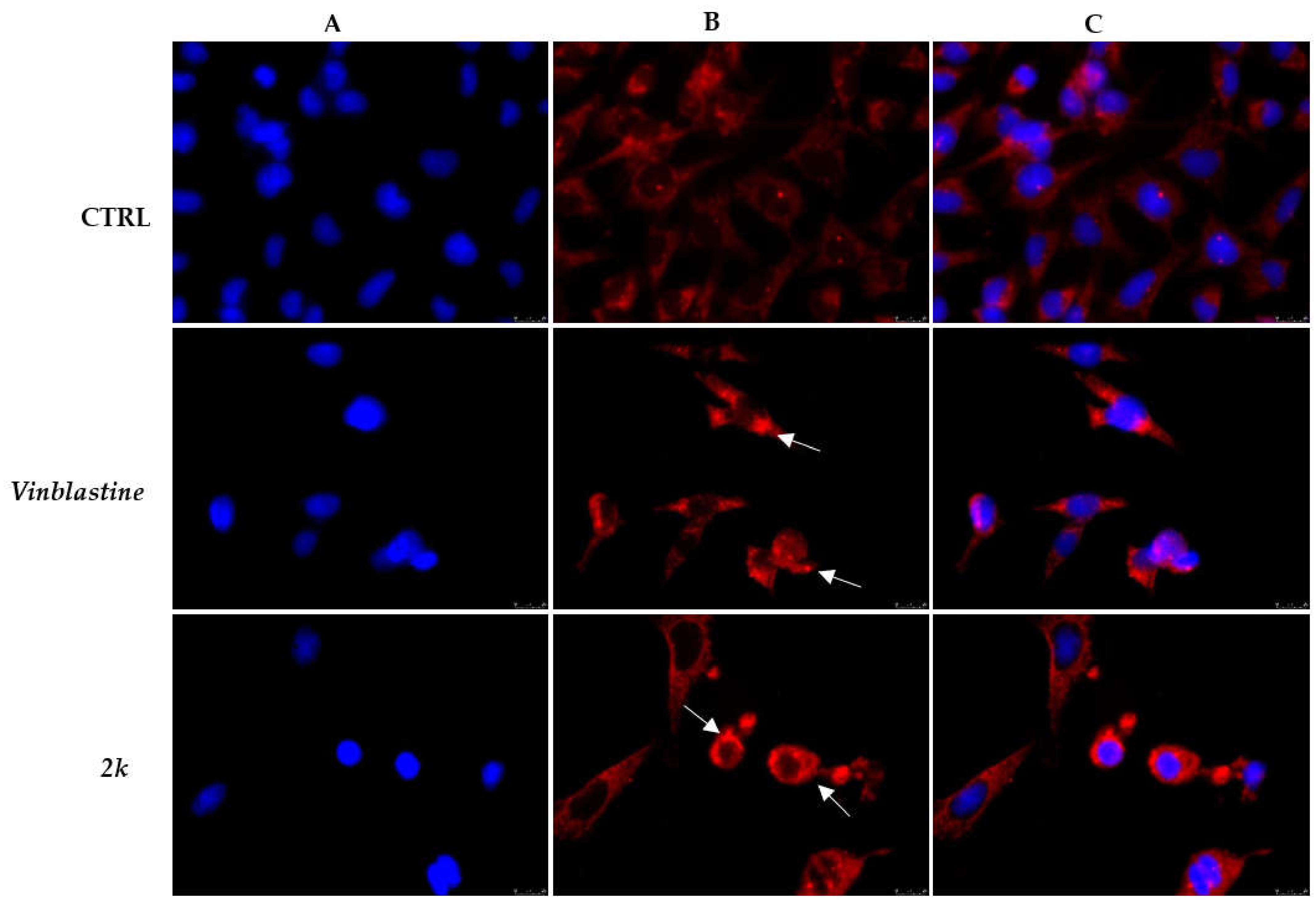
3.2.2. Effect of Compound 2k on Microtubule Dynamics and Cell Death by Apoptosis

Microtubules represent vital cell components shaped by polymerization of α and β tubulin heterodimers and play crucial roles in several cellular functions, including cell division [34]. Several classes of naturally occurring, as well as synthetic and semi-synthetic, compounds disrupt microtubule organization, altering the tubulin dynamics. Among them, vinca alkaloids and taxanes are two families of microtubule-binding agents currently used for the treatment of different solid and hematological tumors, and are characterized by strong side effects [35]. Generally, microtubule-binding agents are divided into two main groups: microtubule-destabilizing and microtubule-stabilizing agents, able to suppress microtubule dynamics with different mechanisms. Particularly, microtubule-destabilizing agents, including vinca alkaloids, inhibit the tubulin polymerization and thus destabilize microtubules; instead, taxanes, including paclitaxel, bind to polymerized microtubules, stabilizing tubulin filaments [36]. Recently, agents able to regulate microtubule dynamics, hampering tubulin polymerization or blocking microtubule disassembly, and free of adverse effects, have received great interest in anticancer therapy [37,38,39]. Given that our most active compound, 2k, did not exert any cytotoxicity against the normal cells, we evaluated its ability to interfere with tubulin by using both an immunofluorescence assay and an in vitro tubulin-polymerization inhibition assay. Regarding the immunofluorescence assay, melanoma A2058 cells were exposed to compound 2k or vinblastine, used as a reference molecule, for 24 h and then processed as described in the materials and methods. Figure 2 shows that, in the CTRL cells (vehicle-treated), tubulin resulted widely distributed in the cytoplasm, forming a regular arrangement and organization of the microtubules with normal and well-assembled filaments (Panel B, CTRL). On the contrary, A2058 cells exposed to vinblastine, as well as those treated with compound 2k, reveled a dramatic disorganization of microtubules in which tubulin accumulated in the form of crystals within the cell cytoplasm (see white arrows, Figure 2, Panel B, vinblastine and 2k). Thus, compound 2k is able to perturb tubulin assembly, interfering with normal microtubule organization.
Next, we evaluated if the detected microtubule disorder could be linked to the ability of compound 2k to act as a destabilizing agent, inhibiting tubulin polymerization, or as a stabilizing agent. Thus, we performed an in vitro tubulin-polymerization inhibition assay, using paclitaxel and vinblastine as reference molecules, and compound 2k, all used at a concentration of 10 µM. In this assay, we measured the turbidity at 350 nm for 3600 s at 37 °C to evaluate the tubulin assembly. The obtained results, shown in Figure 3, demonstrated that, in the CTRL reaction, tubulin heterodimers polymerize in a time-dependent manner, reaching a plateau after about 15 min with a final optical density value at 350 nm (OD350) of about 0.48. Instead, the exposure of tubulin to the microtubule-stabilizing agent, paclitaxel, caused an increase in tubulin polymerization, which reached a plateau after about 10 min with a final OD350 higher than CTRL (OD350 paclitaxel = 0.56). Contrarily, the microtubule-destabilizing agent vinblastine strongly blocked the tubulin polymerization and its curve reached the steady state only after 70 min with a very low final OD350 (0.19). Similarly, the exposure of tubulin to the compound 2k exerted an important inhibitory activity on the protein polymerization, even if in a lesser extent if compared with vinblastine. Indeed, the 2k curve reaches the plateau in about 25 min with a turbidity value 1.5-fold less than the control reaction (OD350 2k = 0.32). Taken together, these results suggest that compound 2k perturbs microtubule dynamics due to its capability to inhibit tubulin polymerization.
Due to the great involvement of microtubules in cellular growth, they represent a good target for cancer treatment. Indeed, the disorganization of microtubules can be responsible of cell cycle arrest, subsequently triggering the programmed cell death of tumoral cells [22,23,40,41,42]. In order to investigate if compound 2k induced apoptosis, we performed a TUNEL assay on A2058 cells, exposed for 24 h to this compound, used at its IC50 value. The obtained results indicate that compound 2k induced apoptosis in these cells. Indeed, in the treated cells is notable a clear green nuclear fluorescence due to the formation of fragmented DNA, substrate of the terminal deoxynucleotidyl transferase (TdT) enzyme (Figure 4, Panel B, compound 2k). Contrarily, green fluorescence is totally absent in the nuclei of the CTRL cells (vehicle-treated cells), indicating no DNA breakage (Figure 4, Panel B, CTRL).
The 2k-induced apoptosis of melanoma A2058 cells was accompanied by Poly(ADP-ribose) polymerase-1 (PARP-1) cleavage. During the apoptosis process, cells exhibit several biochemical modifications, including the degradation of many proteins by caspases [43]. Among them, PARP-1 is converted from the 116-kDa form to a fragment of 89 kDa. PARP-1 is an eukaryotic protein that plays a vital role in DNA repair, replication, and differentiation, and is involved in the activation of cellular defense mechanisms against DNA damage [44]. During apoptotic death, caspases cause PARP-1 cleavage and inactivation, making it no longer able to repair DNA damage [45]. Thus, considering the importance of PARP-1 cleavage as a hallmark of apoptosis, we analyzed the status of this protein in A2058 cells treated with compound 2k (used at its IC50 value) at different times (48 h and 72 h), performing a Western blot analysis. Treatment with compound 2k induced proteolytic cleavage of PARP-1. Indeed, an accumulation of the 89-kDa cleavage fragment was observed (Figure 5, 48 h and 72 h) in a time dependent manner: the cleaved form of the protein was already visible 48 h after 2k treatment and it gradually increased until 72 h. Contrarily, the native form of PARP-1 (116-kDa) decreased in a time-dependent manner in cells treated with compound 2k, becoming hardly visible at 72 h. Instead, at time 0 h (Figure 5, 0 h), the uncleaved form is highly represented. Thus, these data indicate that apoptosis cell death induced by compound 2k involves PARP-1 degradation.

3.2.3. Docking Studies

To first evaluate the binding poses and the calculated affinities between our more active compounds (2d, 2i and 2k) and tubulin and in order to screen the molecules for the best lead candidate, we performed molecular docking simulations using, as a target, the three-dimensional structure of tubulin in complex with its well-known ligand vinblastin, a vinca alkaloid that inhibits the formation of microtubules within cells [26]. Affinities of the three compounds to the protein were calculated using Autodock according to the expression Ki = exp (deltaG/(R*T)). As discussed in our previous works [46,47,48], to identify the most promising compound, we adopted a strategy based on the clusterization of the results from our simulation runs, together with the visual inspection of the ligand-protein binding mode. All our compounds share the same protein binding site with vinblastine, at the interface between the β- and α-tubulin subunits (Figure 6) as determined by X-ray crystallography [26]. All the tested compounds seem to be good ligands of the protein. They are almost superimposed over each other and share most of the interactions with the protein amino acids. Particularly, molecule 2d is positioned within the vinblastine binding site of tubulin, forming hydrogen bonds with protein residues Asp β179 and Tyr β224. This binding mode is stabilized by hydrophobic interactions with residues Tyr β210, Phe β214, Val β217, Pro β222, Ile α332, and Phe α351. Ligands 2k and 2i share the same interactions described above with 2d, but form an additional hydrophobic interaction with Val α328. The slightly different activity of 2i in comparison with 2k might be due to the hydrophobic environment surrounding the CH3 group of 2k, unlike the more hydrophilic chlorine atom present in ortho position of the phenoxy portion of 2i.

4. Conclusions

Thalidomide, a drug well-known in the pharmaceutical history for its teratogenicity has been re-evaluated due to its antiangiogenic and immunomodulatory actions, which have made it effective against several malignancies. As a result, in the last decade, many researchers have concentrated their efforts on the design and development of new thalidomide analogs with improved efficacy and no or reduced toxicity. The focal point of our work was the synthesis and the biological evaluation of a series of thalidomide analogs (2al) for their potential anticancer activity toward a series of cell lines, including melanoma cells. Interestingly, compound 2k, bearing a methyl group at the ortho position of the phenoxy moiety, exhibited a noteworthy cytotoxic effect against A2058 cells, thus becoming the most efficient compound of the series. A weak or no activity was detected for the other compounds. All the compounds under investigation have proven to be safe on the non-tumoral human embryonic renal cell line Hek-293. Furthermore, several studies agree on the importance of microtubule dynamics in governing the main cell functions, such as mitosis, maintaining of cell shape, cell division, intracellular transport, and chromosomes segregation. Following this, we tested the ability of compound 2k to target tubulin polymerization performing both an immunofluorescence assay in A2058 cells and an in vitro tubulin-polymerization assay. Data extrapolated from both the experiments evidenced that the compound 2k interferes with the microtubule dynamics because of its capability of preventing tubulin polymerization. These interesting findings were also supported by docking studies that shown the ability of 2k to superpose to vinblastine binding site in tubulin structure. Next, we performed a TUNEL assay proving that compound 2k can induce the apoptotic process in A2058 melanoma cell line. To conclude, although most of the examined compounds did not display an encouraging activity, we aimed to focus our attention on a single compound (2k) from which we obtained auspicious results that make it a promising ally in the fight against cancer. However, further studies must be carried out to optimize this candidate and validate its efficacy in vivo.

Author Contributions

Conceptualization, M.S.S.; methodology, D.I. and A.C. (Alessia Carocci); software, C.R.; validation, C.F., formal analysis, A.B. and A.C. (Alessia Catalano); investigation, A.B. and A.C. (Alessia Catalano); data curation, D.I. and J.C.; writing—original draft preparation, A.C. (Alessia Carocci) and A.B.; supervision, M.S.S. and C.F. All authors have read and agreed to the published version of the manuscript.

Funding

The work of C.R. has been partially supported by a grant from the Italian Ministry of Heatlth (Ricerca Corrente).

Institutional Review Board Statement

Not applicable.

Informed Consent Statement

Not applicable.

Conflicts of Interest

The authors declare no conflict of interest.

References

  1. Sung, H.; Ferlay, J.; Siegel, R.L.; Laversanne, M.; Soerjomataram, I.; Jemal, A.; Bray, F. Global cancer statistics 2020: GLOBOCAN estimates of incidence and mortality worldwide for 36 cancers in 185 countries. CA Cancer J. Clin. 2021, 71, 209–249. [Google Scholar] [CrossRef]
  2. Paolino, G.; Bekkenk, M.W.; Didona, D.; Eibenschutz, L.; Richetta, A.G.; Cantisani, C.; Viti, G.P.; Carbone, A.; Buccini, P.; De Simone, P.; et al. Is the prognosis and course of acral melanoma related to site-specific clinicopathological features? Eur. Rev. Med. Pharmacol. Sci. 2016, 20, 842–848. [Google Scholar] [PubMed]
  3. Scali, E.; Mignogna, C.; Di Vito, A.; Presta, I.; Camastra, C.; Donato, G.; Bottoni, U. Inflammation and macrophage polarization in cutaneous melanoma: Histopathological and immunohistochemical study. Int. J. Immunopathol. Pharmacol. 2016, 29, 715–719. [Google Scholar] [CrossRef] [PubMed] [Green Version]
  4. Chang, Q.; Long, J.; Hu, L.; Chen, Z.; Li, Q.; Hu, G. Drug repurposing and rediscovery: Design, synthesis, and preliminary biological evaluation of 1-arylamino-3-aryloxypropan-2-ols as anti-melanoma agents. Bioorg. Med. Chem. 2020, 28, 115404. [Google Scholar] [CrossRef] [PubMed]
  5. Mercurio, A.; Adriani, G.; Catalano, A.; Carocci, A.; Rao, L.; Lentini, G.; Cavalluzzi, M.M.; Franchini, C.; Vacca, A.; Corbo, F. A mini-review on thalidomide: Chemistry, mechanisms of action, therapeutic potential and anti-angiogenic properties in multiple myeloma. Curr. Med. Chem. 2017, 24, 2736–2744. [Google Scholar] [CrossRef] [PubMed]
  6. Zhang, S.; Li, M.; Gu, Y.; Liu, Z.; Xu, S.; Cui, Y.; Sun, B. Thalidomide influences growth and vasculogenic mimicry channel formation in melanoma. J. Exp. Clin. Cancer Res. 2008, 27, 1–9. [Google Scholar] [CrossRef] [Green Version]
  7. Rashid, A.; Kuppa, A.; Kunwar, A.; Panda, D. Thalidomide (5HPP-33) suppresses microtubule dynamics and depolymerizes the microtubule network by binding at the vinblastine binding site on tubulin. Biochemistry 2015, 54, 2149–2159. [Google Scholar] [CrossRef]
  8. Inatsuki, S.; Noguchi, T.; Miyachi, H.; Oda, S.; Iguchi, T.; Kizaki, M.; Hashimoto, Y.; Kobayashi, H. Tubulin-polymerization inhibitors derived from thalidomide. Bioorg. Med. Chem. Lett. 2005, 15, 321–325. [Google Scholar] [CrossRef]
  9. Ghobrial, I.M.; Rajkumar, S.V. Management of thalidomide toxicity. J. Support. Oncol. 2003, 1, 194–205. [Google Scholar]
  10. Pessoa, C.; Ferreira, P.M.P.; Lotufo, L.V.C.; de Moraes, M.O.; Cavalcanti, S.M.; Coêlho, L.C.D.; Hernandes, M.Z.; Leite, A.C.L.; De Simone, C.A.; Costa, V.M.; et al. Discovery of phthalimides as immunomodulatory and antitumor drug prototypes. ChemMedChem Chem. Enabling Drug Discov. 2010, 5, 523–528. [Google Scholar] [CrossRef]
  11. Da Costa, P.M.; da Costa, M.P.; Carvalho, A.A.; Cavalcanti, S.M.T.; de Oliveira Cardoso, M.V.; de Oliveira Filho, G.B.; Viana, D.D.A.; Fechine-Jamacaru, F.V.; Leite, A.L.; De Moraes, M.O.; et al. Improvement of in vivo anticancer and antiangiogenic potential of thalidomide derivatives. Chem. Biol. Interact. 2015, 239, 174–183. [Google Scholar] [CrossRef] [Green Version]
  12. Saturnino, C.; Caruso, A.; Longo, P.; Capasso, A.; Pingitore, A.; Cristina Caroleo, M.; Cione, E.; Perri, M.; Nicolo, F.; Mollica Nardo, V.; et al. Crystallographic study and biological evaluation of 1, 4-dimethyl-N-alkylcarbazoles. Curr. Top. Med. Chem. 2015, 15, 973–979. [Google Scholar] [CrossRef] [PubMed]
  13. Ceramella, J.; Caruso, A.; Occhiuzzi, M.A.; Iacopetta, D.; Barbarossa, A.; Rizzuti, B.; Dallemagne, P.; Rault, S.; El-Kashef, H.; Saturnino, C.; et al. Benzothienoquinazolinones as new multi-target scaffolds: Dual inhibition of human Topoisomerase I and tubulin polymerization. Eur. J. Med. Chem. 2019, 181, 111583. [Google Scholar] [CrossRef] [PubMed]
  14. Sinicropi, M.S.; Iacopetta, D.; Rosano, C.; Randino, R.; Caruso, A.; Saturnino, C.; Muià, N.; Ceramella, J.; Puoci, F.; Rodriquez, M.; et al. N-thioalkylcarbazoles derivatives as new anti-proliferative agents: Synthesis, characterisation and molecular mechanism evaluation. J. Enzym. Inhib. Med. Chem. 2018, 33, 434–444. [Google Scholar] [CrossRef] [PubMed] [Green Version]
  15. Ceramella, J.; Mariconda, A.; Rosano, C.; Iacopetta, D.; Caruso, A.; Longo, P.; Sinicropi, M.S.; Saturnino, C. α–ω Alkenyl-bis-S-Guanidine Thiourea Dihydrobromide Affects HeLa Cell Growth Hampering Tubulin Polymerization. ChemMedChem 2020, 15, 2306–2316. [Google Scholar] [CrossRef] [PubMed]
  16. Iacopetta, D.; Lappano, R.; Mariconda, A.; Ceramella, J.; Sinicropi, M.S.; Saturnino, C.; Talia, M.; Cirillo, F.; Martinelli, F.; Puoci, F.; et al. Newly Synthesized Imino-Derivatives Analogues of Resveratrol Exert Inhibitory Effects in Breast Tumor Cells. Int. J. Mol. Sci. 2020, 21, 7797. [Google Scholar] [CrossRef]
  17. Iacopetta, D.; Carocci, A.; Sinicropi, M.S.; Catalano, A.; Lentini, G.; Ceramella, J.; Curcio, R.; Caroleo, M.C. Old drug scaffold, new activity: Thalidomide-correlated compounds exert different effects on breast cancer cell growth and progression. ChemMedChem 2017, 12, 381–389. [Google Scholar] [CrossRef]
  18. Aliabadi, A.; Gholamine, B.; Karimi, T. Synthesis and antiseizure evaluation of isoindoline-1,3-dione derivatives in mice. Med. Chem. Res. 2014, 23, 2736–2743. [Google Scholar] [CrossRef]
  19. Assis, S.P.O.; Araújo, T.G.; Sena, V.L.; Catanho, M.T.J.; Ramos, M.N.; Srivastava, R.M.; Lima, V.L. Synthesis, hypolipidemic, and anti-inflammatory activities of arylphthalimides. Med. Chem. Res. 2014, 23, 708–716. [Google Scholar] [CrossRef]
  20. Iacopetta, D.; Grande, F.; Caruso, A.; Mordocco, R.A.; Plutino, M.R.; Scrivano, L.; Ceramella, J.; Muià, N.; Saturnino, C.; Puoci, F.; et al. New insights for the use of quercetin analogs in cancer treatment. Future Med. Chem. 2017, 9, 2011–2028. [Google Scholar] [CrossRef]
  21. Fazio, A.; Iacopetta, D.; La Torre, C.; Ceramella, J.; Muià, N.; Catalano, A.; Carocci, A.; Sinicropi, M.S. Finding solutions for agricultural wastes: Antioxidant and antitumor properties of pomegranate Akko peel extracts and β-glucan recovery. Food Funct. 2018, 9, 6618–6631. [Google Scholar] [CrossRef]
  22. Ceramella, J.; Loizzo, M.R.; Iacopetta, D.; Bonesi, M.; Sicari, V.; Pellicanò, T.M.; Saturnino, C.; Malzert-Fréon, A.; Tundis, R.; Sinicropi, M.S. Anchusa azurea Mill. (Boraginaceae) aerial parts methanol extract interfering with cytoskeleton organization induces programmed cancer cells death. Food Funct. 2019, 10, 4280–4290. [Google Scholar] [CrossRef]
  23. Iacopetta, D.; Rosano, C.; Sirignano, M.; Mariconda, A.; Ceramella, J.; Ponassi, M.; Saturnino, C.; Sinicropi, M.S.; Longo, P. Is the way to fight cancer paved with gold? Metal-based carbene complexes with multiple and fascinating biological features. Pharmaceuticals 2020, 13, 91. [Google Scholar] [CrossRef]
  24. Tundis, R.; Iacopetta, D.; Sinicropi, M.S.; Bonesi, M.; Leporini, M.; Passalacqua, N.G.; Ceramella, J.; Menichini, F.; Loizzo, M. Assessment of antioxidant, antitumor and pro-apoptotic effects of Salvia fruticosa Mill. subsp. thomasii (Lacaita) Brullo, Guglielmo, Pavone & Terrasi (Lamiaceae). Food Chem. Toxicol. 2017, 106, 155–164. [Google Scholar]
  25. Rechoum, Y.; Rovito, D.; Iacopetta, D.; Barone, I.; Andò, S.; Weigel, N.L.; O’Malley, B.W.; Brown, P.H.; Fuqua, S.A.W. AR collaborates with ERα in aromatase inhibitor-resistant breast cancer. Breast Cancer Res. Treat. 2014, 147, 473–485. [Google Scholar] [CrossRef]
  26. Waight, A.B.; Bargsten, K.; Doronina, S.; Steinmetz, M.O.; Sussman, D.; Prota, A.E. Structural Basis of Microtubule Destabilization by Potent Auristatin Anti-Mitotics. PLoS ONE 2016, 11, e0160890. [Google Scholar] [CrossRef] [Green Version]
  27. Morris, G.M.; Huey, R.; Lindstrom, W.; Sanner, M.F.; Belew, R.K.; Goodsell, D.S.; Olson, A.J. Autodock4 and AutoDockTools4: Automated docking with selective receptor flexiblity. J. Comput. Chem. 2009, 30, 2785–2791. [Google Scholar] [CrossRef] [Green Version]
  28. Sanner, M.F.; Duncan, B.S.; Carrillo, C.J.; Olson, A.J. Integrating computation and visualization for biomolecular analysis: An example using python and AVS. Pac. Symp. Biocomput. 1999, 8, 401–412. [Google Scholar]
  29. Cesarini, S.; Spallarossa, A.; Ranise, A.; Schenone, S.; Rosano, C.; La Colla, P.; Sanna, G.; Busonera, B.; Loddo, R. N-acylated and N,N’-diacylated imidazolidine-2-thione derivatives and N,N’-diacylated tetrahydropyrimidine-2(1H)-thione analogues: Synthesis and antiproliferative activity. Eur. J. Med. Chem. 2009, 44, 1106–1118. [Google Scholar] [CrossRef]
  30. Viale, M.; Cordazzo, C.; de Totero, D.; Budriesi, R.; Rosano, C.; Leoni, A.; Ioan, P.; Aiello, C.; Croce, M.; Andreani, A.; et al. Inhibition of MDR1 activity and induction of apoptosis by analogues of nifedipine and diltiazem: An in vitro analysis. Investig. New Drugs 2011, 29, 98–109. [Google Scholar] [CrossRef]
  31. Rosano, C.; Lappano, R.; Santolla, M.F.; Ponassi, M.; Donadini, A.; Maggiolini, M. Recent advances in the rationale design of GPER ligands. Curr. Med. Chem. 2012, 19, 6199–6206. [Google Scholar] [CrossRef]
  32. Saturnino, C.; Iacopetta, D.; Sinicropi, M.S.; Rosano, C.; Caruso, A.; Caporale, A.; Marra, N.; Marengo, B.; Pronzato, M.A.; Parisi, O.I.; et al. N-alkyl carbazole derivatives as new tools for Alzheimer’s disease: Preliminary studies. Molecules 2014, 19, 9307–9317. [Google Scholar] [CrossRef]
  33. Pettersen, E.F.; Goddard, T.D.; Huang, C.C.; Couch, G.S.; Greenblatt, D.M.; Meng, E.C.; Ferrin, T.E. UCSF Chimera- A visualization system for exploratory research and analysis. J. Comput. Chem. 2004, 25, 1605–1612. [Google Scholar] [CrossRef] [Green Version]
  34. Brouhard, G.J.; Rice, L.M. Microtubule dynamics: An interplay of biochemistry and mechanics. Nat. Rev. Mol. Cell Biol. 2018, 19, 451–463. [Google Scholar] [CrossRef]
  35. Zhang, D.; Kanakkanthara, A. Beyond the paclitaxel and Vinca alkaloids: Next generation of plant-derived microtubule-targeting agents with potential anticancer activity. Cancers 2020, 12, 1721. [Google Scholar] [CrossRef] [PubMed]
  36. Dumontet, C.; Jordan, M.A. Microtubule-binding agents: A dynamic field of cancer therapeutics. Nat. Rev. Drug Discov. 2010, 9, 790–803. [Google Scholar] [CrossRef] [Green Version]
  37. Yele, V.; Pindiprolu, S.K.S.; Sana, S.; Ramamurty, D.S.V.N.M.; Madasi, J.R.; Vadlamani, S. Synthesis and preclinical evaluation of indole triazole conjugates as microtubule targeting agents that are effective against MCF-7 breast cancer cell lines. Anti-Cancer Agents Med. Chem. 2021, 21, 1047–1055. [Google Scholar] [CrossRef]
  38. Karahalil, B.; Yardım-Akaydin, S.; Nacak Baytas, S. An overview of microtubule targeting agents for cancer therapy. Arch. Hyg. Rada Toksikol. 2019, 70, 160–172. [Google Scholar] [CrossRef] [Green Version]
  39. Kaur, R.; Kaur, G.; Gill, R.K.; Soni, R.; Bariwal, J. Recent developments in tubulin polymerization inhibitors: An overview. Eur. J. Med. Chem. 2014, 87, 89–124. [Google Scholar] [CrossRef]
  40. Liu, J.; Xue, D.; Zhu, X.; Yu, L.; Mao, M.; Liu, Y. Anticancer evaluation of a novel dithiocarbamate hybrid as the tubulin polymerization inhibitor. Investig. New Drugs 2020, 38, 525–532. [Google Scholar] [CrossRef]
  41. Donthiboina, K.; Anchi, P.; Ramya, P.S.; Karri, S.; Srinivasulu, G.; Godugu, C.; Shankaraiah, N.; Kamal, A. Synthesis of substituted biphenyl methylene indolinones as apoptosis inducers and tubulin polymerization inhibitors. Bioorg. Chem. 2019, 86, 210–223. [Google Scholar] [CrossRef] [PubMed]
  42. Iacopetta, D.; Catalano, A.; Ceramella, J.; Barbarossa, A.; Carocci, A.; Fazio, A.; La Torre, C.; Caruso, A.; Ponassi, M.; Rosano, C.; et al. Synthesis, anticancer and antioxidant properties of new indole and pyranoindole derivatives. Bioorg. Chem. 2020, 105, 104440. [Google Scholar] [CrossRef]
  43. Elmore, S. Apoptosis: A review of programmed cell death. Toxicol. Pathol. 2007, 35, 495–516. [Google Scholar] [CrossRef] [PubMed]
  44. Almahli, H.; Hadchity, E.; Jaballah, M.Y.; Daher, R.; Ghabbour, H.A.; Kabil, M.M.; Al-Shakliah, N.S.; Eldehna, W.M. Development of novel synthesized phthalazinone-based PARP-1 inhibitors with apoptosis inducing mechanism in lung cancer. Bioorg. Chem. 2018, 77, 443–456. [Google Scholar] [CrossRef]
  45. Chaitanya, G.V.; Alexander, J.S.; Babu, P.P. PARP-1 cleavage fragments: Signatures of cell-death proteases in neurodegeneration. Cell. Commun. Signal 2010, 8, 1–11. [Google Scholar] [CrossRef] [PubMed] [Green Version]
  46. Lappano, R.; Rosano, C.; Pisano, A.; Santolla, M.F.; De Francesco, E.M.; De Marco, P.; Dolce, V.; Ponassi, M.; Felli, L.; Cafeo, G.; et al. Calixpyrrole derivative acts as an antagonist to GPER, a G-protein coupled receptor: Mechanisms and models. Dis. Model. Mech. 2015, 8, 1237–1246. [Google Scholar] [CrossRef] [Green Version]
  47. Sinicropi, M.S.; Lappano, R.; Caruso, A.; Santolla, M.F.; Pisano, A.; Rosano, C.; Capasso, A.; Panno, A.; Lancelot, J.C.; Rault, S.; et al. (6-bromo-1,4-dimethyl-9H-carbazol-3-yl-methylene)-hydrazine (carbhydraz) acts as a GPER agonist in breast cancer cells. Curr. Top. Med. Chem. 2015, 15, 1035–1042. [Google Scholar] [CrossRef] [PubMed]
  48. Stec-Martyna, E.; Ponassi, M.; Miele, M.; Parodi, S.; Felli, L.; Rosano, C. Structural comparison of the interaction of tubulin with various ligands affecting microtubule dynamics. Curr. Cancer Drug Targets 2012, 12, 658–666. [Google Scholar] [CrossRef]
Figure 1. Structure of thalidomide and correlated compounds (2al).
Applsci 11 05823 g001
Scheme 1. Synthesis of tested compounds. Reagents and conditions: (i) Et3N, toluene, Δ, 5 h.
Applsci 11 05823 sch001
Figure 2. Immunofluorescence studies. Human melanoma A2058 cells were incubated with compound 2k, with vinblastine (both used at their IC50 values), or with DMSO (CTRL) for 24 h. Cells were observed under the inverted fluorescence microscope at 40× magnification. CTRL cells displayed a normal arrangement and organization of the tubulin cytoskeleton. Instead, A2058 cells treated with compound 2k and vinblastine exhibited an irregular microtubules organization (see white arrows). (A) nuclear stain with DAPI (λex = 350 nm/λem = 460 nm); (B) β-tubulin (Alexa Fluor® 568; λex = 644 nm/λem = 665 nm); (C) overlay channel. Representative fields are shown.
Applsci 11 05823 g002
Figure 3. Tubulin polymerization assay. The in vitro tubulin polymerization was evaluated after tubulin incubation with compound 2k used at the concentration of 10 µM. Vinblastine and paclitaxel (10 µM) were used as positive controls, while vehicle (DMSO) was used as a negative control. Tubulin polymerization was measured by determining the turbidity at 350 nm for 3600 s at 37 °C.
Applsci 11 05823 g003
Figure 4. TUNEL assay. A2058 cells were treated with compound 2k used at its IC50 value or with the vehicle (CTRL) for 24 h. Apoptotic death is clearly indicated by the green nuclear fluorescence, present in the A2058 cells treated with compound 2k and absent in the CTRL cells. Images were acquired under an inverted fluorescence microscope at 20× magnification. (A) DAPI (CTRL and compound 2k) λex = 350 nm/λem = 460 nm. (B) CF™488A (CTRL and compound 2k) λex = 490 nm/λem = 515 nm. (C) Overlay channel. Fields are representative of three separate experiments.
Applsci 11 05823 g004
Figure 5. PARP-1 cleavage. A2058 melanoma were treated with compound 2k (used at its IC50 value) for 48 and 72 h and then the total protein extracts were analyzed by Western blot. PARP-1 native form (116-kDa) and the cleaved form (89 kDa) were visualized. Time points 0 h, 48 h and 72 h for protein extraction. GAPDH was used for loading normalization. Blots are representative of three independent experiments.
Applsci 11 05823 g005
Figure 6. A schematic representation of the tetrameric 2α2β tubulin structure. α chain is reported as cyan ribbons, β as Golden ribbons (A). Compounds 2d, 2k and 2i superpose to vinblastine binding site (B). (CE) report the compounds 2d, 2k and 2i, respectively, drawn as white, green and dark magenta sticks, bound to the vinblastine binding task of the protein.
Applsci 11 05823 g006
Table 1. IC50 values of compounds 2al and thalidomide expressed in µM.
IC50 (µM)
CompoundA2058Sk-Mel28MDA-MB-231MCF-7HeLaHek-293
2a>200>200>200>200>200>200
2b>200>200>200>200>200>200
2c>200>200>200>200>20065.63 ± 0.8
2d124.7 ± 0.9 >20076.5 ± 1.2177.0 ± 0.742.1 ± 1.1>200
2e>200>200>200>200>200>200
2f>200>200>200 >200>200>200
2g>200>200>200>200>200>200
2h>200>200>200>200>200 >200
2i23.26 ± 1.1>20025.34 ± 1.031.2 ± 1.254.5 ± 0.8149.2 ± 0.9
2j>200>200>200>200>200>200
2k15.37 ± 0.7>20020.99 ± 0.822.72 ± 0.9>200>200
2l>200>200>200>200>200>200
thalidomide>200>200>200>200>200>200
Publisher’s Note: MDPI stays neutral with regard to jurisdictional claims in published maps and institutional affiliations.

