Article

Cytokine-Related Effect of Buccal-Delivered Collagen Peptide Incorporated in Mucoadhesive Films to Improve Female Skin Conditions

1
Department of Food and Nutrition, Kyung Hee University, 26 Kyungheedae-ro, Dongdaemun-gu, Seoul 02447, Korea
2
Wooshin Labottach Co., Ltd., Seoul 08390, Korea
3
College of Pharmacy, Chosun University, Pilmundaero 309, Ding-gu, Gwangju 61452, Korea
*
Author to whom correspondence should be addressed.
Submission received: 20 July 2021 / Revised: 10 August 2021 / Accepted: 11 August 2021 / Published: 15 August 2021

Abstract

:
Recently, interest in collagen products has increased in the industries However, collagen products that are taken orally have the problem of being degraded by digestive enzymes. Therefore, a collagen peptide buccal delivery film (C-BDF) was developed to enhance the absorption without destruction and a clinical trial was conducted. A C-BDF was developed as a double layer and the permeation of collagen peptide (CP) through swine mucosa was investigated. This clinical study was performed on 43 healthy women, who were divided into either a control (n = 21) or test group (n = 22), over the course of 4 weeks. Skin assessments analyzed the hydration, elasticity, and roughness. In addition, the production of peroxynitrite and IL-1α in RAW 264.7 cells in supernatant media was conducted. A total of 1 kDa of CP in BDF showed significantly stronger permeation through swine mucosa compared to 3 kDa of CP in BDF. The C-BDF significantly enhanced skin hydration, elasticity, and roughness, and it removed wrinkles with no side effects after 2 weeks of intake. In addition, the production of peroxynitrite and IL-1α after the treatment with CP was significantly increased. Therefore, this study showed that collagen peptides could be completely absorbed into mucosa via a buccal delivery system and homeopathic effects might occur.

1. Introduction

Collagen is a polypeptide and is the main component of tissues [1]. It is an abundant protein in many tissues of organisms and 26 types of collagen are found in tissues [2]. Collagen is the major structural protein of connective tissues and is found in skin, bones, and ligaments because of its biocompatibility, good attachment ability to cells, and wide range of physiological roles [3,4]. It is formed of three α polypeptide chains as a fibrous protein [5]. Collagen can be extracted from various origins, such as bovine, porcine, and fish origins, and it has different characteristics depending on its molecular weight and function in cosmetics or functional foods. Although collagen from bovine and porcine sources is used most commonly in the industries these sources carry the risk of zoonotic diseases, such as bovine spongiform encephalopathy (BSE), transmissible spongiform encephalopathy (TSE), and foot and mouth disease (FMD). Using these animal products also goes against some religious teachings [5]. Therefore, to solve these problems, the demand for collagen from marine sources is increasing.
Recently, there has been increasing global interest in collagen, ginseng, and milk thistle in functional foods. Notably, various functional foods and cosmetic products using collagen are being developed. Collagen can be used in cosmetic personal care as an anti-wrinkling and anti-aging product [6,7,8] because it is a natural moisturizer and humectant, and it has a high substantivity on the skin [9,10,11]. However, collagen is a high-molecular-weight protein and cannot be absorbed by the skin [10]. Thus, lower-molecular-weight collagen under 500 Da is composed of at most three amino acids, such as Gly-Pro-Hyp and Gly-Pro-Ala, as well as Hyp-Gly and Gly-Pro [12], called collagen hydrolysate, are frequently used to enhance the absorption of cosmetics [13] and effectiveness of collagen capsules as functional foods. On the other hand, The lower-molecular-weight collagen can be another protein due to it being just part of the peptide in whole collagen and when taken orally, as collagen passes through the gastrointestinal tract, it is mostly destroyed by digestive enzymes during first-pass metabolism [14]. Therefore, new technology for enhancing the absorption of high-molecular-weight protein collagen peptides without destroying them is needed.
The buccal delivery system is a direct absorption method for materials to enter the body. This pathway has been widely used in the pharmaceutical industry because of its rapid absorption potential and high bioavailability, as it avoids the first-pass metabolism. Oral transmucosal material delivery was intensively explored regarding its local or systemic effects because the oral cavity has various advantages, including high vascularization of venous blood to the heart directly through the internal jugular vein [15]. Buccal films are easily attached to the mucosa, and the permeability of buccal mucosa is 4–4000 times better than that of skin [16]. In previous studies, low-molecular-weight polypeptides, such as collagen hydrolysate, which were under several thousand daltons, were passed through the buccal mucosa [13,17,18,19,20]. However, the buccal film attached to the mucosa is easily inhibited by saliva, leading to the destruction and loss of functional materials and rapid crumbling of the film. Therefore, double-layer film technology was applied in our study. The double-layer film was a composite film with a muco-adhesive layer (containing collagen peptides) and a protective layer, which prevented the enzymes in the saliva from destroying the collagen peptides and releasing them in one direction into the mucosa.
Although various collagen functional food products are on the market, clinical studies on the functions of collagen are lacking. Liane et al. asserted that a collagen drink significantly improved skin hydration, elasticity, roughness, and density compared to a placebo group after 12 weeks of clinical study [21]. Those who drank the collagen peptide drink every day for 3 months had an increased density of skin collagen and exhibited anti-wrinkle effects around the eyes and neck [22]. However, a clinical study on a buccal film containing collagen and studies investigating the improvement of human skin using collagen peptide have never been conducted. Thus, the aims of this study were (i) to develop an optimized buccal delivery film to enhance the adsorption of collagen peptides without destroying them, (ii) to confirm the effectiveness of a collagen peptide buccal delivery film on the human skin using a clinical trial, and (iii) to analyze the inflammation effects caused by collagen peptide using an in vitro test.

2. Materials and Methods

2.1. Preparation of Collagen Peptide Buccal Delivery Film

The collagen peptide buccal delivery film comprised two layers: a muco layer and a protective layer. The composition of the film is shown in Table 1. To develop the protective layer, an aqueous dispersion was formed by dissolving glycerin (MUSIM MAS, Medan, Indonesia) in distilled water and ethanol (Daejung, Soul, Korea) using a homogenizer (SSC811EA, Matsushita Electric Industrial Co., Ltd., Osaka, Japan) at 680 rpm. After that, hydroxypropyl cellulose (HPC, Ashland Co., Ltd., Seoul, Korea) and hydroxypropylmethyl cellulose (HPMC, LOTTE Fine Chemical Co., Ltd., Seoul, Korea) were added to the solution under continuous stirring using an overhead stirrer (PL-SS20D, Technopoong Lim. Co., Ltd., Seoul, Korea) for 1 h. To remove the air bubbles, the aqueous dispersion was set using a pump (Laboport N 820.3. FT.18, KNF Co., Ltd., Freibug, Germany) for 30 min. The aqueous dispersion was poured onto a polyethylene film, cast using a knife-casting device (KP-3000V, KipaENT Co., Ltd., Suwan, Korea) set to 35 µm, and then was dried in an oven at 80 °C for 8 min.
The formation of the muco layer followed the same process as the protective layer. The aqueous dispersion was created by dissolving glycerin and polyacrylate (Carbopol 971P, Lubrizol, OH, USA) in distilled water and ethanol using a homogenizer at 680 rpm. When all materials were dispersed well, 1 kDa or 3 kDa of fish collagen (Geltech, Busan, Korea) was added to the aqueous dispersion. The aqueous dispersion was transferred from the homogenizer to an overhead stirrer. After that, hydroxypropylmethyl cellulose (HPMC, Cheminex Co., Ltd., Sung-nam, Korea), carrageenan (Serimfood, Bucheon, Korea), and sucrose distearate (Dubois, OH, USA) were added to the aqueous dispersion for 1 h at maximum rpm. The aqueous dispersion was set using a pump to remove air bubbles for 30 min. The aqueous dispersion was poured onto the protective layer, cast using the knife-casting device set to 75 µm, and then dried in the oven at 80 °C for 13 min. Finally, the collagen peptide buccal delivery films were cut into 18.75 × 20 m2 sections. The final amount of collagen peptide in each film was 30 mg.

2.2. Permeation of the Collagen Peptide Buccal Delivery Film

Permeation of the collagen peptide buccal delivery film was analyzed using an automated transdermal diffusion cell sampling system (SYSTEM 918-6, LOGAN Instruments Co. Ltd., Somerset, NJ, USA). A swine head was purchased from a local market. The buccal tissue was taken using a lancet and the flesh and fat were removed so that only buccal tissue remained. To make artificial saliva (pH 7.4 PBS), 14.9 g of dipotassium phosphate (Daejung, Soul, Korea) and 1.8 g of monopotassium phosphate (Daejung, Soul, Korea) were mixed in 1 L of distilled water. The Franz diffusion cell with fully poured artificial saliva was covered with buccal tissue. Each film (1 kDa or 3 kDa) was put onto buccal tissue and fixed. The flow rate was set to 100 mL/min and the time interval that was set ranged from 10 min to 40 min. To analyze the concentration of collagen peptide in artificial saliva after permeation through buccal tissue, 200 µL of each collected sample was transferred to a sterilized 1.5 mL microtube with 1 mL of 8 M HCl (Daejung, Soul, Korea). This EP tube was centrifuged at 14,000 rpm for 3 min. Then, 200 µL of supernatant liquid was transferred to a new 1.5 mL microtube. A total of 100 µL of chloramine-T (Sigma-Aldrich, St. Louis, MO, USA) solution was added to the microtube to promote oxidation and was stored at room temperature for 20 min. After that, 100 µL of DMAB (Sigma-Aldrich, St. Louis, MO, USA) was added to the mixture at 65 °C for 20 min. Then, 300 µL of the mixture was transferred to a 96-well plate and measured using a Multiskan GO (Thermo Scientific, Waltham, MA, USA) at 560 nm.

2.3. Study Design and Ethical Aspects

As this was a cosmetic-related product, the skin effect test was primarily conducted for women rather than men. This clinical study followed a randomized, single-blind, and placebo-controlled design. It was conducted at Kyung Hee University Skin Biotechnology Center (no. KHUSBC 2020-008) according to national regulations from the Korean Ministry of Food and Drug Safety and good clinical practice (GCP) [23]. All volunteers provided informed consent.

2.4. Study Volunteers and Study Schedule

A total of 44 healthy female participants aged 30–59 years were recruited in this study. The exclusion criteria were as follows: a person who (i) had used a steroid-containing cosmetic product to treat skin diseases for at least 1 month, (ii) had taken the same test within the last 6 months, (iii) had sensitive and irritable skin, (iv) continuously used products containing collagen or ceramide within 3 months of taking the test, (v) was an employee of this clinical research institute or its requesting agency, and (vi) had drug-sensitive or allergic reactions.
All subjects were instructed to conduct this study by themselves. Any intake of other products during this study was not allowed. The recruited subjects were randomly divided into two groups: a control group that received a placebo film or a test group that received the collagen peptide (1 kDa) buccal film from the formulated buccal permeation test.
The study duration was four weeks. Skin data were collected at 3 time points: (i) before the first intake of the sample, (ii) after a follow-up period of 2 weeks of intake, and (iii) 4 weeks of intake.

2.5. Test Product and Placebo

The test product was categorized as a food supplement. Both the placebo and test products had two layers of film (15 mm × 25 mm) in individual packaging (Figure 1a). The other ingredients were hydroxypropyl methylcellulose, lambda carrageenan, glycerin, and sucrose ester, which was also in the placebo. The placebo did not contain any functional materials. Two sheets of the placebo or test product were taken per day (60 mg/d of collagen, Figure 1b).

2.6. Measurements

All measurements and evaluations were conducted with the skin stabilized in a controlled space where there was no air movement, no direct sunlight, and there was a constant temperature (22 ± 2 °C) and humidity (50 ± 10%). The evaluation was done by randomly selecting the left or right cheek.

2.6.1. Skin Hydration

Hydration of the epidermis was analyzed using a Corneometer CM 825 (Courage and Khazaka, Cologne, Germany). For the measurement, the probe (Ø = 10 mm) was put in contact with the skin, and the pressure value was set to 1.1–1.5 N. Measurements were taken at different locations in the test area and were performed three times.

2.6.2. Skin Elasticity

To assess skin elasticity, a Custometer MP 580 (Courage and Khazaka, Cologne, Germany) was used. This test is based on suctioning the skin at 400–450 mb of pressure using a probe, where the skin elasticity can be assessed by the degree of deformation and reformation of the skin [24]. The elasticity tests (“R2” and “R7”) were analyzed at three different places in the test area.

2.6.3. Skin Roughness

Skin roughness on the face of the subject was observed using an Antera 3D CS (Miravex, Dublin, Ireland). Parameters such as wrinkles, roughness, melanin, and redness of the skin were determined using 3D imaging and means of the roughness (“Ra”) using spot-on mode at three different places in the test area.

2.6.4. Questionnaire

The safety of the test materials for the clinical trial was assessed using two methods: (i) monitoring the skin condition by collecting information during interviews and from questionnaires during the study and (ii) using a questionnaire at the end of the clinical test.
In order to evaluate the effectiveness of the collagen peptide film, the volunteers in both groups were asked about the following areas: (i) the degree of skin hydration, (ii) the degree of reduction of sagging skin, (iii) the degree of elasticity of the skin, and (iv) the degree of smoothness of the skin. A 6-point hedonic scale was used, from 1 (indicating no effect) to 6 (extremely good effect).

2.7. Cytokine Test

2.7.1. Cell Line

LRRK2 parental RAW 264.7 cells (SC-6003, 5 × 10⁵ cells/mL) were incubated in Dulbecco’s High Glucose Modified Eagle’s Medium (DMEM, Thermo Fisher Scientific, Waltham, MA, USA) with 10% fetal bovine serum (FBS, Thermo Fisher Scientific, Waltham, MA, USA) in a humidified 5% CO2/95% air atmosphere at 37 °C. For the cytokine tests, the fully active cells were divided into five groups: (i) DMEM media (vehicle) for 48 h; (ii) 10, 30, and 100 µg/mL of collagen peptide for 48 h; (iii) 1, 3, and 10 µg/mL of lipopolysaccharide (LPS); (iv) DMEM media for 24 h and adding 1, 3, and 10 µg/mL of LPS for 24 h; and (v) 10, 30, and 100 µg/mL of collagen peptide for 24 h and adding 1, 3, and 10 µg/mL of LPS, respectively, for 24 h. After that, the supernatant of each sample was collected after centrifugation at 4000 rpm for 10 min.

2.7.2. Bradford, Peroxynitrite (ONOO), and Interleukin-1α (IL-1α) Assay

To measure the total protein concentrations in the test groups, the Bradford assay was used [25]. The production of peroxynitrite (ONOO) was evaluated with a Peroxynitrite Assay Kit (ab233469, Abcam, Cambridge, UK). The fluorescence of the solution was analyzed with a FLUOstar Omega (BMG Labtech, Offenburg, Germany) at an excitation of 480 nm and an emission of 530 nm. The quantitative production of interleukin-1α after incubation was detected using a Mouse IL-1α SimpleStep ELISA Kit (ab199076, Abcam, Cambridge, UK). The optical densities of the colored supernatant from enzymatic reactions in the Mouse IL-1α SimpleStep ELISA Kit were analyzed using a Multiskan Go Microplate Spectrophotometer (Thermo Fisher Scientific, Inc., Waltham, MA, USA) at 450 nm.

2.8. Statistical Analysis

All statistical analyses were performed in triplicate and presented as mean ± SD (standard deviation). Normality was measured using the Shapiro–Wilk test. Comparisons before and after the clinical test and cytokine test were analyzed using a paired t-test or Wilcoxon signed-rank test for normal and non-normal distributions, respectively (p < 0.05, 0.01, and 0.001). Statistical significance was assessed using an independent t-test or Mann–Whitney U test for normal and non-normal distributions, respectively (p < 0.05, 0.01, and 0.001) using the SPSS statistical analysis computer program (SPSS Inc., Chicago, IL, USA).

3. Results

3.1. Permeation of the Collagen Peptide Buccal Delivery Film

The collagen peptide in the buccal delivery film could permeate through the swine mucosa regardless of the molecular weight (Figure 2). In addition, the concentration of the permeated collagen peptide was directly proportional with time. However, the degree of collagen peptide permeation differed by molecular weight. The concentration of the 1 kDa collagen peptide that permeated through the buccal mucosa was significantly higher than that of the 3 kDa collagen peptide at all test points (p < 0.001). For example, at 40 min, 152 µg/mL of the 3 kDa collagen peptide permeated the mucosa, while the concentration of the 1 kDa collagen peptide was 487 µg/mL. This difference in concentration was a factor of more than 3 times.

3.2. Characteristics of Volunteers
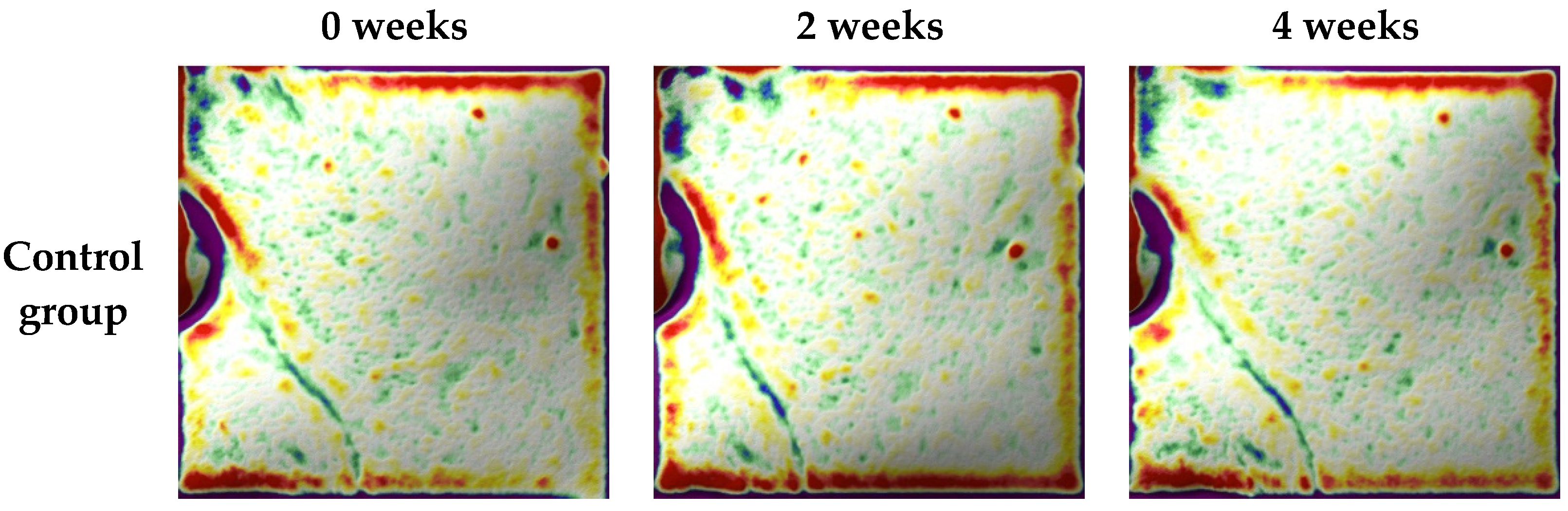
The final number of volunteers was 43 because one person dropped out, where those that remained were randomly divided into two groups: a control group (n = 21) or a test group (n = 22). The characteristics of volunteers are presented in Table 2. The average age of the control group was 51.6 ± 4.4 years and that of the test group was 46.8 ± 4.7 years. The most common skin type in both the control and test groups was dry skin, with n = 15 and n = 11, respectively. All participants reported no skin irritation due to the environment, such as prickling, itching, or side effects. Only one person reported a skin condition change, which was due to menstruation. All subjects applied sunscreen. The characteristics of the volunteers were suitable according to national regulations from the Korean Ministry of Food and Drug Safety and good clinical practice (GCP) [23].

3.3. Skin Hydration

Figure 3 shows the skin hydration value of the control and test groups before and after using the buccal collagen peptide film for 2 weeks and 4 weeks. No significant differences in the mean initial skin hydration (at 0 weeks) between the control group and the test group were found (43.97 ± 13.32 AU vs. 42.03 ± 11.00 AU, respectively). The mean skin hydration of the control group did not significantly change after 4 weeks. However, after 2 weeks, the mean skin hydration of the test group significantly increased (p < 0.001) by 16.56% compared with 0 weeks (48.99 ± 9.19 AU), and there was a significant increase (p < 0.001) in the test group compared with the control group (48.99 ± 9.19 AU vs. 44.89 ± 13.36 AU, respectively). The final measurements after 4 weeks showed a significant increase (p < 0.001) in skin hydration in the test group by 33.43% compared with before the buccal collagen peptide film was used (56.08 ± 9.38 AU). A significant difference (p < 0.001) between the test group and the control group after 4 weeks was also shown (56.08 ± 9.38 AU vs. 47.27 ± 13.56 AU). Therefore, the buccal collagen peptide film improved skin hydration.

3.4. Skin Elasticity

The results of skin elasticity are shown in Figure 4. At the start (0 weeks), the R2 skin elasticity in the control and test groups were 0.6541 ± 0.0844 and 0.6835 ± 0.0599 and those of R7 were 0.3207 ± 0.0512 and 0.3015 ± 0.0498, respectively. The R2 skin elasticity in the test group significantly increased at 2 weeks (0.6898 ± 0.0598, +5.46%, p < 0.01) and 4 weeks (0.7353 ± 0.0644, +12.41%, p < 0.001), and that of R7 in the test group also significantly increased at 2 weeks (0.3291 ± 0.0389, +9.16%, p < 0.01) and 4 weeks (0.3528 ± 0.0431, +17.01%, p < 0.001). The control group’s R2 skin elasticity was only significantly enhanced at 4 weeks (0.7084 ± 0.0553, 3.63%, p < 0.01), and the rate of increase in R2 in the test group at 4 weeks was better (12.41%) than that in the control group (3.63%). Furthermore, the skin elasticity in the test group, compared to the control group, was significantly different at both 2 weeks (p < 0.01) and 4 weeks (p < 0.001) but only for R2.

3.5. Skin Roughness

In line with the findings for skin hydration and elasticity, skin roughness (Ra) decreased after using the buccal delivery collagen peptide film (Figure 5). The baseline Ra values in the control and test groups were similar: 10.43 ± 1.50 and 10.93 ± 1.53, respectively. Only Ra in the test group significantly decreased, with a decrease of 10.62 ± 1.43 (−2.77%, p < 0.05) after 2 weeks and 10.57 ± 1.55 (−3.25%, p < 0.05) after 4 weeks. The Ra values between the control and test groups were significantly different at 2 weeks (p < 0.01) and 4 weeks (p < 0.01).
Furthermore, 3D imaging after 4 weeks showed the effect of using the buccal delivery collagen peptide film on skin wrinkles (Figure 6). The green lines indicate wrinkles. The wrinkles on the cheek faded after only 2 weeks in subjects in the test group. Conversely, the depth of wrinkles on the cheek in the control group did not change.

3.6. Questionnaire

The assessment of side effects in all subjects, at both 2 weeks and 4 weeks, is not reported.
The results of the questionnaire are shown in Table 3. The subjects in the control group only reported significant improvement in sagging skin between 2 and 4 weeks. However, all results of the questionnaire in the test group were significantly different between 2 and 4 weeks. Although significant differences in the questionnaire between the control and test groups were not observed, all mean scores in the test group were higher than those in the control group at both 2 and 4 weeks.

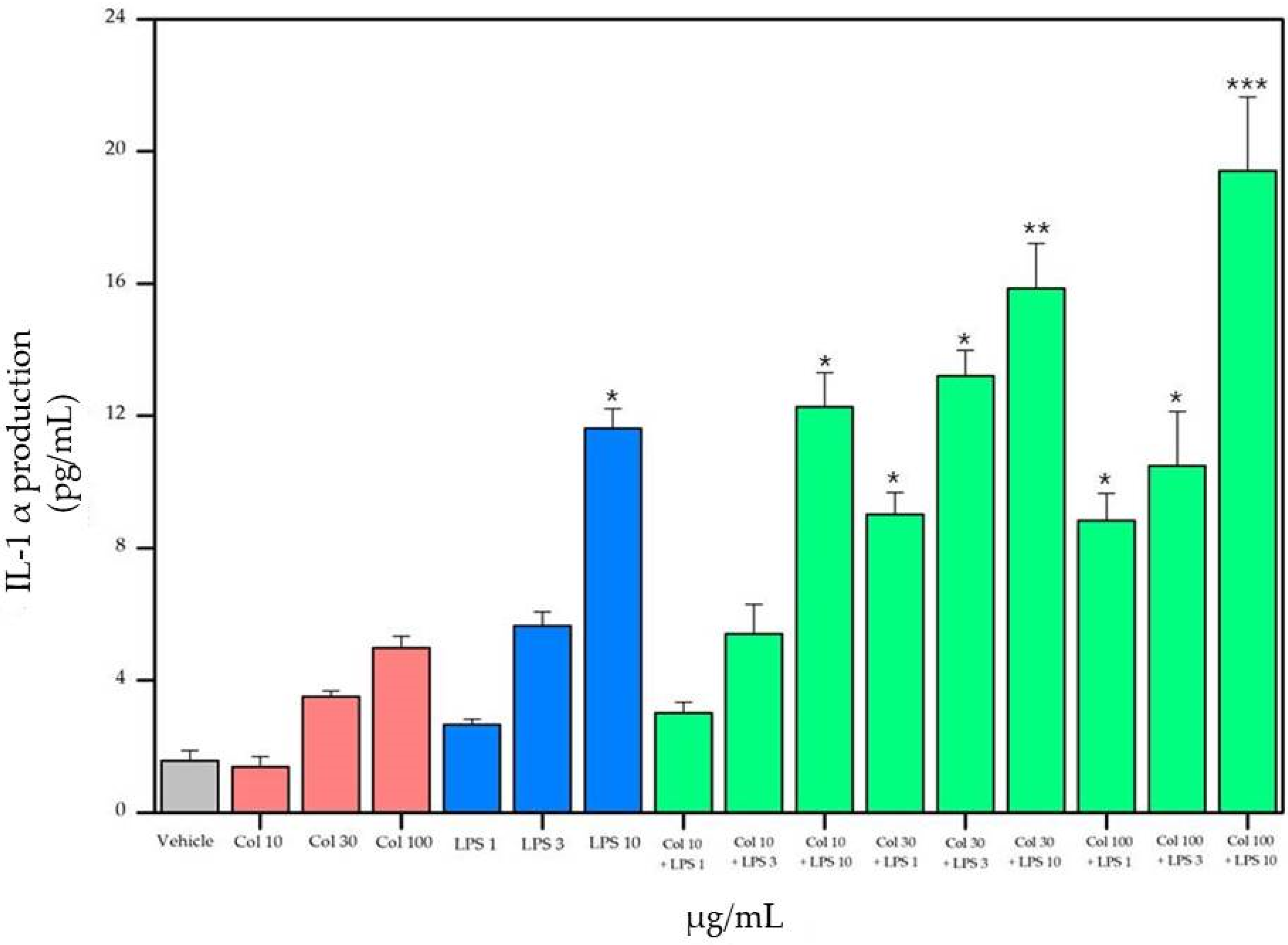
3.7. Cytokine Results

3.7.1. Bradford Assay

The residual protein content in the bioprocessed supernatants of RAW 264.7 cells treated with collagen and LPS was evaluated using the Bradford assay (Figure 7). To precisely measure the residual protein content in the cell culture supernatants, cells were cultivated in serum-free media when collagen and LPS were supplied. The concentration of protein in the vehicle was 2.22 ± 0.41 µg/mL. Treatments in the 10 and 30 µg/mL collagen-only groups, as well as the 1 and 3 µg/mL LPS-only groups, showed no significant differences. However, both 100 µg/mL of collagen and 10 µg/mL of LPS significantly increased the protein concentration, 2.42 ± 0.62 µg/mL and 2.42 ± 0.35 µg/mL, respectively (p < 0.05). In addition, synergic effects of collagen with LPS were observed, where the protein concentrations treated with the combined samples of collagen with LPS were significantly increased (p < 0.05). Groups with 100 µg/mL of collagen, regardless of LPS concentrations, were significantly different (p < 0.05); notably, the protein concentration of 100 µg/mL of collagen with 10 µg/mL of LPS was the highest at 2.55 ± 0.28 µg/mL.

3.7.2. Peroxynitrite (ONOO) and Interleukin-1α

Figure 8 and Figure 9 display the effects of collagen and LPS on the secretion of inflammatory mediators, namely, peroxynitrite (ONOO) and IL-1α. Overall, the relative levels of peroxynitrite increased after treatment in all groups (Figure 8). The fluorescence of the ONOO vehicle was 87.17 ± 4.67. The relative levels of 30 and 100 µg/mL of the collagen-only and 3 and 10 µg/mL of the LPS-only treatment groups were significantly increased (p < 0.05). Collagen and LPS showed synergic effects on ONOO production in the media. In addition, a significant increase in peroxynitrite levels was observed after the treatment with a combination of 10 µg/mL collagen and 10 µg/mL LPS (p < 0.05); notably, the combination of 100 µg/mL of collagen and 10 µg/mL of LPS showed the highest levels (p < 0.001).
Overall, the trends in IL-1α levels increased after the treatment with collagen and LPS (Figure 9). The concentration of IL-1α in the vehicle was 1.57 ± 0.31 pg/mL; 10 µg/mL of collagen (only) did not affect the IL-1α levels. Although the IL-1α levels increased in the collagen-only treatment group (30 and 100 µg/mL) and the LPS-only treatment group (1 and 3 µg/mL), the differences were not significant. The highest concentration of LPS (10 µg/mL) significantly increased the production of IL-1α in the media (p < 0.05) to 11.63 ± 0.59 pg/mL. Combined samples of collagen and LPS showed a synergic effect on IL-1α production. The IL-1α levels significantly increased in all concentrations in the combination groups with higher than 10 µg/mL of collagen and 10 µg/mL of LPS (p < 0.05). As expected, the highest concentration of IL-1α was exhibited in the 100 µg/mL of collagen with 10 µg/mL of LPS group and was 19.42 ± 2.23 pg/mL.

4. Discussion

Although many collagen products, such as drinks, tablets/capsules, and cosmetics, have been developed in the foods and cosmetics industries recently, the buccal delivery technology that is applied to collagen peptide delivery is unique. Collagen products have usually faced absorption issues because (i) when they are taken orally, the polypeptide is broken down by digestive enzymes, and (ii) when it is applied on the skin’s surface in the form of cosmetics, the collagen polymer cannot efficiently permeate the skin surface. Therefore, new technology for enhancing the absorption of collagen peptides is needed.
The buccal permeation results suggest the possibility of improving collagen peptide absorption without breaking the peptide. Koo et al. investigated hydrophilic polymer-based buccal films containing 2–3 kDa of collagen hydrolysate, finding that they can permeate mucosa via an in vitro human buccal tissue permeation test [13]. A concentration of 300 µg/cm2 of collagen hydrolysate was detected in permeated human buccal tissues after 4 h in their study. The permeation results in our study are in good agreement with Koo et al.’s study. We demonstrated that the small molecular weight of the collagen peptide facilitated easier penetration through buccal tissues compared to a large peptide. The film method is more efficient than taking a capsule, tablet, or drink because the collagen peptide can permeate directly through the buccal mucosa; however, collagen taken orally needs to be digested and absorbed again.
Many systematic reviews on the effectiveness of oral collagen supplementation have recently confirmed that they can enhance skin moisture, elasticity, and roughness [21]. The effects of collagen peptide use on skin hydration were already reported in previous studies. Inoue et al. found that the skin moisture of subjects taking 10 mg/d of collagen was significantly higher than those taking 0.5 mg/d of collagen after 8 weeks, with 30.99 ± 16.78% for high intake and 18.79 ± 12.85% for low intake [26]. In addition, skin hydration among woman volunteers significantly increased by 16% after daily oral intake of 10 g of collagen for 4 weeks compared with the placebo group [27].
The R2 skin elasticity significantly improved from 0.69 ± 0.05 at 0 weeks to 0.71 ± 0.06 at 12 weeks in the test group, which drunk 2.5 g/d of collagen peptide [21]. Inoue et al. induced the skin elasticity (R2) of volunteers taking a collagen capsule containing 10 mg collagen per day, and they found significant enhancement on the cheek after 4 weeks (25.67 ± 23.55%) and canthus after 8 weeks (20.84 ± 10.75%) [26].
Compared to a placebo, a significant decrease in skin wrinkle depth was seen in the test group, which took a drink product every day for 3 months and procollagen type I containing 2.5 g of collagen every day for 56 days [21,28,29]. In addition, Asserin reported that a daily oral intake of 10 g collagen for 12 weeks led to a significant reduction in collagen network fragmentation [27].
This study was conducted as a clinical trial with 43 healthy women who consumed either a placebo (n = 21) or functional supplement (n = 22) for 4 weeks. An improvement of human skin treated with the buccal delivery collagen peptide film was demonstrated. Although the effectiveness of collagen taken orally, such as capsules and drinks, on skin improvement was good in various studies, the collagen peptide film has only been tested in this study, and positive effects were shown. Most former studies on other oral collagen products report human skin improvement after 4, 8, and 12 weeks [21,26,27,28,29]. However, buccal delivery of the collagen peptide film showed effects after 2 weeks. In addition, the wrinkles on the cheeks of volunteers were reduced by the collagen buccal film after 2 weeks. Thus, using buccal delivery technology for skin enhancement was more efficient than other oral products, such as tablets/capsules and drinks.
One reason for the significant skin improvement was the difference in absorption of collagen peptides. Collagen products taken orally use specific amino acids as building blocks of human collagen, such as proline, hydroxyproline, glycine, alanine, glutamic acid, and arginine [30,31,32]. However, the mechanisms for the re-synthesis of amino acids that are broken by digestive enzymes have not been shown. To overcome these problems, transdermal and trans-buccal routes have been investigated [33]. We previously showed that a buccal delivery film can directly absorb a completely formed collagen peptide (2000–3000 Da) through the human buccal tissue [13]. Tomoko et al. demonstrated that collagen peptides, such as prolyl-hydroxyproline and hydroxyprolyl-glycine, triggered the growth of fibroblasts combined with collagen gel in blood, which were associated with improved skin conditions [34]. Therefore, the buccal delivery film shows fast and strong effects on the enhancement of skin condition.
However, the results in this clinical study, especially in terms of reducing the wrinkles within 2 weeks, were not completely explained by the advantages of the buccal delivery film. Therefore, we suggest that collagen peptides absorbed through the buccal mucosa had a cytokine-related homeopathic effect, similar to Botox®.
Homeopathy is a systemic therapy that is focused on health and wholeness rather than disease [35]. When taken at a higher dose, any substance can cause symptoms; however, when taken as a small dose, it can revitalize the immune system with no cell death [35]. This effect is usually applied in immunology. One of the most representative products using homeopathy in the cosmetic field is Botox® [36]. However, at a high dose, collagen is known to induce arthritis [37,38,39]. Jo et al. reported that a filler injection containing collagen led to inflammatory granulomas under the eye after 7 years [40]. Therefore, according to previous studies, collagen can induce local inflammation. IL-1, an inflammation mediator, can be produced when cells are damaged [41]. Peroxynitrite (ONOO) use can also lead to skin inflammation, arthritis, oxidation, and vascular dilatation [42,43,44]. According to the results of this study, total protein, relative levels of peroxynitrite (ONOO), and IL-1α production in the supernatant of RAW 264.7 cells were increased after collagen peptide use. Additionally, similar increases were seen after LPS use, which is a well-known inflammatory inducer. Because cytokine materials, such as interleukin and TNF, are proteins, the increase in total protein after the collagen treatment could be due to the production of cytokine factors. In addition, the increased levels of IL-1α and peroxynitrite could be due to some damage to the cells by the collagen peptide. Many previous studies reported that collagen could produce peroxynitrite [45,46,47] and interleukin-1 [48,49]. These phenomena suggest that collagen peptide delivered via buccal film enhances the reduction in wrinkles and skin hydration owing to the slight edema and vascular dilatation from IL-1α and peroxynitrite, which showed no side effects in the clinical test. The results from the cytokine experiment can be the basis for future studies. Thus, collagen enhancement via buccal film delivery followed the local and temporary actions of (i) homeopathic effects induced by weak inflammation, (ii) vascular dilatation, and (iii) enhancement of interstitial fluid hydration.

5. Conclusions

This study showed the absorption of collagen peptides in the human body without being destroyed using a buccal delivery system. The collagen peptide permeated the buccal mucosa of swine, and significantly different concentrations of collagen were also analyzed after permeation according to molecular weight. Furthermore, the present study is the first clinical trial that used a collagen peptide buccal delivery method. The collagen peptide buccal delivery film demonstrated a significant improvement in skin hydration, elasticity, roughness, and wrinkle removal on cheeks with no side effects after 2 weeks of use. To investigate these results, the production of peroxynitrite and interleukin-1α was analyzed, and the effectiveness may have been due to the complete absorption of collagen peptides via the buccal delivery system and homeopathic effects.

6. Patents

The test product, Collagen Oral Bio Film®, was registered in patent “High-efficiency buccal drug delivery film (application number: 10-2020-0084388)”.

Author Contributions

Y.H.K., writing the manuscript. Y.B.H., supervision. D.I., designed the clinical trial and review. K.-H.L. and Y.H.K., designed and conducted the cytokine experiment. S.Y., development of the collagen peptide buccal delivery film. All authors read and agreed to the published version of the manuscript.

Funding

This research received no external funding.

Institutional Review Board Statement

The study was conducted according to the guidelines of the Declaration of Helsinki and Good Clinical Practice of Ministry of Food and Drug Safety, and approved by the Institutional Review Board of Kyung-Hee University (protocol code KHSP-011 V.1 and approval in 25 May 2020).

Informed Consent Statement

Informed consent was obtained from all subjects involved in the study.

Conflicts of Interest

The authors declare no conflict of interest.

References

  1. Sorushanova, A.; Delgado, L.M.; Wu, Z.; Shologu, N.; Kshirsagar, A.; Raghunath, R.; Mullen, A.M.; Bayon, Y.; Pandit, A.; Raghunath, M.; et al. The collagen suprafamily: From bio-synthesis to advanced biomaterial development. Adv. Mater. 2019, 31, e1801651. [Google Scholar] [CrossRef] [Green Version]
  2. Rodríguez, M.I.A.; Barroso, L.G.R.; Sánchez, M.L. Collagen: A review on its sources and potential cosmetic applications. J. Cosmet. Dermatol. 2017, 17, 20–26. [Google Scholar] [CrossRef] [PubMed]
  3. Rizk, M.A.; Mostafa, N.Y. Extraction and characterization of collagen from buffalo skin for bio-medical applications. Orient. J. Chem. 2016, 32, 1601–1609. [Google Scholar] [CrossRef]
  4. Blackburn, N.J.; Sofrenovic, T.; Kuraitis, D.; Ahmadi, A.; McNeill, B.; Deng, C.; Rayner, K.; Zhong, Z.; Ruel, M.; Suuronen, E.J. Timing underpins the benefits associated with injectable collagen biomaterial therapy for the treatment of myocardial infarction. Biomaterials 2014, 39, 182–192. [Google Scholar] [CrossRef] [PubMed]
  5. Alves, A.L.; Marques, A.L.P.; Martins, E.; Silva, T.H.; Reis, R.L. Cosmetic Potential of Marine Fish Skin Collagen. Cosmetics 2017, 4, 39. [Google Scholar] [CrossRef] [Green Version]
  6. Guillerme, J.-B.; Couteau, C.; Coiffard, L. Applications for Marine Resources in Cosmetics. Cosmetics 2017, 4, 35. [Google Scholar] [CrossRef] [Green Version]
  7. Xhauflaire-Uhoda, E.; Fontaine, K.; Piérard, G. Kinetics of moisturizing and firming effects of cosmetic formulations. Int. J. Cosmet. Sci. 2008, 30, 131–138. [Google Scholar] [CrossRef] [PubMed]
  8. Hayashi, Y.; Yamada, S.; Ikeda, T.; Yanagiguchi, K. Marine Cosmeceuticals: Trends and Prospects; CRC Press: Boca Raton, FL, USA, 2011. [Google Scholar]
  9. Morgantii, P.; Randazzo, S.; Cardilloi, A. Role of insoluhle and soluhle collagen as skin moisturizer. J. Appl. Cosmetol. 1986, 4, 141–152. [Google Scholar]
  10. Fluhr, J.W.; Elsner, P.; Berardesca, E.; Maibach, H.I. Bioengineering of the Skin: Water and the Stratum Corneum; CRC Press: Boca Raton, FL, USA, 2004. [Google Scholar]
  11. Peng, Y.; Glattauer, V.; Werkmeister, J.A.; Ramshaw, J.A.M. Evaluation for collagen products for cosmetic application. Int. J. Cosmet. Sci. 2004, 26, 313. [Google Scholar] [CrossRef]
  12. Nikolaeva, T.I.; Laurinavichus, K.S.; Kaptsov, V.V.; Molchanov, M.V.; Shekhovtsov, P.V. Development of Low-Molecular Weight Collagen Peptide Complex with Glycosaminoglycan Components. Bull. Exp. Biol. Med. 2018, 165, 629–634. [Google Scholar] [CrossRef]
  13. Koo, J.; Lee, S.; Yeo, S.; Kim, D.; Park, M.; Nam, T.S.; Lee, J. Viscosity Effects of Hydrophilic Polymers on Transport of Collagen Hydrolysate Across Reconstructed Human Buccal Tissue. Bull. Korean Chem. Soc. 2020, 41, 290–295. [Google Scholar] [CrossRef]
  14. Kiela, P.R.; Ghishan, F.K. Physiology of Intestinal Absorption and Secretion. Best Pr. Res. Clin. Gastroenterol. 2016, 30, 145–159. [Google Scholar] [CrossRef] [Green Version]
  15. Martin, L.; Wilson, C.G.; Koosha, F.; Uchegbu, I.F. Sustained buccal delivery of the hydrophobic drug denbufylline using physically cross-linked palmitoyl glycol chitosan hydrogels. Eur. J. Pharm. Biopharm. 2003, 55, 35–45. [Google Scholar] [CrossRef]
  16. Siddiqui, M.; Garg, G.; Sharma, P.K. A short review on “A novel approach in oral fast dissolving drug delivery system and their patents”. Adv. Biol. Res. 2011, 5, 291–303. [Google Scholar]
  17. Castro, P.M.; Baptista, P.; Madureira, A.R.; Sarmento, B.; Pintado, M.E. Combination of PLGA nanoparticles with mucoadhesive guar-gum films for buccal delivery of antihypertensive peptide. Int. J. Pharm. 2018, 547, 593–601. [Google Scholar] [CrossRef] [PubMed]
  18. Metia, P.K.; Bandyopadhyay, A.K. In vitro and in vivo evaluation of a novel mucoadhesive buccal oxytocin tablet prepared with Dillenia indica fruit mucilage. Die Pharm. Int. J. Pharm. Sci. 2008, 270–274. [Google Scholar] [CrossRef]
  19. Langoth, N.; Bernkop-Schnürch, A.; Kurka, P. In Vitro Evaluation of Various Buccal Permeation Enhancing Systems for PACAP (Pituitary Adenylate Cyclase-Activating Polypeptide). Pharm. Res. 2005, 22, 2045–2050. [Google Scholar] [CrossRef]
  20. Bird, A.P.; Faltinek, J.R.; Shojaei, A.H. Transbuccal peptide delivery: Stability and in vitro per-meation studies on endomorphin-1. J. Control. Release 2001, 73, 31–36. [Google Scholar] [CrossRef]
  21. Bolke, L.; Schlippe, G.; Gerß, J.; Voss, W. A Collagen Supplement Improves Skin Hydration, Elasticity, Roughness, and Density: Results of a Randomized, Placebo-Controlled, Blind Study. Nutrients 2019, 11, 2494. [Google Scholar] [CrossRef] [Green Version]
  22. Faocd, D.O.; Kathy, L.A. Clinical Evidence of the Anti-Aging Effects of a Collagen Peptide Nutraceutical Drink on the Skin. J Drugs Dermatol. 2020, 19, s5–s10. [Google Scholar]
  23. Anonymous International Council for Harmonisation of Technical Requirements for Pharmaceuticals for Human Use ICH Harmonised Guideline: Integrated addendum to ICH E6; Clinical Practice, European Medicines Agency: London, UK, 2016; R1: Guideline for Good, E6(R2).
  24. Stroumza, N.; Bosc, R.; Hersant, B.; Hermeziu, O.; Meningaud, J.P. Benefits of using the cut-ometer to evaluate the effectiveness of skin treatments in plastic and maxillofacial surgery. Rev. Stomatol. Chir. Maxillo-Faciale Chir. Orale 2015, 116, 77–81. [Google Scholar] [CrossRef] [PubMed]
  25. Willy, K.; Girndt, M.; Voelkl, J.; Fiedler, R.; Martus, P.; Storr, M.; Schindler, R.; Zickler, D. Expanded haemodialysis therapy of chronic haemodialysis patients prevents calcification and apoptosis of vascular smooth muscle cells in vitro. Blood Purif. 2018, 45, 131–138. [Google Scholar] [CrossRef] [PubMed]
  26. Inoue, N.; Sugihara, F.; Wang, X. Ingestion of bioactive collagen hydrolysates enhance facial skin moisture and elasticity and reduce facial ageing signs in a randomised double-blind placebo-controlled clinical study. J. Sci. Food Agric. 2016, 96, 4077–4081. [Google Scholar] [CrossRef] [PubMed]
  27. Asserin, J.; Lati, E.; Shioya, T.; Prawitt, J. The effect of oral collagen peptide supplementation on skin moisture and the dermal collagen network: Evidence from an ex vivo model and randomized, placebo-controlled clinical trials. J. Cosmet. Dermatol. 2015, 14, 291–301. [Google Scholar] [CrossRef] [Green Version]
  28. Proksch, E.; Schunck, M.; Zague, V.; Segger, D.; Degwert, J.; Oesser, S. Oral Intake of Specific Bioactive Collagen Peptides Reduces Skin Wrinkles and Increases Dermal Matrix Synthesis. Ski. Pharmacol. Physiol. 2014, 27, 113–119. [Google Scholar] [CrossRef] [PubMed]
  29. Proksch, E.; Segger, D.; Degwert, J.; Schunck, M.; Zague, V.; Oesser, S. Oral Supplementation of Specific Collagen Peptides Has Beneficial Effects on Human Skin Physiology: A Double-Blind, Placebo-Controlled Study. Ski. Pharmacol. Physiol. 2014, 27, 47–55. [Google Scholar] [CrossRef]
  30. Zague, V.; de Freitas, V.; da Costa Rosa, M.d.C.; de Castro, G.Á; Jaeger, R.G.; Machado-Santelli, G.M. Collagen hydrolysate intake increases skin collagen expression and suppresses matrix metalloproteinase 2 activity. J. Med. Food 2011, 14, 618–624. [Google Scholar] [CrossRef]
  31. Iwai, K.; Hasegawa, T.; Taguchi, Y.; Morimatsu, F.; Sato, K.; Nakamura, Y.; Higashi, A.; Kido, Y.; Nakabo, Y.; Ohtsuki, K. Identification of Food-Derived Collagen Peptides in Human Blood after Oral Ingestion of Gelatin Hydrolysates. J. Agric. Food Chem. 2005, 53, 6531–6536. [Google Scholar] [CrossRef] [PubMed]
  32. Ichikawa, S.; Morifuji, M.; Ohara, H.; Matsumoto, H.; Takeuchi, Y.; Sato, K. Hydroxypro-line-containing dipeptides and tripeptides quantified at high concentration in human blood after oral administration of gelatin hydrolysate. Int. J. Food Sci. Nutr. 2010, 61, 52–60. [Google Scholar] [CrossRef]
  33. Dubey, V.; Mishra, D.; Asthana, A.; Jain, N.K. Transdermal delivery of a pineal hormone: Melatonin via elastic liposomes. Biomaterials 2006, 27, 3491–3496. [Google Scholar] [CrossRef] [PubMed]
  34. Asai, T.T.; Oikawa, F.; Yoshikawa, K.; Inoue, N.; Sato, K. Food-derived collagen peptides, prolyl-hydroxyproline (Pro–Hyp), and hydroxyprolyl-glycine (Hyp–Gly) enhance growth of primary cultured mouse skin fibroblast using fetal bovine serum free from hydroxyprolyl peptide. Int. J. Mol. Sci. 2019, 21, 229. [Google Scholar] [CrossRef] [PubMed] [Green Version]
  35. Seeley, B.M.; Denton, A.B.; Ahn, M.S.; Maas, C.S. Effect of homeopathic Arnica montana on bruising in face-lifts: Results of a randomized, double-blind, placebo-controlled clinical trial. Arch. Facial Plast. Surg. 2006, 8, 54–59. [Google Scholar] [CrossRef] [PubMed] [Green Version]
  36. Goldschmidt, M.; Clemow, J.B. Neuromodulators (Botox). In Applied Head and Neck Anatomy for the Facial Cosmetic Surgeon; Springer: Berlin/Heidelberg, Germany, 2021; pp. 123–129. [Google Scholar]
  37. Boilard, E.; Nigrovic, P.A.; Larabee, K.; Watts, G.; Coblyn, J.S.; Weinblatt, M.E.; Massarotti, E.M.; Remold-O’Donnell, E.; Farndale, R.W.; Ware, J.; et al. Platelets Amplify Inflammation in Arthritis via Collagen-Dependent Microparticle Production. Science 2010, 327, 580–583. [Google Scholar] [CrossRef] [Green Version]
  38. Yamanishi, Y.; Boyle, D.L.; Pinkoski, M.J.; Mahboubi, A.; Lin, T.; Han, Z.; Zvaifler, N.J.; Green, D.; Firestein, G.S. Regulation of Joint Destruction and Inflammation by p53 in Collagen-Induced Arthritis. Am. J. Pathol. 2002, 160, 123–130. [Google Scholar] [CrossRef] [Green Version]
  39. Lubberts, E.; Joosten, L.A.B.; Oppers, B.; Bersselaar, L.V.D.; Roo, C.J.J.C.-D.; Kolls, J.K.; Schwarzenberger, P.; Van De Loo, F.A.J.; Berg, W.B.V.D. IL-1-Independent Role of IL-17 in Synovial Inflammation and Joint Destruction During Collagen-Induced Arthritis. J. Immunol. 2001, 167, 1004–1013. [Google Scholar] [CrossRef] [PubMed] [Green Version]
  40. Jo, Y.J.; Lee, D.G.; Lee, S.B. Late-onset migrated inflammatory granuloma after collagen-containing filler injection. J. Korean Ophthalmol. Soc. 2008, 49, 1330–1334. [Google Scholar] [CrossRef] [Green Version]
  41. Park, Y.S.; Han, H. Anti-inflammatory activity Effects of Mori Folium Water Extraction IL-1α, IL-6 and IL-10 on mouse macrophages. J. Digit. Converg. 2018, 16, 613–620. [Google Scholar]
  42. Chung, H.Y.; Kim, H.J.; Kim, K.W.; Choi, J.S.; Yu, B.P. Molecular inflammation hypothesis of aging based on the anti-aging mechanism of calorie restriction. Microsc. Res. Tech. 2002, 59, 264–272. [Google Scholar] [CrossRef] [PubMed]
  43. Chung, H.; Soung, D.; Kim, A.; Choi, H.; Kim, H.; Choi, J.; Yang, R.; Lee, K.; Yu, B. Generation, toxicity and scavenging of ONOO−: Its involvement in the aging process. Kor. J. Gerontol. 2000, 10, 46–59. [Google Scholar]
  44. Kweon, Y.; Jeong, J. Peroxynitrite scavenging activity of Sabohwanin lipopolysaccha-ride-induced oxidatively-stressed mice. J. Intern. Korean Med. 2007, 28, 80–91. [Google Scholar]
  45. Kato, Y.; Ogino, Y.; Aoki, T.; Uchida, K.; Kawakishi, S.; Osawa, T. Phenolic Antioxidants Prevent Peroxynitrite-Derived Collagen Modification in Vitro. J. Agric. Food Chem. 1997, 45, 3004–3009. [Google Scholar] [CrossRef]
  46. Evans, L.C.; Liu, H.; Pinkas, G.A.; Thompson, L.P. Chronic hypoxia increases peroxynitrite, MMP9 expression, and collagen accumulation in fetal guinea pig hearts. Pediatr. Res. 2011, 71, 25–31. [Google Scholar] [CrossRef] [PubMed]
  47. Dooley, A.; Gao, B.; Shi-Wen, X.; Abraham, D.J.; Black, C.M.; Jacobs, M.; Bruckdorfer, K.R. Effect of nitric oxide and peroxynitrite on type I collagen synthesis in normal and scleroderma dermal fibroblasts. Free. Radic. Biol. Med. 2007, 43, 253–264. [Google Scholar] [CrossRef]
  48. Tingstrom, A.; Heldin, C.; Rubin, K. Regulation of fibroblast-mediated collagen gel contraction by platelet-derived growth factor, interleukin-1 alpha and transforming growth factor-beta 1. J. Cell Sci. 1992, 102, 315–322. [Google Scholar] [CrossRef] [PubMed]
  49. Yasuda, T.; Poole, A.R. A fibronectin fragment induces type II collagen degradation by colla-genase through an interleukin-1—mediated pathway. Arthritis Rheum. 2002, 46, 138–148. [Google Scholar] [CrossRef]
Figure 1. The schematic image of the double-layer buccal film (a) and intake method (b).
Applsci 11 07486 g001
Figure 2. The concentration of collagen peptide in the buccal delivery film that permeated the buccal mucosa of swine as a function of the molecular weight and time. The changes were significantly different between 1 kDa and 3 kDa with p < 0.001 (***).
Applsci 11 07486 g002
Figure 3. Skin hydration before (0 weeks) and after (2 and 4 weeks) using the collagen peptide buccal delivery film of the control group (gray) and test group (white). The boxplot shows the average (•), median ( Applsci 11 07486 i001), and max–min whiskers (I). The changes were significantly different between the control and test groups, and before and after intake, with p < 0.001 (***).
Applsci 11 07486 g003
Figure 4. (a) R2 and (b) R7 skin elasticity before (0 weeks) and after (2 and 4 weeks) using the collagen peptide buccal delivery film of the control group (gray) and test group (white). The boxplot shows the average (•), median ( Applsci 11 07486 i001), and max–min whiskers (I). The changes were significantly different between the control and test groups, before and after intake, with p < 0.05 (*), p < 0.01 (**), and p < 0.001 (***).
Applsci 11 07486 g004
Figure 5. Skin roughness before (0 weeks) and after (2 and 4 weeks) using the collagen peptide buccal delivery film of the control group (gray) and test group (white). The boxplot shows the average (•), median ( Applsci 11 07486 i001), and max–min whiskers (I). The changes were significantly different between the control and test groups, before and after intake, with p < 0.05 (*) and p < 0.01 (**).
Applsci 11 07486 g005
Figure 6. 3D images of wrinkles on the cheek before (0 weeks) and after (2 and 4 weeks) using the collagen peptide buccal delivery film.
Applsci 11 07486 g006aApplsci 11 07486 g006b
Figure 7. Effect of collagen peptide and LPS on total protein production in supernatant media without RAW 264.7 cells for 48 h. The values are presented as a mean ± standard deviation. The changes were significantly different between the vehicle and test samples, with p < 0.05 (*).
Applsci 11 07486 g007
Figure 8. Effect of collagen peptide and LPS on peroxynitrite production in supernatant media without RAW 264.7 cells for 48 h. The values are presented as a mean ± standard deviation. The changes were significantly different between the vehicle and test samples, with p < 0.05 (*), p < 0.01 (**), and p < 0.001 (***).
Applsci 11 07486 g008
Figure 9. Effect of collagen peptide and LPS on interleukin-1α production in the supernatant media without RAW 264.7 cells for 48 h. The values are presented as a mean ± standard deviation. The changes were significantly different between the vehicle and test samples, with p < 0.05 (*), p < 0.01 (**), and p < 0.001 (***).
Applsci 11 07486 g009
Table 1. Composition of the collagen peptide buccal delivery film.
Protective LayerMuco Layer
ComponentsProportion (%)ComponentsProportion (%)
HPMC10.0Collagen peptide10.7
HPC7.7HPMC8.8
Glycerin0.7Carrageenan0.4
Water29.4Sucrose distearate0.2
Ethanol52.2Glycerin0.7
--Polyacrylate0.7
--Water62.4
--Ethanol16.1
Table 2. Characteristics of the volunteers.
ParametersClassificationControl Group (n = 21)Test Group (n = 22)
Frequency (n)Proportion (%)Frequency (n)Proportion (%)
Age 30s00.0014.55
40s1047.621568.18
50s1152.38627.27
Skin typeDry skin1571.431150.00
Oily skin00.0014.55
Normal skin419.05313.64
Combination skin29.52731.82
Skin irritation reaction to the
environment
Yes00.0000.00
No21100.0022100.00
Prickle and itchYes00.0000.00
No21100.0022100.00
Side effectsYes00.0000.00
No21100.0022100.00
Change in skin condition during menstruationYes14.7614.55
No2095.242195.45
Applied
sunscreen
Daily use838.101045.45
Used when going out1361.901254.55
Not used00.0000.00
Table 3. The questionnaire scores after 2 and 4 weeks of using the collagen peptide buccal delivery film.
Sensory Question (1)Control GroupTest Group
2 w4 w2 w4 w
The degree of skin hydration3.95 ± 0.744.38 ± 0.744.14 ± 0.64 *4.73 ± 0.83 *
The degree of reduction in sagging skin3.62 ± 0.67 *4.14 ± 0.91 *3.86 ± 0.83 *4.45 ± 0.91 *
The degree of elasticity of the skin3.81 ± 0.814.33 ± 0.914.18 ± 0.85 *4.68 ± 0.78 *
The degree of smoothness of the skin4.14 ± 0.574.43 ± 0.874.27 ± 0.704.73 ± 0.83
(1) Evaluated on a 6-point hedonic sale, from 1 = no effect to 6 = extremely good effect. The changes were significantly different between intakes after 2 and 4 weeks with p < 0.05 (*).
Publisher’s Note: MDPI stays neutral with regard to jurisdictional claims in published maps and institutional affiliations.

