Review

Is Diffusion Tensor Imaging-Guided Radiotherapy the New State-of-the-Art? A Review of the Current Literature and Technical Insights

1
UCL Queen Square Institute of Neurology, University College London, London WC1N 3BG, UK
2
The Lysholm Department of Neuroradiology, National Hospital for Neurology and Neurosurgery, Queen Square, Holborn, London WC1N 3BG, UK
3
Neuroradiological Academic Unit, Department of Brain Repair and Rehabilitation, UCL Queen Square Institute of Neurology, London WC1N 3BG, UK
4
Department of Medical Physics & Biomedical Engineering, University College London, Malet Place Engineering Building, London WC1E 6BT, UK
5
Department of Radiotherapy, University College London Hospitals NHS Foundation Trust, London NW1 2PG, UK
6
Department of Clinical Oncology, University College London Hospitals NHS Foundation Trust, London NW1 2PG, UK
7
National Institute for Health Research University College London Hospitals (UCLH) Biomedical Research Centre, London W1T 7HA, UK
*
Author to whom correspondence should be addressed.
Submission received: 16 December 2021 / Revised: 5 January 2022 / Accepted: 11 January 2022 / Published: 13 January 2022
(This article belongs to the Special Issue New Vistas in Radiotherapy)

Abstract

:
Despite the increasing precision of radiotherapy delivery, it is still frequently associated with neurological complications. This is in part due to damage to eloquent white matter (WM) tracts, which is made more likely by the fact they cannot be visualised on standard structural imaging. WM is additionally more vulnerable than grey matter to radiation damage. Primary brain malignancies also are known to spread along the WM. Diffusion tensor imaging (DTI) is the only in vivo method of delineating WM tracts. DTI is an imaging technique that models the direction of diffusion and therefore can infer the orientation of WM fibres. This review article evaluates the current evidence for using DTI to guide intracranial radiotherapy and whether it constitutes a new state-of-the-art technique. We provide a basic overview of DTI and its known applications in radiotherapy, which include using tractography to reduce the radiation dose to eloquent WM tracts and using DTI to detect or predict tumoural spread. We evaluate the evidence for DTI-guided radiotherapy in gliomas, metastatic disease, and benign conditions, finding that the strongest evidence is for its use in arteriovenous malformations. However, the evidence is weak in other conditions due to a lack of case-controlled trials.

1. Introduction

1.1. Intracranial Radiotherapy

Radiotherapy continues to advance to be able to target areas with higher conformality. The introduction of stereotactic radiotherapy (SRT), sometimes referred to as stereotactic radiosurgery, has meant that radiation can be directed with increasing precision. This results in better outcomes by enabling a higher dose to the area of pathology while reducing the radiation dose to healthy brain structures. Popular implementations include the Gamma Knife and Cyberknife platforms [1].
Intracranial radiotherapy requires planning the target area, which is typically done using MRI to identify the target region and eloquent brain structures that need to be avoided. This is usually performed with structural MRI sequences such as T1w, T2w, and FLAIR. This is achieved by manual segmentation of the pathology, with eloquent areas additionally segmented as organs at risk (OAR). Examples of OAR that are currently advised to be included are the brain stem, optic chiasm, cochlea, and others, with each OAR having an advised maximum radiation dose measured in Gray (Gy) to avoid significant late dysfunction and neurological toxicities [2]. Then, a CT with a frame is required for dose treatment planning.
With the advent of intensity-modulated radiation therapy (IMRT), the dose to pathological structures can be conformed to in 3D with excellent conformality. IMRT splits the beams into many smaller beamlets, enabling radiation to be directed into complex shapes and have a non-uniform radiation intensity [3]. IMRT additionally utilises computerised inverse planning where the radiation dose to the target region and dose limits to healthy brain and OAR are specified and optimised in advance. Both Cyber and Gamma Knife SRT delivery platforms include treatment planning software that enables integrated SRT planning allow for a combination of frameless MRI and the planning CT [4].
When targeting primary intracranial malignancies, the tumour visible on imaging is referred to as the gross tumour volume (GTV); this is then expanded by a standard margin (dependent on the tumour type) to incorporate any peritumoral infiltration that is unseen on imaging and is referred to as the clinical target volume (CTV). Therefore, the expansion of the CTV is important to target these migrating cells to reduce the chance of recurrence. Then, the CTV is typically expanded further by a few mm to allow for patient set-up errors that can occur despite immobilisation and be used as the final planning target volume (PTV) [5]. An example of the differing target volumes is shown in Figure 1. These expansion steps are typically done isotropically unless there are barriers to tumour spread e.g., skull or dura when delineating the CTV. The European Organization for Research and Treatment of Cancer (EORTC) current guidance for SRT in glioblastoma treatment is to define the GTV as the T1w contrast-enhancing tumour, or resection cavity, and then, the CTV is defined as the GTV expanded isotropically by 2 cm. Then, 3–5 mm is additionally added to define the PTV [2]. The CTV is set at a 2 cm isotropic expansion, as roughly 80% or more recurrences are within 2 cm of the enhancing tumour core.
Radiotherapy to the brain can lead to many neurological complications due to the damage of healthy brain structures and is usually dependent on dose, fraction size, volume of irradiated tissue, and concurrent chemotherapy use [6]. Complications include vascular abnormalities such as radiation necrosis and ischemia but additionally include progressive neurocognitive decline. This can lead to many long-term side effects in addition to short-term complications [7]. WM is additionally more vulnerable than grey matter, and some of the cognitive and functional negative effects are thought to be due to WM tract damage [8,9]. Individual WM tracts are not visible on structural imaging, and therefore, eloquent tracts may be unknowingly given high doses of radiation when in close proximity to the target region.
Gliomas as mentioned are infiltrative, and target volume expansion is included to allow for this while weighing up the risks of radiation. However, glioma cells are thought to spread preferentially along white matter (WM) tracts [10]. Therefore, it has been suggested that the modelling of WM properties such as their orientation may mean the CTV can be expanded anisotropically in order to reduce the overall radiation does while still covering the same proportion of potential recurrence sites [11].
The only in vivo method of delineating WM tracts is diffusion tensor imaging (DTI). DTI can be used to infer the isotropic and anisotropic properties of white matter and its orientation. This can be extended to produce virtually dissected WM tracts, the technique being called tractography. The incorporation of information on WM orientation and WM tracts appears to have potential for improving PTV delineation in SRT.

1.2. Diffusion Tensor Imaging

In diffusion-weighted MRI, a gradient is applied in a given direction; if protons diffuse along the gradient direction, dephasing will be accelerated, attenuating the MR signal. Areas with more free diffusion of water will have a reduced MR signal due to the greater movement of the protons; e.g., CSF will appear dark compared to grey matter as CSF contains minimal obstructive microstructure. Some tissues such as white matter will have preferential diffusion in a particular direction, which is known as anisotropic diffusion. This is due to the WM axons restricting the diffusion of water perpendicular to them, but diffusion parallel to their orientation being unrestricted. The imaging gradient direction can be changed, and the DWI signal would be expected to be higher if perpendicular to a WM tract and lower if parallel to it [12].
If DWI images are obtained with at least six gradient directions, the orientation or overall direction of diffusion in the voxel can be calculated and is known as diffusion tensor imaging (DTI). If the voxel is in WM, then the diffusion direction can be assumed to be the orientation of the WM tract. The amount to which that diffusion is preferential to one direction can be quantified and is known as fraction anisotropy (FA), where 0 is completely equal diffusion in all directions and 1 is completely uniform diffusion in only one direction. The overall diffusion of a voxel is known as mean diffusivity (MD). Another set of similar metrics to MD and FA are p and q maps, which refer to the isotropic and anisotropy components of the voxel, respectively [13]. A diagram displaying the effects of tissue microstructure on diffusion measurements is shown in Figure 2.
As displayed in Figure 2, DTI can capture microstructural changes of white matter, as damage leads to reduced FA and increased MD values due to a reduction in axon density and reduced myelination. This has been shown to be useful in detecting tumoural invasion, which can be observed as reduced FA or q values and has additionally been shown to be potentially useful in differentiating glioma reoccurrence from radiation necrosis after radiotherapy [14]. MD or p values will also increase with tumour invasion and have been suggested to be useful in defining glioma margins [15]. Other DTI measures have additionally been suggested to have potential in the diagnosis of intracranial malignancies, with ADC being shown to be useful in differentiating brain stem gliomas from medulloblastoma [16,17].
The information from DTI can be used to model whole WM tracts and is known as tractography. This is where each voxel is followed from a seed point and walks in the orientation or tract direction of each voxel; this is repeated for a set number of steps, and a tract is formed. Then, this is done for each voxel in a seed region. When a single tract is taken from each seed voxel, this is known as deterministic tractography. An issue with this method is that a false tract may be followed by accident. Another method known as probabilistic tractography repeats the tract formation multiple times, and a probability distribution of potential tracts is taken. Probabilistic tractography is usually more accurate, as it incorporates an amount of error, and tracts with a lower probability, that are likely false positives, can be filtered out [18]. Figure 3 displays an example of tractography forming a tract.
Selecting a seed point to form a desired tract is usually done based on structural anatomy likely to only contain the tract. An example is using the precentral gyrus as a seed point for the corticospinal tracts, as is contains the primary motor cortex. Way points can additionally be used, which only includes tracts that pass through them from the seed point. An example for the corticospinal tract could be the cerebral peduncles.
A limitation of tractography is that when the voxel contains multiple WM tracts with different orientations, then the mean direction of the two tracts may be taken or a smaller tract can be lost, as only the overall diffusion direction is taken. This is a particular issue when two tracts cross or “kiss” in the same voxel and may lead to false positive and false negative tracts. There are several techniques that attempt to model multiple WM tracts in one voxel, all of which require a larger number of directions [18]. The main drawback of these methods is that image acquisition and processing will take longer, but the sequence acquisitions and the processing software are continuously improved, and with more advanced hardware and software, these advanced acquisitions and processing have become clinically feasible.

2. Review Aims and Objectives

DTI can provide additional information on WM anatomy and therefore has the potential to improve radiotherapy accuracy. We decided to undertake a literature review on the subject of DTI-guided intracranial radiotherapy and whether its use can be considered a new state-of-the-art for clinical practice. The MEDLINE database via PubMed was searched for relevant articles along with references from a recent systematic review article [19]. A systematic review was not performed, and instead, a general overview of the key literature can be undertaken. We provide an overview of the current uses of DTI for guiding radiotherapy, which includes the virtual dissection of WM tracts and modelling tumoural invasion. Then, we go onto discuss the evidence for application of DTI guidance in radiotherapy treatment of gliomas, brain metastasis, and in benign conditions. We additionally provide our own suggestions for future work that should be undertaken based on the findings in the literature and outline the limitations for the use of DTI in radiotherapy.

3. Review Results

Table 1 provides an overview of the key articles discussed in this review and the subsections to which they are relevant. The articles are discussed in detail below.

3.1. Virtual Dissection of Eloquent WM Tracts

The benefits of knowing the position of eloquent WM tracts are immediately clear, as if known, their radiation exposure can be minimised, and more of their function preserved. This concept has already been used in stereotactic neurosurgery to avoid important WM tracts [18]. This can be applied to radiotherapy to include eloquent tracts as OAR and minimise their radiation dose. However, the possible tracts passing near the area of pathology may not be obvious, and disease processes such as tumours may alter or obscure the normal anatomy. Therefore, the application of tractography requires a highly skilled operator and is potentially subjective. An example of altering a CTV by including a tract as an OAR is shown in Figure 4.
A study used deterministic tractography in Gamma Knife radiosurgery to model suspected tracts at risk, such as the optic radiations and arcuate fasciculus, including them as OAR in target volume planning, showing that this is possible. However, one limitation is that the study does not make any comparison to a control group to show that the tracts dose is reduced [20]. Additionally, it remains unclear whether reducing radiation dose to this part of the standard clinical target volume leads to higher rates of local recurrence or not.
Several examples in the literature focus on methods of minimising radiation to the pyramidal tract/corticospinal tract (CST), due to it being one of the most salient WM pathways and being easily identified with the more basic DTI processing. A study compared the predicted radiation does to the pyramidal tract with and without the use of including the tracts. The study found there was a significantly reduced dose to the pyramidal tract without dose reductions in the area of pathology [21]. The study used the cerebral peduncles, posterior limb of the internal capsule, and primary motor cortex selected on T1w MRI as the seed points for tractography. Another retrospective study that used a non-randomised control method with 52 subjects in total found that modelling of the corticospinal tracts reduced the risk or motor complications in patients treated with SRT, although not all non-control subjects had the CST dissected, and it was operator dependent [22].
A limitation of using structural anatomy for selecting seed points is that functional areas are not always anatomically consistent. A paper attempted to overcome this issue by using fMRI to define the seed region of the pyramidal tract. They compared radiotherapy plans with and without the inclusion of the CST as an OAR and found that their inclusion reduce the average radiation dose by 22.7% [23]. However, a limitation of this study is that fMRI and anatomically defined pyramidal tracts were not compared. Additionally, the two techniques were not compared when used clinically, so the clinical outcomes of the two techniques could not be compared.
One paper attempted to model multiple tracts, including those that are important for cognition. The study used tractography to segment several tracts bilaterally including the superior longitudinal fasciculus (SLF), the arcuate fasciculus, the inferior fronto-occipital fasciculus (IFOF), and the uncinate fasciculus. The study compared treatment plans with and without including the dissected tracts as OAR and found that radiations doses were significantly reduced if included [9].

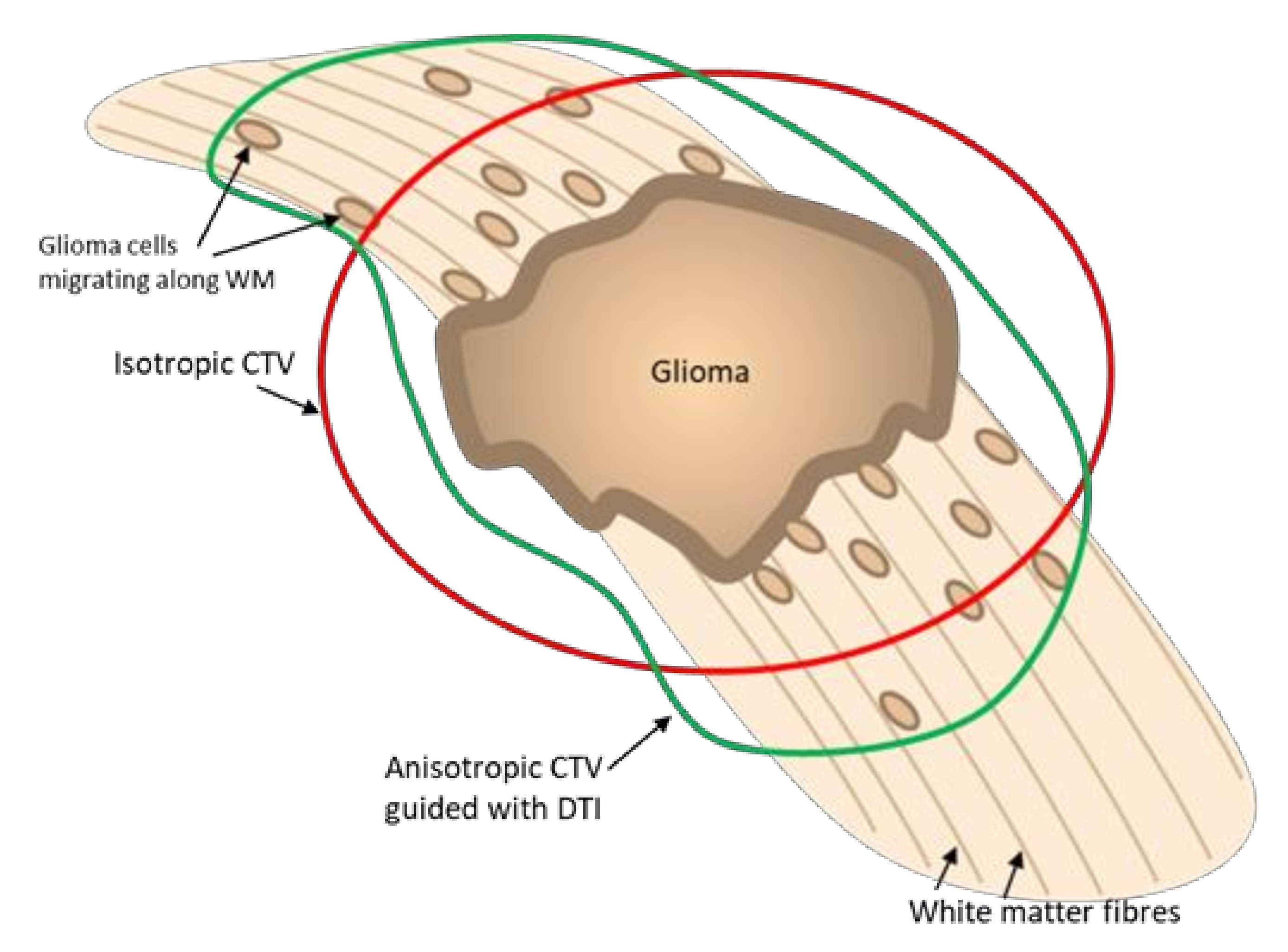
3.2. DTI Modelled Tumoral Invasion

Gliomas typically spread along the white matter tracts with tumour cells migrating with the orientation of the axons [32,33]. Typically, the tumoral core in high-grade tumours is defined as the contrast-enhancing tumour often with necrotic tissue within it [2]. However, glioma cells are found outside this core region due to the tumoral spread. As glioma cells migrate, it is thought to cause a loss of axonal integrity, leading to increased diffusivity and the T2 signal, and it is difficult to differentiate from vasogenic edema [34,35]. It has been suggested that diffusion imaging can improve the identification of non-enhancing tumour infiltration. For example, Pavlisa et al., 2009 find that the ADC is lower in the peritumour tissue of infiltrative tumours compared to non-infiltrative tumours [36]. Another study additionally found FA reductions in regions prior to visible tumour recurrence [35].
Therefore, DTI has been proposed to improve the identification of peritumour infiltration by better characterising diffusion abnormalities. Several studies have used the p (isotropic) and q (anisotropic) maps which are derived from the diffusion tensor to identify tumour and peritumour infiltration as it is proposed that the tumour core has a reduced q and increased p and the infiltrative tumour has an increased p and unchanged q. Defining the infiltrative tumour with p and q maps has been suggested as a replacement for defining the CTV on standard anatomic imaging in order to reduce the overall radiation dose in radiotherapy plans [24]. The CTV will typically need to be manually drawn; however, a study has proposed a semi-automated method that automatically expands the GTV drawn on the q map using a level set function on a p map [15].
Another proposed method utilises FA maps to detect tumour infiltration, as FA has been found to be reduced in infiltrative regions. Study groups have used deep learning to correct FA maps for free water to increase FA maps’ accuracy, finding that a model utilising these corrected FA maps could predict recurrence with an area under the curve (AUC) of 0.9 (compared to 0.77 with uncorrected maps) in a retrospective study [27]. The group additionally used the method to create the CTV and found that it correlated with the typical method of CTV definition, although it was larger in volume and adequately covered reoccurrences in follow-up scans [28].
A limitation of using p and q maps or FA maps to expand the GTV is that they do not take directionality into account, and information on fibre orientation is not used. Some studies have used the whole tensor to predict tumour spread. An article used a 3D-based DTI model that uses the fibre orientation to predict glioma growth [25]. However, this study did not evaluate the model accuracy quantitatively. Another article used white matter path length function to map the shortest path along the WM from a given region back to the tumour core and expanded the GTV from this by 1, 2, or 3 cm to form an anisotropic CTV. However, this method is only displayed in two cases, and the results of a retrospective study using the software are awaited [26]. The group additionally released the software to be used openly, therefore making translational studies using the method much easier.
Figure 5 shows a diagram showing how a DTI-guided anisotropic CTV could theoretically capture more areas of tumoral spread in gliomas by expanding along the WM.

3.3. Applications in Gliomas

The most promising use of DTI in gliomas as discussed is to define the CTV in areas of tumour infiltration not visible on anatomical MR sequences or to predict areas of tumour reoccurrence. Several studies already discussed have developed methods to perform this with one getting an AUC accuracy of 0.9 for predicting tumour recurrence sites [27]. These methods have been applied to producing SRT treatment CTVs. Virtual dissection would additionally be of benefit for gliomas in order to reduce radiation dose to the eloquent WM tracts with a small number of theoretical SRT planning studies making use of this technique, showing that it would be effective [9,29]. Unfortunately, there have not been any prospective studies using a DTI-guided SRT treatment plan to treat patients with gliomas, as found by a recent systematic review [19]. As far as the authors are aware, there have not been studies on DTI-guided radiotherapy in any primary brain malignancies other than gliomas [19], although there are studies using DTI to differentiate gliomas from other primary brain malignancies, which is an important step for management planning [16,17].

3.4. Applications in Brain Metastases

Brain metastases develop from the haematogenous spread of cancer cells and unlike gliomas are typically not infiltrative [36,37]. Therefore, the main use of DTI in radiotherapy treatment planning would be to avoid salient WM tracts by including them as OAR. This method has been evaluated in several studies of cohorts including brain metastasis along with other benign neurological conditions. The largest identified study contained 20 patients with brain metastasis (and three patients with other conditions), and they compared SRT plans with and without including the CST as an OAR, finding that this significantly reduced the maximum radiation dose to the CST but not the total dose to 95% of the tract [21]. However, this study did not use the tractography-guided treatment plan for administration of the dose to the patients. There are studies that include patients with metastasis that use radiotherapy-guided treatment plans for the patients’ therapy, with all of the studies finding that the maximum dose was significantly reduced to the dissected tracts [20,23,30]. These studies found no complications in the patients during short follow-up periods (all < 1.5 years); however, the subject numbers were small (three to nine subjects each), and no studies included a control group. However, these studies show the feasibility on integrating tractography into SRT treatment of brain metastases.

3.5. Application in Benign Conditions

SRT can be used in a range of non-malignant neurological conditions, and therefore, reducing the dose to nearby eloquent WM tracts to the pathology would likely reduce the risk of complications. DTI integration is most widely explored in the benign condition, arteriovenous malformation (AVM). A control study included 24 AVM patients, which used tractography in SRT treatment planning and 28 subjects with AVM without using tractography [22]. The subjects were followed up for 4 years, and the study found there were no significant differences in the success of treatment on angiography and that the use of tractography to model the CST significantly reduced the risk of motor complications. A limitation of this study is that it was retrospective with the control group being subjects from before the institution introduced tractography into SRT treatment planning. Therefore, the groups were not randomised, and the improved outcomes may be due to other factors such as improved operator skill over time. The same institution released a review of all patients they treated for AVM since the routine implementation of tractography with SRT (from 2004 to 2009), which included 144 patients. They found that 46% of cases utilised tractography, showing that it can be introduced effectively to a clinical setting, and a large proportion of patients may benefit from its integration. However, the use of tractography integration was operator dependent and may have not been necessary in some of the patients it was used in—or necessary in some of the cases when not integrated. Although it was found that in 60% of cases utilising tractography, the tract was within 5 mm [31].
DTI-integrated SRT has been explored in a small number of other neurological conditions, which include meningiomas and vestibular schwannomas. These studies include cohorts with multiple conditions and find that integrating tractography reduces the maximum radiation dose to the eloquent tracts [20,23]. However, the evidence for tractography implementation in the individual conditions is limited.

3.6. Future Considerations

The most important future work needed is randomised control trials on DTI-guided IMRT and SRT, as currently, none have been identified, although there is a non-randomised trial on tractography use in AVM [22]. The PRaM-GBM study, which is aiming to recruit 120 patients in the UK and performing DTI prior to surgery and IMRT of glioblastomas, is currently ongoing and promising [38]. However, the outline of this study is for it to be observational, and therefore, it likely will not use DTI-guided therapy plans in treatment. At the same time, the study should give important information on using DTI to predict areas of recurrence.
Work is additionally needed on the combined use of DTI-guided CTV definition in gliomas and inclusion of dissected tracts such as OAR, and software that optimises both simultaneously may be required. Additionally, further work is needed on the extent to which the CTV should be expanded along WM tracts, as it could potentially be expanded further than the typical 2 cm and potentially reduce recurrence rates; Jordan et al., 2019 have developed software that is able to perform this. The study produced CTVs that were expanded by 1, 2, and 3 cm along WM tracts; however, it does not compare the potential effectiveness of each, as this would likely require a controlled trial [26].
Subjectivity may be able to be reduced in the future by using automated whole brain tractography, which automatically dissects multiple eloquent tracts. This has previously been shown to be useful in tractography-guided stereotactic neurosurgery [39]. It may additionally increase accuracy by including tracts not immediately thought to be near to the pathology due to distorted anatomy. It may also reduce the doses to contralateral WM tracts in large lesions such as those shown in Altabella et al., 2018 on DTI-guided SRT plans [9]. One limitation of automated whole brain tractography is that there is still subjectivity regarding which tracts are included, and atlas-based approaches may be prone to errors, particularly in the presence of anatomy-distorting lesions. However, this method may play a role in reducing the time required for pre-radiotherapy planning and increasing standardisation, although the tracts would still need to be quality controlled by a highly skilled practitioner.
There is additionally much progress in automated lesion segmentation due to improvements in deep learning. Automated lesion segmentation may be able to be utilised to delineate the initial GTV and previously discussed algorithms used to expand it to the CTV. This could additionally remove subjectivity and speed up the SRT planning process.
An idealised fully automated pipeline for DTI-guided radiotherapy planning is shown in Figure 6, which could potentially be achieved with further research as suggested.

4. Limitations

The biggest limitation for using DTI to guide radiotherapy planning is the lack of controlled trials. The only controlled trial the authors, and a recent systematic review, have identified is in AVM, and this is a non-randomised retrospective study [22]. This poses an issue, as while the majority of the SRT pre-treatment plans using DTI guidance deliver the same dose to the pathology, this does not necessarily mean the treatment will have the same success rate on follow up. Additionally, while reduced radiation doses overall to eloquent WM tracts will likely mean reduced complications, control trials are still highly desirable.
An issue posed by gliomas is that due to infiltration along WM tracts, if it infiltrates into an eloquent tract that is included as an OAR, then the dose may be reduced and increase the risk of recurrence. Therefore, further research in gliomas is required to evaluate when and when not to include WM tracts as OAR.
An additional issue with gliomas is that their infiltration leads to reductions in FA, which may reduce the tractography algorithm’s accuracy. This is due to tractography algorithms typically using an FA threshold, with tracts not including voxels below the threshold. This could be accounted for by reducing the FA threshold, but this may lead to an increase in false positive tracts. These downsides can be overcome using a combination of more advanced diffusion acquisitions (i.e., with more directions and b-values), diffusion models (e.g., spherical deconvolution), and probabilistic tractography.
Another limitation is that the accuracy of the dissected tracts is dependent on the registration of the MRI images to the planning CT, as CT is used for the radiotherapy planning. CT MRI fusion is more difficult than simply registering different MRI sequences, and quality control is likely difficult due to different tissue contrasts. A recent systematic review on the topic found many different methods for CT/MRI registration with no standardisation and poor comparability of the validation of methods [40]. Although this article was assessing the issue in Stereotactic Electroencephalography, and there are only a small number of radiotherapy treatment planning systems that have their own in-built co-registration algorithms.
A practical limitation is that the integration of dissected WM tracts into the SRT treatment workstation may be difficult. Gavin et al., 2016 give a detailed step-by-step example of how they integrated the tractography results into their ‘GammaPlan’ workstation [20]; however, due to varying software, standardisation will likely be difficult.
A consideration also required is the additional resources and time needed. Logistics may be particularly difficult, as the DTI MRI scan will need to happen shortly before the SRT to reduce error from brain changes over time and that DTI requires incredibly computationally complex processing, which can take a long time. DTI application will additionally require highly skilled both technical and clinical operators to apply it correctly. The decision of which tracts to include is additionally highly subjective, as this is operator-dependent. However, despite these limitations, an institution in Japan introduced tractography prior to the SRT of AVMs into routine clinical practice, using it in almost 50% of cases. One way the group achieved this is by performing the DTI sequences the day prior [22].

5. Conclusions

DTI-guided radiotherapy is particularly promising for the modelling of eloquent tracts using tractography to reduce the radiation dose by their inclusion as OAR in radiotherapy treatment planning. This has already been shown to improve motor complications in a case-controlled trial and is reported to be routinely clinically implemented as part of SRT treatment of AVMs in one institution, and therefore, it could potentially be referred to as a state-of-the-art method. However, replication and further case-controlled clinical trials using this method are of a high priority before this method is used routinely, especially in conditions other than AVM. There is additionally considerable promise in using DTI to produce anisotropic CTV in treatment of gliomas. This could potentially improve current radiotherapy practice, as it may not only reduce the radiation dose but has the potential to more accurately target invasive tumours. However, research into this is in the initial phase, as there are currently only theoretical planning studies published with no consensus on the method of optimally delineating the CTV boundary. Therefore, further theoretical studies and case control trials are required before it can be considered viable for inclusion into clinical practice.

Author Contributions

Conceptualisation, J.C. and S.B.; writing—original draft preparation, J.C.; writing—review and editing, L.M., S.M., M.G., M.K. and S.B.; creation of figures, J.C.; supervision, S.B. All authors have read and agreed to the published version of the manuscript.

Funding

This research received no external funding.

Institutional Review Board Statement

Not applicable.

Informed Consent Statement

Not applicable.

Conflicts of Interest

The authors declare no conflict of interest.

References

  1. Halasz, L.M.; Rockhill, J.K. Stereotactic radiosurgery and stereotactic radiotherapy for brain metastases. Surg. Neurol. Int. 2013, 4, S185. [Google Scholar] [CrossRef] [PubMed]
  2. Niyazi, M.; Brada, M.; Chalmers, A.J.; Combs, S.E.; Erridge, S.C.; Fiorentino, A.; Grosu, A.L.; Lagerwaard, F.J.; Minniti, G.; Mirimanoff, R.O.; et al. ESTRO-ACROP guideline “target delineation of glioblastomas”. Radiother. Oncol. 2016, 118, 35–42. [Google Scholar] [CrossRef]
  3. Taylor, A.; Powell, M.E.B. Intensity-modulated radiotherapy—What is it? Cancer Imaging 2004, 4, 68–73. [Google Scholar] [CrossRef] [PubMed] [Green Version]
  4. Mamalui-Hunter, M.; Yaddanapudi, S.; Zhao, T.; Mutic, S.; Low, D.A.; Drzymala, R.E. Patient-specific independent 3D GammaPlan quality assurance for Gamma Knife Perfexion radiosurgery. J. Appl. Clin. Med. Phys. 2013, 14, 62–70. [Google Scholar] [CrossRef] [Green Version]
  5. Burnet, N.G.; Thomas, S.J.; Burton, K.E.; Jefferies, S.J. Defining the tumour and target volumes for radiotherapy. Cancer Imaging 2004, 4, 153–161. [Google Scholar] [CrossRef] [Green Version]
  6. Armstrong, C.L.; Gyato, K.; Awadalla, A.W.; Lustig, R.; Tochner, Z.A. A Critical Review of the Clinical Effects of Therapeutic Irradiation Damage to the Brain: The Roots of Controversy. Neuropsychol. Rev. 2004, 14, 65–86. [Google Scholar] [CrossRef]
  7. Dhermain, F.; Barani, I.J. Complications from radiotherapy. Handb. Clin. Neurol. 2016, 134, 219–234. [Google Scholar] [CrossRef] [PubMed]
  8. Chapman, C.H.; Zhu, T.; Nazem-Zadeh, M.; Tao, Y.; Buchtel, H.A.; Tsien, C.I.; Lawrence, T.S.; Cao, Y. Diffusion tensor imaging predicts cognitive function change following partial brain radiotherapy for low-grade and benign tumors. Radiother. Oncol. 2016, 120, 234–240. [Google Scholar] [CrossRef] [Green Version]
  9. Altabella, L.; Broggi, S.; Mangili, P.; Conte, G.M.; Pieri, V.; Iadanza, A.; del Vecchio, A.; Anzalone, N.; di Muzio, N.; Calandrino, R.; et al. Integration of Diffusion Magnetic Resonance Tractography into tomotherapy radiation treatment planning for high-grade gliomas. Phys. Med. 2018, 55, 127–134. [Google Scholar] [CrossRef] [PubMed]
  10. Price, S.J.; Gillard, J.H. Imaging biomarkers of brain tumour margin and tumour invasion. Br. J. Radiol. 2011, 84, S159. [Google Scholar] [CrossRef] [Green Version]
  11. Trip, A.K.; Jensen, M.B.; Kallehauge, J.F.; Lukacova, S. Individualizing the radiotherapy target volume for glioblastoma using DTI-MRI: A phase 0 study on coverage of recurrences. Acta Oncol. 2019, 58, 1532–1535. [Google Scholar] [CrossRef]
  12. O’Donnell, L.J.; Westin, C.-F. An introduction to diffusion tensor image analysis. Neurosurg. Clin. N. Am. 2011, 22, 185. [Google Scholar] [CrossRef] [Green Version]
  13. Peña, A.; Green, H.A.L.; Carpenter, T.A.; Price, S.J.; Pickard, J.D.; Gillard, J.H. Enhanced visualization and quantification of magnetic resonance diffusion tensor imaging using the p:q tensor decomposition. Br. J. Radiol. 2014, 79, 101–109. [Google Scholar] [CrossRef] [PubMed]
  14. Razek, A.A.K.A.; El-Serougy, L.; Abdelsalam, M.; Gaballa, G.; Talaat, M. Differentiation of residual/recurrent gliomas from postradiation necrosis with arterial spin labeling and diffusion tensor magnetic resonance imaging-derived metrics. Neuroradiology 2018, 60, 169–177. [Google Scholar] [CrossRef] [PubMed]
  15. Rahmat, R.; Brochu, F.; Li, C.; Sinha, R.; Price, S.J.; Jena, R. Semi-automated construction of patient individualised clinical target volumes for radiotherapy treatment of glioblastoma utilising diffusion tensor decomposition maps. Br. J. Radiol. 2020, 93, 93. [Google Scholar] [CrossRef] [PubMed]
  16. Thong, P.M.; Duc, N.M. The Role of Apparent Diffusion Coefficient in the Differentiation between Cerebellar Medulloblastoma and Brainstem Glioma. Neurol. Int. 2020, 12, 34–40. [Google Scholar] [CrossRef]
  17. Duc, N.M. The role of diffusion tensor imaging metrics in the discrimination between cerebellar medulloblastoma and brainstem glioma. Pediatr. Blood Cancer 2020, 67, e28468. [Google Scholar] [CrossRef]
  18. Essayed, W.I.; Zhang, F.; Unadkat, P.; Cosgrove, G.R.; Golby, A.J.; O’Donnell, L.J. White matter tractography for neurosurgical planning: A topography-based review of the current state of the art. NeuroImage Clin. 2017, 15, 659–672. [Google Scholar] [CrossRef]
  19. Yahya, N.; Manan, H.A. Utilisation of Diffusion Tensor Imaging in Intracranial Radiotherapy and Radiosurgery Planning for White Matter Dose Optimization: A Systematic Review. World Neurosurg. 2019, 130, e188–e198. [Google Scholar] [CrossRef]
  20. Gavin, C.G.; Sabin, H.I. Stereotactic diffusion tensor imaging tractography for Gamma Knife radiosurgery. J. Neurosurg. 2016, 125, 139–146. [Google Scholar] [CrossRef] [Green Version]
  21. Kawasaki, K.; Matsumoto, M.; Kase, M.; Nagano, O.; Aoyagi, K.; Kageyama, T. Quantification of the radiation dose to the pyramidal tract using tractography in treatment planning for stereotactic radiosurgery. Radiol. Phys. Technol. 2017, 10, 507–514. [Google Scholar] [CrossRef] [PubMed] [Green Version]
  22. Koga, T.; Shin, M.; Maruyama, K.; Kamada, K.; Ota, T.; Itoh, D.; Kunii, N.; Ino, K.; Aoki, S.; Masutani, Y.; et al. Integration of Corticospinal Tractography Reduces Motor Complications After Radiosurgery. Int. J. Radiat. Oncol. 2012, 83, 129–133. [Google Scholar] [CrossRef] [PubMed]
  23. Sun, L.; Qu, B.; Wang, J.; Ju, Z.; Zhang, Z.; Cui, Z.; Jack, Y.; Ling, Z.; Yu, X.; Pan, L. Integration of Functional MRI and White Matter Tractography in CyberKnife Radiosurgery. Technol. Cancer Res. Treat. 2017, 16, 850–856. [Google Scholar] [CrossRef] [PubMed] [Green Version]
  24. Berberat, J.; McNamara, J.; Remonda, L.; Bodis, S.; Rogers, S. Diffusion tensor imaging for target volume definition in glioblastoma multiforme. Strahlenther. Onkol. 2014, 190, 939–943. [Google Scholar] [CrossRef]
  25. Hathout, L.; Patel, V.; Wen, P. A 3-dimensional DTI MRI-based model of GBM growth and response to radiation therapy. Int. J. Oncol. 2016, 49, 1081–1087. [Google Scholar] [CrossRef] [Green Version]
  26. Jordan, K.; Morin, O.; Wahl, M.; Amirbekian, B.; Chapman, C.; Owen, J.; Mukherjee, P.; Braunstein, S.; Henry, R. An Open-Source Tool for Anisotropic Radiation Therapy Planning in Neuro-oncology Using DW-MRI Tractography. Front. Oncol. 2019, 9, 810. [Google Scholar] [CrossRef] [PubMed]
  27. Metz, M.-C.; Molina-Romero, M.; Lipkova, J.; Gempt, J.; Liesche-Starnecker, F.; Eichinger, P.; Grundl, L.; Menze, B.; Combs, S.E.; Zimmer, C.; et al. Predicting Glioblastoma Recurrence from Preoperative MR Scans Using Fractional-Anisotropy Maps with Free-Water Suppression. Cancers 2020, 12, 728. [Google Scholar] [CrossRef] [Green Version]
  28. Peeken, J.C.; Molina-Romero, M.; Diehl, C.; Menze, B.H.; Straube, C.; Meyer, B.; Zimmer, C.; Wiestler, B.; Combs, S.E. Deep learning derived tumor infiltration maps for personalized target definition in Glioblastoma radiotherapy. Radiother. Oncol. 2019, 138, 166–172. [Google Scholar] [CrossRef]
  29. Igaki, H.; Sakumi, A.; Mukasa, A.; Saito, K.; Kunimatsu, A.; Masutani, Y.; Hanakita, S.; Ino, K.; Haga, A.; Nakagawa, K.; et al. Corticospinal tract-sparing intensity-modulated radiotherapy treatment planning. Rep. Pr. Oncol. Radiother. 2014, 19, 310–316. [Google Scholar] [CrossRef] [Green Version]
  30. Conti, A.; Pontoriero, A.; Ricciardi, G.K.; Granata, F.; Vinci, S.; Angileri, F.F.; Pergolizzi, S.; Alafaci, C.; Rizzo, V.; Quartarone, A.; et al. Integration of functional neuroimaging in CyberKnife radiosurgery: Feasibility and dosimetric results. Neurosurg. Focus 2013, 34, E5. [Google Scholar] [CrossRef] [Green Version]
  31. Koga, T.; Maruyama, K.; Kamada, K.; Ota, T.; Shin, M.; Itoh, D.; Kunii, N.; Ino, K.; Terahara, A.; Aoki, S.; et al. Outcomes of Diffusion Tensor Tractography–Integrated Stereotactic Radiosurgery. Int. J. Radiat. Oncol. 2012, 82, 799–802. [Google Scholar] [CrossRef] [PubMed]
  32. Mair, D.B.; Ames, H.M.; Li, R. Mechanisms of invasion and motility of high-grade gliomas in the brain. Mol. Biol. Cell 2018, 29, 2509–2515. [Google Scholar] [CrossRef]
  33. Engwer, C.; Hillen, T.; Knappitsch, M.; Surulescu, C. Glioma follow white matter tracts: A multiscale DTI-based model. J. Math. Biol. 2014, 71, 551–582. [Google Scholar] [CrossRef]
  34. Lu, S.; Ahn, D.; Johnson, G.; Law, M.; Zagzag, D.; Grossman, R.I. Diffusion-Tensor MR Imaging of Intracranial Neoplasia and Associated Peritumoral Edema: Introduction of the Tumor Infiltration Index. Radiology 2004, 232, 221–228. [Google Scholar] [CrossRef] [PubMed]
  35. Jin, Y.; Randall, J.W.; Elhalawani, H.; Al Feghali, K.A.; Elliott, A.M.; Anderson, B.M.; Lacerda, L.; Tran, B.L.; Mohamed, A.S.; Brock, K.K.; et al. Detection of Glioblastoma Subclinical Recurrence Using Serial Diffusion Tensor Imaging. Cancers 2020, 12, 568. [Google Scholar] [CrossRef] [PubMed] [Green Version]
  36. Pavlisa, G.; Rados, M.; Pavlisa, G.; Pavic, L.; Potocki, K.; Mayer, D. The differences of water diffusion between brain tissue infiltrated by tumor and peritumoral vasogenic edema. Clin. Imaging 2009, 33, 96–101. [Google Scholar] [CrossRef]
  37. Eichler, A.F.; Chung, E.; Kodack, D.P.; Loeffler, J.S.; Fukumura, D.; Jain, R.K. The biology of brain metastases—Translation to new therapies. Nat. Rev. Clin. Oncol. 2011, 8, 344. [Google Scholar] [CrossRef] [Green Version]
  38. Predicting Sites of Tumour Progression in the Invasive Margin of Glioblastomas (PRaM-GBM Study)—Full Text View—ClinicalTrials.gov. Available online: https://clinicaltrials.gov/ct2/show/NCT03294434 (accessed on 6 October 2021).
  39. Zakaria, H.; Haider, S.; Lee, I. Automated Whole Brain Tractography Affects Preoperative Surgical Decision Making. Cureus 2017, 9, e1656. [Google Scholar] [CrossRef] [Green Version]
  40. Perez, J.; Mazo, C.; Trujillo, M.; Herrera, A. MRI and CT Fusion in Stereotactic Electroencephalography: A Literature Review. Appl. Sci. 2021, 11, 5524. [Google Scholar] [CrossRef]
Figure 1. Diagram displaying an example of the gross tumour volume (GTV) with the isotropically expanded clinical target volume (CTV) and the planned target volume (PTV) on a temporal lobe glioma. Addition of the brain stem as an organ at risk (OAR) on the right and how the PTV could be altered using inverse planning to reduce radiation dose to the brain stem.
Applsci 12 00816 g001
Figure 2. Diagram displaying tensors from different microstructures and how this would affect the isotropic tensor derivatives (p and MD) and the anisotropic components (q and FA).
Applsci 12 00816 g002
Figure 3. Diagram explaining the tractography process showing an example of how the voxel tensors (left) can be tracked from a seed point to form a tract (middle) and performed multiple times to produce a virtually dissected white matter tract (right).
Applsci 12 00816 g003
Figure 4. Diagram displaying (A) Example without tractography dissected corticospinal tract (CST) included as an OAR, and (B) CST included as an OAR during dose planning, resulting in a reduced radiation dose.
Applsci 12 00816 g004
Figure 5. Diagram showing glioma cells migrating along white matter tracts and how a DTI-based anisotropic clinical target volume (CTV) could better capture areas of tumoral spread than isotropic CTV and potentially reduce the overall radiation dose.
Applsci 12 00816 g005
Figure 6. A proposed idealised pipeline for DTI-guided SRT planning, which would be fully automated.
Applsci 12 00816 g006
Table 1. This table outlines the key articles on DTI-guided intracranial radiotherapy discussed in the review broken down into subtopics. AVM = arteriovenous malformation, CST = corticospinal tract, CM = cerebral metastasis, Menig = Meningioma, OAR = organ at risk, HGG = high-grade glioma, VS = vestibular schwannoma.
ReferenceType of StudyNumber of SubjectsSummaryMain Outcomes
Virtual dissection of Eloquent WM Tracts
Altabella et al., 2018 [9]Theoretical planning study19 (all with HGG)Tractography used to dissect multiple eloquent tracts bilaterally (CST, SLF, IFOF, UNF). Tracts included as OAR during inverse planning with TomoTherapy.Significant reduction in maximum and mean radiation dose to the tracts, particularly to contralateral tracts.
Gavin and Sabin 2016 [20]Clinical feasibility study20 (5 VS, 5 AVM, 9 CM, 1 Menig) DTI tractography performed using ‘StealthViz’ used on user selected tracts which were included as OAR into GammaPlan prior to GKRS on a series of cases.Methodologies outlined and shown to be clinically feasible.
Kawasaki et al., 2017 [21]Theoretical planning study23 (20 CM, 3 AVM)Tractography used to include pyramidal tract as OAR in GKRS planning and plans with and without tractography compared.Maximum radiation dose to the pyramidal tracts significantly reduced when tractography used in planning.
Koga et al., 2012 [22]Retrospective cohort study52 (all with AVM, 24 tractography, 28 control)Integration of tractography into radiotherapy planning for AVM at a single centre with 28 control cases being prior and 24 test cases after this. Patients followed up for a minimum of 3 years.Significantly less motor complication in patients with tractography integration. No significant difference in treatment success.
Sun et al., 2017 [23]Theoretical planning study16 (6 AVM, 8 CM, 2 Menig)Integration of fMRI to with tractography to model the corticospinal tracts and ‘sensory pathway’.Significant reduction of the maximum radiation dose to the included cortical areas and tracts.
DTI modelled tumoural invasion
Berberat et al., 2014 [24]Theoretical planning study13 (all with glioblastoma)Use of DTI p and q maps and tractography to define the CTV. Comparison to CTV defined by oedema on T2w MRI or isotropic expansion of the GTV.The DTI-defined CTV was significantly smaller than the T2w MRI-defined CTV and still included sites of tumour recurrence.
Hathout et al., 2016 [25]Theoretical modelling studyNADTI-based 3D mathematical model of glioblastoma growth.Produces model using DTI to model tumour infiltration shown to predict glioma growth in example cases.
Jordan et al., 2019 [26]Theoretical modelling studyNAStudy that develops open-source DTI based fibre-tracking software that produces anisotropic CTV to better capture likely areas of tumour infiltration.The software is shown to qualitatively capture areas of recurrence well in a few example cases.
Metz et al., 2020 [27]Retrospective observational study35 (all with glioblastoma)Use of deep learning to correct FA maps for free water and used to predict areas of glioma recurrence on follow-up imaging.Area under the curve for recurrence prediction of 0.77 using FA and 0.9 using free water-corrected FA values.
Peeken et al., 2019 [28]Theoretical planning study33 (all with glioblastoma)Use of deep-learning-based free water-corrected FA maps to define the GTV and compared to traditionally defined GTV.Free water corrected FA map-based GTV were significantly smaller than traditionally defined GTV but still include the recurrence area of all but one of the 14 subjects with recurrence.
Rahmat et al., 2020 [15]Theoretical planning study50 (all with glioblastoma)Semi-automated DTI-defined tumour volume. GTV manually defined using q map, which was automatically expanded using an expansion model on the p map to model tumour infiltration. Comparison made to manual segmentation of p and q maps.Mean Dice coefficient of 74% between manual and semi-automated method over all 50 patients.
Applications in Gliomas
Altabella et al., 2018 [9]Theoretical planning study19 (all with HGG)Tractography used to dissect multiple eloquent tracts bilaterally (CST, SLF, IFOF, UNF). Tracts included as OAR during inverse planning with TomoTherapy. Treatment plans with and without tractography were compared.Significant reduction in maximum and mean radiation dose to the tracts, particularly to contralateral tracts.
Igaki et al., 2014 [29]Clinical feasibility studyNAIntegration of tractography into radiotherapy planning in glioblastoma by including the corticospinal tracts as OAR in planning.Integration of tractography shown to be feasible and reduce radiation does in two cases.
Metz et al., 2020 [27]Retrospective observational study35 (all with glioblastoma)Use of deep learning to correct FA maps for free water and used to predict areas of glioma recurrence on follow up imaging.Area under the curve for recurrence prediction of 0.77 using FA and 0.9 using free water-corrected FA values.
Yahya and Manan 2019 [19]Systematic reviewNASystematic review of literature on DTI in intracranial radiotherapy.Finds only three articles evaluating DTI in radiotherapy that includes gliomas, none of which were case control trials or used prospective integration.
Applications in Brain Metastases
Conti et al., 2013 [30]Theoretical planning study25 (10 AVM, 3 CM, 12 ‘brain tumours’fMRI and tractography of eloquent structures in close proximity to the lesions. Comparison of radiotherapy plans with and without integration.Found an average reduction in radiation dose by 17% to eloquent regions when fMRI and tractography integrated.
Gavin and Sabin 2016 [20]Clinical feasibility study20 (5 VS, 5 AVM, 9 CM, 1 Menig) DTI tractography used to guide GKRS.Applied to nine cases of cerebral metastasis successfully; however, there was no comparison to a control group.
Kawasaki et al., 2017 [21]Theoretical planning study23 (20 CM, 3 AVM)Tractography used to include pyramidal tract as OAR in GKRS planning and plans with and without tractography were compared.Twenty out of 23 subjects had CM. The maximum radiation dose to the pyramidal tracts significantly reduced when tractography was used in planning.
Sun et al., 2017 [23]Theoretical planning study16 (6 AVM, 8 CM, 2 Menig)Integration of fMRI to select tractography seed point to model the corticospinal tracts and ‘sensory pathway’. Functional regions and tracts included as OAR during planning for CyberKnife radiosurgery. Treatment plans with and without integration were compared.Significant reduction of the maximum radiation dose to the included cortical areas and tracts.
Yahya and Manan 2019 [19]Systematic reviewNASystematic review of literature on DTI in intracranial radiotherapy.Finds five articles evaluating DTI in radiotherapy that includes CM, one of which uses prospective integration.
Applications in Benign Conditions
Gavin and Sabin 2016 [20]Clinical feasibility study20 (5 VS, 5 AVM, 9 CM, 1 Menig) DTI tractography used to guide GKRS.Applied to five cases of vestibular schwannoma, five cases of AVM, and one case of meningioma successfully; however, there was no comparison to a control group.
Kawasaki et al., 2017 [21]Theoretical planning study23 (20 CM, 3 AVM)Tractography used to include pyramidal tract as OAR in GKRS planning and plans with and without tractography were compared.Three out of 23 subjects had AVM. The maximum radiation dose to the pyramidal tracts significantly reduced when tractography was used in planning. However, the plan not used for treatment.
Koga et al., 2012 [22]Retrospective cohort study52 (all with AVM, 24 tractography, 28 control)Integration of tractography into radiotherapy planning for AVM at a single centre in 2004 with 28 control cases being prior to this and 24 test cases after this. Patients followed up for minimum of 3 years.Significantly less motor complication in patients with tractography integration. No significant difference in treatment success.
Koga et al., 2012 [31]Prospective integration155 (all with AVM, 71 of which had tractography)Routine integration of tractography in radiotherapy of AVMs at a single centre. Selected tracts user selected if suspected to be close to the pathology.Tractography used in 71 out of 155 radiotherapy cases during the study period with 60% of cases using tractography finding the critical tracts within 5 mm of the lesion.
Sun et al., 2017 [23]Theoretical planning study16 (6 AVM, 8 CM, 2 Menig)Integration of fMRI to select tractography seed point to model the corticospinal tracts and ‘sensory pathway’. Functional regions and tracts included as OAR during planning for CyberKnife radiosurgery. Treatment plans with and without integration were compared.Significant reduction of the maximum radiation dose to the included cortical areas and tracts.
Yahya and Manan 2019 [19]Systematic reviewNASystematic review of literature on DTI in intracranial radiotherapy.Finds 13 articles evaluating DTI in radiotherapy that include AVM, many of which use prospective integration.
Publisher’s Note: MDPI stays neutral with regard to jurisdictional claims in published maps and institutional affiliations.

