Article

Relationship between Carotid Artery Calcification and Periodontitis in the Saudi Population: A Retrospective Study

by
Wael I. Ibraheem
1,*,
Ashok Kumar Bhati
1,*,
Manar Ali Alhomood
2,
Omaima Ahmed Allayl
2,
Alzhouor Ali Qsadi
2,
Tahani Ibrahim Mnaji Mohammed
2 and
Yahya Sulaiman Aljabri
2
1
Department of Preventive Dental Sciences, College of Dentistry, Jazan University, Jazan 45142, Saudi Arabia
2
College of Dentistry, Jazan University, Jazan 45142, Saudi Arabia
*
Authors to whom correspondence should be addressed.
Submission received: 9 October 2023 / Revised: 2 November 2023 / Accepted: 13 November 2023 / Published: 15 November 2023

Abstract

:
Background: Atherosclerosis is the primary cause of cardiovascular death, wherein plaque accumulates in the carotid artery, resulting in the formation of carotid artery calcification (CAC). Chronic infections emulate atherosclerosis development. Periodontitis is a chronic disease commenced by biofilm aggregation on the surface of teeth, which then progresses via deregulated local and inflammatory immune responses. Aim: The aim of this study was to determine the prevalence of calcification in periodontitis patients and to determine if there is a relationship between carotid calcification and periodontitis with risk factors such as age, gender, hypertension, diabetes, smoking. Methods: The data and panoramic radiographs (PR) of 3000 patients from January 2017 to February 2022 were examined. On the basis of the inclusion and exclusion criteria, 500 patients were selected for determining carotid calcification. Non-periodontitis patients, patients <40 years of age, patients without any medical data or incomplete medical records and cases where the OPG was not clear were not included in this study. The data were analyzed using R and GraphPad Prism software version 4.1.2 and Microsoft Excel Worksheet Office 2019. Chi-square tests were used to check the dependency among the categorical variables. Mann–Whitney U-tests were used to compare the distribution of the variables across the groups, and p-values < 0.05 were interpreted as statistically significant. Result: Twenty-five (5%) patients showed carotid calcification. Carotid calcification was seen more in females than in males, which was statistically significant. Gender, percentage of alveolar bone loss and periodontal risk were found to be statistically significantly related to carotid artery calcification. Factors such as age, diabetes and hypertension were found to be statistically nonsignificant. Conclusions: More periodontal destruction was present in the CAC patients. Within the limitations of this study, it can be concluded that CAC is associated with periodontitis. Early recognition of carotid calcification is important, and patients in whom carotid artery calcification is observed via their PR should be referred to medical practitioners for further investigation and treatment care.

1. Introduction

Arthrosclerosis is a chronic inflammatory disease of fatty material deposition in the walls of the coronary artery. This fatty deposition leads to the formation of atheroma plaque [1]. Over a period of time, the atheroma plaque thickens with decreasing fibrous and lipid composition and increasing calcification, resulting in carotid artery calcification (CAC) [2]. Calcification can appear in the medial and intimal layers of the intracranial and cervical carotid arteries [3].
Vos et al. in 2018 in a study of 1132 patients observed that predominant intimal calcification was found in 30.9% of intimal calcification, and predominant medial calcification in 46.9% of medial calcification [4]. In a systemic review by Song et al. in 2020, it was found that 75% of males above 75 years and 62% of females above 75 years had carotid artery calcification [5].
Calcification partially or completely reduces the lumen diameter of the coronary artery [6]. With a reduction in the vessel lumen diameter, there is a decrease in the blood and oxygen supply to the designated organs, thus endangering the life of the patient [7,8]. There are no peculiar clinical manifestations of carotid artery calcification. CAC is associated with ischemic symptoms. The manifestations such as bumps, contralateral weakness or numbness in an extremity or face, with significant sparing of the forehead, dysarthria, aphasia or ischemic stroke, appear when the artery is partially or completely occluded. [9]. Atherosclerosis is associated with coronary artery disease (CAD) and cerebrovascular diseases [10]. CAD is one of the primary causes of death globally. According to the WHO Global Health Submit, the leading cause of death is ischemic heart disease accounting for 16% of the world’s total deaths [11]. The number of deaths rose from 2 million in 2000 to 8.9 million in 2019. Stroke is the second leading cause of death, responsible for approximately 11% of the total deaths. According to the WHO, the proportional mortality rate for CVD in Saudi Arabia was 37% in 2018 [12].
In many patients, CAD can progress undetected until the late stages, resulting in morbidity and mortality [7,8]. Therefore, it should be diagnosed as soon as possible. Various invasive and noninvasive procedures have been used to detect the presence and distribution of carotid artery calcification [13]. To date, sonography has been used as “gold standard” noninvasive techniques to determine the arteriosclerotic changes in the carotid artery [14]. Nevertheless, ultrasound examinations are not routinely used as safeguard measure in all patients. Other more time-consuming methods used for diagnosis of carotid artery calcification, such as computer tomography (CT), magnet resonance tomography (MRT) and electronic thermography (ET), as well as the invasive method of angiography, are available, but they are used for special indications and are not commonly used for the assessment of all patients [15]. Furthermore, more advanced methods such as optical coherence tomography (OCT), photoacoustic tomography and infrared thermography are also used to characterize plaques [16].
PET-CT imaging with (18)F-Sodium Fluoride is used for the detection and quantification of microcalcification in atherosclerotic plaques. Due to its association with cardiovascular risk factors, it determines the amount and severity of CAC [17,18,19].
Invasive and advanced methods are more expensive and time-consuming than noninvasive methods. Consequently, different diagnostic methods which are quick and economical are required.
The relationship between cardiovascular disease and periodontitis is well established [20]. Periodontitis is a complex disease marked by clinical attachment loss and alveolar bone loss resulting in tooth loss if not treated at an early stage or treated unsuccessfully [21].
In 2014, the Global Burden of Disease Activity reported periodontitis as the sixth disease worldwide, affecting 743 million people, with the overall prevalence of 11.2% [22]. Inflammatory events in periodontitis occur as a result of increased concentrations of various proinflammatory cytokines as evident by increased levels found in infected periodontal tissues or in the crevicular fluid of periodontitis patients. Similarly, continuous increases in cytokines are found in atherosclerosis. [23]. Patients with periodontitis have a 1.14-time higher relative risk of developing coronary artery disease and a 25% higher risk compared to non-periodontitis patients [24,25].
Furthermore, studies also suggest that atherosclerosis can also be amplified by periodontitis [26,27]. Periodontal disease may stimulate endothelial dysfunction through bacterial products from Porphyromonas gingivalis and Aggregatibacter actinomycetemcomitans and soluble bacterial components and also via systemic inflammation [27,28]. Endothelia dysfunction results in the release of inflammatory cytokines interleukin 1, interleukin 6 and tumor necrosis factor alpha [9]. These inflammatory cytokines promote cell adhesion, permeability and endothelial permeability. The decomposing endothelial layer results in the production of thrombin and formation of fibrinogen, which can result in thrombosis and, consequently, in cerebrovascular or cardiovascular accidents [29,30].
When samples from carotid endarterectomies were studied in a polymerase chain reaction study, a minimum of one periodontal bacterium was found in 44% of atheroma, and Porphyromona gingivalis and Tannerella forsythia were found in 26% and 30% of the specimens, respectively [28]. Porphyromona gingivalis and Tannerella forsythia are the main periodontal pathogens which are associated with bleeding on probing and deep periodontal pockets [31,32].
Panoramic radiographs (PRs) are routinely used for diagnosing periodontitis. A panoramic radiograph is an economical and most commonly used tool in dentistry for diagnostic investigation. In a panoramic radiograph, carotid artery calcification appears as an irregular, circular, linear or heterogeneous radiopaque mass located posterosuperiorly to the angle of the mandible, at approximately the inferior margin of the third cervical vertebra between C3 and C4 [33,34,35,36].
Various studies have shown the importance of panoramic radiographs in the identification of CAC [37,38]. Studies demonstrate that the identification of the calcified plaque of the carotid artery using panoramic radiography is attainable with a high degree of accuracy [11,12,13,37,38,39]. The probability interval of finding carotid calcification in a PR lies between 2.5% and 4.1% [36,40,41].
Ravon et al., 2003, also reported high accuracy in the detection of CAC using panoramic radiographs [39]. In 1981, Friedlander and Lande for the first time detected carotid artery calcification in panoramic radiographs [33]. Friedlander and Baker (1994) also detected CAC in asymptomatic stroke patients [34].
Studies have also shown the prevalence of CAC in the general Saudi population [42,43,44]. But to the best of our knowledge, there are no studies conducted in Jazan region of Saudi Arabia determining the relationship of CAC to periodontitis and periodontal risk. Therefore, the aim of the present study was to determine the prevalence of CAC in panoramic radiographs and to determine the association of CAC with periodontitis by determining the correlation of CAC with the number of missing teeth, percentage of alveolar bone loss (ABL) and periodontal risk and the association of CAC with other risk components such as age, gender, systemic diseases (diabetes, hypertension) and smoking.

2. Material and Methods
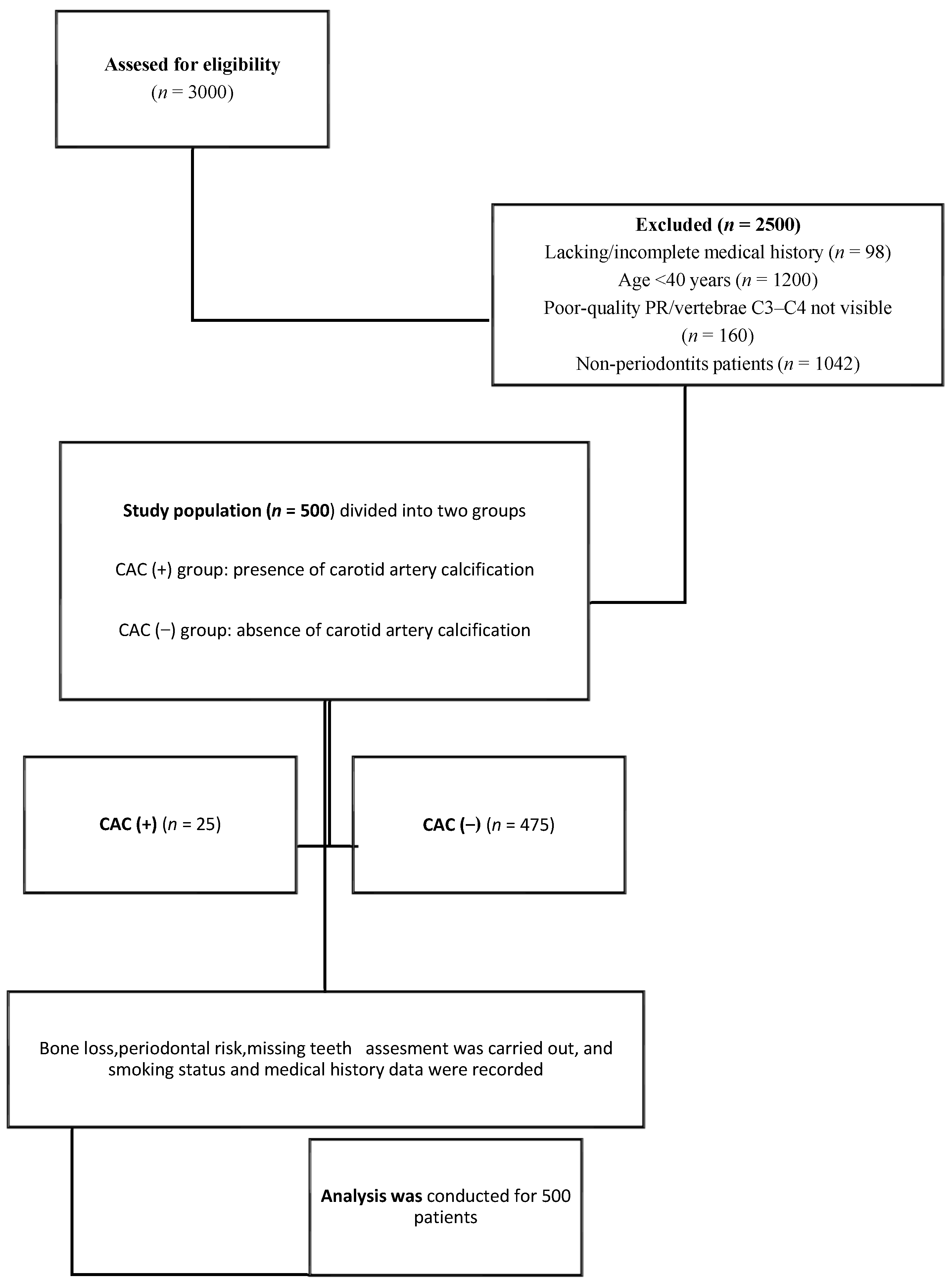
2.1. Study Design

This retrospective cross-sectional study was conducted at the College of Dentistry, Jazan University, Saudi Arabia. Patients who visited the College of Dentistry from January 2017 to February 2022 were included in this study. No written informed consents were obtained from the patients as this study was a retrospective study. This study was started after receiving the approval from the Standing Committee for Scientific Research—Jazan University (Reference no: REC-43/06/131).
A total of 3000 patients were evaluated for this study based on the inclusion and exclusion criteria. Patients with the following criteria were included in this study: patients aged ≥40 years, patients diagnosed as periodontitis cases, patients having complete medical history, panoramic radiographs showing clearly the C3 and C4 vertebra. Patients with the following criteria were excluded from this study: non-periodontitis patients (patients diagnosed as gingivitis), patients <40 years old, patients without any medical history record or incomplete medical data. Panoramic radiographs where C3 and C4 were not visible or where the quality of the PR was not good were excluded from this study. A total of 500 patients met the above inclusion criteria and were included in this study. A total of 2500 patients were excluded from this study due to following reasons: patients with incomplete medical history (n = 98), patient age < 40 years (n = 1200), patients with poor quality of OPG or OPG where vertebrae C3–C4 were not visible (n = 160) and non-periodontitis patients (gingivitis cases, n = 1042) (Figure 1). The selected periodontitis patients were divided into two age groups: ≤60 (40–60 years) and ≥61 years (61–90 years).

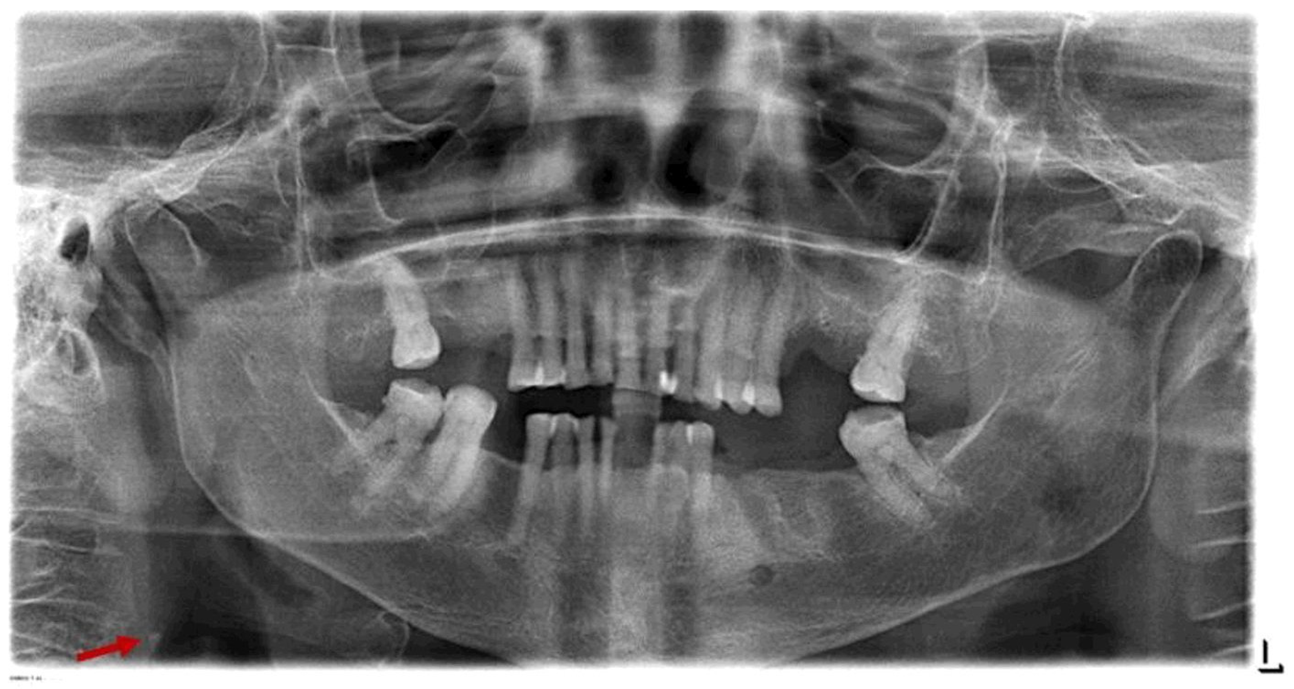
2.2. Assessment of Carotid Artery Calcification in Panoramic Radiographs

All panoramic radiographs were acquired using machine ORTHOPHOS XG 5, (Sirona, Charlotte, NC, USA). All PRs were taken by the same personnel. In the panoramic radiograph, carotid artery calcification was recognized as asymmetrical single or multiple radiopaque masses appearing unilaterally or bilaterally, positioned subjacent to the mandibular angle adjoining the space between the third or fourth cervical vertebrae (C3 and C4) (Figure 2). These calcifications can appear as a single nodule (single radiopaque mass) or multiple nodules (multiple radiopaque masses). Patients with the presence of CAC were categorized as CAC (+), and those without the presence of CAC were categorized as CAC (−). The CAC identification in the PRs were crosschecked by an experienced oral and maxillofacial radiologist with more than 10 years of experience.

2.3. Data Collection

Patient information such as age, gender, medical history (diabetes and hypertension) and smoking status (smoker or nonsmoker) were obtained from the R4 software (CSR4 Clinical+ Practice Management Software 6.1.1, Carestream Dental LLC., Atlanta, GA, USA). Panoramic radiographs were assessed using the digital radiograph imaging software SIDEXIS XG 2.61 (Sirona, Charlotte, NC, USA).

2.4. Assessment of Percentage of Alveolar Bone Loss

Alveolar bone loss (ABL) assessment was performed by calculating the distance from the cementoenamel junction (CEJ) to the alveolar crest (AC) divided by the distance from the cementoenamel junction (CEJ) to the apex [45]. The percentage (ABL%) was obtained by multiplying the result by 100.

2.5. Number of Missing Teeth

The number of missing teeth was recorded for each patient. The means were obtained for CAC (+) and CAC (−). The reason for tooth loss was not recorded for any patient.

2.6. Assessment of Periodontal Risk [46]

Using the number of missing teeth and the bone reduction index (calculated by bone loss percentage divided by age of patient), the periodontal risk was evaluated. According to Lang and Tonnetti, periodontal risk was categorized into low, medium and high risk as follows: low risk, ≤4 missing teeth and/or bone reduction index ≤0.5; moderate risk, 5–8 missing teeth and/or bone reduction index of 0.51–1.0; high risk, ≥9 missing teeth and/or bone reduction index ≥ 1.1. The most adverse factor determined the overall grade of risk.

2.7. Statistical Analysis

The data were analyzed using R and GraphPad Prism software version 4.1.2. Chi-square tests were used to check the dependency among the categorical variables, and Mann–Whitney U-tests were used to compare the distributions of the variables across the groups; p-values of < 0.05 were interpreted as statistically significant.

3. Result

A total of 3000 patients were studied using the inclusion and exclusion criteria.
A total of 500 patients met the inclusion criteria and were included in this study, of which 276 patients were male (54.2%) and 224 were female (44.8%) patients. The patients’ mean age was 54.64 ± 9.91 years. Distribution of the study population according to different variables is given in Table 1.

3.1. Prevalence and Distribution of Carotid Artery Calcification

Out of 500 panoramic radiographs, 475 (95%) showed no CAC upon radiographic evaluation, whereas 25 (5%) panoramic radiographs showed the presence of CAC (Table 1).
Among these 25 CAC instances, 80% were present as single nodule and 20% were present as multiple nodules; 52% of CAC appeared bilaterally (present on both the right and left sides of the PR), and 48% unilaterally (on the right or left side of the PR). In unilateral distribution, more CAC instances were found to be on the left side than on the right side (Table 2).

3.2. Gender and Carotid Artery Calcification

Within the 5% prevalence, 4.6% was represented by female and 0.4% was represented by male patients. Females had twenty-three CAC instances (4.6%, i.e., 92% of the detected CAC instances), and males had two CAC instances (0.4%, i.e., 8% of the detected CAC instances). The chi-square tests showed a significant correlation between CAC and gender (p < 0.0001) (Table 3).

3.3. Age and Carotid Artery Calcification

The mean age of the CAC (−) patients was found to be 54.54 ± 9.97 years, while that of the CAC (+) patients was 55.58 ± 9.49 years. However, the Mann–Whitney U-tests showed no significant differences between age and CAC (p = 0.347) (Table 3).
An increased number of CAC instances was seen with increasing age; 5.08% percent of CAC was seen in the age group of ≥61 years as compared to 4.9% in patients ≤60 years (Table 4).

3.4. Smoking and Carotid Artery Calcification

Out of 500 patients, 72 (14.4%) were smokers (Table 1) with 51 smokers in the age group of ≤60 years and 21 smokers in the age group of ≥61 years (Table 4). Only one smoker had CAC in the PR; 71 smoker patients did not show any CAC in the PR. The chi-square tests showed no significant relationship between smoking and CAC (p = 0.12) (Table 3).

3.5. Percentage of Alveolar Bone Loss and Carotid Artery Calcification

The mean alveolar bone loss percentage in CAC (+) patients was 41.06 ± 15.49, whereas it was 39.99 ± 15.47 in the CAC (−) patients. A greater bone reduction was also seen in the CAC (+) patients compared to the CAC (−) patients (Table 3). A higher percentage of alveolar bone loss was seen with the increase in age. Patients in the age group of ≥61 years had a mean alveolar bone loss percentage of 44.1 ± 15.4, whereas patients in the age group of ≤60 years had a mean alveolar bone loss percentage of 39.6 ± 15.3 (Table 4).

3.6. Number of Missing Teeth

The number of missing teeth was found to be higher in the CAC (+) patients than in the CAC (−) patients. The mean number of missing teeth was 6.1 ± 6.4 in the CAC (+) group and 5.3 ± 5.6 in the CAC (−) group (Table 3).
When the number of missing teeth was assessed according to the age, it was found that the number of missing teeth was higher in the group of ≥61 years compared to the group of ≤60 years (Table 2 and Table 4).

3.7. Periodontal Risk

Periodontal risk was calculated according to Lang and Tonetti’s procedure [46] using the bone reduction index and the number of missing teeth. A statistically significant correlation was found with periodontal risk in the CAC (+) patients (Table 3). High periodontal risk was also found to be more with increasing age (Table 4).

3.8. Systemic Diseases and Carotid Artery Calcification

In the CAC (+) group, 16% were hypertensive, and 8% were diabetic. In the CAC (−) group, 6.7% were hypertensive, 10.5% were diabetic, and 6.1% patients were having both diabetes and hypertension. There was no significant association found between systemic diseases and calcification (Table 3). However, when these systemic diseases were compared according to the age group, they were statistically significant with increasing age (Table 4).

4. Discussion

The aim of the present study was to determine the prevalence of carotid artery calcification and its relationship with other risk factors of cardiovascular disease such as age, gender, systemic diseases (diabetes, hypertension), smoking along with the determination of alveolar bone loss and periodontal risk.
To the best of our knowledge, this is perhaps the first study in Saudi Arabia to determine the relationship between CAC and periodontitis. Prior studies in Saudi Arabia focused on the prevalence of carotid artery calcification and risk factors in the general population [42,43,44]. However, we did not find studies in Saudi Arabia depicting the relationship between CAC and periodontitis in terms of the alveolar bone loss and periodontal risk.
In 1981, Friedlander and Lande first detected carotid artery calcification in panoramic radiographs [33]. They highlighted the importance of assessing the carotid artery calcification in a panoramic radiograph in routine dental practice. Thereafter, many researchers began to study the relationship between CAC-associated risks and cardiovascular lesions using panoramic radiography thereby enhancing its use in detecting CAC [47,48,49,50].
In this study, the prevalence of CAC was found to be 5%. This is in agreement with the findings of Alzoman [42] who also reported similar prevalence of CAC in the general dental population in 2012 with the sample size (n = 555) similar to that in the present study (n = 500). Some studies have also reported lower prevalence of 2.9% [51], 3.1% [3] and 2.43% [52], while others have reported higher prevalence of 9% [53], 6.8% [54] and 9.4% [55]. This variation in prevalence could be due to the differences in the sample size, sample population, age, dietary habits and lifestyle.
In our study, most of the detected cases of CAC were presented bilaterally (52%). A higher bilateral distribution was also reported by Kamak et al., 2015 [56], Naseeha Ibrahim, 2018 [54], and Ariyai et al., 2009 [15]. In terms of unilateral distribution (right side or left side), more cases were observed on the left side compared to the right side (20%). Similar greater left-side distribution was also reported by Alzoman, 2012, Kamak, 2015, and Tamura et al., 2005 [42,56,57]. However, some studies also reported more CAC on the right side [53,58,59].
CAC appears to be more often in female patients. In our study, higher prevalence of CAC (4.92%) was found in female patients as well. High female prevalence was also reported by other studies [44,53,54,56,57]. In our study, the difference between gender and CAC was statistically significant, which is similar to that in the study of Santosh et al. conducted on a Brazilian population [60]. This could be due to the changes in the estrogen level after menopause. Estrogen results in the formation of atheromatous plaque by acting on the lipoprotein metabolism. A reduction in the estrogen level after menopause precipitates this reduction in plaque [34].
Friedlander et al. [59] studied the PRs of female patients with a history of amenorrhea and found that 31% of the radiographs showed CAC. Similar findings were also reported by Patil et al., 2015, with 22.9% prevalence of CAC in women in 1214 panoramic radiographs [61]. However, CAC predominance in males was also reported in some studies [42,62,63]. In their study, Ohba et al. [58] also had twenty-five female and eight male patients with CAC. This is similar to the present study’s findings of twenty-three females and two males having CAC.
The mean age of the patients in which CAC was absent (n = 475) was 54 years, whereas, for the 25 patients in which CAC was found, their mean age was 56 years. This supports the findings of other studies in which the prevalence of CAC increased with age [3,60,64]. Santosh et al. also reported that CAC (+) is associated with increasing age, showing more CAC instances in the 50–70-year age group, with a mean age of 54 years [60]. Similar findings were also reported by Patil in 2015, who found greater prevalence of CAC in the age groups older than 50 years [61].
Diabetes and hypertension are acknowledged factors for arteriosclerosis. When the correlation with systemic diseases was studied, this study did not show any correlation with diabetes and hypertension. Almog et al. and Brito did not find any correlation with systemic diseases either [65,66]. However, studies by Ertas et al. found that CAC was correlated with hypertension but not with diabetes, whereas Ramesh et al. reported a correlation between diabetes and CAC as well [67,68]. Christou et al. (n = 15) also showed positive correlation of hypertension with CAC [69]. The difference in results could be due to the difference in the sample size. A significant difference was found in age with respect to diabetes and hypertension. More patients with systemic diseases were reported with increasing age.
Smoking is one of the factors which affects the cardiovascular disease morbidity and mortality. Smoking results in a two- to fourfold increase in the risk of coronary heart disease. This risk increases in the presence of hypertension and diabetes, due to the symbiotic effect with smoking [70]. In this study we investigated the association of smoking with CAC. The results of this study showed no significant relationship between CAC and smoking. This is in agreement with the findings of Cetin et al. [3], Monterio et al. [71] and Atalay et al. [72] who did not find any association between smoking and CAC either. However, Brito et al. showed that CAC was two times worse in smokers than in nonsmokers [66]. This difference could be due to the sample size. Ertas et al. also showed a positive correlation between smoking duration and CAC. The difference between their results and the results of our study could be due to the lack of information about the duration of smoking and the study sample population [67].
Alveolar bone loss is an import factor that determines the periodontal support. Greater periodontal destruction is seen with increased periodontal disease or the severity of periodontitis resulting in tooth loss if not controlled. In the present study, increased bone loss was seen more in the CAC (+) than in the CAC (−) group. This is an accordance with other studies which showed greater bone loss associated with carotid artery calcification [39,40,73]. However, such findings were not significant in a study by Thanakun [74] who reported no correlation between the alveolar bone loss and CAC. This could be attributed to the differences in the sample size and the method of assessment. However, Engerberston showed a significant association between bone loss and CAC [73].
Tooth loss is a quick and reliable feature to determine the periodontal disease comparable to the clinical periodontal parameters of periodontitis such as pocket dept, clinical attachment loss and alveolar bone loss [75]. A higher number of missing teeth was seen in the CAC (+) group compared to the CAC (−) group. Andrianka et al., 2006, and Kamak, 2015, also showed increased tooth loss in the presence of carotid artery calcification. Increased tooth loss was also seen associated with the increase in age [56,75]. Using the number of missing teeth and the bone loss, periodontal risk was assessed as per Lang and Tonetti’s procedure [46]. A significant correlation was found between CAC (+) and periodontal risk. A similar positive association between periodontal risk and CAC was reported by another study [53].
This study showed that panoramic radiography is an effective tool in determining the CAC. However, it is important to make careful differential diagnosis while assessing CAC in the panoramic radiographs. Various cervical radiopacities such as calcified triticeous cartilage, superior corner of calcified thyroid cartilage, greater corner of hyoid bone, styloid process, stylohyoid ligament, stylomandibular ligament, epiglottis, calcified lymph node, phleboliths, submandibular salivary gland sialoliths, tonsilloliths could be simply jumbled with CAC instances. Proper knowledge and experience are crucial, and, therefore, emphasis should be put on the assessment of carotid artery calcification in panoramic radiographs during undergraduate studies.
The present study had certain limitations. The limitations include a smaller sample size and the fact that this study was based mainly on panoramic findings and did not evaluate the periodontal parameters such as attachment loss and pocket depth which are crucial in assessing the periodontitis. Moreover, the CAC findings based on the PRs were not assessed using further advanced diagnostics such as CT and ultrasonography.

5. Conclusions

In our study, greater periodontal destruction was found in the CAC patients showing a greater number of missing teeth, significant alveolar bone loss and a higher periodontal risk. Based on these findings and within the limitations of this study, it can be concluded that CAC is associated with periodontitis. However, further studies with a larger sample size and the assessment of clinical attachment loss and pocket depth in relation to CAC and the use of advanced diagnostic aids to confirm the CAC findings in the PRs are required.
Moreover, patients with CAC detected in the PRs could be at risk of stroke or cardiovascular disease and should be promptly referred to the physician for further confirmation of CAC through required investigations and provided necessary treatment and care.

Author Contributions

Conceptualization, W.I.I. and A.K.B.; methodology, W.I.I., A.K.B., T.I.M.M., A.A.Q., M.A.A. and O.A.A.; validation, W.I.I., A.K.B., T.I.M.M., A.A.Q., M.A.A. and O.A.A.; formal analysis, A.K.B. and W.I.I.; resources, W.I.I., A.K.B., T.I.M.M., A.A.Q., M.A.A., O.A.A. and Y.S.A.; writing—original draft preparation, A.K.B. and W.I.I.; writing—review and editing, W.I.I., A.K.B., T.I.M.M., A.A.Q., M.A.A., O.A.A. and Y.S.A.; supervision, W.I.I. and A.K.B.; project administration, W.I.I.; funding acquisition, W.I.I., A.K.B., T.I.M.M., A.A.Q., M.A.A., O.A.A. and Y.S.A. All authors have read and agreed to the published version of the manuscript.

Funding

This research was self-funded. There was no external funding received for the study.

Institutional Review Board Statement

This study was approved by the Standing Committee for Scientific Research—Jazan University (Reference no: REC-43/06/131).

Informed Consent Statement

Informed consent was not obtained as this was a retrospective study.

Data Availability Statement

Data are contained within the article.

Conflicts of Interest

The authors declare no conflict of interest.

References

  1. Alves, N.; Deana, N.F.; Garay, I. Detection of common carotid artery calcifications on panoramic radiographs: Prevalence and reliability. Int. J. Clin. Exp. Med. 2014, 7, 1931–1939. [Google Scholar]
  2. Van Gils, M.J.; Vukadinovic, D.; van Dijk, A.C.; Dippel, D.W.; Niessen, W.J.; van der Lugt, A. Carotid atherosclerotic plaque progression and change in plaque composition over time: A 5-year follow-up study using serial CT angiography. AJNR Am. J. Neuroradiol. 2012, 33, 1267–1273. [Google Scholar] [CrossRef]
  3. Shi, X.; Gao, J.; Lv, Q.; Cai, H.; Wang, F.; Ye, R.; Liu, X. Calcification in atherosclerotic plaque vulnerability: Friend or foe? Front. Physiol. 2020, 11, 56. [Google Scholar] [CrossRef]
  4. VVos, A.; Kockelkoren, R.; de Vis, J.B.; van der Schouw, Y.T.; van der Schaaf, I.C.; Velthuis, B.K.; Mali, W.P.; de Jong, P.A.; Majoie, C.B.; Roos, Y.B.; et al. Risk factors for atherosclerotic and medial arterial calcification of theintracranial internal carotid artery. Atherosclerosis 2018, 276, 44–49. [Google Scholar] [CrossRef]
  5. Song, P.; Fang, Z.; Wang, H.; Cai, Y.; Rahimi, K.; Zhu, Y.; Fowkes, F.G.R.; Fowkes, F.J.; Rudan, I. Global and regional prevalence, burden, and risk factors for carotid atherosclerosis: A systematic review, meta-analysis, and modelling study. Lancet Glob. Health 2020, 8, 721–729. [Google Scholar] [CrossRef]
  6. Çetin, M.B.; Sezgin, Y.; Yilmaz, M.N.N.; Seçgin, C.K. Assessment of Carotid Artery Calcifications on Digital Panoramic Radiographs and Their Relationship with Periodontal Condition and Cardiovascular Risk Factors. Int. Dent. J. 2021, 71, 160–166. [Google Scholar] [CrossRef]
  7. Senosiain-Oroquieta, A.; Pardo-Lopez, B.; De Carlos-Villafranca, F.; Cobo-Plana, J. Detección de placas de ateroma mediante radiografías dentales. RCOE 2006, 11, 297–303. [Google Scholar] [CrossRef]
  8. Romano-Sousa, C.M.; Krejci, L.; Medeiros, F.M.M.; Graciosa-Filho, R.G.; Martins, M.F.F.; Guedes, V.N.; Fenyo-Pereira, M. Diagnostic agreement between panoramic radiographs and color doppler images of carotid atheroma. J. Appl. Oral Sci. 2009, 17, 45–48. [Google Scholar] [CrossRef] [PubMed]
  9. Ahmed, M.; McPherson, R.; Abruzzo, A.; Thomas, S.E.; Gorantla, V.R. Carotid Artery Calcification: What We Know So Far. Cureus 2021, 13, e18938. [Google Scholar] [CrossRef] [PubMed]
  10. Cheng, C.; Tempel, D.; van Haperen, R.; van der Baan, A.; Grosveld, F.; Daemen, M.J.; Krams, R.; de Crom, R. Atherosclerotic lesion size and vulnerability are determined by patterns of fluid shear stress. Circulation 2006, 113, 2744–2753. [Google Scholar] [CrossRef] [PubMed]
  11. Top Ten Causes of Death. Available online: https://www.who.int/news-room/fact-sheets/detail/the-top-10-causes-of-death (accessed on 23 May 2023).
  12. World Health Organization—Noncommunicable Diseases (NCD) Country Profiles. 2018. Available online: https://www.who.int/publications/i/item/9789241514620 (accessed on 18 July 2022).
  13. Bos, D.; Leening, M.J.; Kavousi, M.; Hofman, A.; Franco, O.H.; van der Lugt, A.; Vernooij, M.W.; Ikram, M.A. Comparison of atherosclerotic calcification in major vessel beds on the risk of all-cause and cause-specific mortality: The Rotterdam study. Circ. Cardiovasc. Imaging 2015, 8, e003843. [Google Scholar] [CrossRef]
  14. Wyman, R.A.; Fraizer, M.C.; Keevil, J.G.; Busse, K.L.; Aeschlimann, S.E.; Korcarz, C.E. Ultrasound detected carotid plaque as a screening tool for advanced subclinical atherosclerosis. Am. Heart J. 2005, 150, 1081–1085. [Google Scholar] [CrossRef]
  15. Ariayi, A.S.; Berndt, D.; Lambrecht, J.T. Panoramic radiography for diagnosis of soft tissue calcifications—An aid to identify stroke prone patients? Schweiz Monatsschr Zahnmed 2009, 119, 1009–1013. (In German) [Google Scholar]
  16. Saxena, A.; Ng, E.Y.; Lim, S.T. Imaging modalities to diagnose carotid artery stenosis: Progress and prospect. Biomed. Eng. Online 2019, 18, 66. [Google Scholar] [CrossRef]
  17. Derlin, T.; Richter, U.; Bannas, P.; Begemann, P.; Buchert, R.; Mester, J.; Klutmann, S. Feasibility of 18F-sodium fluoride PET/CT for imaging of atherosclerotic plaque. J. Nucleic Med. 2010, 51, 862–865. [Google Scholar] [CrossRef]
  18. Derlin, T.; Wisotzki, C.; Richter, U.; Apostolova, I.; Bannas, P.; Weber, C.; Mester, J.; Klutmann, S. In vivo imaging of mineral deposition in carotid plaque using 18Fsodium fluoride PET/CT: Correlation with atherogenic risk factors. J. Nucl. Med. 2011, 52, 362–368. [Google Scholar] [CrossRef]
  19. Derlin, T.; Tóth, Z.; Papp, L.; Wisotzki, C.; Apostolova, I.; Habermann, C.R.; Mester, J.; Klutmann, S. Correlation of inflammation assessed by 18F-FDG PET, active mineral deposition assessed by 18F-fluoride PET, and vascular calcification in atherosclerotic plaque: A dual-tracer PET/CT study. J. Nucleic Med. 2011, 52, 1020–1027. [Google Scholar] [CrossRef] [PubMed]
  20. Lockhart, P.B.; Bolger, A.F.; Papapanou, P.N.; Osinbowale, O.; Trevisan, M.; Levison, M.E.; Taubert, K.A.; Newburger, J.W.; Gornik, H.L.; Gewitz, M.H.; et al. Periodontal disease and atherosclerotic vascular disease: Does the evidence support an independent association? A scientific statement from the American Heart Association. Circulation 2012, 125, 2520–2544. [Google Scholar] [CrossRef] [PubMed]
  21. Lang, N.; Bartold, P.M. Periodontal health. J. Periodontol. 2018, 89, S9–S16. [Google Scholar] [PubMed]
  22. Kassebaum, N.J.; Bernabe, E.; Dahiya, M.; Bhandari, B.; Murray, C.J.L.; Marcenes, W. Global burden of severe periodontitis in 1990−2010: A systematic review and meta-regression. J. Dent. Res. 2014, 93, 1045–1053. [Google Scholar] [CrossRef] [PubMed]
  23. Zhu, H.; Lin, X.; Zheng, P.; Chen, H. Inflammatory cytokine levels in patients with periodontitis and/or coronary heart disease. Int. J. Clin. Exp. Pathol. 2015, 8, 2214. [Google Scholar]
  24. DeStefano, F.; Anda, R.F.; Kahn, H.S.; Williamson, D.F.; Russell, C.M. Dental disease and risk of coronary heart disease and mortality. BMJ 1993, 306, 688–691. [Google Scholar] [CrossRef] [PubMed]
  25. Bahekar, A.A.; Singh, S.; Saha, S.; Molnar, J.; Arora, R. The prevalence and incidence of coronary heart disease is significantly increased in periodontitis: A meta-analysis. Am. Heart J. 2007, 154, 830–837. [Google Scholar] [CrossRef] [PubMed]
  26. Velsko, I.M.; Chukkapalli, S.S.; Rivera, M.F.; Lee, J.-Y.; Chen, H.; Zheng, D.; Bhattacharyya, I.; Gangula, P.R.; Lucas, A.R.; Kesavalu, L. Active invasion of oral and aortic tissues by Porphyromonas gingivalis in mice causally links periodontitis and atherosclerosis. PLoS ONE 2014, 9, e97811. [Google Scholar] [CrossRef] [PubMed]
  27. Reyes, L.; Herrera, D.; Kozarov, E.; Roldan, S.; Progulske-Fox, A. Periodontal bacterial invasion and infection: Contribution to atherosclerotic pathology. J. Clin. Periodontol. 2013, 40, S30–S50. [Google Scholar] [CrossRef] [PubMed]
  28. AlSakr, A.; Blanchard, S.; Wong, P.; Thyvalikakath, T.; Hamada, Y. Association between intracranial carotid artery calcifications and periodontitis: A cone-beam computed tomography study. J. Periodontol. 2021, 92, 1402–1409. [Google Scholar] [CrossRef] [PubMed]
  29. Rose, L.F.; Genco, R.J.; Cohen, D.W.; Mealey, B.L. Periodontal Medicine; BC Decker: Hamilton, ON, Canada, 2000. [Google Scholar]
  30. Schenkein, H.A.; Loos, B.G. Inflammatory mechanisms linking periodontal diseases to cardiovascular diseases. J. Clin. Periodontol. 2013, 40, S51–S69. [Google Scholar] [CrossRef]
  31. Haraszthy, V.I.; Zambon, J.J.; Trevisan, M.; Zeid, M.; Genco, R.J. Identification of periodontal pathogens in atheromatous plaques. J. Periodontol. 2000, 71, 1554–1560. [Google Scholar] [CrossRef]
  32. Socransky, S.S.; Haffajee, A.D.; Cugini, M.A.; Smith, C. Microbial complexes in subgingival plaque. J. Clin. Periodontol. 1998, 25, 134–144. [Google Scholar] [CrossRef]
  33. Friedlander, A.H.; Lande, A. Panoramic radiographic identification of carotid arterial plaques. Oral Surg. Oral Med. Oral Pathol. 1981, 52, 102–104. [Google Scholar] [CrossRef]
  34. Friedlander, A.H.; Baker, J.D. Panoramic radiography: An aid in detecting patients at risk of cerebrovascular accident. J. Am. Dent. Assoc. 1994, 125, 1598–1603. [Google Scholar] [CrossRef]
  35. Roldán-Chicano, R.; Oñate-Sánchez, R.E.; López-Castaño, F.; Cabrerizo-Merino, M.C.; Martínez López, F. Panoramic radiograph as a method for detecting calcified atheroma plaques. Review of literature. Med. Oral Patol. Oral Cir. Bucal. 2006, 11, E261–E266. [Google Scholar]
  36. Ahmad, M.; Madden, R.; Perez, L. Triticeous cartilage: Prevalence on panoramic radiographs and diagnostic criteria. Oral Surg Oral Med. Oral Pathol. Oral Radiol. Endodontol. 2005, 99, 225–230. [Google Scholar] [CrossRef]
  37. Bengtsson, V.W.; Persson, G.R.; Renvert, S. Assessment of carotid calcifications on panoramic radiographs in relation to other used methods and relationship to periodontitis and stroke: A literature review. Acta Odontol. Scand. 2014, 76, 401–412. [Google Scholar] [CrossRef]
  38. Khosropanah, S.H.; Shahidi, S.H.; Bronoosh, P.; Rasekhi, A. Evaluation of carotid calcification detected using panoramic radiography and carotid Doppler sonography in patients with and without coronary artery disease. Br. Dent. J. 2009, 207, 162–163. [Google Scholar] [CrossRef]
  39. Ravon, N.A.; Hollender, L.G.; McDonald, V.; Persson, G.R. Signs of carotid calcification from dental panoramic radiographs are in agreement with Doppler sonography results. J. Clin. Periodontol. 2003, 30, 1084–1090. [Google Scholar] [CrossRef]
  40. Beckstrom, B.W.; Horsley, S.H.; Scheetz, J.P.; Khan, Z.; Silveira, A.M.; Clark, S.J.; Greenwell, H.; Farman, A.G. Correlation between carotid area calcifications and periodontitis: A retrospective study of digital panoramic radiographic findings in pretreatment cancer patients. Oral Surg. Oral Med. Oral Pathol. Oral Radiol. Endodontol. 2007, 103, 359–366. [Google Scholar] [CrossRef]
  41. Uthman, A.T.; Al-Saffar, A. Prevalence in digital panoramic radiographs of carotid area calcification among Iraqi individuals with stroke-related disease. Oral Surg. Oral Med. Oral Pathol. Oral Radiol. Endodontol. 2008, 105, 68–73. [Google Scholar] [CrossRef]
  42. Alzoman, H.A.; Al-Sadhan, R.E.; Al-Lahem, Z.H.; Al-Sakaker, A.N.; Al-Fawaz, Y.F. Prevalence of carotid calcification detected on panoramic radiographs in a Saudi population from a training institute in Central Saudi Arabia. Saudi Med. J. 2012, 33, 177–181. [Google Scholar]
  43. Alsweed, A.; Farah, R.; Ps, S.; Farah, R. The Prevalence and Correlation of Carotid Artery Calcifications and Dental Pulp Stones in a Saudi Arabian Population. Diseases 2019, 7, 50. [Google Scholar] [CrossRef]
  44. Sinjab, A.Z.; Soman, C.; Sahab, L.A. Prevalence of carotid artery calcifications in Riyadh from digital panoramic radiographs and its relationship with cardiovascular disorders: A retrospective cross-sectional study. Saudi J. Oral Sci. 2021, 8, 28–32. [Google Scholar] [CrossRef]
  45. Bassiouny, M.A.; Grant, A.A. The accuracy of the Schei ruler: A laboratory investigation. J. Periodontol. 1975, 46, 748–752. [Google Scholar] [CrossRef] [PubMed]
  46. Lang, N.P.; Tonetti, M.S. Periodontal risk assessment (PRA) for patients in supportive periodontal therapy (SPT). Oral Health Prev. Dent. 2003, 1, 7–16. [Google Scholar]
  47. Almog, D.M.; Horev, T.; Illig, K.A.; Green, R.M.; Carter, L.C. Correlating carotid artery stenosis detected by panoramic radiography with clinically relevant carotid artery stenosis determined by duplex ultrasound. Oral Surg. Oral Med. Oral Pathol. Oral Radiol. Endodontol. 2002, 94, 768–773. [Google Scholar] [CrossRef] [PubMed]
  48. Garo, M.; Johansson, E.; Ahlqvist, J.; Jaghagen, E.L.; Arnerlov, C.; Wester, P. Detection of calcifications in panoramic radiographs in patients with carotid stenoses ≥50%. Oral Surg. Oral Med. Oral Pathol. Oral Radiol. 2014, 117, 385–391. [Google Scholar]
  49. Lee, J.S.; Kim, O.S.; Chung, H.J.; Kim, Y.J.; Kweon, S.S.; Lee, Y.H.; Shin, M.H.; Yoon, S.J. The correlation of carotid artery calcification on panoramic radiographs and determination of carotid artery atherosclerosis with ultrasonography. Oral Surg. Oral Med. Oral Pathol. Oral Radiol. 2014, 118, 739–745. [Google Scholar] [CrossRef]
  50. Cohen, S.N.; Friedlander, A.H.; Jolly, D.A.; Date, L. Carotid calcification on panoramic radiographs: An important marker for vascular risk. Oral Surg. Oral Med. Oral Pathol. Oral Radiol. Endodontol. 2002, 94, 510–514. [Google Scholar] [CrossRef] [PubMed]
  51. Bayram, B.; Uckan, S.; Acikgoz, A.; Müderrisoglu, H.; Aydinalp, A. Digital panoramic radiography: A reliable method to diagnose carotid artery atheromas? Dentomaxillofac. Radiol. 2006, 35, 266–270. [Google Scholar] [CrossRef]
  52. Ezoddini-Ardakani, F.; Mirzaei, M.; Nayer, S.; Besharati, S.; Moeini, M. Evaluation of Positive Predictive Value for Digital Panoramic Radiography in Comparison to Ultrasound in the Diagnosis of Calcified Carotid Atheroma. Health 2014, 6, 1933–1940. [Google Scholar] [CrossRef]
  53. Tiller, R.; Bengel, W.; Rinke, S.; Ziebolz, D. Association between carotid area calcifications and periodontal risk: A cross sectional study of panoramic radiographic findings. BMC Cardiovasc. Disord. 2011, 11, 67. [Google Scholar] [CrossRef]
  54. Nasseh, I.; Aoun, G. Carotid Artery Calcification: A Digital Panoramic-Based Study. Diseases 2018, 6, 15. [Google Scholar] [CrossRef]
  55. Brand, H.S.; Mekenkamp, W.C.; Baart, J.A. Prevalence of carotid artery calcification on panoramic radiographs. Ned. Tijdschr. Tandheelkd. 2009, 116, 69–73. [Google Scholar]
  56. Kamak, G.; Yildirim, E.; Rencber, E. Evaluation of the relationship between periodontal risk and carotid artery calcifications on panoramic radiographs. Eur. J. Dent. 2015, 9, 483–489. [Google Scholar] [CrossRef]
  57. Tamura, T.; Inui, M.; Nakamura, S.; Nakase, M.; Okumura, K.; Tagawa, T. Clinicostatistical study of carotid calcification on panoramic radiographs. Oral Dis. 2005, 11, 314–317. [Google Scholar] [CrossRef] [PubMed]
  58. Ohba, T.; Takata, Y.; Ansai, T.; Morimoto, Y.; Tanaka, T.; Kito, S.; Awano, S.; Akifusa, S.; Takehara, T. Evaluation of calcified carotid artery atheromas detected by panoramic radiograph among 80-year-olds. Oral Surg. Oral Med. Oral Pathol. Oral Radiol. Endodontol. 2003, 96, 647–650. [Google Scholar] [CrossRef] [PubMed]
  59. Friedlander, A.H.; Altman, L. Carotid artery atheromas in postmenopausal women. Their prevalence on panoramic radiographs and their relationship to atherogenic risk factors. J. Am. Dent. Assoc. 2001, 132, 1130–1136. [Google Scholar] [CrossRef] [PubMed]
  60. Santos, J.M.O.; Soares, G.C.; Kurita, L.M. Prevalence of carotid artery calcifications among 2500 digital panoramic radiographs of an adult Brazilian population. Med. Oral Patol. Oral Cir. Bucal. 2018, 23, e256. [Google Scholar]
  61. Patil, S.R. Prevalence of carotid artery calcification in postmenopausal women and its correlation with atherogenic risk factors. J. Nat. Sci. Biol. Med. 2015, 6, S1. [Google Scholar] [CrossRef]
  62. Dewake, N.; Ishioka, Y.; Uchida, K.; Taguchi, A.; Higashi, Y.; Yoshida, A.; Yoshinari, N. Association between Carotid Artery Calcification and Periodontal Disease Progression in Japanese Men and Women: A Cross-Sectional Study. J. Clin. Med. 2020, 9, 3365. [Google Scholar] [CrossRef] [PubMed]
  63. Azimi, S.; Sarlak, H.; Tofangchiha, M. Determining the prevalence of carotid artery calcification and associations with medical history using dental panoramic radiographs. Dent. Med. Probl. 2016, 53, 29–33. [Google Scholar] [CrossRef]
  64. Lee, J.S.; Kim, O.S.; Chung, H.J.; Kim, Y.J.; Kweon, S.S.; Lee, Y.H.; Shin, M.H.; Yoon, S.J. The prevalence and correlation of carotid artery calcification on panoramic radiographs and peripheral arterial disease in a population from the Republic of Korea: The Dong-gu study. Dentomaxillofac. Radiol. 2013, 42, 29725099. [Google Scholar] [CrossRef]
  65. Almog, D.M.; Tsimidis, K.; Moss, M.E.; Gottlieb, R.H.; Carter, L.C. Evaluation of a training program for detection of carotid artery calcifications on panoramic radiographs. Oral Surg. Oral Med. Oral Pathol. Oral Radiol. Endodontol. 2000, 90, 111–117. [Google Scholar] [CrossRef] [PubMed]
  66. Brito, A.C.; Nascimento, H.A.; Argento, R.; Beline, T.; Ambrosano, G.M.B.; Freitas, D.Q. Prevalence of suggestive images of carotid artery calcifications on panoramic radiographs and its relationship with predisposing factors. Cien. Saude Colet. 2016, 21, 2201–2208. [Google Scholar] [CrossRef]
  67. Ertas, E.T.; Sisman, Y. Detection of incidental carotid artery calcifications during dental examinations: Panoramic radiography as an important aid in dentistry. Oral Surg. Oral Med. Oral Pathol. Oral Radiol. Endodontol. 2011, 112, e11–e17. [Google Scholar] [CrossRef] [PubMed]
  68. Ramesh, A.; Soroushian, S.; Ganguly, R. Coincidence of calcified carotid atheromatous plaque, osteoporosis, and periodontal bone loss in dental panoramic radiographs. Imaging Sci. Dent. 2013, 43, 235–243. [Google Scholar] [CrossRef]
  69. Christou, P.; Leemann, B.; Schimmel, M.; Kiliaridis, S.; Müller, F. Carotid artery calcification in ischemic stroke patients detected in standard dental panoramic radiographs—A preliminary study. Adv. Med. Sci. 2010, 55, 26–31. [Google Scholar] [CrossRef]
  70. Lankier, J.F. Smoking and cardiovascular disease. Am. J. Med. 1992, 93, S8–S12. [Google Scholar] [CrossRef]
  71. Monteiro, I.A.; Ibrahim, C.; Albuquerque, R.; Donaldson, N.; Salazar, F.; Monteiro, L. Assessment of carotid calcifications on digital panoramic radiographs: Retrospective analysis and review of the literature. J. Stomatol. Oral Maxillofac. Surg. 2018, 119, 102–106. [Google Scholar] [CrossRef] [PubMed]
  72. Atalay, Y.; Asutay, F.; Agacayak, K.S.; Koparal, M.; Adali, F.; Gulsun, B. Evaluation of calcified carotid atheroma on panoramic radiographs and Doppler ultrasonography in an older population. Clin. Interv. Aging 2015, 10, 1121–1129. [Google Scholar] [CrossRef] [PubMed]
  73. Engebretson, S.P.; Lamster, I.B.; Elkind, M.S.V.; Rundek, T.; Serman, N.J.; Demmer, R.T. Radiographics measures of chronic periodontitis and carotid artery plaque. Stroke 2005, 36, 561–566. [Google Scholar] [CrossRef]
  74. Thanakun, S.; Pornprasertsuk-Damrongsri, S.; Izumi, Y. C-reactive protein levels and the association of carotid artery calcification with tooth loss. Oral Dis. 2017, 23, 69–77. [Google Scholar] [CrossRef]
  75. Andriankaja, O.M.; Genco, R.J.; Dmochowski, J.D.J.; Hovey, K.; Falkner, K.L.; Scannapieco, F.; Trevisan, M. The use of different measurements and definitions of periodontal disease in the study of the association between periodontal disease and risk of myocardial infarction. J. Periodontol. 2006, 77, 1067–1073. [Google Scholar] [CrossRef]
Figure 1. Study population.
Applsci 13 12370 g001
Figure 2. Unilateral single carotid artery calcification (CAC) on the right side of panoramic radiograph.
Applsci 13 12370 g002
Table 1. Distribution of the study population according to different variables.
VariablesSubcategoryNumber of Subjects (%)
Periodontitis (n = 500)
GenderMale276 (54.2%)
Female224 (44.8%)
Age (years)≤60 (40–60)362 (72.4%)
≥61 (61–90)138 (27.6%)
Mean ± SD54.64 ± 9.91
Carotid artery calcificationPresent25 (5%)
Absent475 (95%)
Systemic diseasesDiabetes52 (10.4%)
Hypertension36 (7.2%)
Both diabetes and
hypertension
29 (5.8%)
SmokerYes72 (14.4%)
No428 (85.6%)
Percentage of alveolar
bone loss
Mean ± SD40.9 ± 15.49
Number of missing teethMean ± SD6.45 ± 5.64
Periodontal riskLow65 (13%)
Medium270 (54%)
High165 (33%)
Table 2. Distribution of carotid artery calcification.
VariablesSubcategoryNumber of Subjects (%)
(n = 25)
Appearance of carotid artery calcificationSingle20 (80%)
Multiple5 (20%)
Distribution of carotid artery calcificationBilateral13 (52%)
Unilateral12 (48%)
Left side—7 (28%)
Right side—5 (20%)
Table 3. Comparison of different variables between CAC (+) and CAC (−).
VariablesSubcategoryCalcificationp-Value
CAC (−)
(n = 475)
CAC (+)
Present (n = 25)
GenderMale274 (57.6%)2 (8%)<0.0001 C
(s)
Female201 (42.3%)23 (92%)
Age (years)≤60344 (72.4%)18 (72%)0.94 C
≥61131 (27.5%)7 (28%)
Mean ± SD54.54 ± 9.9755.58 ± 9.490.3475 MW
DiabeticsYes50 (10.5%)2 (8%)0.6 C
No425 (89.4%)23 (92)
HypertensionYes32 (6.7%)4 (16%)0.08 C
No443 (93.2%)21 (84%)
Combination of diabetes and hypertensionYes29 (6.1%)00.2 C
No446 (93.9%)25 (100%)
SmokerYes71 (14.9%)1 (4%)0.12 C
No404 (85.0%)24 (96%)
Percentage of alveolar
bone loss
Mean ± SD39.99 ± 15.4741.06 ± 15.490.005 MW
(s)
Number of missing teethMean ± SD5.3 ± 5.66.1 ± 6.40.4 MW
Periodontal riskLow58 (12.2%)7 (28%)0.001 C
(s)
Medium259 (54.5%)11 (44%)
High158 (33.2%)7 (28%)
Abbreviations: C, chi-square test; MW, Mann–Whitney U-test; s, statistically significant.
Table 4. Comparison of variables according to age group.
VariablesSubcategoryAge (Years)
≤60
(n = 362)
≥61
(n = 138)
GenderMale191 (52.76%)85 (61.5%)
Female171 (47.23%)53 (38.4%)
CalcificationPresent18 (4.97%)7 (5.08%)
Absent344 (95.03%)131 (94.92%)
Systemic diseasesDiabetes32 (8.83%)20 (14.4%)
Hypertension21 (5.80%)15 (10.86%)
Combination of hypertension and diabetes13 (3.59%)16 (11.59%)
SmokerYes51 (14.08%)21 (15.21%)
No311 (85.91%)117 (84.78%)
Percentage of alveolar
bone loss
Mean ± SD39.6 ± 15.444.1 ± 15.4
Number of missing teethMean ± SD4.6 ± 5.07.2 ± 6.7
Periodontal riskLow43 (11.87%)22 (15.94%)
Medium204 (56.35%)66 (47.82%)
High115 (31.76%)50 (36.23%)
Disclaimer/Publisher’s Note: The statements, opinions and data contained in all publications are solely those of the individual author(s) and contributor(s) and not of MDPI and/or the editor(s). MDPI and/or the editor(s) disclaim responsibility for any injury to people or property resulting from any ideas, methods, instructions or products referred to in the content.

