Article

Influence of Nitrogen Source and Growth Phase on Extracellular Biosynthesis of Silver Nanoparticles Using Cultural Filtrates of Scenedesmus obliquus

1
Agricultural Microbiology Department, National Research Centre, 33 EL-Buhouth St., Dokki 12622, Cairo, Egypt
2
Department of Chemical & Biomolecular Engineering, Pusan National University, Busan 46241, Korea
*
Authors to whom correspondence should be addressed.
Submission received: 11 March 2019 / Revised: 26 March 2019 / Accepted: 2 April 2019 / Published: 8 April 2019
(This article belongs to the Section Nanotechnology and Applied Nanosciences)

Abstract

:
In this study, silver nanoparticles (AgNPs) were green-synthesized extracellularly by the action of bioactive compounds in cultural filtrates of green microalga Scenedesmus obliquus (KY621475). The influences of six different nitrogen sources (i.e., NaNO3, CO(NH4)2, (NH4)2CO3, KNO3, NH4NO3, and (NH4)2SO4) on extracellular biosynthesis of AgNPs were observed by UV–Visible spectroscopy (380–425 nm) and confirmed using high-resolution transmission electron microscopy (HRTEM). The highest biomass production was observed in the case of urea and ammonium carbonate treatments, which, surprisingly, showed negative activity for AgNPs biosynthesis. Considering their coupling and compatible presence in cultural filtrates, reductases (especially nitrate reductase) as reduction agents are assumed to play a key role in the extracellular biosynthesis of AgNPs. The cultural filtrates of the potassium and sodium nitrate treatments produce AgNPs of relatively small size (5–10 and 4–10 nm, respectively), smaller than those produced by filtrate of ammonium nitrate treatment. The antimicrobial activity of produced AgNPs was a function mainly of particle size, which was influenced by the nitrogen source of the microalgal culture. The AgNPs produced from the KNO3 and NaNO3 cultural filtrates performed the best as antimicrobial agents.

1. Introduction

The main objective of microbial biotechnology is to use microorganisms and their biological processes to obtain useful products or services [1]. Such products could be feedstocks for industry, human foods, animal feeds, biofuels, nutraceuticals, or bioactive materials [2,3,4,5]. Also, many biotechnological services such as bioremediation of environmental wastes and hazardous or xenobiotic compounds can be performed using different kinds of microorganisms [5,6,7,8,9].
Recently, the field of nanotechnology research has become more active and attractive due to the promising industrial, agricultural, environmental, and health applications. Nanomaterials with particle sizes less than 100 nm are considered to be significant milestones of rapidly developing field of nanotechnology studies and applications [10,11]. Nanoparticle size plays important roles in catalytic efficiency, thermal conductivity, chemical performance as well as antimicrobial activity [12].
Bio-nanotechnology is a combination of nanotechnology and biotechnology, and one of its key purposes is to develop environmentally friendly technologies for nanomaterials synthesis and applications. Bio-nanotechnology has already been applied in many environmental, industrial, and medical fields [13,14]. Several nano-scale materials, such as iron, selenium, cadmium, platinum, titanium, zinc, copper, gold, magnesium, aminoclays, and silver, have been derived [15,16,17,18].
Nanoparticles can be synthesized through various approaches including physical, chemical, and biological methods. Although chemical approaches for nanoparticle production are the most used and effective methods, some chemicals are toxic or environmentally hazardous [19]. Therefore, environmentally friendly and green biosynthesis of nanoparticles using microorganisms or their extracellular metabolites as well as plant extracts has become more desirable [20,21,22]. Different microbial groups including bacteria, fungi, actinomycetes, and microalgae reportedly have been implemented in green nanoparticle biosynthesis [12,17,23,24]. Those microorganisms can produce intracellular or extracellular (in their cultural filtrates) reducing agents that can mediate the bio-conversion of metal ions into nano-forms [25].
Microalgae cultivation can be performed for wastewater treatment, biofuel (biodiesel, bioethanol, etc.) production, animal feeds, food additives, and in pharmaceutical industries [26,27,28,29]. It offers the ability to photosynthetically convert CO2 into biomass via a series of interior biochemical reactions [29]. Most microalgal species can utilize different nitrogen forms such as nitrate, ammonia, and urea [28,30]. Huge amounts of microalgae culture filtrates from large-scale production systems typically are discarded despite their many beneficial metabolites and bioactive compounds. Such bioactive compounds include enzymes and reducing agents that can be utilized to manufacture different nanometals extracellularly [31]. Extracellular substances produced by microalgae differ according to nitrogen source in the culture medium as well as by algal growth stage [32,33]. Several microalgae such as Scenedesmus sp., Chlorella sp., and others have been reported for biosynthesis of different nanoparticles, especially silver nanoparticles (AgNPs), from appropriate metallic ion sources [19,34,35]. This biogenic synthesis of nanoparticles can occur either intracellularly due to interior biochemical reactions or extracellularly due to excreted polymers and enzymes [12,34].
Silver-based materials are known to cause bacterial membrane damage induced by formed free radicals. The antibacterial activity of nanosilver has been reported to be superior due to greater microbe/nanosilver surface-area contact [12,36]. Therefore, application of AgNPs as antimicrobial agents can be a reliable alternative solution to the problem of antibiotic-resistant pathogens that threaten humanity [35,37]. The antimicrobial efficacy of AgNPs depends on their properties, which include size and shape [38,39]. However, the physicochemical and antibacterial properties of nanoparticles synthesized using microalgae are species-specific; practical applications, therefore, require further and extensive research.
In this study, the potential of the extracellular biosynthesis of AgNPs using exudates-rich cultural filtrates from the green microalga S. obliquus was examined. The influences of nitrogen source and cultural age (growth stage) on microalgal nanoparticle formation were evaluated. As potential candidates for use against antibiotic-resistant microorganisms, the produced AgNPs also were evaluated for their antimicrobial activities.

2. Materials and Methods

2.1. Microalgal Strain and Culture Conditions

S. obliquus (KY621475) was previously isolated from a wastewater-contaminated swamp and identified using physiological, biochemical, and molecular techniques [28]. The microalgal strain was cultivated and maintained in Bold’s Basal Medium (BBM) containing (l−1) KH2PO4 (175 mg), CaCl2·2H2O (25 mg), MgSO4·7H2O (75 mg), NaNO3 (250 mg), K2HPO4 (75 mg), NaCl (25 mg), H3BO3 (11.42 mg), ZnSO4·7H2O (8.82 mg), MnCl2·4H2O (1.44 mg), MoO3 (0.71 mg), CuSO4·5H2O (1.57 mg), Co(NO3)2·6H2O (0.49 mg), Na2EDTA (50 mg), KOH (3.1 mg), FeSO4 (4.98 mg), and 1 µl of concentrated H2SO4 [40].
To evaluate the influence of nitrogen source and incubation period on extracellular biosynthesis of AgNPs by S. obliquus, six different nitrogen sources were individually used in the BBM medium. The main nitrogen source of the BBM medium (NaNO3) was replaced with equal amounts of N from different sources (i.e., CO(NH4)2, (NH4)2CO3, KNO3, NH4NO3, or (NH4)2SO4). S. obliquus was cultured in 2-L air bubble-column photobioreactors under continuous white fluorescent illumination (2000 lux) for 18 days.

2.2. Microalgal Growth Parameters

The cultures’ optical density, nitrogen content, and dry biomass as the main growth parameters were estimated at 6-day intervals. The optical densities of the samples were measured at 680 nm using a spectrophotometer (SHIMADZU UV-2401PC, Japan). The nitrate–nitrogen value was estimated colorimetrically using the modified pyrogallol method used by Eida et al. [28]. Ammonium nitrogen was measured colorimetrically using Nessler’s method [40]. The generated color from samples and ammonium sulfate standards were measured at 410 nm using a spectrophotometer. Urea was estimated based on the quantitative conversion of urea into ammonia by urease enzyme followed by determination of liberated ammonia using the previously mentioned Nessler’s method [30,41].

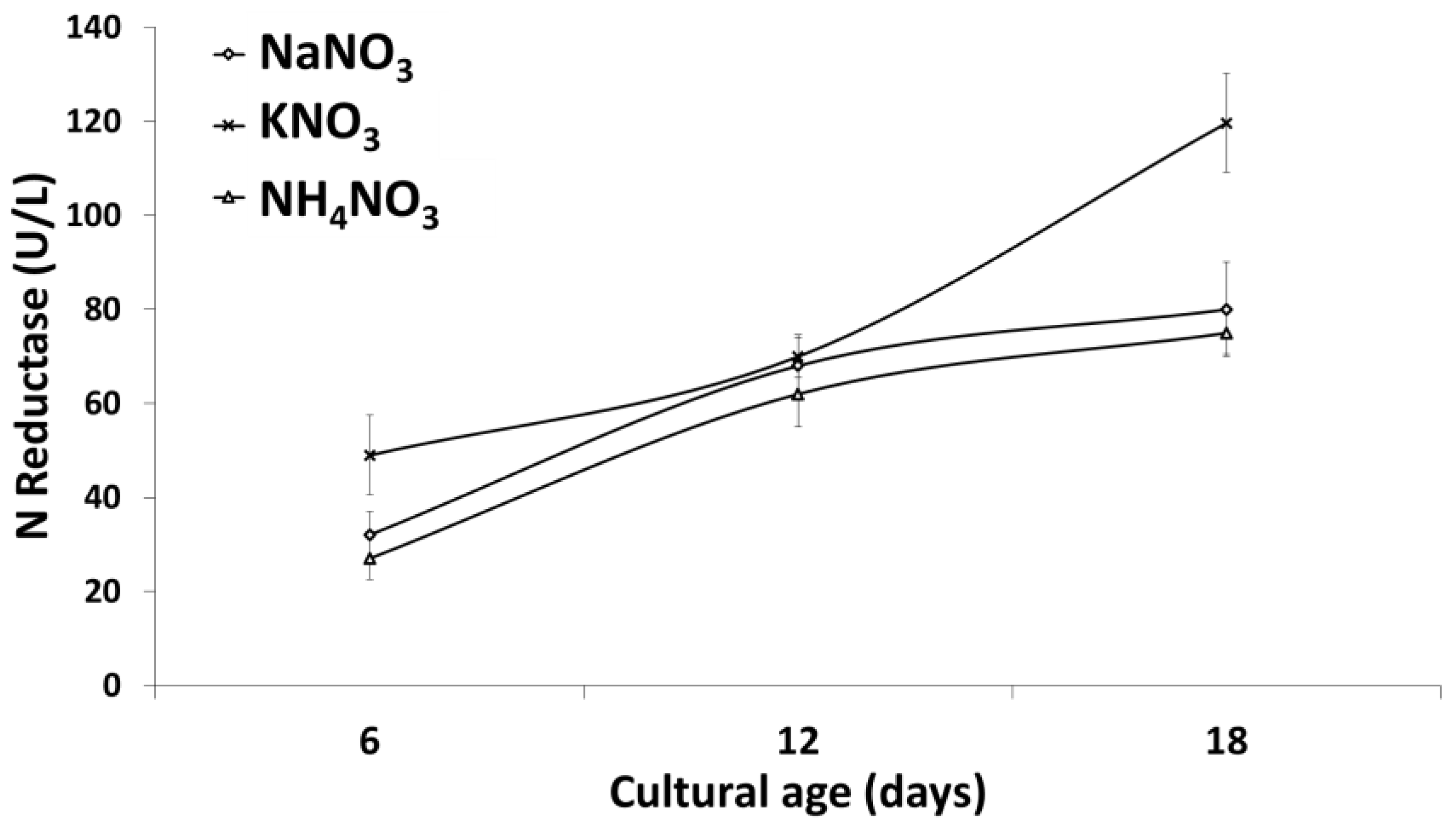
2.3. Nitrate Reductase Activity Assay

As one of the major reduction machineries for nanoparticle synthesis, nitrate reductase activities were measured in culture filtrates of S. obliquus cultivated on different N sources. Assays were conducted according to Redinbaugh and Campbell [42] with some modifications. In brief, 24 mM potassium phosphate buffer containing 0.05 mM EDTA (pH 7.3), 9.5 mM sodium nitrate, 58 mM sulfanilamide, and 0.77 mM N-(1-Naphthyl) Ethylenediamine Dihydrochloride solution (NED) were mixed. The reaction was started by adding a 100 µl enzyme source (the tested culture filtrates), and the volume of the mixture was augmented to 2 mL. The mixture was thoroughly mixed and incubated for 10 min at 25 °C, and then the color was spectrophotometrically determined at 540 nm. Enzyme units were defined as the amount of enzyme that liberates a micromole of nitrite per min.

2.4. Extracellular Biosynthesis and Characterization of AgNPs

The cultural filtrates from S. obliquus cultivated with different nitrogen sources were collected during various growth phases for use in extracellular biosynthesis of AgNPs. After each treatment, the cultural filtrates were collected via centrifugation at 5000 rcf for 10 min (Herarus Megafuge; Thermo ScientificsTM, Langensel Bold, Germany) at 6-day intervals representing the log, early- and late-stationary growth phases. Equal volumes of cell-free cultural filtrates from each treatment were added to silver nitrate solution (0.01 M). The mixtures were incubated at room temperature (28 ± 2 °C) for 24 h to allow biosynthesis of AgNPs. Visible observations (changes in the color from yellow to dark brown) and UV–Visible spectroscopy of the reaction mixture were performed to check the synthesis of the nanoparticles using a UV/VIS spectrophotometer (Jenway UV/Visible- 2605 Spectrophotometer, UK). The biologically synthesized nanoparticles were centrifuged, washed twice with deionized water, and air dried for further examinations. The size and shape of the synthesized AgNPs in selected samples were studied under HRTEM (JEM-2100 TEM, JEOL Ltd., Tokyo, Japan).

2.5. Antimicrobial Activity of AgNPs

The antimicrobial activities of the produced AgNPs were tested against some targeted pathogenic microorganisms obtained from the American type culture collection (ATCC; Rockville, MD, USA) and the culture collection of the National Research Centre (NRC, Cairo, Egypt). The tested organisms were Staphylococcus aurous ATCC- 47077, Bacillus cereus ATCC-12228, Escherichia coli ATCC-25922, Pseudomonas aeruginosa, Candida albicans ATCC-10231, and Saccharomyces cerevisiae ATCC-9763. The stock cultures of microorganisms used in this study were maintained on nutrient agar slants at 4 °C. The Agar well diffusion method was employed to study the antimicrobial activities of the synthesized AgNPs according to the method described by Kheiralla et al. [43] and Darwesh et al. [44].

2.6. Statistical Analysis

The one-way ANOVA analysis of variance followed Duncan’s multiple range test to compare means were carried out using the SPSS software V.20.0 (SPSS, Inc., Chicago, IL, USA). Difference was considered significant at p ≤ 0.05. The data were presented as mean ± standard deviation.

3. Results and Discussion

The current study was carried out to investigate the possibility of extracellular biosynthesis of AgNPs using the green microalga S. obliquus and to explain the effect of N-sources as well as algal growth stages on this process. Cell-free supernatants of S. obliquus cultivated with different nitrogen sources during different growth stages were used as sources of bioactive reductive compounds. The release of bioactive compounds implicated in construction of AgNPs was reported by Patel et al. [12]. Exudates-rich cultural supernatants of S. obliquus grown in BBM medium with various nitrogen sources were collected after 6, 12, and 18 days of algal cultivation to be examined for green biosynthesis of AgNPs. The time intervals were selected to represent the different growth stages of S. obliquus (i.e., log, early- and late-stationary phases).

3.1. Visible Observation and UV–Visible Spectroscopy of AgNPs Biosynthesis

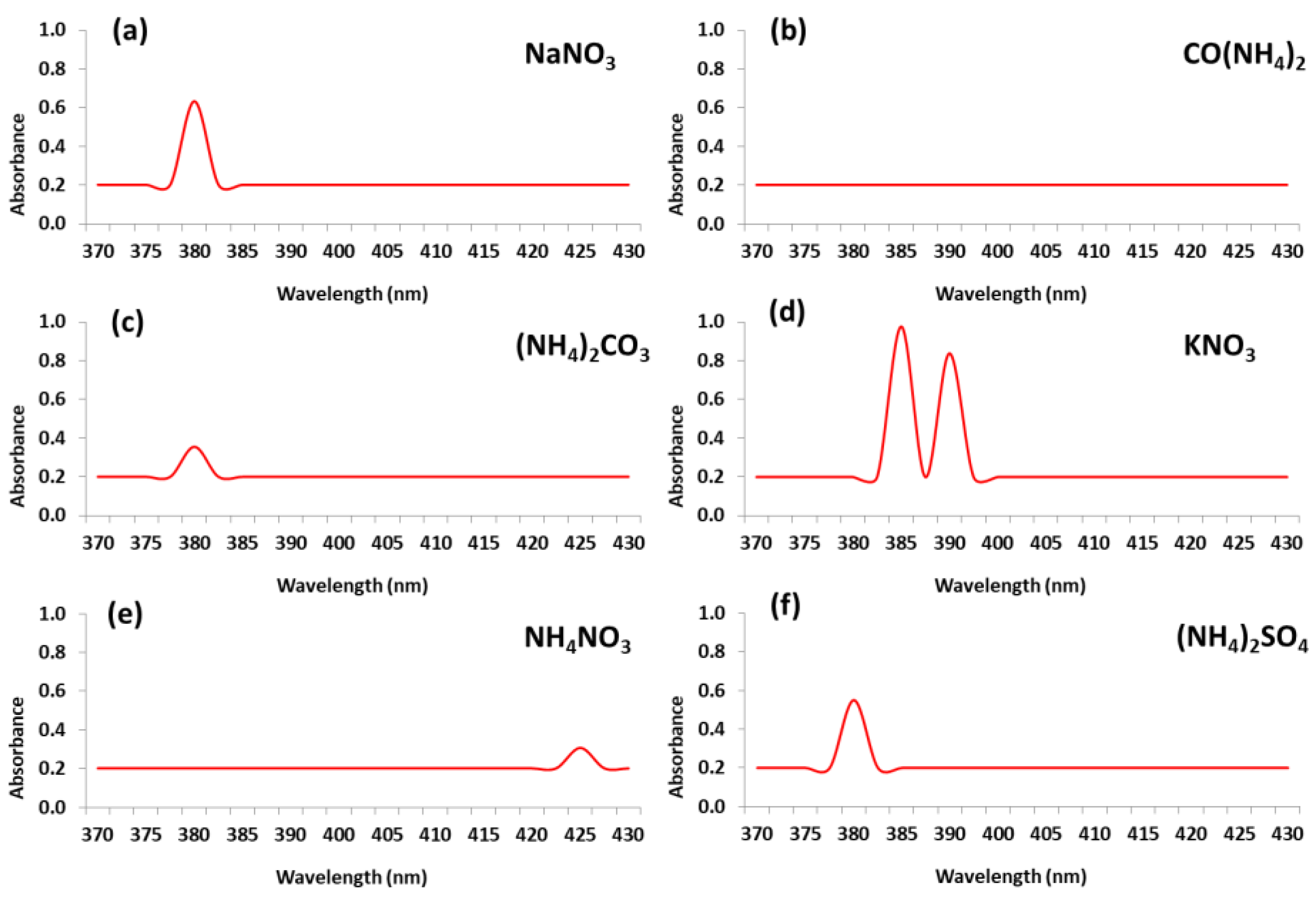
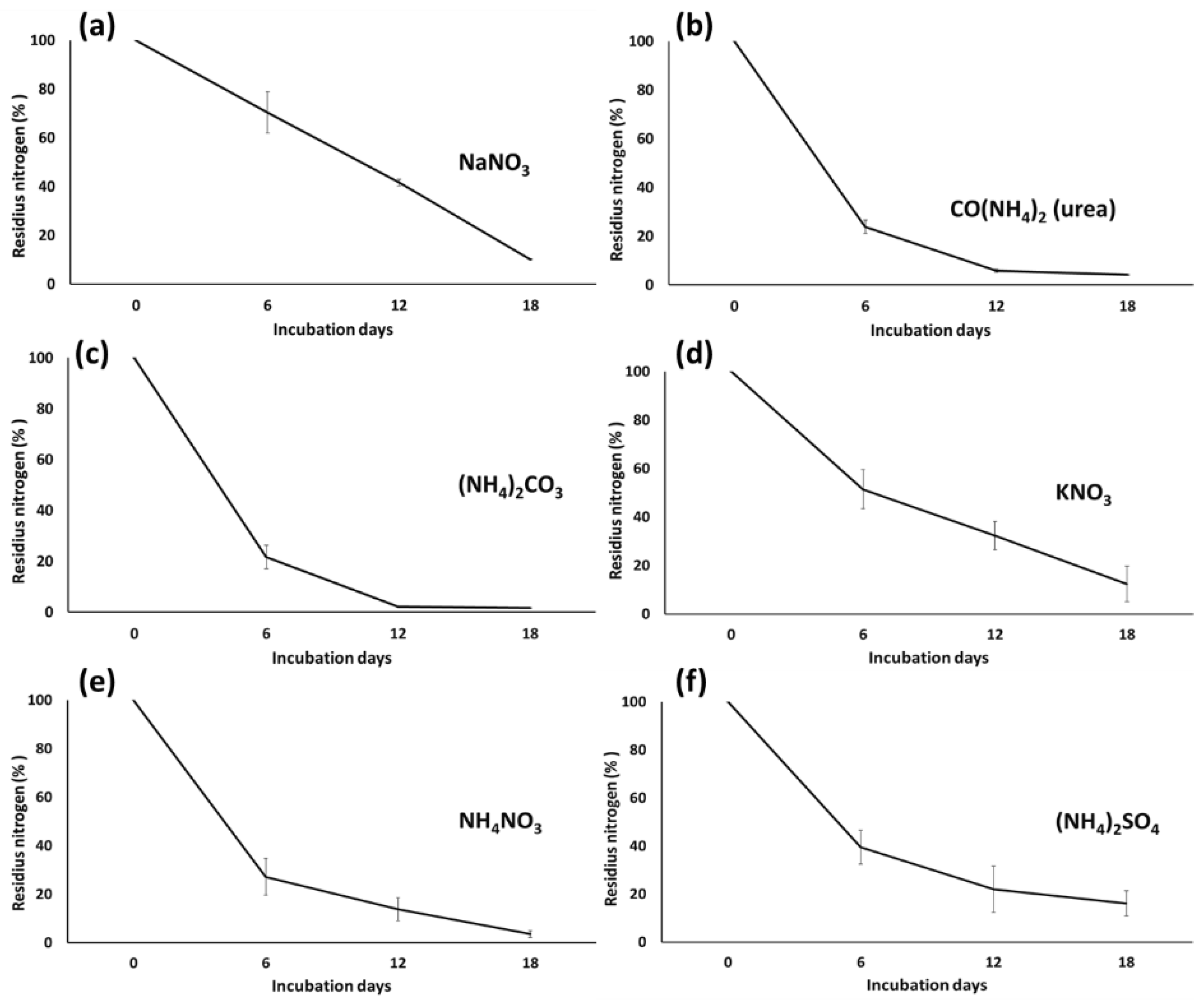
3.1.1. Impact of Nitrogen Source

In this study, S. obliquus was cultivated in BBM medium with six different sources of nitrogen. The cultivation nitrogen source affects microalgal growth through different biochemical pathways that are expected to produce different extracellular active substances [45] and subsequently influence the green biosynthesis of AgNPs. The results for the S. obliquus growth parameters are presented in Table 1. The highest growth was recorded when urea was used as the nitrogen source for S. obliquus. The growth status of a microalgal culture reflects the extracellular compounds that are secreted into the medium, including reducing agents that are responsible for silver nanoparticle biosynthesis [12].
The supernatants containing the metabolites of S. obliquus cultivated for 18 days were examined to reduce silver nitrate salt and produce nano-size silver particles. After 24 h incubation with the cultural filtrates, the colorless silver nitrate solutions were changed from yellow to reddish brown in color for all N treatments, but to lesser levels in the cases of urea and ammonium carbonate. Such brown colors have been reported to be characteristic of the formation of AgNPs [17]. The mixtures were scanned by spectrophotometer and the results are illustrated in Figure 1. According to amounts and particle sizes, several peaks were recorded in the range of 380–425 nm corresponding to AgNPs [17,24]. The highest peaks were detected at 385 and 390 nm for the KNO3 treatment followed by the peaks for the NaNO3 and ammonium sulfate treatments at 380 nm (Figure 1a,d,f). Additionally, the peaks obtained from (NH4)2CO3 and NH4NO3 were recorded at 380 and 425 nm, respectively, and were obviously smaller (Figure 1c,e). It is worth mentioning that the supernatant from the urea treatment did not yield any peaks in the range of AgNPs detection (Figure 1b). These results could be explained according to the superior role of KNO3 to induce nitrate reductase production in the cell-free culture supernatant of the 18-day-old S. obliquus culture.
Nitrogen is an essential and critical macronutrient affecting microalgal growth and cells’ biochemical composition [30,46]. Most microalgae can utilize various nitrogen sources such as nitrate, nitrite, ammonium (inorganic), and urea (organic) [46,47]. Nutrient uptake and growth of microalgae indicate its enzymatic activities either interiorly or through the surrounding medium [48]. For this reason, in the present study, nitrogen uptakes from different sources were determined in algal cultures through measurement of residual nitrogen in the supernatant (Figure 2).
The consumption rates of nitrogen for the three different forms (i.e., urea, nitrate and ammonia) by S. obliquus were recorded at the log, early-stationary, and late-stationary growth phases. Nitrogen in the supernatants was determined in each treatment as the original applied form (nitrate or ammonium). The nitrogen contents in the microalgal cultures gradually decreased through the cultivation period (18 days) due to their utilization for algal growth, as represented in Figure 2.
Visible observations and the UV–Visible spectroscopy results indicated the influence of the nitrogen source on AgNP biosynthesis during the cultivation of S. obliquus. Extracellular AgNP biosynthesis was highest for nitrate followed by sulfate, while it was almost negligible in the cases of the urea and ammonium carbonate supernatants. These results might be attributable to the reductase enzymatic activities, which increased due to the utilization and consumption of nitrate and sulfate in the microalgal culture. The role of reductase enzymes in extracellular biosynthesis of AgNPs was reported in a previous study [49].

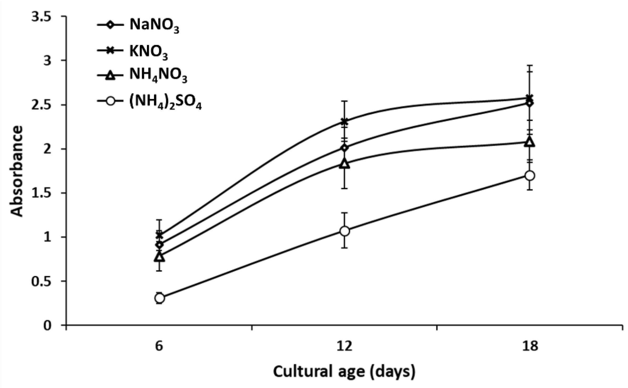
3.1.2. Impact of Microalgal Growth Stage

The aging of microalgae can be a significant factor to the production of their metabolites and to their reduction systems. Therefore, the metabolite composition in the culture filtrate is expected to differ by cultivation time [26]. Microalgae require a long time for desirable cell growth relative to bacteria. In the present study, 6-, 12- and 18-day cultures were regarded as representatives for the log, early- and late-stationary growth stages, respectively [28,50].
As noted earlier, reductase enzymes were expected to play the main role in the extracellular biosynthesis of AgNPs. The production level of such enzymes as well as other reduction systems varied largely according to the growth stage of the microalga and could be improved via utilization of nitrate-N. Hence, reductase enzyme was considered in the study of the influence of microalgal growth stage on extracellular biosynthesis of AgNPs. Samples were collected at 6-day intervals from each nitrate treatment, and the supernatants were used for the green synthesis of AgNPs.
The results obtained for nitrate reductase activity are represented in Figure 3. In the ammonium sulfate treatment, there was no nitrate in the medium, and so no nitrate reductase was detected, but sulfate reductase had been expected to be secreted [51]. On the other hand, the application of nitrate as the N-source induced reductase release in the culture supernatant of S. obliquus. However, the reductase activity varied according to both nitrate salt and the algal growth stage. The highest reductase activity was recorded when KNO3 was applied as the sole N-source, and its values increased with aging of the algal culture. In this case, the nitrate reductase activity reached 49, 70, and 120 U after 6, 12, and 18 days of algal cultivation, respectively (Figure 3). Similar results were observed for the NaNO3 and NH4NO3 treatments, the values being slightly lower than that for KNO3. The nitrate reductase enzymes were induced by application of nitrate-N sources, while ammonium-N sources repressed nitrate reductase activities [52,53,54].
Figure 4 shows the gradual synthesis of the silver nano-form in response to the different nitrate nitrogen sources and cultivation stages. The formation of the silver nano-size in the cultural filtrates of the nitrate as well as sulfate (ammonium sulfate) treatments showed obvious increases with cell aging. This increase was almost linear with the reductase increment. However, the supernatants collected from the cultures fed on KNO3 produced more nano-silver than in the case of NaNO3 or (NH4)NO3. The absorbance curve indicating the AgNPs’ use of the culture supernatant from the KNO3 treatment increased to 1.02, 2.31, and 2.58 with 6-, 12-, and 18-day algae aging, respectively. The obtained results suggested the vital role of cultivation-medium components in the composition of microalgae metabolites and, consequently, nanoparticle biosynthesis. As previously mentioned, the culture filtrates from the urea and ammonia treatments showed no nanoparticle formation, due to the absence of reductases, while the ammonium sulfate treatment aided the synthesis of relatively small amounts of AgNPs, due to the expected role of sulfate reductase.
Extracellular release of reductases enzymes into a surrounding medium is considered to be the main factor for extracellular nanoparticle biosynthesis, especially for AgNPs [55]. Those enzymes are responsible for reduction processes in many microorganisms. They are conjugated with an electron donor (quinine) to reduce metal ions to the elemental form [56]. Moteshafi et al. [57] demonstrated the role of reductases in AgNPs biosynthesis and reported the superiority of nitrate reductase to sulfate reductase in this regard. These results are in line with those in other reports, the authors of which stated that the nitrate reductase enzyme could be used as an indicator of nanometals formation [58,59].

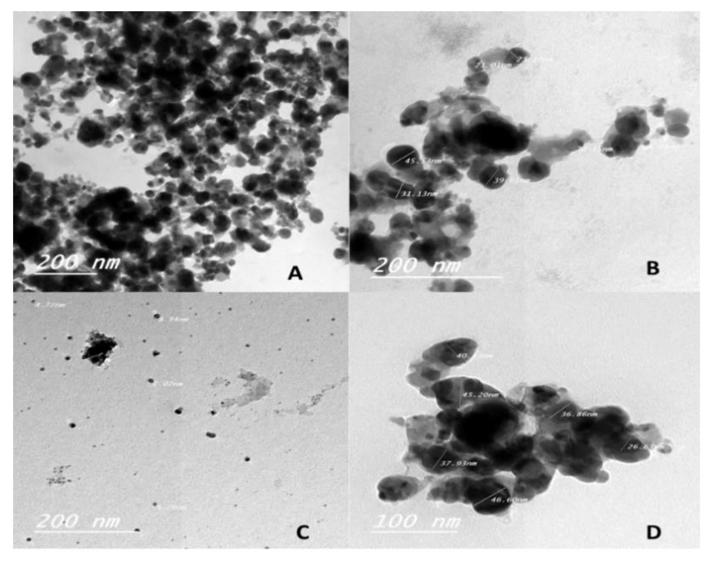
3.2. Characterization of AgNPs by High-Resolution Transmission Electron Microscopy (HRTEM)

The extracellular biosynthesis of AgNPs using microalgal cultural filtrates was confirmed by HRTEM (Figure 5). The HRTEM analysis was performed for the silver nitrate precipitates that had been reduced by the extracellular exudates from the S. obliquus cultivated for 18 days with KNO3, NaNO3, NH4NO3, and (NH4)2SO4. Such samples were selected based on the preliminarily positive visible and UV–Visible spectroscopy data indicative of AgNP biosynthesis. Figure 5 shows the variations in particle size, distribution, and aggregation among the preformed AgNPs for the cultivation nitrogen source. The smallest AgNP formations were obtained by supernatants containing potassium nitrate and sodium nitrate (5–10 and 4–10 nm, respectively). AgNPs produced using ammonium sulfate and ammonium nitrate cultural filtrates, by contrast, were significantly larger (25–50 and 30–50 nm, respectively). This might be related to differences in the metabolic compositions of the culture supernatants due to the use of different nitrogen sources. Cuellar-Bermudez et al. [60] also reported changes in filtrate metabolites by alteration of N-sources. The results in Figure 5 are in agreement with those obtained via spectrophotometric characterization (Figure 1 and Figure 4). Based on these results, the effect of the nitrogen source in the cultivation media was reflected not only in the effectiveness of nanoparticle biosynthesis but also in the properties of the produced AgNPs. The N-source played a crucial role in reducing silver nitrates to the silver nano-form. This might have been due to the reduction of metal silver ions mainly by the action of “nitrate-dependent reductase” and other microalgae-reducing extracellular metabolites [61]. In any case, the role of reductase enzymes seems to influence the formation of AgNPs, as noted also by Ramezani et al. [56].

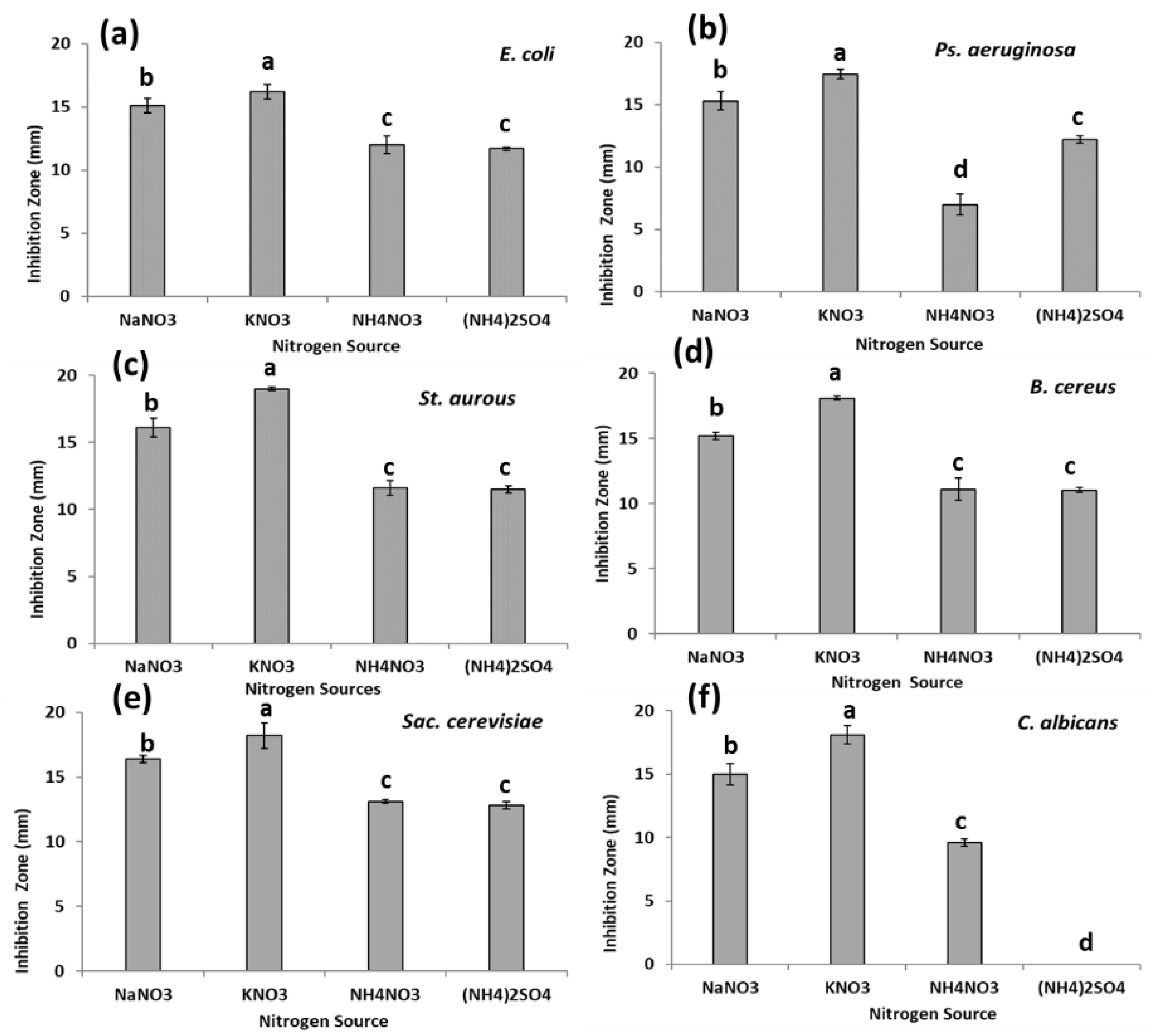
3.3. Antimicrobial Activity of Synthesized AgNPs

Pathogenic microbes exist in all environments, causing many problems and negatively affecting economies and public health [62,63]. Silver nano-forms are successfully applied to inhibit pathogens that are commonly found in medical, industrial, and agricultural fields [21,24]. In the present study, the antimicrobial activities of AgNPs extracellularly produced by supernatants of S. obliquus cultivated with nitrogen sources of different nitrates and sulphate were studied. The targeted microorganisms were: 2 G+ bacteria (St. aurous ATCC- 47077 and B. cereus ATCC- 12228), 2 G- bacteria (E. coli ATCC- 25922 and Ps. aeruginosa) as well as yeasts C. albicans ATCC- 10231 and Sac. cerevisiae ATCC- 9763. Figure 6 shows the inhibitory effects of the different produced AgNPs. All of the synthesised AgNPs exhibited antimicrobial activities against all of the tested microorganisms except that generated by ammonium sulphate against C. albicans. However, the AgNPs produced by the microalgal supernatants of KNO3 or NaNO3 treatment were superior in inhibiting the growth of all of the tested pathogens. This antimicrobial superiority of AgNPs from KNO3 or NaNO3 was consistent with the small size of these synthesized nanoparticles. The largest size of synthesized AgNPs was recorded for the ammonium sulphate supernatant treatment, which exhibited no activity against C. albicans as well as the lowest microbial activity against other targeted microbes. The obtained results indicated that the microbial activity of the synthesized AgNPs was related mainly to the produced particles’ size, which was a function of the nitrogen source of the original culture. Strong correlations between AgNP size and antimicrobial activities were suggested by Patel et al. [12] and Morones et al. [64]. In the present study, AgNPs synthesized using cultural filtrates of S. obliquus could be successfully used as an antimicrobial agent; however, additional studies are needed in order to assess their environmental and health impacts prior to actual applications.

4. Conclusions

Extracellular green biosynthesis of AgNPs using cultural filtrates of S. obliquus was demonstrated. The cultural nitrogen source had an important effect on extracellular substances that mediate the formation of AgNPs and control their size. Cultural age also influenced the synthesis of AgNPs, specifically by affecting the concentration of reducing agents including reductases. The reduction system of S. obliquus could be enhanced through the consumption of nitrates and sulphates during cultivation, thereby enhancing, in turn, the biosynthesis of AgNPs. The biosynthesized AgNPs showed antimicrobial activities that were inversely correlated with nanoparticle size. The sizes of the AgNPs produced by the microalgal filtrates could be controlled by altering the nutrient composition, especially the N-source, of their culture media. Biosafety and risk-assessment studies should be performed to evaluate the environmental and health impacts of green-synthesized-AgNP applications.

Author Contributions

All authors conceived and planned the research; O.M.D., I.A.M., and M.F.E. carried out the experiments and data analysis; All authors discussed the results and commented on the manuscript. I.A.M., O.M.D., and M.F.E. wrote the manuscript in consultation with all authors; I.A.M. performed the writing—review and editing of manuscript; Y.K.O. and H.M. revised and managed the final form of manuscript.

Acknowledgments

The authors would like to express their thanks to the National Research Centre, Egypt, and to the Advanced Biomass R&D Center (ABC) of the Global Frontier Project funded by the Ministry of Science and ICT, Republic of Korea (ABC-2012M3A6A205388) and the National Research Foundation of Korea (NRF) grant (NRF-2019R1A2C1003463) funded by the Ministry of Science and ICT, Republic of Korea, for their support of this work.

Conflicts of Interest

The authors declare no conflict of interest.

References

  1. Hopkins, M.M.; Kraft, A.; Martin, P.A.; Nightingale, P.; Mahdi, S. Is the biotechnology revolution a myth? In Comprehensive Medicinal Chemistry II; Taylor, J.B., Triggle, D.J., Eds.; Elsevier: London, UK, 2007; Volume 1, pp. 591–613. [Google Scholar]
  2. Abou-Shanab, R.A.; Matter, I.A.; Kim, S.N.; Oh, Y.K.; Choi, J.; Jeon, B.H. Characterization and identification of lipid-producing microalgae species isolated from a freshwater lake. Biomass Bioenergy 2011, 35, 3079–3085. [Google Scholar] [CrossRef]
  3. Matter, I.A.; Darwesh, O.M.; Eida, M.F. Harvesting of Scenedesmus obliquus by bioflocculation: Appropriate chitosan concentrations with various pH values at different growth stages. Jordan J. Biol. Sci. 2018, 11, 475–481. [Google Scholar]
  4. Hasanin, M.S.; Mostafa, A.M.; Mwafy, E.A.; Darwesh, O.M. Eco-friendly cellulose nano fibers via first reported Egyptian Humicola fuscoatra Egyptia X4: Isolation and characterization. Environ. Nanotechnol. Monit. Manag. 2018, 10, 409–418. [Google Scholar] [CrossRef]
  5. Hasanin, M.S.; Darwesh, O.M.; Matter, I.A.; El-Saied, H. Isolation and characterization of non-cellulolytic Aspergillus flavus EGYPTA5 exhibiting selective ligninolytic potential. Biocatal. Agric. Biotechnol. 2019, 17, 160–167. [Google Scholar] [CrossRef]
  6. Abou-Shanab, R.A.I.; Khalafallah, M.A.; Emam, N.F.; Aly, M.A.; Abou-Sdera, S.A.; Matter, I.A. Characterisation and identification of carbofuran-utilising bacteria isolated from agricultural soil. Chem. Ecol. 2012, 28, 193–203. [Google Scholar] [CrossRef]
  7. Saber, M.; Hoballah, E.; Matter, I.; Zaghloul, A. Bioremediation of chlorinated hydrocarbons in a sewaged soil by certain remediative amendments followed by phytoremediation. Res. J. Pharm. Biol. Chem. Sci. 2014, 5, 83–90. [Google Scholar]
  8. Hoballah, E.; Saber, M.; Matter, I.; Zaghloul, A. Bioremediation of polychlorinated biphenyl (PCBs) in a sewaged soil by certain remediative amendments followed by phytoremediation. Res. J. Pharm. Biol. Chem. Sci. 2014, 5, 91–103. [Google Scholar]
  9. Barakat, K.M.; Hassan, S.W.M.; Darwesh, O.M. Biosurfactant production by haloalkaliphilic Bacillus strains isolated from Red Sea, Egypt. Egypt. J. Aquat. Res. 2017, 43, 205–211. [Google Scholar] [CrossRef]
  10. Annamalai, J.; Nallamuthu, T. Green synthesis of silver nanoparticles: Characterization and determination of antibacterial potency. Appl. Nanosci. 2016, 6, 259–265. [Google Scholar] [CrossRef]
  11. Marrez, D.A.; Abdelhamid, A.E.; Darwesh, O.M. Eco-friendly cellulose acetate green synthesized silver nano-composite as antibacterial packaging system for food safety. Food Packag. Shelf Life 2019, 20, 100302. [Google Scholar] [CrossRef]
  12. Patel, V.; Berthold, D.; Puranik, P.; Gantar, M. Screening of cyanobacteria and microalgae for their ability to synthesize silver nanoparticles with antibacterial activity. Biotechnol. Rep. 2015, 5, 112–119. [Google Scholar] [CrossRef]
  13. Darwesh, O.M.; Matter, I.A.; Eida, M.F. Development of peroxidase enzyme immobilized magnetic nanoparticles for bioremediation of textile wastewater dye. J. Environ. Chem. Eng. 2019, 7, 102805. [Google Scholar] [CrossRef]
  14. Lee, K.; Lee, S.Y.; Praveenkumar, R.; Kim, B.; Seo, J.Y.; Jeon, S.G.; Na, J.G.; Park, J.Y.; Kim, D.M.; Oh, Y.K. Repeated use of stable magnetic flocculant for efficient harvest of oleaginous Chlorella sp. Bioresour. Technol. 2014, 167, 284–290. [Google Scholar] [CrossRef]
  15. Gu, H.; Ho, P.L.; Tong, E.; Wang, L.; Xu, B. Presenting vancomycin on nanoparticles to enhance antimicrobial activities. Nano Lett. 2003, 3, 1261–1263. [Google Scholar] [CrossRef]
  16. Schabes-Retchkiman, P.S.; Canizal, G.; Herrera-Becerra, R.; Zorrilla, C.; Liu, H.B.; Ascencio, J.A. Biosynthesis and characterization of Ti/Ni bimetallic nanoparticles. Opt. Mater. 2006, 29, 95–99. [Google Scholar] [CrossRef]
  17. El-Naggar, N.E.; Abdelwahed, N.A.M.; Darwesh, O.M. Fabrication of biogenic antimicrobial silver nanoparticles by Streptomyces aegyptia NEAE 102 as eco-friendly nanofactory. J. Microbiol. Biotechnol. 2014, 24, 453–464. [Google Scholar] [CrossRef]
  18. Kim, B.; Praveenkumar, R.; Lee, J.; Nam, B.; Kim, D.M.; Lee, K.; Lee, Y.C.; Oh, Y.K. Magnesium aminoclay enhances lipid production of mixotrophic Chlorella sp. KR-1 while reducing bacterial populations. Bioresour. Technol. 2016, 219, 608–613. [Google Scholar] [CrossRef]
  19. Jena, J.; Pradhan, N.; Nayak, R.R.; Dash, B.P.; Sukla, L.B.; Panda, P.K.; Mishra, B.K. Microalga Scenedesmus sp.: A potential low-cost green machine for silver nanoparticle synthesis. J. Microbiol. Biotechnol. 2014, 24, 522–533. [Google Scholar] [CrossRef]
  20. Marambio-Jones, C.; Hoek, E.M. A review of the antibacterial effects of silver nanomaterials and potential implications for human health and the environment. J. Nanopart. Res. 2010, 12, 1531–1551. [Google Scholar] [CrossRef]
  21. Mahdieh, M.; Zolanvari, A.; Azimee, A.S. Green biosynthesis of silver nanoparticles by Spirulina platensis. Sci. Iran. 2012, 19, 926–929. [Google Scholar] [CrossRef]
  22. Kabary, H.A.; Attia, M.; Easa, S.M.; Awad, N.M.; Abou-Shanab, R.A.; Eida, M.F. Characterization of magnetotactic bacteria (MTBs) isolated from different habitats in Egypt. Res. J. Pharm. Biol. Chem. Sci. 2016, 7, 244–257. [Google Scholar]
  23. Kabary, H.; Eida, M.F.; Attia, M.; Awad, N.; Easa, S.M. Magnetotactic characterization and environmental application P. aeruginosa kb1 isolate. Annu. Res. Rev. Biol. 2017, 20, 1–10. [Google Scholar] [CrossRef]
  24. Elshahawy, I.; Abouelnasr, H.M.; Lashin, S.M.; Darwesh, O.M. First report of Pythium aphanidermatum infecting tomato in Egypt and its control using biogenic silver nanoparticles. J. Plant Prot. Res. 2018, 15, 137–151. [Google Scholar]
  25. Rahman, A.; Kumar, S.; Bafana, A.; Dahoumane, S.; Jeffryes, C. Biosynthetic conversion of Ag+ to highly sable Ag0 nanoparticles by wild type and cell wall deficient strains of Chlamydomonas reinhardtii. Molecules 2019, 24, 98. [Google Scholar] [CrossRef]
  26. El-Baz, F.K.; Gad, M.S.; Abdo, S.M.; Abed, K.A.; Matter, I.A. Performance and exhaust emissions of a diesel engine burning algal biodiesel blends. Int. J. Mech. Mech. Eng. 2016, 16, 151–158. [Google Scholar]
  27. Matter, I.A.; Darwesh, O.M.; Eida, M.F. Harvesting of microalgae Scenedesmus obliquus using chitosan-alginate dual flocculation system. Biosci. Res. 2018, 15, 540–548. [Google Scholar]
  28. Eida, M.F.; Matter, I.A.; Darwesh, O.M. Cultivation of oleaginous microalgae Scenedesmus obliquus on secondary treated municipal wastewater as growth medium for biodiesel production. Ecol. Eng. 2018, 19, 38–50. [Google Scholar] [CrossRef]
  29. Perin, G.; Bellan, A.; Bernardi, A.; Bezzo, F.; Morosinotto, T. The potential of quantitative models to improve microalgae photosynthetic efficiency. Physiol. Plant. 2019, in press. [Google Scholar] [CrossRef]
  30. Amin, N.F.; Khalafallah, M.A.; Ali, M.A.; Abou-Sdera, S.A.; Matter, I.A. Effect of some nitrogen sources on growth and lipid of microalgae Chlorella sp. for biodiesel production. J. Appl. Sci. Res. 2013, 9, 4845–4855. [Google Scholar]
  31. Sahayaraj, K. Bionanomaterials: Synthesis and applications. In Proceedings of the National Seminar on New Materials Research and Nano Technology; John, N.J., Ed.; Govt. Arts College: Ooty, Tamil Nadu, Indian, 2012; pp. 24–29. [Google Scholar]
  32. González-Fernández, C.; Ballesteros, M. Microalgae autoflocculation: An alternative to high-energy consuming harvesting methods. J. Appl. Phycol. 2013, 25, 991–999. [Google Scholar] [CrossRef]
  33. Xiao, R.; Zheng, Y. Overview of microalgal extracellular polymeric substances (EPS) and their applications. Biotechnol. Adv. 2016, 34, 1225–1244. [Google Scholar] [CrossRef]
  34. Rahman, A.; Kumar, S.; Bafana, A.; Dahoumane, S.A.; Jeffryes, C. individual and combined effects of extracellular polymeric substances and whole cell components of Chlamydomonas reinhardtii on silver nanoparticle synthesis and stability. Molecules 2019, 24, 956. [Google Scholar] [CrossRef]
  35. El-Sheekh, M.M.; El-Kassas, H.Y. Algal production of nano-silver and gold: Their antimicrobial and cytotoxic activities: A review. J. Genet. Eng. Biotechnol. 2016, 14, 299–310. [Google Scholar] [CrossRef] [Green Version]
  36. Gong, P.; Li, H.; He, X.; Wang, K.; Hu, J.; Tan, W.; Zhang, S.; Yang, X. Preparation and antibacterial activity of Fe3O4@ Ag nanoparticles. Nanotechnology 2007, 18, 285604. [Google Scholar] [CrossRef]
  37. Khalil, A.M.; Abdel-Monem, R.A.; Darwesh, O.M.; Hashim, A.I.; Nada, A.A.; Rabie, S.T. Synthesis, characterization, and evaluation of antimicrobial activities of chitosan and carboxymethyl chitosan schiff-base/Silver nanoparticles. J. Chem. 2017, 2017, 1434320. [Google Scholar] [CrossRef]
  38. Rai, M.; Yadav, A.; Gade, A. Silver nanoparticles as a new generation of antimicrobials. Biotechnol. Adv. 2009, 27, 76–83. [Google Scholar] [CrossRef]
  39. Pal, S.; Tak, Y.K.; Song, J.M. Does the antibacterial activity of silver nanoparticles depend on the shape of the nanoparticle? A study of the gram-negative bacterium Escherichia coli. Appl. Environ. Microbiol. 2007, 73, 1712–1720. [Google Scholar] [CrossRef]
  40. Barsanti, L.; Gualtieri, P. Algae: Anatomy, Biochemistry, and Biotechnology, 2nd ed.; CRC Press, Taylor & Francis Group, LLC: Boca Raton, FL, USA, 2014; 361p. [Google Scholar]
  41. Basova, E.M.; Bulanova, M.A.; Ivanov, V.M. Photometric detection of urea in natural waters. Mosc. Univ. Chem. Bull. 2011, 66, 345–350. [Google Scholar] [CrossRef]
  42. Redinbaugh, M.G.; Campbell, W.H. Quaternary structure and composition of squash NADH: Nitrate reductase. J. Biol. Chem. 1985, 260, 3380–3385. [Google Scholar]
  43. Kheiralla, Z.H.; Hewedy, M.A.; Mohammed, H.R.; Darwesh, O.M. Isolation of pigment producing actinomycetes from rhizosphere soil and application it in textiles dyeing. Res. J. Pharm. Biol. Chem. Sci. 2016, 7, 2128–2136. [Google Scholar]
  44. Darwesh, O.M.; Sultan, Y.Y.; Seif, M.M.; Marrez, D.A. Bio-evaluation of crustacean and fungal nano-chitosan for applying as food ingredient. Toxicol. Rep. 2018, 5, 348–356. [Google Scholar] [CrossRef] [PubMed]
  45. Sultan, Y.Y.; Ali, M.A.; Darwesh, O.M.; Embaby, M.A.; Marrez, D.A. Influence of nitrogen source in culture media on antimicrobial activity of Microcoleus lacustris and Oscillatoria rubescens. Res. J. Pharm. Biol. Chem. Sci. 2016, 7, 1444–1452. [Google Scholar]
  46. Kim, G.; Mujtaba, G.; Lee, K. Effects of nitrogen sources on cell growth and biochemical composition of marine chlorophyte Tetraselmis sp. for lipid production. Algae 2016, 31, 257–266. [Google Scholar] [CrossRef]
  47. Agwa, O.K.; Abu, G.O. Influence of various nitrogen sources on biomass and lipid production by Chlorella vulgaris. Br. Biotechnol. J. 2016, 15, 1–13. [Google Scholar] [CrossRef]
  48. Ebrahiminezhad, A.; Bagheri, M.; Taghizadeh, S.M.; Berenjian, A.; Ghasemi, Y. Biomimetic synthesis of silver nanoparticles using microalgal secretory carbohydrates as a novel anticancer and antimicrobial. Adv. Nat. Sci. Nanosci. Nanotechnol. 2016, 7, 015018. [Google Scholar] [CrossRef] [Green Version]
  49. Mukherjee, K.; Gupta, R.; Kumar, G.; Kumari, S.; Biswas, S.; Padmanabhan, P. Synthesis of silver nanoparticles by Bacillus clausii and computational profiling of nitrate reductase enzyme involved in production. J. Genet. Eng. Biotechnol. 2018, 16, 527–536. [Google Scholar] [CrossRef]
  50. Matter, I.A.; Darwesh, O.M.; El-baz, F.K. Using the natural polymer chitosan in harvesting Scenedesmus species under different concentrations and cultural pH values. Int. J. Pharm. Biol. Sci. 2016, 7, 254–260. [Google Scholar]
  51. Sarayloo, E.; Tardu, M.; Unlu, Y.S.; Simsek, S.; Cevahir, G.; Erkey, C.; Kavakli, I.H. Understanding lipid metabolism in high-lipid-producing Chlorella vulgaris mutants at the genome-wide level. Algal Res. 2017, 28, 244–252. [Google Scholar] [CrossRef]
  52. Herrero, A.; Muro-Pastor, A.M.; Flores, E. Nitrogen control in cyanobacteria. J. Bacteriol. 2001, 183, 411–425. [Google Scholar] [CrossRef]
  53. Muro-Pastor, M.I.; Reyes, J.C.; Florencio, F.J. Ammonium assimilation in cyanobacteria. Photosynth. Res. 2005, 83, 135–150. [Google Scholar] [CrossRef]
  54. Jha, P.; Ali, A.; Raghuram, N. Nitrate-induction of nitrate reductase and its inhibition by nitrite and ammonium ions in Spirulina platensis. Physiol. Mol. Biol. Plants 2007, 13, 163–167. [Google Scholar]
  55. Khodashenas, B.; Ghorbani, H.R. Optimisation of nitrate reductase enzyme activity to synthesise silver nanoparticles. IET Nanobiotechnol. 2016, 10, 158–161. [Google Scholar] [CrossRef]
  56. Ramezani, F.; Ramezani, M.; Talebi, S. Mechanistic aspects of biosynthesis of nanoparticles by several microbes. Nanocon 2010, 10, 1–7. [Google Scholar]
  57. Moteshafi, H.; Mousavi, S.M.; Shojaosadati, S.A. The possible mechanisms involved in nanoparticles biosynthesis. J. Ind. Eng. Chem. 2012, 18, 2046–2050. [Google Scholar] [CrossRef]
  58. Pradhan, N.; Nayak, R.R.; Pradhan, A.K.; Sukla, L.B.; Mishra, B.K. In situ Synthesis of entrapped silver nanoparticles by a fungus—Penicillium purpurogenum. Nanosci. Nanotechnol. Lett. 2011, 3, 659–665. [Google Scholar] [CrossRef]
  59. Vahabi, K.; Mansoori, G.A.; Karimi, S. Biosynthesis of silver nanoparticles by fungus Trichoderma reesei (a route for large-scale production of AgNPs). Insci. J. 2011, 1, 65–79. [Google Scholar] [CrossRef]
  60. Cuellar-Bermudez, S.P.; Aguilar-Hernandez, I.; Cardenas-Chavez, D.L.; Ornelas-Soto, N.; Romero-Ogawa, M.A.; Parra-Saldivar, R. Extraction and purification of high-value metabolites from microalgae: Essential lipids, astaxanthin and phycobiliproteins. Microb. Biotechnol. 2015, 8, 190–209. [Google Scholar] [CrossRef]
  61. Durán, N.; Marcato, P.D.; Alves, O.L.; De Souza, G.I.; Esposito, E. Mechanistic aspects of biosynthesis of silver nanoparticles by several Fusarium oxysporum strains. J. Nanobiotechnol. 2005, 3, 1–7. [Google Scholar] [CrossRef]
  62. Younes, A.M.; Mohamed, L.A.; Eida, M.F.; Gaafar, A.Y. Characterization and pathogen challenge of Pseudomonas species from Oreochromis niloticus in Egypt. Res. J. Pharm. Biol. Chem. Sci. 2015, 6, 312–317. [Google Scholar]
  63. Fernández, M.; Hudson, J.A.; Korpela, R.; de los Reyes-Gavilán, C.G. Impact on human health of microorganisms present in fermented dairy products: An overview. BioMed Res. Int. 2015, 2015, 412714. [Google Scholar] [CrossRef]
  64. Morones, J.R.; Elechiguerra, J.L.; Camacho, A.; Holt, K.; Kouri, J.B.; Ramírez, J.T.; Yacaman, M.J. The bactericidal effect of silver nanoparticles. Nanotechnology 2005, 16, 2346. [Google Scholar] [CrossRef]
Figure 1. Spectrophotometer scanning of synthesized AgNPs by S. obliquus cultivated in BBM media containing different nitrogen sources (a) NaNO3; (b) urea; (c) (NH4)2CO3; (d) KNO3; (e) NH4NO3; (f) (NH4)2SO4).
Applsci 09 01465 g001
Figure 2. Residual nitrogen (% from initial) in S. obliquus growth medium throughout cultivation period (18 days) (a) NaNO3; (b) urea; (c) (NH4)2CO3; (d) KNO3; (e) NH4NO3; (f) (NH4)2SO4. Data are expressed as mean values ± standard deviation (n = 2).
Applsci 09 01465 g002
Figure 3. Nitrate reductase enzyme activity (U/L) in supernatant of S. obliquus cultivated in media containing different nitrogen sources. Data are expressed as mean values ± standard deviation (n = 3).
Applsci 09 01465 g003
Figure 4. Absorbance of synthesized AgNPs by supernatant of microalgal growth at wavelength of 385 nm during cultivation period (18 days at 6-day intervals). Data are expressed as mean values ± standard deviation (n = 3).
Applsci 09 01465 g004
Figure 5. HRTEM of AgNPs fabricated by supernatant of S. obliquus cultivated in media containing different nitrogen sources (A: KNO3; B: NH4NO3; C: NaNO3; D: (NH4)2SO4).
Applsci 09 01465 g005
Figure 6. Inhibition zone (mm) for antimicrobial activity of AgNPs produced by supernatant of S. obliquus cultivated in media containing different nitrogen sources against (a) E. coli, (b) Ps. Aeruginosa, (c) St. aurous, (d) B. cereus, (e) C. albicans, and Sac. cerevisiae. Data are expressed as mean values ± standard deviation (n = 3).
Applsci 09 01465 g006
Table 1. Changes in growth parameters of S. obliquus with different nitrogen sources at end of incubation period (18 days). Data are expressed as mean values ± standard deviation (n = 2).
Nitrogen SourceOD (Abs 680 nm)Dry Biomass (g/L)
NaNO32.33 ± 0.2 b0.63 ± 0.013 c
CO(NH4)2 (urea)3.20 ± 0.3 a1.09 ± 0.046 a
(NH4)2CO32.66 ± 0.2 b0.76 ± 0.046 b
KNO31.92 ± 0.2 c0.48 ± 0.008 e
NH4NO32.72 ± 0.3 b0.56 ± 0.004 d
(NH4)2SO40.84 ± 0.1 d0.26 ± 0.001 f

