Communication

Quantification of Trans-Resveratrol-Loaded Solid Lipid Nanoparticles by a Validated Reverse-Phase HPLC Photodiode Array

by
Roberta B. Rigon
1,
Naiara Fachinetti
1,
Patrícia Severino
2,
Alessandra Durazzo
3,
Massimo Lucarini
3,
Atanas G. Atanasov
4,5,
Soukaina El Mamouni
6,
Marlus Chorilli
1,
Antonello Santini
6,* and
Eliana B. Souto
7,8,*
1
Department of Drugs and Medicines, School of Pharmaceutical Sciences of Araraquara, Universidade Estadual Paulista “Júlio de Mesquita Filho”, Rod. Araraquara-Jaú, km 1, 14801-902 Araraquara, São Paulo, Araraquara, Brazil
2
Tiradentes Institute, University of Tiradentes (Unit) and Institute of Technology and Research (ITP), Av. Murilo Dantas, 300, 49010-390 Aracaju-SE, Brazil
3
CREA—Research Centre for Food and Nutrition, Via Ardeatina 546, 00178 Rome, Italy
4
Department of Pharmacognosy, University of Vienna, 1090 Vienna, Austria
5
Institute of Neurobiology, Bulgarian Academy of Sciences, 23 Acad. G. Bonchev str., 1113 Sofia, Bulgaria
6
Department of Pharmacy, University of Napoli Federico II, Via D. Montesano 49, 80131 Napoli, Italy
7
Department of Pharmaceutical Technology, Faculty of Pharmacy, University of Coimbra (FFUC), Pólo das Ciências da Saúde, Azinhaga de Santa Comba, 3000-548 Coimbra, Portugal
8
CEB—Centre of Biological Engineering, University of Minho, Campus de Gualtar, 4710-057 Braga, Portugal
*
Authors to whom correspondence should be addressed.
Submission received: 25 September 2019 / Revised: 7 November 2019 / Accepted: 14 November 2019 / Published: 18 November 2019
(This article belongs to the Section Chemical and Molecular Sciences)

Abstract

:
A new method based on reverse-phase HPLC combined with photodiode array (PDA) was developed to quantify the release of trans-resveratrol (tRES) from solid lipid nanoparticles (SLN). The mobile phase was composed of 75:0:25 (V/V) water/methanol/acetonitrile at 0–3.5 min, 32.5:30.0:37.5 (V/V) water/methanol/acetonitrile at 3.6–5.8 min, and 75:0:25 (V/V) water/methanol/acetonitrile at 5.9–10 min. The flow rate was set at 1.0 mL/min, and tRES was detected at the wavelength of 306.6 nm. A concentration range of 1–100 µg/mL was used to obtain the linear calibration curve. SLN were produced by ultrasound technique to load 0.1% (wt/wt) of tRES, and the in vitro release of the drug was run in modified Franz diffusion cells. The mean recovery of tRES was found to be 96.84 ± 0.32%. The intra-assay and inter-assay coefficients of variation were less than 5%. The proposed method was applied to in vitro permeability studies, and the Weibull model was found to be the one that best fits the tRES release, which is characterized by a simultaneous lipid chain relaxation and erosion during drug release.

1. Introduction

Resveratrol (trans-4,3′,5′-trihydroxystilbene) is a natural compound found in grapes and in many other medicinal plants [1]. Resveratrol acts as phytoalexin protecting the plant against virus infections. The high concentration of this compound in red wine is attributed to its high concentration in the skin of red grapes [2]. Resveratrol (C14H12O3) is a whitish color powder with a molecular weight of 228.25 g/mol, its solubility varies from 0.03 g/L in water to 16 g/L in DMSO and 50 g/L in ethanol. Its melting point ranges from 253 to 255 °C, exhibiting a log P of 3.1 [3,4]. Although poorly water soluble, resveratrol shows high membrane permeability and may be considered as a Class II of the Biopharmaceutics Classification System (BCS) [3,5]. Besides its glycoside form, resveratrol occurs in the cis-(Z) and trans-(E) structural isomers, which exhibit distinct biological effects [6,7]. Ultraviolet (UV) radiation induces partial conversion of trans-(E) isomer into cis-(Z), resulting in a mix of the two isomers (Figure 1) [8].
From the perspective of nutraceutical applications [9,10,11,12,13,14,15,16,17], research is being demonstrating that trans-resveratrol (tRES) offers many health benefits e.g., as it: (i) lowers the risk of cardiovascular complications [18,19], (ii) protects the human body against retinal ischemia by downregulating matrix metalloproteinase-9 expression by nitric oxide and by upregulating heme oxygenase-1 [20]; (iii) improves cells sensitivity to insulin, reducing oxidative stress, and activating protein kinase B pathway in type 2 diabetic patients [21,22,23]; (iv) acts as a therapeutic agent for neurodegenerative diseases e.g., in Alzheimer’s disease, Parkinson’s disease, or multiple sclerosis [24,25]; (v) modulates inflammatory arthritis by selectively suppressing key cellular and humoral responses required for disease settling [26]; (vi) inhibits platelet aggregation and synthesis of thromboxane B2 and hydroxyl heptadecatrienoate (HHT) [27]; (vii) exhibits antiherpetic activity [28] and (viii) chemopreventive properties [29].
Scientific literature describes the encapsulation of tRES in different types of lipid nanoparticles [4,30,31], which have demonstrated to be taken up by cells [32], and accumulated around nuclei. Guo et al. [33] showed that tRES concentration was greatly increased in the target tissue when tRES-loaded nanoparticles were injected [33]. Moreover, tRES-loaded nanoparticles promoted greater tumor growth inhibition. Jung et al. [34] demonstrated that tRES loaded in PEGylated polymeric nanoparticles based on polylactic acid cores (PEG–PLA, of MW 5000:5000) exhibited in vitro and in vivo antitumor effects such as the dose-dependent reduction of cell viability, the suppression of glucose metabolism, improved animal survival, and reduced tumor volume. Among the several types of nanoparticles, solid lipid nanoparticles (SLN) have been showing high physical stability, biodegradability, and biocompatibility for a set of administration routes [35,36], the potential to modify the release kinetics of loaded drugs [37,38], as well as potential for crossing the cellular membrane and to accumulate in the perinuclear region without toxic effect [32,39,40].
Methods commonly used for the quantification of resveratrol when loaded in nanoparticles do not promote the separation of the isoforms (trans- and cis-) of the drug. Furthermore, a monograph of tRES in official compendia is not yet available. For this purpose, the validation of a suitable quantitative method for this drug becomes a demand, for which the first step is to identify the critical parameters and to establish the acceptance criteria [41]. For the quantification of tRES, some analytical procedures have already been described, e.g., high-performance liquid chromatography (HPLC) coupled with UV, high-performance thin-layer chromatography (HPTLC), fluorescence and electrochemical detection, liquid chromatography coupled to mass spectrometry, gas chromatography, or capillary electrophoresis [42].
In this work, we describe a reverse-phase HPLC coupled with photodiode array for the quantification of tRES released from SLN in modified Franz diffusion cells. SLN composed of stearic acid as solid lipid and poloxamer 407 as surfactant have been produced by ultrasound (US) technique. The effect of the presence of a co-surfactant (i.e., soy phosphatidylcholine) on the release profile of tRES from SLN was also evaluated and fitted to different mathematical models (Korsmeyer-Peppas, Higuchi, First Order, Weibull and Zero Order kinetics).

2. Material and Methods

2.1. Materials

As standard chemical substance, trans-resveratrol 99%, batch number #030M5216V, from Sigma-Aldrich (Taufkirchen, Germany) was used. Acetonitrile and methanol of HPLC grade were purchased from Fisher Scientifics (Pittsburg, USA) and glacial acetic acid of analytical grade was obtained from Merck GmbH (Darmstadt, Germany).

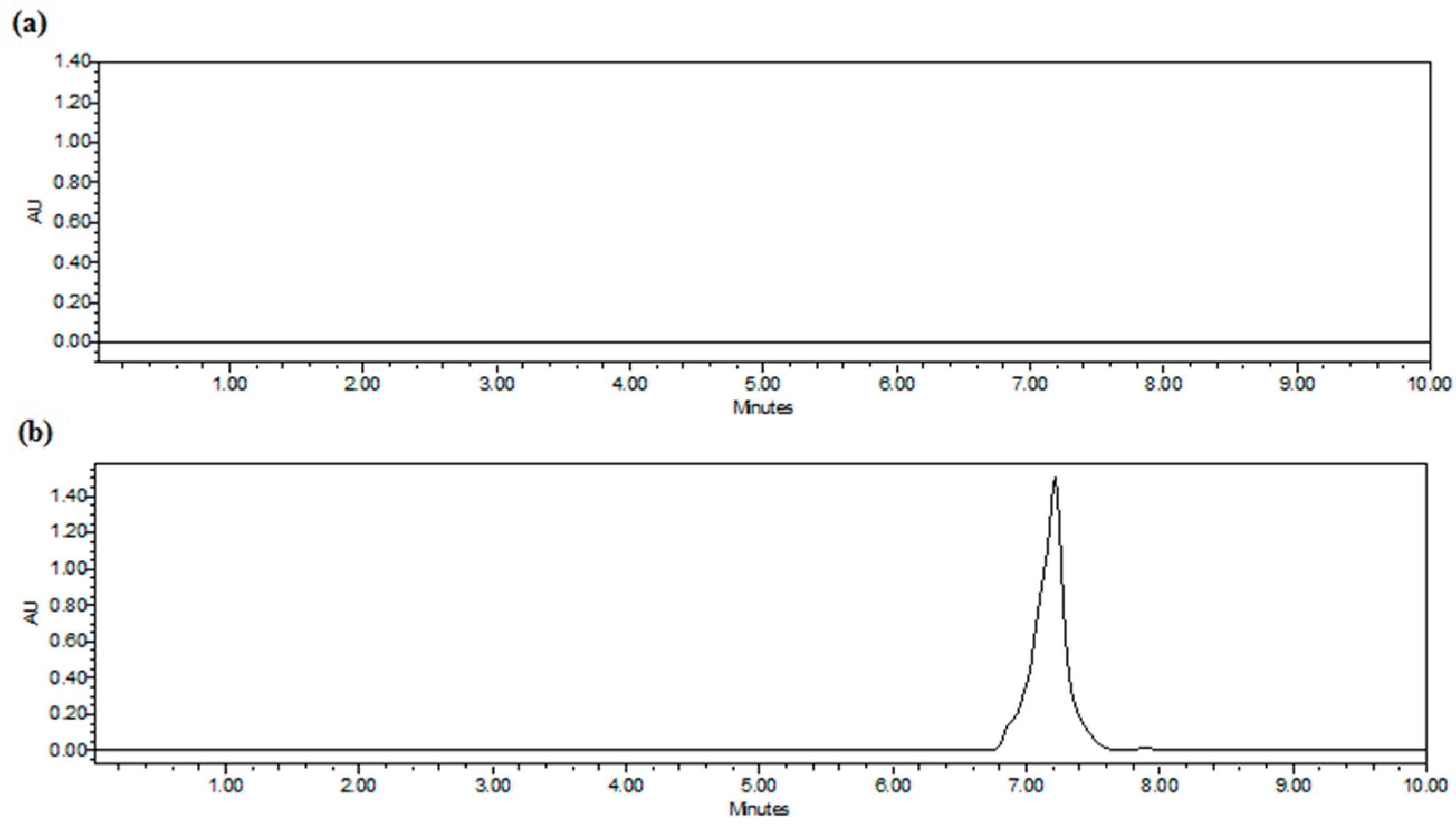
2.2. Chromatographic Parameters

An absorbance spectrum scan was recorded between 280 and 350 nm to determine the wavelength of maximum absorbance of tRES. Detection in the UV range was fixed at 306.6 nm, and the peak area was analyzed automatically. The experiment was conducted by reverse-phase HPLC using the 2996 Waters® photodiode array detector and the 2695 Waters® Alliance quaternary pump for 10 min. A Phenomenex (Torrance, CA, USA) RP-C18 Luna column of 250 mm × 4.6 mm I.D. 5 µm was kept at 24 ± 1 °C. Gradient elution was carried out, and the mobile phase was composed of 75:0:25 (V/V) water/methanol/acetonitrile at 0–3.5 min; 32.5:30.0:37.5 (V/V) water/methanol/acetonitrile at 3.6–5.8 min, and 75:0:25 (V/V) water/methanol/acetonitrile at 5.9–10 min. The selected mobile phase promoted a better separation of trans- and cis-resveratrol without enlarging the chromatogram peak. The flow rate was set at a constant of 1.0 mL/min.

2.3. Method Validation

The International Conference on Harmonization (ICH) [43] and National Agency for Sanitary Vigilance Agency (ANVISA) [44] guidelines were followed for the validation of the proposed method. Accuracy, precision, linearity, specificity, limit of detection (LOD), limit of quantification (LOQ), and robustness were the selected analytical parameters for validation.

2.3.1. Accuracy

Accuracy stands for the nearness of the results obtained experimentally in relation to theoretical values. The recovery percentage of known tRES amounts from three different concentrations of tRES solutions (5.0, 50.0, and 100.0 μg/mL) was calculated [44]. According to Brazilian regulations, the results should not exceed 5%.

2.3.2. Precision

The daily (intraday) and day-to-day (interday) precision was analyzed using three consecutive injections of tRES solution at three different concentrations (1.0, 50.0, and 100.0 μg/mL), followed by determination of the average, the standard deviation (SD), and the relative standard deviation (RSD) [44].

2.3.3. Linearity

Linearity represents a linear relationship between the concentration of the drug and its peak area. Three calibration curves were built with tRES solutions in acetonitrile at different concentrations (1.0–100.0 μg/mL). All solutions were freshly prepared and filtered through a 0.45-μm membrane prior to HPLC injection. The linearity was determined by linear regression analysis [45].

2.3.4. Specificity

Specificity translates the capacity of the method to discriminate between tRES and the other closely related structures present in the samples, such as lipids and surfactants [46]. Specificity was assessed by analyzing the filtrate of drug-free SLN (10.000 NMWL membranes, Millipore, Billerica, MA, USA) and further comparing the resulting chromatogram with that obtained for 50.0 μg/mL tRES standard sample.

2.3.5. Limit of Detection (LOD) and Limit of Quantification (LOQ)

LOD and LOQ are determined using the standard deviations of the slope and Y-intercept of the obtained calibration curve.
Both LOD and LOQ were calculated applying the following equations [44]:
D L = S D 3.3 / S   and   Q L = S D 10 / S ,  
where SD stands for the standard deviation of the Y-intercept and S stands for the slope of the calibration curve.

2.3.6. Robustness

Robustness is defined as the ability of the method to give the same results upon small changes of the chromatographic experimental conditions, indicating its reliability during normal usage. Robustness was checked, stressing the samples at 25, 30 and 35 °C and changes on flow rate at 0.8, 1.0, and 1.2 mL/min.

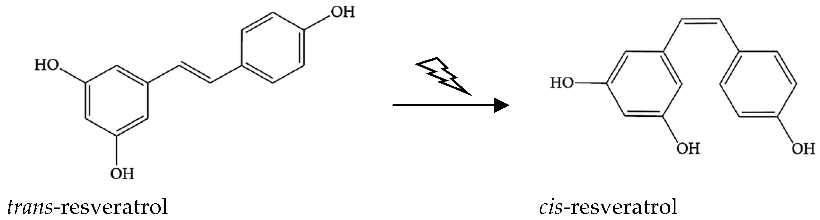
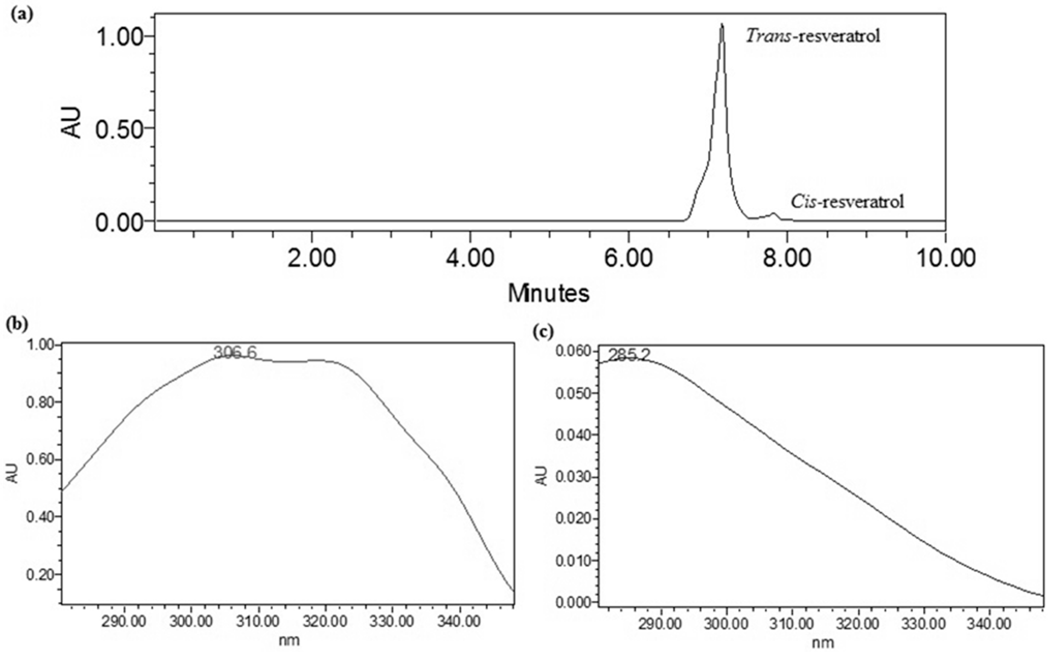
2.4. Measurement of tRES Stability under UV Exposure

A solution of 50.0 μg/mL trans-resveratrol was prepared and exposed for one hour to a 365 nm UVA light in a Benchtop 2UV Transilluminator (LMS-20, UVP Ltd., AnalytikJena, Jena, Germany), at 0 cm distance from the 16 W light source (on the top of the UV light). Resveratrol isomers were quantified by the validated HPLC method (n = 3).

2.5. Production and Characterization of SLN

Blank SLN (tRES-free) and tRES-loaded SLN formulations were prepared using the ultrasound (US) technique, modified after Pimentel-Moral et al. [47]. The developed formulation consisted of 5.0% of stearic acid as solid lipid (Galena, Campinas, SP, Brazil), with or without of 1.2% soy phosphatidylcholine (Epikuron® 200, Cargill, Hamburg, Germany); and the water phase consisted of 3.5% of poloxamer 407 (Pluronic® F127, Sigma–Aldrich, St. Louis, MO, USA); 0.18% of methyl paraben (Nipagin® M, PharmaSpecial, Itapevi, SP, Brazil), and 0.02% of propyl paraben (Nipasol® M, PharmaSpecial, Itapevi, SP, Brazil) (Table 1).
The water phase was firstly heated up to 70 °C, and then added to a previously prepared mixture of solid lipid (stearic acid) and soy phosphatidylcholine (Epikuron® 200) for F2 and F2.RES heated at the same temperature, or to the melted solid lipid in case of F1 and F1.RES, to produce a pre-emulsion. The obtained pre-emulsion was stirred with a 752A-2 magnetic stirrer (Fisatom, São Paulo, SP, Brazil) for 1 min, and heat was kept constant. A sonication probe (Branson Sonifier 250, Branson Ultrasonics Corporation, Frederick, MD, USA) was placed in the pre-emulsion for 20 min, applying a power output of amplitude of 47%. Ice was used to lower the temperature during sonication, because preliminary studies showed that in these conditions, the produced nanoparticles would have a lower mean hydrodynamic diameter and polydispersity index. For tRES-loaded SLN (F1.RES and F2.RES), the drug was added to melted stearic acid prior to sonication of the pre-emulsion. tRES (Resveratrol extract 100%, Galena, Campinas, SP, Brazil) was used in a concentration of 0.1% (wt/wt). All formulations were prepared following the same procedure [48]. After sonication, the formulations were centrifuged for 10 min at 5000 rpm to eventually eliminate the titanium fragments that could be released from the probe tip during sonication [49]. The mean hydrodynamic diameter and polydispersity index were determined by dynamic light scattering (Zetasizer Nano ZS, Malvern Instruments, Malvern UK). Samples were previously diluted with ultra-purified water to suitable concentration and analyzed in triplicate.

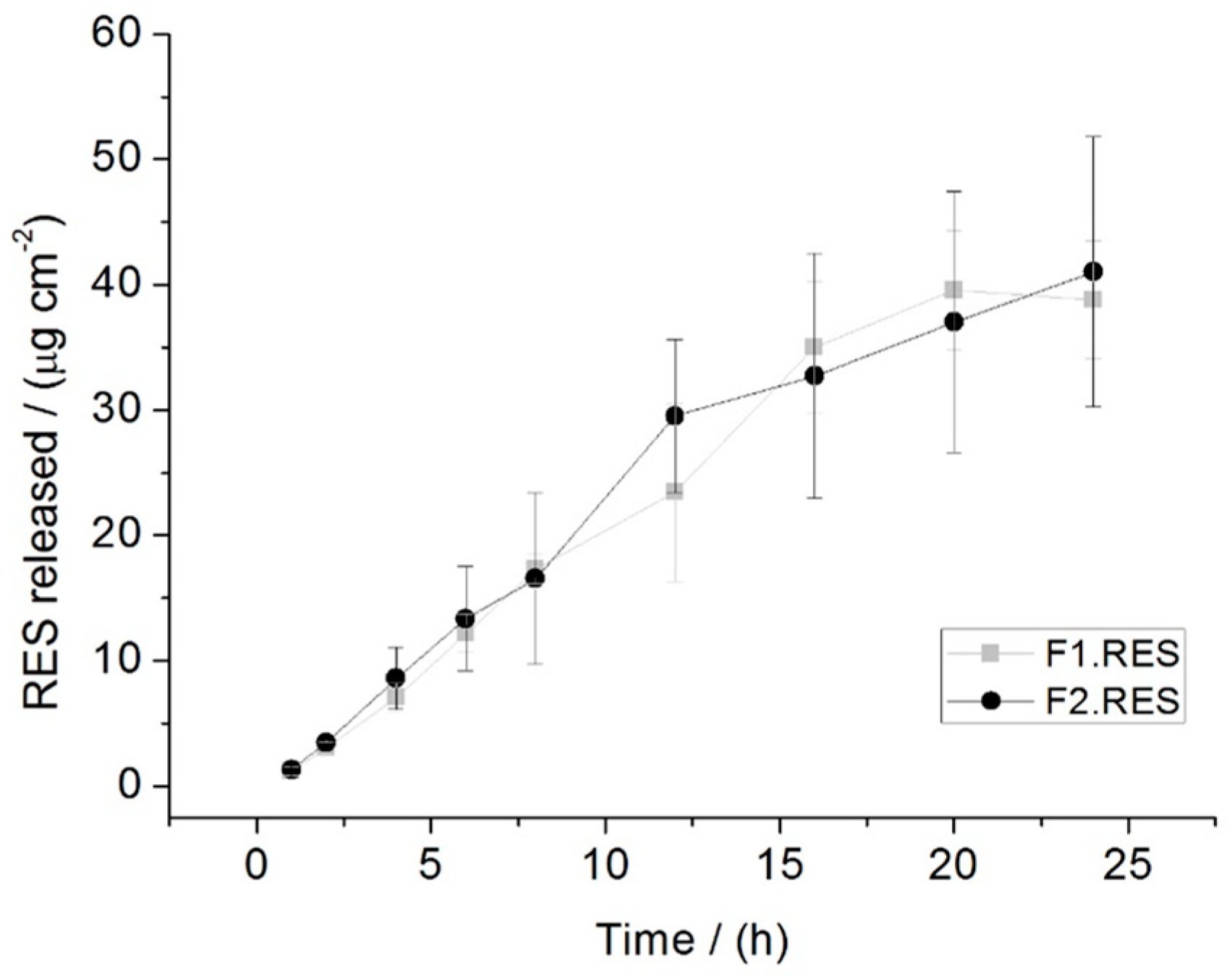
2.6. Drug Release Data Modeling

In vitro tRES release from SLN formulation was carried out in modified Franz diffusion cells (Microette Plus™, Hanson Research, Chatsworth, CA, USA) with a diffusional surface area of 1.77 cm2 and volume of acceptor compartment of 7.0 mL. A pre-hydrated membrane of cellulose acetate with 0.45 µm was assembled between the donor and acceptor compartments, and a volume of 300 μL of SLN with 0.1% tRES was placed onto the membrane surface in the donor compartment (6 cells/formulation). The acceptor compartment was filled with an aqueous solution of 2% (V/V) of polysorbate 80 to assure that the sink conditions were met during the release experiments [48]. The acceptor solution was maintained at a temperature of 32 ± 2 °C and constant stirring rate of 300 rpm. At suitable time intervals (5, 15, and 30 min and 1, 2, 4, 6, 8, 12, 16, 20, and 24 h), a volume of 1.5 mL of each cell was sampled from the acceptor compartment with a previously pre-heated syringe connected with a needle (32 ± 2 °C), followed by the replacement of the medium of same volume with automatic collector system (Auto Multi Fill™, Hanson Research, Chatsworth, CA, USA). Before the quantification of tRES released in the acceptor solution, the sampled volumes were passed through a 0.21 μm syringe filter (Millipore®, Darmstadt, Germany), followed by HPLC analysis applying the validated conditions described in this work.

2.7. Statistical Analysis

For parametric data, ANOVA (analysis of variance) and Tukey test were applied, using Origin 12.5 software. Statistical significance was set at p < 0.05.

3. Results and Discussion

After 1 h of exposure to UV light of standard solution of tRES, partial transformation of trans- to cis-resveratrol was observed i.e., 89.87% of trans isomer in solution was converted to cis form. It is expected that loading tRES into SLN would contribute to enhance the stability of the trans-isoform, limiting the risk of conversion into the cis-isoform. The mean recovery of trans-isoform was 96.84 ± 0.32%, whereas for cis-isoform it was 3.18 ± 0.47%. The decrease in the intensity of the chromatographic peak of trans-resveratrol was proportional to the area of new chromatographic peak, corresponding to cis-resveratrol. The cis-resveratrol may also show health benefits, but has lower biological activity [50]. To overcome these shortcomings, our main purpose was to validate a suitable analytical procedure for the quantification of resveratrol that could be used for both isomers. Since conversion occurs from trans- to cis-, the trans-isomeric form has been selected as the active ingredient to be loaded in SLN. Due to the instability of cis-resveratrol, this isomer is not commercially available.
The identification by high-performance liquid chromatography-diode array detection (HPLC-DAD) of analytes in pharmaceutical formulations was realized by Pragst et al. [51], and it is based on a combination of retention time and spectral matching in the UV region obtained at diode array. UV spectra generated for trans-resveratrol and cis-resveratrol could be easily differentiated due to the high resolution, accuracy, and sensitivity in the wavelength (200 to 400 nm) [52].
For a spectrum to be identified, a mathematical comparison with a spectra library is usually carried out, requiring the calculation of a similarity index within the entire spectra range and wavelengths measured allowing the recognition of very small differences (i.e., 181 points of comparison in the range of 200 to 380 nm) [51].
The distinct retention times of trans- and cis-resveratrol in the RP-HPLC chromatogram are shown in Figure 2a, and the respective UV absorption spectra recorded in the range of 280–350 nm are displayed in Figure 2b,c.
The UV absorbance spectrum of trans-resveratrol (Figure 2b) shows a lambda max (λ max) at 306.6 nm, while the UV absorbance spectrum of cis-resveratrol (Figure 2c) exhibits λ max at 285.2 nm. Similar results have been described in the scientific literature, in which 285 nm and 306 nm of UV absorbance spectra have been reported of cis- and trans-resveratrol, respectively [53,54]. After UV light exposure, a tRES solution of 50 μg/mL exhibited a proportional decrease of the peak area of trans-resveratrol and an increase of the peak area of cis-resveratrol.
The resolution of chromatographic peak (Rs) is a quantitative measure of the peak width, i.e., it measures the separation of two peaks of different retention times t in the chromatogram. The valley between the peaks should touch the baseline, and for complete resolution of the peak, the ideal value should be Rs ≥1.5 [55]. However, values between 1.2 ≥ Rs ≥ 1.5 are often enough. Our results exhibited an Rs value of 1.2; for validation of the analytical method for the quantification of tRES, the UV detection was fixed at 306.6 nm.
The analytical curves were obtained at 12 concentration levels, each recorded in triplicate. According to RDC 899/2003 [44], the acceptable linearity is when the linear correlation coefficients (R2) are greater than 0.99. The mean R2 obtained in the experimental measure was 0.9998, demonstrating that linearity proved to be acceptable for the quantification of tRES. The obtained linear regression equation was y = 400,628x − 144,146, where y stands for the peak area (AU), and x stands for the tRES concentration (μg/mL).
Method specificity was analyzed with respect to the risk of interference between the lipids and surfactants composing SLN and their effect in the tRES retention time [46]. The three distinct samples were filtered before analysis by the proposed HPLC method. When comparing the results with a chromatogram of tRES standard solution of 50 μg/mL (Figure 3a,b), no interferences between the components of SLN could be recorded. As shown in Figure 3a, for all the tested formulations, the filtrate of blank SLN (F1) did not exhibit any peak at the tRES retention time. These results demonstrate the specificity of the proposed HPLC-PDA to identify and to quantify the trans isomeric form (tRES) released from SLN.
Three different tRES standard solutions, each concentration prepared in triplicate (n = 3) (1.0, 50.0, and 100.0 μg/mL), were analyzed in three consecutive days. The results obtained for the assessment of precision are shown in Table 2.
The obtained results document the acceptable intraday and interday precisions of our method. All the data were within the acceptance criteria of RSD lower than 5.0%, which attributes a high degree of agreement between the results when analyzed with the same experimental conditions. The method proved to be accurate, not exceeding ± 10.0% [44], demonstrating the proximity of experimental results with the real value.
Robustness can be measured by assessing the effect of small fluctuations in the chromatographic experimental conditions on the retention time and obtained RSD value [46]. Robustness was evaluated by analyzing the 50.0 μg/mL tRES standard solution. The results are shown in Table 3. The standard solution was prepared in triplicate (n = 3).
No significant changes in the retention time were observed when flow was changed, and a similar result was shown when varying the temperature. The tested variations were among the values allowed by ANVISA [44], for which the RSD is ± 5.0%. The method was shown to be robust for the flow but not for the temperature.
The LOD and LOQ obtained in this work were 1.29 µg/mL and 4.29 µg/mL, respectively. The literature reports that the LOD and LOQ depend on various factors such as the method (e.g., spectrophotometric, fluorometric, mass spectrometry) and type/quantity of sample. Neves et al. developed a methodology using chromatographic separation connected to a fluorometric detector [56]. The objective was to detect the concentration of nanomolars between 1 and 229 nmol/L of tRES loaded in lipid nanoparticles to evaluate in vitro intestinal assays, using Caco-2 cell monolayers. The authors showed a successfully quantification and low-cost analysis, enabling the improvement pharmacokinetic studies. In another study, the LOD and LOQ of tRES in polymeric nanoparticles, as recorded by chromatography with photodiode array, were found to be 1.29 µg/mL and 4.29 µg/mL, respectively [57]. While the sensitivity of the method has not been one of the selected parameters for its validation, the recorded LOD and LOQ confirm the capacity of our RP-HPLC-PDA to identify and quantify resveratrol. In addition, our method is easy, of straightforward implementation, and of low cost. Therefore, it has been used for in vitro studies.
Stearic acid has been selected as the solid lipid from the preliminary screening of the solubility of tRES in a set of solid lipids (data not shown), while the surfactant combination has been based on reports from scientific literature [46]. SLN with a mean hydrodynamic diameter of 237.03 ± 1.80 nm (F1.tRES) and 201 ± 2.04 nm (F2.tRES), and a polydispersity index of 0.206 ± 0.012 nm (F1.tRES) and 0.153 ± 0.061 nm (F2.tRES) were obtained by ultrasound technique, and the validated method was applied for the quantification of tRES released from SLN in Franz diffusion cells. The drug release from SLN may occur either by diffusion from the nanoparticles surface or from the lipid matrix, or by erosion [58]. The in vitro release profiles of tRES from SLN are shown in Figure 4.
There was no statistical difference (p < 0.05) in tRES release between F1.tRES and F2.tRES, demonstrating that the addition of soy phosphatidylcholine in the formulation had no influence on the size and tRES release from the developed SLN. The first release of tRES was observed only 1 h after the beginning of the assay. For F1.tRES formulation, about 38.82 ± 2.30 (μg/cm2) of the drug was released after 24 h, while for F2.tRES, about 41.03 ± 4.28 (μg/cm2) of the drug was released within the same time frame.
The tRES release data were adjusted using the mathematical models (zero-order and first-order kinetic, Higuchi, Weibull, and Korsmeyer–Peppas models) determining the linear regression. Table 4 summarizes the regression coefficients (R2) for tRES released from SLN. The criterion for selecting the most appropriate mathematical model was established to be the highest value of R2.
According to the obtained R2 values (Table 4), the Weibull model was the one that best described the tRES release from SLN, which translates a complex release mechanism, involving simultaneously polymer chain relaxation and erosion during drug release.
Barzegar-Jalali et al. [59] compared 13 mathematical models of kinetic release data of 32 drugs from 106 dispersions published in the literature [60]. The obtained results show that the Weibull model was superior over the other tested models, as it translates the complex release behavior of tRES from nanoparticles. The lipid composition of the formulation is of utmost relevance in the design of the tRES release kinetics from SLN, because the lipid is responsible for the loading of drug in the matrix. We have demonstrated that stearic acid figuring in both formulations (F1.RES and F2.RES) promotes the slow rate of drug release from the developed SLN. However, during the release experiments, the drug is being dissolved, thereby increasing the matrix porosity, which promotes a faster drug release. Research has demonstrated that raising the amount of stearic acid associated with the hydrophilic polymer promotes matrix solubilization increases the matrix porosity and promotes a faster drug release [60]. Due to the lipophilic character of SLN, these particles act as absorption enhancers upon oral administration, contributing to improving the bioavailability of the loaded active ingredients [61,62,63,64]. SLN also show great potential for the oral delivery of nutraceutics [47,65,66,67], which is a research area that is receiving growing attention both from academia and industry [68,69,70,71]. Nutraceuticals bridge the gap from conventional pharmacological therapy, and the use of natural substances able to prevent/delay the onset of long-term pathological conditions. They are placed in a gray area between pharmaceuticals and food, and have been recently named as: “the phytocomplex, if they derive from a food of vegetal origin, and as the pool of the secondary metabolites if they derive from a food of animal origin, concentrated and administered in the more suitable pharmaceutical form” [14,68]. This study addresses the emerging new branch of “Nanonutraceutical Science” [67,72]. Nanonutraceutical formulations are being exploited for their efficacy, safety, and effect, and could be another tool in the arsenal of strategies that are helpful in managing health conditions, especially in patients who are not eligible for a conventional pharmacological therapy. Recent examples are the end points reached on the use of milk thistle (Silybum marianum) in liver diseases, and the use of the apple phytocomplex against hypercholesterolemy [9,73]. The stability of nutraceuticals in new nanoformulations is a recent research area with foreseen important outcomes. SLN can be further processed in foodstuffs to increase their ratio in active ingredients. Together with the chemical protection that SLN may offer, the modified release profile of resveratrol can also be exploited for other administration routes to reduce cytotoxicity, improve cell-stress response, offer photoprotection, and increase drug solubility and antitumor response.

4. Conclusions

The developed and validated HPLC-PDA detection method here described has been shown to be appropriate for the quantification of the tRES content in SLN, and for the determination of their release profile from the lipid matrices. Our analytical method has demonstrated to be reliable and feasible for the aim of this work, triggering interest in easy-to-use applications.

Author Contributions

Conceptualization and experimental design: R.B.R., N.F., P.S., M.C., and E.B.S.; methodology, data acquisition: R.B.R., N.F., P.S., A.D., M.L., M.C., A.S., and E.B.S.; resources, A.G.A., M.C., A.S., and E.B.S.; data curation: all authors; writing—original draft preparation, R.B.R., and A.D.; writing, review, and editing, R.B.R., A.D., P.S., S.E.M., M.C., A.S., and E.B.S.; supervision, P.S., M.C., A.S., and E.B.S.; project administration and funding acquisition, P.S., M.C., A.S., and E.B.S.; data analysis and scientific discussion, all authors.

Funding

The authors wish to acknowledge the financial support from CAPES (Coordenação de Aperfeiçoamento de Pessoal de Nível Superior) and FAPESP (Fundação de Amparo à Pesquisa do Estado de São Paulo). The financial support was also received from The Portuguese Science and Technology Foundation (FCT/MCT) and from European Funds (PRODER/COMPETE) under the project M-ERA-NET-0004/2015-PAIRED, co-financed by FEDER, under the Partnership Agreement PT2020. Authors also thank the support of the project: Nutraceutica come supporto nutrizionale nel paziente oncologico; CUP: B83D18000140007.

Acknowledgments

Thanks are due to Anselmo, G. Oliveira, and Maria Palmira Daflon Gremião by the partnership in the use of equipment.

Conflicts of Interest

The authors declare no conflict of interest.

Abbreviations

ANOVAAnalysis of variance
ANVISANational Agency for Sanitary Vigilance Agency
BCSBiopharmaceutics Classification System
DLDetection limit
DMSODimethyl sulfoxide
HPLCHigh-performance liquid chromatography
HPTLCHigh-performance thin-layer chromatography
HPLC-DADHigh-performance liquid chromatography-diode array detection
HHTHydroxyl heptadecatrienoate
ICHInternational Conference on Harmonization
LODLimit of detection
LOQLimit of quantification
NPsNanoparticles
PDAPhotodiode array
QLQuantification limit
R2Regression coefficient
RSDrelative standard deviation
RDCResolução da Diretoria Colegiada
RP-C18Reverse phase C18
SLNSolid lipid nanoparticles
SDStandard deviation
tREStrans-Resveratrol
USUltrasound
UVUltraviolet

References

  1. Yeung, A.W.K.; Aggarwal, B.B.; Orhan, I.E.; Barreca, D.; Battino, M.; Belwal, T.; Bishayee, A.; Daglia, M.; Devkota, H.P.; Echeverría, J.; et al. Resveratrol, a popular dietary supplement for human and animal health. Anim. Sci. Papers Rep. 2019, 37, 103–118. [Google Scholar]
  2. Gehm, B.D.; McAndrews, J.M.; Chien, P.-Y.; Jameson, J.L. Resveratrol, a polyphenolic compound found in grapes and wine, is an agonist for the estrogen receptor. Proc. Natl. Acad. Sci. USA 1997, 94, 14138–14143. [Google Scholar] [CrossRef]
  3. Amri, A.; Chaumeil, J.; Sfar, S.; Charrueau, C. Administration of resveratrol: What formulation solutions to bioavailability limitations? J. Control. Release 2012, 158, 182–193. [Google Scholar] [CrossRef]
  4. Kristl, J.; Teskač, K.; Caddeo, C.; Abramović, Z.; Šentjurc, M. Improvements of cellular stress response on resveratrol in liposomes. Eur. J. Pharm. Biopharm. 2009, 73, 253–259. [Google Scholar] [CrossRef] [PubMed]
  5. Amidon, G.L.; Lennernäs, H.; Shah, V.P.; Crison, J.R. A theoretical basis for a biopharmaceutic drug classification: The correlation of in vitro drug product dissolution and in vivo bioavailability. Pharm. Res. 1995, 12, 413–420. [Google Scholar] [CrossRef] [PubMed]
  6. Orallo, F. Comparative studies of the antioxidant effects of cis-and trans-resveratrol. Curr. Med. Chem. 2006, 13, 87–98. [Google Scholar] [CrossRef] [PubMed]
  7. Yáñez, M.; Fraiz, N.; Cano, E.; Orallo, F. Inhibitory effects of cis-and trans-resveratrol on noradrenaline and 5-hydroxytryptamine uptake and on monoamine oxidase activity. Biochem. Bioph. Res. Co. 2006, 344, 688–695. [Google Scholar] [CrossRef]
  8. Figueiras, T.S.; Neves-Petersen, M.T.; Petersen, S.B. Activation energy of light induced isomerization of resveratrol. J. Fluoresc. 2011, 21, 1897–1906. [Google Scholar] [CrossRef]
  9. Santini, A.; Novellino, E. Nutraceuticals in hypercholesterolaemia: An overview. Br. J. Pharmacol. 2017, 174, 1450–1463. [Google Scholar] [CrossRef]
  10. Santini, A.; Novellino, E. To Nutraceuticals and Back: Rethinking a Concept. Foods 2017, 6, 74. [Google Scholar] [CrossRef]
  11. Durazzo, A.; D’Addezio, L.; Camilli, E.; Piccinelli, R.; Turrini, A.; Marletta, L.; Marconi, S.; Lucarini, M.; Lisciani, S.; Gabrielli, P.; et al. From Plant Compounds to Botanicals and Back: A Current Snapshot. Molecules 2018, 23, 1844. [Google Scholar] [CrossRef] [PubMed]
  12. Durazzo, A.; Lucarini, M. Extractable and Non-Extractable Antioxidants. Molecules 2019, 24, 1933. [Google Scholar] [CrossRef] [PubMed]
  13. Santini, A.; Novellino, E. Nutraceuticals - shedding light on the grey area between pharmaceuticals and food. Expert Rev. Clin. Pharmacol. 2018, 11, 545–547. [Google Scholar] [CrossRef] [PubMed]
  14. Santini, A.; Cammarata, S.M.; Capone, G.; Ianaro, A.; Tenore, G.C.; Pani, L.; Novellino, E. Nutraceuticals: Opening the debate for a regulatory framework. Br. J. Clin. Pharmacol. 2018, 84, 659–672. [Google Scholar] [CrossRef] [PubMed]
  15. Daliu, P.; Santini, A.; Novellino, E. From pharmaceuticals to nutraceuticals: Bridging disease prevention and management. Expert Rev. Clin. Pharmacol. 2019, 12, 1–7. [Google Scholar] [CrossRef]
  16. Durazzo, A.; Lucarini, M.; Souto, E.B.; Cicala, C.; Caiazzo, E.; Izzo, A.A.; Novellino, E.; Santini, A. Polyphenols: A concise overview on the chemistry, occurrence, and human health. Phytother. Res. 2019, 33, 2221–2243. [Google Scholar] [CrossRef] [PubMed]
  17. Lucarini, M.; Durazzo, A.; Romani, A.; Campo, M.; Lombardi-Boccia, G.; Cecchini, F. Bio–Based Compounds from Grape Seeds: A Biorefinery Approach. Molecules 2018, 23, 1888. [Google Scholar] [CrossRef]
  18. Dolinsky, V.W.; Dyck, J.R. Calorie restriction and resveratrol in cardiovascular health and disease. BBA-Mol. Basis. Dis. 2011, 1812, 1477–1489. [Google Scholar] [CrossRef]
  19. Petrovski, G.; Gurusamy, N.; Das, D.K. Resveratrol in cardiovascular health and disease. Ann. N. Y. Acad. Sci. 2011, 1215, 22–33. [Google Scholar] [CrossRef]
  20. Liu, X.-Q.; Wu, B.-J.; Pan, W.H.; Zhang, X.-M.; Liu, J.-H.; Chen, M.-M.; Chao, F.-P.; Chao, H.-M. Resveratrol mitigates rat retinal ischemic injury: The roles of matrix metalloproteinase-9, inducible nitric oxide, and heme oxygenase-1. J. Ocul. Pharmacol. Ther. 2013, 29, 33–40. [Google Scholar] [CrossRef]
  21. Brasnyó, P.; Molnár, G.A.; Mohás, M.; Markó, L.; Laczy, B.; Cseh, J.; Mikolás, E.; Szijártó, I.A.; Mérei, A.; Halmai, R. Resveratrol improves insulin sensitivity, reduces oxidative stress and activates the Akt pathway in type 2 diabetic patients. Br. J. Nutr. 2011, 106, 383–389. [Google Scholar] [CrossRef] [PubMed]
  22. Vieira, R.; Souto, S.B.; Sanchez-Lopez, E.; Machado, A.L.; Severino, P.; Jose, S.; Santini, A.; Fortuna, A.; Garcia, M.L.; Silva, A.M.; et al. Sugar-Lowering Drugs for Type 2 Diabetes Mellitus and Metabolic Syndrome-Review of Classical and New Compounds: Part-I. Pharmaceuticals (Basel) 2019, 12, 152. [Google Scholar] [CrossRef] [PubMed]
  23. Vieira, R.; Souto, S.B.; Sanchez-Lopez, E.; Machado, A.L.; Severino, P.; Jose, S.; Santini, A.; Silva, A.M.; Fortuna, A.; Garcia, M.L.; et al. Sugar-Lowering Drugs for Type 2 Diabetes Mellitus and Metabolic Syndrome-Strategies for In Vivo Administration: Part-II. J. Clin. Med. 2019, 8, 1332. [Google Scholar] [CrossRef] [PubMed]
  24. Sun, A.Y.; Wang, Q.; Simonyi, A.; Sun, G.Y. Resveratrol as a therapeutic agent for neurodegenerative diseases. Mol. Neurobiol. 2010, 41, 375–383. [Google Scholar] [CrossRef]
  25. Wight, R.D.; Tull, C.A.; Deel, M.W.; Stroope, B.L.; Eubanks, A.G.; Chavis, J.A.; Drew, P.D.; Hensley, L.L. Resveratrol effects on astrocyte function: Relevance to neurodegenerative diseases. Biochem. Biophys. Res. Commun. 2012, 426, 112–115. [Google Scholar] [CrossRef]
  26. Xuzhu, G.; Komai-Koma, M.; Leung, B.P.; Howe, H.S.; McSharry, C.; McInnes, I.B.; Xu, D. Resveratrol modulates murine collagen-induced arthritis by inhibiting Th17 and B-cell function. Ann. Rheum. Dis. 2012, 71, 129–135. [Google Scholar] [CrossRef]
  27. Pace-Asciak, C.R.; Hahn, S.; Diamandis, E.P.; Soleas, G.; Goldberg, D.M. The red wine phenolics trans-resveratrol and quercetin block human platelet aggregation and eicosanoid synthesis: Implications for protection against coronary heart disease. Clin. Chim. Acta 1995, 235, 207–219. [Google Scholar] [CrossRef]
  28. Chen, X.; Qiao, H.; Liu, T.; Yang, Z.; Xu, L.; Xu, Y.; Ge, H.M.; Tan, R.-X.; Li, E. Inhibition of herpes simplex virus infection by oligomeric stilbenoids through ROS generation. Antivir. Res. 2012, 95, 30–36. [Google Scholar] [CrossRef]
  29. Shukla, Y.; Singh, R. Resveratrol and cellular mechanisms of cancer prevention. Ann. N. Y. Acad. Sci. 2011, 1215, 1–8. [Google Scholar] [CrossRef]
  30. Bazzo, K.O.; Souto, A.A.; Lopes, T.G.; Zanin, R.F.; Gomez, M.V.; Souza, A.H.; Campos, M.M. Evidence for the analgesic activity of resveratrol in acute models of nociception in mice. J. Nat. Prod. 2013, 76, 13–21. [Google Scholar] [CrossRef]
  31. Shimojo, A.A.M.; Fernandes, A.R.V.; Ferreira, N.R.E.; Sanchez-Lopez, E.; Santana, M.H.A.; Souto, E.B. Evaluation of the Influence of Process Parameters on the Properties of Resveratrol-Loaded NLC Using 2(2) Full Factorial Design. Antioxidants (Basel) 2019, 8, 272. [Google Scholar] [CrossRef] [PubMed]
  32. Jose, S.; Anju, S.S.; Cinu, T.A.; Aleykutty, N.A.; Thomas, S.; Souto, E.B. In vivo pharmacokinetics and biodistribution of resveratrol-loaded solid lipid nanoparticles for brain delivery. Int. J. Pharm. 2014, 474, 6–13. [Google Scholar] [CrossRef] [PubMed]
  33. Guo, L.; Peng, Y.; Yao, J.; Sui, L.; Gu, A.; Wang, J. Anticancer Activity and Molecular Mechanism of Resveratrol–Bovine Serum Albumin Nanoparticles on Subcutaneously Implanted Human Primary Ovarian Carcinoma Cells in Nude Mice. Cancer Biother. Radiopharma. 2010, 25, 471–477. [Google Scholar] [CrossRef] [PubMed] [Green Version]
  34. Jung, K.-H.; Lee, J.H.; Park, J.W.; Quach, C.H.T.; Moon, S.-H.; Cho, Y.S.; Lee, K.-H. Resveratrol-loaded polymeric nanoparticles suppress glucose metabolism and tumor growth in vitro and in vivo. Int. J. Pharm. 2015, 478, 251–257. [Google Scholar] [CrossRef] [PubMed]
  35. Doktorovova, S.; Kovacevic, A.B.; Garcia, M.L.; Souto, E.B. Preclinical safety of solid lipid nanoparticles and nanostructured lipid carriers: Current evidence from in vitro and in vivo evaluation. Eur. J. Pharm. Biopharm. 2016, 108, 235–252. [Google Scholar] [CrossRef]
  36. Doktorovova, S.; Souto, E.B.; Silva, A.M. Nanotoxicology applied to solid lipid nanoparticles and nanostructured lipid carriers - a systematic review of in vitro data. Eur. J. Pharm. Biopharm. 2014, 87, 1–18. [Google Scholar] [CrossRef]
  37. Souto, E.B.; Doktorovova, S. Chapter 6 - Solid lipid nanoparticle formulations pharmacokinetic and biopharmaceutical aspects in drug delivery. Methods Enzymol 2009, 464, 105–129. [Google Scholar] [CrossRef]
  38. Souto, E.B.; Muller, R.H. Lipid nanoparticles: Effect on bioavailability and pharmacokinetic changes. Handb. Exp. Pharmacol. 2010, 115–141. [Google Scholar] [CrossRef]
  39. Teskač, K.; Kristl, J. The evidence for solid lipid nanoparticles mediated cell uptake of resveratrol. Int. J. Pharm. 2010, 390, 61–69. [Google Scholar] [CrossRef]
  40. Dianzani, C.; Zara, G.P.; Maina, G.; Pettazzoni, P.; Pizzimenti, S.; Rossi, F.; Gigliotti, C.L.; Ciamporcero, E.S.; Daga, M.; Barrera, G. Drug delivery nanoparticles in skin cancers. Biomed. Res. Int. 2014, 2014. [Google Scholar] [CrossRef]
  41. Singh, R. HPLC method development and validation-an overview. J. Pharm. Educ. Res. 2013, 4, 26–33. [Google Scholar]
  42. Paulo, L.; Domingues, F.; Queiroz, J.A.; Gallardo, E. Development and Validation of an Analytical Method for the Determination of trans- and cis-Resveratrol in Wine: Analysis of Its Contents in 186 Portuguese Red Wines. J. Agric. Food Chem. 2011, 59, 2157–2168. [Google Scholar] [CrossRef] [PubMed]
  43. ICH. Harmonised Tripartite Guideline, Validation of Analytical Procedures: Text and Methodology Q2 (R1), International Conference on Harmonisation of Technical Requirements for Registration of Pharmaceuticals for Human Use. 2015. Available online: http://www.ich.org/products/guidelines/quality/article/quality-guidelines.html (accessed on 20 October 2019).
  44. ANVISA-Brasil. Guia Para Validação de Métodos Analíticos e Bioanalíticos; ANVISA, Agência Nacional de Vigilância Sanitária, Ed.; Diário Oficial da União: Brasília, Brasil, 2003; p. 47.
  45. Nemen, D.; Lemos-Senna, E. Preparação e caracterização de suspensões coloidais de nanocarreadores lipídicos contendo resveratrol destinados à administração cutânea. Quim. Nova 2011, 34, 408–413. [Google Scholar] [CrossRef]
  46. Silva, A.C.; Lopes, C.M.; Fonseca, J.; Soares, M.E.; Santos, D.; Souto, E.B.; Ferreira, D. Risperidone release from solid lipid nanoparticles (SLN): Validated HPLC method and modelling kinetic profile. Curr. Pharm. Anal. 2012, 8, 307–316. [Google Scholar] [CrossRef]
  47. Pimentel-Moral, S.; Teixeira, M.C.; Fernandes, A.R.; Borrás-Linares, I.; Arráez-Román, D.; Martínez-Férez, A.; Segura-Carretero, A.; Souto, E.B. Polyphenols-enriched Hibiscus sabdariffa extract-loaded nanostructured lipid carriers (NLC): Optimization by multi-response surface methodology. J. Drug Deliv. Sci. Technol. 2019, 49, 660–667. [Google Scholar] [CrossRef]
  48. Detoni, C.B.; Souto, G.D.; da Silva, A.L.M.; Pohlmann, A.R.; Guterres, S.S. Photostability and Skin Penetration of Different E-Resveratrol-Loaded Supramolecular Structures. Photochem. Photobiol. 2012, 88, 913–921. [Google Scholar] [CrossRef]
  49. Agayan, R.R.; Horvath, T.; McNaughton, B.H.; Anker, J.N.; Kopelman, R. Optical manipulation of metal-silica hybrid nanoparticles. In Proceedings of the Optical Science and Technology, International Society for Optics and Photonics, the SPIE 49th Annual Meeting, Denver, CO, USA, 18 October 2004; pp. 502–513. [Google Scholar]
  50. Bertelli, A.; Giovannini, L.; Bernini, W.; Migliori, M.; Fregoni, M.; Bavaresco, L.; Bertelli, A. Antiplatelet activity of cis-resveratrol. Drug Exp. Clin. Res. 1995, 22, 61–63. [Google Scholar]
  51. Pragst, F.; Herzler, M.; Erxleben, B.T. Systematic toxicological analysis by high-performance liquid chromatography with diode array detection (HPLC-DAD). Clin. Chem. Lab. Med. 2004, 42, 1325–1340. [Google Scholar] [CrossRef]
  52. Ortiz, R.S.; Antunes, M.V.; Linden, R. Determinação de citrato de sildenafila e de tadalafila por cromatografia líquida de ultraeficiência com detecção por arranjo de diodos (CLUE-DAD). Quim. Nova 2010, 33, 389–393. [Google Scholar] [CrossRef] [Green Version]
  53. Bonda, C.; Zhang, J.; Pavlovic, A. The photostability and photostabilization of trans-resveratrol. Cosmet. Toiletries 2011, 126, 652. [Google Scholar]
  54. Rossi, D.; Guerrini, A.; Bruni, R.; Brognara, E.; Borgatti, M.; Gambari, R.; Maietti, S.; Sacchetti, G. trans-Resveratrol in nutraceuticals: Issues in retail quality and effectiveness. Molecules 2012, 17, 12393–12405. [Google Scholar] [CrossRef] [PubMed] [Green Version]
  55. Snyder, L.R.; Kirkland, J.J.; Glajch, J.L. Practical HPLC Method Development, 2nd ed.; John Wiley & Sons: Hoboken, NJ, USA, 2012. [Google Scholar]
  56. Neves, A.R.; Reis, S.; Segundo, M.A. Development and Validation of a HPLC Method Using a Monolithic Column for Quantification of trans-Resveratrol in Lipid Nanoparticles for Intestinal Permeability Studies. J. Agric. Food Chem. 2015, 63, 3114–3120. [Google Scholar] [CrossRef] [PubMed]
  57. da Rocha Lindner, G.; Khalil, N.M.; Mainardes, R.M. Resveratrol-loaded polymeric nanoparticles: Validation of an HPLC-PDA method to determine the drug entrapment and evaluation of its antioxidant activity. Sci. World J. 2013, 2013, 506083. [Google Scholar] [CrossRef] [PubMed] [Green Version]
  58. Agnihotri, S.A.; Mallikarjuna, N.N.; Aminabhavi, T.M. Recent advances on chitosan-based micro-and nanoparticles in drug delivery. J. Control. Release 2004, 100, 5–28. [Google Scholar] [CrossRef] [PubMed]
  59. Barzegar-Jalali, M.; Adibkia, K.; Valizadeh, H.; Shadbad, M.R.S.; Nokhodchi, A.; Omidi, Y.; Mohammadi, G.; Nezhadi, S.H.; Hasan, M. Kinetic analysis of drug release from nanoparticles. J. Pharm. Pharm. Sci 2008, 11, 167–177. [Google Scholar] [CrossRef] [PubMed] [Green Version]
  60. Grassi, M.; Voinovich, D.; Franceschinis, E.; Perissutti, B.; Filipovic-Grcic, J. Theoretical and experimental study on theophylline release from stearic acid cylindrical delivery systems. J. Control. Release 2003, 92, 275–289. [Google Scholar] [CrossRef]
  61. Teixeira, M.C.; Carbone, C.; Souto, E.B. Beyond liposomes: Recent advances on lipid based nanostructures for poorly soluble/poorly permeable drug delivery. Prog. Lipid Res. 2017, 68, 1–11. [Google Scholar] [CrossRef]
  62. Severino, P.; Andreani, T.; Macedo, A.S.; Fangueiro, J.F.; Santana, M.H.; Silva, A.M.; Souto, E.B. Current State-of-Art and New Trends on Lipid Nanoparticles (SLN and NLC) for Oral Drug Delivery. J. Drug Deliv. 2012, 2012, 750891. [Google Scholar] [CrossRef]
  63. Muller, R.H.; Runge, S.; Ravelli, V.; Mehnert, W.; Thunemann, A.F.; Souto, E.B. Oral bioavailability of cyclosporine: Solid lipid nanoparticles (SLN) versus drug nanocrystals. Int. J. Pharm. 2006, 317, 82–89. [Google Scholar] [CrossRef]
  64. Souto, E.B.; Severino, P.; Basso, R.; Santana, M.H. Encapsulation of antioxidants in gastrointestinal-resistant nanoparticulate carriers. Methods Mol. Biol. 2013, 1028, 37–46. [Google Scholar] [CrossRef]
  65. Lopes, C.M.; Martins-Lopes, P.; Souto, E.B. Nanoparticulate carriers (NPC) for oral pharmaceutics and nutraceutics. Pharmazie 2010, 65, 75–82. [Google Scholar] [PubMed]
  66. Santos, I.S.; Ponte, B.M.; Boonme, P.; Silva, A.M.; Souto, E.B. Nanoencapsulation of polyphenols for protective effect against colon–rectal cancer. Biotechnol. Adv. 2013, 31, 514–523. [Google Scholar] [CrossRef] [PubMed]
  67. Pimentel-Moral, S.; Teixeira, M.C.; Fernandes, A.R.; Arráez-Román, D.; Martínez-Férez, A.; Segura-Carretero, A.; Souto, E.B. Lipid nanocarriers for the loading of polyphenols—A comprehensive review. Adv. Colloid Interface Sci. 2018, 260, 85–94. [Google Scholar] [CrossRef] [PubMed]
  68. Daliu, P.; Santini, A.; Novellino, E. A decade of nutraceutical patents: Where are we now in 2018, Expert Opin. Ther. Pat. 2018, 28, 875–882. [Google Scholar] [CrossRef]
  69. Zielinska, A.; Ferreira, N.R.; Durazzo, A.; Lucarini, M.; Cicero, N.; Mamouni, S.E.; Silva, A.M.; Nowak, I.; Santini, A.; Souto, E.B. Development and Optimization of Alpha-Pinene-Loaded Solid Lipid Nanoparticles (SLN) Using Experimental Factorial Design and Dispersion Analysis. Molecules 2019, 24, 2683. [Google Scholar] [CrossRef] [PubMed] [Green Version]
  70. Zielinska, A.; Martins-Gomes, C.; Ferreira, N.R.; Silva, A.M.; Nowak, I.; Souto, E.B. Anti-inflammatory and anti-cancer activity of citral: Optimization of citral-loaded solid lipid nanoparticles (SLN) using experimental factorial design and LUMiSizer(R). Int. J. Pharm. 2018, 553, 428–440. [Google Scholar] [CrossRef]
  71. Pereira, I.; Zielinska, A.; Ferreira, N.R.; Silva, A.M.; Souto, E.B. Optimization of linalool-loaded solid lipid nanoparticles using experimental factorial design and long-term stability studies with a new centrifugal sedimentation method. Int. J. Pharm. 2018, 549, 261–270. [Google Scholar] [CrossRef]
  72. Watkins, R.; Wu, L.; Zhang, C.; Davis, R.M.; Xu, B. Natural product-based nanomedicine: Recent advances and issues. Int. J. Nanomed. 2015, 10, 6055–6074. [Google Scholar] [CrossRef] [Green Version]
  73. Abenavoli, L.; Izzo, A.A.; Milić, N.; Cicala, C.; Santini, A.; Capasso, R. Milk thistle (Silybum marianum): A concise overview on its chemistry, pharmacological, and nutraceutical uses in liver diseases. Phytother. Res. 2018, 32, 2202–2213. [Google Scholar] [CrossRef]
Figure 1. Conversion of trans- to cis-resveratrol after exposure to ultraviolet radiation.
Applsci 09 04961 g001
Figure 2. Retention times of trans- (7.194 min) and cis-resveratrol (7.847 min) in reverse-phase (RP)-HPLC chromatogram of standard solution of (50 μg/mL) after 1 h of exposition to UV light (a); UV absorption spectrum of trans-resveratrol (b) and cis-resveratrol (c) recorded in the range of 280–350 nm from the diode array.
Applsci 09 04961 g002
Figure 3. Retention time recorded in the chromatogram of the (a) filtrated drug-free SLN formulation (F1) (b) and in the chromatogram of trans-resveratrol (tRES) standard solution (50.0 μg/mL).
Applsci 09 04961 g003
Figure 4. Trans-resveratrol (tRES) release profile from SLN after 24 h of analysis (n = 6). F1.RES comprised 5.0% SA, 3.5% P407, 0.18% MP, 0.02% PP, and 0.1% tRES, and F2.RES comprised 5.0% SA, 1.2% SPC, 3.5% P407, 0.18% MP, 0.02% PP, and 0.1% tRES.
Applsci 09 04961 g004
Table 1. Composition of solid lipid nanoparticles (SLN) formulations.
Ingredients/%
SASPCP407MPPPRESMQ Water g
F15.0-3.50.180.02-Sq *
F25.01.23.50.180.02-Sq *
F1.RES5.0-3.50.180.020.1Sq *
F2.RES5.01.23.50.180.020.1Sq *
SA: Stearic acid; SPC: Soy phosphatidylcholine; P407: Polaxamer 407; MP: Methylparaben; PP: Propylparaben; RES: Trans-resveratrol; MQ g: Mili-Q; sq *: sufficient quantity.
Table 2. Results achieved for the intraday and interday precision of the method and results found for method accuracy.
PrecisionAccuracy
DaytRES Concentration/(μg/mL)IntradayInterdayExperimental Concentration/(μg/mL) ***Recovery/(%)
SD */(AU)RSD **/(%)SD */(AU)RSD **/(%)
11.010,9162.87191504.834.626 ± 0.00992.53 ± 0.19
298672.35
36320.16
150.096230.056623463.1750.57 ± 0.228101.15 ± 0.46
291,4040.45
3205,2530.95
1100.066,3720.1614399473.4799.64 ± 0.17099.64 ± 0.170
268,2120.17
384,3380.19
* Standard deviation (n = 3); **Relative standard deviation; ***Determined by equation of the line (y = 400651x − 145812).
Table 3. Results of the robustness for quantification of tRES.
ParametersRetention Time/(min.)RSD/(%)
Flow0.8 mL/min8.22 ± 0.007 *0.52
1.0 mL/min7.24 ± 0.009 *0.64
1.2 mL/min6.53 ± 0.015 *0.52
Column temperature25 °C7.24 ± 0.009 **0.64
30 °C7.15 ± 0.100 **0.26
35 °C7.06 ± 0.006 **0.70
* Not statistically significant (p < 0.05); ** Statistically significant (p > 0.05).
Table 4. Regression coefficient obtained after adjusted the drug release data of tRES-loaded SLN with five mathematical models.
Mathematical ModelsRegression Coefficient (R2)
F1.RESF2.RES
Korsmeyer-Peppas0.96800.9752
Higuchi0.84690.8734
First Order0.97570.9814
Weibull0.99160.9939
Zero Order0.97000.9683

