Review

Applied Nanotechnologies in Anticoagulant Therapy: From Anticoagulants to Coagulation Test Performance of Drug Delivery Systems

by
Yuri B. G. Patriota
1,
Luíse L. Chaves
1,
Evren H. Gocke
2,
Patricia Severino
3,4,5,
Mônica F. R. Soares
1,
José L. Soares-Sobrinho
1,* and
Eliana B. Souto
6,7,*
1
Quality Control Core of Medicines and Correlates, Department of Pharmaceutical Sciences, Federal University of Pernambuco, Recife 50740-521, PE, Brazil
2
Department of Pharmaceutical Technology, Faculty of Pharmacy, Ege University, Bornova-Izmir 35100, Turkey
3
Laboratory of Nanotechnology and Nanomedicine (LNMed), Institute of Technology and Research (ITP), Av. Murilo Dantas, 300, Aracaju 49010-390, SE, Brazil
4
Institute of Technology and Research (ITP), University of Tiradentes (Unit), Av. Murilo Dantas, 300, Aracaju 49010-390, SE, Brazil
5
Tiradentes Institute, 150 Mt Vernon St, Dorchester, MA 02125, USA
6
Faculty of Pharmacy, University of Coimbra, Pólo das Ciências da Saúde, Azinhaga de Santa Comba, 3000-548 Coimbra, Portugal
7
CEB-Centre of Biological Engineering, Campus de Gualtar, University of Minho, 4710-057 Braga, Portugal
*
Authors to whom correspondence should be addressed.
Submission received: 9 March 2021 / Revised: 20 April 2021 / Accepted: 22 April 2021 / Published: 1 May 2021
(This article belongs to the Collection Feature Papers for Applied Nano)

Abstract

:
Heparin-based delivery systems have been explored to improve their therapeutic efficacy and to reduce toxicity for different administration routes. Regardless of the applied drug delivery system (DDS), the evaluation of anticoagulant performance is instrumental for the development of a suitable DDS. The understanding of the range of anticoagulant assays, together with their key applications and limitations, is essential both within the context of scientific research and for clinical usage. This review provides an overview of the current anticoagulant therapy and discusses the advantages and limitations of currently available anticoagulant assays. We also discuss studies involving low-molecular-weight heparin (LMWH)-based nanocarriers with emphasis on their anticoagulation performance. Conventional anticoagulants have been used for decades for the treatment of many diseases. Direct oral anticoagulants have overcome some limitations of heparins and vitamin K antagonists. However, the lack of an accurate laboratory assessment, as well as the lack of a factor “xaban” (Xa) inhibitor reversal agent, remains a major problem associated with these anticoagulants. LMWHs represent anticoagulant agents with noteworthy efficacy and safety, and they have been explored to improve their outcomes with various nanocarriers through several administration routes. The main problems related to LMWHs have been surmounted, and improved efficiency may be achieved through the use of DDSs.

1. Introduction

Investments in terms of money and time in the development of new chemical compounds have led pharmaceutical companies to explore several delivery approaches in order to improve the efficacy and minimize the side effects of drugs that are already on the market [1]. One example is heparin. This natural compound has been clinically used for the treatment and prophylaxis of coagulation disorders (e.g., venous thromboembolism (VTE) [2]) for more than 60 years [3]. Though heparins, including unfractionated heparin (UFH) and low-molecular-weight heparins (LMWHs), have also been clinically used for a long time, they show limitations because they are available only for parenteral administration, a costly and invasive method that leads to low patient compliance [4]. Non-invasive drug delivery systems are being explored to overcome these limitations. Nanotechnology-based drug carriers (nanocarriers), such as liposomes, nanogels, nanospheres, and nanocapsules, have gained increasing attention in the last few decades as a promising strategy for improving drug delivery [5] (Figure 1). Compared to conventional systems, nanocarriers have a number of advantages: (i) They are able to pass through the capillary vessels due to their submicron size and avoid clearance by phagocytes, prolonging the bloodstream circulation time; (ii) they can permeate cells and tissues to reach target organs; (iii) they can deliver the drugs in a controlled manner, reducing the frequency of administration and side effects, and they improve patient compliance [6]. Several heparin-based drug delivery systems have been developed to enhance the therapeutic efficacy of these drugs [7,8]. Although the oral route is the most desirable, it is noteworthy that there have been efforts to investigated heparin delivery through other pathways, such as nasal [9], pulmonary [10], and transdermal [11] routes.
The evaluation of nanocarrier efficacy comprises a critical step in the development of DDSs. In particular, heparin-based nanocarriers’ performance is evaluated through several anticoagulant assays based on the type of drug used. The suitability and relevance of a specific anticoagulant assay for a certain objective are also important questions [12], thus stressing the need to know their advantages and limitations [13]. Anticoagulant assays, however, have not been comprehensively described from the perspective of DDS performance. The aim of our work was to provide a comprehensive overview of anticoagulant drugs, together with an outline of the techniques used for monitoring the anticoagulant plasma level. The most important heparin-based DDSs are also addressed, highlighting the most relevant techniques used to evaluate their anticoagulant performance.

2. Anticoagulant Therapy

The main anticoagulant drugs are categorized into four classes: heparins, vitamin K antagonists (VKAs), direct thrombin inhibitors (DTIs), and direct factor “xaban” (Xa) inhibitors. For clarity, we subdivided this section into conventional anticoagulant agents (heparins and VKAs) and novel anticoagulant drugs (DTIs and direct factor Xa inhibitors).

2.1. Conventional Anticoagulant Agents

2.1.1. Unfractionated Heparin (UFH), Low-Molecular-Weight Heparin (LMWH), and Ultra-Low-Molecular-Weight Heparin (ULMWH)

Unfractionated heparin (UFH) is one of the oldest biopolymeric drugs that is still used in therapeutics. It is a highly sulfated glycosaminoglycan (GAG), presenting the highest negative charge density of any known biological macromolecule [14]. The anticoagulant activity of UFH is related to its affinity with a naturally occurring serine protease inhibitor, antithrombin III (ATIII), resulting in an increase in the ATIII thrombin inhibition rate, as well as an inhibition of other serine proteases involved in the coagulation process [15]. UFH is indicated for treatment and prophylaxis of several conditions, including VTE, a dangerous disorder that includes deep vein thrombosis (DVT) and pulmonary embolism (PE) [2]. UFH is administered parenterally through either intravenous or subcutaneous injection; the latter is associated with a poor bioavailability. The non-specific binding of UFH to plasma proteins explains the unpredictable anticoagulant activity among individuals, which requires continuous monitorization. UFH is rapidly cleared from the body through endothelial and macrophage cells’ depolymerization mechanisms, while the kidneys are responsible for a slower UFH clearance mechanism [16]. UFH is an effective, inexpensive, and relatively safe anticoagulant agent; however, it has numerous limitations. Hemorrhage is its main life-threatening adverse effect [17]. Although bleeding can be fatal, protamine sulfate administration could reverse this adverse event. Moreover, the binding of UFH to osteoblasts and platelet factor 4 leads to the tendency to induce osteoporosis and thrombocytopenia, respectively [18,19].
The discovery of the pentasaccharide sequence [20,21], the most active core, which is involved in ATIII binding and activation and associated with the need to overcome some heparin limitations, resulted in the development of LMWH in the late 1970s to early 1980s [22,23]. LMWHs are oligosaccharide fragments that are highly sulfated, water-soluble, and negatively charged compounds, and like UFH, they are indicated for the treatment and prophylaxis of VTE [24]. LMWHs show a mean molecular weight (Mw) between 3500 and 6000 Da, with at least 60% of the material with Mw below 8000 Da. Hence, as the minimum Mw for anti-IIa activity is approximately 5000 Da, the LMWH anti-IIa activity is dramatically reduced [17]. Currently available LMWHs are produced by several controlled chemical or enzymatic depolymerization reactions of UFH, as seen in Table 1.
These controlled reactions afford drugs with more predictable pharmacokinetic and pharmacodynamic profiles, making them more dose-dependent than UFH. As a consequence, LMWHs require less monitorization [25]. Nevertheless, LMWHs are heterogeneous compounds and have different pharmacological and biochemical features; thus, they are not clinically interchangeable [26]. Additionally, LMWHs present less adverse effects and higher bioavailability when administered subcutaneously in comparison with UFH. However, protamine sulfate administration partially reverts the anticoagulant effect, thus increasing the risk of bleeding due to overdose. LMWHs are eliminated by the kidneys; consequently, the administration in patients with renal failure prolongs its half-life [22,27].
The theory that originated the therapeutic interest in ULMWH was based on the idea that compounds with a high anti-activated factor X (FXa) to anti-activated factor II (FIIa) activity ratio would achieve similar or better efficacy than LMWH products, but with a lower risk of bleeding and thrombocytopenia [28]. ULMWHs are obtained through a more extensive controlled depolymerization reaction, which preserves the pentasaccharide active site. They contain higher percentages of short chains (Mw < 3000 Da), showing better efficacy and safety profiles. Fondaparinux was the first synthetic ULMWH, which is an analogue of the pentasaccharide sequence marketed by Sanofi in 2002 and now sold by GlaxoSmithKline under the trade name Arixtra© [29]. It has an Mw of 1728 Da, with a higher specific anti-FXa activity and longer half-life after subcutaneous injection than LMWHs. It is rapidly absorbed and 100% bioavailable after subcutaneous administration and has a half-life of approximately 14 to 18 h. It has a minimal non-specific binding and does not require monitorization. It is cleared only through the renal route; therefore, clearance may be diminished in elderly patients [30]. An advantage of fondaparinux is that it presents a low risk of virus or prion contamination because it is synthetic. On the other hand, it requires an expensive synthetic process (approximately 50 steps) with a low yield [15]. This fact has stimulated research on less expensive synthetic routes.
Idraparinux is a long-acting hypermethylated fondaparinux analogue developed by Sanofi-Aventis that binds to AT with high affinity. After subcutaneous injection, idraparinux is almost completely absorbed and shows a plasma half-life of 80 h, allowing once-weekly injection without monitoring. Idraparinux has no antidote and is excreted unaltered through the renal route. The development of idraparinux was discontinued in phase III trials due to excessive intracranial bleeding [22,31]. The absence of an antidote for the clinical use of a long-acting anticoagulant is too dangerous. To solve this problem, a biotinylated idraparinux version was synthesized and clinically investigated. Idrabiotaparinux is a long-acting synthetic pentasaccharide analogue developed by Sanofi-Aventis. It displays the same anticoagulant properties as idraparinux. The biotin moiety allows rapid anticoagulant neutralization following the avidin infusion [31,32,33]. There is only one commercially available ULMWH (bemiparin). It is obtained through alkaline depolymerization from UFH, and it has been available since 1998. Bemiparin is approved for once-daily subcutaneous use in the prophylaxis of VTE. Pharmacokinetics studies reported that bemiparin showed a dose-dependent effect in healthy volunteers [34,35]. Semuloparin is a hemisynthetic ULMWH obtained through selective and controlled depolymerization. It is designed for once-daily subcutaneous administration in VTE prophylaxis in patients with cancer and to produce a higher anti-factor Xa and minimal anti-factor IIa activity compared with LWMHs. However, in 2012, the manufacturer suspended the development of semuloparin [28].
Table 1. Comparative features of LMWHs, ULMWHs, and their synthetic analogues.
AgentsMw a (Da b)Production ProcessTrade Name (Pharma Company)Anti-Factor Xa:IIa RatioReferences
LMWH
Ardeparin6000Peroxidative depolymerizationNormiflo©
(Pfizer)
1.9[15,22,36]
Certoparin6000Deamine cleavage with isoamyl nitrate degradationSandoparin©, Mono-Embolex©, Sandoz© (Novartis)2.4[15,22,36]
Dalteparin6000Nitrous acid depolymerizationFragmin©
(Pfizer)
2.7[15,22,36]
Enoxaparin4500Benzylation and alkaline depolymerizationClexane©/Lovenox©
(Sanofi-Aventis)
3.8[15,22,37]
Nadroparin5000Nitrous acid depolymerizationFraxiparin© (Choay/Aspen)3.6[15,22]
Parnaparin4500Hydrogen peroxide and cupric salt depolymerizationFluxum© (Wasserman)3.0[22]
Reviparin4000Nitrous acid depolymerization followed by chromatographic purificationClivarine©
(Abbott)
3.5[15,22]
Tinzaparin6000Heparinase digestionInnohep© (Novo/Leo)1.5[15,22,24]
ULMWH
Bemiparin3600β-eliminative cleavage through alkaline depolymerizationHibor©, Ivor©, Zivor©
(Sigma Tau)
8.1[15,22,24]
Semuloparin2400β-eliminative cleavage through selective and controlled depolymerization using a phosphazene base(Sanofi-Aventis)80[22,28]
Synthetic analogues
Fondaparinux1728Chemical synthesisArixtra© (GlaxoSmithKline)~850 UI anti-Xa/mg[22,28]
Idraparinux1728Chemical synthesis(Sanofi-Aventis)~1600 UI anti-Xa/mg[22]
Idrabiotaparinux2052Chemical synthesis(Sanofi-Aventis)~1600 UI anti-Xa/mg[22]
a Mean molecular weight (Mw); b Daltons (Da).

2.1.2. Vitamin K Antagonists (VKAs)

VKAs, such as warfarin, have been the standard anticoagulant therapy for more than 60 years, and they remain the most commonly prescribed oral anticoagulants worldwide [38]. VKAs are indicated for the long-term prevention of arterial and venous thromboembolic disorders [39]. Antagonizing the vitamin K, VKAs inhibit the formation of clotting factors dependent on vitamin K, such as factor II (FII), factor VII (FVII), factor IX (FIX), factor X (FX), and proteins C and S [40]. After oral administration, VKAs are rapidly absorbed from the gastrointestinal tract; they show high bioavailability and reach the plasma concentration peak within a few hours. Furthermore, VKAs have a mean plasma half-life of 40 h, and are highly bound to plasma proteins during circulation. Biotransformation and inactivation occur through different enzymes of the cytochrome P450 system, including CYP2C9, CYP1A2, and CYP34A [41,42,43]. Excretion of unchanged VKAs is negligible; thus, its elimination relies on hepatic metabolism [44]. Moreover, VKA antidotes are available, and a reversal effect could be achieved through vitamin K administration or clotting factor infusion [45]. Although the VKAs’ advantages are well established in several thromboembolic disorders, they have several remarkable drawbacks. Bleeding is the most common and severe complication of VKAs [46]. In addition, the delay of onset and offset of action narrows the therapeutic index, and high inter- and intra-variability in the dose–response relationship are other issues presented by VKAs, leading to the need for frequent monitoring and dose adjustment [27,47].

2.2. Novel Anticoagulant Drugs

Over the last two decades, several direct oral anticoagulants (DOACs) have been developed. The research has moved directly toward the search for small molecules that bind reversibly with thrombin or FXa and are suitable for oral administration [48]. Megalatran (the prodrug was ximelagatran), developed by Hassle, followed by Astra and, subsequently, Astra-Zeneca, became the first available oral thrombin inhibitor. It shows an efficacy and bleeding rate comparable to those of LMWH and had the advantage of not needing parenteral administration or laboratory monitoring. However, ximelagatran was removed from the market due to liver toxicity [49]. Currently, there are two types of DOACs licensed for use in thromboembolic disorders: FXa inhibitors (apixaban, betrixaban, edoxaban, and rivaroxaban) and a thrombin inhibitor: dabigatran. The comparable characteristics of these agents are shown in Table 2. Dabigatran etexilate (Pradaxa®), the only direct thrombin inhibitor (DTI) licensed [47], is a selective and reversible oral nonpeptide prodrug developed by Boehringer Ingelheim Pharmaceuticals [42]. After being taken orally, dabigatran etexilate is converted into its active form, dabigatran, through esterase-mediated hydrolytic cleavage [50]. It has a rapid-action onset and low bioavailability, and it is predominantly excreted through the renal route [51]. Dabigatran etexilate has been approved for the prevention of venous thromboembolism (VTE), stroke, or systemic embolism [19]. Rivaroxaban (Xarelto®), the first direct oral FXa inhibitor clinically approved by the Food and Drug Administration (FDA) in 2008 [52], is a selective, dose-dependent, and competitive inhibitor of free and clot-based factor Xa, which was developed by Janssen Pharmaceuticals and Bayer HealthCare [53]. It is rapidly absorbed, and the blood concentration peak is achieved after 2–4 h. It has a half-life of 5–13 h, and it is excreted through the renal route (66%) and through the fecal/biliary route (28%) [54]. Rivaroxaban has been approved for the prevention of VTE [19]. Apixaban (Eliquis®) was the second FXa inhibitor to be clinically approved by the European Medicines Agency (EMA) and by the FDA for thromboembolic prophylaxis, as well as for preventing blood clots in DVT and PE [52]. Apixaban is a direct, reversible, competitive, and selective FXa inhibitor designed by Bristol Myers Squibb and Pfizer (US). It shows good oral absorption, reaching its peak plasma concentration in 1–4 h. A total of 25% of the apixaban is excreted through the kidneys, and the remnant by the liver [55]. Edoxaban (Savaysa® in USA and Lixiana® in Canada) is another direct FXa inhibitor developed by Daiichi-Sankyo, approved in Japan and in the US for stroke and VTE prophylaxis [56,57]. It is well absorbed, reaching its plasma concentration peak after 1–2 h. Elimination of edoxaban takes place via the renal and hepatobiliary routes [58]. Betrixaban (Bevixxa®) is a more recent direct FXa inhibitor developed by Portola Pharmaceuticals Inc. (San Francisco, CA, USA). It was approved by the FDA in 2017 for the prevention of thromboembolism in high-risk hospitalized individuals [59]. Betrixaban inhibits FXa in a competitive, reversible, selective, and dose-dependent way. It is rapidly absorbed and shows its peak plasma concentration within 3–4 h. Its bioavailability is 34%, and it shows the longest half-life among the FXa inhibitors. Betrixaban is mainly excreted through the hepatobiliary system and via a P-glycoprotein efflux pump [60,61].
Unlike VKAs, the DOACs have a rapid onset and offset of action and a low potential for food interaction. In addition, DOACs have a wide therapeutic window and predictable anticoagulant response, and there is no need for laboratory monitoring or dose adjustments according to body weight [48]. Despite the enthusiasm for DOACs, a number of situations are noteworthy and should be considered. Pregnant patients were excluded from the randomized trials and, consequently, the efficacy and safety concerns (e.g., teratogenic risk) in this group are unknown [39]. Cost is another issue. Drugs such as UH and VKAs are cheaper compared to DOACs [64]. Moreover, the lack of available reversal agents is another important problem. Currently, dabigatran is the only DOAC with a licensed antidote [48]. Although the DOACs have been licensed without the need for laboratory monitoring, some circumstances may require that the drugs be monitored: (i) unconscious patients, where the presence of anticoagulants may influence the management decision; (ii) in the case of suicide attempts, it may be important to establish whether there was an anticoagulant drug overdose; (iii) bridging from one anticoagulant to another; (iv) establishing whether co-medication effects are altering the anticoagulant efficacy [64]. Therefore, the lack of a reliable method for monitoring the drug level is potentially problematic [65].

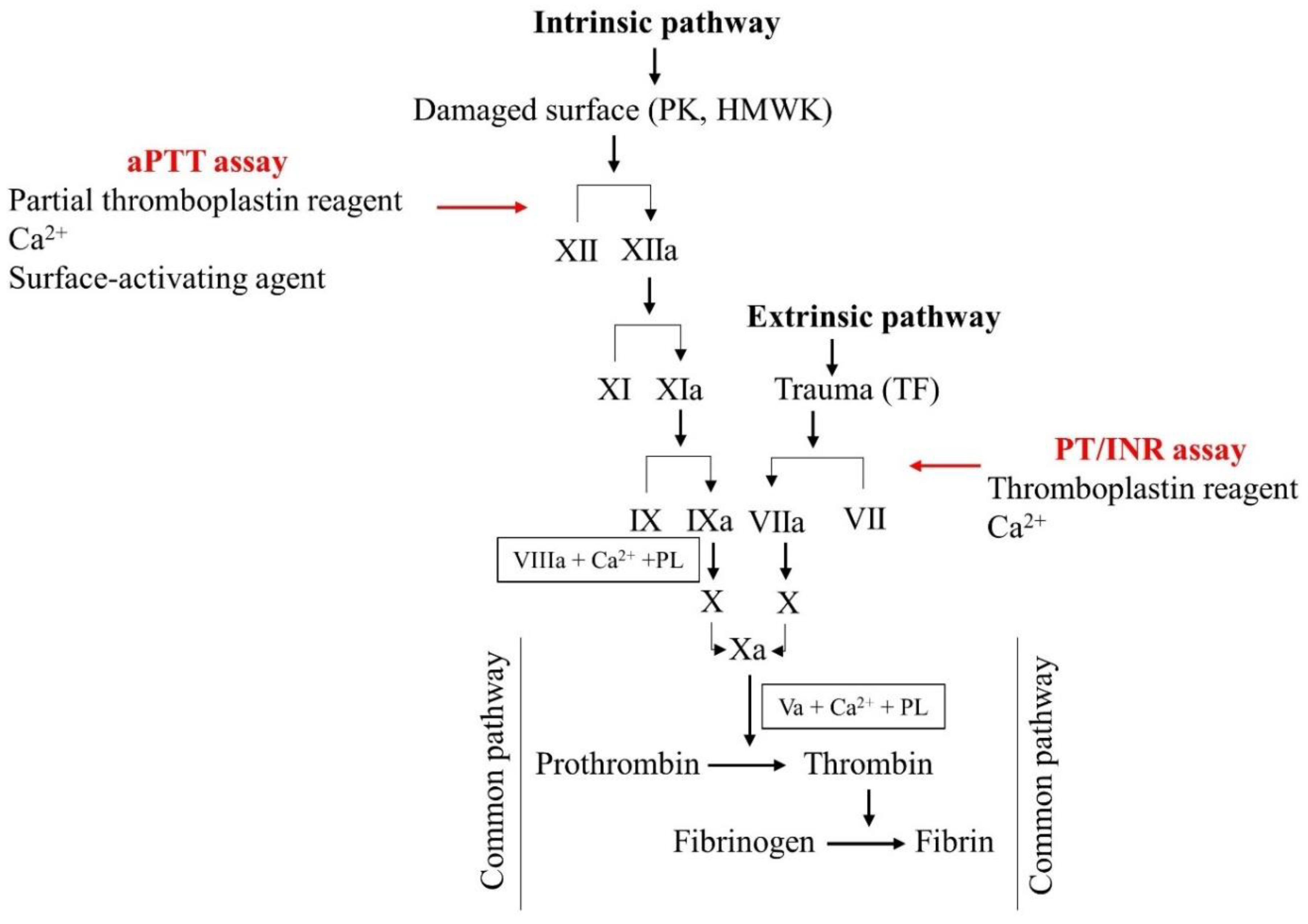
3. Laboratory Assessment of Anticoagulant Therapy

There are several anticoagulant drugs available, as discussed in the previous section. Each anticoagulant agent differs in its effects on coagulation tests, and each drug may require different laboratory assays to measure/monitor drug concentration.

3.1. Monitoring VKAs and UFH

Routine coagulation tests are represented according to their prothrombin time/international normalized ratio (PT/INR) and their activated partial thromboplastin time (aPTT). The PT/INR and aPTT are sometimes obtained to investigate dysfunctions in secondary hemostasis; however, they are more frequently used for monitoring anticoagulant therapy [64]. The PT assay is a straightforward test that evaluates the extrinsic and common pathway factors of coagulation. It is defined as the time in seconds for plasma to clot after the addition of calcium and the thromboplastin reagent, which uses an extrinsic activator pathway (Figure 2) [66]. Commercial thromboplastin reagents have different sources, methods of preparation by their different manufacturers, and, consequently, varying sensitivities to coagulation factors, resulting in uncertainty in PT results [13]. In addition, there are various coagulation analyzers available from several manufacturers. The influence of these two factors is responsible for the variability in PT results among laboratories [66]. To bypass the variability and standardize the PT results, in 1983, the World Health Organization (WHO) introduced the INR concept. The INR is calculated as follows: INR = (patient PT/MNPT)ISI [64], where MNPT is the mean normal PT, which is obtained from at least 20 healthy volunteers, and ISI is the international sensitivity index of the thromboplastin. The ISI reflects the ability of thromboplastin to reduce the vitamin-K-dependent factor in comparison with a standard thromboplastin defined by the WHO [13]. While the INR approach has reduced the variability of patients’ interlaboratory results, on the other hand, the determination of ISI has generated a source of variability depending on the reagent/instrument utilized in the laboratory and the source of plasma used to generate MNPT [66].
Despite the fact that INR has brought an important means of standardization for interlaboratory results, the need for improvements in the technique to ensure more reliable results still exists. The PT/INR is sensitive to the presence or activity of FII, factor V (FV), FVII, FX, and fibrinogen. In that way, anticoagulant agents that alter the levels of these factors can be monitored with the PT/INR if a dose–response relationship could be defined [64]. The PT/INR responds to a lowering of the level of FII, VII, and FX caused by VKAs, leading to a prolongation of PT and, thus, a higher INR [13]. The target INR therapeutic limits are 2.0 to 3.0 for most indications, and it is well defined that subtherapeutic INR levels are related with an increased thrombotic risk. On the other hand, supratherapeutic levels correlate with an increased bleeding risk [48]. Monitoring VKA therapy is crucial due to the highly variable anticoagulant response and the narrow therapeutic window. VKAs interfere with three of the five factors of the extrinsic pathway. Thus, the PT/INR is the most sensitive technique for monitoring the VKA levels, as well as, possible fluctuations in VKA levels due to several interactions presented by these drugs [66].
The aPTT is a clotting assay that evaluates the intrinsic and common pathway factors of coagulation. This test was first developed by Langdell, Wagner, and Brinkhous in 1953 [67] through a modification of the whole blood clotting time test (WBCT) developed by Lee and White to test patients for hemophilia [68]. The aPTT is defined as the time in seconds for the recalcified plasma to form a clot after adding a platelet substitute (partial thromboplastin, named this way because the absence of TF) and a surface-activating agent (such as celite, ellagic acid, kaolin, or micronized silica) (Figure 2) [69,70]. The aPTT is sensitive to the presence or activity of FII, FV, factor VIII (FVIII), FIX, FX, factor XI (FXI), factor XII (FXII), and fibrinogen. Therefore, anticoagulant agents that affect the levels of these factors can be monitored with the aPTT if a dose–response relationship can be determined [64]. Most aPTT reagents shows linear sensitivity to UFH over the therapeutic range, so the aPTT is considered the main assay for monitoring UFH therapy [71]. Monitoring UFH therapy is important for avoiding plasma variation, preventing both toxic levels, which are related with an increased risk of bleeding, thrombocytopenia, and osteoporosis, or subtherapeutic levels, leading to the inefficacy of thrombosis treatment [47]. The aPTT test offers advantages, such as its low cost, ease of performing, wide availability, and automation. These explain why it is widely accepted by clinicians [72]. However, there are some limitations that might influence the technique and are noteworthy. Regarding pre-analytical conditions, several variables might affect the test results, namely, the duration of sample collection, citrate concentration, sample transport temperature, and the time between collection and plasma separation [73]. Concerning analytical conditions, the reagents, instruments, and methods of end-point detection are a source of test variability. The same reagents produced in different lots can yield variability in the test’s sensibility. The partial thromboplastin reagent is available through many suppliers with either vegetable or animal phospholipid sources [74]. Thus, the high phospholipid heterogeneity, either in composition or in concentration, is another source of variation in the sensitivity of aPTT assays. Moreover, the choice of the surface-activating agent modifies the aPTT time in normal plasma [75]. The biological conditions are another factor influencing the aPTT results. Intravascular volume, protein level, coagulation factor concentration, and AT deficiency contribute to the variability in the aPTT [72]. From all of the limitations discussed above, it is remarkable that the major problem of aPTT assays is the absence of standard methods. Consequently, the aPTT shows a high interlaboratory variation, and even in-house analysis should be carefully interpreted [76]. UFH and VKAs require rigorous laboratory control because they have a very narrow therapeutic window; therefore, the correct dose is strictly related to an accurate aPTT or PT/INR assay control, respectively.

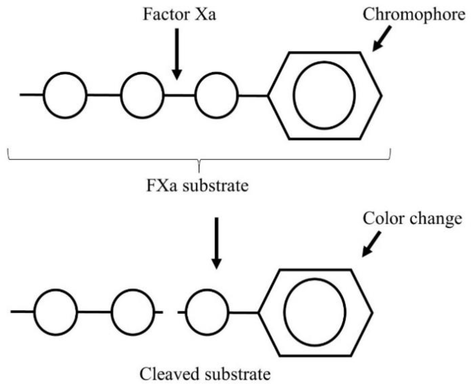
3.2. Monitoring LMWH Therapy

Despite the advantageous pharmacokinetic and pharmacodynamic properties of LMWHs, which grant a lesser need for laboratory monitoring in some circumstances, drug monitoring can bring some benefits for specific populations. The most commonly applied test for LMWH monitoring is the chromogenic anti-factor Xa or anti-Xa assay, mainly due to its low cost and broad availability [77]. The chromogenic anti-factor Xa is considered the gold standard for assessing the plasma LMWH level. In general, the anti-Xa assay measures the enzymatic activity of a patient’s plasma in cleaving an exogenous FXa [77]. A synthetic FXa substrate linked with a chromophore is added to the plasma sample. Endogenous FXa cleaves the chromogenic substrate to release the chromophore unit, leading to a color change that can be detected spectrophotometrically (Figure 3) [78]. In the case of a plasma sample containing LMWH, it will inhibit FXa, reducing the FXa level in order to cleave the synthetic chromophore. By comparing the results with the corresponding standard curve (samples containing known amounts of LMWH), the anticoagulant concentration can be calculated. The extent of colorimetric change is directly proportional to the enzymatic activity and inversely proportional to the anticoagulant concentration [79].
Drawbacks in LMWH monitoring with the anti-factor Xa assay include the lack of standardization in the (1) anti-Xa reagent, (2) standard curve construction, and (3) instrument for analysis [80]. There are numerous commercially available anti-Xa assay kits that use diverse synthetic substrates to measure FXa activity. Thus, analyses of the same sample yield discrepant results [77]. Two different anti-Xa test designs with distinct aims are available. One applies exogenous AT to correct a possibly low in vivo AT concentration. However, this approach may lead to overestimation of the in vivo anticoagulation activity because unbound LMWH, which does not produce anticoagulant activity, could bind to exogenous AT, causing a false positive result [12]. On the other hand, excess dextran sulfate may be added to mitigate in vivo protein binding to the drugs. This strategy may result in overestimated anticoagulant activity, although this is a minor concern for LMWH due to their reduced binding to plasma proteins [77]. The construction of standard curves to calculate the drug’s concentration in the plasma is essential. The same brand and lot number of LMWH administered to patients should be utilized in order to prepare the standard curve. Moreover, different reagents and instrument systems produce variable results [12,80]. The anti-FXa/anti-FIIa ratios among LMWH formulations and the time needed to obtain a blood sample are another pitfall related to the anti-factor Xa assay. In fact, because the peak plasma concentration of LMWH is at about 4 h, blood samples should be collected as close as possible to 4 h after LMWH administration [78,79]. The target therapeutic range for anti-Xa activity depends on the dosage regime and the LMWH agent utilized. With once-daily dosing, the therapeutic range of LMWH is 1.0 to 2.0 U/mL, while for a twice-daily regime, the therapeutic range of LMWH is between 0.5 and 1.0 U/mL [81]. Although anti-Xa is considered the main choice for plasma LMWH level measurement, the anti-Xa level may not correlate with the clinical results, so the results should be evaluated with caution.

3.3. Monitoring DOAC Therapy

Although routine laboratory monitoring of DOACs is not necessary, in some special situations, laboratory measurements may be helpful [82], such as for emergency surgeries, overdoses, extreme body weights, pregnancies, drug interactions, renal impairments, and treatment failures [83]. An ideal assay that measures DOACs accurately should show a great level of linearity within a wide range of drug concentrations. Moreover, the assay should be sensitive to low therapeutic concentrations and should be specific for the drug of interest so that no interferences affect the drug’s determination. Finally, the assay should be widely available and able to give results in a short time. Currently, there is no test with these ideal characteristics [82].
Liquid chromatography/mass spectrometry (LC/MS), calibrated for each drug to be measured, is considered the standard method for DOAC measurement [84]. This technique has a superior accuracy and precision over a broad concentration range compared to coagulation-activity-based assays [85,86]. However, this technique has limited availability, is time consuming and expensive, and requires high technical expertise. Therefore, although LC/MS is a gold-standard method, it is unsuitable for routine DOAC measurement. Conventional anticoagulant assays, such as PT and aPTT, are inappropriate for accurate DOAC measurement. However, recent guidelines and recommendations for DOAC assessment state that aPTT and PT may be utilized as a screening test for DTI and anti-Xa inhibitor (only rivaroxaban), respectively, particularly when rapid and reliable information about a possible overcoagulation is necessary [87].
Conventional monitoring tests are not suitable for apixaban, edoxaban, and betrixaban. Thrombin time (TT) is a rapid, simple, and inexpensive test that assesses the thrombin activity through conversion of fibrinogen into fibrin [88,89]. TT is highly sensitive to dabigatran, and it is not used for accurate measurement of it. Depending on the reagent and the dabigatran concentration, TT may be unmeasurable. Nevertheless, TT has an important use: Normal values are suggestive of little or no clinically relevant dabigatran levels [82]. The plasma dabigatran level can be measured with different assays, such as diluted thrombin time (dTT) and ecarin-based assays: ecarin clotting time (ECT) and ecarin chromogenic assay (ECA) [71,87]. The dTT, or its commercial version, Hemoclot® Thrombin Inhibitor (Hyphen Biomed, Neuville-sur-Oise, France), is a TT modification assay for overcoming the exquisite sensitivity to dabigatran. Diluting the test plasma with normal plasma and purified human α-thrombin (to initiate clotting) ensures a rapid, simple, and commercially available quantitative method for determining plasma DTI concentration [88]. When dTT is calibrated for dabigatran measurement, the assay shows a high linearity with clinical dabigatran concentration with a good reproducibility [90]. The dTT assay has been considered the gold standard for measurement of plasma dabigatran concentration [91]. ECT (Ecarin Reagent, Diagnostica Stago, Asnieres, France) is a meizothrombin generation assay that enables accurate DTI measurement [92]. Ecarin is a metalloproteinase purified from snake venom, Echis carinatus, which, when added to citrated plasma, specifically cleaves prothrombin into an active intermediate, meizothrombin, and the time until clot formation is measured [82]. With DTI therapy, the meizothrombin generated is inactivated as long as DTI is present in the plasma. Thus, coagulation will not occur until all DTI is consumed, thus prolonging the time until clot formation [88,92]. In the ECA (ECA-T Kit, Diagnostica Stago, Asnieres, France), the meizothrombin cleaves a chromogenic substrate, which releases the chromophore unit (paranitroalanine), resulting in absorbance changes. With DTI therapy, the cleavage reaction is inhibited, and by comparing the results with the corresponding standard curve, the anticoagulant concentration can be measured [93]. Direct FXa inhibitors can be accurately measured through a modified chromogenic anti-Xa assay. It is advocated that the anti-Xa assay calibrated with the same drug being taken by the patient is the most reliable technique for accurately measuring the anticoagulation effect of direct oral FXa inhibitors [82,87].
Although the development and introduction of DOACs in the market have revolutionized anticoagulant therapy due to their several advantages over VKAs and heparins, they have brought some challenges in laboratory monitoring [85]. Due to the differences in the sensitivity of routine coagulation tests, none of them are able to accurately measure DOACs. Nevertheless, qualitative information about the presence of DOACs or lack thereof could be obtained [88]. The absence of FDA-approved calibrators and reliable methods for quantifying DOACs has generated a lack of interest in the development of a quantitative assay. Alternative methods, such as ECT and ECA, may be useful, but have not been widely implemented. The use of commercial calibrators, as well as the use of specialty coagulation tests, to measure DOACs should be interpreted with attention [85].

4. Coagulation Test Performance of Drug Delivery Systems
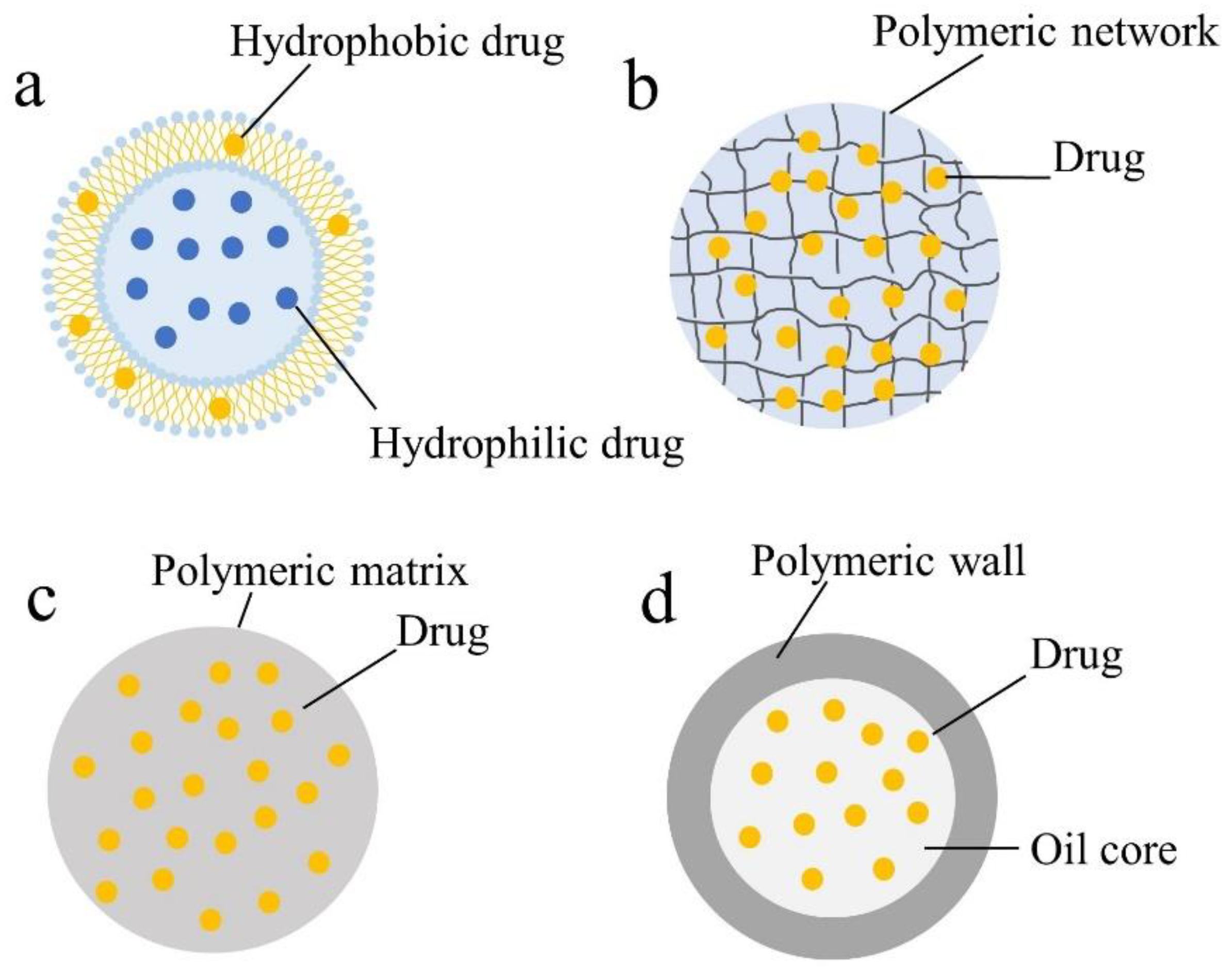
4.1. Liposomes

Liposomes are defined as phospholipid vesicles formed by concentric lipid bilayers surrounding an aqueous inner phase [94]. Their special structure, hydrophobic and hydrophilic characteristics, and biocompatibility make liposomes a promising system for drug delivery [95].
LMWH-loaded flexible liposomes (flexosomes) (size~83 nm) and LMWH-loaded ethosomes (size~85 nm) were prepared for transdermal delivery [96]. Their physicochemical features and pharmacokinetic parameters were compared. The plasma anti-Xa activity of flexosomes, ethosomes, and LMWH solution was measured through a chromogenic anti-Xa assay (Coatest®, Chromogenix, Instrumentation Laboratory, Milano, Italy). After transdermal application, the [anti-Xa]max of the LMWH-loaded flexosomes was 1.11 IU/mL, while the [anti-Xa]max of the LMWH-loaded ethosomes was 0.32 UI/mL. The authors stated that the improved skin permeability of the flexosomes was due to their reduced size and the higher deformability than that of ethosomes (76.7% versus 46.8%). Moreover, they explained that flexosomes are able to change their shape to pass through skin barriers. In another study, Bai and Ahsan [97] prepared ardeparin-loaded conventional liposomes and PEGylated liposomal formulations with the hydration method for the respiratory route. Pulmonary absorption of formulations was monitored through plasma anti-Xa activity and compared with LMWH saline formulations and subcutaneous administration. The antifactor Xa levels (>0.2 UI/mL) for all liposomal formulations were at therapeutic levels for more than 6 h. Moreover, the prolongation of the half-life of the drug was significantly higher when compared to that for the plain LMWH or subcutaneous route.

4.2. Hydrogels

Hydrogels are three-dimensional cross-linked hydrophilic polymer networks that are capable of retaining large amounts of water or biological fluids while remaining insoluble in these media [98]. Their hydrophilicity is responsible for some of the desirable features of hydrogels, such as biocompatibility and the capability of encapsulating hydrophilic molecules [99]. Because the hydrogels were mostly prepared in aqueous solutions, the drug denaturation and aggregation possibilities were mitigated. Moreover, the hydrogel network was able to protect the entrapped drug against the harsh environment [100]. Loira-Pastoriza et al. prepared nanogels to incorporate four commercial LMWHs for topical delivery [11]. For bemiparin measurement in plasma after topical application, they used a chromogenic anti-Xa assay (Stachrom® heparin, Diagnostica Stago, Asnières-sur-Seine, France). The authors could not detect bemiparin in plasma after 24 h of topical application. However, they stated that, for the route of administration and for the treatment of superficial thrombosis and hematomas, the drug should not pass into the systemic blood circulation in order to avoid a systemic effect. Matanovic et al. developed a thermoresponsive poloxamer-based platform that formed in situ combined with pH-responsive polyelectrolyte heparin/chitosan nanocomplexes for prolonged subcutaneous release of heparin. The plasma heparin concentration was measured after subcutaneous injection of selected individual formulations (thermoresponsive hydrogel and heparin/chitosan nanocomplexes) and a dual system (nanocomplexes incorporated into thermoresponsive hydrogel) through a chromogenic FXa assay and Berichrom® Heparin kit (Siemens Healthcare Diagnostics Inc., Erlanger, Germany). Plasma heparin concentration–time profiles showed a double-peak phenomenon, and this dual behavior was probably related to diffusion of heparin inside the polymer network and gel dissolution. It was demonstrated that thermoresponsive hydrogel with heparin/chitosan nanocomplexes showed sustained release of heparin for 5 days [101]. The aPTT is the most preferred assay for monitoring the therapeutic dose of UFH in many hospitals and laboratories, mainly due to its low cost, wide availability, and ease of performing, rather than the specific advantages of this technique. However, the anti-Xa assay may be necessary either in some patients (e.g., elevated baseline aPTT or FXII deficiency) or because it offers another option with fewer interferences [71,87].

4.3. Polymeric Nanoparticles (PNPs)

PNPs are colloidal dispersions that have been used for drug and biomolecule delivery [102]. PNPs have several advantages: (i) They have improved stability; (ii) they can avoid the phagocyte system, thus prolonging their blood stream duration; (iii) because of their sub-cellular size, they can pass through capillary vessels; (iv) they can improve the drug properties and reduce side effects; (v) they can release the drug or biomolecule in a controlled/sustained fashion [103,104,105]. Dong et al. prepared chitosan-based polymer–lipid hybrid nanoparticles (PLNs) with a self-assembly method to incorporate and improve the oral absorption of enoxaparin (Enox). The PLNs were composed of chitosan as the polymer and glyceryl monooleate (GMO) as the lipid, with an optimized lipid/polymer mass ratio of 0.2 and pluronic® F127 as a stabilizer. Nanoparticles with the optimal composition were submitted to an in vivo study. The Enox anticoagulant activity was measured through the aPTT assay. The optimized Enox/PLNs showed a 4.5-fold increase in oral Enox bioavailability in comparison with an Enox solution. The authors claimed that the stability of nanoparticles is a crucial parameter for improved oral drug delivery efficiency [7]. The authors utilized the aPTT assay to measure plasma LMWH concentration; however, the aPTT was not helpful for this purpose because the anticoagulant effect of LMWH is produced mainly through FXa inhibition, and the prolongation of the aPTT is highly dependent on low thrombin activity [77]. In another work, Ramadan et al. produced and characterized cationic lipid nanocapsules (cLNCs) with a phase-inversion method intended for fondaparinux (Fp) oral absorption. In vivo studies were performed after a single oral dose in rats, and the plasma Fp measurements were carried out with the anti-Xa activity assay (HYPHEN, Biomed). The authors reported that the cLNCs significantly increased oral Fp bioavailability and improved pharmacokinetic profile in a dose-dependent manner [106].

4.4. Other Drug Delivery Systems

Solid lipid nanoparticles (SLNs) are lipid-based colloidal nanocarriers consisting of a hydrophobic core composed of lipids that are solid at both room and body temperatures and stabilized by a surfactant layer. SLNs are capable of affording a controlled drug release because the release of the active compound from the solid lipid matrix is much slower when compared to liquid oils [107,108]. Paliwal et al. synthetized biomimetic LMWH–lipid conjugate-loaded SLNs for oral bioavailability studies. An in vivo study was performed in albino rats, and the plasma LMWH concentration was measured with a colorimetric anti-Xa assay using a Chromogenix Coatest LMW heparin kit (Diapharma). The authors demonstrated that SLNs were within the nanometric range and were safe for oral administration. Furthermore, improved bioavailability of LMWH–lipid conjugates was observed in comparison to LMWH-loaded SLNs and LMWH solution. They attributed this result to the high loading capacity and better lymphatic drainage of the LMWH–lipid conjugate SLNs, thus achieving an improved systemic LMWH concentration [109]. Self-nano emulsifying drug delivery systems (SNEDDSs) have been explored as drug delivery systems because they are able to spontaneously form oil-in-water (o/w) nanoemulsions after administration in an aqueous environment, followed by mild agitation (e.g., the gastric motility) [110,111]. Zupancic et al. prepared and characterized various enoxaparin-loaded self-emulsifying drug delivery systems (SEDDSs) with lipids of different chain lengths for oral administration [112]. An in vivo study was carried out in rats, and the plasma enoxaparin concentration was measured with a Biophen® Heparin anti-Xa assay kit. Medium-chain lipids (MC-SEDDS) and no lipids (NL-SEDDS) showed greater mucus diffusion. MC-SEDDS and NL-SEDDS showed sustained enoxaparin release in vitro and an oral absolute enoxaparin bioavailability of 2.02% and 2.25%, respectively. A literature review focusing on anticoagulant therapies using different drug delivery systems is given in Table 3.

5. Conclusions

Conventional anticoagulant agents are well-known drugs with reliable anticoagulant assays that have been used for decades. On the other hand, after a long time without advances in anticoagulation management, DOACs have emerged in the last decade. Despite the remarkable advances made in anticoagulant therapy with the introduction of DOACs, there are still some important issues that should be addressed, such as the lack of specific antidotes and the absence of an accurate and standardized coagulation test. The anticoagulant tests were found to be suitable for measuring the anticoagulant performance of heparin-based nanocarriers through several administration routes.

Author Contributions

All authors substantially contributed to the conceptualization, methodology, formal analysis, investigation, resources, and data curation. Y.B.G.P., L.L.C., M.F.R.S., and J.L.S.-S. contributed to the writing—original draft, data search, and literature research. M.F.R.S., J.L.S.-S., E.H.G., P.S., and E.B.S. contributed to the writing—review, validation, and editing. E.B.S., P.S., M.F.R.S., and J.L.S.-S. contributed to the project administration, supervision, and funding acquisition. All authors have read and agreed to the published version of the manuscript.

Funding

This work was supported by the Banco do Nordeste (grant FUNDECI/2016.0015), Conselho Nacional de Desenvolvimento Científico e Tecnológico (CNPq), Fundação de Apoio à Pesquisa e à Inovação Tecnológica do Estado de Sergipe (Fapitec), and Coordenação de Aperfeiçoamento de Pessoal de Nível Superior (CAPES). Eliana B. Souto would like to acknowledge the Portuguese Science and Technology Foundation (FCT/MCT) and the European Funds (PRODER/COMPETE) for the project UIDB/04469/2020 (strategic fund), co-financed by FEDER, under the Partnership Agreement PT2020.

Institutional Review Board Statement

Not applicable.

Informed Consent Statement

Not applicable.

Data Availability Statement

Not applicable.

Conflicts of Interest

The authors declare no conflict of interest.

Abbreviations

CSchitosan
Cholcholesterol
CTABhexadecyltrimethyl ammonium bromide
DSPE1,2-distearoyl-sn-glycero-3-phosphoethanolamine
DSPE–PEG1,2-distearoyl-sn-glycero-3-phosphoethanolamine polyethyleneglycol
E80egg yolk lecithin
Egg PCphosphatidylcholine from egg yolk
GMOglyceryl monooleate
HPMChydroxypropylmethylcellulose
IPMisopropyl myristate
MBAN,N-methylenebis (acrylamide)
NAnot available
PEIpolyethylenimine
PCphosphatidylcholine
Plysε-polylysine
PNPpolymeric nanoparticle
SAstearyl amine
SEDDSself-emulsifying drug delivery system
SLNsolid lipid nanoparticles
SNEDDSself-nanoemulsifying drug delivery system
TEAtriethylamine

References

  1. Patel, V.F.; Liu, F.; Brown, M.B. Advances in oral transmucosal drug delivery. J. Control. Release 2011, 153, 106–116. [Google Scholar] [CrossRef] [PubMed] [Green Version]
  2. Aláez-Versón, C.R.; Lantero, E.; Fernàndez-Busquets, X. Heparin: New life for an old drug. Nanomedicine 2017, 12, 1727–1744. [Google Scholar] [CrossRef] [PubMed]
  3. Motlekar, N.A.; Youan, B.-B.C. The quest for non-invasive delivery of bioactive macromolecules: A focus on heparins. J. Control. Release 2006, 113, 91–101. [Google Scholar] [CrossRef] [PubMed] [Green Version]
  4. Das Kurmi, B.; Tekchandani, P.; Paliwal, R.; Rai Paliwal, S. Nanocarriers in Improved Heparin Delivery: Recent Updates. Curr. Pharm. Des. 2015, 21, 4509–4518. [Google Scholar] [CrossRef] [PubMed]
  5. Lee, J.H.; Yeo, Y. Controlled drug release from pharmaceutical nanocarriers. Chem. Eng. Sci. 2015, 125, 75–84. [Google Scholar] [CrossRef] [PubMed] [Green Version]
  6. Shi, J.; Votruba, A.R.; Farokhzad, O.C.; Langer, R. Nanotechnology in drug delivery and tissue engineering: From discovery to applications. Nano Lett. 2010, 10, 3223–3230. [Google Scholar] [CrossRef] [Green Version]
  7. Dong, W.; Wang, X.; Liu, C.; Zhang, X.; Zhang, X.; Chen, X.; Kou, Y.; Mao, S. Chitosan based polymer-lipid hybrid nanoparticles for oral delivery of enoxaparin. Int. J. Pharm. 2018, 547, 499–505. [Google Scholar] [CrossRef]
  8. Charoongchit, P.; Suksiriworapong, J.; Mao, S.; Sapin-Minet, A.; Maincent, P.; Junyaprasert, V.B. Investigation of cationized triblock and diblock poly(ε-caprolactone)-co-poly(ethylene glycol) copolymers for oral delivery of enoxaparin: In vitro approach. Acta Biomater. 2017, 61, 180–192. [Google Scholar] [CrossRef]
  9. Rawat, A.; Majumder, Q.H.; Ahsan, F. Inhalable large porous microspheres of low molecular weight heparin: In vitro and in vivo evaluation. J. Control. Release 2008, 128, 224–232. [Google Scholar] [CrossRef] [Green Version]
  10. Patel, B.; Gupta, V.; Ahsan, F. PEG–PLGA based large porous particles for pulmonary delivery of a highly soluble drug, low molecular weight heparin. J. Control. Release 2012, 162, 310–320. [Google Scholar] [CrossRef]
  11. Loira-Pastoriza, C.; Sapin-Minet, A.; Diab, R.; Grossiord, J.L.; Maincent, P. Low molecular weight heparin gels, based on nanoparticles, for topical delivery. Int. J. Pharm. 2012, 426, 256–262. [Google Scholar] [CrossRef] [PubMed]
  12. Walenga, J.; Hoppensteadt, D. Monitoring the new antithrombotic drugs. Semin. Thromb. Hemost. 2005, 30, 683–695. [Google Scholar] [CrossRef] [PubMed]
  13. Samama Meyer, M.; Guinet, C. Laboratory assessment of new anticoagulants. Clin. Chem. Lab. Med. 2011, 49, 761–772. [Google Scholar] [CrossRef]
  14. Mulloy, B.; Hogwood, J.; Gray, E.; Lever, R.; Page, C.P. Pharmacology of heparin and related drugs. Pharmacol. Rev. 2016, 68, 76–141. [Google Scholar] [CrossRef] [PubMed]
  15. Torri, G.; Naggi, A. Heparin centenary—An ever-young life-saving drug. Int. J. Cardiol. 2016, 212, S1–S4. [Google Scholar] [CrossRef] [Green Version]
  16. Onishi, A.; St Ange, K.; Dordick, J.S.; Linhardt, R.J. Heparin and anticoagulation. Front. Biosci. (Landmark Ed). 2016, 21, 1372–1392. [Google Scholar] [CrossRef] [PubMed]
  17. Gray, E.; Mulloy, B.; Barrowcliffe, T.W. Heparin and low-molecular-weight heparin. Thromb. Haemost. 2008, 99, 807–818. [Google Scholar] [CrossRef] [PubMed] [Green Version]
  18. Harter, K.; Levine, M.; Henderson, S.O. Anticoagulation drug therapy: A review. West. J. Emerg. Med. 2015, 16, 11–17. [Google Scholar] [CrossRef] [PubMed]
  19. Vene, N.; Mavri, A. An overview of the anticoagulant drugs used in routine clinical practice. In Anticoagulant Drugs; Božič-Mijovski, M., Ed.; IntechOpen: London, UK, 2018; p. 10. [Google Scholar] [CrossRef]
  20. Casu, B.; Oreste, P.; Torri, G.; Zoppetti, G.; Choay, J.; Lormeau, J.C.; Petitou, M.; Sinaɕ, P. The structure of heparin oligosaccharide fragments with high anti-(factor Xa) activity containing the minimal antithrombin III-binding sequence. Biochem. J. 1981, 197, 599–609. [Google Scholar] [CrossRef] [PubMed]
  21. Lindahl, U.; Bäckström, G.; Höök, M.; Thunberg, L.; Fransson, L.-A.; Linker, A. Structure of the antithrombin-binding site in heparin. Proc. Natl. Acad. Sci. USA 1979, 76, 3198–3202. [Google Scholar] [CrossRef] [Green Version]
  22. Cosmi, B.; Palareti, G. Old and new heparins. Thromb. Res. 2012, 129, 388–391. [Google Scholar] [CrossRef] [PubMed]
  23. Kakkar, A.K. Low- and ultra-low-molecular-weight heparins. Best Pract. Res. Clin. Haematol. 2004, 17, 77–87. [Google Scholar] [CrossRef] [PubMed]
  24. Ibrahim, S.S.; Osman, R.; Awad, G.A.S.; Mortada, N.D.; Geneidy, A.-S. Low molecular weight heparins for current and future uses: Approaches for micro- and nano-particulate delivery. Drug. Deliv. 2016, 23, 2661–2667. [Google Scholar] [CrossRef] [PubMed]
  25. Park, J.; Byun, Y. Recent advances in anticoagulant drug delivery. Expert Opin. Drug Deliv. 2016, 13, 421–434. [Google Scholar] [CrossRef]
  26. Merli, G.J.; Vanscoy, G.J.; Rihn, T.L.; Groce Iii, J.B.; McCormick, W. Applying scientific citeria to therapeutic interchange: A balanced analysis of low-molecular-weight heparins. J. Thromb. Thrombolysis 2001, 11, 247–259. [Google Scholar] [CrossRef]
  27. Hirsh, J.; O’Donnell, M.; Eikelboom, J.W. Beyond unfractionated heparin and warfarin. Circulation 2007, 116, 552–560. [Google Scholar] [CrossRef] [Green Version]
  28. Walenga, J.M.; Lyman, G.H. Evolution of heparin anticoagulants to ultra-low-molecular-weight heparins: A review of pharmacologic and clinical differences and applications in patients with cancer. Crit. Rev. Oncol. Hematol. 2013, 88, 1–18. [Google Scholar] [CrossRef]
  29. Masuko, S.; Linhardt, R.J. Chemoenzymatic synthesis of the next generation of ultralow MW heparin therapeutics. Future Med. Chem. 2012, 4, 289–296. [Google Scholar] [CrossRef] [Green Version]
  30. Bauer, K.A. Fondaparinux sodium: A selective inhibitor of factor Xa. Am. J. Health Syst. Pharm. 2001, 58, S14–S17. [Google Scholar] [CrossRef]
  31. Gómez-Outes, A.; Suárez-Gea, M.L.; Lecumberri, R.; Rocha, E.; Pozo-Hernández, C.; Vargas-Castrillón, E. New parenteral anticoagulants in development. Ther. Adv. Cardiovasc. Dis. 2011, 5, 33–59. [Google Scholar] [CrossRef]
  32. Savi, P.; Herault, J.P.; Duchaussoy, P.; Millet, L.; Schaeffer, P.; Petitou, M.; Bono, F.; Herbert, J.M. Reversible biotinylated oligosaccharides: A new approach for a better management of anticoagulant therapy. J. Thromb. Haemost. 2008, 6, 1697–1706. [Google Scholar] [CrossRef]
  33. Paty, I.; Trellu, M.; Destors, J.-M.; Cortez, P.; Boëlle, E.; Sanderink, G. Reversibility of the anti-FXa activity of idrabiotaparinux (biotinylated idraparinux) by intravenous avidin infusion. J. Thromb. Haemost. 2010, 8, 722–729. [Google Scholar] [CrossRef]
  34. Falkon, L.; Garí, M.; Barbanoj, M.; Amiral, J.; Fontcuberta, J. Tissue factor pathway inhibitor and anti-FXa kinetic profiles of a new low-molecular-mass heparin, bemiparin, at therapeutic subcutaneous doses. Blood Coagul. Fibrinolysis 1998, 9, 137–141. [Google Scholar] [CrossRef]
  35. Ciccone, M.M.; Cortese, F.; Corbo, F.; Corrales, N.E.; Al-Momen, A.K.; Silva, A.; Zito, A.; Pinto, M.; Gesualdo, M.; Scicchitano, P. Bemiparin, an effective and safe low molecular weight heparin: A review. Vasc. Pharmacol. 2014, 62, 32–37. [Google Scholar] [CrossRef]
  36. Akhtar, F.; Wan, X.; Wu, G.; Kesse, S.; Wang, S.; He, S. Low-Molecular-Weight Heparins: Reduced Size Particulate Systems for Improved Therapeutic Outcomes. Molecules 2018, 23, 1757. [Google Scholar] [CrossRef] [Green Version]
  37. Schluter, A.; Lamprecht, A. Current Developments for the Oral Delivery of Heparin. Curr. Pharm. Biotechnol. 2014, 15, 640–649. [Google Scholar] [CrossRef]
  38. Pirmohamed, M.; Kamali, F.; Daly, A.K.; Wadelius, M. Oral anticoagulation: A critique of recent advances and controversies. Trends Pharm. Sci. 2015, 36, 153–163. [Google Scholar] [CrossRef]
  39. Ferreira, J.L.; Wipf, J.E. Pharmacologic therapies in anticoagulation. Med. Clin. N. Am. 2016, 100, 695–718. [Google Scholar] [CrossRef]
  40. Ibrahim, T.F.; Maxwell, S.; Iqbal, O. Current anticoagulation drugs and mechanisms of action. In Anticoagulation and Hemostasis in Neurosurgery; Loftus, C.M., Ed.; Springer: Cham, Switzerland, 2016; pp. 33–46. [Google Scholar] [CrossRef]
  41. Hirsh, J. Oral anticoagulant drugs. N. Engl. J. Med. 1991, 324, 1865–1875. [Google Scholar] [CrossRef]
  42. Ageno, W.; Gallus, A.S.; Wittkowsky, A.; Crowther, M.; Hylek, E.M.; Palareti, G. Oral anticoagulant therapy: Antithrombotic therapy and prevention of thrombosis, 9th ed: American college of chest physicians evidence-based clinical practice guidelines. Chest 2012, 141, e44S–e88S. [Google Scholar] [CrossRef] [Green Version]
  43. Wadelius, M.; Pirmohamed, M. Pharmacogenetics of warfarin: Current status and future challenges. Pharmacogenomics J. 2006, 7, 99–111. [Google Scholar] [CrossRef] [Green Version]
  44. Ufer, M. Comparative pharmacokinetics of vitamin K antagonists. Clin. Pharmacokinet. 2005, 44, 1227–1246. [Google Scholar] [CrossRef] [PubMed]
  45. Mega, J.L.; Simon, T. Pharmacology of antithrombotic drugs: An assessment of oral antiplatelet and anticoagulant treatments. Lancet 2015, 386, 281–291. [Google Scholar] [CrossRef]
  46. Wysowski, D.K.; Nourjah, P.; Swartz, L. Bleeding complications with warfarin use: A prevalent adverse effect resulting in regulatory action. Arch. Intern. Med. 2007, 167, 1414–1419. [Google Scholar] [CrossRef]
  47. Franchini, M.; Liumbruno, G.M.; Bonfanti, C.; Lippi, G. The evolution of anticoagulant therapy. Blood. Transfus. 2016, 14, 175–184. [Google Scholar] [CrossRef]
  48. Riva, N.; Ageno, W. Pros and cons of vitamin K antagonists and non–vitamin K antagonist oral anticoagulants. Semin. Thromb. Hemost. 2015, 41, 178–187. [Google Scholar] [CrossRef]
  49. Schulman, S. Advantages and limitations of the new anticoagulants. J. Intern. Med. 2014, 275, 1–11. [Google Scholar] [CrossRef]
  50. Bauer, K.A. Pros and cons of new oral anticoagulants. Hematol. Am. Soc. Hematol. Educ. Program. 2013, 2013, 464–470. [Google Scholar] [CrossRef] [Green Version]
  51. Stangier, J. Clinical pharmacokinetics and pharmacodynamics of the oral direct thrombin inhibitor dabigatran etexilate. Clin. Pharmacokinet. 2008, 47, 285–295. [Google Scholar] [CrossRef] [PubMed]
  52. Zacconi, F.C. FXa direct synthetic inhibitors. In Anticoagulant Drugs; Božič-Mijovski, M., Ed.; IntechOpen: London, UK, 2018; pp. 11–37. [Google Scholar] [CrossRef]
  53. Hinojar, R.; Jiménez-Natcher, J.J.; Fernández-Golfín, C.; Zamorano, J.L. New oral anticoagulants: A practical guide for physicians. Eur. Heart J. Cardiovasc. Pharmacother. 2015, 1, 134–145. [Google Scholar] [CrossRef]
  54. Laux, V.; Perzborn, E.; Kubitza, D.; Misselwitz, F. Preclinical and clinical characteristics of rivaroxaban: A novel, oral, direct factor Xa inhibitor. Semin. Thromb. Hemost. 2007, 33, 515–523. [Google Scholar] [CrossRef] [PubMed] [Green Version]
  55. He, K.; He, B.; Grace, J.E.; Xin, B.; Zhang, D.; Pinto, D.J.; Luettgen, J.M.; Knabb, R.M.; Lam, P.Y.S.; Wexler, R.R.; et al. Preclinical pharmacokinetic and metabolism of apixaban, a potent and selective factor Xa inhibitor. Blood 2006, 108, 910. [Google Scholar] [CrossRef]
  56. Minguet, J.; Sims, H.M.; Smith, K.H.; Bramlage, P. The factor Xa inhibitor edoxaban for the prevention of stroke and systemic embolism in patients with atrial fibrillation. Expert Rev. Clin. Pharmacol. 2017, 10, 5–15. [Google Scholar] [CrossRef]
  57. Poulakos, M.; Walker, J.N.; Baig, U.; David, T. Edoxaban: A direct oral anticoagulant. Am. J. Health Syst. Pharm. 2017, 74, 117–129. [Google Scholar] [CrossRef] [PubMed]
  58. Parasrampuria, D.A.; Truitt, K.E. Pharmacokinetics and pharmacodynamics of edoxaban, a non-vitamin K antagonist oral anticoagulant that inhibits clotting factor Xa. Clin. Pharmacokinet. 2016, 55, 641–655. [Google Scholar] [CrossRef] [PubMed] [Green Version]
  59. Garland, S.G.; DeRemer, C.E.; Smith, S.M.; Gums, J.G. Betrixaban: A new oral factor Xa inhibitor for extended venous thromboembolism prophylaxis in high-risk hospitalized patients. Ann. Pharmacother. 2018, 52, 554–561. [Google Scholar] [CrossRef]
  60. Huisman, M.V.; Klok, F.A. Pharmacological properties of betrixaban. Eur. Heart J. Suppl. 2018, 20, E12–E15. [Google Scholar] [CrossRef]
  61. Thoenes, M.; Minguet, J.; Bramlage, K.; Bramlage, P.; Ferrero, C. Betrixaban—the next direct factor Xa inhibitor? Expert Rev. Hematol. 2016, 9, 1111–1117. [Google Scholar] [CrossRef]
  62. Cosmi, B. An update on the pharmaceutical management of thrombosis. Expert Opin. Pharmacother. 2016, 17, 2149–2164. [Google Scholar] [CrossRef]
  63. Gómez-Outes, A.; Suárez-Gea, M.L.; Lecumberri, R.; Terleira-Fernández, A.I.; Vargas-Castrillón, E. Direct-acting oral anticoagulants: Pharmacology, indications, management, and future perspectives. Eur. J. Haematol. 2015, 95, 389–404. [Google Scholar] [CrossRef] [Green Version]
  64. Favaloro, E.J.; Lippi, G.; Koutts, J. Laboratory testing of anticoagulants: The present and the future. Pathology 2011, 43, 682–692. [Google Scholar] [CrossRef] [PubMed]
  65. Czuprynska, J.; Patel, J.P.; Arya, R. Current challenges and future prospects in oral anticoagulant therapy. Br. J. Haematol. 2017, 178, 838–851. [Google Scholar] [CrossRef] [PubMed] [Green Version]
  66. Moffat, K.; Lewis, C. Laboratory monitoring of oral vitamin K anticoagulation. Semin. Thromb. Hemost. 2017, 43. [Google Scholar] [CrossRef]
  67. Langdell, R.D.; Wagner, R.H.; Brinkhous, K.M. Effect of antihemophilic factor on one-stage clotting tests. A presumptive test for hemophilia and a simple one-stage antihemophilic factor assay procedure. J. Lab. Clin. Med. 1953, 41, 637–647. [Google Scholar]
  68. Kitchens, C.S. To bleed or not to bleed? Is that the question for the PTT? J. Thromb. Haemost. 2005, 3, 2607–2611. [Google Scholar] [CrossRef]
  69. Chee, Y.L.; Greaves, M. Role of coagulation testing in predicting bleeding risk. Hematol. J. 2003, 4, 373–378. [Google Scholar] [CrossRef]
  70. Bonhomme, F.; Fontana, P. Laboratory testing of hemostasis. In Perioperative hemostasis; Marcucci, C.E., Schoettker, P., Eds.; Springer: Berlin/Heidelberg, Germany, 2015; pp. 13–24. [Google Scholar] [CrossRef]
  71. Avecilla, S.T.; Roshal, M.; Reyes Gil, M.; Erdman, P.A. Laboratory monitoring for heparins, fondaparinux, direct thrombin inhibitors, and oral anti-Xa medications. In Transfusion Medicine and Hemostasis, 3rd ed.; Shaz, B.H., Hillyer, C.D., Reyes Gil, M., Eds.; Elsevier: Amsterdam, The Netherlands, 2019; pp. 933–938. [Google Scholar] [CrossRef]
  72. Cuker, A. Unfractionated heparin for the treatment of venous thromboembolism: Best practices and areas of uncertainty. Semin. Thromb. Hemost. 2012, 38, 593–599. [Google Scholar] [CrossRef]
  73. Marlar, R.A.; Clement, B.; Gausman, J. Activated partial thromboplastin time monitoring of unfractionated heparin therapy: Issues and recommendations. Semin. Thromb. Hemost. 2017, 43, 253–260. [Google Scholar] [CrossRef]
  74. Shapiro, G.A.; Huntzinger, S.W.; Wilson, J.E. Variation among commercial activated partial thromboplastin time reagents in response to heparin. Am. J. Clin. Pathol. 1977, 67, 477–480. [Google Scholar] [CrossRef]
  75. Watson, H.G.; Greaves, M. Can we predict bleeding? Semin. Thromb. Hemost. 2008, 34, 97–103. [Google Scholar] [CrossRef]
  76. Cuker, A.; Raby, A.; Moffat, K.; Flynn, G.; Crowther, M. Interlaboratory variation in heparin monitoring: Lessons from the quality management program of Ontario coagulation surveys. Thromb. Haemost. 2010, 104, 837–844. [Google Scholar] [CrossRef] [PubMed]
  77. Babin, J.L.; Traylor, K.; Witt, D. Laboratory monitoring of low-molecular-weight heparin and fondaparinux. Semin. Thromb. Hemost. 2017, 43, 261–269. [Google Scholar] [CrossRef] [PubMed]
  78. Gehrie, E.; Laposata, M. Test of the month: The chromogenic antifactor Xa assay. Am. J. Hematol. 2012, 87, 194–196. [Google Scholar] [CrossRef] [PubMed]
  79. Bates, S.M.; Weitz, J.I. Coagulation assays. Circulation 2005, 112, 53–60. [Google Scholar] [CrossRef] [PubMed] [Green Version]
  80. Newall, F. Anti-factor Xa (anti-Xa) assay. In Haemostasis. Methods in Molecular Biology (Methods and Protocols); Monagle, P., Ed.; Humana Press: Totowa, NJ, USA, 2013; Volume 992, pp. 265–272. [Google Scholar]
  81. Funk, D.M. Coagulation assays and anticoagulant monitoring. Hematol. Am. Soc. Hematol. Educ. Program. 2012, 2012, 460–465. [Google Scholar] [CrossRef]
  82. Samuelson, B.T.; Cuker, A. Measurement and reversal of the direct oral anticoagulants. Blood Rev. 2017, 31, 77–84. [Google Scholar] [CrossRef] [Green Version]
  83. Samuelson, B.T.; Cuker, A.; Siegal, D.M.; Crowther, M.; Garcia, D.A. Laboratory assessment of the anticoagulant activity of direct oral anticoagulants: A systematic review. Chest 2017, 151, 127–138. [Google Scholar] [CrossRef] [Green Version]
  84. Eby, C. Novel anticoagulants and laboratory testing. Int. J. Lab. Hematol. 2013, 35, 262–268. [Google Scholar] [CrossRef]
  85. Adcock, D.M.; Gosselin, R. Direct oral anticoagulants (DOACs) in the laboratory: 2015 review. Thromb. Res. 2015, 136, 7–12. [Google Scholar] [CrossRef] [Green Version]
  86. Schmitz, E.M.H.; Boonen, K.; van den Heuvel, D.J.A.; van Dongen, J.L.J.; Schellings, M.W.M.; Emmen, J.M.A.; van der Graaf, F.; Brunsveld, L.; van de Kerkhof, D. Determination of dabigatran, rivaroxaban and apixaban by ultra-performance liquid chromatography—tandem mass spectrometry (UPLC-MS/MS) and coagulation assays for therapy monitoring of novel direct oral anticoagulants. J. Thromb. Haemost. 2014, 12, 1636–1646. [Google Scholar] [CrossRef]
  87. Lippi, G.; Favaloro, E. Recent guidelines and recommendations for laboratory assessment of the direct oral anticoagulants (DOACs): Is there consensus? Clin. Chem. Lab. Med. 2015, 53, 185–197. [Google Scholar] [CrossRef]
  88. Dale, B.J.; Chan, N.C.; Eikelboom, J.W. Laboratory measurement of the direct oral anticoagulants. Br. J. Haematol. 2016, 172, 315–336. [Google Scholar] [CrossRef] [Green Version]
  89. van Ryn, J.; Stangier, J.; Härtter, S.; Liesenfeld, K.-H.; Wienen, W.; Feuring, M.; Clemens, A. Dabigatran etexilate—a novel, reversible, oral direct thrombin inhibitor: Interpretation of coagulation assays and reversal of anticoagulant activity. Thromb. Haemost. 2010, 103, 1116–1127. [Google Scholar] [CrossRef] [PubMed]
  90. Pollack, C.V. Coagulation assessment with the new generation of oral anticoagulants. Emerg. Med. J. 2016, 33, 423–430. [Google Scholar] [CrossRef] [PubMed] [Green Version]
  91. Douxfils, J.; Mullier, F.; Robert, S.; Chatelain, C.; Chatelain, B.; Dogne, J.M. Impact of dabigatran on a large panel of routine or specific coagulation assays. Laboratory recommendations for monitoring of dabigatran etexilate. Thromb. Haemost. 2012, 107, 985–997. [Google Scholar] [CrossRef]
  92. Nowak, G. The ecarin clotting time, a universal method to quantify direct thrombin inhibitors. Pathophysiol. Haemost. Thromb. 2003, 33, 173–183. [Google Scholar] [CrossRef] [PubMed] [Green Version]
  93. Gosselin, R.C.; Dwyre, D.M.; Dager, W.E. Measuring dabigatran concentrations using a chromogenic ecarin clotting time assay. Ann. Pharmacother. 2013, 47, 1635–1640. [Google Scholar] [CrossRef]
  94. Ahmed, K.S.; Hussein, S.A.; Ali, A.H.; Korma, S.A.; Lipeng, Q.; Jinghua, C. Liposome: Composition, characterisation, preparation, and recent innovation in clinical applications. J. Drug Target. 2019, 27, 742–761. [Google Scholar] [CrossRef]
  95. Liu, G.; Hou, S.; Tong, P.; Li, J. Liposomes: Preparation, Characteristics, and Application Strategies in Analytical Chemistry. Crit. Rev. Anal. Chem. 2020, 1–21. [Google Scholar] [CrossRef]
  96. Song, Y.-K.; Hyun, S.Y.; Kim, H.-T.; Kim, C.-K.; Oh, J.-M. Transdermal delivery of low molecular weight heparin loaded in flexible liposomes with bioavailability enhancement: Comparison with ethosomes. J. Microencapsul. 2011, 28, 151–158. [Google Scholar] [CrossRef]
  97. Bai, S.; Ahsan, F. Inhalable liposomes of low molecular weight heparin for the treatment of venous thromboembolism. J. Pharm. Sci. 2010, 99, 4554–4564. [Google Scholar] [CrossRef] [PubMed]
  98. Croisfelt, F.M.; Tundisi, L.L.; Ataide, J.A.; Silveira, E.; Tambourgi, E.B.; Jozala, A.F.; Souto, E.M.B.; Mazzola, P.G. Modified-release topical hydrogels: A ten-year review. J. Mater. Sci. 2019, 54, 10963–10983. [Google Scholar] [CrossRef]
  99. Narayanaswamy, R.; Torchilin, V.P. Hydrogels and Their Applications in Targeted Drug Delivery. Molecules (Basel, Switzerland) 2019, 24, 603. [Google Scholar] [CrossRef] [PubMed] [Green Version]
  100. Jacob, S.; Nair, A.B.; Shah, J.; Sreeharsha, N.; Gupta, S.; Shinu, P. Emerging Role of Hydrogels in Drug Delivery Systems, Tissue Engineering and Wound Management. Pharmaceutics 2021, 13, 357. [Google Scholar] [CrossRef] [PubMed]
  101. Matanović, M.R.; Grabnar, P.A.; Voinovich, D.; Golob, S.; Mijovski, M.B.; Grabnar, I. Development and preclinical pharmacokinetics of a novel subcutaneous thermoresponsive system for prolonged delivery of heparin. Int. J. Pharm. 2015, 496, 583–592. [Google Scholar] [CrossRef] [PubMed] [Green Version]
  102. Sur, S.; Rathore, A.; Dave, V.; Reddy, K.R.; Chouhan, R.S.; Sadhu, V. Recent developments in functionalized polymer nanoparticles for efficient drug delivery system. Nano-Struct. Nano-Objects 2019, 20, 100397. [Google Scholar] [CrossRef]
  103. Singh, R.; Lillard, J.W. Nanoparticle-based targeted drug delivery. Exp. Mol. Pathol. 2009, 86, 215–223. [Google Scholar] [CrossRef] [Green Version]
  104. Liu, Z.; Jiao, Y.; Wang, Y.; Zhou, C.; Zhang, Z. Polysaccharides-based nanoparticles as drug delivery systems. Adv. Drug Deliv. Rev. 2008, 60, 1650–1662. [Google Scholar] [CrossRef]
  105. Rizvi, S.A.A.; Saleh, A.M. Applications of nanoparticle systems in drug delivery technology. Saudi Pharma. J. 2018, 26, 64–70. [Google Scholar] [CrossRef]
  106. Ramadan, A.; Lagarce, F.; Tessier-Marteau, A.; Thomas, O.; Legras, P.; Macchi, L.; Saulnier, P.; Benoit, J.-P. Oral fondaparinux: Use of lipid nanocapsules as nanocarriers and in vivo pharmacokinetic study. Int. J. Nanomed. 2011, 6, 2941–2951. [Google Scholar] [CrossRef] [Green Version]
  107. Souto, E.B.; Baldim, I.; Oliveira, W.P.; Rao, R.; Yadav, N.; Gama, F.M.; Mahant, S. SLN and NLC for topical, dermal and transdermal drug delivery. Expert Opin. Drug Deliv. 2020, 17, 357–377. [Google Scholar] [CrossRef] [PubMed]
  108. Mahant, S.; Rao, R.; Souto, E.B.; Nanda, S. Analytical tools and evaluation strategies for nanostructured lipid carrier based topical delivery systems. Expert Opin. Drug Deliv. 2020. [Google Scholar] [CrossRef]
  109. Paliwal, R.; Paliwal, S.R.; Agrawal, G.P.; Vyas, S.P. Biomimetic solid lipid nanoparticles for oral bioavailability enhancement of low molecular weight heparin and its lipid conjugates: In vitro and in vivo evaluation. Mol. Pharm. 2011, 8, 1314–1321. [Google Scholar] [CrossRef] [PubMed]
  110. Rehman, F.U.; Shah, K.U.; Shah, S.U.; Khan, I.U.; Khan, G.M.; Khan, A. From nanoemulsions to self-nanoemulsions, with recent advances in self-nanoemulsifying drug delivery systems (SNEDDS). Expert Opin. Drug Deliv. 2017, 14, 1325–1340. [Google Scholar] [CrossRef] [PubMed]
  111. Cherniakov, I.; Domb, A.J.; Hoffman, A. Self-nano-emulsifying drug delivery systems: An update of the biopharmaceutical aspects. Expert Opin. Drug Deliv. 2015, 12, 1121–1133. [Google Scholar] [CrossRef]
  112. Zupančič, O.; Grieβinger, J.A.; Rohrer, J.; Pereira de Sousa, I.; Danninger, L.; Partenhauser, A.; Sündermann, N.E.; Laffleur, F.; Bernkop-Schnürch, A. Development, in vitro and in vivo evaluation of a self-emulsifying drug delivery system (SEDDS) for oral enoxaparin administration. Eur. J. Pharm. Biopharm. 2016, 109, 113–121. [Google Scholar] [CrossRef] [PubMed]
  113. Lavanya, N.; Muzib, Y.I.; Aukunuru, J.; Balekari, U. Preparation and evaluation of a novel oral delivery system for low molecular weight heparin. Int. J. Pharm. Investig. 2016, 6, 148–157. [Google Scholar] [CrossRef] [PubMed] [Green Version]
  114. Fang, G.; Zhou, J.; Qian, Y.; Gou, J.; Yang, X.; Tang, B. Development and evaluation of thermo-sensitive hydrogel system with nanocomplexes for prolonged subcutaneous delivery of enoxaparin. J. Drug Deliv. Sci. Technol. 2018, 48, 118–124. [Google Scholar] [CrossRef]
  115. Gritsch, L.; Motta, F.L.; Contessi Negrini, N.; Yahia, L.H.; Farè, S. Crosslinked gelatin hydrogels as carriers for controlled heparin release. Mater. Lett. 2018, 228, 375–378. [Google Scholar] [CrossRef]
  116. Eleraky, N.E.; Swarnakar, N.K.; Mohamed, D.F.; Attia, M.A.; Pauletti, G.M. Permeation-Enhancing Nanoparticle Formulation to Enable Oral Absorption of Enoxaparin. AAPS PharmSciTech 2020, 21, 88. [Google Scholar] [CrossRef]
  117. Tang, B.; Qian, Y.; Fang, G. Development of Lipid-Polymer Hybrid Nanoparticles for Improving Oral Absorption of Enoxaparin. Pharmaceutics 2020, 12, 607. [Google Scholar] [CrossRef] [PubMed]
  118. Soltani, Y.; Goodarzi, N.; Mahjub, R. Preparation and characterization of self nano-emulsifying drug delivery system (SNEDDS) for oral delivery of heparin using hydrophobic complexation by cationic polymer of β-cyclodextrin. Drug Dev. Ind. Pharm. 2017, 43, 1899–1907. [Google Scholar] [CrossRef] [PubMed]
  119. Xue, X.; Cao, M.; Ren, L.; Qian, Y.; Chen, G. Preparation and Optimization of Rivaroxaban by Self-Nanoemulsifying Drug Delivery System (SNEDDS) for Enhanced Oral Bioavailability and No Food Effect. AAPS PharmSciTech 2018, 19, 1847–1859. [Google Scholar] [CrossRef] [PubMed]
  120. Abouhussein, D.M.N.; Bahaa El Din Mahmoud, D.; Mohammad, F.E. Design of a liquid nano-sized drug delivery system with enhanced solubility of rivaroxaban for venous thromboembolism management in paediatric patients and emergency cases. J. Liposome Res. 2019, 29, 399–412. [Google Scholar] [CrossRef]
Figure 1. Schematic representation of different types of nanocarriers: (a) liposomes, (b) nanogels, (c) nanospheres, and (d) nanocapsules.
Applnano 02 00009 g001
Figure 2. Intrinsic and extrinsic activator pathways.
Applnano 02 00009 g002
Figure 3. Schematic representation of the endogenous FXa cleavage of the chromogenic substrate, thus releasing the chromophore unit and leading to a color change.
Applnano 02 00009 g003
Table 2. Pharmacokinetic and pharmacodynamic properties of DOACs.
FeaturesFactor IIa InhibitorFactor Xa InhibitorReferences
DabigatranApixabanRivaroxabanEdoxabanBetrixaban
Mw a (Da b)628460436548452[62]
Bioavailability (%)65080–1006234[62,63]
Protein binding (%)358792–955560[61,63]
Tmax c (h)1–31–42–41–23–4[61]
Half-life (h)12–17125–139–1119–27[53,60]
Reversal agentsYesNoNoNoNo[62]
Renal clearance (%)>80256635–50<7[61]
a Mean molecular weight (Mw); b Daltons (Da); c Time to reach the maximum plasma concentration (Tmax).
Table 3. Anticoagulant drug delivery systems for different routes of administration.
Drug Delivery SystemActive (Class)CompositionSize RangeMajor OutcomesRef
Liposomes(LMWH)Egg PC
Tween® 20
Ethanol
80–90 nmLMWH-loaded flexosomes showed higher antifactor Xa (Anti-Xa) max than LMWH-loaded ethosome.[96]
Ardeparin (LMWH)PC
Chol
DSPE
DSPE-PEG-2000 and DSPE–PEG-5000
100–150 nmLiposomal formulations showed sustained release and longer half-life compared to the plain solution or subcutaneous route.[97]
Enoxaparin (LMWH)Soybean PC
Chol
SA
Eudragit® S 100
100–200 nmEudragit-coated liposomes showed higher permeation and oral bioavailability when compared to uncoated liposomes.[113]
HydrogelsBemiparin (LMWH)
Nadroparin (LMWH)
Tinzaparin (LMWH)
Eudragit® RS 30D130 nmGel formulations were able to deliver LMWHs across the skin barrier, and after 24 h, the drug was not detected in plasma.[11]
Heparin (UFH)CS hydrochloride
Lutrol® F127
Lutrol® F68
HPMC
150–400 nmThe dual system enabled the lowest absorption rate of heparin into systemic circulation and provided heparin concentrations above the prophylaxis threshold for 5 days. [101]
Enoxaparin (LMWH) CS
Pluronic® F127
PEI
Plys
100–1000 nmThermo-sensitive hydrogels were able to prolong the enoxaparin release.[114]
Heparin (UFH)Gelatin
MBA
TEA
NAHeparin-loaded hydrogels showed sustained release for 60 h and platelet adhesion was significantly reduced. [115]
PNPEnoxaparin (LMWH)CS
GMO
Pluronic® F127
290–320 nmThe optimized formulation showed higher oral bioavailability compared with the drug solution.[7]
Fondaparinux (Synthetic analogues)Labrafac® WL 1349
Lipoïd® S75-3
Lipoïd® GMBH
Solutol® HS 15
CTAB
SA
40–65 nmCationic lipid nanocapsules showed increased oral bioavailability and longer half-life when compared to fondaparinux control solutions (oral and intravenous).[106]
Enoxaparin (LMWH)Pluronic® F-68
CTAB
Dextran sulfate
PLGA
Precirol ATO 5
E80
Tween 80
Poloxamer 407
180–195 nmEnoxaparin/CTAB nanoparticles showed three-fold improved gastrointestinal permeation when compared with the drug solution. [116]
Enoxaparin (LMWH) 145–160 nmLPHNs improved the drug’s intestinal permeation, enhanced the oral bioavailability, and showed therapeutic efficacy. [117]
SLN(LMWH)Compritol 888 ATO
Stearic, palmitic and myristic acid
PC
280–380 nmThe SLNs were able to improve the LMWH bioavailability in comparison to the free drug solution. [109]
SNEDDDSEnoxaparin (LMWH)Capmul MCM EP, Capmul PG-8 EP/NF
Captex 8000, Peceol
Labrafil M 1944 CS, Labrasol
Maisine 35-1, Transcutol HP
Myglyol 840, Cremophor EL
PEG, triacetin, olive and sesame oil
30–245 nmSEDDS formulations showed sustained enoxaparin release and two-fold bioavailability.[118]
Rivaroxaban (Factor Xa inhibitor)IPM
Ethyl oleate, Tween20, and Tween80
Cremophor, Cremophor HEL, and Transutol
50–105 nmSNEDDS showed higher dissolution than the commercial formulation. The SNEDDS technology used in
rivaroxaban successfully enhanced drug bioavailability in
fasting conditions, and no food effects were observed in the rivaroxaban–SNEDDS formulation.
[119]
Rivaroxaban (Factor Xa inhibitor)Transcutol HP
Capryol TM 90
Maisine TM 35-1
Castor oil, oleic acid, triacetin, IPM
Cremophore EL
PEG 300 and PEG 400
Tween 20, Tween 80, and Span 80
10–115 nmSafe SNEDDS formulations enhanced oral and intravenous bioavailability in comparison to the drug suspension. Moreover, SNEDDS exhibited anticoagulant efficacy in a rat thrombosis model.[120]
Publisher’s Note: MDPI stays neutral with regard to jurisdictional claims in published maps and institutional affiliations.

