
Response of Pluripotent Stem Cells to Environmental Stress and Its Application for Directed Differentiation


1
School of Pharmacy at Fukuoka, International University of Health and Welfare, 137-1 Enokizu, Okawa, Fukuoka 831-8501, Japan
2
Biogen Pharmaceutical Company, 225 Binney Street, Cambridge, MA 02142, USA
*
Author to whom correspondence should be addressed.
Biology 2021, 10(2), 84; https://0-doi-org.brum.beds.ac.uk/10.3390/biology10020084
Submission received: 18 December 2020
/
Revised: 14 January 2021
/
Accepted: 20 January 2021
/
Published: 23 January 2021
(This article belongs to the Special Issue Response of Pluripotent Stem Cells to Environmental Stresses)
Abstract
:Simple Summary
Environmental changes in oxygen concentration, temperature, and mechanical stimulation lead to the activation of specific transcriptional factors and induce the expression of each downstream gene. In general, these responses are protective machinery against such environmental stresses, while these transcriptional factors also regulate cell proliferation, differentiation, and organ development in mammals. In the case of pluripotent stem cells, similar response mechanisms normally work and sometimes stimulate the differentiation cues. Up to now, differentiation protocols utilizing such environmental stresses have been reported to obtain various types of somatic cells from pluripotent stem cells. Basically, environmental stresses as hypoxia (low oxygen), hyperoxia, (high oxygen) and mechanical stress from cell culture plates are relatively safer than chemicals and gene transfers, which affect the genome irreversibly. Therefore, protocols designed with such environments in mind could be useful for the technology development of cell therapy and regenerative medicine. In this manuscript, we summarize recent findings of environmental stress-induced differentiation protocols and discuss their mechanisms.
Abstract
Pluripotent stem cells have unique characteristics compared to somatic cells. In this review, we summarize the response to environmental stresses (hypoxic, oxidative, thermal, and mechanical stresses) in embryonic stem cells (ESCs) and their applications in the differentiation methods directed to specific lineages. Those stresses lead to activation of each specific transcription factor followed by the induction of downstream genes, and one of them regulates lineage specification. In short, hypoxic stress promotes the differentiation of ESCs to mesodermal lineages via HIF-1α activation. Concerning mechanical stress, high stiffness tends to promote mesodermal differentiation, while low stiffness promotes ectodermal differentiation via the modulation of YAP1. Furthermore, each step in the same lineage differentiation favors each appropriate stiffness of culture plate; for example, definitive endoderm favors high stiffness, while pancreatic progenitor favors low stiffness during pancreatic differentiation of human ESCs. Overall, treatments utilizing those stresses have no genotoxic or carcinogenic effects except oxidative stress; therefore, the differentiated cells are safe and could be useful for cell replacement therapy. In particular, the effect of mechanical stress on differentiation is becoming attractive for the field of regenerative medicine. Therefore, the development of a stress-mediated differentiation protocol is an important matter for the future.
1. Introduction
Embryonic stem cells (ESCs) and induced pluripotent stem cells (iPSCs) have a capacity to differentiate into any cell type [1,2,3] and are utilized for developmental research, disease modeling, drug development, and regenerative medicine. Presently, these pluripotent stem cells are revealed to have unique characteristics, such as pluripotency, naïve epigenetic state, and open chromatin (which means less condensed chromatin); the ratio between euchromatin and heterochromatin is also higher than it is in somatic cells [4]. This state allows transcriptional programs to switch their stem cell character rapidly upon induction of differentiation [4]. Therefore, these cells can have special functions such as infinite growth and pluripotency.
Cells respond to a variety of stresses introduced by the environment to maintain their normal function, namely cellular homeostasis. These include hypoxic, oxidative, thermal, mechanical, physical, metabolic stress and others. In general, environmental stresses also include exposure to hormones, drugs, toxic substances, pollutants, and others; however, we focused only on stresses caused from extracellular space such as oxygen, temperature, and mechanical forces in this review. Such environmental stresses lead to dramatic cellular events through signal transduction and transcription of specific genes. In general, hypoxia activates hypoxia-inducible factor-1α (HIF-1α), and the resulting complex with HIF-1α binds to hypoxia response elements (HREs) and transcriptionally activates hundreds of genes involved in low oxygen adaptation [5,6,7]. In the case of oxidative stress, excess reactive oxygen species (ROS) and electrophiles activate NF-E2-related factor 2 (Nrf2), the complex with Jun and small Maf proteins binds to the antioxidant-responsive element (ARE) or the electrophile-response element (EpRE) and transcriptionally activates gene-coding antioxidant, antiapoptotic, metabolic, and detoxification proteins [8,9]. Elevated temperature activates heat shock factor 1 (HSF1), and it binds to the heat shock element (HSE) and transcriptionally activates genes encoding protein chaperones [10]. Furthermore, mechanical forces like extracellular stiffness lead to mechanotransduction via a remodeling of the cytoskeleton and activation of specific genes by YAP/TAZ transcriptional co-regulators, which bind primarily to enhancer elements by using TEAD factors as DNA-binding platforms [11,12]. YAP/TAZ-TEAD usually act in combination with other transcriptional factors (MYC, AP-1, etc.) bound at neighboring cis-regulatory elements [11]. Therefore, diverse downstream genes are regulated by those complexes. They target genes that promote cell proliferation, survival, and maintenance of stem cell fate [11,13]. All of these stress-inducible transcriptional factors are constantly expressed in pluripotent stem cells due to a low amount of each stress or other factors and may influence their proliferation, pluripotency and differentiation (Figure 1A). In fact, that stress pathway regulates the development of organs, as reviewed in some papers [14,15,16,17].
As mentioned above, pre-existing stress pathways support the maintenance of pluripotency via transcription of pluripotency genes, and mild stress exposure sometimes reinforces it. When a stressor is applied, those pathways are fully activated, and the direction and speed of differentiation are influenced (Figure 1A). Actually, some stress-inducible pathways are reported to enhance differentiation of pluripotent stem cells into specific lineages, as growth factors, hormones, culture environment, and 3D structures act on them. In this review, we summarize the response to such environmental stresses in pluripotent stem cells and its difference from somatic cells, and the differentiation protocol utilizing such stress responses. In general, these studies could be useful for developing differentiation protocols and elucidating the properties of stress response in pluripotent stem cells.
2. Hypoxic Stress for Directed Differentiation
Hypoxia is a consequence of a decrease in cellular oxygen. The ambient oxygen concentration is 21%. Hypoxic stress is defined as less than 5% of cases in which a molecular event in response to hypoxia is initiated [18,19]. When a cell is subjected to hypoxic stress, a cascade of hypoxic signaling is initiated through a family of transcription factors known as hypoxia-inducible factors (HIFs): HIF-1α, HIF-2α, and HIF-1β [18] (Figure 1A). During mouse embryogenesis, cellular O2 concentration is 1% to 5%, and this hypoxic state acts as a morphogen in many developmental systems via activation of HRE-containing genes [19]. Thus, the hypoxia–HIFs signal regulates organ development. Supporting this theory, HIF-1α deficient mice die by embryonic day 10.5 with cardiac malformations, vascular defects, and impaired erythropoiesis [20], showing that HIF-1α is critical for the development of such organs.
2.1. Involvement of Hypoxia Signaling in Pluripotency of Pluripotent Stem Cells
In pluripotent stem cells, HIFs have an important role in maintaining pluripotency and proliferation. Culturing human ESCs at a lower oxygen tension of 2–5% O2 is advantageous for their maintenance in terms of reduced spontaneous differentiation, improved proliferation and increased expression of key pluripotent markers [21,22,23,24,25,26]. In these functions of hypoxic signaling, HIF-2α is thought to be more predominant than HIF-1α, and HIF-2α was shown to directly regulate the expression of pluripotency genes Oct4 and Nanog [24,27,28,29].
2.2. Effect of Hypoxic Stress on Directed Differentiation of Pluripotent Stem Cells
On the other hand, O2 tension, the partial pressure of O2, has been shown to regulate the embryonic development of organs, including the trachea, heart, lung, limb bud, and bone [14,30,31,32,33]. Recently, several reports have shown the utilization of modified O2 tension (hyperoxic or hypoxic stress) for the differentiation of pluripotent stem cells. High concentration of oxygen (more than 50%) is defined as hyperoxia and induces a cellular event inhibiting HIF-1α in the culture of endocrine progenitors [34,35,36]. In a previous study, we and some colleagues reported that a high oxygen condition (60% O2) improved the pancreatic differentiation via the inhibition of HIF-1α followed by repressed Notch-dependent gene Hes1 expression [37]. Hes1 represses Ngn3, an important factor for endocrine cell differentiation, by directly binding to this gene [38]. Additionally, severe hypoxia treatment (0.5–1% O2) during spontaneous differentiation in embryoid bodies (EBs) enhanced vascular-lineage differentiation [39] and mesoderm and cardiac differentiation [40] of mouse ESCs. Furthermore, cardiac and chondrogenic differentiation of human ESCs-derived EBs under 2–4% O2 was reported [41,42]. Hypoxic treatment (1% O2) also promotes the differentiation from mouse ESCs to arterial endothelial cells with endothelial differentiation medium [43] and 3% O2 promotes haemato-endothelial progenitor cells [44]. As a mechanism, hypoxia induces HIF-1α and regulates the expression of differentiation-guiding genes like VEGF, Cripto-1, and the genes involved in NOTCH1 signaling; it then promotes subsequent differentiation. In particular, Tsang et al. showed not only the usefulness of hypoxia treatment for the differentiation but also the machinery of the biphasic and sequential role of HIF-1α signaling in ESCs to arterial endothelial cells [43]. Initially, HIF-1α induces the transcription factor Etv2 expression and then enhances the generation of endothelial cell progenitors; then, HIF-1α induces Dll4 expression and activates NOTCH1 signaling, resulting in the maturation of their progenitors to an arterial endothelial cell fate. Thus, NOTCH1 signaling is supposed to be a key factor for HIF-1α-mediated differentiation. Recently, it was shown that a mild hypoxic condition (10% O2) promoted hepatocyte differentiation in liver buds from human iPSCs in combination with organoid technology [45]. They concluded that the inhibition of the transforming growth factor beta (TGFB) signal was involved in this effect because TGFB isoforms are known to affect fetal hepatocyte development [46]. From the above reports, hypoxic treatment cannot be a start switch for the differentiation, but can act like a handle to direct differentiating cells at specific lineages. In addition, interestingly, severe hypoxic treatment (less than 5% O2) promotes differentiation while mild hypoxic treatments (more than 5%) are used for strengthening pluripotency (Figure 1B). We summarized these reports in Table 1.
According to the above reports, hypoxic stress promotes the differentiation to mesodermal lineages and sometimes endodermal lineages. Another treatment for chemical hypoxia, such as CoCl2 and deferoxamine, could be useful for such vascular, cardiac, pancreatic, and hepatic differentiation of pluripotent stem cells, because such treatments stabilize HIF-1α via inhibition of prolyl hydroxylase domain proteins (PHDs). CoCl2 is easily used by adding it to regular cell culture media at a final concentration of 100 μM and incubating the cultures for 24 h in a conventional incubator.
3. Oxidative Stress for Directed Differentiation
Oxidative stress caused by reactive oxygen species (ROS) was initially presumed to cause cell damage and apoptotic cell death. They are now recognized as important molecules that regulate many cell signaling and biological processes, such as activation of transcription factors, induction of defense genes, phosphorylation of kinases, and mobilization of ions in transport systems [61,62].
3.1. Involvement of Oxidative Stress Signaling in Pluripotency of Pluripotent Stem Cells
Oxidative stress signaling is also an essential process in pluripotent stem cells. In fact, the genetic stability of ESCs requires moderate levels of ROS expression [63,64]. Surprisingly, Li and Marban showed that the addition of high-dose antioxidant to the medium of human ESCs increases aneuploidy, suggesting that physiological levels of intracellular ROS are required for the DNA repair pathway to maintain genomic stability [64]. Pluripotent stem cells are sensitive to excess oxidative stress; however, Guo et al. reported that mouse ESCs are resistant to oxidative-stress-induced senescence compared to differentiated cells, but not to oxidative stress-induced apoptosis [65]. They suggested that ESCs might have unique mechanisms to protect self-renewal capacity against such stress. In the case of multipotent stem cells, low levels of ROS are reported to enhance mesenchymal stem cells (MSCs) proliferation and migration through the activation of extracellular-signal-regulated kinases (ERK) 1/2 and Jun-1/2 pathways [63]. Thus, moderate levels of ROS are key molecules to maintain the potency in both pluripotent and multipotent stem cells.
3.2. Effect of Oxidative Stress on Directed Differentiation of Pluripotent Stem Cells
Concerning the effect of oxidative stress on differentiation, treatment with oxidizing agent paraquat, which induces cellular ROS followed by oxidative stress, leads to the spontaneous differentiation to neuronal cells of human ESCs via suppressed expression of stemness genes and enhanced expression of neuronal differentiation markers PAX6, NEUROD1, HOXA1, NCAM, GFRA1, and TUJ1 [47]. H2O2 treatment induced a similar effect, in which the activation of MAPK-ERK1/2 pathways was shown to be involved [47]. In addition, treatment with buthionine sulfoximine, which inhibits glutathione, induces ROS and causes oxidative stress in human ESCs because glutathione usually reduces H2O2 with catalysis by glutathione peroxidase. This treatment promoted the differentiation of human ESCs towards mesendodermal lineages with enhanced expression of mesodermal genes T and MYOG and endodermal genes HNF3B and SOX17 [48,63]. Furthermore, ROS production by icariin or by NADPH oxidase-4 (NOX-4) promotes ESC differentiation into cardiomyocytes [49,50]. For all these effects, MAPK-ERK1/2 pathways are shown to be activated by oxidative stress and such activation leads to differentiation. Nrf2 is known to control self-renewal and pluripotency in human ESCs as described elsewhere [66], and additionally, Jang et al. reported an interesting machinery, namely that the primary cilium, which is a microtubule-based organelle, and autophagy-Nrf2 control axis decide cell fate to neuroectoderm in human ESCs [51]. They defined Nrf2-binding site in OCT4 and NANOG promoter and showed that Nrf2 directly regulates their expressions. In addition, the differentiation potential of each iPSC line to neuroectoderm can be predicted by the levels of Nrf2 expression and the suppression of Nrf2 in the iPSC line, which has poor differentiation potential to rescue the differentiation to neural fate, supporting the key role of Nrf2 in early lineage determination [51]. If this is true, the suppression of Nrf2 is a useful method for neural differentiation of human iPSCs. We summarized these reports in Table 1.
4. Thermal Stress for Directed Differentiation
Elevated temperature (generally exceeding 40 °C in cultured cells) causes denaturation of proteins and leads to protein aggregation, which results in cellular toxicity and cell death [67]. To prevent such a crisis, heat shock factor 1 (HSF1) is activated upon cell stress and stimulates transcription of genes encoding molecular chaperones [68]. HSF1 is constitutively expressed in most tissues and cell types and appears to be regulated primarily through protein–protein interactions and posttranslational mechanisms [69,70]. In the absence of stress, the DNA-binding activity of HSF1 is repressed through the interaction with chaperones such as HSP70, HSP90, TRiC, and others, and the majority of HSF1 exists in an inert monomeric form [70,71,72,73]. Upon heat stress, the influx of misfolded proteins prevents chaperones from binding to HSF1 monomers, and this leads to the de-repression of HSF1 from chaperones, followed by conversion of monomer to DNA binding-competent trimers [71,72,73]. Then HSF1, transcriptionally activates genes encoding molecular chaperones, which are essential for protein folding, preventing misfolding and restoring the native conformation of misfolded proteins, and components of the ubiquitin proteasome system [68]. The coordinated action of these protein quality-control genes restores protein homeostasis when it is disrupted by heat shock [68,74]. In almost all cells, this pathway is essential for protein folding of denatured proteins, which is called heat shock response (HSR).
4.1. Involvement of Thermal Stress Signaling in Pluripotency of Pluripotent Stem Cells
In pluripotent stem cells, HSR normally occurs, and it was shown that human and mouse ESCs are more resistant against heat stress than differentiated cells [75]. Global protein synthesis in ESCs is enhanced to maintain its pluripotency, showing the huge amount of proteins constantly produced and utilized for cell life [76]. Accordingly, human pluripotent stem cells exhibit enhanced assembly of the TRiC/CCT complex, which is a chaperonin that facilitates the folding of proteins [77]. Thus, it is assumed that pluripotent stem cells might have a higher chaperon function than somatic cells to prevent protein-aggregation-induced toxicity. Furthermore, it was reported that thermal stress (42 °C) changes the expression of hundreds of genes via the activation of decommissioning of their enhancers mediated by not only HSF1 and AP-1, but also pluripotency factors such as NANOG, KLF4, and OCT4 [78].
4.2. Effect of Thermal Stress on Directed Differentiation of Pluripotent Stem Cells
Concerning the utilization of thermal stress signaling, Byun et al. reported that heat shock treatment (46 °C) caused differentiation of human ESCs via repression of OCT4 expression by HSF1 [79]. In this report, they showed that HSF1 negatively regulates OCT4 expression and SAPK/JNK mediates its effect via phosphorylation. In addition, Koga et al. reported unique research in which mild electrical stimulation (1 V/cm, 55 pps) with heat shock (42 °C) facilitated the differentiation of mouse ESCs to definitive endoderm, with an upregulation of heat shock protein 72 [52]. Those reports are summarized in Table 1.
5. Mechanical Stress for Directed Differentiation
Mechanical forces have been revealed to regulate many physiological process of the cells [80,81,82]. There are many types of mechanical forces, such as tension, compression, pressure, and shear [81]. The properties of a material as stiffness, compliance, elasticity, and rigidity also affect mechanotransduction in the cells [81]. Overall, the response to mechanical forces is known to regulate cell growth, differentiation, shape changes, and cell death [82].
5.1. Involvement of Mechanical Stress Signaling in Pluripotency of Pluripotent Stem Cells
There are two mechanical forces involved in stem cell function and differentiation: fluid shear stress and a signal from the stiffness of the culture environment. Theoretically, cells sense a mechanical environment mainly via the actin cytoskeleton tension and integrin-mediated focal adhesion, which interact with external biophysical stimuli to elicit downstream signaling (mechanotransductive signaling) [80,81,82]. In addition to those two pathways, pluripotent stem cells also use mechanosensitive ion channels Piezo1 and their primary cilium to regulate mechanotransduction [83,84]. In fact, mechanical signals promote osteogenic fate through a primary cilia-mediated mechanism [85]. Recently, it was shown that stiff substrate leads to engagement of integrins and activates focal adhesions as focal adhesion kinase (FAK) and steroid receptor coactivator (SRC). Then, FAK phosphorylates and activates YAP, leading to the activation and nuclear translocation of YAP/TAZ transcription factor, which is known to be involved in such cellular mechanoresponses [13,86]. In pluripotent stem cells, it was shown that YAP binds to promoters of pluripotent genes and is required for the pluripotency of mouse ESCs [87].
5.2. Effect of Mechanical Stress on Directed Differentiation of Pluripotent Stem Cells
Fluid shear stress in endothelial cells is a physical force from flowing blood in the vasculature [88]. It has major effects on vascular development and function. Previously, fluid shear stress has been shown to induce the vascular endothelial cell differentiation of ESCs via tyrosine phosphorylation of Flk-1 [53]. This stress also promoted endothelial and hematopoietic differentiation of ESCs via Flk1 activation [54]. In addition, Adamo et al. showed that fluid shear stress increases the expression of Runx1 in the hematopoietic progenitor cells differentiated from ESCs, concomitantly augmenting their hematopoietic colony-forming potential [55].
Another type of exogenous mechanical force could affect the differentiation of pluripotent stem cells to several cell lineages. Tissues have a variation of stiffness [17], known as hard and soft tissues. Thus, stiffness of culture plate and extracellular matrix (ECM) affect the cell fate of differentiation, because each tissue has unique elasticity, as explained above [89]. Therefore, the differentiation potentials of stem cells to distinct lineages could be improved if the cells are cultured in the mechanical microenvironment mimicking their tissue elasticity in vivo [17,90,91]. Regarding the differentiation method considering such stiffness and elasticity, mechanical stress led to Oct4 gene downregulation in mouse ESCs, showing that small forces might play important roles in the early development of soft embryos [92]. In addition, it was reported that an increase in capsule stiffness enhanced differentiation of human ESCs to definitive endoderm via an increase in pSMAD/pAkt levels, while suppressing differentiation to pancreatic progenitor [56]. Another group has shown that decreased stiffness of capsule enhanced endodermal differentiation of mouse ESCs [59]. Others reported that mesodermal differentiation was upregulated when stiffness increased on fibronectin-coated polydimethylsiloxane (PDMS) substrate [57]. Interestingly, Zoldan et al. compared the effect of scaffold elasticity on the differentiation to three germ layers of human ESCs and showed that high elasticity promoted mesodermal, intermediate-elasticity endodermal, and low-elasticity ectodermal differentiation [58]. These reports suggest an implication of tissue-specific stiffness to such elasticity-dependent differentiation; however, the precise mechanism has not been elucidated. Furthermore, cell confinement was recently revealed to increase high PDX1-expressing cells differentiated from human ESCs, suggesting that loss of YAP1 expression was involved in cell-confinement-induced differentiation to pancreatic progenitors [60]. We summarized these reports in Table 1.
6. Physical Stimulation for Directed Differentiation
6.1. Involvement of Physical Stimulation in Pluripotency of Pluripotent Stem Cells
There are other unique environmental stimuli such as microgravity and the electromagnetic effect. Up to the present, research about the effect of space flight on organ development is increasing with the use of pluripotent stem cells [93]. Regarding pluripotency, the effect of space microgravity on the self-renewal capacity of mouse iPSCs was studied via live-imaging of Oct4-GFP reporter in spacecraft [94]. In microgravity condition, cells in iPSC clones spread out more rapidly than those in ground 1 g condition and easily recovered Oct4 expression, suggesting that microgravity leads to more dynamic behavior of iPSCs, even while they maintain pluripotency [94]. Furthermore, Blaber et al. showed that exposure to microgravity inhibited mouse ESCs differentiation in embryoid bodies and cells recovered from microgravity-unloaded embryoid bodies showed greater stemness, indicating that the condition of microgravity maintains their pluripotency [95]. Generally, microgravity seems to maintain pluripotency than ground gravity without the signals directing to differentiation.
6.2. Effect of Physical Stimuli on Directed Differentiation of Pluripotent Stem Cells
Regarding the utilization of physical stimulation for stem cell differentiation, Lei et al. showed that rotary suspension culture, which gives a microgravity to cells, promoted mesodermal differentiation of mouse ESCs via Wnt/β-catenin pathway, while this protocol repressed ectodermal differentiation [96,97]. Additionally, Li et al. reported that microgravity promoted myocardial differentiation of mouse iPSCs, which was shown by down-regulation of Oct4 reporter and upregulation of α-myosin heavy chain reporter via time-lapse imaging of the cells in a bioreactor during space flight [98]. Furthermore, another group reported that microgravity and 3D culture enhanced the differentiation of cardiac progenitor from human ESCs and iPSCs with the production of enriched cardiomyocytes (99% purity) and high viability [99]. Regarding endodermal differentiation, the culturing embryoid bodies were placed in a rotary bioreactor, which simulates microgravity, and upregulated all of the definitive endoderm markers as Foxa2 and Sox17 in mouse ESCs, indicating that this biophysical stimulation enhanced directed endodermal differentiation [100]. A rotating bioreactor with a biodegradable polymer scaffold also can yield functional and transplantable hepatocyte from mouse ESCs [101]. Another stimuli to consider is the electromagnetic field (EMF) environment, which is reported to regulate cell fate conversion of several types of stem cells [102]. It was reported that extremely low-frequency EMF (50 Hz, 1 mT), which is usually generated from power lines and household electric appliances, promoted neuronal differentiation of neural stem cells via up-regulation of TRPC1 expression [103]. Additionally, Huang et al. reported that a pulsed EMF with magnetic nanoparticle composite scaffold induced osteogenic differentiation of bone marrow mesenchymal stem cells [104]. Regarding the mechanism of the effect of EMF, it was reported that single electrical field pulse of 500 V/m promoted cardiomyocyte differentiation of mouse ESCs via intracellular ROS generation and nuclear factor κB (NF-κB) activation [105]. Furthermore, treatment of differentiating mouse ESCs with static EMF (0.4–2 mT) was reported to stimulate vasculogenesis and chondro-osteogenesis via ROS generation and vascular endothelial growth factor (VEGF) induction which is regulated by ERK1/2 [106]. The protocol mentioned above is summarized in Table 2.
7. Other Candidates for the Method of Directed Differentiation with Environmental Stresses
As mentioned above, there are many reports about the utilization of environmental stresses and physical stimulation for the directed differentiation to specific lineages of human and mouse pluripotent stem cells (Figure 2). Generally, hypoxia treatment is more apt to direct those stem cells toward the mesoderm and its derived lineages. ROS generation via oxidative stress-inducing chemicals or treatments promotes the differentiation to mesodermal and ectodermal lineages. The stiffness of culture substrate is influential in the differentiation to each 3 germ layers, and could be more important in the differentiation to endoderm and its derived lineages, especially the pancreas. Simulated microgravity seems to promote both mesodermal and endodermal lineages with each defined differentiation media. In addition to type and intensity of the stress, the timing of stress exposure is also important and should be considered to direct differentiating progenitors to its mature tissues. The relationship between capsule stiffness and each stage of pancreatic differentiation was well studied by Richardson et al. [56]. However, which timing of stress exposure is effective on each stage of the process of differentiating cells to other lineages has not been fully studied throughout all stress types. Furthermore, any protocol with the combination of multiple environmental stresses for the directed differentiation has not been reported. Recently, 3D culture and organoid formation were revealed to be effective in getting mature tissues such as cerebral cortex, liver bud, pancreatic islet, and others [107,108,109]. The combination of stress exposure and such 3D culture technology seems to be a more powerful tool for the directed differentiation at least to the above tissues. These could be attractive research fields for developmental biology and regenerative medicine.
As with other types of stresses, metabolic stress (overnutrition or starvation) and environmental pH are also reported to be utilized for stem cell technology. Starvation with single-cell plating enhanced transfection efficiency of siRNAs and plasmids into human ESCs [110]. In addition, intracellular pH influences proliferation and differentiation of pluripotent stem cells [111]. Inhibition of Na+/H+ exchanger 1 (NHE1), which plays a key role in intracellular pH regulation, prevents cardiomyocyte differentiation of mouse ESCs, while increased expression of NHE1 facilitates that differentiation [112]. Ulmschneider et al. also reported that intracellular pH increased with differentiation of mouse ESCs and when prevented, attenuated spontaneous differentiation of naïve cells [113]. Furthermore, Kim et al. examined the effect of medium pH on the differentiation of mouse ESCs and showed that the high pH level of 7.8 enhanced mesendodermal differentiation [114].
8. Conclusions
Environmental stimuli, such as hypoxic, thermal, mechanical, and physical stimuli, are not especially harmful and do not have genotoxic or carcinogenic effects except oxidative stress. Therefore, they are relatively safe compared to artificial chemicals and gene transfer for the differentiation of clinical-grade cells and organoids. In particular, mechanical forces could be infinitely modified by a variation of materials and coated ECM for culture plates and could be an attractive research field for regenerative medicine. In addition, the effect of exposure to gravity and the EMF on the differentiation of pluripotent stem cells has not been fully studied; therefore, those are promising research areas for the development of the differentiation method via physical stimulation.
Author Contributions
Writing—original draft preparation, T.K.; writing—review and editing, T.K. & F.H.; funding acquisition, T.K. All authors have read and agreed to the published version of the manuscript.
Funding
This research was funded by the Japan Society for the Promotion of Science, grant number KAKENHI 18K06876.
Institutional Review Board Statement
Not applicable.
Informed Consent Statement
Not applicable.
Data Availability Statement
Not applicable.
Conflicts of Interest
The authors declare no conflict of interest.
References
- Thomson, J.A.; Itskovitz-Eldor, J.; Shapiro, S.S.; Waknitz, M.A.; Swiergiel, J.J.; Marshall, V.S.; Jones, J.M. Embryonic Stem Cell Lines Derived from Human Blastocysts. Science 1998, 282, 1145–1147. [Google Scholar] [CrossRef] [Green Version]
- Takahashi, K.; Tanabe, K.; Ohnuki, M.; Narita, M.; Ichisaka, T.; Tomoda, K.; Yamanaka, S. Induction of Pluripotent Stem Cells from Adult Human Fibroblasts by Defined Factors. Cell 2007, 131, 861–872. [Google Scholar] [CrossRef] [PubMed] [Green Version]
- Weinberger, L.; Ayyash, M.; Novershtern, N.; Hanna, J.H. Dynamic Stem Cell States: Naive to Primed Pluripotency in Rodents and Humans. Nat. Rev. Mol. Cell Biol. 2016, 17, 155–169. [Google Scholar] [CrossRef] [PubMed]
- Gaspar-Maia, A.; Alajem, A.; Meshorer, E.; Ramalho-Santos, M. Open Chromatin in Pluripotency and Reprogramming. Nat. Rev. Mol. Cell Biol. 2011, 12, 36–47. [Google Scholar] [CrossRef] [PubMed] [Green Version]
- Ivan, M.; Kaelin, W.G. The EGLN-HIF O2-Sensing System: Multiple Inputs and Feedbacks. Mol. Cell 2017, 66, 772–779. [Google Scholar] [CrossRef] [PubMed] [Green Version]
- Kaelin, W.G.; Ratcliffe, P.J. Oxygen Sensing by Metazoans: The Central Role of the HIF Hydroxylase Pathway. Mol. Cell 2008, 30, 393–402. [Google Scholar] [CrossRef]
- Semenza, G.L. Hypoxia-Inducible Factors in Physiology and Medicine. Cell 2012, 148, 399–408. [Google Scholar] [CrossRef] [Green Version]
- Kansanen, E.; Kuosmanen, S.M.; Leinonen, H.; Levonen, A.-L. The Keap1-Nrf2 Pathway: Mechanisms of Activation and Dysregulation in Cancer. Redox Biol. 2013, 1, 45–49. [Google Scholar] [CrossRef] [Green Version]
- Tebay, L.E.; Robertson, H.; Durant, S.T.; Vitale, S.R.; Penning, T.M.; Dinkova-Kostova, A.T.; Hayes, J.D. Mechanisms of Activation of the Transcription Factor Nrf2 by Redox Stressors, Nutrient Cues, and Energy Status and the Pathways through Which It Attenuates Degenerative Disease. Free Radic. Biol. Med. 2015, 88 Pt B, 108–146. [Google Scholar] [CrossRef] [Green Version]
- Gomez-Pastor, R.; Burchfiel, E.T.; Thiele, D.J. Regulation of Heat Shock Transcription Factors and Their Roles in Physiology and Disease. Nat. Rev. Mol. Cell Biol. 2018, 19, 4–19. [Google Scholar] [CrossRef]
- Totaro, A.; Panciera, T.; Piccolo, S. YAP/TAZ Upstream Signals and Downstream Responses. Nat. Cell Biol. 2018, 20, 888–899. [Google Scholar] [CrossRef] [PubMed]
- Panciera, T.; Azzolin, L.; Cordenonsi, M.; Piccolo, S. Mechanobiology of YAP and TAZ in Physiology and Disease. Nat. Rev. Mol. Cell Biol. 2017, 18, 758–770. [Google Scholar] [CrossRef] [PubMed]
- Dasgupta, I.; McCollum, D. Control of Cellular Responses to Mechanical Cues through YAP/TAZ Regulation. J. Biol. Chem. 2019, 294, 17693–17706. [Google Scholar] [CrossRef] [Green Version]
- Simon, M.C.; Keith, B. The Role of Oxygen Availability in Embryonic Development and Stem Cell Function. Nat. Rev. Mol. Cell Biol. 2008, 9, 285–296. [Google Scholar] [CrossRef] [PubMed]
- Akerfelt, M.; Morimoto, R.I.; Sistonen, L. Heat Shock Factors: Integrators of Cell Stress, Development and Lifespan. Nat. Rev. Mol. Cell Biol. 2010, 11, 545–555. [Google Scholar] [CrossRef] [PubMed]
- Chen, Q.M.; Maltagliati, A.J. Nrf2 at the Heart of Oxidative Stress and Cardiac Protection. Physiol. Genom. 2018, 50, 77–97. [Google Scholar] [CrossRef]
- Discher, D.E.; Mooney, D.J.; Zandstra, P.W. Growth Factors, Matrices, and Forces Combine and Control Stem Cells. Science 2009, 324, 1673–1677. [Google Scholar] [CrossRef] [Green Version]
- Sridharan, S.; Varghese, R.; Venkatraj, V.; Datta, A. Hypoxia Stress Response Pathways: Modeling and Targeted Therapy. IEEE J. Biomed. Health Inform. 2017, 21, 875–885. [Google Scholar] [CrossRef]
- Dunwoodie, S.L. The Role of Hypoxia in Development of the Mammalian Embryo. Dev. Cell 2009, 17, 755–773. [Google Scholar] [CrossRef] [Green Version]
- Iyer, N.V.; Kotch, L.E.; Agani, F.; Leung, S.W.; Laughner, E.; Wenger, R.H.; Gassmann, M.; Gearhart, J.D.; Lawler, A.M.; Yu, A.Y.; et al. Cellular and Developmental Control of O2 Homeostasis by Hypoxia-Inducible Factor 1 Alpha. Genes Dev. 1998, 12, 149–162. [Google Scholar] [CrossRef] [Green Version]
- Arthur, S.A.; Blaydes, J.P.; Houghton, F.D. Glycolysis Regulates Human Embryonic Stem Cell Self-Renewal under Hypoxia through HIF-2α and the Glycolytic Sensors CTBPs. Stem Cell Rep. 2019, 12, 728–742. [Google Scholar] [CrossRef] [PubMed] [Green Version]
- Chen, H.-F.; Kuo, H.-C.; Lin, S.-P.; Chien, C.-L.; Chiang, M.-S.; Ho, H.-N. Hypoxic Culture Maintains Self-Renewal and Enhances Embryoid Body Formation of Human Embryonic Stem Cells. Tissue Eng. Part A 2010, 16, 2901–2913. [Google Scholar] [CrossRef] [PubMed]
- Ezashi, T.; Das, P.; Roberts, R.M. Low O2 Tensions and the Prevention of Differentiation of HES Cells. Proc. Natl. Acad. Sci. USA 2005, 102, 4783–4788. [Google Scholar] [CrossRef] [PubMed] [Green Version]
- Forristal, C.E.; Wright, K.L.; Hanley, N.A.; Oreffo, R.O.C.; Houghton, F.D. Hypoxia Inducible Factors Regulate Pluripotency and Proliferation in Human Embryonic Stem Cells Cultured at Reduced Oxygen Tensions. Reproduction 2010, 139, 85–97. [Google Scholar] [CrossRef] [PubMed] [Green Version]
- Prasad, S.M.; Czepiel, M.; Cetinkaya, C.; Smigielska, K.; Weli, S.C.; Lysdahl, H.; Gabrielsen, A.; Petersen, K.; Ehlers, N.; Fink, T.; et al. Continuous Hypoxic Culturing Maintains Activation of Notch and Allows Long-Term Propagation of Human Embryonic Stem Cells without Spontaneous Differentiation. Cell Prolif. 2009, 42, 63–74. [Google Scholar] [CrossRef] [PubMed]
- Westfall, S.D.; Sachdev, S.; Das, P.; Hearne, L.B.; Hannink, M.; Roberts, R.M.; Ezashi, T. Identification of Oxygen-Sensitive Transcriptional Programs in Human Embryonic Stem Cells. Stem Cells Dev. 2008, 17, 869–881. [Google Scholar] [CrossRef] [Green Version]
- Gustafsson, M.V.; Zheng, X.; Pereira, T.; Gradin, K.; Jin, S.; Lundkvist, J.; Ruas, J.L.; Poellinger, L.; Lendahl, U.; Bondesson, M. Hypoxia Requires Notch Signaling to Maintain the Undifferentiated Cell State. Dev. Cell 2005, 9, 617–628. [Google Scholar] [CrossRef] [Green Version]
- Petruzzelli, R.; Christensen, D.R.; Parry, K.L.; Sanchez-Elsner, T.; Houghton, F.D. HIF-2α Regulates NANOG Expression in Human Embryonic Stem Cells Following Hypoxia and Reoxygenation through the Interaction with an Oct-Sox Cis Regulatory Element. PLoS ONE 2014, 9, e108309. [Google Scholar] [CrossRef] [Green Version]
- Covello, K.L. HIF-2 Regulates Oct-4: Effects of Hypoxia on Stem Cell Function, Embryonic Development, and Tumor Growth. Genes Dev. 2006, 20, 557–570. [Google Scholar] [CrossRef] [Green Version]
- Jarecki, J.; Johnson, E.; Krasnow, M.A. Oxygen Regulation of Airway Branching in Drosophila Is Mediated by Branchless FGF. Cell 1999, 99, 211–220. [Google Scholar] [CrossRef] [Green Version]
- Provot, S.; Zinyk, D.; Gunes, Y.; Kathri, R.; Le, Q.; Kronenberg, H.M.; Johnson, R.S.; Longaker, M.T.; Giaccia, A.J.; Schipani, E. Hif-1alpha Regulates Differentiation of Limb Bud Mesenchyme and Joint Development. J. Cell Biol. 2007, 177, 451–464. [Google Scholar] [CrossRef] [PubMed]
- Schipani, E.; Ryan, H.E.; Didrickson, S.; Kobayashi, T.; Knight, M.; Johnson, R.S. Hypoxia in Cartilage: HIF-1alpha Is Essential for Chondrocyte Growth Arrest and Survival. Genes Dev. 2001, 15, 2865–2876. [Google Scholar] [CrossRef] [PubMed]
- Tian, H.; Hammer, R.E.; Matsumoto, A.M.; Russell, D.W.; McKnight, S.L. The Hypoxia-Responsive Transcription Factor EPAS1 Is Essential for Catecholamine Homeostasis and Protection against Heart Failure during Embryonic Development. Genes Dev. 1998, 12, 3320–3324. [Google Scholar] [CrossRef] [PubMed] [Green Version]
- Bhandari, V.; Choo-Wing, R.; Lee, C.G.; Zhu, Z.; Nedrelow, J.H.; Chupp, G.L.; Zhang, X.; Matthay, M.A.; Ware, L.B.; Homer, R.J.; et al. Hyperoxia Causes Angiopoietin 2-Mediated Acute Lung Injury and Necrotic Cell Death. Nat. Med. 2006, 12, 1286–1293. [Google Scholar] [CrossRef] [PubMed] [Green Version]
- Heinis, M.; Simon, M.-T.; Ilc, K.; Mazure, N.M.; Pouysségur, J.; Scharfmann, R.; Duvillié, B. Oxygen Tension Regulates Pancreatic Beta-Cell Differentiation through Hypoxia-Inducible Factor 1alpha. Diabetes 2010, 59, 662–669. [Google Scholar] [CrossRef] [Green Version]
- Fraker, C.A.; Alvarez, S.; Papadopoulos, P.; Giraldo, J.; Gu, W.; Ricordi, C.; Inverardi, L.; Domínguez-Bendala, J. Enhanced Oxygenation Promotes Beta-Cell Differentiation in Vitro. Stem Cells 2007, 25, 3155–3164. [Google Scholar] [CrossRef]
- Hakim, F.; Kaitsuka, T.; Raeed, J.M.; Wei, F.-Y.; Shiraki, N.; Akagi, T.; Yokota, T.; Kume, S.; Tomizawa, K. High Oxygen Condition Facilitates the Differentiation of Mouse and Human Pluripotent Stem Cells into Pancreatic Progenitors and Insulin-Producing Cells. J. Biol. Chem. 2014, 289, 9623–9638. [Google Scholar] [CrossRef] [Green Version]
- Lee, J.C.; Smith, S.B.; Watada, H.; Lin, J.; Scheel, D.; Wang, J.; Mirmira, R.G.; German, M.S. Regulation of the Pancreatic Pro-Endocrine Gene Neurogenin3. Diabetes 2001, 50, 928–936. [Google Scholar] [CrossRef] [Green Version]
- Lee, S.; Jeong, H.; Lee, J.; Yang, J.; Lee, E.J.; Kim, S.; Youn, S.; Lee, J.; Kim, W.J.; Kim, K.; et al. Hypoxic Priming of MESCs Accelerates Vascular-lineage Differentiation through HIF1-mediated Inverse Regulation of Oct4 and VEGF. EMBO Mol. Med. 2012, 4, 924–938. [Google Scholar] [CrossRef]
- Bianco, C.; Cotten, C.; Lonardo, E.; Strizzi, L.; Baraty, C.; Mancino, M.; Gonzales, M.; Watanabe, K.; Nagaoka, T.; Berry, C.; et al. Cripto-1 Is Required for Hypoxia to Induce Cardiac Differentiation of Mouse Embryonic Stem Cells. Am. J. Pathol. 2009, 175, 2146–2158. [Google Scholar] [CrossRef] [Green Version]
- Niebruegge, S.; Bauwens, C.L.; Peerani, R.; Thavandiran, N.; Masse, S.; Sevaptisidis, E.; Nanthakumar, K.; Woodhouse, K.; Husain, M.; Kumacheva, E.; et al. Generation of Human Embryonic Stem Cell-Derived Mesoderm and Cardiac Cells Using Size-Specified Aggregates in an Oxygen-Controlled Bioreactor. Biotechnol. Bioeng. 2009, 102, 493–507. [Google Scholar] [CrossRef]
- Koay, E.J.; Athanasiou, K.A. Hypoxic Chondrogenic Differentiation of Human Embryonic Stem Cells Enhances Cartilage Protein Synthesis and Biomechanical Functionality. Osteoarthr. Cartil. 2008, 16, 1450–1456. [Google Scholar] [CrossRef] [PubMed] [Green Version]
- Tsang, K.M.; Hyun, J.S.; Cheng, K.T.; Vargas, M.; Mehta, D.; Ushio-Fukai, M.; Zou, L.; Pajcini, K.V.; Rehman, J.; Malik, A.B. Embryonic Stem Cell Differentiation to Functional Arterial Endothelial Cells through Sequential Activation of ETV2 and NOTCH1 Signaling by HIF1α. Stem Cell Rep. 2017, 9, 796–806. [Google Scholar] [CrossRef] [PubMed] [Green Version]
- Ramírez-Bergeron, D.L.; Runge, A.; Dahl, K.D.C.; Fehling, H.J.; Keller, G.; Simon, M.C. Hypoxia Affects Mesoderm and Enhances Hemangioblast Specification during Early Development. Development 2004, 131, 4623–4634. [Google Scholar] [CrossRef] [PubMed] [Green Version]
- Ayabe, H.; Anada, T.; Kamoya, T.; Sato, T.; Kimura, M.; Yoshizawa, E.; Kikuchi, S.; Ueno, Y.; Sekine, K.; Camp, J.G.; et al. Optimal Hypoxia Regulates Human IPSC-Derived Liver Bud Differentiation through Intercellular TGFB Signaling. Stem Cell Rep. 2018, 11, 306–316. [Google Scholar] [CrossRef] [PubMed] [Green Version]
- Antoniou, A.; Raynaud, P.; Cordi, S.; Zong, Y.; Tronche, F.; Stanger, B.Z.; Jacquemin, P.; Pierreux, C.E.; Clotman, F.; Lemaigre, F.P. Intrahepatic Bile Ducts Develop According to a New Mode of Tubulogenesis Regulated by the Transcription Factor SOX9. Gastroenterology 2009, 136, 2325–2333. [Google Scholar] [CrossRef] [Green Version]
- Hu, Q.; Khanna, P.; Ee Wong, B.S.; Lin Heng, Z.S.; Subhramanyam, C.S.; Thanga, L.Z.; Sing Tan, S.W.; Baeg, G.H. Oxidative Stress Promotes Exit from the Stem Cell State and Spontaneous Neuronal Differentiation. Oncotarget 2018, 9, 4223–4238. [Google Scholar] [CrossRef] [Green Version]
- Ji, A.-R.; Ku, S.-Y.; Cho, M.S.; Kim, Y.Y.; Kim, Y.J.; Oh, S.K.; Kim, S.H.; Moon, S.Y.; Choi, Y.M. Reactive Oxygen Species Enhance Differentiation of Human Embryonic Stem Cells into Mesendodermal Lineage. Exp. Mol. Med. 2010, 42, 175–186. [Google Scholar] [CrossRef] [Green Version]
- Ding, L.; Liang, X.-G.; Hu, Y.; Zhu, D.-Y.; Lou, Y.-J. Involvement of P38MAPK and Reactive Oxygen Species in Icariin-Induced Cardiomyocyte Differentiation of Murine Embryonic Stem Cells in Vitro. Stem Cells Dev. 2008, 17, 751–760. [Google Scholar] [CrossRef]
- Li, J.; Stouffs, M.; Serrander, L.; Banfi, B.; Bettiol, E.; Charnay, Y.; Steger, K.; Krause, K.-H.; Jaconi, M.E. The NADPH Oxidase NOX4 Drives Cardiac Differentiation: Role in Regulating Cardiac Transcription Factors and MAP Kinase Activation. Mol. Biol. Cell 2006, 17, 3978–3988. [Google Scholar] [CrossRef] [Green Version]
- Jang, J.; Wang, Y.; Lalli, M.A.; Guzman, E.; Godshalk, S.E.; Zhou, H.; Kosik, K.S. Primary Cilium-Autophagy-Nrf2 (PAN) Axis Activation Commits Human Embryonic Stem Cells to a Neuroectoderm Fate. Cell 2016, 165, 410–420. [Google Scholar] [CrossRef] [PubMed] [Green Version]
- Koga, T.; Shiraki, N.; Yano, S.; Suico, M.A.; Morino-Koga, S.; Sato, T.; Shuto, T.; Kume, S.; Kai, H. Mild Electrical Stimulation with Heat Shock Guides Differentiation of Embryonic Stem Cells into Pdx1-Expressing Cells within the Definitive Endoderm. BMC Biotechnol. 2017, 17, 14. [Google Scholar] [CrossRef] [Green Version]
- Yamamoto, K.; Sokabe, T.; Watabe, T.; Miyazono, K.; Yamashita, J.K.; Obi, S.; Ohura, N.; Matsushita, A.; Kamiya, A.; Ando, J. Fluid Shear Stress Induces Differentiation of Flk-1-Positive Embryonic Stem Cells into Vascular Endothelial Cells in Vitro. Am. J. Physiol. Heart Circ. Physiol. 2005, 288, H1915–H1924. [Google Scholar] [CrossRef] [Green Version]
- Wolfe, R.P.; Ahsan, T. Shear Stress during Early Embryonic Stem Cell Differentiation Promotes Hematopoietic and Endothelial Phenotypes. Biotechnol. Bioeng. 2013, 110, 1231–1242. [Google Scholar] [CrossRef] [PubMed] [Green Version]
- Adamo, L.; Naveiras, O.; Wenzel, P.L.; McKinney-Freeman, S.; Mack, P.J.; Gracia-Sancho, J.; Suchy-Dicey, A.; Yoshimoto, M.; Lensch, M.W.; Yoder, M.C.; et al. Biomechanical Forces Promote Embryonic Haematopoiesis. Nature 2009, 459, 1131–1135. [Google Scholar] [CrossRef] [PubMed] [Green Version]
- Richardson, T.; Barner, S.; Candiello, J.; Kumta, P.N.; Banerjee, I. Capsule Stiffness Regulates the Efficiency of Pancreatic Differentiation of Human Embryonic Stem Cells. Acta Biomater. 2016, 35, 153–165. [Google Scholar] [CrossRef] [PubMed] [Green Version]
- Eroshenko, N.; Ramachandran, R.; Yadavalli, V.K.; Rao, R.R. Effect of Substrate Stiffness on Early Human Embryonic Stem Cell Differentiation. J. Biol. Eng. 2013, 7, 7. [Google Scholar] [CrossRef] [PubMed] [Green Version]
- Zoldan, J.; Karagiannis, E.D.; Lee, C.Y.; Anderson, D.G.; Langer, R.; Levenberg, S. The Influence of Scaffold Elasticity on Germ Layer Specification of Human Embryonic Stem Cells. Biomaterials 2011, 32, 9612–9621. [Google Scholar] [CrossRef] [PubMed] [Green Version]
- Wilson, J.L.; Najia, M.A.; Saeed, R.; McDevitt, T.C. Alginate Encapsulation Parameters Influence the Differentiation of Microencapsulated Embryonic Stem Cell Aggregates: Alginate Encapsulation Influences Differentiation of ESC Aggregates. Biotechnol. Bioeng. 2014, 111, 618–631. [Google Scholar] [CrossRef] [PubMed] [Green Version]
- Mamidi, A.; Prawiro, C.; Seymour, P.A.; de Lichtenberg, K.H.; Jackson, A.; Serup, P.; Semb, H. Mechanosignalling via Integrins Directs Fate Decisions of Pancreatic Progenitors. Nature 2018, 564, 114–118. [Google Scholar] [CrossRef] [PubMed]
- da Costa, R.M.; Rodrigues, D.; Pereira, C.A.; Silva, J.F.; Alves, J.V.; Lobato, N.S.; Tostes, R.C. Nrf2 as a Potential Mediator of Cardiovascular Risk in Metabolic Diseases. Front. Pharmacol. 2019, 10, 382. [Google Scholar] [CrossRef] [PubMed] [Green Version]
- Dröge, W. Free Radicals in the Physiological Control of Cell Function. Physiol. Rev. 2002, 82, 47–95. [Google Scholar] [CrossRef] [PubMed]
- Sart, S.; Song, L.; Li, Y. Controlling Redox Status for Stem Cell Survival, Expansion, and Differentiation. Oxidative Med. Cell. Longev. 2015, 2015, 1–14. [Google Scholar] [CrossRef] [PubMed] [Green Version]
- Li, T.-S.; Marbán, E. Physiological Levels of Reactive Oxygen Species Are Required to Maintain Genomic Stability in Stem Cells. Stem Cells 2010, 28, 1178–1185. [Google Scholar] [CrossRef] [PubMed] [Green Version]
- Guo, Y.-L.; Chakraborty, S.; Rajan, S.S.; Wang, R.; Huang, F. Effects of Oxidative Stress on Mouse Embryonic Stem Cell Proliferation, Apoptosis, Senescence, and Self-Renewal. Stem Cells Dev. 2010, 19, 1321–1331. [Google Scholar] [CrossRef] [PubMed]
- Jang, J.; Wang, Y.; Kim, H.-S.; Lalli, M.A.; Kosik, K.S. Nrf2, a Regulator of the Proteasome, Controls Self-Renewal and Pluripotency in Human Embryonic Stem Cells. Stem Cells 2014, 32, 2616–2625. [Google Scholar] [CrossRef] [PubMed] [Green Version]
- Kim, Y.E.; Hipp, M.S.; Bracher, A.; Hayer-Hartl, M.; Hartl, F.U. Molecular Chaperone Functions in Protein Folding and Proteostasis. Annu. Rev. Biochem. 2013, 82, 323–355. [Google Scholar] [CrossRef]
- Li, J.; Labbadia, J.; Morimoto, R.I. Rethinking HSF1 in Stress, Development, and Organismal Health. Trends Cell Biol. 2017, 27, 895–905. [Google Scholar] [CrossRef]
- Wu, C. Heat Shock Transcription Factors: Structure and Regulation. Annu. Rev. Cell Dev. Biol. 1995, 11, 441–469. [Google Scholar] [CrossRef]
- Anckar, J.; Sistonen, L. Regulation of H SF 1 Function in the Heat Stress Response: Implications in Aging and Disease. Annu. Rev. Biochem. 2011, 80, 1089–1115. [Google Scholar] [CrossRef]
- Zou, J.; Guo, Y.; Guettouche, T.; Smith, D.F.; Voellmy, R. Repression of Heat Shock Transcription Factor HSF1 Activation by HSP90 (HSP90 Complex) That Forms a Stress-Sensitive Complex with HSF1. Cell 1998, 94, 471–480. [Google Scholar] [CrossRef] [Green Version]
- Guo, Y.; Guettouche, T.; Fenna, M.; Boellmann, F.; Pratt, W.B.; Toft, D.O.; Smith, D.F.; Voellmy, R. Evidence for a Mechanism of Repression of Heat Shock Factor 1 Transcriptional Activity by a Multichaperone Complex. J. Biol. Chem. 2001, 276, 45791–45799. [Google Scholar] [CrossRef] [PubMed] [Green Version]
- Neef, D.W.; Jaeger, A.M.; Gomez-Pastor, R.; Willmund, F.; Frydman, J.; Thiele, D.J. A Direct Regulatory Interaction between Chaperonin TRiC and Stress-Responsive Transcription Factor HSF1. Cell Rep. 2014, 9, 955–966. [Google Scholar] [CrossRef] [PubMed] [Green Version]
- Labbadia, J.; Morimoto, R.I. The Biology of Proteostasis in Aging and Disease. Annu. Rev. Biochem. 2015, 84, 435–464. [Google Scholar] [CrossRef] [Green Version]
- Prinsloo, E.; Setati, M.M.; Longshaw, V.M.; Blatch, G.L. Chaperoning Stem Cells: A Role for Heat Shock Proteins in the Modulation of Stem Cell Self-Renewal and Differentiation? Bioessays 2009, 31, 370–377. [Google Scholar] [CrossRef]
- You, K.T.; Park, J.; Kim, V.N. Role of the Small Subunit Processome in the Maintenance of Pluripotent Stem Cells. Genes Dev. 2015, 29, 2004–2009. [Google Scholar] [CrossRef] [Green Version]
- Noormohammadi, A.; Khodakarami, A.; Gutierrez-Garcia, R.; Lee, H.J.; Koyuncu, S.; König, T.; Schindler, C.; Saez, I.; Fatima, A.; Dieterich, C.; et al. Somatic Increase of CCT8 Mimics Proteostasis of Human Pluripotent Stem Cells and Extends C. Elegans Lifespan. Nat. Commun. 2016, 7, 13649. [Google Scholar] [CrossRef] [Green Version]
- Lyu, X.; Rowley, M.J.; Corces, V.G. Architectural Proteins and Pluripotency Factors Cooperate to Orchestrate the Transcriptional Response of HESCs to Temperature Stress. Mol. Cell 2018, 71, 940–955. [Google Scholar] [CrossRef] [Green Version]
- Byun, K.; Kim, T.-K.; Oh, J.; Bayarsaikhan, E.; Kim, D.; Lee, M.Y.; Pack, C.-G.; Hwang, D.; Lee, B. Heat Shock Instructs HESCs to Exit from the Self-Renewal Program through Negative Regulation of OCT4 by SAPK/JNK and HSF1 Pathway. Stem Cell Res. 2013, 11, 1323–1334. [Google Scholar] [CrossRef] [Green Version]
- Weng, S.; Shao, Y.; Chen, W.; Fu, J. Mechanosensitive Subcellular Rheostasis Drives Emergent Single-Cell Mechanical Homeostasis. Nat. Mater. 2016, 15, 961–967. [Google Scholar] [CrossRef]
- Chen, C.S. Mechanotransduction—A Field Pulling Together? J. Cell. Sci. 2008, 121 Pt 20, 3285–3292. [Google Scholar] [CrossRef] [Green Version]
- Vogel, V.; Sheetz, M. Local Force and Geometry Sensing Regulate Cell Functions. Nat. Rev. Mol. Cell Biol. 2006, 7, 265–275. [Google Scholar] [CrossRef] [PubMed]
- Pathak, M.M.; Nourse, J.L.; Tran, T.; Hwe, J.; Arulmoli, J.; Le, D.T.T.; Bernardis, E.; Flanagan, L.A.; Tombola, F. Stretch-Activated Ion Channel Piezo1 Directs Lineage Choice in Human Neural Stem Cells. Proc. Natl. Acad. Sci. USA 2014, 111, 16148–16153. [Google Scholar] [CrossRef] [PubMed] [Green Version]
- Hoey, D.A.; Tormey, S.; Ramcharan, S.; O’Brien, F.J.; Jacobs, C.R. Primary Cilia-Mediated Mechanotransduction in Human Mesenchymal Stem Cells. Stem Cells 2012, 30, 2561–2570. [Google Scholar] [CrossRef] [PubMed] [Green Version]
- Chen, J.C.; Hoey, D.A.; Chua, M.; Bellon, R.; Jacobs, C.R. Mechanical Signals Promote Osteogenic Fate through a Primary Cilia-Mediated Mechanism. FASEB J. 2016, 30, 1504–1511. [Google Scholar] [CrossRef] [PubMed] [Green Version]
- Dupont, S.; Morsut, L.; Aragona, M.; Enzo, E.; Giulitti, S.; Cordenonsi, M.; Zanconato, F.; Le Digabel, J.; Forcato, M.; Bicciato, S.; et al. Role of YAP/TAZ in Mechanotransduction. Nature 2011, 474, 179–183. [Google Scholar] [CrossRef] [PubMed]
- Lian, I.; Kim, J.; Okazawa, H.; Zhao, J.; Zhao, B.; Yu, J.; Chinnaiyan, A.; Israel, M.A.; Goldstein, L.S.B.; Abujarour, R.; et al. The Role of YAP Transcription Coactivator in Regulating Stem Cell Self-Renewal and Differentiation. Genes Dev. 2010, 24, 1106–1118. [Google Scholar] [CrossRef] [Green Version]
- Souilhol, C.; Serbanovic-Canic, J.; Fragiadaki, M.; Chico, T.J.; Ridger, V.; Roddie, H.; Evans, P.C. Endothelial Responses to Shear Stress in Atherosclerosis: A Novel Role for Developmental Genes. Nat. Rev. Cardiol. 2020, 17, 52–63. [Google Scholar] [CrossRef]
- Vining, K.H.; Mooney, D.J. Mechanical Forces Direct Stem Cell Behaviour in Development and Regeneration. Nat. Rev. Mol. Cell Biol. 2017, 18, 728–742. [Google Scholar] [CrossRef]
- Engler, A.J.; Sen, S.; Sweeney, H.L.; Discher, D.E. Matrix Elasticity Directs Stem Cell Lineage Specification. Cell 2006, 126, 677–689. [Google Scholar] [CrossRef] [Green Version]
- Sun, Y.; Chen, C.S.; Fu, J. Forcing Stem Cells to Behave: A Biophysical Perspective of the Cellular Microenvironment. Annu. Rev. Biophys. 2012, 41, 519–542. [Google Scholar] [CrossRef] [PubMed] [Green Version]
- Chowdhury, F.; Na, S.; Li, D.; Poh, Y.-C.; Tanaka, T.S.; Wang, F.; Wang, N. Material Properties of the Cell Dictate Stress-Induced Spreading and Differentiation in Embryonic Stem Cells. Nat. Mater. 2010, 9, 82–88. [Google Scholar] [CrossRef] [PubMed] [Green Version]
- Grimm, D.; Wehland, M.; Corydon, T.J.; Richter, P.; Prasad, B.; Bauer, J.; Egli, M.; Kopp, S.; Lebert, M.; Krüger, M. The Effects of Microgravity on Differentiation and Cell Growth in Stem Cells and Cancer Stem Cells. Stem Cells Transl. Med. 2020, 9, 882–894. [Google Scholar] [CrossRef] [PubMed]
- Zhou, J.; Dong, X.; Zhang, F.; Zhu, H.; Hao, T.; Jiang, X.; Zheng, W.; Zhang, T.; Wang, P.; Li, H.; et al. Real Microgravity Condition Promoted Regeneration Capacity of Induced Pluripotent Stem Cells during the TZ-1 Space Mission. Cell Prolif. 2019, 52, e12574. [Google Scholar] [CrossRef] [PubMed]
- Blaber, E.A.; Finkelstein, H.; Dvorochkin, N.; Sato, K.Y.; Yousuf, R.; Burns, B.P.; Globus, R.K.; Almeida, E.A.C. Microgravity Reduces the Differentiation and Regenerative Potential of Embryonic Stem Cells. Stem Cells Dev. 2015, 24, 2605–2621. [Google Scholar] [CrossRef] [PubMed] [Green Version]
- Lei, X.; Deng, Z.; Zhang, H.; Zhao, H.; Zhou, J.; Liu, S.; Chen, Q.; Ning, L.; Cao, Y.; Wang, X.; et al. Rotary Suspension Culture Enhances Mesendoderm Differentiation of Embryonic Stem Cells through Modulation of Wnt/β-Catenin Pathway. Stem Cell Rev. Rep. 2014, 10, 526–538. [Google Scholar] [CrossRef] [PubMed]
- Lei, X.; Deng, Z.; Duan, E. Uniform Embryoid Body Production and Enhanced Mesendoderm Differentiation with Murine Embryonic Stem Cells in a Rotary Suspension Bioreactor. Methods Mol. Biol. 2016, 1502, 63–75. [Google Scholar] [CrossRef]
- Li, H.; Zhu, H.; Zhang, F.; Dong, X.; Hao, T.; Jiang, X.; Zheng, W.; Zhang, T.; Chen, X.; Wang, P.; et al. Spaceflight Promoted Myocardial Differentiation of Induced Pluripotent Stem Cells: Results from Tianzhou-1 Space Mission. Stem Cells Dev. 2019, 28, 357–360. [Google Scholar] [CrossRef]
- Jha, R.; Wu, Q.; Singh, M.; Preininger, M.K.; Han, P.; Ding, G.; Cho, H.C.; Jo, H.; Maher, K.O.; Wagner, M.B.; et al. Simulated Microgravity and 3D Culture Enhance Induction, Viability, Proliferation and Differentiation of Cardiac Progenitors from Human Pluripotent Stem Cells. Sci. Rep. 2016, 6, 30956. [Google Scholar] [CrossRef]
- Oss-Ronen, L.; Redden, R.A.; Lelkes, P.I. Enhanced Induction of Definitive Endoderm Differentiation of Mouse Embryonic Stem Cells in Simulated Microgravity. Stem Cells Dev. 2020, 29, 1275–1284. [Google Scholar] [CrossRef]
- Wang, Y.; Zhang, Y.; Zhang, S.; Peng, G.; Liu, T.; Li, Y.; Xiang, D.; Wassler, M.J.; Shelat, H.S.; Geng, Y. Rotating Microgravity-Bioreactor Cultivation Enhances the Hepatic Differentiation of Mouse Embryonic Stem Cells on Biodegradable Polymer Scaffolds. Tissue Eng. Part A 2012, 18, 2376–2385. [Google Scholar] [CrossRef] [PubMed]
- Baek, S.; Choi, H.; Park, H.; Cho, B.; Kim, S.; Kim, J. Effects of a Hypomagnetic Field on DNA Methylation during the Differentiation of Embryonic Stem Cells. Sci. Rep. 2019, 9, 1333. [Google Scholar] [CrossRef] [PubMed]
- Ma, Q.; Chen, C.; Deng, P.; Zhu, G.; Lin, M.; Zhang, L.; Xu, S.; He, M.; Lu, Y.; Duan, W.; et al. Extremely Low-Frequency Electromagnetic Fields Promote In Vitro Neuronal Differentiation and Neurite Outgrowth of Embryonic Neural Stem Cells via Up-Regulating TRPC1. PLoS ONE 2016, 11, e0150923. [Google Scholar] [CrossRef]
- Huang, J.; Wang, D.; Chen, J.; Liu, W.; Duan, L.; You, W.; Zhu, W.; Xiong, J.; Wang, D. Osteogenic Differentiation of Bone Marrow Mesenchymal Stem Cells by Magnetic Nanoparticle Composite Scaffolds under a Pulsed Electromagnetic Field. Saudi Pharm. J. 2017, 25, 575–579. [Google Scholar] [CrossRef]
- Sauer, H.; Rahimi, G.; Hescheler, J.; Wartenberg, M. Effects of Electrical Fields on Cardiomyocyte Differentiation of Embryonic Stem Cells. J. Cell. Biochem. 1999, 75, 710–723. [Google Scholar] [CrossRef]
- Bekhite, M.M.; Finkensieper, A.; Abou–Zaid, F.A.; El-Shourbagy, I.K.; Omar, K.M.; Figulla, H.-R.; Sauer, H.; Wartenberg, M. Static Electromagnetic Fields Induce Vasculogenesis and Chondro-Osteogenesis of Mouse Embryonic Stem Cells by Reactive Oxygen Species-Mediated Up-Regulation of Vascular Endothelial Growth Factor. Stem Cells Dev. 2010, 19, 731–743. [Google Scholar] [CrossRef]
- Sasai, Y. Next-Generation Regenerative Medicine: Organogenesis from Stem Cells in 3D Culture. Cell Stem Cell 2013, 12, 520–530. [Google Scholar] [CrossRef] [Green Version]
- Takebe, T.; Sekine, K.; Enomura, M.; Koike, H.; Kimura, M.; Ogaeri, T.; Zhang, R.-R.; Ueno, Y.; Zheng, Y.-W.; Koike, N.; et al. Vascularized and Functional Human Liver from an IPSC-Derived Organ Bud Transplant. Nature 2013, 499, 481–484. [Google Scholar] [CrossRef]
- Kim, Y.; Kim, H.; Ko, U.H.; Oh, Y.; Lim, A.; Sohn, J.-W.; Shin, J.H.; Kim, H.; Han, Y.-M. Islet-like Organoids Derived from Human Pluripotent Stem Cells Efficiently Function in the Glucose Responsiveness in Vitro and in Vivo. Sci. Rep. 2016, 6, 35145. [Google Scholar] [CrossRef] [Green Version]
- Liu, H.; Ren, C.; Zhu, B.; Wang, L.; Liu, W.; Shi, J.; Lin, J.; Xia, X.; Zeng, F.; Chen, J.; et al. High-Efficient Transfection of Human Embryonic Stem Cells by Single-Cell Plating and Starvation. Stem Cells Dev. 2016, 25, 477–491. [Google Scholar] [CrossRef]
- Charruyer, A.; Ghadially, R. Influence of PH on Skin Stem Cells and Their Differentiation. In Current Problems in Dermatology; Surber, C., Abels, C., Maibach, H., Eds.; S. Karger AG: Basel, Switzerland, 2018; Volume 54, pp. 71–78. [Google Scholar] [CrossRef]
- Li, X.; Karki, P.; Lei, L.; Wang, H.; Fliegel, L. Na+/H+ Exchanger Isoform 1 Facilitates Cardiomyocyte Embryonic Stem Cell Differentiation. Am. J. Physiol. Heart Circ. Physiol. 2009, 296, H159–H170. [Google Scholar] [CrossRef] [PubMed]
- Ulmschneider, B.; Grillo-Hill, B.K.; Benitez, M.; Azimova, D.R.; Barber, D.L.; Nystul, T.G. Increased Intracellular PH Is Necessary for Adult Epithelial and Embryonic Stem Cell Differentiation. J. Cell Biol. 2016, 215, 345–355. [Google Scholar] [CrossRef] [PubMed]
- Kim, N.; Minami, N.; Yamada, M.; Imai, H. Immobilized PH in Culture Reveals an Optimal Condition for Somatic Cell Reprogramming and Differentiation of Pluripotent Stem Cells. Reprod. Med. Biol. 2017, 16, 58–66. [Google Scholar] [CrossRef] [PubMed]
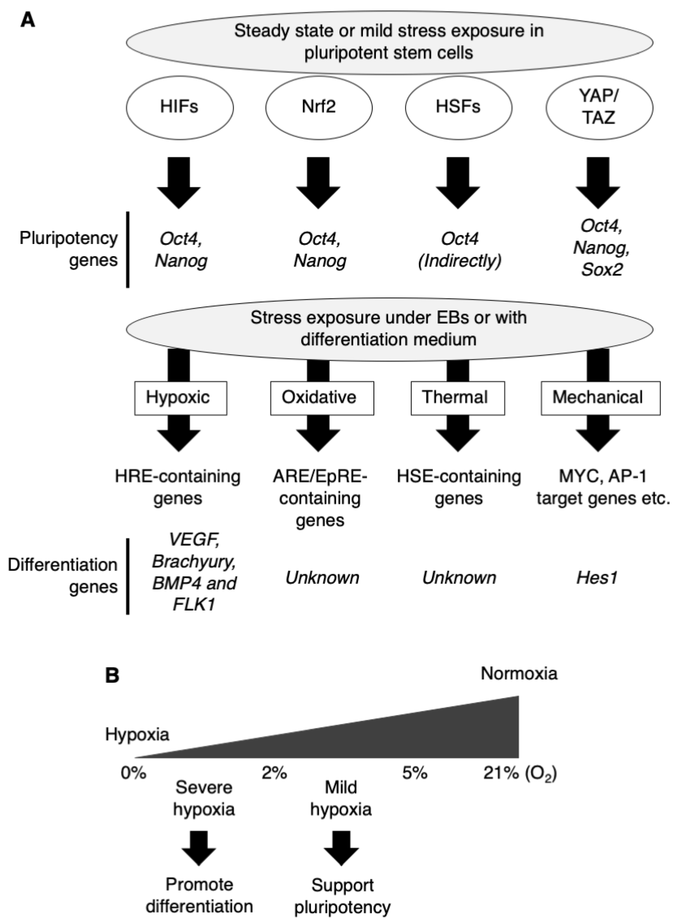
Figure 1.
Environmental stresses and cellular response with transcriptional activation. (A) Steady-state levels of transcription factors as hypoxia-inducible factors (HIFs), Nrf2, HSFs, and YAP/TAZ involved in stress pathways support the maintenance of pluripotency in pluripotent stem cells independently of a stressor. Upon stress exposure, hypoxic stress leads to activation of HIFs (mainly HIF-1α and HIF-2α), followed by induction of hypoxia response element (HRE)-containing genes. Oxidative stress leads to activation of Nrf2, followed by induction of ARE/EpRE-containing genes. Thermal stress leads to activation of heat shock factors (HSFs) (mainly HSF1 and HSF2) followed by induction of HSE-containing genes. Mechanical stress leads to the activation of the YAP/TAZ transcriptional regulator of TEAD followed by induction of MYC and AP-1 target genes. In combination with embryoid body (EB) formation or differentiation medium, those stresses promote the differentiation to each specific lineage via activation of each target genes. EB, embryoid body; HRE, hypoxia response element; ARE, antioxidant-responsive element; EpRE, electrophile-responsive element; HSE, heat shock-responsive element. (B) About the intensity of hypoxic stress, mild hypoxia strengthens pluripotency in stem cell-maintaining medium, while severe hypoxia promote differentiation in differentiation medium.

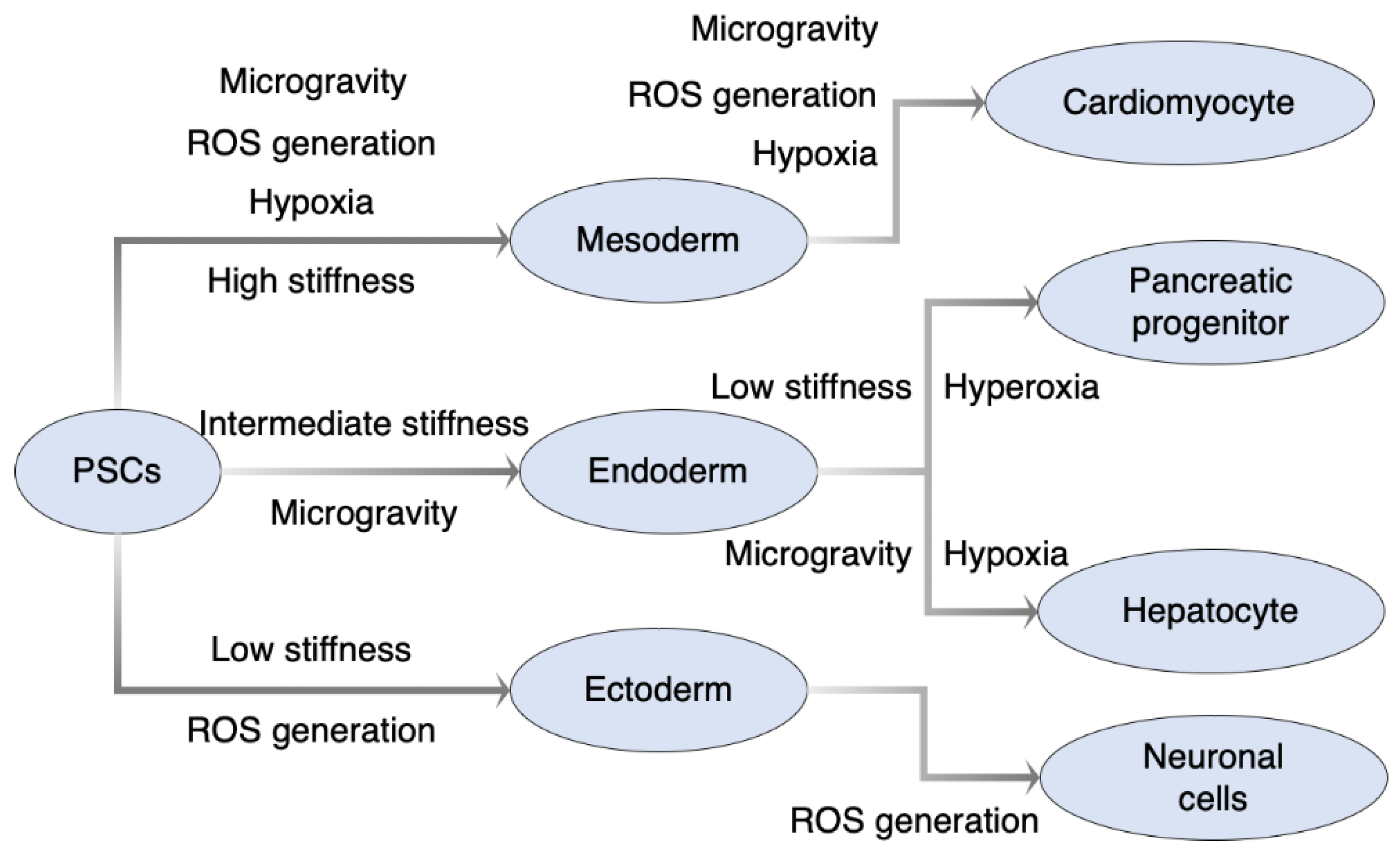
Figure 2.
Summary of directed differentiation with environmental stresses. Hypoxia treatment tends to promote mesodermal differentiation followed by mesoderm-derived lineages like cardiomyocytes. ROS generation by ROS-inducing agents as paraquat, icariin, and H2O2, and treatment as EMF could also promote both mesodermal and ectodermal differentiation. Regarding mechanical stress, there is suitable stiffness of the culture substrate to the differentiation to each 3 germ layers, and it is also utilized for further differentiation like pancreatic progenitors from definitive endoderm. PSCs, pluripotent stem cells; ROS, reactive oxygen species; EMF, electromagnetic field.


Table 1.
The differentiation protocol of pluripotent stem cells using a stress response pathway.
| Stress | Cell Type | Directed Cell Type | Mechanism | References |
|---|---|---|---|---|
| Hypoxic stress | ||||
| Hyperoxia (60% O2) | Mouse ESCs 1, human iPSCs 2 | Pancreatic beta cell | Inhibition of HIF-1αfollowed by Hes1 repression | [37] |
| Hypoxia (1% O2) | Mouse ESCs | Vascular lineage | HIF-1-mediated inverse regulation of Oct4 (down) and VEGF (up) | [39] |
| Hypoxia (0.5% O2) | Mouse ESCs | Mesoderm and cardiomyocyte | HIF-1α mediated Cripto-1 expression | [40] |
| Hypoxia (2% O2) | Human ESCs | Chondrocyte | Undescribed | [42] |
| Hypoxia (1% O2) | Mouse ESCs | Arterial endothelial cells | Activation of ETV2 and NOTCH1 signaling by HIF-1α | [43] |
| Hypoxia (4% O2) | Human ESCs | Cardiomyocytes | Undescribed | [41] |
| Hypoxia (3% O2) | Mouse ESCs | Mesoderm and hemangioblast | Accelerated expression of Brachyury, BMP4 and FLK1 via Arnt | [44] |
| Mild hypoxia (10% O2) | Human iPSCs | Hepatocyte | TGFB signal inhibition | [45] |
| Oxidative stress | ||||
| Paraquat (25 µM) | Human ESCs | Neuronal cells | ROS 3 and activation of MAPK-ERK1/2 | [47] |
| Buthionine sulfoximine (0.2 mM) | Human ESCs | Mesodermal and endodermal lineages | Inactivation of p38 and AKT as well as concomitant transient increase in JNK and ERK signaling | [48] |
| Icariin | Mouse ESCs | Cardiomyocyte | ROS generation and the subsequent activation of p38 MAPK | [49] |
| H2O2 (1~100 nM) | Mouse ESCs | Cardiomyocyte | p38 activation and MEF2C nuclear translocation | [50] |
| Nrf2 shRNA | Human iPSCs | Neuroectoderm | Suppression of Nrf2 binding to pluripotency genes OCT4 and NANOG | [51] |
| Thermal stress | ||||
| Heat shock with mild electrical stimulation (42 °C, 55 pps) | Mouse ESCs | Pdx1-expressing pancreatic progenitors from definitive endoderm | Upregulation of Hsp72 and activation of Akt, ERK, p38 and JNK (putative). | [52] |
| Mechanical stress | ||||
| Fluid shear stress | Mouse ESCs | Vascular endothelial cell | Flk-1 activation and VEGF production | [53] |
| Fluid shear stress | Mouse ESCs | Endothelial and hematopoietic cell | Flk1 activation | [54] |
| Fluid shear stress | Mouse ESCs | Hematopoietic cell | Increased Runx1 expression | [55] |
| High stiffness (BAlg 4 capsule, ~22 kPa) | Human ESCs | Definitive endoderm | Increase in pSMAD/pAkt | [56] |
| Low stiffness (BAlg capsule, ~4 kPa) | Human ESCs | Pancreatic progenitor | Decrease in SHH signaling | [56] |
| High stiffness | Human ESCs | Mesoderm | Undescribed | [57] |
| High stiffness (3D scaffold, 1.5–6 MPa) | Human ESCs | Mesoderm | Undescribed (similar elasticity during gastrulation could be related) | [58] |
| Intermediate stiffness (3D scaffold, 0.1–1 MPa) | Human ESCs | Endoderm | Undescribed (similar elasticity during gastrulation could be related) | [58] |
| Low stiffness (3D scaffold, <0.1 MPa) | Human ESCs | Ectoderm | Undescribed (similar elasticity during gastrulation could be related) | [58] |
| Low stiffness (encapsulated by alginate microbeads) | Mouse ESCs | Endoderm | Undescribed | [59] |
| Confinement (~300 µm2) | Human ESCs | Pancreatic endocrine progenitor | Inhibition of YAP1 | [60] |
1 ESCs, embryonic stem cells; 2 iPSCs, induced pluripotent stem cells; 3 ROS, reactive oxygen species; 4 BAlg, barium alginate.
Table 2.
The differentiation protocol of pluripotent stem cells using physical stimuli.
| Stress | Cell Type | Directed Cell Type | Mechanism | References |
|---|---|---|---|---|
| Microgravity | ||||
| Rotary suspension culture | Mouse ESCs 1 | Mesoderm | Enhancement of Wnt/β-catenin signaling | [96,97] |
| Spaceflight | Mouse iPSCs 2 | Cardiomyocyte | Undescribed | [98] |
| Simulated microgravity and 3D culture | Human ESCs and iPSCs | Cardiomyocyte | Increased proliferation and viability of cardiac progenitors via up-regulation of heat shock proteins and BIRC5 | [99] |
| Simulated microgravity in rotary bioreactor | Mouse ESCs (embryoid body) | Definitive endoderm | Undescribed | [100] |
| Simulated microgravity in rotary bioreactor | Mouse ESCs | Hepatocyte | Undescribed | [101] |
| EMF3 | ||||
| Single electrical field (500 V/m) | Mouse ESCs | Cardiomyocyte | Intracellular ROS 4 generation and NF-κB 5 activation | [105] |
| Static EMF (0.4–2 mT) | Mouse ESCs | Vasculogenesis and chondro-osteogenesis | Intracellular ROS generation and VEGF 6 induction | [106] |
1 ESCs, embryonic stem cells; 2 iPSCs, induced pluripotent stem cells; 3 EMF, electromagnetic field; 4 ROS, reactive oxygen species; 5 NF-κB, nuclear factor κB; 6 VEGF, vascular endothelial growth factor.
Publisher’s Note: MDPI stays neutral with regard to jurisdictional claims in published maps and institutional affiliations. |
© 2021 by the authors. Licensee MDPI, Basel, Switzerland. This article is an open access article distributed under the terms and conditions of the Creative Commons Attribution (CC BY) license (http://creativecommons.org/licenses/by/4.0/).
Share and Cite
MDPI and ACS Style
Kaitsuka, T.; Hakim, F. Response of Pluripotent Stem Cells to Environmental Stress and Its Application for Directed Differentiation. Biology 2021, 10, 84. https://0-doi-org.brum.beds.ac.uk/10.3390/biology10020084
AMA Style
Kaitsuka T, Hakim F. Response of Pluripotent Stem Cells to Environmental Stress and Its Application for Directed Differentiation. Biology. 2021; 10(2):84. https://0-doi-org.brum.beds.ac.uk/10.3390/biology10020084
Chicago/Turabian StyleKaitsuka, Taku, and Farzana Hakim. 2021. "Response of Pluripotent Stem Cells to Environmental Stress and Its Application for Directed Differentiation" Biology 10, no. 2: 84. https://0-doi-org.brum.beds.ac.uk/10.3390/biology10020084
Note that from the first issue of 2016, this journal uses article numbers instead of page numbers. See further details here.