Article

Phytochemicals in Garlic Extract Inhibit Therapeutic Enzyme DPP-4 and Induce Skeletal Muscle Cell Proliferation: A Possible Mechanism of Action to Benefit the Treatment of Diabetes Mellitus

by
Poonam Kalhotra
1,
Veera C.S.R. Chittepu
2,
Guillermo Osorio-Revilla
2 and
Tzayhri Gallardo-Velazquez
1,*
1
Departamento de Biofísica, Escuela Nacional de Ciencias Biológicas, Instituto Politécnico Nacional, Prolongación de Carpio y Plan de Ayala S/N, Col. Santo Tomás, CP. Ciudad de Mexico 11340, Mexico
2
Departamento de Ingeniería Bioquímica, Escuela Nacional de Ciencias Biológicas, Instituto Politecnico Nacional, Av. Wilfrido Massieu S/N, Col. Unidad Profesional Adolfo López Mateos, Zacatenco, CP. Ciudad de Mexico 07738, Mexico
*
Author to whom correspondence should be addressed.
Submission received: 23 December 2019 / Revised: 7 February 2020 / Accepted: 10 February 2020 / Published: 14 February 2020

Abstract

:
Diabetes mellitus is a severe health problem in Mexico, and its prevalence is increasing exponentially every year. Recently, DPP-4 (dipeptidyl peptidase-4) inhibitors have become attractive oral anti-hyperglycemic agents to reduce the pathology of diabetes. Gliptin’s family, such as sitagliptin, vildagliptin, and alogliptin, are in clinical use to treat diabetes mellitus but possess side effects. Therefore, there is a specific need to look for new therapeutic scaffolds (biomolecules). Garlic bulb is widely used as a traditional remedy for the treatment of diabetes. The garlic extracts are scientifically proven to control glucose levels in patients with diabetes, despite the unknown mechanism of action. The aim of the study is to investigate the antidiabetic effects of ultrasonication assisted garlic bulb extract. To achieve this, in-vitro assays such as DPP-4 inhibitory and antioxidant activities were investigated. Further, functional group analysis using FTIR and identification of phytochemicals using mass spectrometry analysis was performed. The results showed that 70.9 µg/mL of garlic bulb extract inhibited 50% DPP-4 activity. On top of that, the garlic extract exhibited a 20% scavenging activity, equivalent to 10 µg/mL of ascorbic acid. Molecular docking simulations on identified phytochemicals using mass spectrometry revealed their potential binding at the DPP-4 druggable region, and therefore the possible DPP-4 inhibition mechanism. These results suggest that prepared garlic extract contains phytochemicals that inhibit DPP-4 and have antioxidant activity. Also, the prepared extract induces skeletal muscle cell proliferation that demonstrates the antidiabetic effect and its possible mechanism of action.

Graphical Abstract

1. Introduction

Diabetes mellitus (DM) is a condition in people with elevated levels of blood glucose and is a common and major health concern in Mexico [1] and around the world [2]. In clinical use, the majority of antidiabetic drugs typically exist with distinct mechanisms of action but unfortunately possess side effects such as nutritional disorders, weight gain, hypoglycemia, liver damage, and allergic reactions [3]. The elevated incidence of side effects undoubtedly makes investigators and companies look for novel antidiabetic agents with fewer side effects. Recent developments in the pharmacology of diabetes mellitus have led to the discovery of more than one therapeutic drug target to adequately reduce glucose levels and, among these, dipeptidyl peptidase-4 was found to be more effective [4].
Dipeptidyl peptidase 4 (DPP-4) is a naturally occurring serine protease expressed on the cell surface and thoroughly known to regulate glucose metabolism. DPP-4 rapidly degrades glucagon-like peptide (GLP-1 and GLP-2), glucose-dependent insulin releasing polypeptide GIP [5], stromal-cell derived factor (SDF-2) [6], chemokine (C-C motif) ligand 5 (RANTES) [7], and eotaxin [8]. Among these specific substrates, incretin hormones secreted by intestinal cells are responsible for adequately controlling blood glucose levels [9]. The fundamental purposes of incretin hormones are to naturally stimulate insulin production and inhibit secreted postprandial glucagon, merely delaying gastric emptying and progressively improving pancreatic β-cell function (shown in Figure 1) [10]. DPP-4 inhibitors are scientifically proven to prolong the incretin effect for the successful treatment of diabetes mellitus.
Currently, GLP-1 receptors have been proven to possess a direct effect on skeletal muscle cells [11] and, upon successful treatment with DPP-4 inhibitors such as vildagliptin, an improvement in energy metabolism was observed [12]. Furthermore, DPP-4 inhibitors of natural origin are proven to stimulate skeletal muscle cell proliferation [13,14]. This promptly led academics and industries to look for the treatment and possible prevention of human diseases by using natural bioactive compounds. There is positively a vast market in the development of functional products due to the growing interest of “healthy,” “pure,” and “clean” foods. Plants and animals serve as a source of enormous, diverse scaffolds [15]. Phytochemicals in plants possess DPP-4 inhibition and are proven to have fewer side effects, are cheaper, and easier to produce at an industrial level [16,17,18,19,20].
In addition, bioactive chemicals from food have proven to be beneficial for improving health because of their functional properties. The bioactive compounds can be naturally antioxidant [21], anti-inflammatory [22], and antifungal [23]. These days, extensive studies on bioactive compounds have promptly led health care sectors and food-based companies to focus collaboratively on the development of functional foods. This functional food contains biomolecules that target specific mechanisms to manage, prevent, and/or treat communicable and non-communicable diseases. Supercritical fluid extraction (SFE) [24], ultrasonic-assisted extraction (UAE) [25], microwave-assisted extraction (MAE) [26], pressurized-liquid extraction (PLE) [27], and enzyme-assisted extraction (EAE) [28] are some of the technologies utilized by academics and industries to extract bioactive molecules from natural sources [29].
Traditionally, plants are used to manage chronic diseases, and recently extracts in combinations with existing therapeutics are recommended to improve the therapeutic effect. Garlic (Allium sativum) naturally belongs to a member of the Alliaceae family, is well recognized as a principal spice, and is additionally used as a remedy for specific ailments and physiological disorders [30]. Figure 2 shows the beneficial effects of garlic proven to lower blood pressure [31], cholesterol [32], combat infections [33], treat metabolic disorders [34], and prevent cancer [35]. Furthermore, garlic extracts are proven to be antidiabetic [36], hepatoprotective [37], anthelmintic [38], anti-inflammatory [39], antioxidant [40], antifungal [41] and wound-healing [42]. The possible mechanism of direct action is still unknown despite the well-known benefits of garlic. The benefits related to diabetes mellitus are reduced blood glucose levels, improved insulin sensitivity, and cardiovascular protection.
In the present study, in vitro anti-diabetic effects of garlic extract were investigated upon dipeptidyl peptidase-4 inhibition and skeletal muscle cell proliferative metabolism. In addition, functional group analysis by Fourier-transform infrared spectroscopy (FTIR), chemical identification using mass spectrometry, and computational studies on identified phytochemicals in garlic extract revealed the possibility to inhibit DPP-4 and its role in the proliferation of skeletal muscle cells.

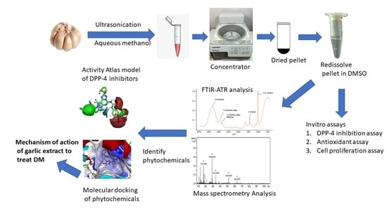
2. Materials and Methods

2.1. Plant Material

Garlic bulbs (Allium sativum L.) were purchased from a local market in Mexico City, Mexico. Garlic bulbs were frozen using liquid nitrogen and crushed into a fine powder. The fine powder was immediately used for biomolecule extraction.

2.2. Chemicals and Reagents

HPLC grade methanol, dimethyl sulfoxide, 2,2-diphenyl-1-picrylhydrazyl (DPPH), and a human DPP-4 inhibitor screening kit were purchased from Sigma Aldrich (St. Louis, MO, USA).

2.3. Preparation of Garlic Extract

A digital ultrasonic bath (G.T.Sonic brand, Model: VGT-1730, Power: 100 W, GuangDong GT Ultrasonic Co., Ltd., Shenzhen, China) was used for the extraction purpose in this study with the frequency of 40 kHz. The extraction was carried out in methanol:water (8:2 v/v) using a ratio garlic powder:solvent of 1:10 in a 100 mL glass beaker. The beaker was incubated for 30 min in an ultrasonic bath and then filtered using syringe filters of 0.45 µm. The solvent was evaporated under vacuum using an Eppendorf Concentrator 5301 (Eppendorf, Hamburg, Germany), and the resulting residue was dissolved in dimethyl sulfoxide (DMSO) for biological activity assays.

2.4. Fourier-Transform Infrared Spectroscopy (FTIR)

Fourier-transform infrared spectroscopy (FTIR) spectra of garlic extract in aqueous methanol (8:2) and DMSO were recorded at room temperature using a Perkin Elmer Frontier spectrum spectrophotometer, scanning over the frequency range of 4000 to 400 cm−1. The spectrum of each sample was acquired using spectrum software version 5.3.1 and a diamond ATR (Perkin Elmer, MA, USA) and each spectrum was an average of 64 scans, with a resolution of 4 cm−1. The aqueous methanol aliquots and DMSO aliquots of 10 µL were uniformly spread directly onto the ATR crystal before each spectrum was collected. Functional group analysis was carried out using Know-it-all software (Bio-Rad, Life Science, Hercules, CA, USA).

2.5. DPPH Radical Scavenging Activity

The 2,2-diphenyl-1-picrylhydrazyl (DPPH) radical scavenging activity was performed as previously reported [43]. Briefly, 4 mg of DPPH was dissolved in 100 mL methanol. In a 96-well plate, 10 µL plant extract and 190 µL DPPH solution were added to each well and incubated for 30 min at room temperature in the dark. The absorbance at 595 nm wavelength was recorded at room temperature. The degree of scavenging activity was calculated using Equation (1).
%   s c a v e n g i n g   a c t i v i t y =   A b s o r b a n c e   o f   c o n t r o l A b s o r b a n c e   o f   p l a n t   e x t r a c t A b s o r b a n c e   o f   c o n t r o l   × 100

2.6. Human Dipeptidyl Peptidase-4 (DPP-4) Inhibition Assay

Human dipeptidyl peptidase-4 (DPP-4) inhibition assay was performed in-vitro using a human inhibitor screening kit. The assay was specific to the fluorescent product Gly-Pro that is cleaved upon the binding of DPP-4 and the fluorescence was monitored (λexcitation = 360 nm and λemission = 460 nm) using a Thermo Scientific Varioskan Flash Multimode Reader (Thermo Scientific, Waltham, MA, USA). All the readings were recorded in a dynamic mode for 30 min. Equation (2) was utilized to calculate the relative percent of DPP-4 inhibition, where ΔF/ΔT represents changes in fluorescence over time.
                    R e l a t i v e   p e r c e n t   o f   D P P 4   i n h i b i t i o n =   F T e n z y m e F T s a m p l e F T e n z y m e × 100

2.7. Identification of Phytochemicals in Garlic Extract

The polyphenols in garlic bulb extract were identified using a mass spectrometer (ESI-micrOTOF-QTM, Bruker Daltonics, Bremen, Germany). Negative ion mode was used to acquire the spectra over a mass range of 50–3000 m/z. The other parameters were capillary voltage 2700 V; dry gas temperature 180 °C; dry gas flow 4.0 L/min; nebulizer pressure 0.4 bar; and spectra rate 1 Hz. Bruker Data Analysis 4.0 software (Bruker Daltonics Inc., Billerica, MA, USA) was used to analyze the data. The native phenolic compounds were assigned based on the matching of molecular weight, isotope pattern, ion mode collected, and considering the polarity of compounds information available in the literature and plant database.

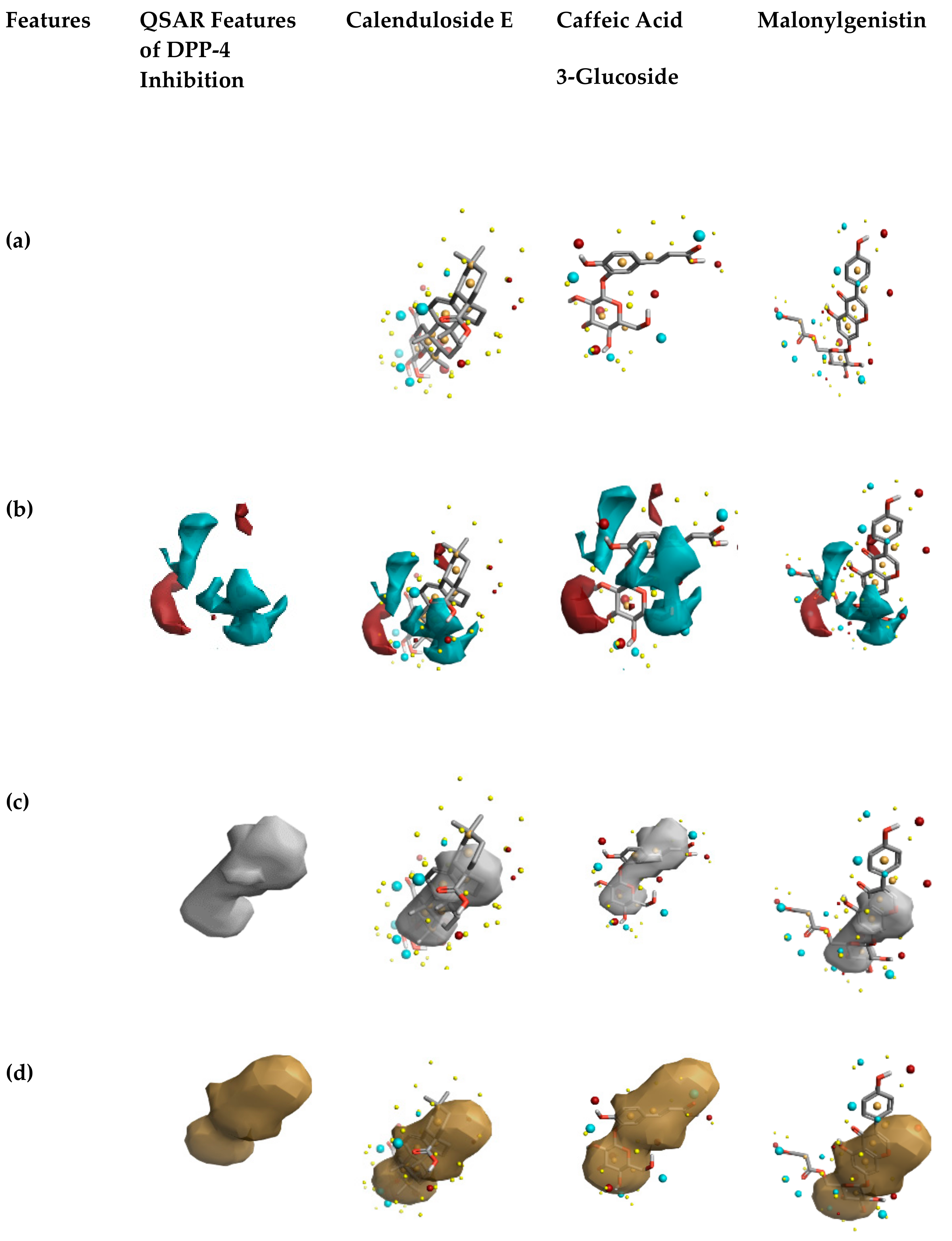
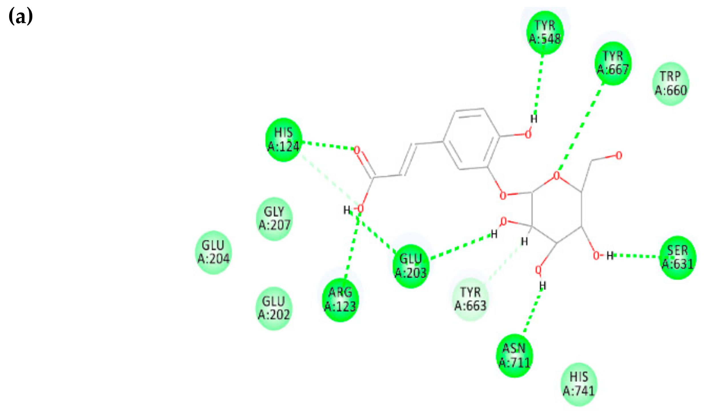
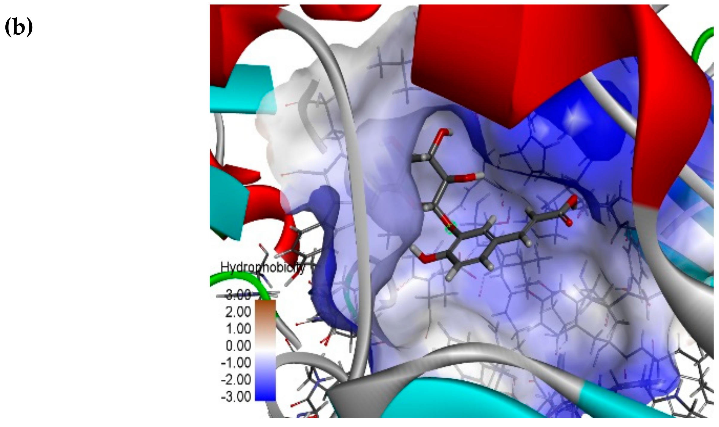
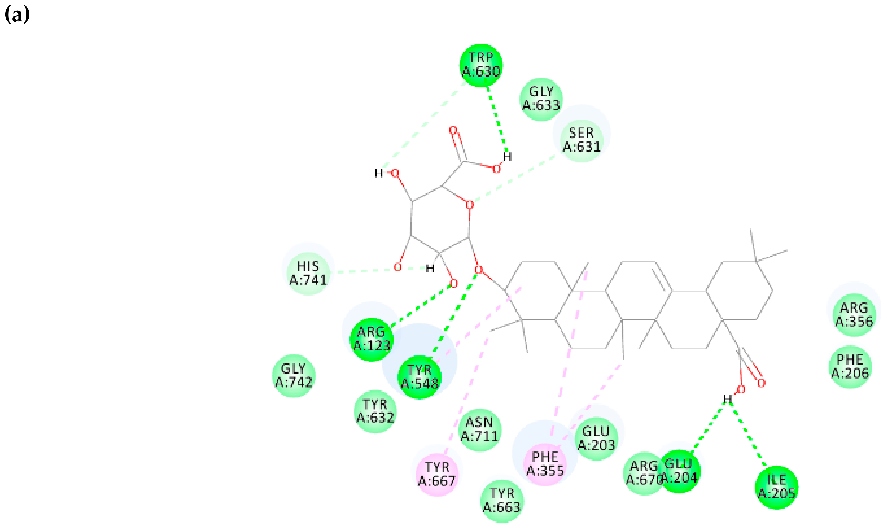
2.8. Activity Atlas Model and Molecular Docking Simulations

In our earlier study [44], the activity atlas model of DPP-4 inhibitors was constructed and validated using Forge software (Cresset Inc., Cambridgeshire, UK). This model was utilized to understand the features of phytochemicals identified in the garlic bulb extract as possible DPP-4 inhibitors.
To perform molecular docking simulations, ensemble docking methodology was utilized in this study using the Flare tool provided by Cresset software (Cresset Inc., Cambridgeshire, UK) [45]. In this study, the DPP-4 in complex with vildagliptin (6B1E), sitagliptin (PDB ID: 4FFW) and anagliptin (PDB ID: 3WQH) were validated initially using structure validation server SAVES and the errors in protein structures were corrected using Flare protein preparation wizard, and the resulting complexes were superimposed.
The DPP-4 binding site is comprised of a pocket where reference ligands, vildagliptin, sitagliptin and anagliptin, are bound. All the atomics coordinates within the grid box were converged before performing the docking of phytochemicals identified in garlic extract. Discovery studio visualizer 2.5.5 was utilized to understand all the interacting residues and binding pose views between phytochemicals and DPP-4.

2.9. Cell Culture, Treatment, and Cell Proliferation Studies

Rat skeletal muscle cells (L6) were cultured as per the procedure described elsewhere [46]. The rat skeletal muscle cells (L6) were cultured in EMEM media, penicillin (100 units/mL), streptomycin (100 µg/mL), and L-glutamine (sterile filtered) at 37 °C in 5% CO2 atmosphere. Rat L6 cells with density 3 × 10³ cells/well were seeded in a normal 96-well microplate and cultured with 2% horse serum for 2 days until semi confluent was reached, and then the cell was allowed to differentiate for 5 days to form myotubes (80–90% myotubes formed). Resultant myotubes were treated with 5 ng/mL garlic bulb extract for 24 h to determine cell proliferation using sulforhodamine B (SRB) dye assay [47]. After 24 h exposure, 50% aqueous trichloroacetic acid (50 µL) was added to treated cells to fix to plastic substratum, and the resultant plates were incubated for 1 h at temperature 4 °C. Then, the plates were washed with water, air-dried, and then stained with 0.4% SRB dye. The unbound or free SRB dye was removed by washing (with 1% aqueous acetic acid) and bound dye was solubilized by adding 100 µL unbuffered Tris base (10 mM). The absorbance was recorded at 515 nm using Synergy HT Microplate Reader (BioTek Instruments, Winooski, VT, USA). All the experiments were carried out in triplicates.

3. Results and Discussion

The use of traditional medicine utilizes a broad range of plants and herbs for the treatment of diabetes mellitus in Mexico and other countries for many years. These plants are considered to play a significantly important role in providing alternative medicine, preventive agents, and as a source of new leads in the process of drug discovery and drug development. Metformin is one of the widely used natural therapeutic drugs to control blood glucose levels in diabetes mellitus, isolated from the Galega officinalis plant [48] and, indeed, oral delivery of metformin improves incretin effects by inhibiting the DPP-4 enzyme [49].
In recent years, DPP-4 has emerged as an important therapeutic drug target to treat diabetes mellitus, and great effort by academics and the pharmaceutical industry have naturally led to the development of DPP-4 inhibitors with few side effects in comparison to current FDA approved therapeutics. During the process of plant screening, garlic was found to be more attractive because of its antidiabetic activity, even though the mechanism of action is unknown. A highly specific fluorometric assay was utilized in this study to determine precisely the DPP-4 inhibition activity by garlic bulb extract. The DPP-4 inhibitory activity of prepared garlic bulb extract in this study is shown in Figure 3 It is observed that the garlic extract showed 60.5% DPP-4 inhibition at 100 µg/mL and the inhibition capacity is concentration-dependent. The results obtained demonstrated a stronger activity in DPP-4 inhibition by prepared garlic bulb extract whose IC50 value is 70.9 µg/mL.
In comparison with methanol extract of green tea (IC50 = 5.3 mg/mL), black tea (IC50 = 6.4 mg/mL) and white tea (IC50 = 0.2 mg/mL), the garlic bulb extract obtained in this work possessed significantly higher DPP-4 inhibitory capacity (IC50 = 70.88 µg/mL). The inhibition of human serine protease dipeptidyl peptidase-4 activity obtained in this study could explain the possible mechanism of the already-observed reduction in blood glucose levels by garlic bulb extract. This inhibition progressively improved the incretin effect and helped in the production of insulin [50].
On the other hand, it is well known that free radicals oxidize many biological components of cells such as proteins, DNA, and lipids, which leads to cell death [51] and finally damages the tissues [52]. Antioxidant activity neutralizes the free radicals and prevents pathogenesis as well as diabetic complications [53,54]. Hence, the garlic bulb extract scavenging ability using the DPPH radical was investigated. It was observed that the prepared garlic bulb extract showed a 20% scavenging activity, which is equivalent to 10 µg/mL ascorbic acid.
Traditional medicinal plants are used to maintain health and serve as an excellent source of new scaffolds for investigators [55,56]. Besides these advantages, plant extracts could exhibit a cytotoxic effect [57]. Thus, the potential cytotoxic studies are necessary to demonstrate the safe use of traditional medicinal plants. To investigate the cytotoxicity of garlic bulb extract in this work, cell proliferation studies were carried out on differentiated skeletal muscle cells. Figure 4 shows the percent of cell proliferation corresponding to differentiated skeletal muscle cells when exposed to 5 ng/mL of prepared garlic bulb extract.
The results In Figure 4 reveal that, upon exposure to prepared garlic bulb extract, there is an increase in cell proliferation by 25% when compared to untreated cells, and this increase behavior is significant (p < 0.05). Based on these, it can be concluded that garlic bulb extract is not acutely toxic to cells. Since garlic extract participates in proliferating differentiated skeletal muscle cells, it can be proposed that it altered cellular metabolism by increasing the rate of glycolysis [58]. This is supported by a few investigations that revealed that garlic extract helps in glucose uptake in skeletal muscle cells [59] and reduces blood glucose levels in diabetic animal models [36,60]. On this account, prepared garlic bulb extract provides a therapeutic opportunity to treat diabetes mellitus by proliferating skeletal muscle cells.
To understand the functional groups present in garlic bulb extract, FTIR spectroscopy was carried out. The spectrum of garlic extract is presented in Figure 5. The band between 1800–800 reveals the chemical fingerprint of garlic extract specifically to polyphenols, which is also supported by Xiaonan Lu et al. [61]. The bands between 1125–1000 cm−1 (C–O stretch), 1475–1415 cm−1 (C–H bend), 2975–2845 cm−1 (C–H stretch), 3450–3225 cm−1 (O–H stretch) reveal the presence of a hydroxy group (R–OH) in garlic extract. Also, the bands between 1060–1020 cm−1 (C–S=O sulfoxy stretch) reveal the presence of aliphatic ether (R–O–R) or sulfoxide (R–(S=O)–R) functional groups in the garlic extract. In addition, the band at 1589 cm−1 and 1559 cm−1 correspond to the C–C stretch of phenyl and ring base [62].
Based on the functional group analysis in garlic bulb extract, it is clear that hydroxyl and phenolics functional groups contributed to the biological activity DPP-4 inhibition, scavenging activity, and cell proliferative metabolism. Bozin et al. [63] and Rasul et al. [64] also demonstrated the presence of phytochemicals in aqueous methanol extract of garlic. Hence, the mass spectrometer was utilized to identify the phytochemicals in the prepared extract. The resulting extract was directly injected into the mass spectrometer to identify phytochemicals. The qualitative analysis showed the main compounds present in garlic extract detected in negative mode (Figure 6). The following compounds were identified and assigned based on existing phytochemical database: catechin ([M − H]−1 m/z 289.07), caffeic acid 4-O-glucoside ([M − H]−1 m/z 341.10), malonylgenistin ([M − H]−1 m/z 518.11), and calenduloside E ([M − H]−1 m/z 631.3). Since it was already proven that garlic extract inhibits DPP-4, and the main compounds in this extract are the ones mentioned above, this led us to hypothesize that these phytochemicals could probably inhibit DPP-Among these compounds, polyphenol catechin was already proven to inhibit DPP-4 [65].
To understand the potential DPP-4 inhibition of the remaining identified phytochemicals in garlic extract, in silico methodologies such as field-based virtual screening and molecular docking simulations were carried out based on our earlier works [44,66], where the qualitative structure-activity relationship model of DPP-4 inhibitors was constructed and validated experimentally.
The prebuilt QSAR model was utilized to demonstrate the potential DPP-4 inhibition capacity of the identified phytochemicals in garlic bulb extract. Initially, the activity cliff summary had assigned novelty scores to the natural compounds. Results revealed all the chemicals maintained a novelty score of very high, and Table 1 shows the similarity score of the identified phytochemicals predicted by the QSAR model for DPP-4 inhibition.
The activity cliff summary of positive and negative electrostatic features of the identified phytochemicals such as the DPP-4 inhibitor is shown in Figure 7, compared to already-built QSAR features of DPP-4 inhibition [44,66]. The red color site represents positive electrostatics features, and blue color denotes negative electrostatic features of DPP-4 inhibition. A comparison of the field differences among the three phytochemicals reveals that caffeic acid 3-glucoside possesses increased electronegative and electropositive features. The remaining two phytochemicals possess similar features responsible for DPP-4 inhibition (shown in Figure 7b).
Finally, comparing the features of the hydrophobic shapes of the constructed activity atlas model [44,66] and the identified phytochemicals in the garlic extract, it is observed that they possess increased hydrophobic shape features (shown in Figure 7d). Regarding the features corresponding to average activity shape, Figure 7c shows that the phytochemicals in the garlic extract and QSAR model possess similar shapes. It is clear that the phytochemicals in garlic extract possess features similar to already existing DPP-4 inhibitors. To understand further the phytochemicals interactions with DPP-4, molecular docking simulation was performed.
In this study, ensemble docking was employed, and the docking protocol used was validated using a redocking methodology. It was observed that the Lead Finder docking algorithm [67] could replicate interacting residues to the structures determined using crystallography of vildagliptin, sitagliptin, and anagliptin. The scoring extra precision docking method was used to calculate free energy (ΔG) values of 10 different poses of the remaining three phytochemicals identified in garlic bulb extract. The calculated binding energies are shown in Table 2.
Caffeic acid 3-glucoside possesses the binding free energy of −7.436 kcal/mol and the interacting residues involved at the binding site are TYR 548, TYR 667, TRP 660, SER 631, ASN 711, HIS 741, TYR 663, GLU 203, ARG 123, GLU 202, GLY 207, and GLU 204 (shown in Figure 8). Further non-bonding interaction analysis between caffeic acid 3-glucoside and the DPP-4 active site revealed strong hydrogen-bonding interactions with residues ASN 711, SER 631, GLU 203, TYR 548, GLU 203, TYR 663, ARG 123, HIS 124, and TYR 667. Since caffeic acid 3-glucoside possesses a binding affinity to DPP-4 and the interacting residues belong to the druggable region of DPP-4 (Figure 8), it can be proposed as a new lead to inhibit DPP-4 and treat diabetes mellitus.
Regarding the polyphenol malonylgenistein, it possesses a binding free energy of −7.438 kcal/mol, which is similar to caffeic acid 3-glucoside and the interacting residues involved at the binding site are TRP 630, GLY 633, SER 631, ARG 356, PHE 206, GLU 204, ILE 205, ARG 670, GLU 203, PHE 355, TYR 663, ASN 711, TYR 667, TYR 548, ARG 123, GLY 742, and HIS 741 (shown in Figure 9). Studies on non-bonding interacting analysis of polyphenol malonylgenistein and the DPP-4 interacting site revealed the following: hydrogen bond interactions with residues VAL 207, ARG 358, SER 630, SER 209, PHE 357, and TYR 662; negative electrostatic interactions in specific π-anion with residues GLU 205; hydrophobic interactions with residues TYR 662 and HIS 740 (π-π stacked), TYR 666 (π-π T shaped), and VAL 711 (π-alkyl). Based on the above, it can be proposed that malonylgenistein has a high probability to inhibit DPP-This can be supported by the fact that genistein has been proven to inhibit DPP-4 (IC50 = 0.48 µM), and have antidiabetic properties [68]. Malonylgenistein being a derivative of genistein, it is very probable that it also has the same functional properties.
Finally, the phytochemical calenduloside E (oleanolic acid 3-O-beta-d-glucosiduronic acid) possesses a binding free energy of −10.172 kcal/mol and the interacting residues involved at the binding pocket of DPP-4 are HIS 741, ARG 123, TYR 548, TYR 632, PHE 355, GLU 203, ARG 670, GLU 204, ILE 205, PHE 206, ARG 356, SER 631, GLY 633, TRP 630, and TYR 667 (Figure 10). Besides, a non-bonding interacting residue analysis of the calenduloside E and DPP-4 interacting site revealed the following: stronger hydrogen bond interactions with residues GLU 204, ILE 205, TRP 630, ARG123, and TYR 548 (conventional hydrogen bonding); carbon-hydrogen bonding with interaction residues HIS 747 and SER 631; hydrophobic interactions with PHE 355, TYR 548 and TYR 667 (π-alkyl). Based on the above, it can be proposed that calenduloside E also has a high probability to inhibit DPP-4.
Calenduloside E is a derivative of oleanolic acid, which has been proven to be present in garlic extract [69] and to have antidiabetic properties [70]. The findings in this study show that calenduloside E present in garlic extract probably possesses DPP-4 inhibition activity, which could be the possible mechanism of action to reduce glucose levels in diabetes mellitus.

4. Conclusions

Traditional plants are a source of bioactive compounds with diverse scaffolds, well known to treat and manage metabolic disorders like diabetes mellitus. In this study, ultrasonic-assisted garlic extract has been demonstrated as a promising anti-diabetic agent by inhibiting therapeutic drug target dipeptidyl peptidase 4, also having scavenging activity, with the capacity to induce skeletal muscle cell proliferation. FTIR, mass spectrometry and in silico studies reveal the presence of phytochemicals in garlic extract that are probably responsible for DPP-4 inhibition and provide a new mechanism of action of garlic extract to be used for treatment and management of diabetes mellitus. However, further studies related to the inhibition of DPP-4 and hypoglycemic activity of individual compounds present in the prepared garlic extract need to be evaluated for the possible antidiabetic usage.

Author Contributions

P.K., T.G.-V., V.C.S.R.C., and G.O.-R. designed the experiments; P.K. and V.C.S.R.C. are responsible for in silico studies and in vitro studies. T.G.-V. and G.O.-R. contributed to analysis tools and in the “results and discussion section”; P.K., wrote the manuscript; All authors read and approve the manuscript. All authors have read and agreed to the published version of the manuscript.

Funding

This research received no external funding.

Acknowledgments

Authors wish to express their gratitude to Escuela Nacional de Ciencias Biológicas-Instituto Politécnico Nacional (ENCB-IPN) for providing the financial support for the present study. Authors would like to thank CNMN-IPN for analytical services. P.K. and V.C.S.R.C. would like to thank to Consejo Nacional de Ciencia y Tecnología (CONACYT) for the grant provided.

Conflicts of Interest

The authors declare no conflict of interest.

References

  1. Dávila-Cervantes, C.A.; Pardo-Montaño, A.M. Diabetes mellitus: Contribution to changes in the life expectancy in Mexico 1990, 2000, and 2010. Revista de Salud Publica 2014, 16, 910–923. [Google Scholar] [PubMed]
  2. Forouhi, N.G.; Wareham, N.J. Epidemiology of diabetes. Med. (United Kingdom) 2019, 47, 22–27. [Google Scholar] [CrossRef]
  3. Chaudhury, A.; Duvoor, C.; Dendi, V.S.R.; Kraleti, S.; Chada, A.; Ravilla, R.; Marco, A.; Shekhawat, N.S.; Montales, M.T.; Kuriakose, K.; et al. Clinical Review of Antidiabetic Drugs: Implications for Type 2 Diabetes Mellitus Management. Front. Endocrinol. 2017, 8, 28. [Google Scholar] [CrossRef] [PubMed] [Green Version]
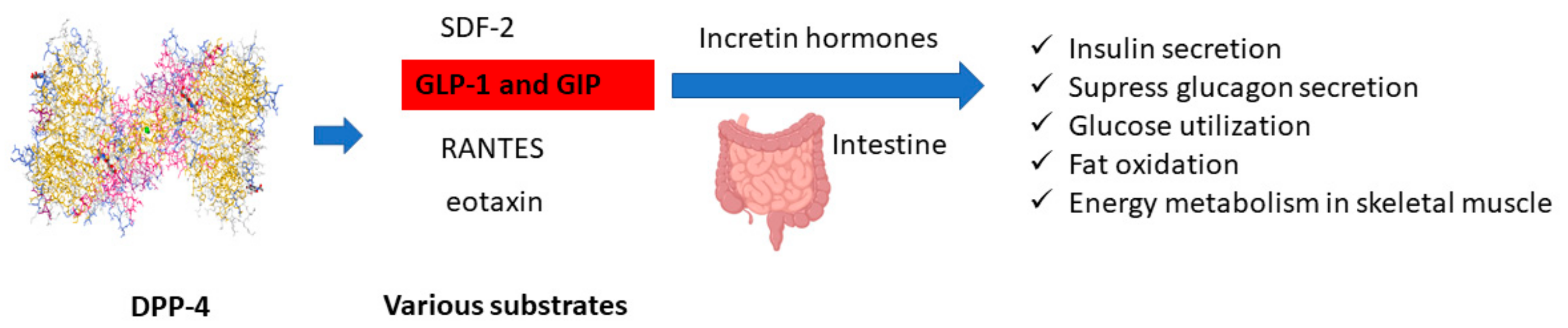
  4. Ross, S.A.; Ekoé, J.-M. Incretin agents in type 2 diabetes. Can. Fam. Physician Med. Fam. Can. 2010, 56, 639–648. [Google Scholar]
  5. Matheeussen, V.; Jungraithmayr, W.; De Meester, I. Dipeptidyl peptidase 4 as a therapeutic target in ischemia/reperfusion injury. Pharmacol. Ther. 2012, 136, 267–282. [Google Scholar] [CrossRef]
  6. Fujiwara, K.; Inoue, T.; Henmi, Y.; Hirata, Y.; Naka, Y.; Hara, A.; Kakimoto, K.; Nouda, S.; Okada, T.; Kawakami, K.; et al. Sitagliptin, a dipeptidyl peptidase-4 inhibitor, suppresses CXCL5 and sdf-1 and does not accelerate intestinal neoplasia formation in Apc min/+ mice fed a high-fat diet. Oncol. Lett. 2017, 14, 4355–4360. [Google Scholar] [CrossRef]
  7. Iwata, S.; Yamaguchi, N.; Munakata, Y.; Ikushima, H.; Lee, J.F.; Hosono, O.; Schlossman, S.F.; Morimoto, C. CD26/dipeptidyl peptidase IV differentially regulates the chemotaxis of T cells and monocytes toward RANTES: possible mechanism for the switch from innate to acquired immune response. Int. Immunol. 1999, 11, 417–426. [Google Scholar] [CrossRef] [Green Version]
  8. Struyf, S.; Proost, P.; Schols, D.; De Clercq, E.; Opdenakker, G.; Lenaerts, J.P.; Detheux, M.; Parmentier, M.; De Meester, I.; Scharpé, S.; et al. CD26/dipeptidyl-peptidase IV down-regulates the eosinophil chemotactic potency, but not the anti-HIV activity of human eotaxin by affecting its interaction with CC chemokine receptor. J. Immunol. 1999, 162, 4903–4909. [Google Scholar]
  9. Nauck, M.A. Incretin-Based Therapies for Type 2 Diabetes Mellitus: Properties, Functions, and Clinical Implications. Am. J. Med. 2011, 124, S3–S18. [Google Scholar] [CrossRef]
  10. Al-Sabah, S. Molecular Pharmacology of the Incretin Receptors. Med. Princ. Pract. 2016, 25, 15–21. [Google Scholar] [CrossRef] [Green Version]
  11. Luque, M.; Gonzalez, N.; Marquez, L.; Acitores, A.; Redondo, A.; Morales, M.; Valverde, I.; Villanueva-Penacarrillo, M. Glucagon-like peptide-1 (GLP-1) and glucose metabolism in human myocytes. J. Endocrinol. 2002, 173, 465–473. [Google Scholar] [CrossRef] [PubMed] [Green Version]
  12. Boschmann, M.; Engeli, S.; Dobberstein, K.; Budziarek, P.; Strauss, A.; Boehnke, J.; Sweep, F.C.G.J.; Luft, F.C.; He, Y.L.; Foley, J.E.; et al. Dipeptidyl-peptidase-IV inhibition augments postprandial lipid mobilization and oxidation in type 2 diabetic patients. J. Clin. Endocrinol. Metab. 2009, 94, 846–852. [Google Scholar] [CrossRef] [PubMed] [Green Version]
  13. Kalhotra, P.; Chittepu, V.C.S.R.; Osorio-Revilla, G.; Gallardo-Velazquez, T. Chrysin in Combination with Insulin Promotes Glucose Uptake in Skeletal Muscle Cell: Impact of Combination Therapy in Diabetes Myopathy (P01-031-19). Curr. Dev. Nutr. 2019, 3, 3. [Google Scholar] [CrossRef] [Green Version]
  14. Kalhotra, P.; Chittepu, V.C.S.R.; Osorio-Revilla, G.; Gallardo-Velázquez, T. Discovery of Galangin as a Potential DPP-4 Inhibitor That Improves Insulin-Stimulated Skeletal Muscle Glucose Uptake: A Combinational Therapy for Diabetes. Int. J. Mol. Sci. 2019, 20, 1228. [Google Scholar] [CrossRef] [Green Version]
  15. Finley, J. Functional Foods. In Adequate Food for All; Informa UK Limited: Colchester, UK, 2009; pp. 201–216. [Google Scholar]
  16. Ekayanti, M.; Sauriasari, R.; Elya, B. Dipeptidyl peptidase IV inhibitory activity of fraction from white tea ethanolic extract (Camellia sinensis (L.) Kuntze) ex vivo. Pharmacogn. J. 2018, 10, 190–193. [Google Scholar] [CrossRef] [Green Version]
  17. Elya, B.; Handayani, R.; Sauriasari, R.; Hasyyati, U.S.; Permana, I.T.; Permatasar, Y.I. Antidiabetic Activity and Phytochemical Screening of Extracts from Indonesian Plants by Inhibition of Alpha Amylase, Alpha Glucosidase and Dipeptidyl Peptidase IV. Pak. J. Biol. Sci. 2015, 18, 279–284. [Google Scholar] [CrossRef]
  18. Rachpirom, M.; Ovatlarnporn, C.; Thengyai, S.; Sontimuang, C.; Puttarak, P. Dipeptidyl peptidase-IV (DPP-IV) inhibitory activity, antioxidant property and phytochemical composition studies of herbal constituents of thai folk anti-diabetes remedy. Walailak J. Sci. Technol. 2016, 13, 803–814. [Google Scholar]
  19. Purnomo, Y.; Soeatmadji, D.W.; Sumitro, S.B.; Widodo, M.A. Anti-diabetic potential of Urena lobata leaf extract through inhibition of dipeptidyl peptidase IV activity. Asian Pac. J. Trop. Biomed. 2015, 5, 645–649. [Google Scholar] [CrossRef] [Green Version]
  20. Bhat, S.H.; Ullah, M.F.; Abu-Duhier, F.M. Bioactive extract of Artemisia judaica causes in vitro inhibition of dipeptidyl peptidase IV and pancreatic/intestinal enzymes of the carbohydrate absorption cascade: Implication for anti-diabetic new molecular entities (NMEs). Orient. Pharm. Exp. Med. 2019, 19, 71–80. [Google Scholar] [CrossRef]
  21. Rice-Evans, C.; Miller, N.; Paganga, G. Antioxidant properties of phenolic compounds. Trends Plant Sci. 1997, 2, 152–159. [Google Scholar] [CrossRef]
  22. Moro, C.; Palacios, I.; Lozano, M.; D’Arrigo, M.; Guillamón, E.; Villares, A.; Martínez, J.A.; Garcia-Lafuente, A. Anti-inflammatory activity of methanolic extracts from edible mushrooms in LPS activated RAW 264.7 macrophages. Food Chem. 2012, 130, 350–355. [Google Scholar] [CrossRef]
  23. Arif, T.; Bhosale, J.; Kumar, N.; Mandal, T.; Bendre, R.; Lavekar, G.; Dabur, R. Natural products—Antifungal agents derived from plants. J. Asian Nat. Prod. Res. 2009, 11, 621–638. [Google Scholar] [CrossRef] [PubMed]
  24. Khaw, K.-Y.; Parat, M.-O.; Shaw, P.N.; Falconer, J.R. Solvent Supercritical Fluid Technologies to Extract Bioactive Compounds from Natural Sources: A Review. Molecules 2017, 22, 1186. [Google Scholar] [CrossRef] [PubMed]
  25. Wu, J.-Y. Ultrasound-assisted extraction of ginseng saponins from ginseng roots and cultured ginseng cells. Ultrason. Sonochem. 2001, 8, 347–352. [Google Scholar] [CrossRef]
  26. Zhang, H.-F.; Yang, X.-H.; Wang, Y. Microwave assisted extraction of secondary metabolites from plants: Current status and future directions. Trends Food Sci. Technol. 2011, 22, 672–688. [Google Scholar] [CrossRef]
  27. Mustafa, A.; Turner, C. Pressurized liquid extraction as a green approach in food and herbal plants extraction: A review. Anal. Chim. Acta 2011, 703, 8–18. [Google Scholar] [CrossRef] [PubMed]
  28. Puri, M.; Sharma, D.; Barrow, C.J. Enzyme-assisted extraction of bioactives from plants. Trends Biotechnol. 2012, 30, 37–44. [Google Scholar] [CrossRef] [PubMed]
  29. Gil-Chávez, G.J.; Villa, J.A.; Ayala-Zavala, J.F.; Heredia, J.B.; Sepúlveda, D.; Yahia, E.M.; González-Aguilar, G.A. Technologies for Extraction and Production of Bioactive Compounds to be Used as Nutraceuticals and Food Ingredients: An Overview. Compr. Rev. Food Sci. Food Saf. 2013, 12, 5–23. [Google Scholar] [CrossRef]
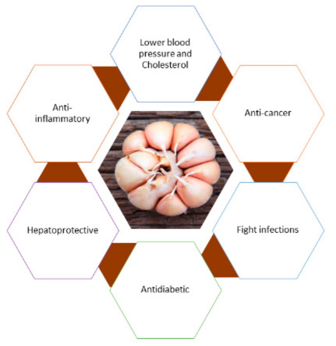
  30. Bayan, L.; Koulivand, P.H.; Gorji, A. Garlic: A review of potential therapeutic effects. Avicenna J. Phytomed. 2014, 4, 1–14. [Google Scholar]
  31. Ried, K.; Fakler, P. Potential of garlic (Allium sativum) in lowering high blood pressure: Mechanisms of action and clinical relevance. Integr. Blood Press. Control 2014, 7, 71–82. [Google Scholar] [CrossRef] [Green Version]
  32. Yeh, Y.-Y.; Liu, L. Cholesterol-lowering effect of garlic extracts and organosulfur compounds: human and animal studies. J. Nutr. 2001, 131, 989–993. [Google Scholar] [CrossRef] [PubMed] [Green Version]
  33. Abubakar, E.M.M. Efficacy of crude extracts of garlic (Allium sativum Linn.) against nosocomial Escherichia coli, Staphylococcus aureus, Streptococcus pneumoniea and Pseudomonas aeruginosa. J. Med. Plants Res. 2009, 3, 179–185. [Google Scholar]
  34. Padiya, R.; Khatua, T.N.; Bagul, P.K.; Kuncha, M.; Banerjee, S.K. Garlic improves insulin sensitivity and associated metabolic syndromes in fructose fed rats. Nutr. Metab. 2011, 8, 53. [Google Scholar] [CrossRef] [PubMed] [Green Version]
  35. Rivlin, R.S.; Budoff, M.; Amagase, H. Significance of Garlic and Its Constituents in Cancer and Cardiovascular Disease. J. Nutr. 2006, 136. [Google Scholar] [CrossRef] [Green Version]
  36. Eidi, A.; Eidi, M.; Esmaeili, E. Antidiabetic effect of garlic (Allium sativum L.) in normal and streptozotocin-induced diabetic rats. Phytomedicine 2006, 13, 624–629. [Google Scholar] [CrossRef] [PubMed]
  37. Shin, J.H.; Lee, C.W.; Oh, S.J.; Yun, J.; Kang, M.R.; Han, S.-B.; Park, H.; Jung, J.C.; Chung, Y.H.; Kang, J.S. Hepatoprotective Effect of Aged Black Garlic Extract in Rodents. Toxicol. Res. 2014, 30, 49–54. [Google Scholar] [CrossRef] [Green Version]
  38. Worku, M.; Franco, R.; Baldwin, K. Efficacy of garlic as an anthelmintic in adult Boer goats. Arch. Biol. Sci. 2009, 61, 135–140. [Google Scholar] [CrossRef]
  39. Arreola, R.; Quintero-Fabián, S.; Lopez-Roa, R.I.; Flores-Gutiérrez, E.O.; Reyes-Grajeda, J.P.; Carrera-Quintanar, L.; Ortuño-Sahagun, D. Immunomodulation and Anti-Inflammatory Effects of Garlic Compounds. J. Immunol. Res. 2015, 2015, 1–13. [Google Scholar] [CrossRef]
  40. Rahman, M.M.; Fazlic, V.; Saad, N.W. Antioxidant properties of raw garlic (Allium sativum) extract. Int. Food Res. J. 2012, 19, 589–591. [Google Scholar]
  41. Hughes, B.G.; Lawson, L.D. Antimicrobial effects of Allium sativum L. (garlic), Allium ampeloprasum L. (elephant garlic), and Allium cepa L. (onion), garlic compounds and commercial garlic supplement products. Phyther. Res. 1991, 5, 154–158. [Google Scholar] [CrossRef]
  42. Ejaz, S.; Chekarova, I.; Cho, J.W.; Lee, S.Y.; Ashraf, S.; Lim, C.W. Effect of aged garlic extract on wound healing: A new frontier in wound management. Drug Chem. Toxicol. 2009, 32, 191–203. [Google Scholar] [CrossRef] [PubMed]
  43. Khatua, S.; Ghosh, S.; Acharya, K. Simplified methods for microtiter based analysis of in vitro antioxidant activity. Asian J. Pharm. 2017, 11, S327–S335. [Google Scholar]
  44. Kalhotra, P.; Chittepu, V.; Osorio-Revilla, G.; Gallardo-Velázquez, T. Structure–Activity Relationship and Molecular Docking of Natural Product Library Reveal Chrysin as a Novel Dipeptidyl Peptidase-4 (DPP-4) Inhibitor: An Integrated In Silico and In Vitro Study. Molecules 2018, 23, 1368. [Google Scholar] [CrossRef] [PubMed] [Green Version]
  45. Amaro, R.E.; Baudry, J.; Chodera, J.; Demir, Ö.; McCammon, J.A.; Miao, Y.; Smith, J.C. Ensemble Docking in Drug Discovery. Biophys. J. 2018, 114, 2271–2278. [Google Scholar] [CrossRef]
  46. Chittepu, V.C.S.R.; Kalhotra, P.; Gallardo-Velázquez, T.; La Torre, R.R.-D.; Osorio-Revilla, G.; La Torre, R.R.R.-D. Designed Functional Dispersion for Insulin Protection from Pepsin Degradation and Skeletal Muscle Cell Proliferation: In Silico and In Vitro Study. Nanomaterials 2018, 8, 852. [Google Scholar] [CrossRef] [Green Version]
  47. Orellana, E.A.; Kasinski, A.L. Sulforhodamine B (SRB) Assay in Cell Culture to Investigate Cell Proliferation. BIO-PROTOCOL 2016, 6, 7–13. [Google Scholar] [CrossRef] [Green Version]
  48. Bailey, C.J. Metformin: Historical overview. Diabetologia 2017, 60, 1566–1576. [Google Scholar] [CrossRef] [Green Version]
  49. Lindsay, J.R.; Duffy, N.A.; McKillop, A.M.; Ardill, J.; O’Harte, F.P.M.; Flatt, P.R.; Bell, P.M. Inhibition of dipeptidyl peptidase IV activity by oral metformin in Type 2 diabetes. Diabet. Med. 2005, 22, 654–657. [Google Scholar] [CrossRef]
  50. Deacon, C.F.; Hughes, T.E.; Holst, J.J. Dipeptidyl peptidase IV inhibition potentiates the insulinotropic effect of glucagon-like peptide 1 in the anesthetized pig. Diabetes 1998, 47, 764–769. [Google Scholar] [CrossRef]
  51. Maritim, A.C.; Sanders, R.A.; Watkins, J.B. Diabetes, oxidative stress, and antioxidants: A review. J. Biochem. Mol. Toxicol. 2003, 17, 24–38. [Google Scholar] [CrossRef]
  52. Rao, P.S.; Kalva, S.; Yerramilli, A.; Mamidi, S. Free Radicals and Tissue Damage: Role of Antioxidants. Free Radic. Antioxidants 2011, 1, 2–7. [Google Scholar]
  53. Singh, R.; Devi, S.; Gollen, R. Role of free radical in atherosclerosis, diabetes and dyslipidaemia: Larger-than-life. Diabetes/Metabol. Res. Rev. 2015, 31, 113–126. [Google Scholar] [CrossRef] [PubMed]
  54. Zatalia, S.R.; Sanusi, H. The role of antioxidants in the pathophysiology, complications, and management of diabetes mellitus. Acta Med. Indones. 2013, 45, 141–147. [Google Scholar]
  55. Hamilton, A.C. Medicinal plants, conservation and livelihoods. Biodivers. Conserv. 2004, 13, 1477–1517. [Google Scholar] [CrossRef]
  56. Balunas, M.J.; Kinghorn, A.D. Drug discovery from medicinal plants. Life Sci. 2005, 78, 431–441. [Google Scholar] [CrossRef]
  57. Zaynab, M.; Fatima, M.; Abbas, S.; Sharif, Y.; Umair, M.; Zafar, M.H.; Bahadar, K. Role of secondary metabolites in plant defense against pathogens. Microb. Pathog. 2018, 124, 198–202. [Google Scholar] [CrossRef]
  58. Guo, X.; Li, H.; Xu, H.; Woo, S.; Dong, H.; Lu, F.; Lange, A.J.; Wu, C. Glycolysis in the control of blood glucose homeostasis. Acta Pharm. Sin. B 2012, 2, 358–367. [Google Scholar] [CrossRef] [Green Version]
  59. Noipha, K.; Ratanachaiyavong, S.; Ninla-aesong, P. Enhancement of glucose transport by selected plant foods in muscle cell line L6. Diabetes Res. Clin. Pract. 2010, 89, e22–e26. [Google Scholar]
  60. Thomson, M.; Al-Amin, Z.M.; Al-Qattan, K.K.; Shaban, L.H.; Ali, M. Anti-diabetic and hypolipidaemic properties of garlic (Allium sativum) in streptozotocin-induced diabetic rats. Int. J. Diabetes Metab. 2007, 15, 108–115. [Google Scholar]
  61. Lu, X.; Ross, C.F.; Powers, J.R.; Aston, D.E.; Rasco, B.A. Determination of Total Phenolic Content and Antioxidant Activity of Garlic (Allium sativum) and Elephant Garlic (Allium ampeloprasum) by Attenuated Total Reflectance–Fourier Transformed Infrared Spectroscopy. J. Agric. Food Chem. 2011, 59, 5215–5221. [Google Scholar] [CrossRef]
  62. Schulz, H.; Baranska, M. Identification and quantification of valuable plant substances by IR and Raman spectroscopy. Vib. Spectrosc. 2007, 43, 13–25. [Google Scholar] [CrossRef]
  63. Bozin, B.; Mimica-Dukić, N.; Samojlik, I.; Goran, A.; Igic, R. Phenolics as antioxidants in garlic (Allium sativum L., Alliaceae). Food Chem. 2008, 111, 925–929. [Google Scholar] [CrossRef]
  64. Rasul Suleria, H.A.; Sadiq Butt, M.; Muhammad Anjum, F.; Saeed, F.; Batool, R.; Nisar Ahmad, A. Aqueous garlic extract and its phytochemical profile; Special reference to antioxidant status. Int. J. Food Sci. Nutr. 2012, 63, 431–439. [Google Scholar] [CrossRef] [PubMed]
  65. Kempegowda, P.K.; Zameer, F.; Murari, S.K. Delineating antidiabetic proficiency of catechin from Withania somnifera and its Inhibitory action on dipeptidyl peptidase-4 (DPP-4). Biomed. Res. 2018, 29, 3192–3200. [Google Scholar] [CrossRef] [Green Version]
  66. Chittepu, V.C.S.R.; Kalhotra, P.; Osorio-Gallardo, T.; Gallardo-Velázquez, T.; Osorio-Revilla, G. Repurposing of FDA-Approved NSAIDs for DPP-4 Inhibition as an Alternative for Diabetes Mellitus Treatment: Computational and in Vitro Study. Pharmaceutics 2019, 11, 238. [Google Scholar] [CrossRef] [PubMed] [Green Version]
  67. Stroganov, O.V.; Novikov, F.N.; Stroylov, V.S.; Kulkov, V.; Chilov, G.G. Lead Finder: An Approach To Improve Accuracy of Protein−Ligand Docking, Binding Energy Estimation, and Virtual Screening. J. Chem. Inf. Model. 2008, 48, 2371–2385. [Google Scholar] [CrossRef] [PubMed]
  68. Yousefi, H.; Karimi, P.; Alihemmati, A.; Reza Alipour, M.; Habibi, P.; Ahmadiasl, N. Therapeutic potential of genistein in ovariectomy-induced pancreatic injury in diabetic rats: The regulation of mapk pathway and apoptosis. Iran. J. Basic Med. Sci. 2017, 20, 1009–1015. [Google Scholar]
  69. Meshram, G.A.; Khamkar, S.S. Effect Of Oleanolic Acid Isolated From Garlic Leaves On Carbohydrate Metabolizing Enzymes, In Vitro. Int. J. Pharma Sci. Res. 2014, 5, 988–991. [Google Scholar]
  70. Wang, X.; Li, Y.L.; Wu, H.; Liu, J.Z.; Hu, J.X.; Liao, N.; Peng, J.; Cao, P.P.; Liang, X.; Hai, C.X. Antidiabetic effect of oleanolic acid: A promising use of a traditional pharmacological agent. Phyther. Res. 2011, 25, 1031–1040. [Google Scholar] [CrossRef]
Figure 1. Potential role of protecting incretin hormone degradation by DPP-4 enzyme.
Biomolecules 10 00305 g001
Figure 2. Overall beneficial effects of garlic.
Biomolecules 10 00305 g002
Figure 3. Percent of DPP-4 inhibition of ultrasonic-assisted extraction of garlic in aqueous methanol. Data are expressed as mean ± S.D.
Biomolecules 10 00305 g003
Figure 4. Cell proliferation studies of skeletal muscle cell lines treated with garlic bulb extract (5 ng/mL) and untreated cells for 24 h. All data in the graph are represented as the mean ± S.D of triplicates. Two-tailed unpaired t-test was used to calculate significant difference at p < 0.05.
Biomolecules 10 00305 g004
Figure 5. FTIR bands of garlic bulb extract.
Biomolecules 10 00305 g005
Figure 6. Garlic bulb extract m/z peaks detected in mass spectrometer (in negative mode).
Biomolecules 10 00305 g006
Figure 7. The activity atlas model revealing insights into DPP-4 inhibitory activity. (a) 3D structures of phytochemicals identified in garlic bulb extract. (b) Activity cliff summary features of DPP-4 inhibitors and phytochemicals of garlic bulb extract. In this, red color shows the positive electrostatic field, and blue color represents the negative electrostatic field responsible for DPP-4 inhibition. (c) Average shapes of activity of DPP-4 inhibitors and natural compounds of garlic bulb extract. (d) Average hydrophobics of active shape of natural compounds of garlic bulb extract and DPP-4 inhibitors.
Biomolecules 10 00305 g007
Figure 8. Phytochemical caffeic acid 3-glucoside bound structure on the DPP-4 druggable region with active site residues interacting analysis. (a) Two-dimensional interaction analysis, (b) binding pose view.
Biomolecules 10 00305 g008aBiomolecules 10 00305 g008b
Figure 9. Phytochemical malonylgenistein bound structure on DPP-4 druggable region with active site residue interacting analysis. (a) Two-dimensional interaction analysis, (b) binding pose view.
Biomolecules 10 00305 g009aBiomolecules 10 00305 g009b
Figure 10. Phytochemical calenduloside E bound structure on DPP-4 druggable region with active site residue interacting analysis. (a) Two-dimensional study interaction analysis, (b) binding pose view.
Biomolecules 10 00305 g010
Table 1. Similarity score and novelty score of phytochemicals in garlic extract predicted by activity atlas model for DPP-4 inhibition.
Phytochemical Name Similarity Score to Field TemplateNovelty Score
Caffeic acid 3-glucoside0.43Very high
Calenduloside E0.4Very high
Malonylgenistin0.41Very high
Table 2. Calculated binding energies between DPP-4 and phytochemicals of garlic bulb extract.
Phytochemical NameProtein–Ligand Binding Free Energy ΔG (kcal/mol)
Caffeic acid 3-glucoside−7.436
Calenduloside E−10.172
Malonylgenistin−7.438

