Amaryllidaceae Alkaloids of Belladine-Type from Narcissus pseudonarcissus cv. Carlton as New Selective Inhibitors of Butyrylcholinesterase

 ,
,  ,
,  , and
, and
Abstract
:
1. Introduction
2. Results and Discussion
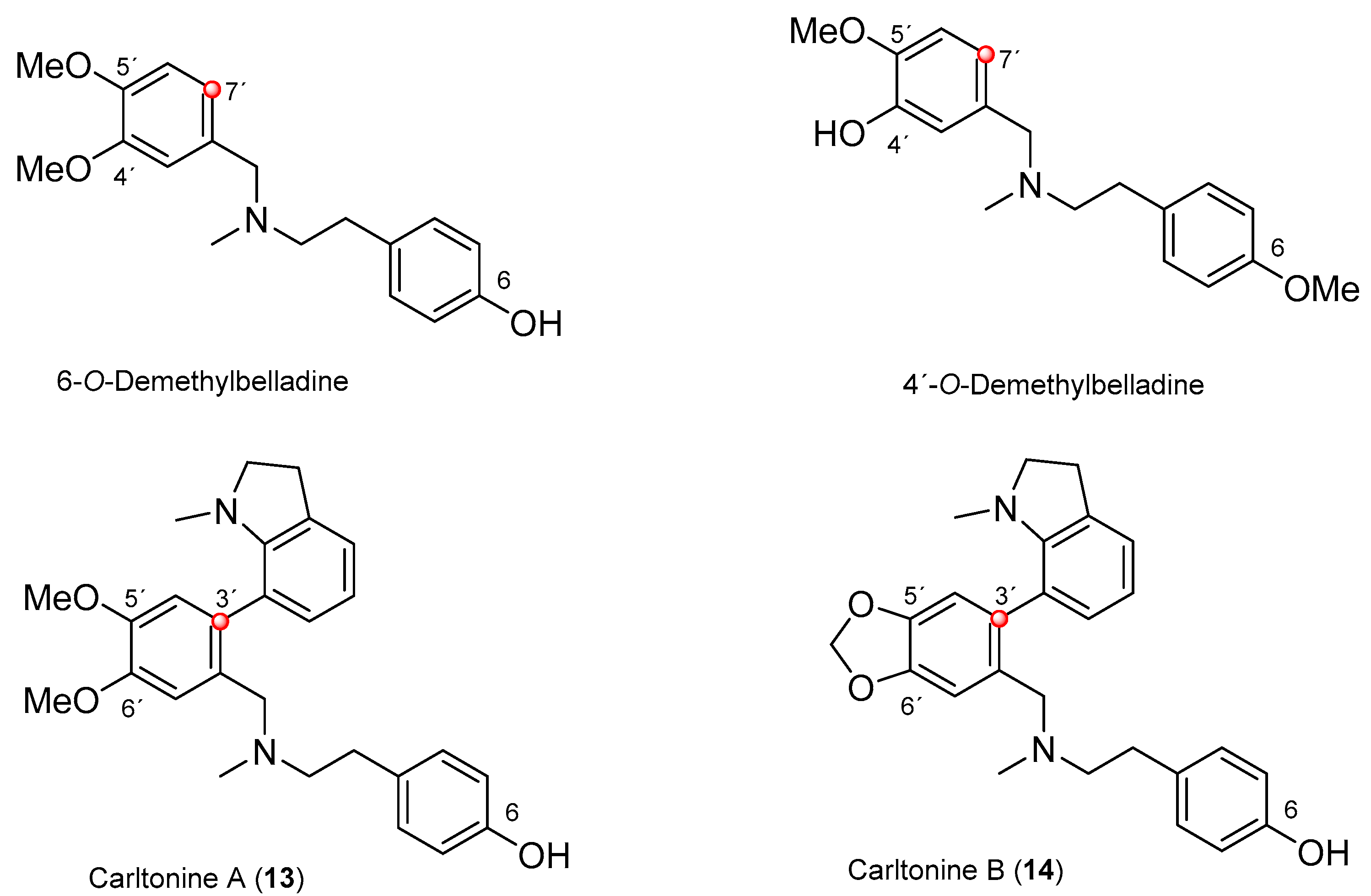
2.1. Phytochemical Study of Narcissus pseudonarcissus cv. Carlton
2.2. Biological Activity Determination of Isolated Alkaloids
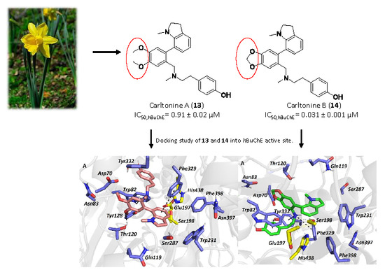
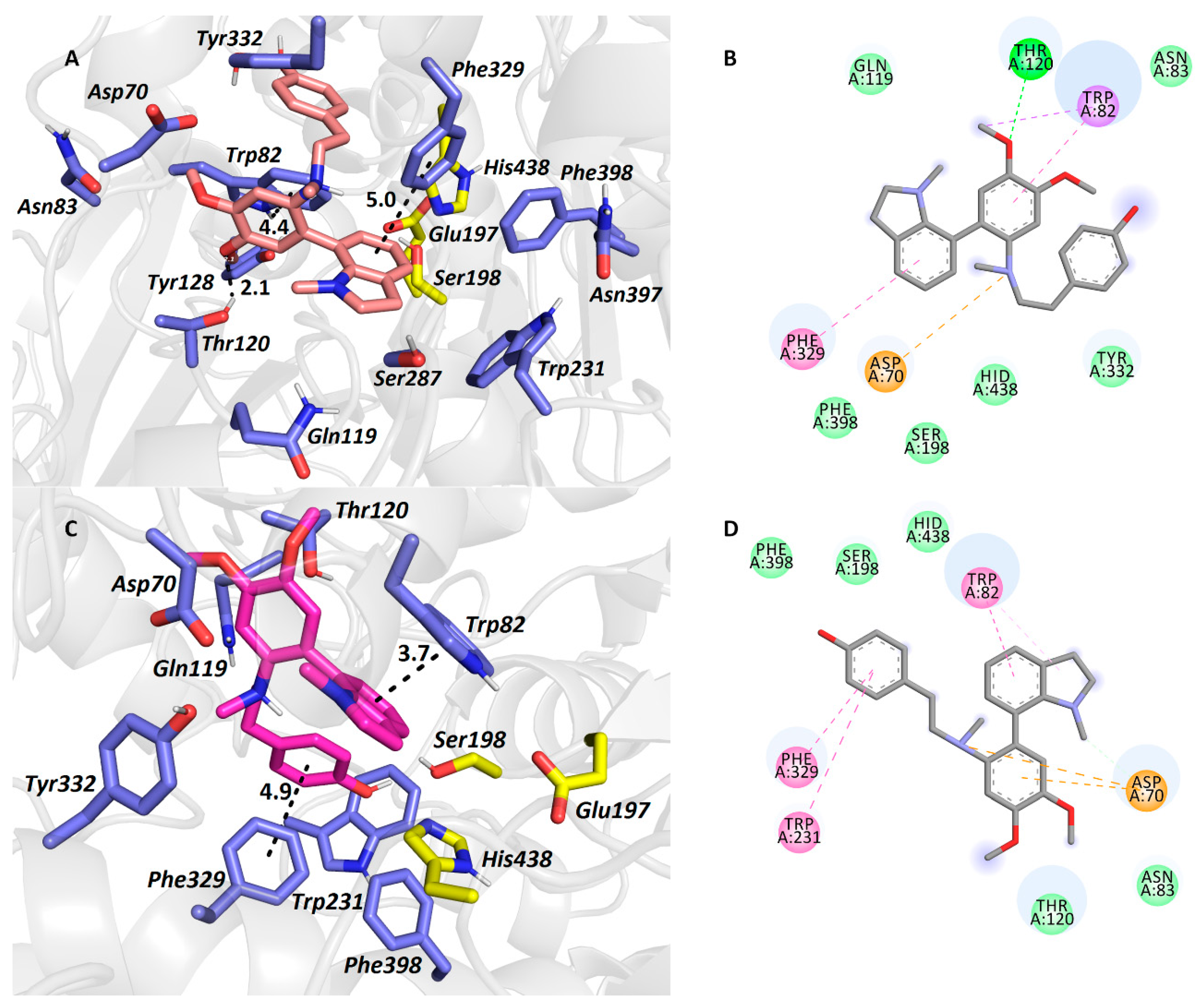
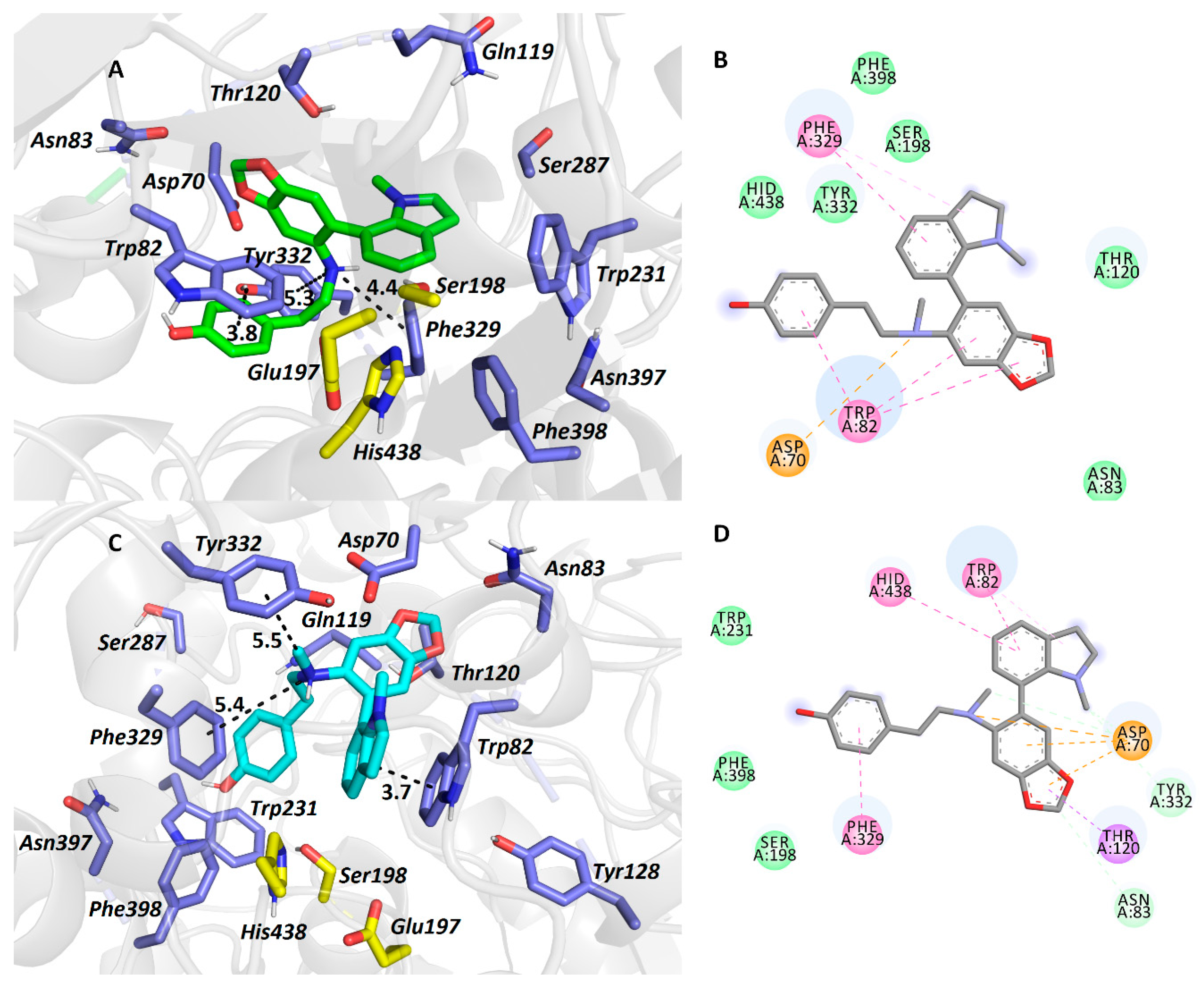
2.3. Docking Study of Carltonine A (13) and Carltonine B (14)
3. Experimental
3.1. General Experimental Procedures
3.2. Plant Material
3.3. Extraction and Isolation of Alkaloids
3.4. Biological Assays
3.4.1. hAChE and hBuChE Inhibition Assay
3.4.2. POP Inhibition Assay
3.4.3. Molecular Modelling Studies
4. Conclusions
Supplementary Materials
Author Contributions
Funding
Acknowledgments
Conflicts of Interest
References
- Burns, A.; Jacoby, R.; Levy, R. Psychiatric phenomena in Alzheimer’s disease. I: Disorders of thought content. Br. J. Psychiatry J. Ment. Sci. 1990, 157, 72–76, 92–94. [Google Scholar] [CrossRef] [PubMed]
- Nichols, E.; Szoeke, C.E.I.; Vollset, S.E.; Abbasi, N.; Abd-Allah, F.; Abdela, J.; Aichour, M.T.E.; Akinyemi, R.O.; Alahdab, F.; Asgedom, S.W.; et al. Global, regional, and national burden of Alzheimer’s disease and other dementias, 1990–2016: A systematic analysis for the Global Burden of Disease Study 2016. Lancet Neurol. 2019, 18, 88–106. [Google Scholar] [CrossRef] [Green Version]
- Cimler, R.; Maresova, P.; Kuhnova, J.; Kuca, K. Predictions of Alzheimer’s disease treatment and care costs in European countries. PLoS ONE 2019, 14, e0210958. [Google Scholar] [CrossRef] [PubMed] [Green Version]
- Kumar, A.; Singh, A.; Ekavali. A review on Alzheimer’s disease pathophysiology and its management: An update. Pharmacol. Rep. PR 2015, 67, 195–203. [Google Scholar] [CrossRef]
- Hampel, H.; Mesulam, M.-M.; Cuello, A.C.; Farlow, M.R.; Giacobini, E.; Grossberg, G.T.; Khachaturian, A.S.; Vergallo, A.; Cavedo, E.; Snyder, P.J.; et al. The cholinergic system in the pathophysiology and treatment of Alzheimer’s disease. Brain J. Neurol. 2018, 141, 1917–1933. [Google Scholar] [CrossRef]
- Zemek, F.; Drtinova, L.; Nepovimova, E.; Sepsova, V.; Korabecny, J.; Klimes, J.; Kuca, K. Outcomes of Alzheimer’s disease therapy with acetylcholinesterase inhibitors and memantine. Expert Opin. Drug Saf. 2014, 13, 759–774. [Google Scholar] [CrossRef]
- Nachon, F.; Brazzolotto, X.; Trovaslet, M.; Masson, P. Progress in the development of enzyme-based nerve agent bioscavengers. Chem. Biol. Interact. 2013, 206, 536–544. [Google Scholar] [CrossRef]
- Nordberg, A.; Ballard, C.; Bullock, R.; Darreh-Shori, T.; Somogyi, M. A Review of Butyrylcholinesterase as a Therapeutic Target in the Treatment of Alzheimer’s Disease. Prim. Care Companion CNS Disord. 2013, 15. [Google Scholar] [CrossRef]
- Inestrosa, N.C.; Alvarez, A.; Pérez, C.A.; Moreno, R.D.; Vicente, M.; Linker, C.; Casanueva, O.I.; Soto, C.; Garrido, J. Acetylcholinesterase accelerates assembly of amyloid-beta-peptides into Alzheimer’s fibrils: Possible role of the peripheral site of the enzyme. Neuron 1996, 16, 881–891. [Google Scholar] [CrossRef] [Green Version]
- Babkova, K.; Korabecny, J.; Soukup, O.; Nepovimova, E.; Jun, D.; Kuca, K. Prolyl oligopeptidase and its role in the organism: Attention to the most promising and clinically relevant inhibitors. Future Med. Chem. 2017, 9, 1015–1038. [Google Scholar] [CrossRef]
- Szeltner, Z.; Polgár, L. Structure, function and biological relevance of prolyl oligopeptidase. Curr. Protein Pept. Sci. 2008, 9, 96–107. [Google Scholar] [CrossRef] [PubMed]
- Wang, S.; Dong, G.; Sheng, C. Structural Simplification of Natural Products. Chem. Rev. 2019, 119, 4180–4220. [Google Scholar] [CrossRef] [PubMed]
- Iranshahy, M.; Quinn, R.J.; Iranshahi, M. Biologically active isoquinoline alkaloids with drug-like properties from the genus Corydalis. RSC Adv. 2014, 4, 15900–15913. [Google Scholar] [CrossRef]
- Nair, J.J.; van Staden, J. Pharmacological and toxicological insights to the South African Amaryllidaceae. Food Chem. Toxicol. Int. J. Publ. Br. Ind. Biol. Res. Assoc. 2013, 62, 262–275. [Google Scholar] [CrossRef]
- Goietsenoven, G.V.; Mathieu, V.; Lefranc, F.; Kornienko, A.; Evidente, A.; Kiss, R. Narciclasine as well as other Amaryllidaceae Isocarbostyrils are Promising GTP-ase Targeting Agents against Brain Cancers. Med. Res. Rev. 2013, 33, 439–455. [Google Scholar] [CrossRef]
- Stafford, G.I.; Pedersen, M.E.; van Staden, J.; Jäger, A.K. Review on plants with CNS-effects used in traditional South African medicine against mental diseases. J. Ethnopharmacol. 2008, 119, 513–537. [Google Scholar] [CrossRef]
- Olin, J.; Schneider, L. Galantamine for Alzheimer’s disease. Cochrane Database Syst. Rev. 2002, CD001747. [Google Scholar] [CrossRef]
- Bastida, J.; Viladomat, F.; Codina, C. Narcissus alkaloids. In Studies in Natural Products Chemistry; Atta-ur-Rahman, Ed.; Structure and Chemistry (Part F); Elsevier: Amsterdam, The Netherlands, 1997; Volume 20, pp. 323–405. [Google Scholar]
- Nair, J.J.; Rárová, L.; Strnad, M.; Bastida, J.; van Staden, J. Mechanistic insights to the cytotoxicity of Amaryllidaceae alkaloids. Nat. Prod. Commun. 2015, 10, 171–182. [Google Scholar] [CrossRef] [Green Version]
- Bastida, J.; Lavilla, R.; Viladomat, F. Chemical and biological aspects of Narcissus alkaloids. Alkaloids Chem. Biol. 2006, 63, 87–179. [Google Scholar] [CrossRef]
- Hulcová, D.; Maříková, J.; Korábečný, J.; Hošťálková, A.; Jun, D.; Kuneš, J.; Chlebek, J.; Opletal, L.; De Simone, A.; Nováková, L.; et al. Amaryllidaceae alkaloids from Narcissus pseudonarcissus L. cv. Dutch Master as potential drugs in treatment of Alzheimer’s disease. Phytochemistry 2019, 165, 112055. [Google Scholar] [CrossRef]
- Torras-Claveria, L.; Berkov, S.; Codina, C.; Viladomat, F.; Bastida, J. Daffodils as potential crops of galanthamine. Assessment of more than 100 ornamental varieties for their alkaloid content and acetylcholinesterase inhibitory activity. Ind. Crops Prod. 2013, 43, 237–244. [Google Scholar] [CrossRef]
- Breiterová, K.; Ločárek, M.; Kohelová, E.; Talácková, M.; Hulcová, D.; Opletal, L.; Cahlíková, L. Daffodils as Potential Crops of Biologically-active Compounds: Assessment of 40 Ornamental Taxa for their Alkaloid Profile and Cholinesterases Inhibition Activity. Nat. Prod. Commun. 2018, 13. [Google Scholar] [CrossRef] [Green Version]
- Pellegrino, S.; Meyer, M.; Zorbas, C.; Bouchta, S.A.; Saraf, K.; Pelly, S.C.; Yusupova, G.; Evidente, A.; Mathieu, V.; Kornienko, A.; et al. The Amaryllidaceae Alkaloid Haemanthamine Binds the Eukaryotic Ribosome to Repress Cancer Cell Growth. Structure 2018, 26, 416–425. [Google Scholar] [CrossRef] [Green Version]
- Sener, B.; Orhan, I.; Satayavivad, J. Antimalarial activity screening of some alkaloids and the plant extracts from Amaryllidaceae. Phytother. Res. PTR 2003, 17, 1220–1223. [Google Scholar] [CrossRef] [PubMed]
- Havelek, R.; Seifrtova, M.; Kralovec, K.; Bruckova, L.; Cahlikova, L.; Dalecka, M.; Vavrova, J.; Rezacova, M.; Opletal, L.; Bilkova, Z. The effect of Amaryllidaceae alkaloids haemanthamine and haemanthidine on cell cycle progression and apoptosis in p53-negative human leukemic Jurkat cells. Phytomedicine Int. J. Phytother. Phytopharm. 2014, 21, 479–490. [Google Scholar] [CrossRef] [PubMed]
- Kohelová, E.; Peřinová, R.; Maafi, N.; Korábečný, J.; Hulcová, D.; Maříková, J.; Kučera, T.; Martínez González, L.; Hrabinova, M.; Vorčáková, K.; et al. Derivatives of the β-Crinane Amaryllidaceae Alkaloid Haemanthamine as Multi-Target Directed Ligands for Alzheimer’s Disease. Molecules 2019, 24, 1307. [Google Scholar] [CrossRef] [PubMed] [Green Version]
- Yang, Y.; Huang, S.-X.; Zhao, Y.-M.; Zhao, Q.-S.; Sun, H.-D. Alkaloids from the Bulbs of Lycoris aurea. Helv. Chim. Acta 2005, 88, 2550–2553. [Google Scholar] [CrossRef]
- Fan-Chiang, T.-T.; Wang, H.-K.; Hsieh, J.-C. Synthesis of phenanthridine skeletal Amaryllidaceae alkaloids. Tetrahedron 2016, 72, 5640–5645. [Google Scholar] [CrossRef]
- Bozkurt, B.; Çoban, G.; Kaya, G.; Onur, M.; Unver-Somer, N. Alkaloid profiling, anticholinesterase activity and molecular modeling study of Galanthus elwesii. South Afr. J. Bot. 2017. [Google Scholar] [CrossRef]
- Huang, S.-D.; Zhang, Y.; He, H.-P.; Li, S.-F.; Tang, G.-H.; Chen, D.-Z.; Cao, M.-M.; Di, Y.-T.; Hao, X.-J. A new Amaryllidaceae alkaloid from the bulbs of Lycoris radiata. Chin. J. Nat. Med. 2013, 11, 406–410. [Google Scholar] [CrossRef]
- Pigni, N.B.; Ríos-Ruiz, S.; Martínez-Francés, V.; Nair, J.J.; Viladomat, F.; Codina, C.; Bastida, J. Alkaloids from Narcissus serotinus. J. Nat. Prod. 2012, 75, 1643–1647. [Google Scholar] [CrossRef] [PubMed]
- Chen, J.-Q.; Xie, J.-H.; Bao, D.-H.; Liu, S.; Zhou, Q.-L. Total Synthesis of (−)-Galanthamine and (−)-Lycoramine via Catalytic Asymmetric Hydrogenation and Intramolecular Reductive Heck Cyclization. Org. Lett. 2012, 14, 2714–2717. [Google Scholar] [CrossRef] [PubMed]
- Berkov, S.; Bastida, J.; Sidjimova, B.; Viladomat, F.; Codina, C. Phytochemical differentiation of Galanthus nivalis and Galanthus elwesii (Amaryllidaceae): A case study. Biochem. Syst. Ecol. 2008, 8, 638–645. [Google Scholar] [CrossRef]
- Bohno, M.; Sugie, K.; Imase, H.; Yusof, Y.B.; Oishi, T.; Chida, N. Total synthesis of Amaryllidaceae alkaloids, (+)-vittatine and (+)-haemanthamine, starting from d-glucose. Tetrahedron 2007, 63, 6977–6989. [Google Scholar] [CrossRef]
- Ellman, G.L.; Courtney, K.D.; Andres, V.; Feather-Stone, R.M. A new and rapid colorimetric determination of acetylcholinesterase activity. Biochem. Pharmacol. 1961, 7, 88–95. [Google Scholar] [CrossRef]
- Vaněčková, N.; Hošt‘álková, A.; Šafratová, M.; Kuneš, J.; Hulcová, D.; Hrabinová, M.; Doskočil, I.; Štěpánková, Š.; Opletal, L.; Nováková, L.; et al. Isolation of Amaryllidaceae alkaloids from Nerine bowdenii W. Watson and their biological activities. RSC Adv. 2016, 6, 80114–80120. [Google Scholar] [CrossRef]
- Tarrago, T.; Kichik, N.; Seguí, J.; Giralt, E. The Natural Product Berberine is a Human Prolyl Oligopeptidase Inhibitor. ChemMedChem 2007, 2, 354–359. [Google Scholar] [CrossRef]
- Breiterová, K.; Koutová, D.; Maříková, J.; Havelek, R.; Kuneš, J.; Majorošová, M.; Opletal, L.; Hošťálková, A.; Jenčo, J.; Řezáčová, M.; et al. Amaryllidaceae Alkaloids of Different Structural Types from Narcissus L. cv. Professor Einstein and Their Cytotoxic Activity. Plants Basel Switz. 2020, 9, 137. [Google Scholar] [CrossRef] [Green Version]
- Cahlíková, L.; Hrabinová, M.; Kulhánková, A.; Benesová, N.; Chlebek, J.; Jun, D.; Novák, Z.; Macáková, K.; Kunes, J.; Kuca, K.; et al. Alkaloids from Chlidanthus fragrans and their acetylcholinesterase, butyrylcholinesterase and prolyl oligopeptidase activities. Nat. Prod. Commun. 2013, 8, 1541–1544. [Google Scholar] [CrossRef] [Green Version]
- Šafratová, M.; Hošťálková, A.; Hulcová, D.; Breiterová, K.; Hrabcová, V.; Machado, M.; Fontinha, D.; Prudêncio, M.; Kuneš, J.; Chlebek, J.; et al. Alkaloids from Narcissus poeticus cv. Pink Parasol of various structural types and their biological activity. Arch. Pharm. Res. 2018, 41, 208–218. [Google Scholar] [CrossRef]
- Nachon, F.; Carletti, E.; Ronco, C.; Trovaslet, M.; Nicolet, Y.; Jean, L.; Renard, P.-Y. Crystal structures of human cholinesterases in complex with huprine W and tacrine: Elements of specificity for anti-Alzheimer’s drugs targeting acetyl- and butyryl-cholinesterase. Biochem. J. 2013, 453, 393–399. [Google Scholar] [CrossRef] [PubMed] [Green Version]
- Pohanka, M.; Karasova, J.Z.; Kuca, K.; Pikula, J.; Holas, O.; Korabecny, J.; Cabal, J. Colorimetric dipstick for assay of organophosphate pesticides and nerve agents represented by paraoxon, sarin and VX. Talanta 2010, 81, 621–624. [Google Scholar] [CrossRef] [PubMed]
- Hostalkova, A.; Marikova, J.; Opletal, L.; Korabecny, J.; Hulcova, D.; Kunes, J.; Novakova, L.; Perez, D.I.; Jun, D.; Kucera, T.; et al. Isoquinoline Alkaloids from Berberis vulgaris as Potential Lead Compounds for the Treatment of Alzheimer’s Disease. J. Nat. Prod. 2019, 82, 239–248. [Google Scholar] [CrossRef] [PubMed]
- Cheung, J.; Rudolph, M.J.; Burshteyn, F.; Cassidy, M.S.; Gary, E.N.; Love, J.; Franklin, M.C.; Height, J.J. Structures of human acetylcholinesterase in complex with pharmacologically important ligands. J. Med. Chem. 2012, 55, 10282–10286. [Google Scholar] [CrossRef] [PubMed]
- Pettersen, E.F.; Goddard, T.D.; Huang, C.C.; Couch, G.S.; Greenblatt, D.M.; Meng, E.C.; Ferrin, T.E. UCSF Chimera--a visualization system for exploratory research and analysis. J. Comput. Chem. 2004, 25, 1605–1612. [Google Scholar] [CrossRef] [PubMed] [Green Version]
- Morris, G.M.; Huey, R.; Lindstrom, W.; Sanner, M.F.; Belew, R.K.; Goodsell, D.S.; Olson, A.J. AutoDock4 and AutoDockTools4: Automated docking with selective receptor flexibility. J. Comput. Chem. 2009, 30, 2785–2791. [Google Scholar] [CrossRef] [Green Version]
- Panek, D.; Więckowska, A.; Wichur, T.; Bajda, M.; Godyń, J.; Jończyk, J.; Mika, K.; Janockova, J.; Soukup, O.; Knez, D.; et al. Design, synthesis and biological evaluation of new phthalimide and saccharin derivatives with alicyclic amines targeting cholinesterases, beta-secretase and amyloid beta aggregation. Eur. J. Med. Chem. 2017, 125, 676–695. [Google Scholar] [CrossRef]
- Hepnarova, V.; Korabecny, J.; Matouskova, L.; Jost, P.; Muckova, L.; Hrabinova, M.; Vykoukalova, N.; Kerhartova, M.; Kucera, T.; Dolezal, R.; et al. The concept of hybrid molecules of tacrine and benzyl quinolone carboxylic acid (BQCA) as multifunctional agents for Alzheimer’s disease. Eur. J. Med. Chem. 2018, 150, 292–306. [Google Scholar] [CrossRef]
- Svobodova, B.; Mezeiova, E.; Hepnarova, V.; Hrabinova, M.; Muckova, L.; Kobrlova, T.; Jun, D.; Soukup, O.; Jimeno, M.L.; Marco-Contelles, J.; et al. Exploring Structure-Activity Relationship in Tacrine-Squaramide Derivatives as Potent Cholinesterase Inhibitors. Biomolecules 2019, 9, 379. [Google Scholar] [CrossRef] [Green Version]
- O’Boyle, N.M.; Banck, M.; James, C.A.; Morley, C.; Vandermeersch, T.; Hutchison, G.R. Open Babel: An open chemical toolbox. J. Cheminformatics 2011, 3, 33. [Google Scholar] [CrossRef] [Green Version]
- Trott, O.; Olson, A.J. AutoDock Vina: Improving the speed and accuracy of docking with a new scoring function, efficient optimization and multithreading. J. Comput. Chem. 2010, 31, 455–461. [Google Scholar] [CrossRef] [PubMed] [Green Version]


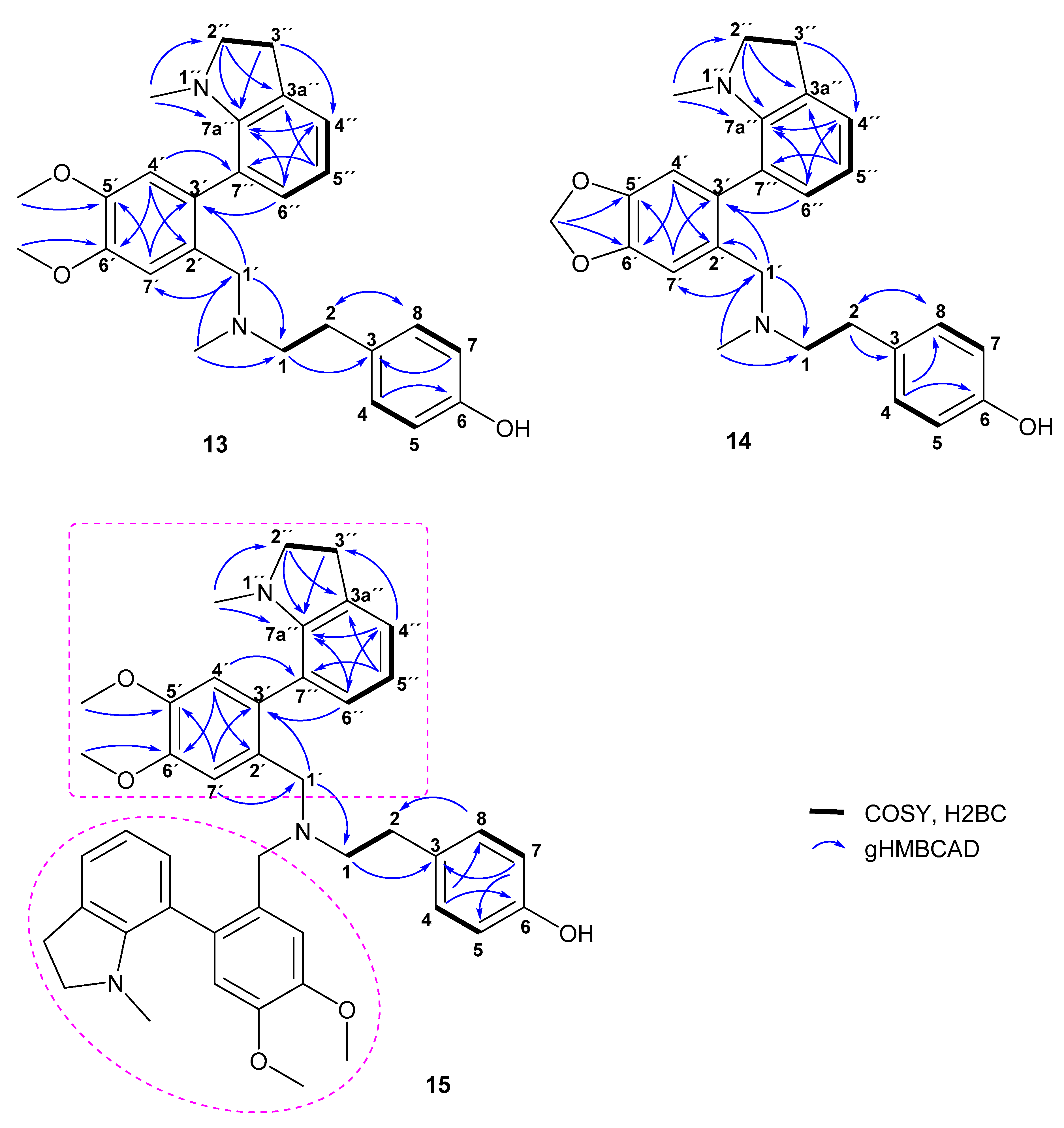
| Carltonine A (13) | Carltonine B (14) | Carltonine C (15) | ||||
|---|---|---|---|---|---|---|
| Position | δC | δH | δC | δH | δC | δH |
| 1 | 59.2 | 2.58–2.46, m | 59.3 | 2.56–2.45, m | 55.2; 55.4 | 2.66–2.42, m |
| 2 | 32.8 | 2.65, t (7.4) | 32.7 | 2.66–2.58, m | 32.7; 32.8 | 2.66–2.42, m |
| 3 | 132.5 | 132.2 | 132.8 | |||
| 4 | 129.8 | 7.00–6.96, m | 129.7 | 7.00–6.94, m | 129.9 | 6.93–6.86, m |
| 5 | 115.1 | 6.72–6.68, m | 115.2 | 6.75–6.72, m | 114.9 | 6.71–6.65, m |
| 6 | 154.0 | 153.9 | 153.7 | |||
| 7 | 115.1 | 6.72–6.68, m | 115.2 | 6.75–6.72, m | 114.9 | 6.71–6.65, m |
| 8 | 129.8 | 7.00–6.96, m | 129.7 | 7.00–6.94, m | 129.9 | 6.93–6.86, m |
| 1′ | 58.5 | 3.45, d (14.0) 3.29, d, overlap (14.0) | 58.3 | 3.46, d (13.2) 3.27, d (13.2) | 55.1; 55.0 | 3.43, d (14.5) 3.29, d (14.5); 3.37, s |
| 2′ | 130.1 | 130.6 | 131.2; 131.0 | |||
| 3′ | 132.1 | 133.3 | 131.9; 131.8 | |||
| 4′ | 113.2 | 6.76, s | 110.2 | 6.75–6.72, m | 113.23; 113.15 | 6.74, s |
| 5′ | 146.9 | 145.9 | 146.7 | |||
| 6′ | 148.1 | 147.0 | 147.9 | |||
| 7′ | 111.1 | 7.10, s, overlap | 108.6 | 7.07, s, overlap | 110.6; 110.5 | 7.15, s; 7.14, s |
| 2″ | 57.1 | 3.32, dt, overlap (17.3, 8.8) 3.20, dt (17.3, 8.8) | 57.1 | 3.35, dt (17.5, 8.7) 3.16, dt (17.5, 8.7) | 57.1; 57.0 | 3.27–3.17, m |
| 3″ | 28.6 | 2.98, t (8.8) | 28.6 | 3.00–2.93, m | 28.6 | 2.99–2.91, m |
| 3a″ | 131.2 | 131.2 | 131.1 | |||
| 4″ | 123.3 | 7.09, dd, overlap (7.4, 1.0) | 123.4 | 7.08, d, overlap (7.4) | 123.3 | 7.07, d (7.4) |
| 5″ | 117.9 | 6.72, t, overlap (7.4) | 118.1 | 6.70, t, overlap (7.4) | 117.9; 117.8 | 6.71–6.65, m |
| 6″ | 130.5 | 6.82, dd (7.4, 1.0) | 130.4 | 6.79, d (7.4) | 130.3; 130.4 | 6.77, d (7.4) |
| 7″ | 123.0 | 122.9 | 122.8 | |||
| 7a″ | 150.4 | 150.4 | 150.3; 150.2 | |||
| N-Me | 42.3 | 2.20, s | 41.9 | 2.18, s | - | - |
| N1″-Me | 38.6 | 2.21, s | 38.8 | 2.23, s | 38.51; 38.48 | 2.14, s; 2.12, s |
| 5′-OMe | 55.8 or 55.9 | 3.34, s | - | - | 55.9 | 3.84, s |
| 6′-OMe | 55.8 or 55.9 | 3.34, s | - | - | 55.6 | 3.82, s; 3.81, s |
| -OCH2O- | - | - | 101.0 | 5.99, s 5.98, s | - | - |
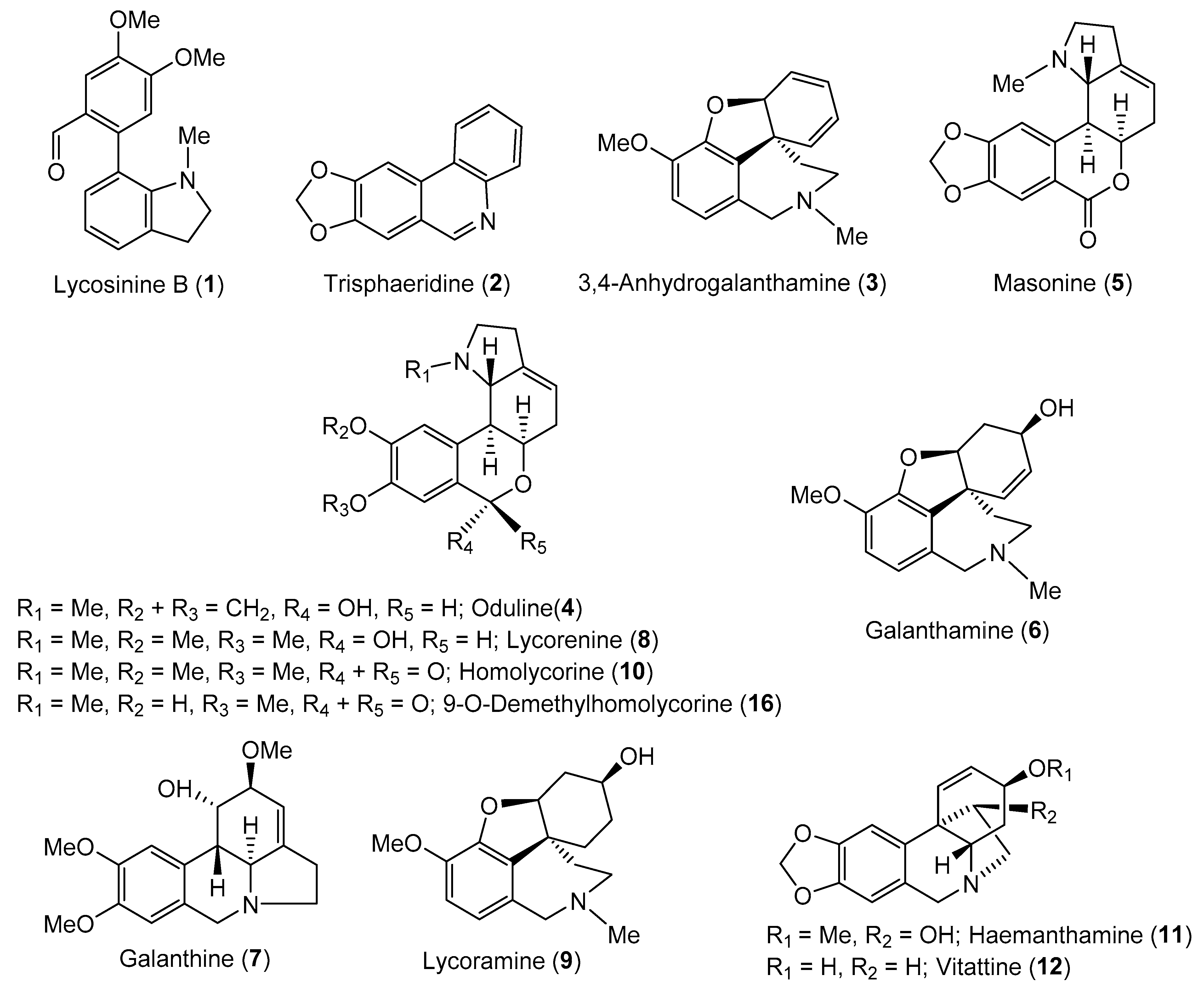
| Compound | %Inhibition hAChE ± SD a | hAChE IC50 ± SD (µM) b | % inhibition hBuChE ± SD a | hBuChE IC50 ± SD (µM) b | SI for hBuChE c | POP IC50 ± SD (µM) b |
|---|---|---|---|---|---|---|
| Lycosinine B (1) | 28 ± 1 | >100 | 42 ± 1 | >100 | nc | 258 ± 14 |
| Trispheridine (2) | 6 ± 1 | >100 | 13 ± 1 | >100 | nc | nm |
| 3,4-Anhydrogalanthamine (3) | 4 ± 0 | >100 | 28 ± 1 | >100 | nc | nm |
| Carltonine A (13) | 2 ± 0 | >100 | 98 ± 1 | 0.91 ± 0.02 | >110 | 143 ± 12 |
| Carltonine B (14) | 40 ± 1 | >100 | 99 ± 1 | 0.031 ± 0.001 | >3226 | nm |
| Carltonine C (15) | 9 ± 0 | >100 | 78 ± 1 | 14.8 ± 1.1 | >7 | nm |
| Galanthamine d | nm | 1.72 ± 0.12 | nm | 42 ± 1 | 0.04 | nm |
| Eserine d | nm | 0.063 ± 0.005 | nm | 0.13 ± 0.01 | 0.48 | nm |
| Berberine d | nm | 0.72 ± 0.11 | nm | 31 ± 4 | 0.02 | 142 ± 21 |
| Carltonine Enantiomer | Calculated Binding Energy (kcal/mol) |
|---|---|
| (R)-13 | −10.6 |
| (S)-13 | −10.6 |
| (R)-14 | −11.6 |
| (S)-14 | −10.9 |
© 2020 by the authors. Licensee MDPI, Basel, Switzerland. This article is an open access article distributed under the terms and conditions of the Creative Commons Attribution (CC BY) license (http://creativecommons.org/licenses/by/4.0/).
Share and Cite
Al Mamun, A.; Maříková, J.; Hulcová, D.; Janoušek, J.; Šafratová, M.; Nováková, L.; Kučera, T.; Hrabinová, M.; Kuneš, J.; Korábečný, J.; et al. Amaryllidaceae Alkaloids of Belladine-Type from Narcissus pseudonarcissus cv. Carlton as New Selective Inhibitors of Butyrylcholinesterase. Biomolecules 2020, 10, 800. https://0-doi-org.brum.beds.ac.uk/10.3390/biom10050800
Al Mamun A, Maříková J, Hulcová D, Janoušek J, Šafratová M, Nováková L, Kučera T, Hrabinová M, Kuneš J, Korábečný J, et al. Amaryllidaceae Alkaloids of Belladine-Type from Narcissus pseudonarcissus cv. Carlton as New Selective Inhibitors of Butyrylcholinesterase. Biomolecules. 2020; 10(5):800. https://0-doi-org.brum.beds.ac.uk/10.3390/biom10050800
Chicago/Turabian StyleAl Mamun, Abdullah, Jana Maříková, Daniela Hulcová, Jiří Janoušek, Marcela Šafratová, Lucie Nováková, Tomáš Kučera, Martina Hrabinová, Jiří Kuneš, Jan Korábečný, and et al. 2020. "Amaryllidaceae Alkaloids of Belladine-Type from Narcissus pseudonarcissus cv. Carlton as New Selective Inhibitors of Butyrylcholinesterase" Biomolecules 10, no. 5: 800. https://0-doi-org.brum.beds.ac.uk/10.3390/biom10050800