1. Introduction
Peptide-modified scaffolds are receiving a great amount of attention as support devices for tissue engineering applications [
1]. Scaffolds containing peptide sequences offer to seeded cells a biomimetic environment, mimicking the biomolecular signals of the extracellular matrix (ECM). In native tissues, the interactions between cell surface receptors and bioactive peptide sequences exposed by ECM proteins regulate cell behavior by influencing cell adhesion, proliferation, and differentiation. Many bioactive peptide motifs have been identified over the past years to be used for scaffold functionalization [
2]. The inclusion of peptide sequences into biomaterials is traditionally achieved by surface modification or by bulk incorporation [
3].
Molecular imprinting technology is a totally new approach, originally proposed by our laboratory [
4,
5], to obtain peptide-modified matrices for tissue engineering. Molecularly imprinted particles with recognition properties toward a peptide sequence of interest (the so-called template) can be obtained through an appropriate copolymerization process, carried out in the presence of a functional monomer, the peptide chosen as template, and a cross-linker. The removal of the template after the polymerization process results in the formation of imprinted cavities, which are physically and chemically complementary toward the template. According to the “epitope approach”, proposed by Rachkov and colleagues, when the imprinted particles will come in contact with the whole protein (containing the peptide used as template), this last will be recognized and rebound [
6]. Therefore, after deposition on the scaffold, when the imprinted particles will be exposed to the biological environment or cell culture environment, they will produce an enrichment of a desired ECM protein on the scaffold. Consequently, cell receptors could interact with the rebound protein, promoting a desired cellular response. The feasibility of such approach, as well as the biocompatibility of the imprinted particles, have been already demonstrated in two previous works [
4,
5].
Molecular Imprinting can offer several advantages with respect to traditional functionalization strategies, such as the covalent attachment of proteins or peptides. Imprinted particles can be deposited on all types of scaffolds, and the presence of specific functional groups for peptide covalent binding is not required. Additionally, the deposition of imprinted particles on the scaffold can be achieved without the use of chemical reagents or solvents, which may induce cell damage. Moreover, the bioactive molecule used as template (usually expensive) could be recovered during the extraction process and reused. Furthermore, if the ambitious goal of a complete removal of the template is achieved, a functional scaffold free of bioactive molecules can be obtained. This could represent a significant advantage from a regulatory point of view, because such a scaffold would fall in the medical device category.
In this work, new molecularly imprinted particles with recognition properties toward two different peptides from ECM proteins were synthesized and characterized. The peptide sequences chosen as templates are H-Gly-Arg-Gly-Asp-Ser-Pro-OH (GRGDSP) from fibronectin and H-Tyr-Ile-Gly-Ser-Arg-OH (YIGSR) from laminin.
Laminin and fibronectin are the main noncollagenous ECM proteins in connective tissues endowed with the ability to regulate cellular phenotype. In particular, fibronectin, the prototype of cell adhesive proteins, has been shown to modulate cell growth, cell shape, cytoskeletal organization, differentiation, migration, and the apoptosis of almost all cells [
7,
8,
9]. Laminins are a large family of glycoproteins, which are the most bioactive components of basement membranes, playing a crucial role in the survival and differentiation of adherent cells [
10,
11].
The peptide sequences from fibronectin and laminin, used as templates in this work, have demonstrated ability to promote cell adhesion [
12]. In addition, the GRDGSP sequence is able to stimulate integrins, such as α5β1 and αvβ3, that are relevant in early cardiac development [
13,
14], and the YIGSR sequence can increase the ability of stem cells to differentiate into beating cardiomyocytes [
15,
16,
17].
While GRGDSP-imprinted particles were synthetized for the first time in this study, imprinted particles with recognition properties toward the YIGSR sequence were already reported in a previous work [
5]. Here, however, with the aim to improve the rebinding performance, a different cross-linker was used during particle synthesis.
Imprinted particles underwent morphological, physicochemical, and functional characterization to assess their recognition and rebinding capability.
Then, both GRGDSP- and YIGSR-functionalized scaffolds were implanted to the rat heart for cardiac tissue engineering. An innovative combination of three natural polymers (alginate/gelatin/elastin, hereafter indicated as AGE) was used as scaffold material to mimic the composition as well as the interactions between components in the native cardiac extracellular matrix [
18]. A complete morphological, physicochemical, mechanical, functional, and biological characterization was performed on functionalized sponges.
2. Materials and Methods
2.1. Materials
Methacrylic acid (MAA > 99%), from Sigma Aldrich (Milan, Italy), was purified by distillation in vacuum to remove the polymerization inhibitor. Azobis(isobutyronitrile) (AIBN > 98%), pentaerythritol triacrylate (PETRA), trifluoroacetic acid (> 99.9%), and phosphate-buffered saline (PBS) were from Sigma Aldrich (Milan, Italy) and used as supplied. Acetonitrile (ACN > 99.9%) from Carlo Erba Reagenti (Milan, Italy) was of HPLC grade purity. The peptides H-GRGDSP-OH and H-YIGSR-OH were from Cambridge Research Biochemicals (Billingham, Cleveland, United Kingdom) and used as supplied. Alginate (viscosity of 2% solution at 25 °C = 250 cps), gelatin (type B from bovine skin), elastin (from bovine neck ligament), and glutaraldehyde (GTA, 25% aqueous solution) were acquired from Sigma Aldrich (Milan, Italy). Calcium chloride was purchased from Carlo Erba Reagenti (Milan, Italy).
2.2. Imprinted Particles: Synthesis Procedure
Two groups of molecularly imprinted particles, with recognition properties toward GRGDSP peptide sequence (hereafter indicated as MIP-GRGDSP) and YIGSR peptide sequence (hereafter indicated as MIP-YIGSR), were prepared using a precipitation polymerization method, at the compositions shown in
Table 1, following a procedure already reported in the literature [
4,
5].
Briefly, the template molecule was dissolved in a 70/30 (v/v) ACN/bi-distilled water (ddH2O) solution and introduced into borosilicate glass tubes (40 mL). The functional monomer (MAA), the cross-linker (PETRA), and the initiator (AIBN) were added to the polymerization solution. The tubes were sealed under dry nitrogen. The polymerization was thermally initiated at 60 °C and carried out for 20 h under constant stirring. At the end of the polymerization process, the residual polymerization solution was removed and subjected to chromatographic analysis. Collected particles were washed with ACN/ddH2O mixture to remove the residual monomer, cross-linker, and not-imprinted template. The template molecule was subsequently extracted by repeated washing with PBS, under vigorous stirring, in order to create the recognition sites.
Control particles (CP) were prepared with the same procedure of the imprinted ones but in the absence of the template molecule.
2.3. Alginate/Gelatin/Elastin Sponges: Preparation and Functionalization
AGE sponges were prepared following a procedure similar to that previously used by us to fabricate alginate/gelatin sponges [
18]. Briefly, alginate, gelatin and elastin 2% (w/v) bi-distilled water solutions were prepared separately at 50 °C.
Different volumes of natural polymer solutions were mixed together, under stirring at room temperature, in order to obtain an AGE blend with a A:G:E = 10:80:10 weight ratio.
A known volume of the blend was poured into polystyrene Petri dishes and freeze-dried. Then, the obtained sponges were treated with GTA vapors at 37 °C for 18 h for proteins cross-linking and then immersed for 30 min in a solution containing 2% (w/v) CaCl2 in bi-distilled water for polysaccharide cross-linking.
After cross-linking treatments, samples were immersed for 16 h in a coagulation bath (0.5 M acetic acid) to promote ionic interactions among the components.
Since GTA and acetic acid could be dangerous for cells, cross-linked sponges were washed repeatedly with bi-distilled water to remove excess GTA and acetic acid, until UV-spectrophotometric and pH analysis of wash waters did not reveal any residual trace.
At the end of the washing procedure, the scaffolds were freeze-dried again.
Sponges were functionalized by the deposition of MIP on their surface. According to the literature, the minimal ligand spacing for cell adhesion in two-dimensional cultures turns out to be 440 nm [
19]. This value was extrapolated to three dimensions (calculations based on a body centered cubic unit cell) and correlates to a ligand density of approximately 34 × 10
−12 mol ligand/cm
3 [
20]. It was decided to modify the sponges using an amount of MIP offering a ligand density at four orders of magnitude above this reference value. In this calculation, both peptide cavities and non-extracted peptides were considered. The calculated quantity of MIP was dispersed in bi-distilled water and deposited on preformed scaffolds.
2.4. Morphological Analysis by Scanning Electron Microscopy
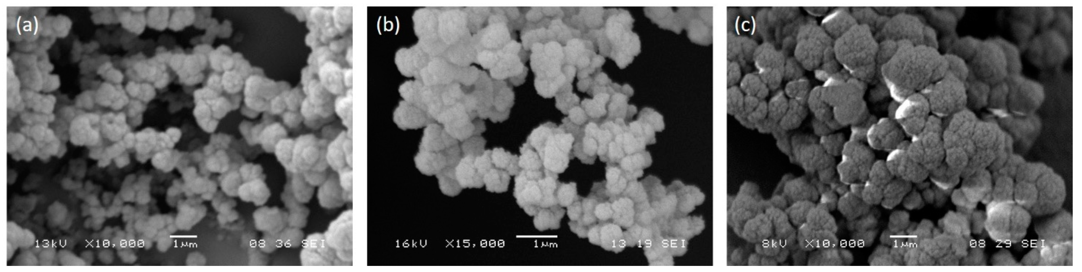
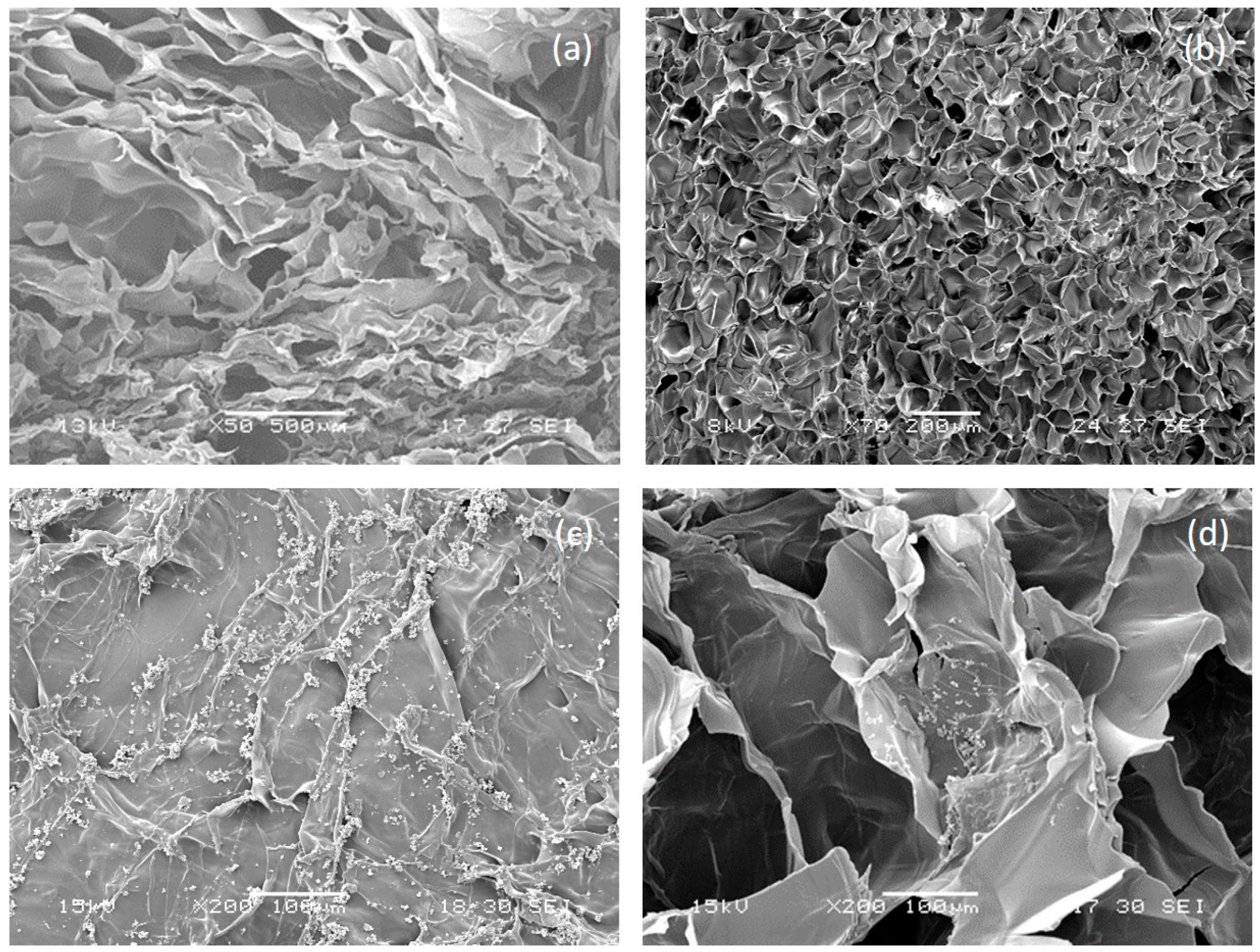
The morphology of imprinted and control particles and of AGE sponges, before and after functionalization, was examined by a JSM 5600 scanning electron microscope (SEM, Jeol Ltd., Tokyo, Japan). Before analysis, samples were coated with a gold layer (thickness of 200–500 Å) and mounted on metal stubs. The percentage of porosity and the average pore size were measured analyzing SEM images by the Image J software (National Institutes of Health). The percentage of porosity was calculated from the ratio between the pore area and the total scaffold area.
2.5. Chromatographic Analysis
The percentage of monomer conversion during MIP synthesis was determined by high-performance liquid chromatography (HPLC, 200 Series HPLC system, Perkin Elmer, Waltham, Massachusetts, United States), with a UV/VIS detector, following a procedure similar to that already described in previous papers [
4,
5]. In order to determine the monomer residual amount in the polymerization solution, an Alltima C18 5u column (250 mm length × 4.5 mm i.d.) was used. The mobile phase was A = 0.085% (w/v) trifluoroacetic acid in ACN; B = 0.1% (w/v) trifluoroacetic acid in water. The elution condition was an isocratic elution for 15 min with a mobile phase composition of 54% A and 46% B at a flow rate of 1 mL/min. The injection volume was 100 µl. The detector wavelength was set at λ = 215 nm.
The amount of template entrapped by the polymeric particles and the amount of extracted template were determined by HPLC measuring the corresponding residual amount in the polymerization solution and in the extraction solution, respectively. To determine the peptide concentrations, a Prosphere HP C4 300 Å 5u column (250 mm length × 4.5 mm i.d.) was used. The elution condition was a linear binary gradient at a flow rate of 1 mL/min, and the gradient was from 30% A and 70% B to 60% A and 40% B in 15 min. The injection volume was 100 µl. The detector wavelength was set at λ = 280 nm.
2.6. Infrared Spectroscopy
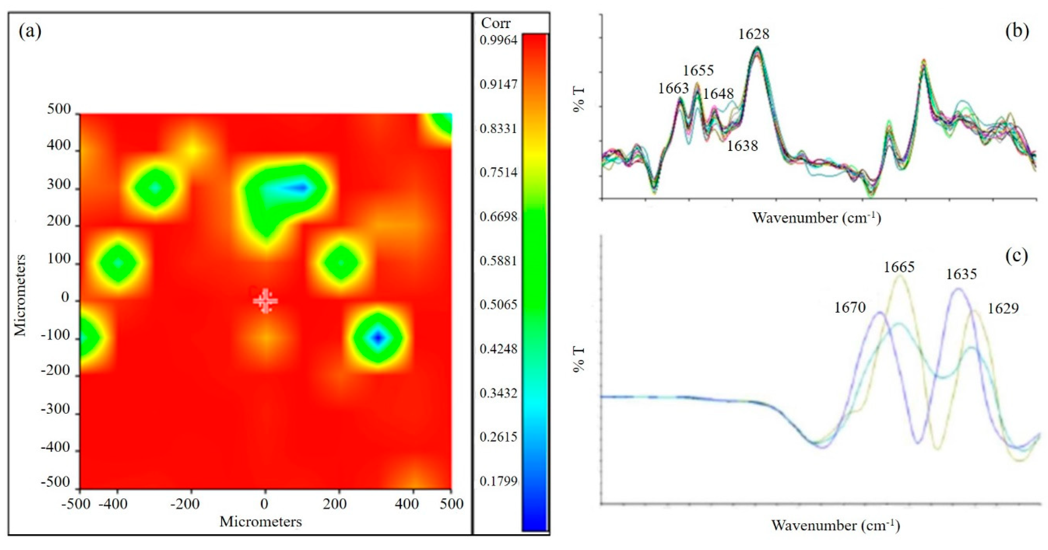
Fourier transformed infrared spectroscopy (FT-IR) spectra were recorded on a Spectrum-One instrument, Perkin Elmer (Waltham, Massachusetts, United States). The spectrometer was equipped with an attenuated total reflectance (ATR) objective lens with a penetration depth of less than 1 µm. All spectra were obtained at 4 cm−1 and represented the average of 16 scans. Spectral images were also acquired using the infrared imaging system of the instrument (Spotlight 300, Perkin Elmer, Waltham, Massachusetts, United States). The spectral resolution was 4 cm−1. The spatial resolution was 100 × 100 µm.
2.7. Recognition and Selectivity Tests
Recognition and selectivity tests were carried out for MIP-GRGDSP, MIP-YIGSR, and relative controls, CP. The procedure followed was the same, but the composition of the rebinding solution was varied, as described hereafter.
To evaluate the rebinding capacity of MIP, 10 mg of MIP and 10 mg of CP samples were put in contact with 1 mL of a rebinding solution consisting of a template solution (0.25 mg/mL) in water/ACN 30/70 (v/v). The template of the rebinding solution was GRGDSP for MIP-GRGDSP and relative control and YIGSR for MIP-YIGSR and relative control. The tubes, containing the rebinding solution and MIP or CP, were maintained under constant stirring for 30 min. The supernatant was separated from the polymer by centrifugation for 30 min at 14,000 rpm. The procedure was repeated three times, replacing every time the supernatant with fresh solution. Both for MIP and for CP, the amount of rebound template was determined on three samples, and the average values were reported. The residual peptide concentration in the rebinding solution was determined by HPLC at a wavelength of 280 nm.
The amount of template rebound by MIP was calculated as follows:
where
V,
Ci, and
Cf represent, respectively: the volume of the rebinding solution, the initial rebinding solution concentration, and the rebinding solution concentration after adsorption.
The imprinting effect was evaluated through the rebinding factor, which was calculated according to the following equation:
When the value of the rebinding factor is higher than one, the recognition capability of MIP is demonstrated.
Selectivity was also verified by incubating fixed amounts of both MIP and CP with 0.25 mg of a template analogue in 1 mL of water/ACN 30/70 (v/v) and then proceeding as for rebinding experiments. The molecules used as analogue were YIGSR for MIP-GRGDSP and GRGDSP for MIP-YIGSR. The selectivity factor
α’ was calculated as indicated in the equation below:
Particles selectivity is demonstrated when α’ is higher than 1.
Recognition experiments were also performed on MIP-modified sponges to evaluate if the rebinding capacity was maintained by MIP after deposition on the scaffolds. The procedure followed was the same used to test MIP recognition: 1 cm2 of each MIP-modified sponge was put in contact with 1 mL of the rebinding solution. Scaffolds modified with CP were used as controls.
2.8. Thermal Analysis
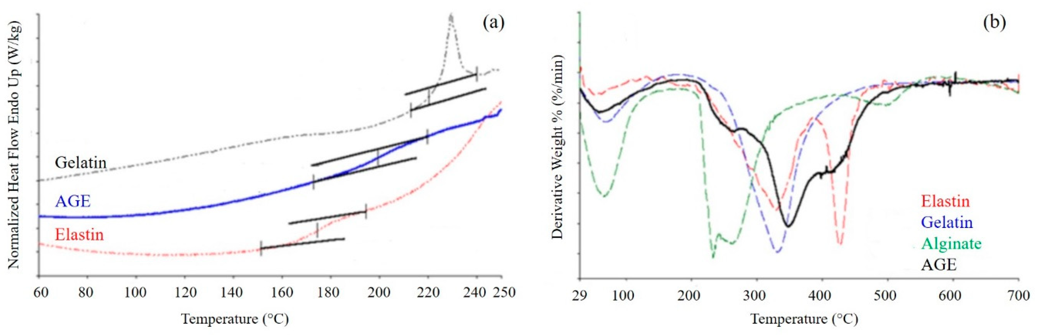
The thermal properties of AGE sponge were studied by carrying out differential scanning calorimetry (DSC) and thermogravimetric analysis (TGA). The DSC thermogram of the sponge was acquired and compared with those of pure alginate, gelatin, and elastin.
A known amount of the scaffold (about 10 mg) was encapsulated in standard aluminum pans and used for the study. All tests were carried out in triplicate. The samples were heated/cooled/heated at a heating rate of 10 °C/min between −60 and 300 °C, under inert nitrogen. The endothermic peaks were measured using the DSC software.
TGA allowed us to study the thermal degradation process of the three pure polymers and their blend. Samples of ca. 10 mg, in triplicate, were heated from 30 to 700 °C at a rate of 10 °C/min, under nitrogen flow.
2.9. Mechanical Analysis
A dynamic mechanical analyzer (DMA8000, Perkin Elmer, Waltham, Massachusetts, United States) was used to investigate the viscoelastic properties of the scaffold. A protocol similar to that described in our previous papers [
18,
21] was followed. Briefly, the sponge was cut in the form of rectangular strips (20 mm in length, 10 mm in width, 5 mm in thickness). Tests were performed under wet conditions. Before analysis, samples were maintained for 3 h in a solution simulating body fluids (PBS) at 37 °C. Wet conditions were reproduced during analysis. The samples were tested in a single cantilever mode. Storage modulus (E’), loss modulus (E’’), and tangent delta (tan δ) were evaluated by performing a single strain/multi-frequency test. The applied strain had an amplitude of 10 µm; frequencies were set at 1, 3.5, and 10 Hz, reproducing, respectively, a healthy human heart rate (1 Hz, corresponding to 60 bpm), a pathological human heart rate (3.5 Hz, corresponding to 210 bpm), and a supraphysiological pulse rate (10 Hz, corresponding to 600 bpm) [
18].
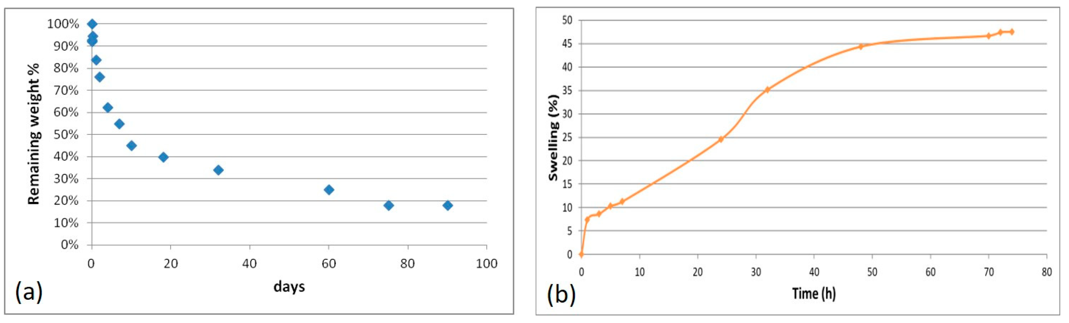
2.10. Degradation Test
In vitro hydrolytic degradation tests were performed in PBS, in agreement with the ISO norm 10993-13 “Biological evaluation of medical devices. Part 13: Identification and quantification of degradation products from polymeric medical devices” [
22].
Scaffold weight loss during degradation was measured by changes in dry weight after incubation for specified time periods. All the experiments were done in triplicate; the results are the mean (±SE) of three determinations. A separate container was used for each sample.
Samples were dried at room temperature to a constant mass. The starting dry weight,
W0, was determined for each sample by using a balance with adequate precision. Then, samples were fully immersed in a closed container, together with the degradation solution, and maintained in an agitating bath at 37 ± 1 °C. At appointed times, samples were removed from the degradation solution. The samples were rinsed in ddH
2O and dried to a constant mass. The dry weight at time
t of degradation,
Wt, was determined for each sample. The percentage of remaining weight was evaluated according to the following equation:
2.11. Swelling Test
The swelling properties of the scaffold were evaluated by exposure to aqueous vapor. Samples cut into squares of 1 cm
2, were dried in oven at 37 °C up to a constant weight and the starting dry weight,
Wd, was registered for each sample. Then, samples were introduced in a closed container, with 100 mL of water in the lower part. The container was maintained in an oven at 37 °C for all the duration of the test. At fixed times, the swollen weight,
Ws, was determined for each sample. The percentage of swelling was calculated as:
2.12. In Vitro Biological Characterization
2.12.1. Rat Cardiac Progenitor Cells Isolation and Culture
Cardiac progenitor cells (rCPCs) were isolated from rats constitutively expressing green fluorescence protein (GFP) [
23] and cultured as previously described [
24,
25]. rCPCs at passages 4 and 5 (P4–P5) were employed for the experiments described in this study.
2.12.2. DiI Cell Labeling
In order to optimize the detection of GFP
pos rCPCs cultured on the sponges, cells were stained with CellTracker CM-DiI (ThermoFischer, C-7001) before seeding in agreement with manufacturer instructions [
26]. CellTracker CM-DiI is a DiI derivative that is somewhat more water-soluble than DiI, thus facilitating the preparation of staining solutions for cell suspensions. CellTracker CM-DiI contains a thiol-reactive chloromethyl moiety (CM) that allows the dye to covalently bind to cellular thiols. Thus, unlike other membrane stains, this label is well retained in the cells throughout several mitotic divisions, and cell-to-cell contact does not allow dye diffusion.
2.12.3. Cell Culture on AGE Scaffolds
AGE scaffolds were sterilized by UV exposure for 15 min on each side, and cells were seeded on their surface at 45 × 10
3 cells/cm
2 density, according to a previously described methodology [
21].
Cells loaded to CTRL, MIP-GRGDSP, and MIP-YIGSR sponges were evaluated 72 h after cell plating. The quantification of cell adhesion was performed by “Image Pro Plus 4.0” software [
26].
2.13. In Vivo Biological Characterization
In order to test the in vivo properties of functionalized AGE scaffolds, experiments were performed on a cryoinjury (CI) rat model. In particular, the suturability, cell adhesion to the sponges, cell migration toward the scaffolds, and cell differentiation were evaluated.
The study population consisted of male Wistar rats (Rattus norvegicus, Charles River, Italy) bred at the University of Parma departmental animal facility. The investigation was conformed to the National Ethical Guidelines (Italian Ministry of Health; D.L.vo 116, January 27, 1992) and the Guide for the Care and Use of Laboratory Animals (NIH publication no. 85–23, revised 1996) and approved by the Veterinary Animal Care and Use Committee of the University of Parma.
In vivo studies in the CI model were performed employing AGE not functionalized (NF) sponges or functionalized with MIP-GRGDSP and MIP-YIGSR sequences. rCPC-seeded (+rCPCs) or unseeded configurations were tested. Each experimental group consisted of four animals.
Cell seeding was performed as described in the previous paragraph. After 48 h, AGE + rCPCs were surgically sutured to cover the damaged area of the rat hearts.
The control group was represented by animals subjected to myocardial damage without scaffolds application.
2.13.1. Surgical Procedure
The surgical procedure and the subsequent macroscopic examination of the rat myocardium were performed according to a methodology well established in our laboratory and detailed in several publications [
21,
27,
28,
29].
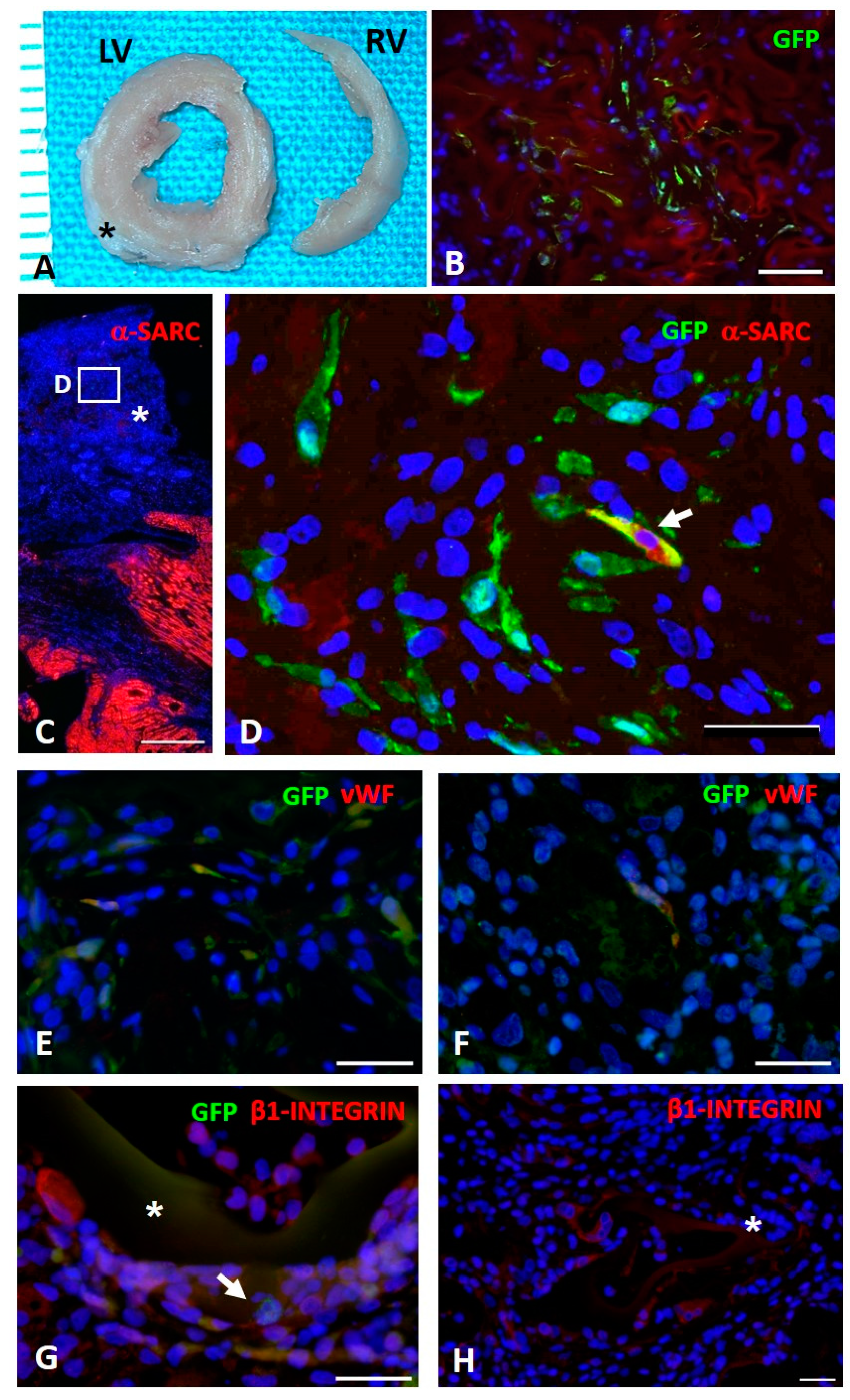
At sacrifice, the heart was excised and fixed in 10% formalin. After 24 h, LV transverse sections corresponding to the base, equatorial portion, and apex were embedded in paraffin, and 5-μm-thick sections were cut for immunohistochemical studies.
2.13.2. Immunohistochemical Analysis
The adhesion of GFPpos rCPCs seeded on scaffolds and their differentiation toward cardiomyogenic phenotypes were determined by the immunofluorescence detection of GFP, alpha-sarcomeric actin (α-SARC), which is typically expressed by cardiomyocytes, and von Willebrand Factor (vWF), which is a specific endothelial cell marker. Moreover, unseeded sponge colonization by resident myocardial cell was evaluated by β1-integrin expression.
For these purposes, LV sections from different experimental groups were incubated with primary antibodies (polyclonal goat anti-GFP (ab6673), dilution 1:100, Abcam; monoclonal mouse anti-α-SARC (A2172), dilution 1:100, Sigma Aldrich; polyclonal rabbit anti-vWF (A0082), dilution 1:100, Dako; monoclonal rabbit anti- β1-integrin (ab52971), dilution 1:250, Abcam). FITC- and TRITC-conjugated specific secondary antibodies were used to simultaneously detect epitopes.
Nuclei were recognized by the blue fluorescence of 4′,6-diamindine-2-phenyndole (DAPI, Sigma Aldrich, Milan, Italy) staining.
2.13.3. Data Management and Statistics
The SPSS statistical software was employed (SPSS, Chicago, IL, USA) to perform data management and statistics. A paired Student t-test and one-way analysis of variance (ANOVA, post-hoc analyses: Tukey test or Holm–Sidak test) were applied. Statistical significance was set at p < 0.05.
4. Conclusions
The development of scaffolds containing biomolecular signals typically present in the ECM of native tissues is of fundamental importance for successful tissue engineering strategies. Scaffold functionalization with peptides is normally achieved by surface or bulk modification, creating a covalent binding between peptide and polymer functional groups. In the present investigation, we propose an innovative approach for scaffolds functionalization, based on Molecular Imprinting technology, which was reported for the first time by our research group a few years ago. Molecularly imprinted particles with recognition properties toward bioactive molecules of interest (such as peptide sequences) can be deposited on the scaffold surface, where they maintain their recognition properties, promoting an enrichment on the scaffold of a chosen protein and consequently a desired interaction at the cellular and tissue level. In this work, molecularly imprinted particles with recognition properties toward peptide sequences from laminin and fibronectin were obtained by the copolymerization of MAA with the template molecule, in the presence of a cross-linker (PETRA). GRGDSP, from fibronectin, and YIGSR, from laminin, were used as templates. Particles with a spherical shape and an average diameter of 1 µm were obtained for both formulations. High values of monomer conversion and template entrapment were observed. The template molecule was only partially removed from the particles (percentage of extracted template below 50%), as typically occurs with a protein template. Infrared analysis confirmed the desired chemical structure of MIP, as well as successful template rebinding. Good recognition and rebinding capabilities were demonstrated as recognition factor and selectivity factors resulted much higher than one. Therefore, MIP-YIGSR and MIP-GRGDSP were used for the functionalization of an innovative biomimetic scaffold, based on a blend of three natural polymers. This scaffold, obtained by blending a polysaccharide (alginate) and two proteins (gelatin and elastin), represents a valid substitute of native ECM, mimicking its chemical composition and the interactions that occur among components. The results of morphological, physicochemical, functional, and mechanical characterization showed adequate properties of the produced scaffold for bioengineered applications to cardiac repair. Moreover, the results obtained in the characterization of MIP-modified scaffolds show interesting properties of the synthesized MIP as functionalization devices for the development of bioactive scaffolds. Overall, thanks to the combination of a biomimetic polysaccharide/protein substrate with imprinted particles capable of imparting to the system recognition properties, it is possible to get a functionalized scaffold that is able to promote desired cellular responses, which represent an innovative and promising approach in the field of cardiac tissue engineering.

 ,
, 


