Cutting-Edge Advances in Electrochemical Affinity Biosensing at Different Molecular Level of Emerging Food Allergens and Adulterants

 ,
, 
Abstract
:1. Introduction
2. Food Safety: Allergens and Adulterations
3. Conventional Methods to Determine Food Allergens and Adulterants
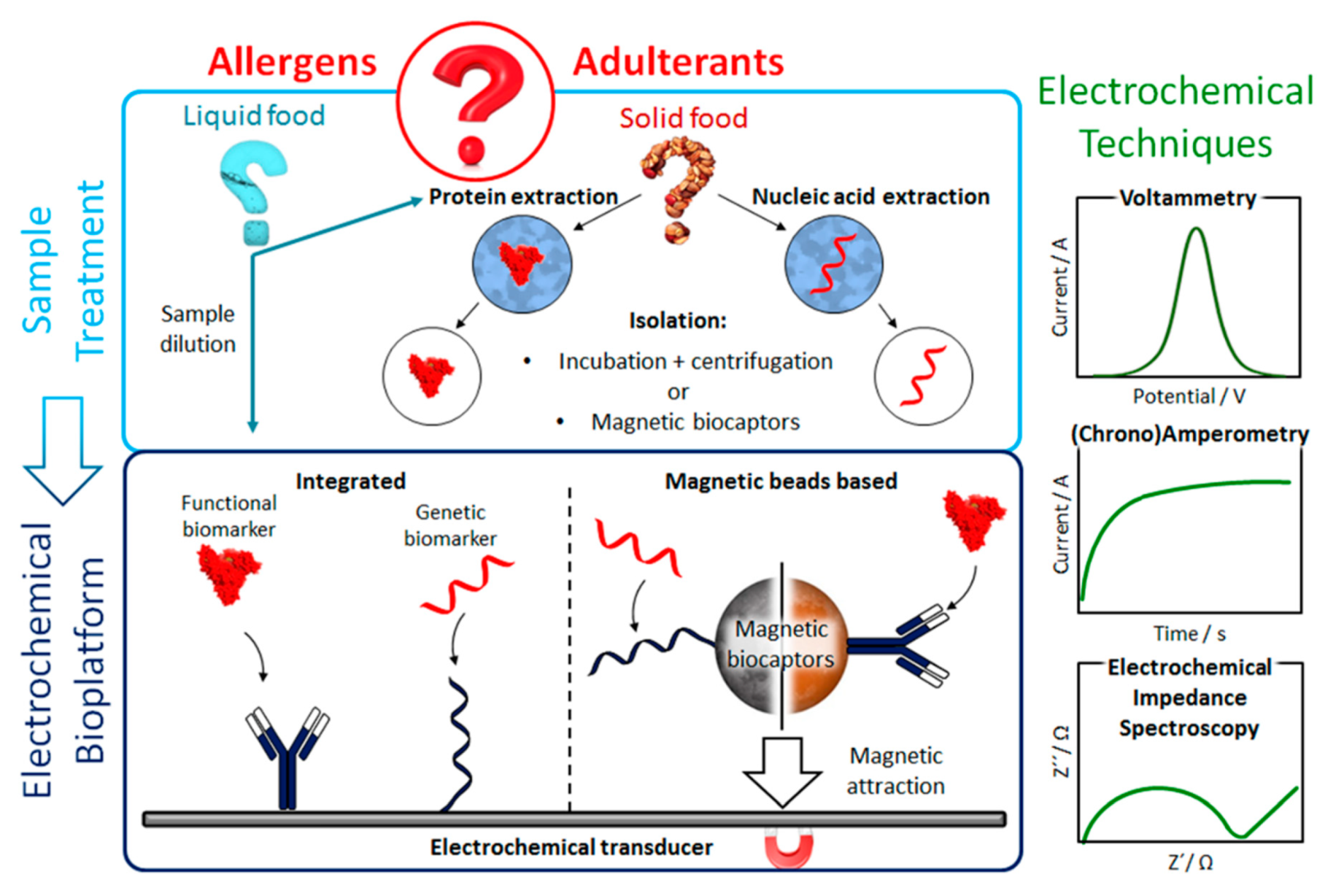
4. Electrochemical Affinity Biosensors for the Determination of Food Allergens and Adulterants
4.1. Electrochemical Immunosensing Methods
4.2. Nucleic Acid-Based Biosensing Methods
5. General Considerations, Challenges to Face and Future Prospects
- (i)
- (ii)
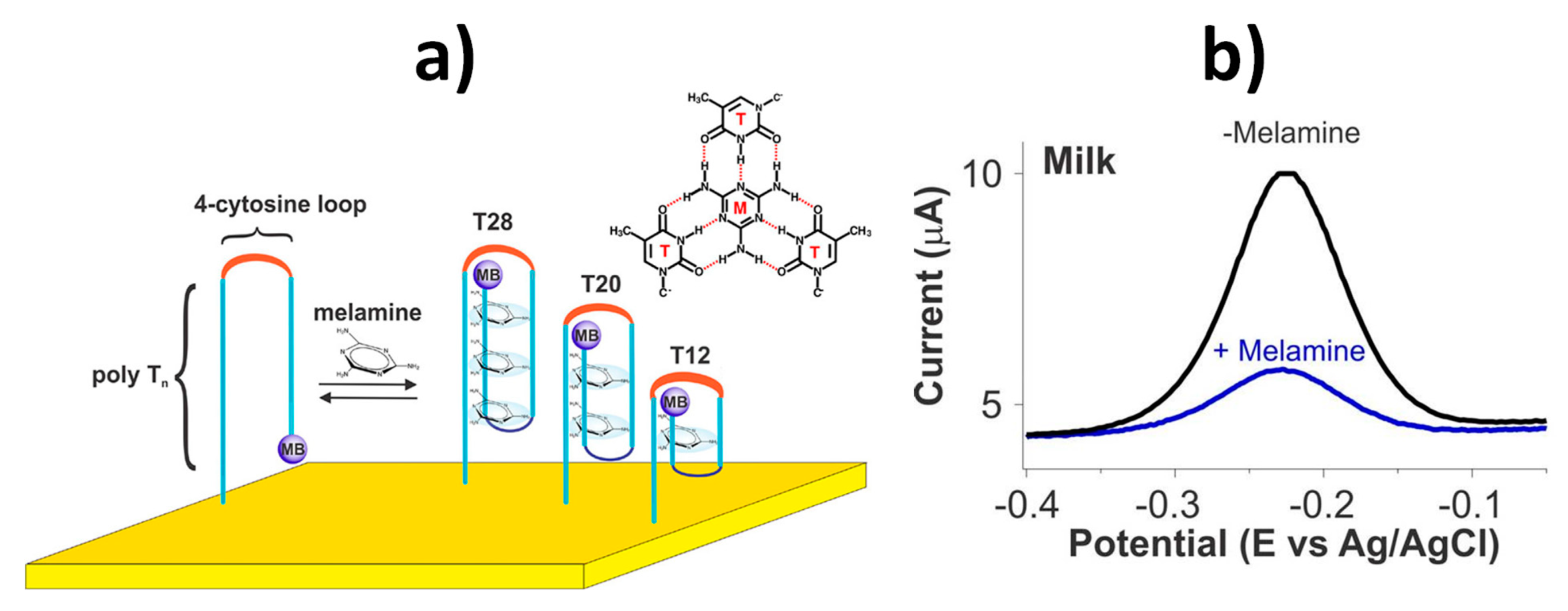
- The continuous detection of targets near real-time and in flowing samples using reagentless switch-based electrochemical biosensors (e.g., melamine in milk) [35].
- (iii)
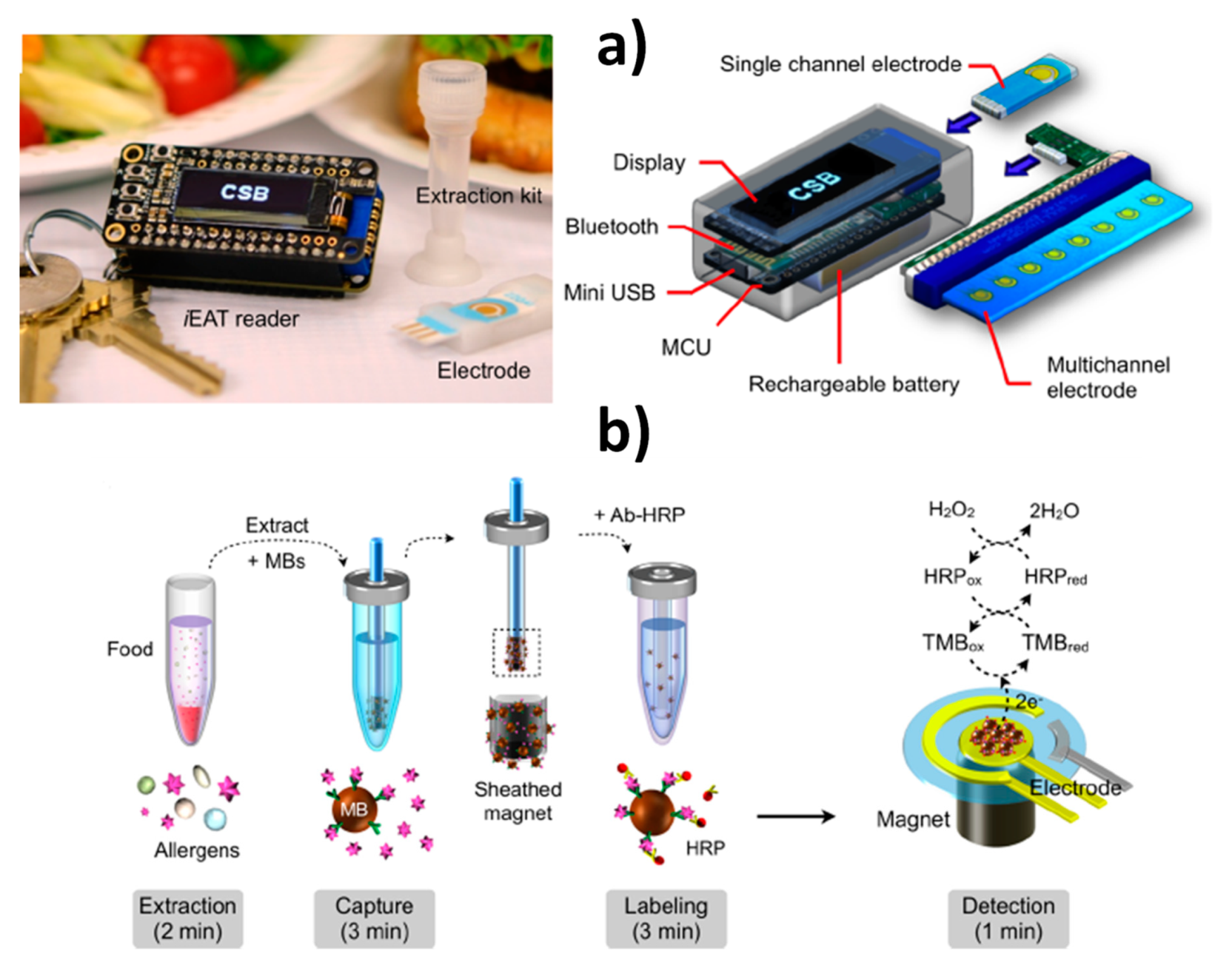
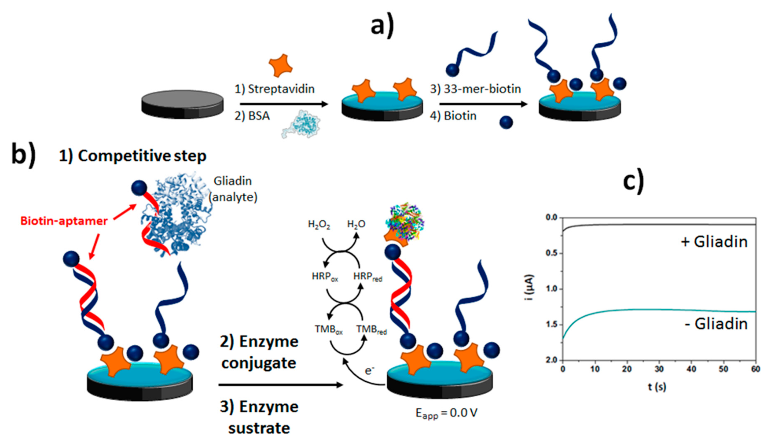
- The reliable multidetermination of 5 relevant allergens (gliadin, Ara h1, Cor a 1, casein, ovalbumin) in only 6 min using an affordable POC system integrating a disposable allergen extraction device and an electronic keychain reader [21].
- (iv)
- The detection of specific fruit allergens (the nonspecific lipid transfer protein Sola l 7 in tomato seeds) by targeting characteristic DNA sequences directly in 100 ng gDNA [57].
- (v)
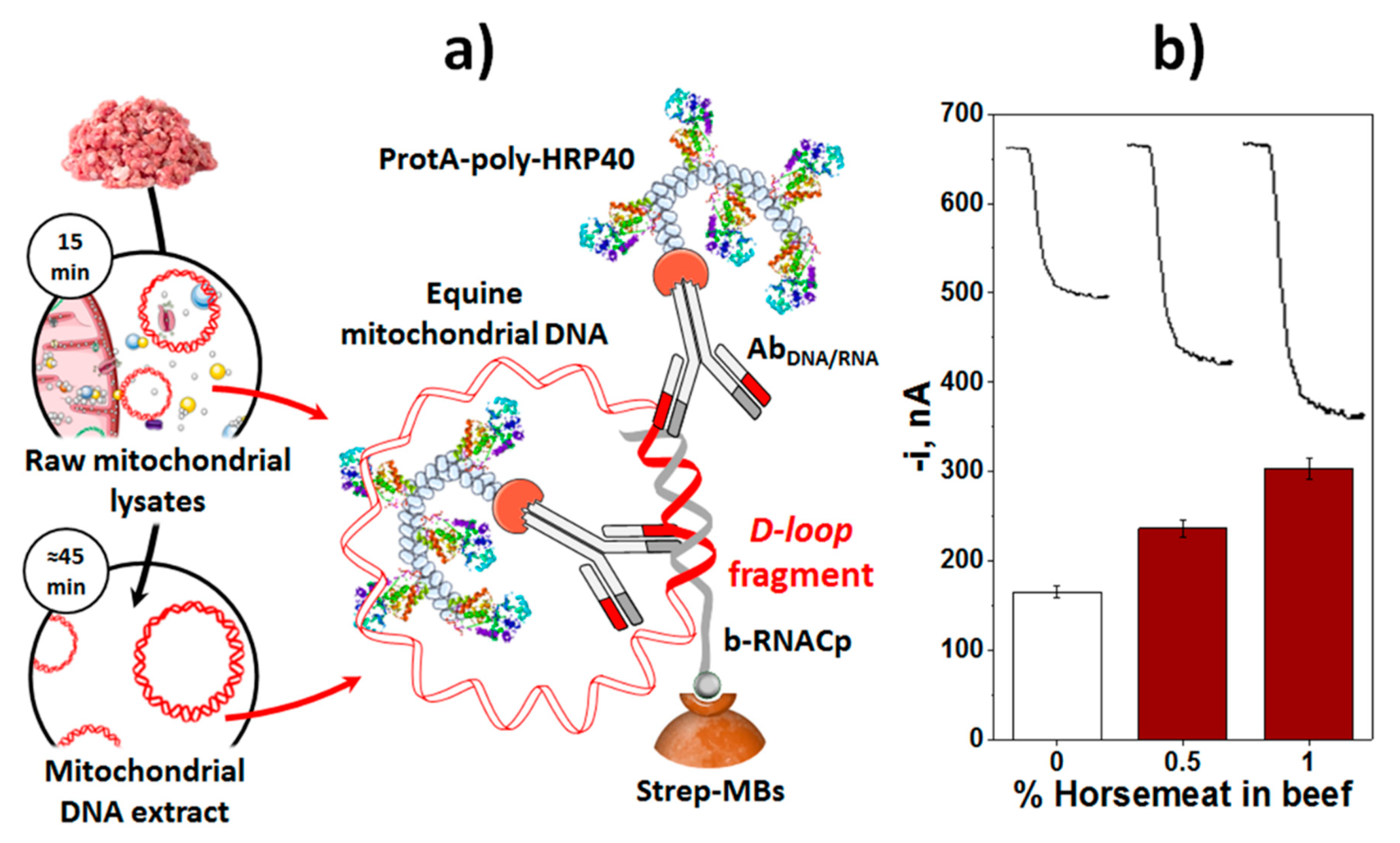
- The detection of meat adulterations directly in raw mitochondrial lysates obtained from a raw meat (detection of horse meat in beef meat) with no need for extracting the genetic material beforehand and in only 75 min [37].
- (vi)
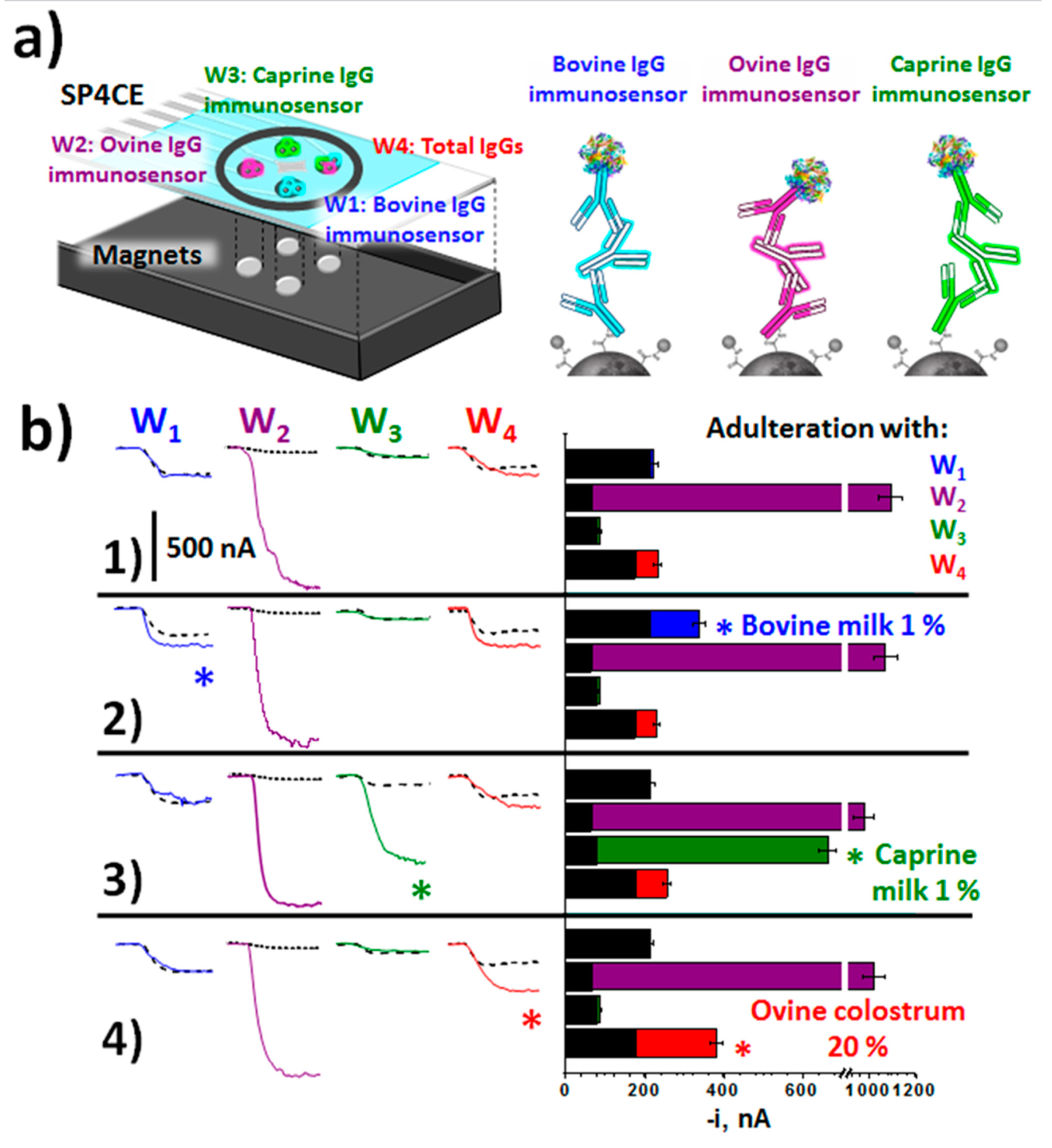
- The detection of milk adulterations with milk of other animal origin or colostrum by targeting IgGs specific to cow, sheep and goat in only 30 min [43].
Author Contributions
Funding
Acknowledgments
Conflicts of Interest
References
- Neethirajan, S.; Weng, X.; Tah, A.; Cordero, J.O.; Ragavan, K.V. Nano-biosensor platforms for detecting food allergens–New trends. Sens. Bio Sens. Res. 2018, 18, 13–30. [Google Scholar] [CrossRef]
- Borková, M.; Snáselová, J. Possibilities of different animal milk detection in milk and dairy products—A review. Czech J. Food Sci. 2005, 23, 41–50. [Google Scholar] [CrossRef] [Green Version]
- Santos, P.M.; Pereira-Filho, E.R.; Rodríguez-Saona, L.E. Rapid detection and quantification of milk adulteration using infrared microspectroscopy and chemometrics analysis. Food Chem. 2013, 138, 19–24. [Google Scholar] [CrossRef] [PubMed]
- Alves, R.C.; Barroso, M.F.; González-García, M.B.; Oliveira, M.B.P.P.; Delerue-Matos, C. New trends in food allergens detection: Toward biosensing strategies. Crit. Rev. Food Sci. Nutr. 2016, 56, 2304–2319. [Google Scholar] [CrossRef] [PubMed]
- Vasilescu, A.; Nunes, G.; Hayat, A.; Latif, U.; Marty, J.-L. Electrochemical Affinity Biosensors Based on Disposable Screen-Printed Electrodes for Detection of Food Allergens. Sensors 2016, 16, 1863. [Google Scholar] [CrossRef] [Green Version]
- Campuzano, S.; Ruiz-Valdepeñas Montiel, V.; Torrente-Rodríguez, R.M.; Reviejo, Á.J.; Pingarrón, J.M. Electrochemical biosensors for food security: Allergens and adulterants detection. In Biosensors for Security and Bioterrorism Applications; Nikolelis, D.P., Nikoleli, G.-P., Eds.; Springer: Berlin/Heidelberg, Germany, 2016; ISBN 978-3-319-28924-3. [Google Scholar]
- Rotariu, L.; Lagarde, F.; Jaffrezic-Renault, N.; Bala, C. Electrochemical biosensors for fast detection of food contaminants-trends and perspective. TrAC Trends Anal. Chem. 2016, 79, 80–87. [Google Scholar] [CrossRef]
- Martín-Fernández, B.; Manzanares-Palenzuela, C.L.; Sánchez-Paniagua López, M.; de-los-Santos-Álvarez, N.; López-Ruiz, B. Electrochemical genosensors in food safety assessment. Crit. Rev. Food Sci. Nutr. 2017, 57, 2758–2774. [Google Scholar] [CrossRef]
- Mishra, G.K.; Barfidokht, A.; Tehrani, F.; Mishra, R.K. Food Safety Analysis Using Electrochemical Biosensors. Foods 2018, 7, 141. [Google Scholar] [CrossRef] [Green Version]
- Gómez-Arribas, L.N.; Benito-Peña, E.; Hurtado-Sánchez, M.C.; Moreno-Bondi, M.C. Biosensing based on nanoparticles for food allergens detection. Sensors 2018, 18, 1087. [Google Scholar] [CrossRef] [Green Version]
- Tsagkaris, A.S.; Nelis, J.L.D.; Ross, G.M.S.; Jafari, S.; Guercetti, J.; Kopper, K.; Zhao, Y.; Rafferty, K.; Salvador, J.P.; Migliorelli, D.; et al. Critical assessment of recent trends related to screening and confirmatory analytical methods for selected food contaminants and allergens. Trends Anal. Chem. 2019, 121, 115688. [Google Scholar] [CrossRef]
- Eissa, S.; Chinnappan, R.; Zourob, M. Food allergy methods of detection and clinical studies. In Advances in Biosensor Technologies for Food Allergen Monitoring and Diagnosis; Abdel Rahman, A.M., Ed.; Taylor & Francis Group: Abington, UK, 2017. [Google Scholar]
- Monaci, L.; Visconti, A. Immunochemical and DNA-based methods in food allergen analysis and quality assurance perspectives. Trends Food Sci. Technol. 2010, 21, 272–283. [Google Scholar] [CrossRef]
- Hourihane, J.B.; Kilburn, S.A.; Nordlee, J.A.; Hefle, S.L.; Taylor, S.L.; Warner, J.O. An evaluation of the sensitivity of subjects with peanut allergy to very low doses of peanut protein: A randomized, double-blind, placebo-controlled food challenge study. J. Allergy Clin. Immunol. 1997, 100, 596–600. [Google Scholar] [CrossRef]
- Taylor, S.L.; Hefle, S.L.; Bindslev-Jensen, C.; Bock, S.A.; Burks, A.W., Jr.; Christie, L.L.; Hill, D.J.; Host, A.; Hourihane, J.O.B.; Lack, G.; et al. Factors affecting the determination of threshold doses for allergenic foods: How much is too much? J. Allergy Clin. Immunol. 2002, 109, 24–30. [Google Scholar] [CrossRef] [PubMed] [Green Version]
- López-López, L.; Miranda-Castro, R.; de-los-Santos-Álvarez, N.; Miranda-Ordieres, A.J.; Lobo-Castañón, M.J. Disposable electrochemical aptasensor for gluten determination in food. Sens. Actuat. B Chem. 2017, 241, 522–527. [Google Scholar] [CrossRef]
- Zhou, J.; Qi, Q.; Wang, C.; Qian, Y.; Liu, G.; Wang, Y.; Fu, L. Surface plasmon resonance (SPR) biosensors for food allergen detection in food matrices. Biosens. Bioelectron. 2019, 142, 111449. [Google Scholar] [CrossRef] [PubMed]
- Ye, Y.; Guo, H.; Sun, X. Recent progress on cell-based biosensors for analysis of food safety and quality control. Biosens. Bioelectron. 2019, 126, 389–404. [Google Scholar] [CrossRef] [PubMed]
- Bunney, J.; Williamson, S.; Atkin, D.; Jeanneret, M.; Cozzolino, D.; Chapman, J.; Power, A.; Chandra, S. The use of electrochemical biosensors in food analysis. Curr. Res. Nutr. Food Sci. 2017, 5, 183–195. [Google Scholar] [CrossRef]
- Malvano, F.; Pilloton, R.; Albanese, D. Label-free impedimetric biosensors for the control of food safety—A review. Int. J. Environ. Anal. Chem. 2019. [Google Scholar] [CrossRef]
- Lin, H.-Y.; Huang, C.-H.; Park, J.; Pathania, D.; Castro, C.M.; Fasano, A.; Weissleder, R.; Lee, H. Integrated magneto-chemical sensor for on-site food allergen detection. ACS Nano 2017, 1, 10062–10069. [Google Scholar] [CrossRef]
- Rama, E.C.; Costa-García, A. Screen-printed electrochemical immunosensors for the detection of cancer and cardiovascular biomarkers. Electroanalysis 2016, 28, 1700–1715. [Google Scholar] [CrossRef]
- Rocha-Santos, T.A.P. Sensors and biosensors based on magnetic nanoparticles. TrAC Trends Anal. Chem. 2014, 62, 28–36. [Google Scholar] [CrossRef]
- Reverté, L.; Prieto-Simón, B.; Campàs, M. New advances in electrochemical biosensors for the detection of toxins: Nanomaterials, magnetic beads and microfluidics systems. A review. Anal. Chim. Acta 2016, 908, 8–21. [Google Scholar] [CrossRef] [PubMed]
- Kudr, J.; Klejdus, B.; Adam, V.; Zitka, O. Magnetic solids in electrochemical analysis. Trac Trends Anal. Chem. 2018, 98, 104–113. [Google Scholar] [CrossRef]
- Pastucha, M.; Farka, Z.; Lacina, K.; Mikušová, Z.; Skládal, P. Magnetic nanoparticles for smart electrochemical immunoassays: A review on recent developments. Microchim. Acta 2019, 186, 312. [Google Scholar] [CrossRef]
- Wang, Z.; Dai, Z. Carbon nanomaterial-based electrochemical biosensors: An overview. Nanoscale 2015, 7, 6420–6431. [Google Scholar] [CrossRef]
- Power, A.C.; Gorey, B.; Chandra, S.; Chapman, J. Carbon nanomaterials and their application to electrochemical sensors: A review. Nanotechnol. Rev. 2018, 7, 19–41. [Google Scholar] [CrossRef]
- Pingarrón, J.M.; Yañez-Sedeño, P.; González-Cortés, A. Gold nanoparticle-based electrochemical biosensors. Electrochim Acta. 2008, 53, 5848–5866. [Google Scholar] [CrossRef]
- Campuzano, S.; Yáñez-Sedeño, P.; Pingarrón, J.M. Nanoparticles for nucleic-acid-based biosensing: Opportunities, challenges, and prospects. Anal. Bioanal. Chem. 2019, 411, 1791–1806. [Google Scholar] [CrossRef]
- Zhang, Y.; Chen, X. Nanotechnology and nanomaterials-based no-wash electrochemical biosensors: From design to application. Nanoscale 2019, 11, 19105–19118. [Google Scholar] [CrossRef]
- Alsager, O.A.; Eissa, S.; Zourob, M. Antibodies versus aptamers: A comparative view. In Immunosensors. Detection Science Series No. 14.; Uddin Ahmed, M., Zourob, M., Tamiya, E., Eds.; The Royal Society of Chemistry: London, UK, 2019; ISBN 978-1-78801-437-3. [Google Scholar]
- Titoiu, A.M.; Porumb, R.; Fanjul-Bolado, P.; Epure, P.; Zamfir, M.; Vasilescu, A. Detection of allergenic lysozyme during winemaking with an electrochemical aptasensor. Electroanalysis 2019, 31, 2262–2273. [Google Scholar] [CrossRef]
- Miranda-Castro, R.; de los Santos Álvarez, N.; Lobo-Castañón, M.J. Aptamer-based magnetoassay for gluten determination. In Laboratory Methods in Dynamic Electroanalysis; Elsevier: Amsterdam, The Netherlands, 2020; pp. 221–229. [Google Scholar]
- Li, H.; Somerson, J.; Xia, F.; Plaxco, K.W. Electrochemical DNA-based sensors for molecular quality control: Continuous, real-time melamine detection in flowing whole milk. Anal. Chem. 2018, 90, 10641–10645. [Google Scholar] [CrossRef] [PubMed] [Green Version]
- Ruiz-Valdepeñas Montiel, V.; Torrente-Rodríguez, R.M.; González de Rivera, G.; Reviejo, A.J.; Cuadrado, C.; Linacero, R.; Gallego, F.J.; Campuzano, S.; Pingarrón, J.M. Amperometric determination of hazelnut traces by means of Express PCR coupled to magnetic beads assembled on disposable DNA sensing scaffolds. Sens. Actuat. B Chem. 2017, 245, 895–902. [Google Scholar] [CrossRef]
- Ruiz-Valdepeñas Montiel, V.; Gutiérrez, M.L.; Torrente-Rodríguez, R.M.; Povedano, E.; Vargas, E.; Reviejo, Á.J.; Linacero, R.; Gallego, F.J.; Campuzano, S.; Pingarrón, J.M. Disposable amperometric Polymerase Chain Reaction-free biosensor for direct detection of adulteration with horsemeat in raw lysates targeting mitochondrial DNA. Anal. Chem. 2017, 89, 9474–9482. [Google Scholar]
- Ruiz-Valdepeñas Montiel, V.; Povedano, E.; Vargas, E.; Torrente-Rodríguez, R.M.; Pedrero, M.; Reviejo, A.J.; Campuzano, S.; Pingarrón, J.M. Comparison of different strategies for the development of highly sensitive electrochemical nucleic acid biosensors using neither nanomaterials nor nucleic acid amplification. ACS Sens. 2018, 3, 211–221. [Google Scholar] [CrossRef] [PubMed]
- Campuzano, S.; Yañez-Sedeño, P.; Pingarrón, J.M. Tailoring sensitivity in electrochemical nucleic acid hybridization biosensing: Role of surface chemistry and labeling strategies. ChemElectroChem 2019, 6, 60–72. [Google Scholar] [CrossRef]
- Alves, R.C.; Pimentel, F.B.; Nouws, H.P.; Silva, T.H.; Oliveira, M.B.P.; Delerue-Matos, C. Improving the extraction of Ara h 6 (a peanut allergen) from a chocolate based matrix for immunosensing detection: Influence of time, temperature and additives. Food Chem. 2017, 218, 242–248. [Google Scholar] [CrossRef] [PubMed] [Green Version]
- Angulo-Ibáñez, A.; Eletxigerra, U.; Lasheras, X.; Campuzano, S.; Merino, S. Electrochemical tropomyosin allergen immunosensor for complex food matrix analysis. Anal. Chim. Acta 2019, 1079, 94–102. [Google Scholar] [CrossRef]
- Marín-Barroso, E.; Messina, G.A.; Bertolino, F.A.; Raba, J.; Pereira, S.V. Electrochemical immunosensor modified with carbon nanofibers coupled to a paper platform for the determination of gliadins in food samples. Anal. Methods 2019, 11, 2170–2178. [Google Scholar] [CrossRef]
- Ruiz-Valdepeñas Montiel, V.; Povedano, E.; Benedé, S.; Mata, L.; Galán-Malo, P.; Gamella, M.; Reviejo, A.J.; Campuzano, S.; Pingarrón, J.M. Disposable amperometric immunosensor for the detection of adulteration in milk through single or multiplexed determination of bovine, ovine, or caprine immunoglobulins G. Anal. Chem. 2019, 91, 11266–11274. [Google Scholar] [CrossRef]
- Benedé, S.; Ruiz-Valdepeñas Montiel, V.; Povedano, E.; Villalba, M.; Mata, L.; Galán-Malo, P.; Torrente-Rodríguez, R.M.; Vargas, E.; Reviejo, A.J.; Campuzano, S.; et al. Fast amperometric immunoplatform for ovomucoid traces determination in fresh and baked foods. Sens. Actuat. B Chem. 2018, 265, 421–428. [Google Scholar] [CrossRef]
- Owen, G.R.H.; Häkkinen, L.; Wu, C.; Larjava, H. A reproducible technique for specific labeling of antigens using preformed fluorescent molecular IgG-F(ab’)2 complexes from primary antibodies of the same species. Microsc. Res. Tech. 2010, 73, 623–630. [Google Scholar] [PubMed] [Green Version]
- MacPhee, D.J. Methodological considerations for improving Western blot analysis. Toxicol. Methods 2010, 61, 171–177. [Google Scholar] [CrossRef] [PubMed]
- González-Álvarez, M.J.; Pérez-Ruiz, E.; Miranda-Castro, R.; de-los-Santos-Álvarez, N.; Miranda-Ordieres, A.J.; Lobo-Castañón, M.J. Effect of tags and labels on the performance of enzyme-amplified electrochemical genomagnetic assays. Electroanalysis 2013, 25, 147–153. [Google Scholar] [CrossRef]
- Miranda-Castro, R.; de-los-Santos-Álvarez, N.; Lobo-Castañón, M.J. Understanding the factors affecting the analytical performance of sandwich-hybridization genosensors on gold electrodes. Electroanalysis 2018, 30, 1229–1240. [Google Scholar] [CrossRef]
- Escamilla-Gómez, V.; Hernández-Santos, D.; González-García, M.B.; Pingarrón-Carrazón, J.M.; Costa-García, A. Simultaneous detection of free and total prostate specific antigen on a screen-printed electrochemical dual sensor. Biosens. Bioelectron. 2009, 24, 2678–2683. [Google Scholar] [CrossRef]
- Benedé, S.; López-Expósito, I.; Molina, E.; Lopez-Fandino, R. Egg proteins as allergens and the effects of the food matrix and processing. Food Funct. 2015, 6, 694–713. [Google Scholar] [CrossRef]
- De Luis, R.; Mata, L.; Estopañán, G.; Lavilla, M.; Sánchez, L.; Pérez, M.D. Evaluation of indirect competitive and double antibody sandwich ELISA tests to determine β-lactoglobulin and ovomucoid in model processed foods. Food Agric. Immunol. 2008, 19, 339–350. [Google Scholar] [CrossRef] [Green Version]
- López-Calleja, I.M.; de la Cruz, S.; Pegels, N.; González, I.; García, T.; Martín, R. Development of a real time PCR assay for detection of allergenic trace amounts of peanut (Arachis hypogaea) in processed foods. Food Control 2013, 30, 480–490. [Google Scholar] [CrossRef]
- Ross, G.M.S.; Bremer, M.G.E.G.; Nielen, M.W.F. Consumer-friendly food allergen detection: Moving towards smartphone-based immunoassays. Anal. Bioanal. Chem. 2018, 410, 5353–5371. [Google Scholar] [CrossRef] [Green Version]
- Alves, R.C.; Pimentel, F.B.; Nouws, H.P.A.; Correr, W.; González-García, M.B.; Oliveira, M.B.P.P.; Delerue-Matos, C. Detection of the peanut allergen Ara h 6 in foodstuffs using a voltammetric biosensing approach. Anal. Bioanal. Chem. 2015, 407, 7157–7163. [Google Scholar] [CrossRef]
- Bottari, F.; Moretto, L.M.; Ugo, P. Impedimetric sensing of the immuno-enzymatic reaction of gliadin with a collagen-modified electrode. Electrochem. Commun. 2018, 97, 51–55. [Google Scholar] [CrossRef]
- Sobhan, A.; Oh, J.-H.; Park, M.-K.; Kim, S.W.; Park, C.; Lee, J. Single walled carbon nanotube based biosensor for detection of peanut allergy-inducing protein ara h1. Korean J. Chem. Eng. 2018, 35, 172–178. [Google Scholar] [CrossRef]
- Pereira-Barros, M.A.; Barroso, M.F.; Martín-Pedraza, L.; Vargas, E.; Benedé, S.; Villalba, M.; Rocha, J.M.; Campuzano, S.; Pingarrón, J.M. Direct PCR-free electrochemical biosensing of plant-food derived nucleic acids in genomic DNA extracts. Application to the determination of the key allergen Sola l 7 in tomato seeds. Biosens. Bioelectron. 2019, 137, 171–177. [Google Scholar] [CrossRef] [PubMed]
- Valat, C.; Limoges, B.; Huet, D.; Romette, J.-L. A disposable Protein A-based immunosensor for flow-injection assay with electrochemical detection. Anal. Chim. Acta 2000, 404, 187–194. [Google Scholar] [CrossRef]
- Akram, M.; Stuart, M.C.; Wong, D.K.Y. Direct application strategy to immobilise a thioctic acid self-assembled monolayer on a gold electrode. Anal. Chim. Acta 2004, 504, 243–251. [Google Scholar] [CrossRef]
- Idili, A.; Parolo, C.; Ortega, G.; Plaxco, K.W. Calibration-free measurement of phenylalanine levels in the blood using an electrochemical aptamer-based sensor suitable for point-of-care applications. ACS Sens. 2019, 4, 3227–3233. [Google Scholar] [CrossRef]
- Li, H.; Li, S.; Dai, J.; Li, C.; Zhu, M.; Li, H.; Lou, X.; Xia, F.; Plaxco, K.W. High frequency, calibration-free molecular measurements in situ in the living body. Chem. Sci. 2019, 10, 10843–10848. [Google Scholar] [CrossRef] [Green Version]
- Lin, P.-H.; Li, B.-R. Antifouling strategies in advanced electrochemical sensors and biosensors. Analyst 2020, 145, 1110–1120. [Google Scholar] [CrossRef]


| Electrode | Methodology | Target Allergen/Adulterant (Food) | Detection Technique | Linear Range | LOD | Sample | Steps */Assay Time | Ref. |
|---|---|---|---|---|---|---|---|---|
| Allergens | ||||||||
| AuNPs-SPCE | Sandwich immunosensor involving a bDAb further conjugated with Strep-AP | Ara h 6 (Peanut) | LSV (3-IP+AgNO3) | 1–100 ng mL−1 | 0.27 ng mL−1 | Complex chocolate-based product containing peanut | 3/3 h starting from CAb- AuNPs-SPCE | [40,54] |
| SPCE | Sandwich immunoassay implemented onto HOOC-MBs involving the same Ab unmodified as capture and labeled with HRP as detector | Ovomucoid (Egg) | Amperometry (H2O2/HQ) | 0.3–25 ng mL−1 | 0.1 ng mL−1 | Eggs, wheat flour and baked bread | 1/30 min starting from CAb-HOOC-MBs | [44] |
| GCE | Crosslinking of gliadin on the surface of a collagen-modified GCE, catalyzed by transglutaminase and recognition with a specific anti-gliadin antibody | Gliadin (Gluten) | EIS [Fe(CN)6]3-/4- | 5–20 mg L−1 | - | - | 4/4 h | [55] |
| SWCNTs-Au-Cr deposited silicon wafer | Direct immunoassay onto and CAb covalently immobilized onto 1-PBSE-modified SWCNTs | Ara h 1 (Peanut) | LSV | 1–1000 ng mL−1 | 1 ng mL−1 | - | 1/30 min starting from the SWCNT-based biosensor | [56] |
| Au-SPEs (individual and array of 8 × SPEs) | Sandwich immunoassays implemented on the surface of Epoxy-MBs | Gliadin, Ara h1, Cor a 1, Casein, Ovalbumin/Gluten, peanuts, hazelnuts, milk, and eggs. | Chronoamperometry (H2O2/TMB) | - | Gliadin: 0.075 mg Kg−1 Ara h1: 0.007 mg Kg−1 Cor a 1: 0.089 mg Kg−1 Casein: 0.170 mg Kg−1 Ovalbumin: 0.003 mg Kg−1 | Bread, milk, cereal, cookie, ice cream, burger, salads with dressing, pizza, beer and gluten-free menu items from restaurants | 2/6 min starting from CAb-Epoxy-MBs | [21] |
| SPCE | Sandwich immunoassay implemented onto HOOC-MBs involving specific CAb and DAb and enzymatic labeling with an HRP-secondary Ab | Shrimp TPM (Shrimp) | Amperometry (H2O2/HQ) | Up to 218.7 ng mL−1 | 47 pg mL−1 | Raw and cooked shrimp and squid samples | 3/3 h starting from CAb-HOOC-MBs | [41] |
| CNFs/SPCE coupled to a paper immunoaffinity platform | Sandwich immunoassay implemented onto paper microzone | Gliadin (Gluten) | Amperometry (H2O2/HQ) | Up to 80 μg kg−1 | Gliadin: 0.005 mg Kg−1 | Flour samples (manioc, rice, gluten free, common wheat) | 2/15 min starting from CAb-paper | [42] |
| Adulterants | ||||||||
| SPCE and SP4CEs | Sandwich immunoassays implemented onto HOOC-MBs involving the same Ab unmodified as capture and labeled with HRP as detector | Bovine, Ovine and Caprine IgGs (Milk) | Amperomety (H2O2/HQ) | Bovine IgGs: 2.6−250 ng mL−1, Ovine IgGs: 2.7−250 ng mL−1, Caprine IgGs 2.2−250 ng mL−1 | Bovine IgGs: 0.74 ng mL−1, Ovine IgGs: 0.82 ng mL−1, Caprine IgGs 0.66 ng mL−1 | Colostrum, raw, pasteurized and UHT milk samples | 1/30 min starting from CAb-HOOC-MBs | [43] |
| Electrode | Methodology | Target Allergen/Adulterant (Food) | Detection Technique | Linear Range | LOD | Sample | Steps */Assay Time | Ref. |
|---|---|---|---|---|---|---|---|---|
| Allergens | ||||||||
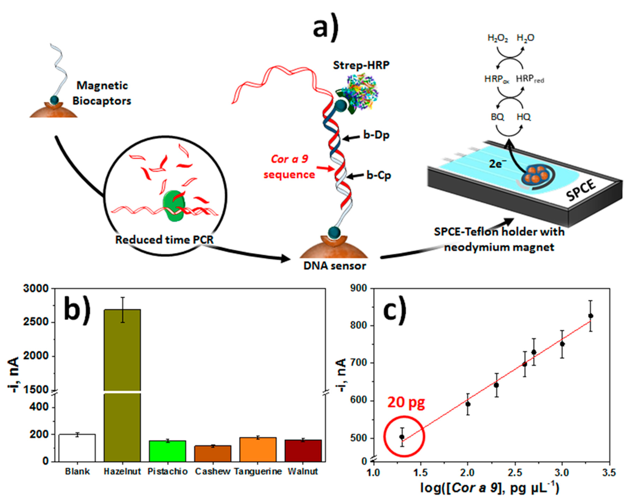
| SPCE | Sandwich hybridization assay involving biotinylated DNA Cp and Dp implemented on the surface of Strep-MBs and coupled to Express-PCR (100 bp-amplicon) | Cor a 9 (Hazelnut) | Amperometry (H2O2/HQ) | 0.0024–0.75 nM (synthetic target DNA) | 0.72 pM (synthetic target DNA) 20 pg gDNA extracted from hazelnut | hazelnut varieties and other species of similar families (pistachio, cashew, walnut and tangerine) | 1/15 min starting from bCp-Strep-MBs | [36] |
| SPCE | Indirect competitive approach between a biotinylated peptide (b-Pep) immobilized onto Strep-SPCE and gluten proteins for a defined concentration of biotinylated Gli4 aptamer further labeling with Strep-HRP | Gliadin (Gluten) | Chronoamperometry (H2O2/TMB) | 1–100 μg L−1 (four-parameter logistic equation) | 0.113 μg L−1 Gliadin (< >380 μg kg−1 gluten) | Food samples (Fixamyl, Rolled oats, Fit Snack) | 2/60 min starting from b-Pep-Strep-SPCE | [16] |
| SPCE | Sandwich hybridization assay involving a b-RNACp and a RNADp implemented on the surface of Strep-MBs and recognition of the RNA/DNA heterohybrids with AbRNA/DNA further conjugated with an HRP-secondary antibody | Sola l 7 (Tomato) | Amperometry (H2O2/HQ) | 0.8–50 pM (synthetic target) | 0.2 pM (synthetic target) | Tomato and corn (100 ng extracted gDNA) | 2/1.5 h starting from b-RNACp-Strep-MBs | [57] |
| AuNPs-modified Au-SPE | Direct aptasensing approach at AuNPs-Au-SPE modified with a SH-Aptamer | Lysozyme | CV ([Fe(CN)6]3-) | 1–10 μg mL−1 | 0.32 μg mL−1 | Wines | 1/1 h starting from SH-Aptamer-AuNPs-Au-SPE | [33] |
| SPCE | Indirect competitive approach between a biotinylated peptide (b-Pep) immobilized onto Strep-MBs and gluten proteins for a defined concentration of biotinylated Gli1 aptamer further labeling with Strep-HRP | Gliadin (Gluten) | Chronoamperometry (H2O2/TMB) | - | - | - | 2/60 min starting from b-Pep-Strep-MBs | [34] |
| Adulterants | ||||||||
| SPCE | Direct hybridization assay at b-RNACp-Strep-MBs and and recognition of the RNA/DNA heterohybrids with AbRNA/DNA further conjugated with ProtA-polyHRP40 | Specific fragment of horse mitochondrial DNA D-loop region (Meat) | Amperomety (H2O2/HQ) | 0.39–75 pM | 0.12 pM (synthetic target DNA) | Beef meat spiked with horse meat | 2/1 h 30 min starting from b-RNACp-Strep-MBs | [37] |
| Au disk(2-mm) | E-DNA based on a MB-modified thiolated DNA (Signal-off) | Melamine (Milk) | SWV (MB) | - | 150 μM(~19 ppm) in buffered solutions,20 μM (~2.5 ppm) in whole milk | Milk | Continuous, real-time mesurements in flowing samples | [35] |
© 2020 by the authors. Licensee MDPI, Basel, Switzerland. This article is an open access article distributed under the terms and conditions of the Creative Commons Attribution (CC BY) license (http://creativecommons.org/licenses/by/4.0/).
Share and Cite
Campuzano, S.; Ruiz-Valdepeñas Montiel, V.; Serafín, V.; Yáñez-Sedeño, P.; Pingarrón, J.M. Cutting-Edge Advances in Electrochemical Affinity Biosensing at Different Molecular Level of Emerging Food Allergens and Adulterants. Biosensors 2020, 10, 10. https://0-doi-org.brum.beds.ac.uk/10.3390/bios10020010
Campuzano S, Ruiz-Valdepeñas Montiel V, Serafín V, Yáñez-Sedeño P, Pingarrón JM. Cutting-Edge Advances in Electrochemical Affinity Biosensing at Different Molecular Level of Emerging Food Allergens and Adulterants. Biosensors. 2020; 10(2):10. https://0-doi-org.brum.beds.ac.uk/10.3390/bios10020010
Chicago/Turabian StyleCampuzano, Susana, Víctor Ruiz-Valdepeñas Montiel, Verónica Serafín, Paloma Yáñez-Sedeño, and José Manuel Pingarrón. 2020. "Cutting-Edge Advances in Electrochemical Affinity Biosensing at Different Molecular Level of Emerging Food Allergens and Adulterants" Biosensors 10, no. 2: 10. https://0-doi-org.brum.beds.ac.uk/10.3390/bios10020010