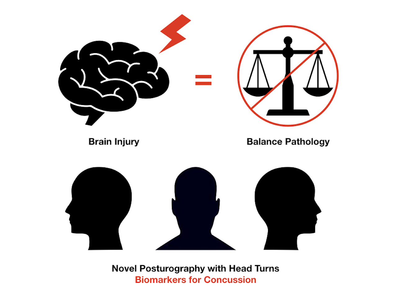
Head Position and Posturography: A Novel Biomarker to Identify Concussion Sufferers

 , ,
, ,
Abstract
:
1. Introduction
2. Materials and Methods
3. Results

4. Discussion
5. Conclusions
Author Contributions
Funding
Acknowledgments
Conflicts of Interest
References
- Balasubramaniam, R.; Wing, A.M. The dynamics of standing balance. Trends Cogn. Sci. 2002, 6, 531–536. [Google Scholar] [CrossRef]
- Yim-Chiplis, P.K.; Talbot, L.A. Defining and measuring balance in adults. Biol. Res. Nurs. 2000, 1, 321–331. [Google Scholar] [CrossRef] [PubMed]
- Marigold, D.S.; Eng, J.J.; Tokuno, C.D.; Donnelly, C.A. Contribution of muscle strength and integration of afferent input to postural instability in persons with stroke. Neurorehabilit. Neural Repair 2004, 18, 222–229. [Google Scholar] [CrossRef] [PubMed] [Green Version]
- Guskiewicz, K.M. Postural stability assessment following concussion: One piece of the puzzle. Clin. J. Sport Med. 2001, 11, 182–189. [Google Scholar] [CrossRef] [PubMed]
- Arifin, N.; Abu Osman, N.A.; Wan Abas, W.A. Intrarater test-retest reliability of static and dynamic stability indexes measurement using the Biodex Stability System during unilateral stance. J. Appl. Biomech. 2014, 30, 300–304. [Google Scholar] [CrossRef]
- Massingale, S.; Alexander, A.; Erickson, S.; McQueary, E.; Gerkin, R.; Kisana, H.; Silvestri, B.; Schodrof, S.; Nalepa, B.; Pardini, J. Comparison of uninjured and concussed adolescent athletes on the concussion balance Test (COBALT). J. Neurol. Phys. Ther. 2018, 42, 149–154. [Google Scholar] [CrossRef] [PubMed]
- Valovich McLeod, T.C.; Hale, T.D. Vestibular and balance issues following sport-related concussion. Brain Inj. 2015, 29, 175–184. [Google Scholar] [CrossRef]
- Pagnacco, G.; Oggero, E.; Carrick, F.R. Repeatability of posturographic measures of the mctsib static balance tests a preliminary investigation. Biomed. Sci. Instrum. 2008, 44, 41–46. [Google Scholar]
- Carrick, F.R.; Clark, J.F.; Pagnacco, G.; Antonucci, M.M.; Hankir, A.; Zaman, R.; Oggero, E. Head-Eye Vestibular Motion Therapy Affects the Mental and Physical Health of Severe Chronic Postconcussion Patients. Front. Neurol. 2017, 8, 414. [Google Scholar] [CrossRef] [Green Version]
- Carrick, F.R.; Hankir, A.; Zaman, R.; Antonucci, M.M.; Pagnacco, G.; Azzolino, S.; Oggero, E. Improvement of Saccadic Eye Movements after Head-Eye Vestibular Motion (HEVM) Therapy and Neuro-Psychiatric Considerations. Psychiatr. Danub. 2019, 31 (Suppl. 3), 318–323. [Google Scholar]
- Carrick, F.R.; Hankir, A.; Zaman, R.; Wright, C.H. Metrological Performance of Instruments used in Clinical Evaluation of Balance. Psychiatr. Danub. 2019, 31 (Suppl. 3), 324–330. [Google Scholar] [PubMed]
- Oggero, E.; Carrick, F.R.; Pagnacco, G. Frequency content of standard posturographic measures—Biomed 2013. Biomed. Sci. Instrum. 2013, 49, 48–53. [Google Scholar] [PubMed]
- Pagnacco, G.; Carrick, F.R.; Wright, C.H.; Oggero, E. In-situ verification of accuracy, precision and resolution of force and balance platforms. Biomed. Sci. Instrum. 2014, 50, 171–178. [Google Scholar] [PubMed]
- Carrick, F.R.; Oggero, E.; Pagnacco, G.; Brock, J.B.; Arikan, T. Posturographic testing and motor learning predictability in gymnasts. Disabil. Rehabil. 2007, 29, 1881–1889. [Google Scholar] [CrossRef]
- Pagnacco, G.; Carrick, F.R.; Pascolo, P.B.; Rossi, R.; Oggero, E. Learning effect of standing on foam during posturographic testing preliminary findings. Biomed. Sci. Instrum. 2012, 48, 332–339. [Google Scholar] [PubMed]
- Barry, R.J.; De Blasio, F.M. EEG differences between eyes-closed and eyes-open resting remain in healthy ageing. Biol. Psychol. 2017, 129, 293–304. [Google Scholar] [CrossRef] [Green Version]
- Barry, R.J.; Clarke, A.R.; Johnstone, S.J.; Magee, C.A.; Rushby, J.A. EEG differences between eyes-closed and eyes-open resting conditions. Clin. Neurophysiol. 2007, 118, 2765–2773. [Google Scholar] [CrossRef]
- Lynall, R.C.; Blackburn, J.T.; Guskiewicz, K.M.; Marshall, S.W.; Plummer, P.; Mihalik, J.P. Functional balance assessment in recreational college-aged individuals with a concussion history. J. Sci. Med. Sport 2019, 22, 503–508. [Google Scholar] [CrossRef]
- Alonso, A.C.; Luna, N.M.; Mochizuki, L.; Barbieri, F.; Santos, S.; Greve, J.M. The influence of anthropometric factors on postural balance: The relationship between body composition and posturographic measurements in young adults. Clinics 2012, 67, 1433–1441. [Google Scholar] [CrossRef]
- Webb, P.U.S. NASA Life Sciences Data; N.A.a.S. Administration: Washington, DC, USA, 1964. [Google Scholar]
- Pagnacco, G.; Carrick, F.R.; Wright, C.H.; Oggero, E. Between-subjects differences of within-subject variability in repeated balance measures: Consequences on the minimum detectable change. Gait Posture 2015, 41, 136–140. [Google Scholar] [CrossRef]
- Freese, J.S.L.a.J. Regression Models for Categorical Dependent Variables Using Stata, 3rd ed.; Stata Press: College Station, TX, USA, 2014; p. 589. [Google Scholar]
- Cruise, D.R.; Chagdes, J.R.; Liddy, J.J.; Rietdyk, S.; Haddad, J.M.; Zelaznik, H.N.; Raman, A. An active balance board system with real-time control of stiffness and time-delay to assess mechanisms of postural stability. J. Biomech. 2017, 60, 48–56. [Google Scholar] [CrossRef] [PubMed]
- Feddermann-Demont, N.; Echemendia, R.J.; Schneider, K.J.; Solomon, G.S.; Hayden, K.A.; Turner, M.; Dvořák, J.; Straumann, D.; Tarnutzer, A.A. What domains of clinical function should be assessed after sport-related concussion? A systematic review. Br. J. Sports Med. 2017, 51, 903–918. [Google Scholar] [CrossRef] [PubMed]
- Scorza, K.A.; Raleigh, M.F.; O’Connor, F.G. Current concepts in concussion: Evaluation and management. Am. Fam. Physician 2012, 85, 123–132. [Google Scholar] [PubMed]
- Brodsky, J.R.; Lipson, S.; Bhattacharyya, N. Prevalence of Pediatric Dizziness and Imbalance in the United States. Otolaryngol. Head Neck Surg. 2020, 162, 241–247. [Google Scholar] [CrossRef]
- Guskiewicz, K.M.; Register-Mihalik, J.K. Postconcussive impairment differences across a multifaceted concussion assessment protocol. PM R 2011, 3 (Suppl. 2), S445–S451. [Google Scholar] [CrossRef]
- Hammerle, M.; Swan, A.A.; Nelson, J.T.; Treleaven, J.M. Retrospective Review: Effectiveness of Cervical Proprioception Retraining for Dizziness After Mild Traumatic Brain Injury in a Military Population With Abnormal Cervical Proprioception. J. Manip. Physiol. Ther. 2019, 42, 399–406. [Google Scholar] [CrossRef]
- Hides, J.A.; Franettovich Smith, M.M.; Mendis, M.D.; Treleaven, J.; Rotstein, A.H.; Sexton, C.T.; Low Choy, N.; McCrory, P. Self-reported Concussion History and Sensorimotor Tests Predict Head/Neck Injuries. Med. Sci. Sports Exerc. 2017, 49, 2385–2393. [Google Scholar] [CrossRef]
- Junn, C.; Bell, K.R.; Shenouda, C.; Hoffman, J.M. Symptoms of Concussion and Comorbid Disorders. Curr. Pain Headache Rep. 2015, 19, 46. [Google Scholar] [CrossRef]
- Matuszak, J.M.; McVige, J.; McPherson, J.; Willer, B.; Leddy, J. A Practical Concussion Physical Examination Toolbox. Sports Health 2016, 8, 260–269. [Google Scholar] [CrossRef] [Green Version]
- Reiley, A.S.; Vickory, F.M.; Funderburg, S.E.; Cesario, R.A.; Clendaniel, R.A. How to diagnose cervicogenic dizziness. Arch Physiother. 2017, 7, 12. [Google Scholar] [CrossRef] [Green Version]
- Schneider, K.J.; Meeuwisse, W.H.; Palacios-Derflingher, L.; Emery, C.A. Changes in Measures of Cervical Spine Function, Vestibulo-ocular Reflex, Dynamic Balance, and Divided Attention Following Sport-Related Concussion in Elite Youth Ice Hockey Players. J. Orthop. Sports Phys. Ther. 2018, 48, 974–981. [Google Scholar] [CrossRef] [PubMed]
- Tiwari, D.; Goldberg, A.; Yorke, A.; Marchetti, G.F.; Alsalaheen, B. Characterization of Cervical Spine Impairments in Children and Adolescents Post-Concussion. Int. J. Sports Phys. Ther. 2019, 14, 282–295. [Google Scholar] [CrossRef] [PubMed]
- Treleaven, J. Dizziness, Unsteadiness, Visual Disturbances, and Sensorimotor Control in Traumatic Neck Pain. J. Orthop. Sports Phys. Ther. 2017, 47, 492–502. [Google Scholar] [CrossRef] [PubMed]
- Salahzadeh, Z.; Maroufi, N.; Ahmadi, A.; Behtash, H.; Razmjoo, A.; Gohari, M.; Parnianpour, M. Assessment of forward head posture in females: Observational and photogrammetry methods. J. Back Musculoskelet. Rehabil. 2014, 27, 131–139. [Google Scholar] [CrossRef]
- Raine, S.; Twomey, L.T. Head and shoulder posture variations in 160 asymptomatic women and men. Arch Phys. Med. Rehabil. 1997, 78, 1215–1223. [Google Scholar] [CrossRef]
- Szczygieł, E.; Fudacz, N.; Golec, J.; Golec, E. The impact of the position of the head on the functioning of the human body: A systematic review. Int. J. Occup. Med. Environ. Health 2020, 33, 559–568. [Google Scholar] [CrossRef]
- Chaturvedi, P.; Singh, A.K.; Tiwari, V.; Thacker, A.K. Post-stroke BDNF concentration changes following proprioceptive neuromuscular facilitation (PNF) exercises. J. Family Med. Prim. Care 2020, 9, 3361–3369. [Google Scholar]
- Guyot, M.A.; Agnani, O.; Peyrodie, L.; Samantha, D.; Donze, C.; Catanzariti, J.F. Cervicocephalic relocation test to evaluate cervical proprioception in adolescent idiopathic scoliosis. Eur. Spine J. 2016, 25, 3130–3136. [Google Scholar] [CrossRef] [PubMed]
- Abedi Khoozani, P.; Blohm, G. Neck muscle spindle noise biases reaches in a multisensory integration task. J. Neurophysiol. 2018, 120, 893–909. [Google Scholar] [CrossRef] [PubMed]
- Silva, A.G.; Cruz, A.L. Standing balance in patients with whiplash-associated neck pain and idiopathic neck pain when compared with asymptomatic participants: A systematic review. Physiother. Theory Pract. 2013, 29, 1–18. [Google Scholar] [CrossRef]
- Treleaven, J. Sensorimotor disturbances in neck disorders affecting postural stability, head and eye movement control–Part 2: Case studies. Man. Ther. 2008, 13, 266–275. [Google Scholar] [CrossRef] [PubMed]
- Whitehead, K.; Meek, J.; Fabrizi, L.; Smith, B.A. Long-range temporal organisation of limb movement kinematics in human neonates. Clin. Neurophysiol. Pract. 2020, 5, 194–198. [Google Scholar] [CrossRef] [PubMed]
- Saavedra, S.; Woollacott, M.; van Donkelaar, P. Head stability during quiet sitting in children with cerebral palsy: Effect of vision and trunk support. Exp. Brain Res. 2010, 201, 13–23. [Google Scholar] [CrossRef] [PubMed] [Green Version]
- Forbes, J.; Cronovich, H. Romberg Test. In StatPearls; StatPearls Publishing: Treasure Island, FL, USA, 2020. [Google Scholar]


| Age (Years) | Height (m) | Mass (kg) | BMI (kg/m2) | |
|---|---|---|---|---|
| Males (35) | 33.06 ± 1.80 20–64 | 1.79 ± 0.01 1.62–2.00 | 87.33 ± 2.61 65.00–149.30 | 27.34 ± 0.66 21.69–41.14 |
| Females (25) | 29.84 ± 1.86 19–52 | 1.65 ± 0.01 1.52–1.80 | 70.15 ± 2.65 51.54–101.39 | 25.84 ± 1.01 18.89–40.61 |
| 18–25 (20) | 23.55 ± 0.41 19–25 | 1.72 ± 0.02 1.55–1.94 | 75.81 ± 3.47 53.97–101.39 | 25.70 ± 1.10 19.92–40.61 |
| 26–34 (24) | 29.46 ± 0.62 26–34 | 1.74 ± 0.02 1.52–2.00 | 85.04 ± 3.99 57.24–149.30 | 27.73 ± 0.92 20.37–41.14 |
| 35–65 (16) | 45.31 ± 2.41 35–64 | 1.72 ± 0.02 1.57–1.86 | 78.27 ± 3.08 53.96–104.01 | 26.45 ± 0.86 18.89–31.10 |
| All (60) | 31.72 ± 1.31 19–64 | 1.73 ± 0.01 1.52–2.00 | 80.16 ± 2.17 53.96–149.30 | 26.71 ± 0.57 18.89–41.14 |
| Age (Years) | Height (m) | Mass (kg) | BMI (kg/m2) | |
|---|---|---|---|---|
| Males (335) | 33.88 ± 0.67 18–65 | 1.82 ± 0.01 1.52–2.23 | 86.60 ± 0.83 52.02–173.66 | 26.17 ± 0.214 15.38–47.84 |
| Females (240) | 37.61 ± 0.87 18–65 | 1.66 ± 0.01 1.48–1.88 | 68.13 ± 0.93 40.03–122.90 | 24.77 ± 0.32 15.15–42.94 |
| 18–25 (175) | 22.31 ± 0.16 18–25 | 1.79 ± 0.01 1.52–2.23 | 78.38 ± 1.40 48.62–173.66 | 24.40 ± 0.29 16.79–42.94 |
| 26–34 (152) | 29.72 ± 0.21 26–34 | 1.76 ± 0.01 1.50–2.12 | 78.77 ± 1.41 46.11–124.47 | 25.22 ± 0.35 17.20–39.37 |
| 35–65 (248) | 48.21 ± 0.55 35–65 | 1.72 ± 6.84 1.48–2.06 | 79.33 ± 1.06 40.03–131.77 | 26.65 ± 0.30 15.15–47.84 |
| All (575) | 35.44 ± 0.54 18–65 | 1.75 ± 0.01 1.48–2.23 | 78.89 ± 0.73 40.03–173.66 | 25.59 ± 0.19 15.15–47.84 |
| Head Position | Mean | Std Error | Significance p | Partial Eta Squared | Observed Power | |
|---|---|---|---|---|---|---|
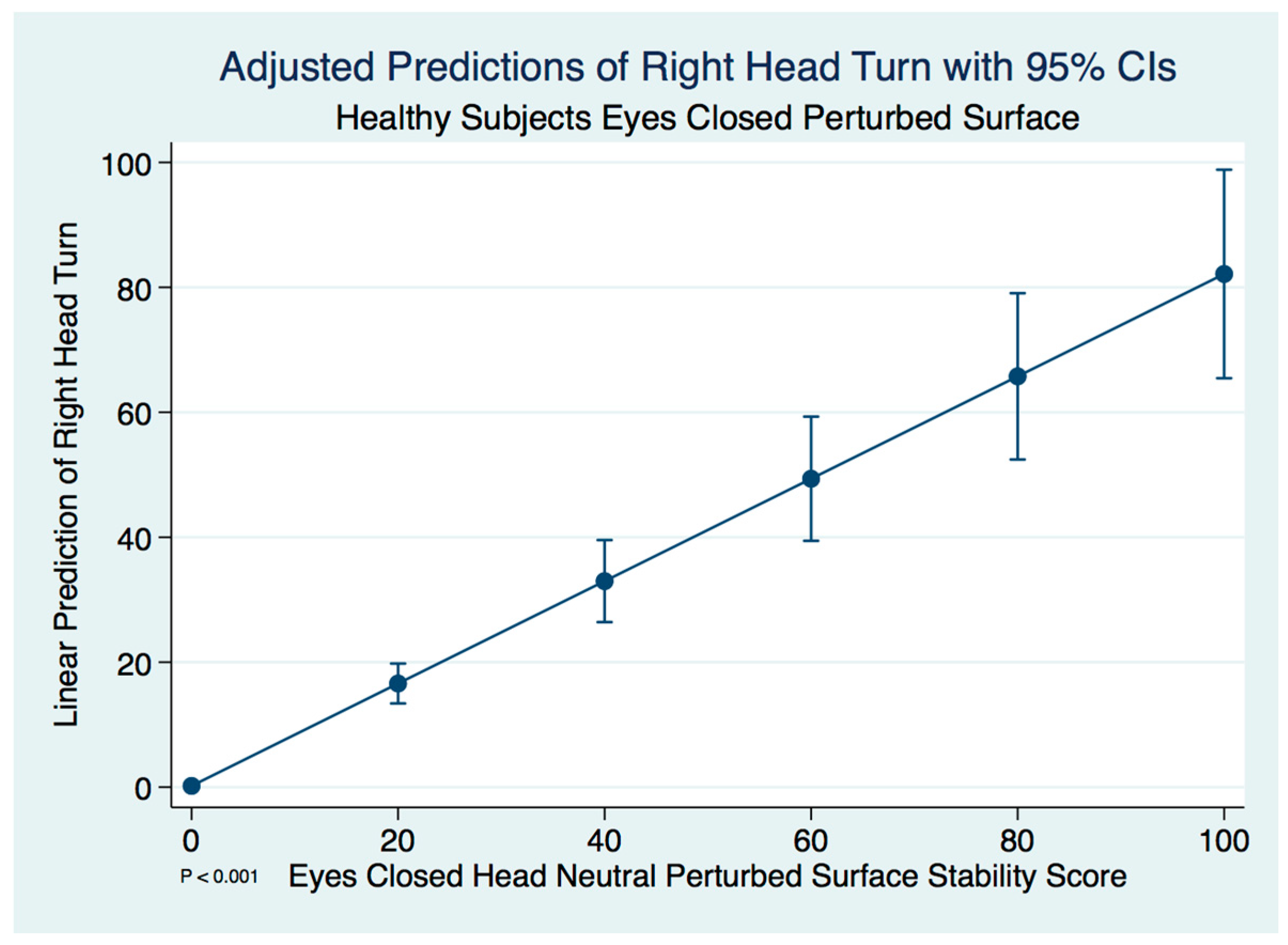
| Controls (n = 60) | Head neutral (reference) | 81.679 | 0.447 | |||
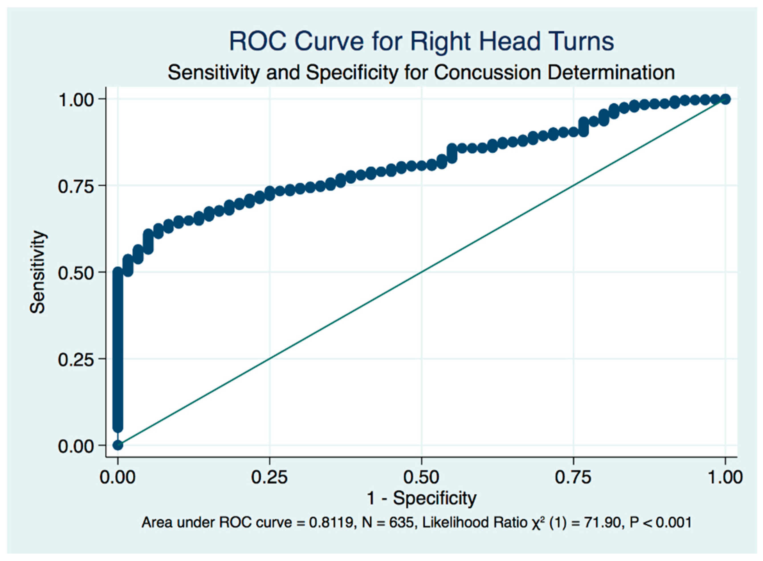
| Head Right | 82.007 | 0.476 | 0.305 | 0.019 | 0.174 | |
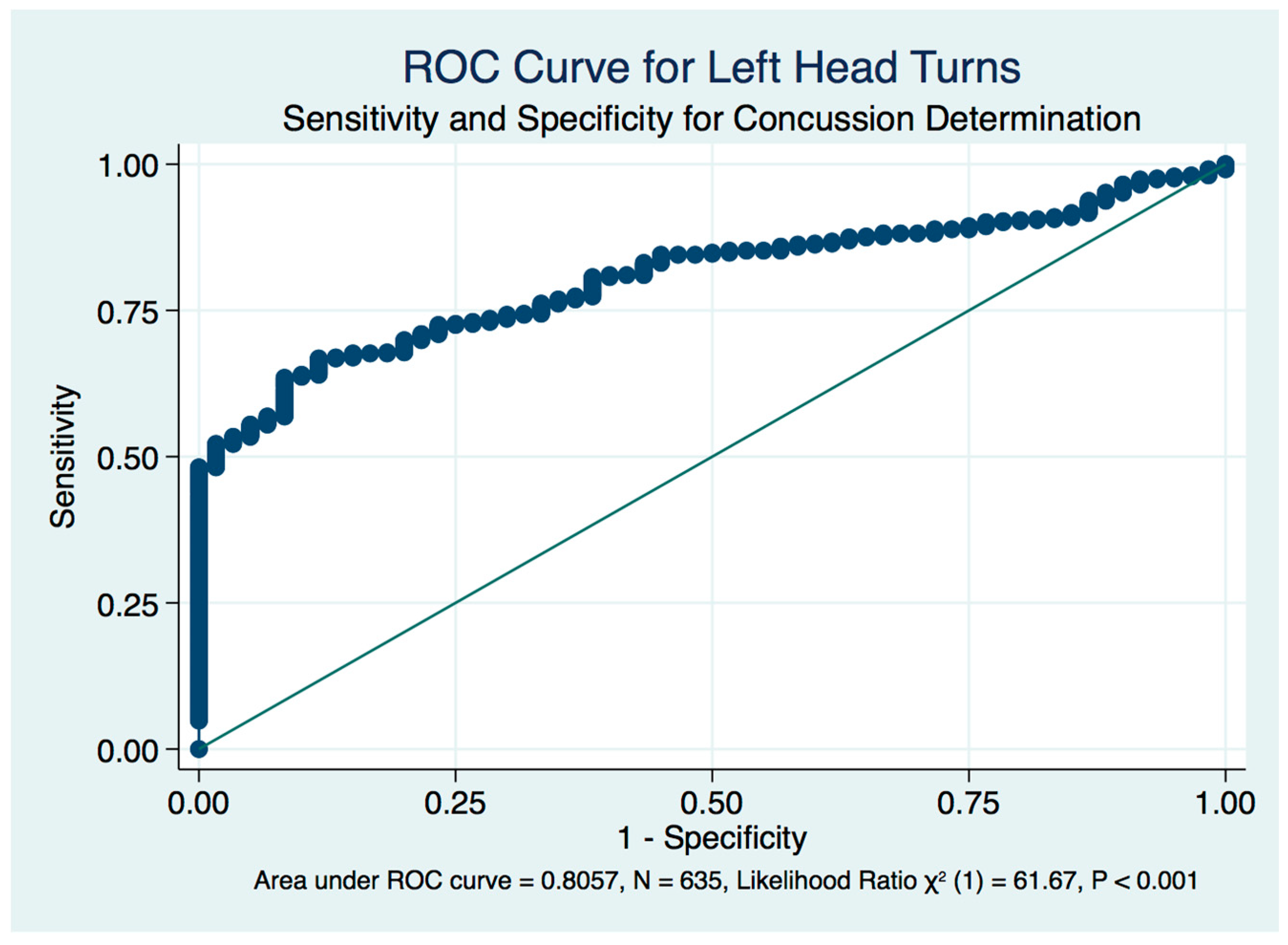
| Head Left | 81.769 | 0.422 | 0.794 | 0.001 | 0.058 | |
| Head Flexed | 82.761 | 0.428 | 0.002 | 0.152 | 0.891 | |
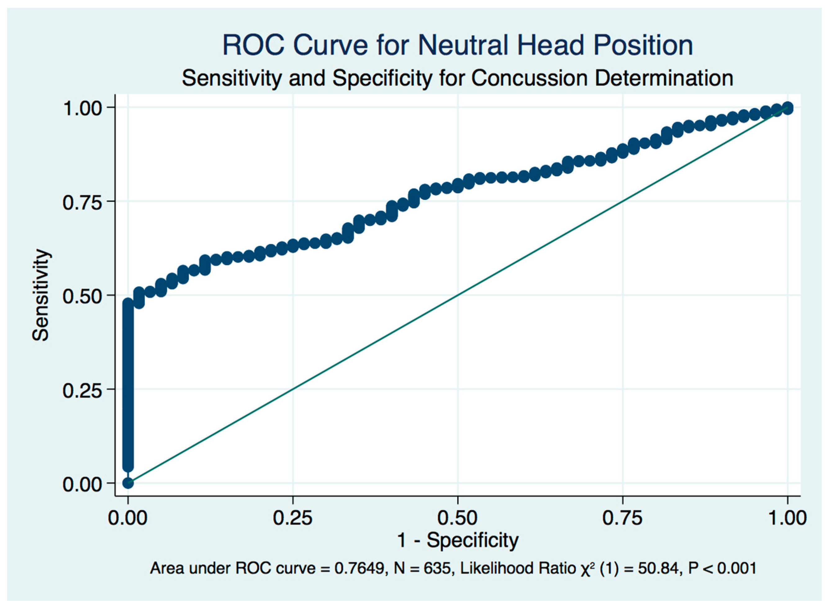
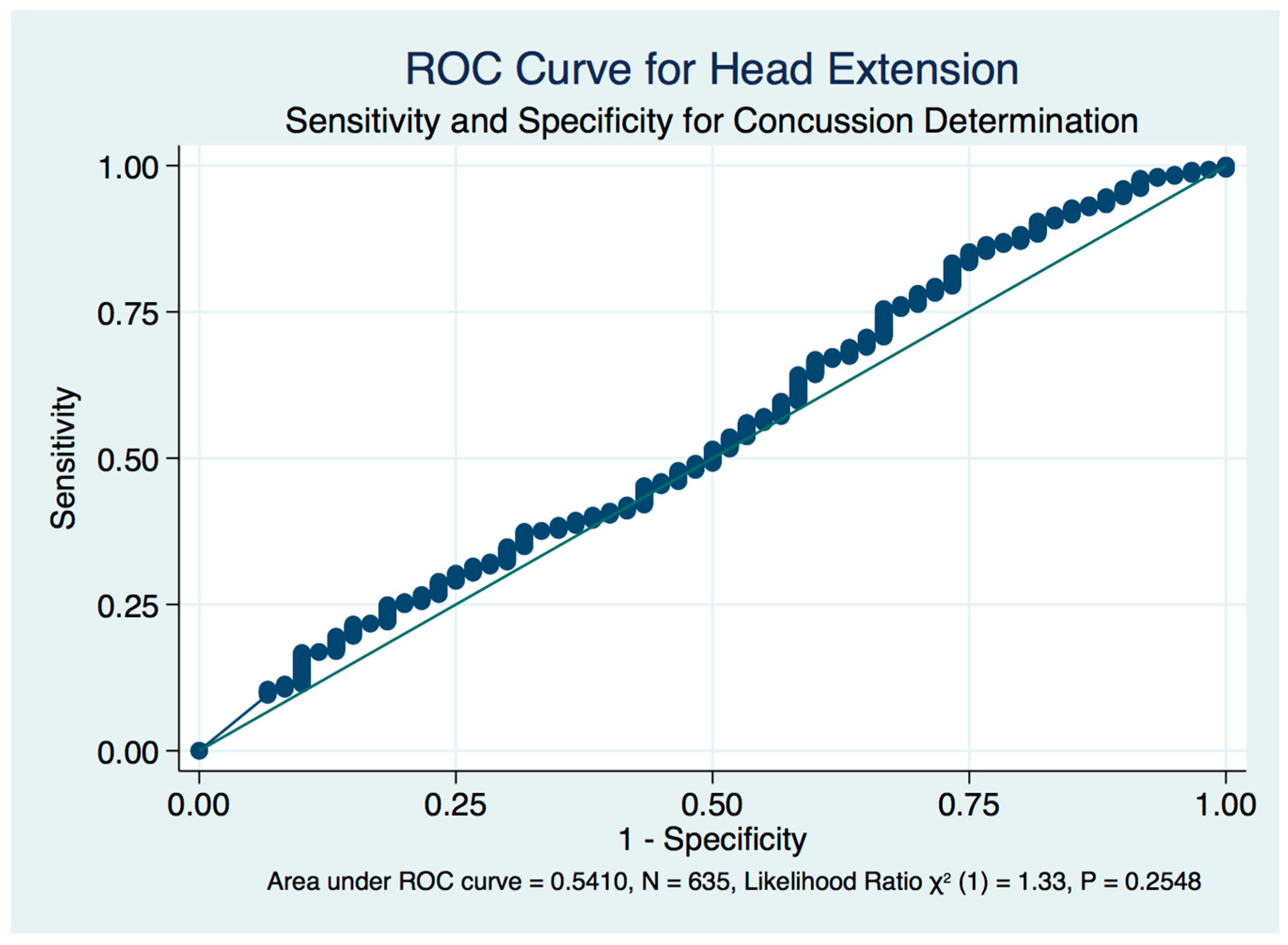
| Head Extended | 75.164 | 0.847 | 0.000 | 0.529 | 1.000 | |
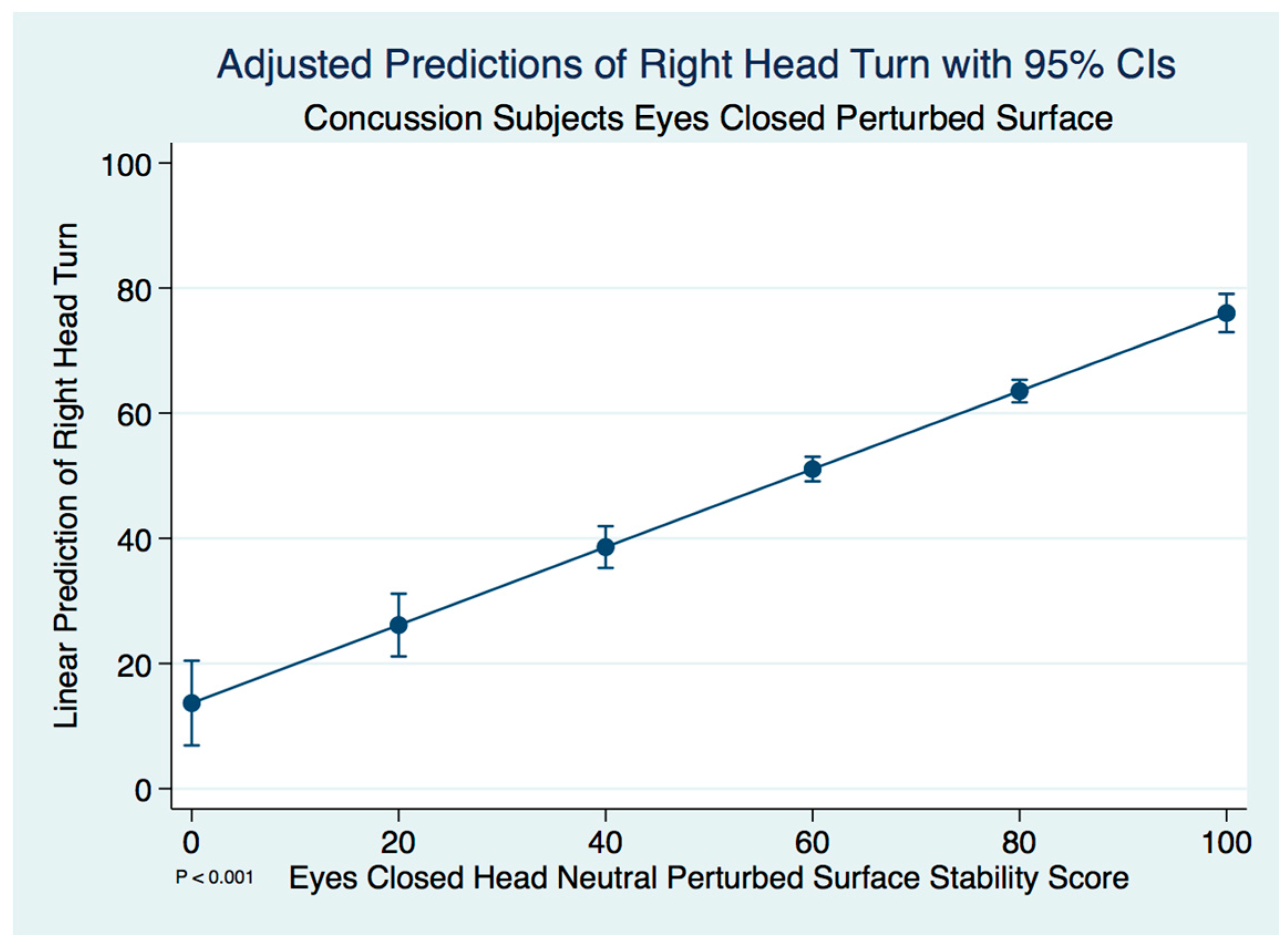
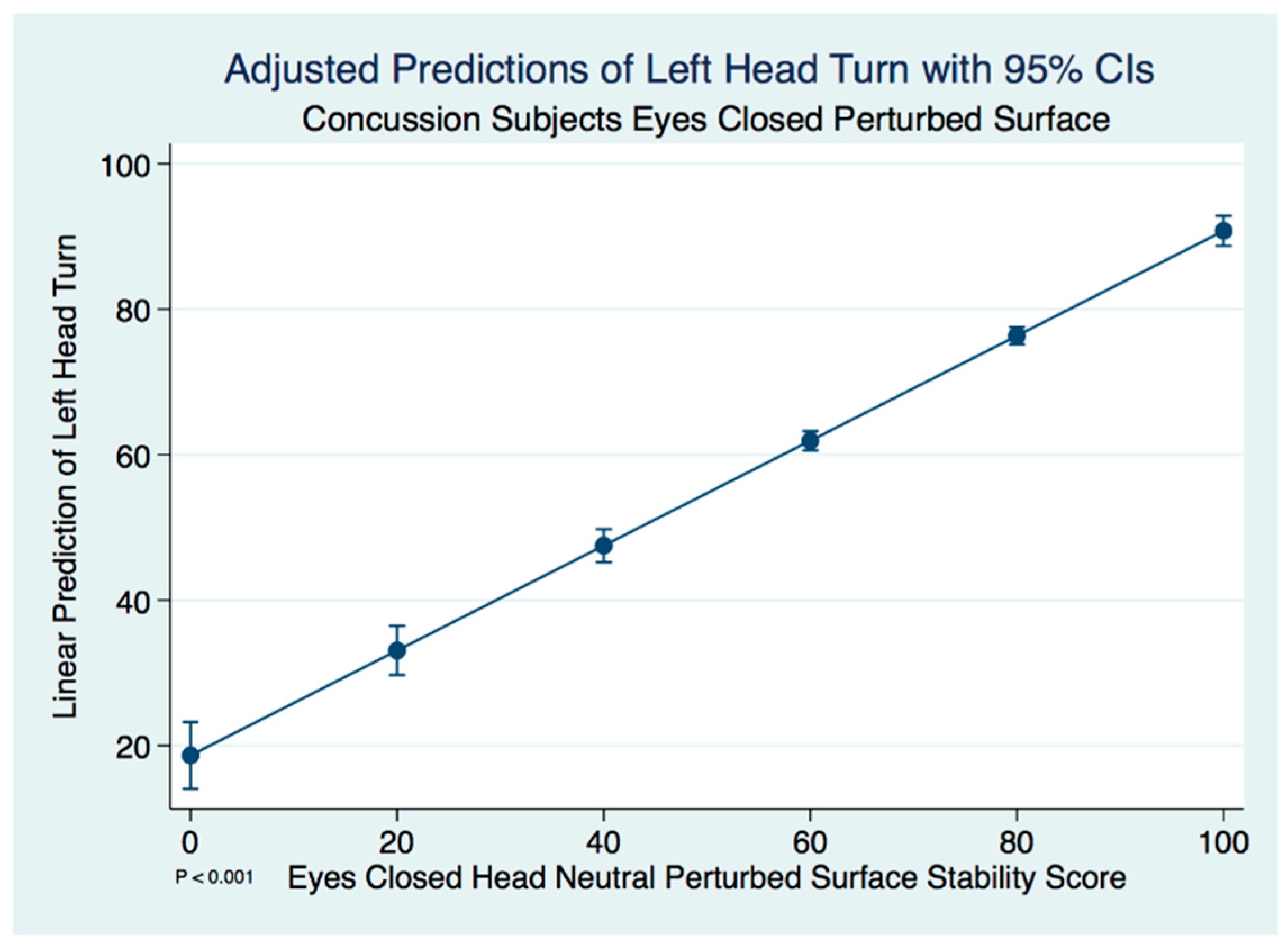
| Patients (n = 575) | Head neutral (reference) | 71.753 | 0.747 | |||
| Head Right | 70.632 | 0.769 | 0.043 | 0.007 | 0.525 | |
| Head Left | 70.401 | 0.779 | 0.025 | 0.009 | 0.614 | |
| Head Flexed | 72.236 | 0.707 | 0.408 | 0.001 | 0.131 | |
| Head Extended | 58.391 | 0.954 | 0.000 | 0.287 | 1.000 |
| Test | Mean | Std. Error | 95% Confidence Interval | |
|---|---|---|---|---|
| Lower Bound | Upper Bound | |||
| NSEO-HN | 94.550 | 0.227 | 94.096 | 95.003 |
| NSEC-HN | 92.651 | 0.268 | 92.115 | 93.187 |
| PSEO-HN | 89.968 | 0.267 | 89.433 | 90.503 |
| PSEC-HN | 81.679 | 0.447 | 80.785 | 82.574 |
| Source | Test | Type III Sum of Squares | df | Mean Square | F | Sig. | Partial Eta Squared | Noncent. Parameter | Observed Power |
|---|---|---|---|---|---|---|---|---|---|
| Test | NSEO-HN vs. NSEC-HN | 216.376 | 1 | 216.376 | 49.873 | 0.000 | 0.458 | 49.873 | 1.000 |
| NSEC-HN vs. PSEO-HN | 431.840 | 1 | 431.840 | 55.045 | 0.000 | 0.483 | 55.045 | 1.000 | |
| PSEO-HN vs. PSEC-HN | 4121.937 | 1 | 4121.937 | 325.927 | 0.000 | 0.847 | 325.927 | 1.000 | |
| Error (Test) | NSEO-HN vs. NSEC-HN | 255.974 | 59 | 4.339 | |||||
| NSEC-HN vs. PSEO-HN | 462.863 | 59 | 7.845 | ||||||
| PSEO-HN vs. PSEC-HN | 746.161 | 59 | 12.647 |
| Test | Mean | Std. Error | 95% Confidence Interval | |
|---|---|---|---|---|
| Lower Bound | Upper Bound | |||
| NSEO-HN | 90.412 | 0.359 | 89.708 | 91.117 |
| NSEC-HN | 89.075 | 0.476 | 88.140 | 90.009 |
| PSEO-HN | 82.693 | 0.474 | 81.761 | 83.624 |
| PSEC-HN | 71.753 | 0.747 | 70.286 | 73.220 |
| Source | Test | Type III Sum of Squares | df | Mean Square | F | Sig. | Partial Eta Squared | Noncent. Parameter | Observed Power a |
|---|---|---|---|---|---|---|---|---|---|
| Test | NSEO-HN vs. NSEC-HN | 1028.855 | 1 | 1028.855 | 19.339 | 0.000 | 0.033 | 19.339 | 0.992 |
| NSEC-HN vs. PSEO-HN | 23,418.979 | 1 | 23,418.979 | 249.292 | 0.000 | 0.303 | 249.292 | 1.000 | |
| PSEO-HN vs. PSEC-HN | 68,812.928 | 1 | 68,812.928 | 310.301 | 0.000 | 0.351 | 310.301 | 1.000 | |
| Error (Test) | NSEO-HN vs. NSEC-HN | 30,537.599 | 574 | 53.201 | |||||
| NSEC-HN vs. PSEO-HN | 53,922.698 | 574 | 93.942 | ||||||
| PSEO-HN vs. PSEC-HN | 127,291.478 | 574 | 221.762 |
| Test | Mean | Std. Error | 95% Confidence Interval | |
|---|---|---|---|---|
| Lower Bound | Upper Bound | |||
| PSEC-HN | 81.679 | 0.447 | 80.785 | 82.574 |
| PSEC-HR | 82.007 | 0.476 | 81.054 | 82.960 |
| PSEC-HL | 81.769 | 0.422 | 80.924 | 82.614 |
| PSEC-HF | 82.761 | 0.428 | 81.905 | 83.617 |
| PSEC-HE | 75.164 | 0.847 | 73.470 | 76.858 |
| Source | Test | Type III Sum of Squares | df | Mean Square | F | Sig. | Partial Eta Squared | Noncent. Parameter | Observed Power a |
|---|---|---|---|---|---|---|---|---|---|
| Test | PSEC-HR vs. PSEC-HN | 6.426 | 1 | 6.426 | 1.069 | 0.305 | 0.018 | 1.069 | 0.174 |
| PSEC-HL vs. PSEC-HN | 0.483 | 1 | 0.483 | 0.069 | 0.794 | 0.001 | 0.069 | 0.058 | |
| PSEC-HF vs. PSEC-HN | 70.179 | 1 | 70.179 | 10.546 | 0.002 | 0.152 | 10.546 | 0.891 | |
| PSEC-HE vs. PSEC-HN | 2547.391 | 1 | 2547.391 | 66.317 | 0.000 | 0.529 | 66.317 | 1.000 | |
| Error (Test) | PSEC-HR vs. PSEC-HN | 354.697 | 59 | 6.012 | |||||
| PSEC-HL vs. PSEC-HN | 413.943 | 59 | 7.016 | ||||||
| PSEC-HF vs. PSEC-HN | 392.633 | 59 | 6.655 | ||||||
| PSEC-HE vs. PSEC-HN | 2266.327 | 59 | 38.412 |
| Measure: SS | ||||
|---|---|---|---|---|
| Test | Mean | Std. Error | 95% Confidence Interval | |
| Lower Bound | Upper Bound | |||
| PSEC-HN | 71.753 | 0.747 | 70.286 | 73.220 |
| PSEC-HR | 70.632 | 0.769 | 69.122 | 72.142 |
| PSEC-HL | 70.401 | 0.779 | 68.872 | 71.931 |
| PSEC-HF | 72.236 | 0.707 | 70.847 | 73.624 |
| PSEC-HE | 58.391 | 0.954 | 56.516 | 60.265 |
| Source | Test | Type III Sum of Squares | df | Mean Square | F | Sig. | Partial Eta Squared | Noncent. Parameter | Observed Power a |
|---|---|---|---|---|---|---|---|---|---|
| Test | PSEC-HR vs. PSEC-HN | 722.992 | 1 | 722.992 | 4.104 | 0.043 | 0.007 | 4.104 | 0.525 |
| PSEC-HL vs. PSEC-HN | 1050.861 | 1 | 1050.861 | 5.079 | 0.025 | 0.009 | 5.079 | 0.614 | |
| PSEC-HF vs. PSEC-HN | 133.792 | 1 | 133.792 | 0.685 | 0.408 | 0.001 | 0.685 | 0.131 | |
| PSEC-HE vs. PSEC-HN | 102,672.138 | 1 | 102,672.138 | 230.938 | 0.000 | 0.287 | 230.938 | 1.000 | |
| Error (Test) | PSEC-HR vs. PSEC-HN | 101,111.042 | 574 | 176.152 | |||||
| PSEC-HL vs. PSEC-HN | 118,773.031 | 574 | 206.922 | ||||||
| PSEC-HF vs. PSEC-HN | 112,114.141 | 574 | 195.321 | ||||||
| PSEC-HE vs. PSEC-HN | 255,192.758 | 574 | 444.587 |
Publisher’s Note: MDPI stays neutral with regard to jurisdictional claims in published maps and institutional affiliations. |
© 2020 by the authors. Licensee MDPI, Basel, Switzerland. This article is an open access article distributed under the terms and conditions of the Creative Commons Attribution (CC BY) license (http://creativecommons.org/licenses/by/4.0/).
Share and Cite
Carrick, F.R.; Pagnacco, G.; Hunfalvay, M.; Azzolino, S.; Oggero, E. Head Position and Posturography: A Novel Biomarker to Identify Concussion Sufferers. Brain Sci. 2020, 10, 1003. https://0-doi-org.brum.beds.ac.uk/10.3390/brainsci10121003
Carrick FR, Pagnacco G, Hunfalvay M, Azzolino S, Oggero E. Head Position and Posturography: A Novel Biomarker to Identify Concussion Sufferers. Brain Sciences. 2020; 10(12):1003. https://0-doi-org.brum.beds.ac.uk/10.3390/brainsci10121003
Chicago/Turabian StyleCarrick, Frederick Robert, Guido Pagnacco, Melissa Hunfalvay, Sergio Azzolino, and Elena Oggero. 2020. "Head Position and Posturography: A Novel Biomarker to Identify Concussion Sufferers" Brain Sciences 10, no. 12: 1003. https://0-doi-org.brum.beds.ac.uk/10.3390/brainsci10121003