Hyaluronic Acid-Based Theranostic Nanomedicines for Targeted Cancer Therapy


1
Department of Organic Materials Science and Engineering, College of Engineering, Pusan National University, 2 Busandaehak-ro 63 beon-gil, Geumjeong-gu, Busan 46241, Korea
2
Department of Cogno-Mechatronics Engineering, College of Nanoscience and Nanotechnology, Pusan National University, 2 Busandaehak-ro 63 beon-gil, Geumjeong-gu, Busan 46241, Korea
*
Authors to whom correspondence should be addressed.
†
These authors contributed equally to this paper.
Cancers 2020, 12(4), 940; https://0-doi-org.brum.beds.ac.uk/10.3390/cancers12040940
Submission received: 12 March 2020
/
Revised: 3 April 2020
/
Accepted: 8 April 2020
/
Published: 10 April 2020
(This article belongs to the Special Issue Novel Nanobiotechnologies for the Discovery and Delivery of Anti-cancer Drugs)
Abstract
:Hyaluronic acid (HA) is a natural mucopolysaccharide and has many useful advantages, including biocompatibility, non-immunogenicity, chemical versatility, non-toxicity, biodegradability, and high hydrophilicity. Numerous tumor cells overexpress several receptors that have a high binding affinity for HA, while these receptors are poorly expressed in normal body cells. HA-based drug delivery carriers can offer improved solubility and stability of anticancer drugs in biological environments and allow for the targeting of cancer treatments. Based on these benefits, HA has been widely investigated as a promising material for developing the advanced clinical cancer therapies in various formulations, including nanoparticles, micelles, liposomes, and hydrogels, combined with other materials. We describe various approaches and findings showing the feasibility of improvement in theragnosis probes through the application of HA.
1. Introduction
Cancer is a leading cause of death in the United States and numerous other parts of the globe. The new cancer cases worldwide are predicted to increase from approximately 14 million in 2012 to more than 22 million in 2030. The number of deaths from cancer is expected to continue rising and to reach 13.1 million in 2030 [1,2,3,4]. Therefore, successful cancer treatment is one of the most important goals of current medical science. Recently, advances in nanotechnology have made it possible to understand the fundamental biology of cancer, and to develop new and promising therapies [5]. Various materials and methods have been reported for producing drug carriers that can protect and deliver therapeutic molecules to tumors [6].
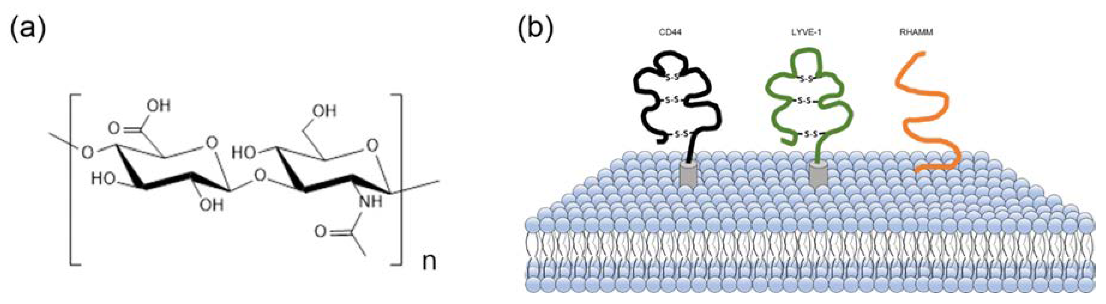
Hyaluronic acid (HA) is a natural mucopolysaccharide comprising alternately repeating disaccharide units of D-glucuronic acid and N-acetyl-D-glucosamine (Figure 1a) and is the main constituent of the extracellular matrix (ECM). The HA plays a significant role in cell growth and in maintaining the structural stability of tissue [7,8,9]. In the body, these structural roles are affected by their hydrodynamic properties and interactions with other ECM components. As HA is biocompatible, non-immunogenic, non-toxic, biodegradable, chemically modifiable, highly hydrophilic, and can absorb water, producing viscoelastic gel, HA has attracted considerable interest from researchers for biomedical applications, including drug delivery systems [10,11].
In addition, there are several overexpressed HA binding receptors in cancer cells compared to normal cells, such as cluster of differentiation 44 (CD44) [12,13,14], lymphatic vessel endocytic receptor (LYVE-1) [15], and the receptor for hyaluronic acid-mediated motility (RHAMM) [16] (Figure 1b). These receptors can offer selective tumor targeting. The role of CD44 in the interactions between HA and specific cells has been extensively explored. The CD44 family of proteins belongs to transmembrane glycoproteins and play a crucial role in extracellular adhesion, cell activities, and signal transduction [17]. The CD44 receptor is involved in tumor invasion and metastasis in cancer cells, and has been associated with the cellular adhesion process, including aggregation and migration in normal biological systems [18,19].
There are some differences between normal cells and tumor cells concerning the HA and CD44 receptors. In normal tissue, the CD44 receptor is endogenously expressed with low levels on different cells and requires activation [20]. However, tumor-derived cells do not require an activation process because the expressed CD44 receptor has a high affinity. In this case, HA binding and internalizing are possible without additional procedures. These interactions can encourage tumor cell migration, which is linked with HA levels; HA levels are high at the edges of rapidly growing tumors [21].
As mentioned above, CD44 receptors are overexpressed on the surfaces of various tumor cells, including breast cancer [22,23] and lung cancer [12,24], therefore, CD44 can be utilized as a cancer-targeting biomarker. RHAMM is another well-known HA-specific receptor that mediates cell proliferation and migration and is poorly expressed in the majority of common normal tissues. Conversely, RHAMM shows increased expression in tumor cells, which is related to metastases [25,26].
HA-based drug delivery carriers provide several advantages. First, HA can improve the stability of anticancer drugs in physiological conditions [27]. Secondly, HA can solve the drawbacks of current anticancer drugs, such as low specificity, via several overexpressed receptors that selectively bind to HA [28,29]. Finally, HA can be chemically modified through functional groups. The carboxyl groups on the glucuronic acid unit and the primarily hydroxyl groups on the N-acetyl-D-glucosamine unit are commonly used groups for chemical modification to obtain HA derivatives [30,31].
In this review, we discuss the approaches that utilized various formulations of HA to design drug carriers and advances in HA-based drug delivery systems for improved cancer treatment. Moreover, we present a brief overview of the recent findings and progression in the research to show the promising aspects of HA-based cancer therapies.
2. HA–Drug Conjugates
In anticancer therapy, there are various treatment methods, the most basic of which is chemotherapy using anticancer drugs, such as paclitaxel (PTX), doxorubicin (DOX), cisplatin (cis-diamminedichloroplatinum (II) or CDDP), SN-38, etc. For cancer therapy, these drugs administered into the body can act on cancer cells or tissues by reducing cell viability or expediting a specific immune reaction for the elimination cancer tissues. However, these effects can cause damage to normal cells as well as cancer cells, resulting in various side effects. Furthermore, they can be easily eliminated by physiological or immune reactions in the body, since these drugs are external substances administered into the body.
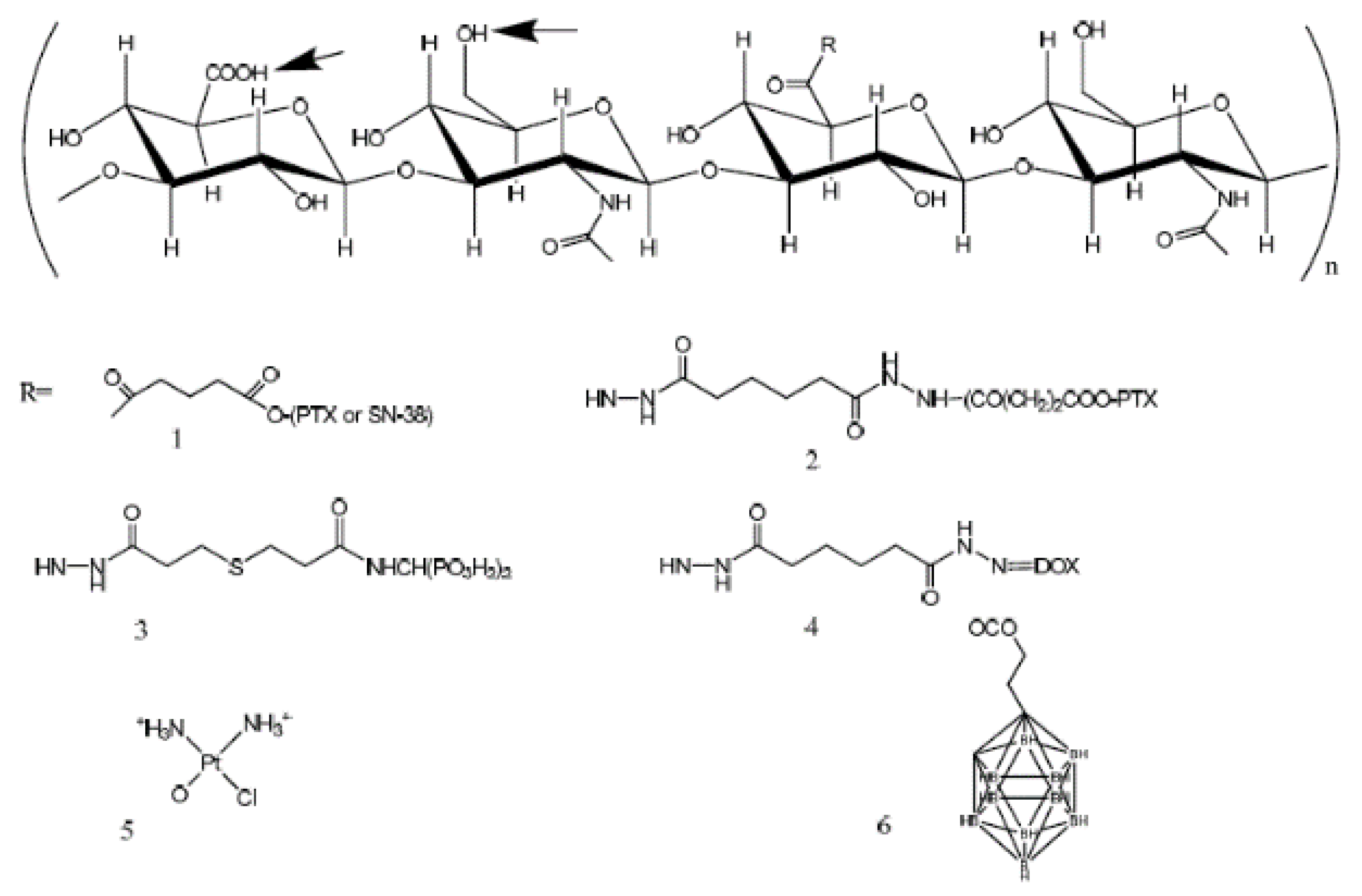
Therefore, it is necessary that administered drugs act on the cancerous tissue without affecting other normal cells and maintain a stable state in in vivo microenvironments until they are delivered. To this end, HA, with the various advantages discussed earlier, has been applied for drug delivery of anticancer drugs [32,33,34,35,36]. In general, anticancer drugs have been conjugated with HA at the carboxyl group and hydroxyl group through functional groups of the drug itself or conjugate linkers, such as ester linkers and amide linkers (Figure 2).
2.1. HA-Conjugated Paclitaxel
Paclitaxel (PTX) is the most well-known anticancer drug for the treatment of numerous tumors, such as breast, ovarian, and melanoma cancer. However, it has several limitations due to poor water solubility, nonselective toxicity, and inactivity against drug-resistant cell lines [37]. For these reasons, HA conjugation can offer improvements to these problems.
Rosato’s group synthesized ONCOFIDTM-P, a novel PTX-HA (MW 200 kDa) conjugate, using 4-bromobuyric acid, which can form two ester linkages between PTX and HA, for treating superficial bladder cancer [37,38]. A subsequent imaging biodistribution analysis of 99mTc-radiolabeled ONCOFIDTM-P by the intravascular method was conducted. As a result, abdominal and gastroenteric tissue after injection showed that the radiolabeled conjugate remained in the cancer tissue. Therefore, these methods can be relevant to local treatment for bladder cancer cells.
Xin et al. conjugated PTX with HA using amino acids between PTX and HA (MW 9.8 kDa), which showed more stability in in vivo conditions [39]. Amino acid linkers were conjugated to the carboxyl group with the hydroxyl group of PTX. Then, intermediates were conjugated to the amino group of the amino acid with the carboxyl group of HA, using carbodiimide activation, and the HA-PTX prodrug was successfully synthesized. Prodrugs exhibited increased cytotoxicity compared to free paclitaxel against the MCF-7 cell line in an in vitro model. Mittapalli et al. conjugated HA (MW 4~5 kDa) and PTX using a polyethylene glycol (PEG)-linker-forming ester linkage, and applied this conjugation to human breast cancer cells (MCF-7) [40]. As a result, the HA–PTX treated group showed that nanoconjugate remained in the body for a longer time compared with the pure PTX treated group. Therefore, HA–PTX improved the drug efficacy in a preclinical model of breast cancer. Moreover, Zhong et al. developed paclitaxel prodrug micelles based on HA (MW 9.5 kDa)-b-dendritic oligoglycerol nanoparticles (HA-dOG-PTX-PM NPs) encapsulating fluorescence dye (DiR) for effective targeting and treatment of xenografted human breast cancer in vitro and in vivo. Through in vivo biodistribution analysis for 48 h, it was confirmed that DiR was significantly accumulated in tumor tissue compared to other organs at 4 h post injection. This demonstrated that a theranostic system via these NPs is possible based on tumor-targetability through HA conjugation and the anti-cancer effect of PTX and bio-imaging through DiR loading [41].
2.2. HA-Conjugated Doxorubicin
Doxorubicin (DOX) is one of the most well-known anthracycline antibiotics utilized for cancer therapy. Despite its wide clinical use for chemotherapy, it can cause a dose-dependent toxicity. Oommen et al. prepared HA-DOX conjugates via covalent linkages between DOX and HA. As HA plays a critical role in cancer targeting, such polymer–drug conjugates can be an effective method to treat metastatic tumors. The HA-DOX particles are stable in serum, as it is known that hyaluronidase activity is significantly reduced in the blood serum of cancer patients. This method could also reduce the in vivo toxicity caused by early release of the drug in vivo [42].
Cai et al. developed HA-DOX conjugates using the HA (MW 35 kDa)-adipic acid dihydrazide (ADH) derivative [43]. In this study, the HA-DOX treatment group showed delayed cancer progression for approximately 10 weeks, and compared with the free DOX treatment group, increased the animals’ survival. HA-DOX showed significant efficacy combined with decreased toxicity, achieving a complete pathologic tumor response. In addition, an antitumor theranostic system using HA (MW 100 kDa)-DOX conjugates was reported by Kim et al. [44]. The HA-DOX conjugates were prepared by chemical reaction between carboxylic groups of HA and amine groups of DOX and formed micelle-like NPs through self-assembling. In this research, the antitumor effect of HA-DOX micelles in in vitro anti-proliferation tests of cancer cells and successful formation of the micelles coated with gold (Au) half-shells to take advantage of NIR-absorbing and electromagnetic properties of Au half-shell for theranostic applications to cancer treatment were demonstrated.
2.3. HA-Conjugated Cisplatin
CDDP is the drug that can be applied to most cancer therapy. However, there are serious side effects such as neurotoxicity, myelosuppression, and nephrotoxicity, which limits its use. To improve tumor targeting and avoid side effects, several approaches have been developed for conjugation with HA. In 2008, Cai et al. reported HA-CDDP conjugates produced by hydrolysis of chloride on CDDP and replacement with carboxylates on HA (MW 35 kDa) using silver nitrate (AgNO3). The resulting conjugates exhibited an increase in local concentration in the drain lymph node basin without decisively affecting the target organ in an intralymphatic delivery model [45]. Xie et al. also demonstrated that the lung instillation of HA (MW 35 kDa)-CDDP conjugates increased Pt accumulations in the lung tissue and peripheral lymph nodes compared to conventional CDDP i.v. infusion and show a sustained-release plasma profile [46]. Moreover, compared with pure CDDP, HA conjugated CDDP has a targeting effect on cancer cells, which can selectively increase the concentrations in cancer cells and tissues. Cohen et al. applied HA-CDDP to head and neck squamous cell carcinoma (HNSCC), and confirmed a significant improvement in the antitumor efficacy, with a lower toxicity compared to free CDDP [47]. In the study, each experimental group was measured for weight loss and given a body conditioning score. The HA-CDDP treated group exhibited the least weight loss compared to the CDDP-only treated group. In addition, the body score was decreased below 2 points in all control groups, but not in any HA-CDDP treated group. Therefore, the HA conjugated groups had no toxic problems compared to the control group.
3. HA-Based Nanomaterials
HA has several functional groups that enable the encapsulation of active pharmaceutical ingredients or adsorption of macromolecular substances [48]. On the other hand, nanomaterials possess excellent potential for cancer therapy owing to their unique physicochemical properties [49]. Therefore, several approaches have been developed to synthesize functional nanomaterials using HA [50,51,52]. HA itself can be synthesized into conjugated polymers, such as dendrimers, micelles, liposomes, and hydrogels. Furthermore, HA can be incorporated into nanomaterials for enhanced water solubility, biocompatibility, and targetability by their specific binding to CD44 overexpressed cancer cells. Moreover, HA does not induce immune reactions and is biocompatible, suggesting that HA can be potentially applied for improved theragnosis probes for cancer treatment.
3.1. Dendrimers
Dendrimers are highly branched synthetic polymers with layered structures around an internal core. Each branch that extends from the internal core has different terminal groups, which determine the characteristics of the dendrimer [53]. Dendrimers have uniform and controllable size/chemical composition and a high loading capacity of payloads but have limited chemical synthesis methods [54]. Therefore, many studies focused on developing new synthesis methods to induce various modifications of the terminal groups of HA dendrimers for cancer therapy.
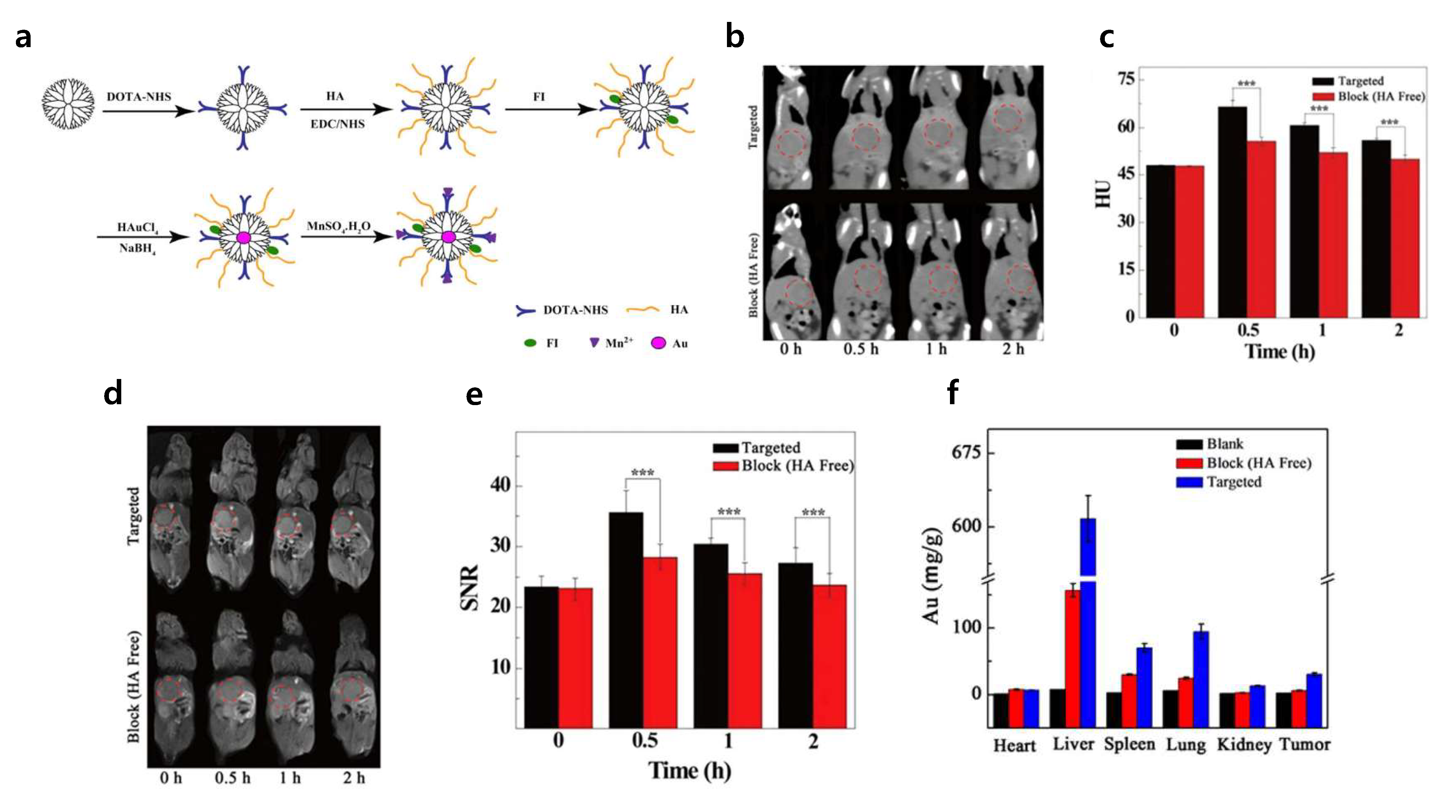
Wang et al. encapsulated Au NPs with a G5.NH2 dendrimer, functionalized with an Mn chelator, 1,4,7,10-tetraazacyclododecane-1,4,7,10-tetraacetic acid (DOTA), fluorescein isothiocyanate (FI), and HA with Mw 6 kDA {(Au0)100G5.NH2-FI-DOTA(Mn)-HA}. The prepared {(Au0)100G5.NH2-FI-DOTA(Mn)-HA} NPs endowed encapsulated Au NPs with several advantages, such as enhanced water solubility, stability under different conditions, and cytocompatibility. The prepared NPs were utilized as an imaging probe for tumor cell imaging [55]. The HA modified Au dendrimers were accumulated 2-fold higher in tumors compared to pure Au-dendrimers, indicating the tumor specificity of HA (Figure 3) [55].
On the other hand, there have been many studies utilizing dendrimer-based drug-delivery strategies exploiting HA as a targeting ligand for the targeted delivery of drugs for cancer. Kesharwani et al. successfully reduced the cationic surface charge of native poly(amidoamine) (PAMAM) by conjugation of HA (10 kDa) with the peripheral amino groups of PAMAM dendrimers through N-(3-dimethylaminopropyl)-N′-ethylcarbodiimide (EDC) coupling chemistry [56]. Then, 3,4-difluorobenzylidene curcumin (CDF) was encapsulated in HA-PAMAM dendrimers and efficiently transferred into the nucleus of tumor cells by CD44 receptor mediated endocytosis [56].
3.2. Micelles
HA also can be formed into polymeric micelles. Polymeric micelles are synthesized by the self-assembly of amphiphilic copolymers in aqueous solution and have a spherical structure with hydrophilic heads at the shell and hydrophobic tails at the core [57]. Owing to their amphiphilic structure, micelles can effectively load water-insoluble drugs and slow down in vivo degradation processes such as uptake by the reticuloendothelial system and blood clearance [57]. Moreover, drug release can be controlled by external stimuli such as pH, temperature, enzymes, ultrasound, etc. [58,59,60,61]. The advantages of micelles for drug delivery are the high dissolution capacity, stability, sustained release, long-term circulation, and the ability to remain in the tumor for a long time due to the enhanced permeability and retention effect (EPR effect), which is the abnormal molecular and fluidic dynamic of certain macromolecules and lipids caused by the specific nature of tumor tissues, such as imperfect vascularization, lack of lymphatic drainage, wider lumen, and so on.
Lee et al. synthesized hydrophobic poly(lactic-co-glycolic acid) (PLGA) multi-cores for doxorubicin encapsulation by the self-assembly of PLGA grafted HA (low and high MW: 17 and 64 kDa, respectively) copolymers. DOX-loaded HA-PLGA micelles exhibited enhanced cellular uptake and greater cytotoxicity to human colon cancer cells (HCT116) [62]. Redox-sensitive amphiphilic HA-deoxycholic acid conjugates were developed for the targeted intracellular delivery of paclitaxel [63]. The conjugates were self-assembled into nanosized micelles in aqueous media and exhibited high drug loading capacity and encapsulation efficiency [63]. HA-deoxycholic acid micelles were sufficiently stable in physiologic conditions but quickly disassembled in the presence of a reducing agent, suggesting that their drug release can be controlled by redox-sensitivity [63].
Similarly, pH-responsive HA-g-poly(L-histidine) (HA-PHis) copolymer micelles were developed with HA (MW 11 kDa) for intracellular DOX delivery [64]. DOX was efficiently loaded into self-assembled HA-PHis-conjugated micelles in aqueous conditions [64]. These micelles were up-taken to the cancer cells by receptor-mediated endocytosis, and drug release could be controlled by pH differences of the intra/extra cellular environments [64].
3.3. Liposomes
Liposomes are vesicles composed of 50–100 nm phospholipid bilayers, similar to biological membranes. The unique amphiphilic properties of liposomes make it possible to transport hydrophilic drugs in aqueous solutions and to dissolve hydrophobic drugs through membranes [65]. In addition, liposomes are synthetic carriers approved by FDA (Food and Drug Administration) and have excellent circulatory, penetration, and controllable diffusion properties depending on their chemical composition [66,67]. However, polymeric liposomes generally have poor in vivo stability and it is difficult for them to maintain their loaded drugs until they reach the targeted site [67].
Taetz et al. synthesized cationic HA modified 1,2-dioleoyl-3-trimethyllammoniumpropane/ dioleoylphosphatidylethanolamine (DOTAP/DOPE) liposomes using an ethanol injection method for the targeted delivery of anti-telomerase small interfering RNAs (siRNAs) to CD44+ lung cancer cells. The presence of HA upregulated siRNA delivery into lung tumor tissues by the enhancement of siRNA conjugation, the protection of siRNA in the presence of RNase V1, and complex stability in the presence of serum [68]. To improve the cancer cell targetability, dual-functionalization, such as pH-responsive cell penetration peptide (CPP)-HA core-shell liposomes, can be utilized [69]. The HA shell primarily delivers liposome complexes to the tumor site and is then removed by hyaluronidase to expose the inner pH-responsive CPP to promote cellular uptake.
4. HA-Coated NPs
Inorganic nanomaterials such as Au nanoparticles, quantum dots, magnetic nanoparticles, ceramic nanoparticles, and carbon-based NPs have attracted considerable attention in the treatment of cancer [70]; however, these systems have drawbacks, including cytotoxicity and non-cell specificity. Therefore, surface modification with biopolymers has been widely investigated to develop further functionalized cancer theranosis probes [71,72,73]. Likewise, surface coating with HA on inorganic NPs has been studied for biocompatibility and biofunctionality, as well as targetability.
4.1. HA-Coated Au NPs
Au NPs can be potentially used in selective photothermal therapies induced by lasers, owing to their surface plasmon resonance. Au NPs are biocompatible and can easily provide surface modification due to their ability to bind amine and thiol groups, optical properties that can be adjusted according to size and shape, and optical quenching ability [74,75,76]. However, since Au NPs are typically cleared slowly, they exhibit longer-term whole-body retention in some cases [77]. Other challenges of the application of Au NPs are their low stability, reactivity, and capacity to load hydrophilic drugs. Therefore, the surface functionalization of Au NPs to solve these problems is currently one of the most intensively researched topics.
HA coatings have several advantages, such as antifouling effects on the prevention of protein adsorption and opsonization due to the hydrophilic and polyanionic characteristics in physiological environments [78,79,80]. HA-conjugated Pheophorbide-A (PheoA) and Au NPs could serve as multifunctional theranostic nanoagents for photodynamic and photothermal therapy. Thiolated HA (MW 7 kDa) was conjugated with PheoA first, then Au NPs were encapsulated within PheoA-HA conjugated by the Au–thiol reaction. This PheoA-HA-Au nanoagent showed excellent colloidal stability under physiological conditions and restored photoactivity in intracellular environments.
Furthermore, tumor specificity and therapeutic efficacy in tumor-bearing mice were significantly increased [81]. On the other hand, HA-Au NPs can be used as protein drug delivery carriers. Lee et al. investigated HA-Au NPs by chemical binding of thiolated HA (MW 12 kDa) and physical binding of interferon α to Au NPs (HA- Au NP/IFNα). Prepared HA-Au NP/IFNα showed enhanced stability and efficiency compared to Au/IFNα and PEGylated IFNα, which is a conventional IFNα carrier [82].
4.2. HA-Coated Quantum Dot
A quantum dot (QD) is a colloidal nano-sized single crystal exhibiting fluorescence. The center of a QD is generally composed of semiconductor materials, such as CdSe, CdTe, CdS, PbSe, ZnS, ZnSe, GaAs, GaN, InP, and InAs. QDs have excellent light stability and show a tunable emission spectrum and high quantum yield depending on their size and composition. In addition, QDs have been widely used in biological applications as imaging contrast agents and labeling agents due to their low photobleaching and low photo- and chemical degradation [83]. However, clinical applications of QDs are still limited because of their composition of toxic heavy materials and low water solubility [83,84,85].
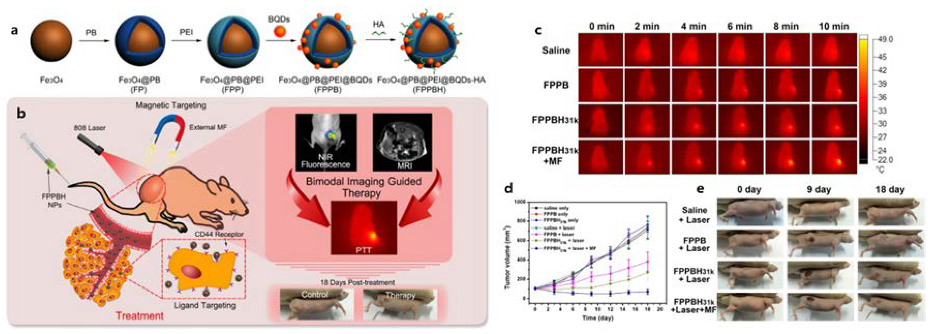
Wang et al. investigated HA-coated QD as a CD44+ cancer cell-targeted imaging probe. A cysteamine-modified HA polymer was employed to coat CdSe (CdZnS) QDs through a convenient one-step reverse micelle method. Prepared HA-QDs showed enhanced stability in PBS and fluorescence stability, and excellent targeting ability to CD44+ breast cancer cells without cytotoxicity, indicating that the stability and low-toxicity of QDs could be achieved through an HA coating [86]. Meanwhile, Yongbo et al. prepared QDs coated by HA-magnetic Prussian Blue conjugates (HA-PB@QDs) for cancer theranosis (Figure 4). The targeting efficiency of HA-PB@QD to lung cancer cells was enhanced by the coexistence of a magnetic core and CD44 ligand HA, which was found to significantly improve the specific uptake by CD44-overexpressed HeLa cells upon external magnetic fields. Moreover, enhanced in vivo photothermal therapy (PTT) efficacy upon NIR laser illumination was observed, indicating the tumor growth inhibition was more than 89.95% [87].
4.3. HA-Coated Superparamagnetic Iron Oxide NPs
Superparamagnetic iron oxide NPs are characterized by biocompatibility, controllable size and shape, and clinically available contrast agents for MRI. However, their clinical application is quite limited due to the poor stability under aqueous conditions, such as the tendency to aggregate due to the large surface area and strong magnetic properties [88]. In addition, magnetic NPs with superparamagnetic properties should be able to maintain a colloidal state for a long time and change surface properties through covalent bonds and be well dispersed in water. However, finding the ideal combination is challenging.
To solve these problems, HA coatings have emerged as a novel method. To prepare HA-coated iron oxide NPs, polyethyleneimine (PEI) drug conjugates were investigated [89]. PEI stabilized Fe3O4 NPs were prepared via a one-pot hydrothermal method, and modified with HA and fluorescein isothiocyanate (HA/FI-Fe3O4 NPs) to be utilized as a cancer diagnosis probe. The HA/FI-Fe3O4 NPs were water-dispersible and cyto/hemo-compatible. The prepared NPs showed enhanced cellular uptake of HeLa cells through CD44 receptor-mediated active targeting pathways and exhibited intracellular green fluorescence.
4.4. HA-Coated Carbon-Based NPs
A wide variety of different nanomaterials based on the allotropic forms of carbon, such as nanotubes, nanohorns, and nanodiamonds, have been explored towards different biomedical applications [90]. To enhance the cancer cell specificity and water stability of carbon-based nanoparticles, HA coatings were performed with several methods. A reduced graphene oxide (rGO) with an HA-based amphiphilic polymer was produced by grafting HA onto poly(maleic anhydride-alt-1-octadecene) (PMAO) [91]. On the other hand, HA-coated rGO nanosheets were obtained by coating with cholesteryl hyaluronic acid (CHA), which was synthesized using cholesteryl-2-aminoethylcarbamate (CAEC) with the carboxyl group of HA [92]. Both nanoprobes exhibited improved stability and cytocompatibility and could potentially be utilized for targeted cancer PTT. Other carbon-based nanomaterials, such as single-walled carbon nanotubes (SWNT), graphene oxide (GO), and fullerene (C60) for anticancer activity, could be HA-surface modified. Prepared HA-SWNT, HA-GO, and HA-C60 showed significantly enhanced water solubility, biocompatibility, and tumor-targeting capacities [93].
5. HA-Based Hydrogel
Hydrogels are three-dimensional hydrated polymeric networks, formed from crosslinked polymer chains, capable of absorbing water by 10–20% up to thousands of times their dry weight [94]. Their highly porous structures enables drug release in a controlled manner depending on the bulk loading of drugs into the gel matrix and the diffusion efficiency of molecules inside the gel network. However, HA hydrogels have some drawbacks as drug delivery probes due to their susceptibility to degradation and low mechanical properties [48,95]. Therefore, chemical modifications, covalent crosslinking, and gelling agents are always needed in order to use HA-based hydrogels as drug delivery systems [96]. In this section, several types of HA hydrogels are introduced for the controlled release of loaded drugs.
5.1. In situ Crosslinked HA Hydrogels
The treatment of localized infections within the body without surgery can be achieved with injectable drug delivery systems. In situ crosslinked hydrogels can be easily injected but maintain a viscose matrix after injection to enable a local drug concentration for a desired time. Recently, an enzymatic crosslinking was used with in situ crosslinking of hydrogels. An HA-tyramine conjugate (HA-Tyr) (MW 90 kDa) was investigated as an injectable and biodegradable drug carrier [97]. HA-Tyr was developed through the oxidative coupling reaction of the Tyr moieties, catalyzed by hydrogen peroxide (H2O2) and horseradish peroxide (HRP). The advantage of enzymatic crosslinking using H2O2 and HRP is the concentration-dependent tuning of the hydrogel stiffness and gelation rate [97].
Rapid gelation was achieved by an optimal concentration of HRP that could effectively encapsulate the proteins within the hydrogel network and thus prevent the undesired leakage of proteins into the surrounding tissues after injection [98]. HA-Tyr also can be used for liver cancer therapy [99]. Interferon-α2a (IFN-α2a) was incorporated into prepared HA-Tyr, which revealed a greatly inhibited proliferation of liver cancer cells and induced apoptosis. Moreover, IFN-α2a incorporated in HA-Tyr highly increased the angiogenesis of mouse tumor tissues, suggesting that HA-Tyr modification can be developed as a method to deliver IFN-α2a into liver tumor tissues [99].
However, there are several studies that indicate that the potential risk of HA in situ hydrogels should be studied. A platinum nanoparticle/HA gel (PtNP/gel) was loaded into a chemically crosslinked HA hydrogel and applied for the local therapy of ovarian cancer. The gel was composed of adipic dihydrazide modified HA (HA-ADH) and oxidized HA (HA-CHO), which can be applied as a liquid and instantly crosslinked via the hydrazone links to form a gel as they mix. The PtNP/gel was maintained in the peritoneal cavity over 4 weeks and the PtNPs release was retained for a prolonged time and locally delivered to CD44+ ovarian cancer cells by receptor-mediated endocytosis. However, the PtNP/gel did not show enhanced anti-cancer efficacy, but rather slightly increased tumor bundles, which was suspected to be due to the effects of the potential involvement of residual empty carriers and degradation products. This study warns that HA carriers may have unwanted biological effects on the residual targets after the drug is released [100].
HA-epigallocatechin-3-O-gallate conjugated injectable hydrogels (HA-EGCG) were synthesized by thiol-mediated reactions, and they exhibited in vivo prolonged properties. HA-EGCG conjugates with tunable degrees of substitution were synthesized by the nucleophilic addition reaction between EGCG quinone and thiolated HA under mild conditions. When injected subcutaneously in mice, HA-EGCG hydrogels were retained much longer than HA-tyramine hydrogels owing to the hyaluronidase inhibitory activities of EGCG [101].
5.2. Thermosensitive HA Hydrogel
Many polymers exhibit a temperature-responsive phase transition property. The common characteristic of thermosensitive polymers is the presence of hydrophobic groups, such as methyl, ethyl, and propyl groups. Thermosensitive polymers, such as poly(N,N-isopropylacrylaminde) (PNIPAAm), feature lower critical solution temperature (LCST) in the body temperature range and are widely clinically used [102]. Thermosensitive HA hydrogels for the delivery of drugs to locally treat tumors were investigated.
HA conjugated with dopamine (HA-DN) (MW 130 kDa) was mixed with thiol end-capped pluronic F127 copolymer (Plu-SH) to produce lightly cross-linked HA/pluronic composite gel structures based on Michael-type catechol-thiol addition reactions. The prepared HA/pluronic hydrogels exhibited temperature-dependent rapid and reversible sol–gel phase transition behaviors, enabling the hardening of the hydrogel at body temperature [103]. Thermosensitive injectable hydrogels composed of a nanocomplex of Dox and HA for the local treatment of cancer disease were also developed. An HA-Dox nanocomplex was synthesized by the addition of divalent metal ions of Mg and was mixed with pluronic F127 to form thermosensitive hydrogels. The prepared hydrogel efficiently inhibited C26 colon cancer cell growth and selectively targeted the lymphatic system by the specific affinity of HA to the lymphatic system [104].
5.3. pH-Sensitive Hydrogels
The pH-sensitive hydrogels possess pendant acidic groups (e.g., carboxylic and sulfonic acid) or basic groups (e.g., ammonium salts) enabling proton absorption or release in accordance with the external pH change [105]. The pH-sensitive hydrogels are widely used as oral medication. The pH values in the mouth, stomach, and intestine are significantly different, therefore, drug release can be efficiently controlled. Moreover, pH values of tumor tissues are generally lower than normal tissues because the glycolysis of tumor cells can cause acidification [106]. Thus, several synthesis approaches for pH-sensitive hydrogels based on HA have been attempted for drug delivery and, furthermore, cancer treatment. Nucleobase pairing was investigated to synthesize pH-sensitive HA hydrogel cross-linking via hydrogen bonding, which was achieved under physiological conditions by cytosine and guanosine complementary base pairing, with 1,6-hexamethylenediamine (HMDA) as a bridging unit between nucleobase and HA (HA-HMDA-C, -G, -C/G according to paired nucleobases [107]). The prepared hydrogel possessed a suitable gelling time, good rheology properties, high swelling ratio, biodegradability, effective drug loading capacity, and sustained drug release ability under physiological conditions [107].
In 2015, Khatun et al. developed a drug delivery system using light-responsive graphene, DOX, and pH-sensitive disulfide-linked HA (MW 7000 kDa) to form a nanoscale hydrogel called nanogel for theranostics of cancer. The prepared nanogel exhibited pH-dependent drug release and enhanced tumor accumulation through a receptor-mediated pathway in optical imaging in vitro and in vivo. Furthermore, nanogels were effective in killing a human lung cancer cell line (A549) and simultaneously limited the toxicity in normal cells [108].
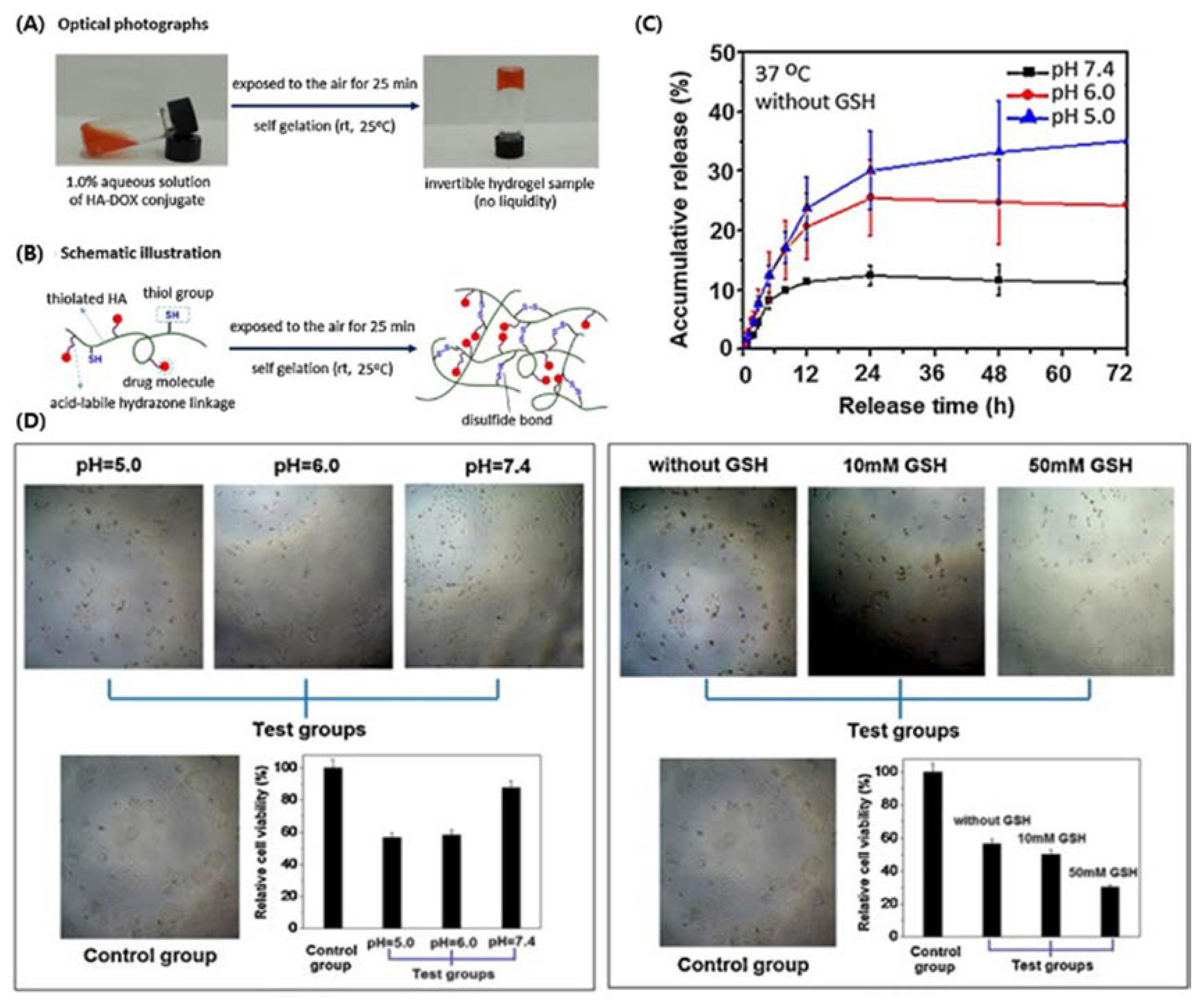
In addition, dual stimuli-responsive hydrogels loaded with DOX were prepared with thiolated HA (MW 320 kDa) crosslinked by an acid-labile hydrazine linker. The hydrogel exhibited a sustained release of DOX in both pH- and reduction-response modes under conditions that mimic the intracellular environments of cancer cells, and effectively inhibited human nasopharyngeal carcinoma CNE2 cells (Figure 5, [109]).
6. Conclusions and Perspectives
Hyaluronic acid is one of the most attractive molecules in the field of developing anticancer drug delivery carriers and imaging agents due to its physicochemical properties that provide various benefits in drug delivery systems. This review summarized the recent progress made with the HA-drug conjugates, HA-based nanomaterials (dendrimers, micelles, and liposomes), hydrogels, and HA-coated NPs, which are used as drug delivery carriers for cancer treatment. Various chemical approaches have been studied to develop HA-based anti-cancer drug delivery methods.
In general, the HA conjugates improved the solubility of hydrophobic drugs and the targeting efficiency to tumor cells with minimum toxicity. From the results described herein, conjugated polymers prepared by themselves or incorporated into other nanomaterials enhanced the solubility, stability, biofunctionality, and ability to target CD44, which is overexpressed in tumor cells. Many studies demonstrated that applying HA can improve theranosis probes for cancer therapy. Moreover, surface coating with HA can be applied to address the issues of inorganic NPs in cancer therapy, as well as provide a more efficient theranostic system.
However, considering such factors as drug loading, in vivo properties, and clinical safety, few studies have been directly compared to clinically available products that provide the best balance. In addition, before attempting clinical applications, well-proven studies such as biodistribution, toxicity, and availability in physiological conditions are essential to estimate the feasibility of the designed cancer therapies. Although HA-based cancer therapy still has several points that should be improved by further research, the results presented in this review show the promising prospects of HA-based theranostic nanomedicine for improved targeted cancer therapies. We expect that further research using HA will provide innovative results and insights into the development of novel cancer therapies.
Funding
This work was supported by the National Research Foundation of Korea (NRF) grant funded by the Korea government (MSIT) (No. 2019R1A4A1024116 and 2019R1F1A1062216).
Conflicts of Interest
The authors declare no conflict of interest and the funders had no role in the design of the study; in the collection, analyses, or interpretation of data; in the writing of the manuscript, or in the decision to publish the results.
References
- Ferlay, J.; Shin, H.R.; Bray, F.; Forman, D.; Mathers, C.; Parkin, D.M. Estimates of worldwide burden of cancer in 2008: GLOBOCAN 2008. Int. J. Cancer 2010, 127, 2893–2917. [Google Scholar] [CrossRef] [PubMed]
- Siegel, R.; DeSantis, C.; Virgo, K.; Stein, K.; Mariotto, A.; Smith, T.; Cooper, D.; Gansler, T.; Lerro, C.; Fedewa, S. Cancer treatment and survivorship statistics, 2012. CA Cancer J. Clin. 2012, 62, 220–241. [Google Scholar] [CrossRef] [PubMed] [Green Version]
- Jemal, A.; Siegel, R.; Xu, J.; Ward, E. Cancer statistics, 2010. CA Cancer J. Clin. 2010, 60, 277–300. [Google Scholar] [CrossRef] [PubMed]
- Stewart, B.; Wild, C. World Cancer Report 2014; Report, International Agency for Research on Cancer; World Health Organization: Paris, France, 2014; pp. 482–494. [Google Scholar]
- Dawidczyk, C.M.; Russell, L.M.; Searson, P.C. Nanomedicines for cancer therapy: State-of-the-art and limitations to pre-clinical studies that hinder future developments. Front. Chem. 2014, 2, 69. [Google Scholar] [CrossRef] [PubMed] [Green Version]
- Mattheolabakis, G.; Rigas, B.; Constantinides, P.P. Nanodelivery strategies in cancer chemotherapy: Biological rationale and pharmaceutical perspectives. Nanomedicine 2012, 7, 1577–1590. [Google Scholar] [CrossRef] [PubMed]
- Robert, L. Hyaluronan, a truly “youthful” polysaccharide. Its medical applications. Pathol. Biol. 2015, 63, 32–34. [Google Scholar] [CrossRef]
- Reed, R.; Lilja, K.; Laurent, T. Hyaluronan in the rat with special reference to the skin. Acta Physiol. Scand. 1988, 134, 405–411. [Google Scholar] [CrossRef]
- Schaefer, L.; Schaefer, R.M. Proteoglycans: From structural compounds to signaling molecules. Cell Tissue Res. 2010, 339, 237. [Google Scholar] [CrossRef]
- Lapčík, L.; Lapcik, L.; De Smedt, S.; Demeester, J.; Chabrecek, P. Hyaluronan: Preparation, structure, properties, and applications. Chem. Rev. 1998, 98, 2663–2684. [Google Scholar] [CrossRef]
- Laurent, T.C.; Fraser, J. Hyaluronan. FASEB J. 1992, 6, 2397–2404. [Google Scholar] [CrossRef]
- Resnick, N.M.; Clarke, M.R.; Siegfried, J.M.; Landreneau, R.; Asman, D.C.; Ge, L.; Kierstead, L.S.; Dougherty, G.D.; Cooper, D.L. Expression of the cell adhesion molecule CD44 in human lung tumors and cell lines. Mol. Diagn. 1998, 3, 93–103. [Google Scholar] [CrossRef]
- Shipitsin, M.; Campbell, L.L.; Argani, P.; Weremowicz, S.; Bloushtain-Qimron, N.; Yao, J.; Nikolskaya, T.; Serebryiskaya, T.; Beroukhim, R.; Hu, M. Molecular definition of breast tumor heterogeneity. Cancer Cell 2007, 11, 259–273. [Google Scholar] [CrossRef] [PubMed] [Green Version]
- Ghosh, S.C.; Neslihan Alpay, S.; Klostergaard, J. CD44: A validated target for improved delivery of cancer therapeutics. Expert Opin. Ther. Targets 2012, 16, 635–650. [Google Scholar] [CrossRef]
- Solis, M.A.; Chen, Y.-H.; Wong, T.Y.; Bittencourt, V.Z.; Lin, Y.-C.; Huang, L.L. Hyaluronan regulates cell behavior: A potential niche matrix for stem cells. Biochem. Res. Int. 2012, 2012, 346972. [Google Scholar] [CrossRef] [PubMed] [Green Version]
- Yang, B.; Yang, B.L.; Savani, R.C.; Turley, E.A. Identification of a common hyaluronan binding motif in the hyaluronan binding proteins RHAMM, CD44 and link protein. EMBO J. 1994, 13, 286–296. [Google Scholar] [CrossRef] [PubMed]
- Ponta, H.; Sherman, L.; Herrlich, P.A. CD44: From adhesion molecules to signalling regulators. Nat. Rev. Mol. Cell Biol. 2003, 4, 33–45. [Google Scholar] [CrossRef]
- Sironen, R.; Tammi, M.; Tammi, R.; Auvinen, P.; Anttila, M.; Kosma, V. Hyaluronan in human malignancies. Exp. Cell Res. 2011, 317, 383–391. [Google Scholar] [CrossRef]
- Sneath, R.; Mangham, D. The normal structure and function of CD44 and its role in neoplasia. Mol. Pathol. 1998, 51, 191–200. [Google Scholar] [CrossRef] [Green Version]
- Cichy, J.; Puré, E. The liberation of CD44. J. Cell Biol. 2003, 161, 839–843. [Google Scholar] [CrossRef] [Green Version]
- Whatcott, C.J.; Han, H.; Posner, R.G.; Hostetter, G.; Von Hoff, D.D. Targeting the tumor microenvironment in cancer: Why hyaluronidase deserves a second look. Cancer Discov. 2011, 1, 291–296. [Google Scholar] [CrossRef] [Green Version]
- Hiscox, S.; Baruah, B.; Smith, C.; Bellerby, R.; Goddard, L.; Jordan, N.; Poghosyan, Z.; Nicholson, R.I.; Barrett-Lee, P.; Gee, J. Overexpression of CD44 accompanies acquired tamoxifen resistance in MCF7 cells and augments their sensitivity to the stromal factors, heregulin and hyaluronan. BMC Cancer 2012, 12, 458. [Google Scholar] [CrossRef] [Green Version]
- Xu, H.; Tian, Y.; Yuan, X.; Liu, Y.; Wu, H.; Liu, Q.; Wu, G.S.; Wu, K. Enrichment of CD44 in basal-type breast cancer correlates with EMT, cancer stem cell gene profile, and prognosis. Onco Targets Ther. 2016, 9, 431–434. [Google Scholar] [PubMed] [Green Version]
- Penno, M.B.; August, J.T.; Baylin, S.B.; Mabry, M.; Linnoila, R.I.; Lee, V.S.; Croteau, D.; Yang, X.L.; Rosada, C. Expression of CD44 in human lung tumors. Cancer Res. 1994, 54, 1381–1387. [Google Scholar] [PubMed]
- Telmer, P.G.; Tolg, C.; McCarthy, J.B.; Turley, E.A. How does a protein with dual mitotic spindle and extracellular matrix receptor functions affect tumor susceptibility and progression? Commun. Integr. Biol. 2011, 4, 182–185. [Google Scholar] [CrossRef]
- Gust, K.M.; Hofer, M.D.; Perner, S.R.; Kim, R.; Chinnaiyan, A.M.; Varambally, S.; Moller, P.; Rinnab, L.; Rubin, M.A.; Greiner, J. RHAMM (CD168) is overexpressed at the protein level and may constitute an immunogenic antigen in advanced prostate cancer disease. Neoplasia 2009, 11, 956–963. [Google Scholar] [CrossRef] [PubMed] [Green Version]
- Han, X.; Li, Z.; Sun, J.; Luo, C.; Li, L.; Liu, Y.; Du, Y.; Qiu, S.; Ai, X.; Wu, C. Stealth CD44-targeted hyaluronic acid supramolecular nanoassemblies for doxorubicin delivery: Probing the effect of uncovalent pegylation degree on cellular uptake and blood long circulation. J. Control. Release 2015, 197, 29–40. [Google Scholar] [CrossRef]
- Misra, S.; Heldin, P.; Hascall, V.C.; Karamanos, N.K.; Skandalis, S.S.; Markwald, R.R.; Ghatak, S. Hyaluronan–CD44 interactions as potential targets for cancer therapy. FEBS J. 2011, 278, 1429–1443. [Google Scholar] [CrossRef] [Green Version]
- Hertweck, M.K.; Erdfelder, F.; Kreuzer, K.-A. CD44 in hematological neoplasias. Ann. Hematol. 2011, 90, 493–508. [Google Scholar] [CrossRef] [Green Version]
- Schanté, C.E.; Zuber, G.; Herlin, C.; Vandamme, T.F. Chemical modifications of hyaluronic acid for the synthesis of derivatives for a broad range of biomedical applications. Carbohydr. Polym. 2011, 85, 469–489. [Google Scholar] [CrossRef]
- Collins, M.N.; Birkinshaw, C. Hyaluronic acid based scaffolds for tissue engineering—A review. Carbohydr. Polym. 2013, 92, 1262–1279. [Google Scholar] [CrossRef]
- Arpicco, S.; Milla, P.; Stella, B.; Dosio, F. Hyaluronic acid conjugates as vectors for the active targeting of drugs, genes and nanocomposites in cancer treatment. Molecules 2014, 19, 3193–3230. [Google Scholar] [CrossRef] [PubMed] [Green Version]
- Mattheolabakis, G.; Milane, L.; Singh, A.; Amiji, M.M. Hyaluronic acid targeting of CD44 for cancer therapy: From receptor biology to nanomedicine. J. Drug Target. 2015, 23, 605–618. [Google Scholar] [CrossRef] [PubMed]
- Dosio, F.; Arpicco, S.; Stella, B.; Fattal, E. Hyaluronic acid for anticancer drug and nucleic acid delivery. Adv. Drug Deliv. Rev. 2016, 97, 204–236. [Google Scholar] [CrossRef]
- Sun, C.-Y.; Zhang, B.-B.; Zhou, J.-Y. Light-activated drug release from a hyaluronic acid targeted nanoconjugate for cancer therapy. J. Mater. Chem. B 2019, 7, 4843–4853. [Google Scholar] [CrossRef]
- Wu, P.; Sun, Y.; Dong, W.; Zhou, H.; Guo, S.; Zhang, L.; Wang, X.; Wan, M.; Zong, Y. Enhanced anti-tumor efficacy of hyaluronic acid modified nanocomposites combined with sonochemotherapy against subcutaneous and metastatic breast tumors. Nanoscale 2019, 11, 11470–11483. [Google Scholar] [CrossRef] [PubMed]
- Banzato, A.; Rondina, M.; Meléndez-Alafort, L.; Zangoni, E.; Nadali, A.; Renier, D.; Moschini, G.; Mazzi, U.; Zanovello, P.; Rosato, A. Biodistribution imaging of a paclitaxel-hyaluronan bioconjugate. Nucl. Med. Biol. 2009, 36, 525–533. [Google Scholar] [CrossRef]
- Rosato, A.; Banzato, A.; De Luca, G.; Renier, D.; Bettella, F.; Pagano, C.; Esposito, G.; Zanovello, P.; Bassi, P. HYTAD1-p20: A new paclitaxel-hyaluronic acid hydrosoluble bioconjugate for treatment of superficial bladder cancer. Urol. Oncol. Semin. Orig. Investig. 2006, 24, 207–215. [Google Scholar] [CrossRef]
- Xin, D.; Wang, Y.; Xiang, J. The use of amino acid linkers in the conjugation of paclitaxel with hyaluronic acid as drug delivery system: Synthesis, self-assembled property, drug release, and in vitro efficiency. Pharm. Res. 2010, 27, 380–389. [Google Scholar] [CrossRef]
- Mittapalli, R.K.; Liu, X.; Adkins, C.E.; Nounou, M.I.; Bohn, K.A.; Terrell, T.B.; Qhattal, H.S.; Geldenhuys, W.J.; Palmieri, D.; Steeg, P.S. Paclitaxel–hyaluronic nanoconjugates prolong overall survival in a preclinical brain metastases of breast cancer model. Mol. Cancer Ther. 2013, 12, 2389–2399. [Google Scholar] [CrossRef] [Green Version]
- Zhong, Y.; Goltsche, K.; Cheng, L.; Xie, F.; Meng, F.; Deng, C.; Zhong, Z.; Haag, R. Hyaluronic acid-shelled acid-activatable paclitaxel prodrug micelles effectively target and treat CD44-overexpressing human breast tumor xenografts in vivo. Biomaterials 2016, 84, 250–261. [Google Scholar] [CrossRef]
- Oommen, O.P.; Garousi, J.; Sloff, M.; Varghese, O.P. Tailored doxorubicin-H yaluronan conjugate as a potent anticancer glyco-D rug: An alternative to prodrug approach. Macromol. Biosci. 2014, 14, 327–333. [Google Scholar] [CrossRef] [PubMed]
- Cai, S.; Thati, S.; Bagby, T.R.; Diab, H.-M.; Davies, N.M.; Cohen, M.S.; Forrest, M.L. Localized doxorubicin chemotherapy with a biopolymeric nanocarrier improves survival and reduces toxicity in xenografts of human breast cancer. J. Control. Release 2010, 146, 212–218. [Google Scholar] [CrossRef] [PubMed] [Green Version]
- Kim, K.S.; Park, S.-J.; Lee, M.-Y.; Lim, K.-G.; Hahn, S.K. Gold half shell coated hyaluronic acid-doxorubicin conjugate micelles for theranostic applications. Macromol. Res. 2012, 20, 277–282. [Google Scholar] [CrossRef]
- Cai, S.; Xie, Y.; Bagby, T.R.; Cohen, M.S.; Forrest, M.L. Intralymphatic chemotherapy using a hyaluronan–cisplatin conjugate. J. Surg. Res. 2008, 147, 247–252. [Google Scholar] [CrossRef] [Green Version]
- Xie, Y.; Aillon, K.L.; Cai, S.; Christian, J.M.; Davies, N.M.; Berkland, C.J.; Forrest, M.L. Pulmonary delivery of cisplatin–hyaluronan conjugates via endotracheal instillation for the treatment of lung cancer. Int. J. Pharm. 2010, 392, 156–163. [Google Scholar] [CrossRef] [PubMed] [Green Version]
- Cohen, S.M.; Rockefeller, N.; Mukerji, R.; Durham, D.; Forrest, M.L.; Cai, S.; Cohen, M.S.; Shnayder, Y. Efficacy and toxicity of peritumoral delivery of nanoconjugated cisplatin in an in vivo murine model of head and neck squamous cell carcinoma. JAMA Otolaryngol. Head Neck Surg. 2013, 139, 382–387. [Google Scholar] [CrossRef] [Green Version]
- Yadav, A.K.; Mishra, P.; Agrawal, G.P. An insight on hyaluronic acid in drug targeting and drug delivery. J. Drug Target. 2008, 16, 91–107. [Google Scholar] [CrossRef]
- Bae, K.H.; Chung, H.J.; Park, T.G. Nanomaterials for cancer therapy and imaging. Mol. Cells 2011, 31, 295–302. [Google Scholar] [CrossRef] [Green Version]
- Kamat, M.; El-Boubbou, K.; Zhu, D.C.; Lansdell, T.; Lu, X.; Li, W.; Huang, X. Hyaluronic acid immobilized magnetic nanoparticles for active targeting and imaging of macrophages. Bioconjug. Chem. 2010, 21, 2128–2135. [Google Scholar] [CrossRef]
- Deng, X.; Cao, M.; Zhang, J.; Hu, K.; Yin, Z.; Zhou, Z.; Xiao, X.; Yang, Y.; Sheng, W.; Wu, Y. Hyaluronic acid-chitosan nanoparticles for co-delivery of MiR-34a and doxorubicin in therapy against triple negative breast cancer. Biomaterials 2014, 35, 4333–4344. [Google Scholar] [CrossRef]
- Yu, M.; Jambhrunkar, S.; Thorn, P.; Chen, J.; Gu, W.; Yu, C. Hyaluronic acid modified mesoporous silica nanoparticles for targeted drug delivery to CD44-overexpressing cancer cells. Nanoscale 2013, 5, 178–183. [Google Scholar] [CrossRef] [PubMed]
- Fréchet, J.M. Functional polymers and dendrimers: Reactivity, molecular architecture, and interfacial energy. Science 1994, 263, 1710–1715. [Google Scholar] [CrossRef] [PubMed]
- Lee, D.-E.; Koo, H.; Sun, I.-C.; Ryu, J.H.; Kim, K.; Kwon, I.C. Multifunctional nanoparticles for multimodal imaging and theragnosis. Chem. Soc. Rev. 2012, 41, 2656–2672. [Google Scholar] [CrossRef] [PubMed]
- Wang, R.; Luo, Y.; Yang, S.; Lin, J.; Gao, D.; Zhao, Y.; Liu, J.; Shi, X.; Wang, X. Hyaluronic acid-modified manganese-chelated dendrimer-entrapped gold nanoparticles for the targeted CT/MR dual-mode imaging of hepatocellular carcinoma. Sci. Rep. 2016, 6, 1–10. [Google Scholar] [CrossRef]
- Kesharwani, P.; Xie, L.; Banerjee, S.; Mao, G.; Padhye, S.; Sarkar, F.H.; Iyer, A.K. Hyaluronic acid-conjugated polyamidoamine dendrimers for targeted delivery of 3, 4-difluorobenzylidene curcumin to CD44 overexpressing pancreatic cancer cells. Colloids Surf. B 2015, 136, 413–423. [Google Scholar] [CrossRef]
- Biswas, S.; Vaze, O.S.; Movassaghian, S.; Torchilin, V.P. Polymeric micelles for the delivery of poorly soluble drugs. In Drug Delivery Strategies for Poorly Water-Soluble Drugs; Wiley: Hoboken, NJ, USA, 2013; pp. 411–476. [Google Scholar]
- Hrubý, M.; Koňák, Č.; Ulbrich, K. Polymeric micellar pH-sensitive drug delivery system for doxorubicin. J. Control. Release 2005, 103, 137–148. [Google Scholar] [CrossRef]
- Lo, C.-L.; Lin, S.-J.; Tsai, H.-C.; Chan, W.-H.; Tsai, C.-H.; Cheng, C.-H.D.; Hsiue, G.-H. Mixed micelle systems formed from critical micelle concentration and temperature-sensitive diblock copolymers for doxorubicin delivery. Biomaterials 2009, 30, 3961–3970. [Google Scholar] [CrossRef]
- Rao, J.; Khan, A. Enzyme sensitive synthetic polymer micelles based on the azobenzene motif. J. Am. Chem. Soc. 2013, 135, 14056–14059. [Google Scholar] [CrossRef]
- Yin, T.; Wang, P.; Li, J.; Zheng, R.; Zheng, B.; Cheng, D.; Li, R.; Lai, J.; Shuai, X. Ultrasound-sensitive siRNA-loaded nanobubbles formed by hetero-assembly of polymeric micelles and liposomes and their therapeutic effect in gliomas. Biomaterials 2013, 34, 4532–4543. [Google Scholar] [CrossRef]
- Lee, H.; Ahn, C.H.; Park, T.G. Poly [lactic-co-(glycolic acid)]-grafted hyaluronic acid copolymer micelle nanoparticles for target-specific delivery of doxorubicin. Macromol. Biosci. 2009, 9, 336–342. [Google Scholar] [CrossRef]
- Li, J.; Huo, M.; Wang, J.; Zhou, J.; Mohammad, J.M.; Zhang, Y.; Zhu, Q.; Waddad, A.Y.; Zhang, Q. Redox-sensitive micelles self-assembled from amphiphilic hyaluronic acid-deoxycholic acid conjugates for targeted intracellular delivery of paclitaxel. Biomaterials 2012, 33, 2310–2320. [Google Scholar] [CrossRef] [PubMed]
- Qiu, L.; Li, Z.; Qiao, M.; Long, M.; Wang, M.; Zhang, X.; Tian, C.; Chen, D. Self-assembled pH-responsive hyaluronic acid–g-poly (l-histidine) copolymer micelles for targeted intracellular delivery of doxorubicin. Acta Biomater. 2014, 10, 2024–2035. [Google Scholar] [CrossRef]
- Rawat, M.; Singh, D.; Saraf, S.; Saraf, S. Lipid carriers: A versatile delivery vehicle for proteins and peptides. Yakugaku Zasshi 2008, 128, 269–280. [Google Scholar] [CrossRef] [PubMed]
- Bobo, D.; Robinson, K.J.; Islam, J.; Thurecht, K.J.; Corrie, S.R. Nanoparticle-based medicines: A review of FDA-approved materials and clinical trials to date. Pharm. Res. 2016, 33, 2373–2387. [Google Scholar] [CrossRef] [PubMed]
- Sharma, A.; Sharma, U.S. Liposomes in drug delivery: Progress and limitations. Int. J. Pharm. 1997, 154, 123–140. [Google Scholar] [CrossRef]
- Taetz, S.; Bochot, A.; Surace, C.; Arpicco, S.; Renoir, J.-M.; Schaefer, U.F.; Marsaud, V.; Kerdine-Roemer, S.; Lehr, C.-M.; Fattal, E. Hyaluronic acid-modified DOTAP/DOPE liposomes for the targeted delivery of anti-telomerase siRNA to CD44-expressing lung cancer cells. Oligonucleotides 2009, 19, 103–116. [Google Scholar] [CrossRef] [PubMed]
- Jiang, T.; Zhang, Z.; Zhang, Y.; Lv, H.; Zhou, J.; Li, C.; Hou, L.; Zhang, Q. Dual-functional liposomes based on pH-responsive cell-penetrating peptide and hyaluronic acid for tumor-targeted anticancer drug delivery. Biomaterials 2012, 33, 9246–9258. [Google Scholar] [CrossRef]
- Huang, H.-C.; Barua, S.; Sharma, G.; Dey, S.K.; Rege, K. Inorganic nanoparticles for cancer imaging and therapy. J. Control. Release 2011, 155, 344–357. [Google Scholar] [CrossRef]
- Gindy, M.E.; Prud’homme, R.K. Multifunctional nanoparticles for imaging, delivery and targeting in cancer therapy. Expert Opin Drug Deliv. 2009, 6, 865–878. [Google Scholar] [CrossRef]
- Xie, J.; Liu, G.; Eden, H.S.; Ai, H.; Chen, X. Surface-engineered magnetic nanoparticle platforms for cancer imaging and therapy. Acc. Chem. Res. 2011, 44, 883–892. [Google Scholar] [CrossRef] [Green Version]
- Chang, D.; Gao, Y.; Wang, L.; Liu, G.; Chen, Y.; Wang, T.; Tao, W.; Mei, L.; Huang, L.; Zeng, X. Polydopamine-based surface modification of mesoporous silica nanoparticles as pH-sensitive drug delivery vehicles for cancer therapy. J. Colloid Interface Sci. 2016, 463, 279–287. [Google Scholar] [CrossRef] [PubMed]
- Sainsbury, T.; Ikuno, T.; Okawa, D.; Pacile, D.; Frechet, J.M.; Zettl, A. Self-assembly of gold nanoparticles at the surface of amine-and thiol-functionalized boron nitride nanotubes. J. Phys. Chem. C 2007, 111, 12992–12999. [Google Scholar] [CrossRef] [Green Version]
- Feng, J.-J.; Zhao, G.; Xu, J.-J.; Chen, H.-Y. Direct electrochemistry and electrocatalysis of heme proteins immobilized on gold nanoparticles stabilized by chitosan. Anal. Biochem. 2005, 342, 280–286. [Google Scholar] [CrossRef] [PubMed]
- Alaqad, K.; Saleh, T. Gold and silver nanoparticles: Synthesis methods, characterization routes and applications towards drugs. J. Environ. Anal. Toxicol. 2016, 6, 525–2161. [Google Scholar] [CrossRef]
- Rancoule, C.; Magné, N.; Vallard, A.; Guy, J.-B.; Rodriguez-Lafrasse, C.; Deutsch, E.; Chargari, C. Nanoparticles in radiation oncology: From bench-side to bedside. Cancer Lett. 2016, 375, 256–262. [Google Scholar] [CrossRef] [PubMed]
- Poon, Z.; Lee, J.B.; Morton, S.W.; Hammond, P.T. Controlling in vivo stability and biodistribution in electrostatically assembled nanoparticles for systemic delivery. Nano Lett. 2011, 11, 2096–2103. [Google Scholar] [CrossRef]
- Oh, E.J.; Park, K.; Kim, K.S.; Kim, J.; Yang, J.-A.; Kong, J.-H.; Lee, M.Y.; Hoffman, A.S.; Hahn, S.K. Target specific and long-acting delivery of protein, peptide, and nucleotide therapeutics using hyaluronic acid derivatives. J. Control. Release 2010, 141, 2–12. [Google Scholar] [CrossRef]
- Ito, T.; Iida-Tanaka, N.; Niidome, T.; Kawano, T.; Kubo, K.; Yoshikawa, K.; Sato, T.; Yang, Z.; Koyama, Y. Hyaluronic acid and its derivative as a multi-functional gene expression enhancer: Protection from non-specific interactions, adhesion to targeted cells, and transcriptional activation. J. Control. Release 2006, 112, 382–388. [Google Scholar] [CrossRef] [PubMed] [Green Version]
- Kang, S.H.; Nafiujjaman, M.; Nurunnabi, M.; Li, L.; Khan, H.A.; Cho, K.J.; Huh, K.M.; Lee, Y.-k. Hybrid photoactive nanomaterial composed of gold nanoparticles, pheophorbide-A and hyaluronic acid as a targeted bimodal phototherapy. Macromol. Res. 2015, 23, 474–484. [Google Scholar] [CrossRef]
- Lee, M.-Y.; Yang, J.-A.; Jung, H.S.; Beack, S.; Choi, J.E.; Hur, W.; Koo, H.; Kim, K.; Yoon, S.K.; Hahn, S.K. Hyaluronic acid–gold nanoparticle/interferon α complex for targeted treatment of hepatitis C virus infection. ACS Nano 2012, 6, 9522–9531. [Google Scholar] [CrossRef] [PubMed]
- Smith, A.M.; Duan, H.; Mohs, A.M.; Nie, S. Bioconjugated quantum dots for in vivo molecular and cellular imaging. Adv. Drug Deliv. Rev. 2008, 60, 1226–1240. [Google Scholar] [CrossRef] [PubMed] [Green Version]
- Qu, L.; Peng, X. Control of photoluminescence properties of CdSe nanocrystals in growth. J. Am. Chem. Soc. 2002, 124, 2049–2055. [Google Scholar] [CrossRef] [PubMed]
- Xie, R.; Kolb, U.; Li, J.; Basché, T.; Mews, A. Synthesis and characterization of highly luminescent CdSe− core CdS/Zn0. 5Cd0. 5S/ZnS multishell nanocrystals. J. Am. Chem. Soc. 2005, 127, 7480–7488. [Google Scholar] [CrossRef] [PubMed]
- Wang, H.; Sun, H.; Wei, H.; Xi, P.; Nie, S.; Ren, Q. Biocompatible hyaluronic acid polymer-coated quantum dots for CD44+ cancer cell-targeted imaging. J. Nanopart. Res. 2014, 16, 2621. [Google Scholar] [CrossRef]
- Yang, Y.; Jing, L.; Li, X.; Lin, L.; Yue, X.; Dai, Z. Hyaluronic acid conjugated magnetic Prussian Blue@ Quantum dot nanoparticles for cancer theranostics. Theranostics 2017, 7, 466. [Google Scholar] [CrossRef]
- Dilnawaz, F.; Singh, A.; Mohanty, C.; Sahoo, S.K. Dual drug loaded superparamagnetic iron oxide nanoparticles for targeted cancer therapy. Biomaterials 2010, 31, 3694–3706. [Google Scholar] [CrossRef] [PubMed]
- Li, J.; He, Y.; Sun, W.; Luo, Y.; Cai, H.; Pan, Y.; Shen, M.; Xia, J.; Shi, X. Hyaluronic acid-modified hydrothermally synthesized iron oxide nanoparticles for targeted tumor MR imaging. Biomaterials 2014, 35, 3666–3677. [Google Scholar] [CrossRef]
- Bianco, A.; Kostarelos, K.; Prato, M. Opportunities and challenges of carbon-based nanomaterials for cancer therapy. Expert Opin. Drug Deliv. 2008, 5, 331–342. [Google Scholar] [CrossRef]
- Miao, W.; Shim, G.; Kang, C.M.; Lee, S.; Choe, Y.S.; Choi, H.-G.; Oh, Y.-K. Cholesteryl hyaluronic acid-coated, reduced graphene oxide nanosheets for anti-cancer drug delivery. Biomaterials 2013, 34, 9638–9647. [Google Scholar] [CrossRef]
- Lima-Sousa, R.; de Melo-Diogo, D.; Alves, C.G.; Costa, E.C.; Ferreira, P.; Louro, R.O.; Correia, I.J. Hyaluronic acid functionalized green reduced graphene oxide for targeted cancer photothermal therapy. Carbohydr. Polym. 2018, 200, 93–99. [Google Scholar] [CrossRef]
- Hou, L.; Yuan, Y.; Ren, J.; Zhang, Y.; Wang, Y.; Shan, X.; Liu, Q.; Zhang, Z. In vitro and in vivo comparative study of the phototherapy anticancer activity of hyaluronic acid-modified single-walled carbon nanotubes, graphene oxide, and fullerene. J. Nanopart. Res. 2017, 19, 286. [Google Scholar] [CrossRef]
- Park, J.-B. The use of hydrogels in bone-tissue engineering. Med. Oral Patol. Oral Cir. Bucal 2011, 16, e115–e118. [Google Scholar] [CrossRef] [PubMed] [Green Version]
- Nimmo, C.M.; Owen, S.C.; Shoichet, M.S. Diels-Alder click cross-linked hyaluronic acid hydrogels for tissue engineering. Biomacromolecules 2011, 12, 824–830. [Google Scholar] [CrossRef] [PubMed] [Green Version]
- Burdick, J.A.; Prestwich, G.D. Hyaluronic acid hydrogels for biomedical applications. Adv. Mater. 2011, 23, H41–H56. [Google Scholar] [CrossRef]
- Lee, F.; Chung, J.E.; Kurisawa, M. An injectable enzymatically crosslinked hyaluronic acid–tyramine hydrogel system with independent tuning of mechanical strength and gelation rate. Soft Matter 2008, 4, 880–887. [Google Scholar] [CrossRef]
- Lee, F.; Chung, J.E.; Kurisawa, M. An injectable hyaluronic acid–tyramine hydrogel system for protein delivery. J. Control. Release 2009, 134, 186–193. [Google Scholar] [CrossRef]
- Xu, K.; Lee, F.; Gao, S.J.; Chung, J.E.; Yano, H.; Kurisawa, M. Injectable hyaluronic acid-tyramine hydrogels incorporating interferon-α2a for liver cancer therapy. J. Control. Release 2013, 166, 203–210. [Google Scholar] [CrossRef]
- Cho, E.J.; Sun, B.; Doh, K.-O.; Wilson, E.M.; Torregrosa-Allen, S.; Elzey, B.D.; Yeo, Y. Intraperitoneal delivery of platinum with in-situ crosslinkable hyaluronic acid gel for local therapy of ovarian cancer. Biomaterials 2015, 37, 312–319. [Google Scholar] [CrossRef] [Green Version]
- Liu, C.; Bae, K.H.; Yamashita, A.; Chung, J.E.; Kurisawa, M. Thiol-Mediated Synthesis of Hyaluronic Acid–Epigallocatechin-3-O-Gallate Conjugates for the Formation of Injectable Hydrogels with Free Radical Scavenging Property and Degradation Resistance. Biomacromolecules 2017, 18, 3143–3155. [Google Scholar] [CrossRef]
- Qiu, Y.; Park, K. Environment-sensitive hydrogels for drug delivery. Adv. Drug Deliv. Rev. 2001, 53, 321–339. [Google Scholar] [CrossRef]
- Lee, Y.; Chung, H.J.; Yeo, S.; Ahn, C.-H.; Lee, H.; Messersmith, P.B.; Park, T.G. Thermo-sensitive, injectable, and tissue adhesive sol–gel transition hyaluronic acid/pluronic composite hydrogels prepared from bio-inspired catechol-thiol reaction. Soft Matter 2010, 6, 977–983. [Google Scholar] [CrossRef]
- Jhan, H.-J.; Liu, J.-J.; Chen, Y.-C.; Liu, D.-Z.; Sheu, M.-T.; Ho, H.-O. Novel injectable thermosensitive hydrogels for delivering hyaluronic acid–doxorubicin nanocomplexes to locally treat tumors. Nanomedicine 2015, 10, 1263–1274. [Google Scholar] [CrossRef] [PubMed]
- Jabeen, S.; Maswal, M.; Chat, O.A.; Rather, G.M.; Dar, A.A. Rheological behavior and Ibuprofen delivery applications of pH responsive composite alginate hydrogels. Colloids Surf. B 2016, 139, 211–218. [Google Scholar] [CrossRef] [PubMed]
- Kenney, R.M.; Boyce, M.W.; Whitman, N.A.; Kromhout, B.P.; Lockett, M.R. A pH-sensing optode for mapping spatiotemporal gradients in 3D paper-based cell cultures. Anal. Chem. 2018, 90, 2376–2383. [Google Scholar] [CrossRef]
- Ye, X.; Li, X.; Shen, Y.; Chang, G.; Yang, J.; Gu, Z. Self-healing pH-sensitive cytosine-and guanosine-modified hyaluronic acid hydrogels via hydrogen bonding. Polymer 2017, 108, 348–360. [Google Scholar] [CrossRef]
- Khatun, Z.; Nurunnabi, M.; Nafiujjaman, M.; Reeck, G.R.; Khan, H.A.; Cho, K.J.; Lee, Y.-k. A hyaluronic acid nanogel for photo–chemo theranostics of lung cancer with simultaneous light-responsive controlled release of doxorubicin. Nanoscale 2015, 7, 10680–10689. [Google Scholar] [CrossRef]
- Fu, C.; Li, H.; Li, N.; Miao, X.; Xie, M.; Du, W.; Zhang, L.-M. Conjugating an anticancer drug onto thiolated hyaluronic acid by acid liable hydrazone linkage for its gelation and dual stimuli-response release. Carbohydr. Polym. 2015, 128, 163–170. [Google Scholar] [CrossRef] [PubMed]
Figure 1.
(a) The structure of hyaluronic acid, composed of alternating units of D-glucuronic acid and N-acetyl-D-glucosamine, (b) hyaluronic acid receptors in the cell: cluster of differentiation 44 (CD44), lymphatic vessel endocytic receptor (LYVE-1), and the receptor for hyaluronic acid-mediated motility (RHAMM).

Figure 2.
Hyaluronic acid (HA) and small molecule conjugates. Arrows indicate chemical modification sites of HA and R is link between HA and the anticancer drugs. Anticancer drugs are conjugated with HA at the carboxyl and hydroxyl groups of HA through ester linkages or amide linkages [32].
Figure 2.
Hyaluronic acid (HA) and small molecule conjugates. Arrows indicate chemical modification sites of HA and R is link between HA and the anticancer drugs. Anticancer drugs are conjugated with HA at the carboxyl and hydroxyl groups of HA through ester linkages or amide linkages [32].

Figure 3.
(a) Schematic diagram of the synthesis of {(Au0)100G5.NH2-FI-DOTA(Mn)-HA} nanoparticles (NPs). (b) In vivo CT images and (c) CT values (Hounsfield units, HU) of orthotopic liver tumors at different times after a 0.3 mL intravenous injection of a {(Au0)100G5.NH2-FI-DOTA(Mn)-HA} NP solution (0.3 mL in PBS, [Au] = 120 mM). (d) In vivo MR images and (e) signal-to-noise ratio (SNR) of orthotopic liver tumors at different times after an intravenous injection of 0.3 mL of a {(Au0)100G5.NH2-FI-DOTA(Mn)-HA} NP (300 μg Mn) solution in PBS. (f) Biodistribution in the major organs of the mice and tumors at 24 h after the intravenous injection of a PBS solution containing {(Au0)100G5.NH2-FI-DOTA(Mn)-HA} NPs (0.3 mL in PBS, [Au] = 120 mM). Reproduced from reference [55], copyright open access by Creative Commons Attribution 4.0 International License 2016.

Figure 4.
Schematic illustration of the (a) Fe3O4@PB@PEI@BQDs-HA (FPPBH) NPs fabrication procedure and (b) NIR fluorescence/MR bimodal imaging-guided cancer photothermal therapy (PTT) through intravenous injection. (c) Infrared thermal images of HeLa-bearing nude mice under the 808 nm laser irradiation taken at different time intervals. (d) Therapeutic effectiveness expressed as tumor volume in each group after treatment in HeLa-bearing nude mice. Data shown as mean SD, n = 8. (e) Photographs of representative mice of the four different groups taken before and after treatment for 9 and 18 days. Reproduced from reference [87], copyright open access by Ivyspring 2017.

Figure 5.
Dual stimuli-responsive drug release hydrogel. Optical photographs (A) and schematic illustration for the self-gelation (B) of aqueous 1.0 wt% HA–DOX conjugate solution when exposed to the air at room temperature (23 °C), and (C) pH-triggered drug release profiles of HA–DOX conjugate hydrogel. The photographs (×5) and relative viabilities of human nasopharyngeal carcinoma CNE2 cells: (D) treated with the release media of HA–DOX conjugate hydrogel under various pH conditions without glutathione (GSH) and treated with the release media of HA–DOX conjugate hydrogel in the absence and presence of GSH (10 and 50 mmol/L) when the pH value was kept at 5.0. Individual PBS (pH 7.4) was used as the control. Reproduced from [109], copyright permission by Elsevier 2017.

© 2020 by the authors. Licensee MDPI, Basel, Switzerland. This article is an open access article distributed under the terms and conditions of the Creative Commons Attribution (CC BY) license (http://creativecommons.org/licenses/by/4.0/).
Share and Cite
MDPI and ACS Style
Lee, S.Y.; Kang, M.S.; Jeong, W.Y.; Han, D.-W.; Kim, K.S. Hyaluronic Acid-Based Theranostic Nanomedicines for Targeted Cancer Therapy. Cancers 2020, 12, 940. https://0-doi-org.brum.beds.ac.uk/10.3390/cancers12040940
AMA Style
Lee SY, Kang MS, Jeong WY, Han D-W, Kim KS. Hyaluronic Acid-Based Theranostic Nanomedicines for Targeted Cancer Therapy. Cancers. 2020; 12(4):940. https://0-doi-org.brum.beds.ac.uk/10.3390/cancers12040940
Chicago/Turabian StyleLee, So Yun, Moon Sung Kang, Woo Yeup Jeong, Dong-Wook Han, and Ki Su Kim. 2020. "Hyaluronic Acid-Based Theranostic Nanomedicines for Targeted Cancer Therapy" Cancers 12, no. 4: 940. https://0-doi-org.brum.beds.ac.uk/10.3390/cancers12040940
Note that from the first issue of 2016, this journal uses article numbers instead of page numbers. See further details here.