Article

Are Pathogenic Germline Variants in Metastatic Melanoma Associated with Resistance to Combined Immunotherapy?

1
Center for Dermatooncology, Department of Dermatology, University Hospital Tuebingen, Eberhard Karls University, 72076 Tuebingen, Germany
2
Portuguese Air Force, Health Care Direction, 1649-020 Lisbon, Portugal
3
Practice for Human Genetics, 72076 Tuebingen, Germany
4
Institute for Clinical Epidemiology and applied Biostatistics (IKEaB), Eberhard Karls University, 72076 Tuebingen, Germany
5
Center for Genomics and Transcriptomics (CeGaT) GmbH, 72076 Tuebingen, Germany
*
Author to whom correspondence should be addressed.
Submission received: 24 March 2020 / Revised: 24 April 2020 / Accepted: 27 April 2020 / Published: 28 April 2020

Abstract

:
Background: Combined immunotherapy has significantly improved survival of patients with advanced melanoma, but there are still patients that do not benefit from it. Early biomarkers that indicate potential resistance would be highly relevant for these patients. Methods: We comprehensively analyzed tumor and blood samples from patients with advanced melanoma, treated with combined immunotherapy and performed descriptive and survival analysis. Results: Fifty-nine patients with a median follow-up of 13 months (inter quartile range (IQR) 11–15) were included. Interestingly, nine patients were found to have pathogenic or likely pathogenic (P/LP) germline variants in one of these genes: BRCA2, POLE, WRN, FANCI, CDKN2A, BAP1, PALB2 and RAD54B. Most of them are involved in DNA repair mechanisms. Patients with P/LP germline variants had a significantly shorter progression-free survival (PFS) and melanoma specific survival (MSS) compared to patients without P/LP germline variants (HR = 2.16; 95% CI: 1.01–4.64; p = 0.048 and HR = 3.21; 95% CI: 1.31–7.87; p = 0.011, respectively). None of the patients with a P/LP germline variant responded to combined immunotherapy. In the multivariate Cox-regression analysis, presence of a P/LP germline variant, S100B and lactate dehydrogenase (LDH) remained independently significant factors for MSS (p = 0.036; p = 0.044 and p = 0.001, respectively). Conclusions: The presence of P/LP germline variants was associated with resistance to combined immunotherapy in our cohort. As genes involved in DNA repair mechanisms are also involved in lymphocyte development and T-cell differentiation, a P/LP germline variant in these genes may preclude an antitumor immune response.

1. Introduction

Cancer is a genetic disease and in the last decades, a huge effort has been made to uncover the implications of genetics in cancer [1,2]. With the increasing availability of next-generation sequencing (NGS) of tumor tissue of cancer patients, a growing body of knowledge has become available. This includes information on somatic variants and the relevance of their total number expressed as tumor mutational burden (TMB) [3,4], and also on pathogenic germline variants identified in different cancer landscapes beyond the well-studied familial cancer syndromes [5,6,7,8,9]. Current methods of tumor DNA sequencing allow the identification of potentially targetable alterations that might have implications in designing new therapies that improve patients’ outcomes. In the last years, anti-PD-1 based immunotherapy has become a part of the standard of care in the advanced setting for cancer patients, particularly for cutaneous melanoma [10,11,12,13]. Additionally, we have experienced a clear shift from nonspecific chemotherapy to a more personalized approach, based not only on the tumor genetic alterations [14,15,16], for example BRAF/MEK inhibition in melanoma patients [17,18], poly-(ADP) ribose polymerase inhibitors (PARPi) in patients with pathogenic germline BRCA1/2 variants [19,20], but also on the individual patients’ characteristics [21,22,23].
Pathogenic germline variants have been found commonly in a variety of tumors from patients that have undergone tumor and normal tissue sequencing [9,24]. In this work, we identified pathogenic and likely pathogenic (P/LP) germline variants in a cohort of patients with advanced melanoma (stage IV of the American Joint Committee on Cancer (AJCC) 8th Edition [25]) and treated with combined immunotherapy (nivolumab and ipilimumab). Germline variants were classified according to the American College of Medical Genetics and Genomics (ACMG) standards and guidelines for the interpretation of sequence variants, representing the gold standard classification system widely used in clinical genetic diagnostics. Here, we focus on high impact germline variants assigned pathogenic or likely pathogenic according to ACMG guidelines [26] and their potential impact on therapy outcome. For RAD54B, a gene involved in homologous recombination the OMIM database (OMIM *604289) currently only lists somatic variants to be of relevance in cancer. However, Zhao et al. [27] described pathogenic germline mutations in RAD54B to be of potentially disease relevance in a Chinese cohort of ovarian cancer patients. Based on this finding, and due the role of RAD54B in homologous repair, we consider RAD54B to represent an important candidate gene in which P/LP germline variants are likely of familial and therapeutic relevance, even though the ACMG criteria is not formally intended to be used to classify variants in genes without (an established/a known) hereditary phenotype.
With the previous considerations, we went on investigating whether the presence of these P/LP germline variants are associated with survival and response to systemic therapy, particularly to combined immunotherapy (nivolumab plus ipilimumab).

2. Materials and Methods

2.1. Patients

In the current analysis, we included all 59 patients who had been enrolled in a prospective study on the value of liquid biopsy and next-generation sequencing and who received combined immunotherapy in the period following enrollment. The patients had a diagnosis of stage IV melanoma, and clinical indication for treatment with systemic therapy. Patients were included only if tumor and normal tissue were available for sequencing. Written consent for study participation was obtained from all patients. Informed consent was also given according to the Gene Diagnostic Law in Germany. The sequencing results were reported to the patients and assisting physician, according to their preferences. Ethical approval was obtained from both the Aerztekammer Baden-Wuerttemberg and the local ethics committee of the Eberhard Karls University (approval numbers F-2016-010 and 827/2018BO2). This study was performed in accordance with the Declaration of Helsinki.

2.2. DNA Extraction, Sequencing and Computational Analysis

For all somatic analyses, DNA from blood was sequenced in parallel as the corresponding normal tissue control. Formalin-fixed paraffin-embedded (FFPE) blocks from the most recently excised metastatic tissue were used for sequencing. Germline mutations were always determined from both tumor and normal tissue. DNA was isolated from FFPE material using black PREP FFPE DNA Kit (Analytik Jena, Jena, Germany). The coding region and flanking intronic regions of 710 tumor relevant genes (CeGaT inhouse design, Supplementary Materials supplement 1) were enriched using in solution hybridization technology (Agilent, Santa Clara, CA, USA or TWIST Bioscience, San Francisco, CA, USA) and were sequenced using the Illumina HiSeq/NovaSeq system (Illumina, San Diego, CA, USA) with an average coverage of 575 reads per base (SE 234.4). Illumina bcl2fastq2 (Version 2.20.0.422, Illumina Inc.) was used to demultiplex sequencing reads. Adapter removal was performed with Skewer (Skewer 0.2.2) [28]. The trimmed reads were mapped to the human reference genome (hg19) using the Burrows Wheeler Aligner (bwa 0.7.2-r351) [29]. Reads mapping to more than one location with identical mapping score were discarded. Read duplicates that likely result from PCR amplification and reads mapping to more than one genomic location were removed. The remaining high-quality sequences were used to determine sequence variants (single nucleotide changes and small insertions/deletions). Only variants (single nucleotide variants (SNVs)/small indels) in the coding region and the flanking intronic regions (±8 bp) with a minor allele frequency (MAF) < 1.5% were evaluated. Known disease-causing variants (according to The Human Gene Mutation Database HGMD® [30]) were evaluated in up to ±30 bp of flanking regions and up to 5% MAF. Minor allele frequencies were taken from public databases (gnomAD and dbSNP) and an in-house database. Copy number variations (CNV) were computed on uniquely mapping, non-duplicate, high quality reads using an internally developed method based on sequencing coverage depth. Briefly, we used reference samples to create a model of the expected coverage that represents wet-lab biases as well as intersample variation. CNV calling was performed by computing the sample’s normalized coverage profile and its deviation from the expected coverage. Genomic regions were called as variant if they deviate significantly from the expected coverage. The expected coverage for each exon is computed from the reference population as the median of the exon-level coverages of each reference sample as follows: Expected coverage for one sample is computed as a number of reads mapping to each exonic region, normalized by total number of on-target reads (as reads-per-million) to remove influences by different enrichment efficiencies. Read counts are further normalized by the expected number of alleles present (2 for all autosomes, 0/1/2 for gonosomes as determined by patient sex), resulting in read-per-million-per-allele (RPMA) values. For each exon, the median RPMA value in the reference population as well as the MAD (median average deviation) is computed. Exon-level RPMA values are also computed for the sample of interest. These are compared to the reference median RPMA resulting in a percentage deviation (e.g., twice as much as expected, half as much as expected) as well as a z-score (as (observed RPMA-expected median RPMA)/expected MAD RPMA). Exons are called as variant if they deviate by at least 2 standard deviations from the model mean (i.e., z-score is ≤−2 or ≥2) and the deviation is concordant with a biologically possible copy number (e.g., +50% for a heterozygous duplication, −50% for a heterozygous deletion).

2.2.1. Tumor Mutation Burden

The methods used for determination of the tumor mutational burden based on comparative sequencing of DNA from tumor and normal tissue were already described elsewhere [31]. TMB was defined as the number of somatic single nucleotide variants, InDel and essential splice changes in the entire coding region (exome) and as mutations (Mut) per million coding bases (Mb). To calculate the tumor mutation burden, firstly somatic variants were counted which affect the protein coding regions of all sequenced genes (synonymous as well as non-synonymous) with a minimal variant frequency of 10%. The variants identified by sequencing 710 gene panels were divided into driver and passenger mutations, and the resulting two counts were used to estimate the number of somatic variants in the entire exome. For this estimation, it was assumed that the passenger mutations occur in all known genes at the same density, i.e., their number was upscaled relative to the difference between the size of the gene panel and the size of the whole exome. It was assumed that the driver mutations are restricted to tumor-associated genes, and their number was not upscaled. The estimated total number of passenger and driver mutations was normalized to the size of the total coding exome. The classification of TMB is usually made in the following categories: "low" (<3.3 mut/Mb) “intermediate” (3.3–23.1 mut/Mb) and "high” (>23.1 mut/Mb) [32,33]. For this study patients were dichotomized into the two sub-groups: intermediate-low TMB (≤23.1 mut/Mb) and high TMB (>23.1 mut/Mb).

2.2.2. Clinical and Biological Interpretation of Germline Alterations, Classification and Assessment of Potential Therapies

For known disease-associated genes, variant classification was performed according to ACMG guidelines [26]. For this study, class 5 (pathogenic), class 4 (likely pathogenic) and variants in genes, which represent risk factors, or which can be of relevance for treatment decision, were reported as P/LP germline variants (Table 1). For each P/LP germline variant, a possible “off-label” treatment option is given in Table 1, considering known interferences and pathways [34,35,36,37] as well as level of evidence. The underlying classification in view of the level of evidence (LOE) reported in Table 1 can be found in detail in Supplementary Materials supplement 2. Please note, the LOE classification reported here is adapted from the LOE classification established by the oncology knowledge base (OncoKB) [38]. For biological interpretation, the corresponding protein names of the nine affected genes were loaded into the STRING Protein–Protein Interaction Networks Functional Enrichment Analysis tool using the mask ‘multiple proteins’ [39]. All reported gene ontology (GO) annotated biological processes have a false discovery rate <0.0005.

2.3. Statistical Methods

Only patients for whom a P/LP germline variant was identified were included in the group of patients with germline variants. Patients with only somatic variants were included in the “non-germline variant group”. Categorical data were compared using the exact version of the Chi-square test as implemented in the statistical software SPSS v.25 (SPSS Inc., Chicago, IL, USA).
The follow-up (FU) time was defined as the time between the beginning of combined immunotherapy and date of patients’ last contact or death from any cause. For the calculation of melanoma specific survival (MSS) only deaths due to melanoma were considered events. MSS was calculated as the time interval between the start of combined immunotherapy and last contact or death from melanoma. Date of data cut-off analysis was 7th July 2019. RECIST 1.1 criteria were used to assess the response [40]. Progression free survival (PFS) was defined as the time between starting combined immunotherapy and date of disease progression or death. Patients for whom response has not yet been assessed at the time of data cut-off analysis were censored in the PFS analysis and excluded from the analysis of response. For both, MSS and PFS, survival curves were obtained according to the Kaplan–Meier (KM) estimators and compared using the log-rank test, hazard ratios with 95% confidence intervals (95% CIs) were obtained from univariate Cox models. Multiple Cox regression models were constructed using forward variable selection (inclusion p = 0.10, exclusion p = 0.20) for covariates significant in the univariate analysis. p-values from the Cox regression models were obtained from two-sided Wald tests. Odds ratios for response were obtained from univariate logistic regression analyses. The level of significance was 0.05 (two-sided) in each analysis. No correction for multiple testing was applied. Statistical analysis was performed with SPSS v.25 (SPSS Inc.). STATA® v15 (StataCorp LLC, College Station, TX, USA) was used to generate the final version of KM survival curves.

3. Results

3.1. Patient Characteristics

A total of 59 patients were included, 9 of which harbored a P/LP germline variant. The median age at the time of starting combined immunotherapy was 61 years (IQR 51–74). The majority of the patients had a normal serum lactate dehydrogenase (LDH) (57%), whereas serum S100B was elevated in 60% of the cases; 49% carried a somatic BRAFV600E/K mutation and 78% of the patients had an intermediate-low TMB. Table 2 presents further details on the cohort characteristics.

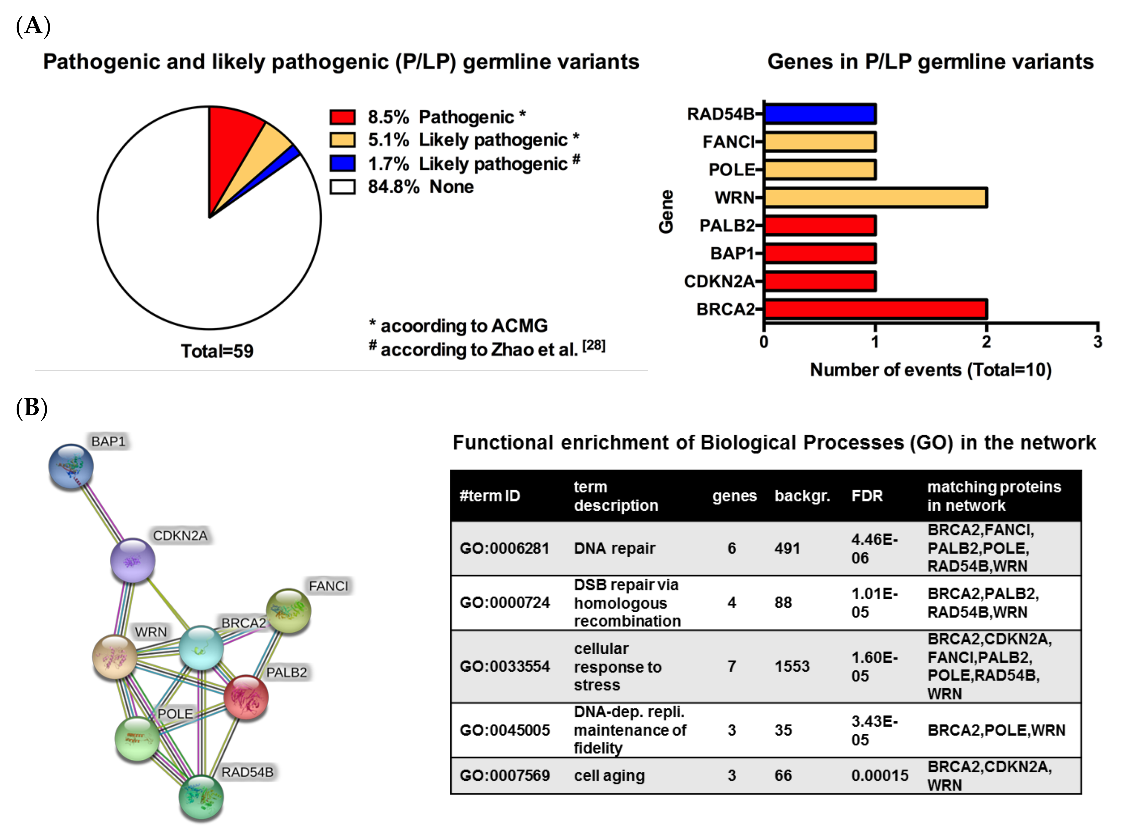
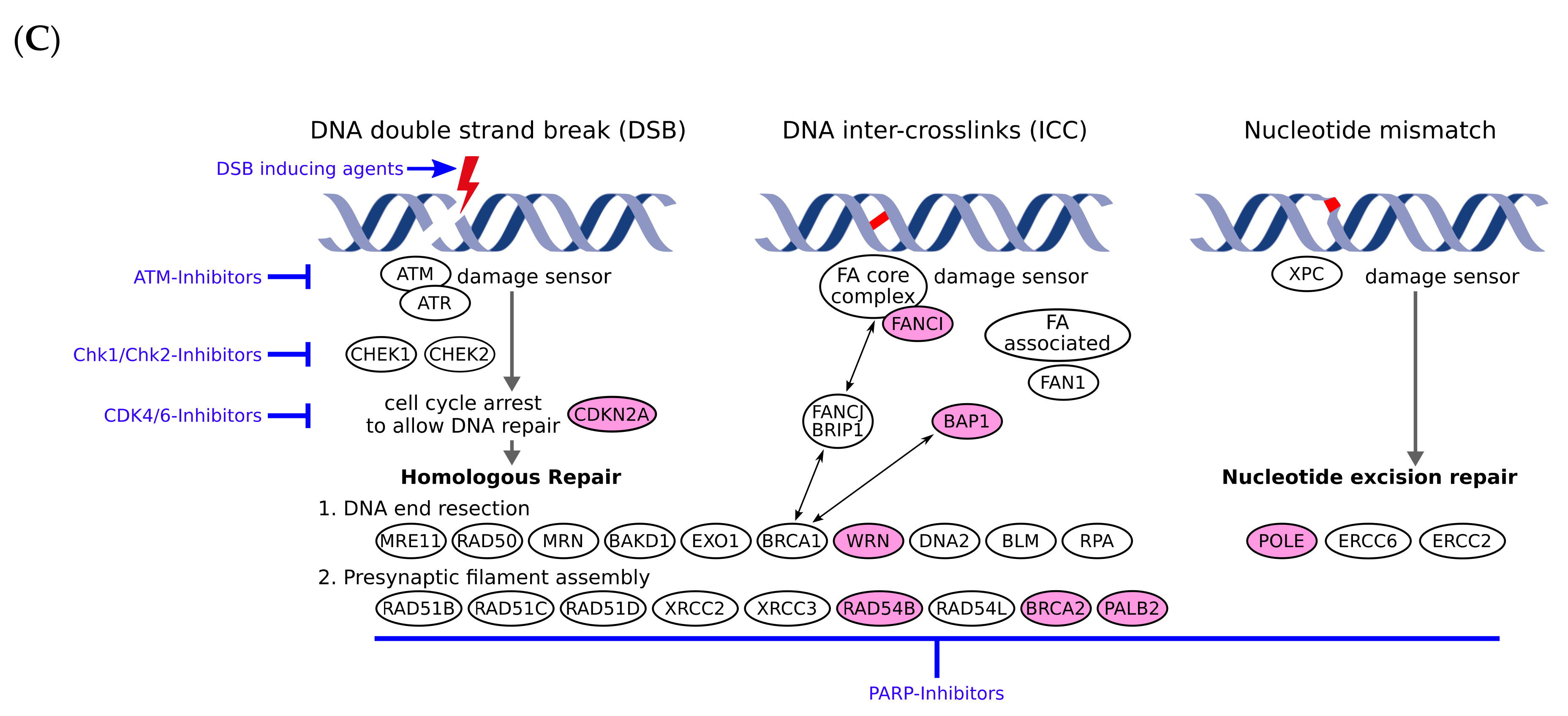
3.2. Pathogenic or Likely Pathogenic Germline Variants Identified

Eight different P/LP germline variants in nine patients were identified, seven of which were involved in DNA repair mechanisms (Table 1). Pathogenic or likely pathogenic germline variants were found in the following genes: BRCA2, CDKN2A, BAP1, PALB2, WRN, POLE, FANCI and RAD54B (Figure 1A). In one patient, two different likely pathogenic germline variants were found: POLE and WRN (Table 1). Both patients with a BRCA2 germline variant had additional cancers, one lung cancer, the other one chronic lymphatic leukemia/small cell lymphocytic lymphoma. Furthermore, the BAP1 mutant patient presented with an additional history of clear cell renal cell carcinoma, invasive ductal breast cancer and basal cell carcinoma. The RAD54B mutant patient had additionally prostate cancer. Using the ACMG classification, we identified four different pathogenic variants (BRCA2, CDKN2A, BAP1 and PALB2) and three likely pathogenic variants (POLE, WRN and FANCI). For reasons described above, we handled the variant in RAD54B as a likely pathogenic germline. An in silico analysis using the STRING database revealed the network of the gene products derived of the eight identified genes. Further functional enrichment analysis using the GO biological process terms revealed that indeed the eight genes are majorly involved in DNA repair but also aging and DNA replication and stress response as top enriched processes (Figure 1B).

3.3. Response to Combined Immunotherapy

Thirty-four of the patients treated with combined immunotherapy had progressive disease (PD) and 21 had disease control (DC) that included stable disease (SD), partial response (PR) or complete response (CR), according to RECIST 1.1 criteria (Table 3). Patients included in these two groups (PD and DC) differed significantly in terms of presence of P/LP germline variants (p = 0.010) and TMB (p < 0.0001), but no significant differences were observed when serum biomarkers S100B and LDH were analyzed (Table 3). All patients with P/LP germline variants that received combined immunotherapy (n = 9) had progressive disease.

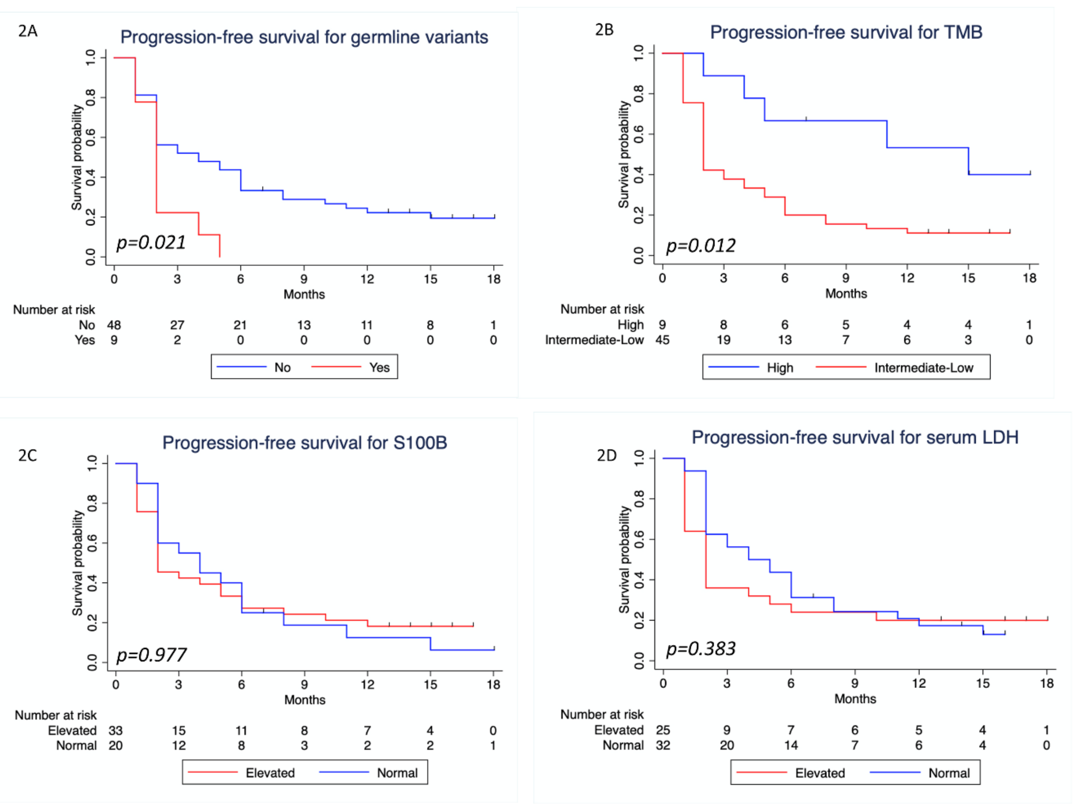
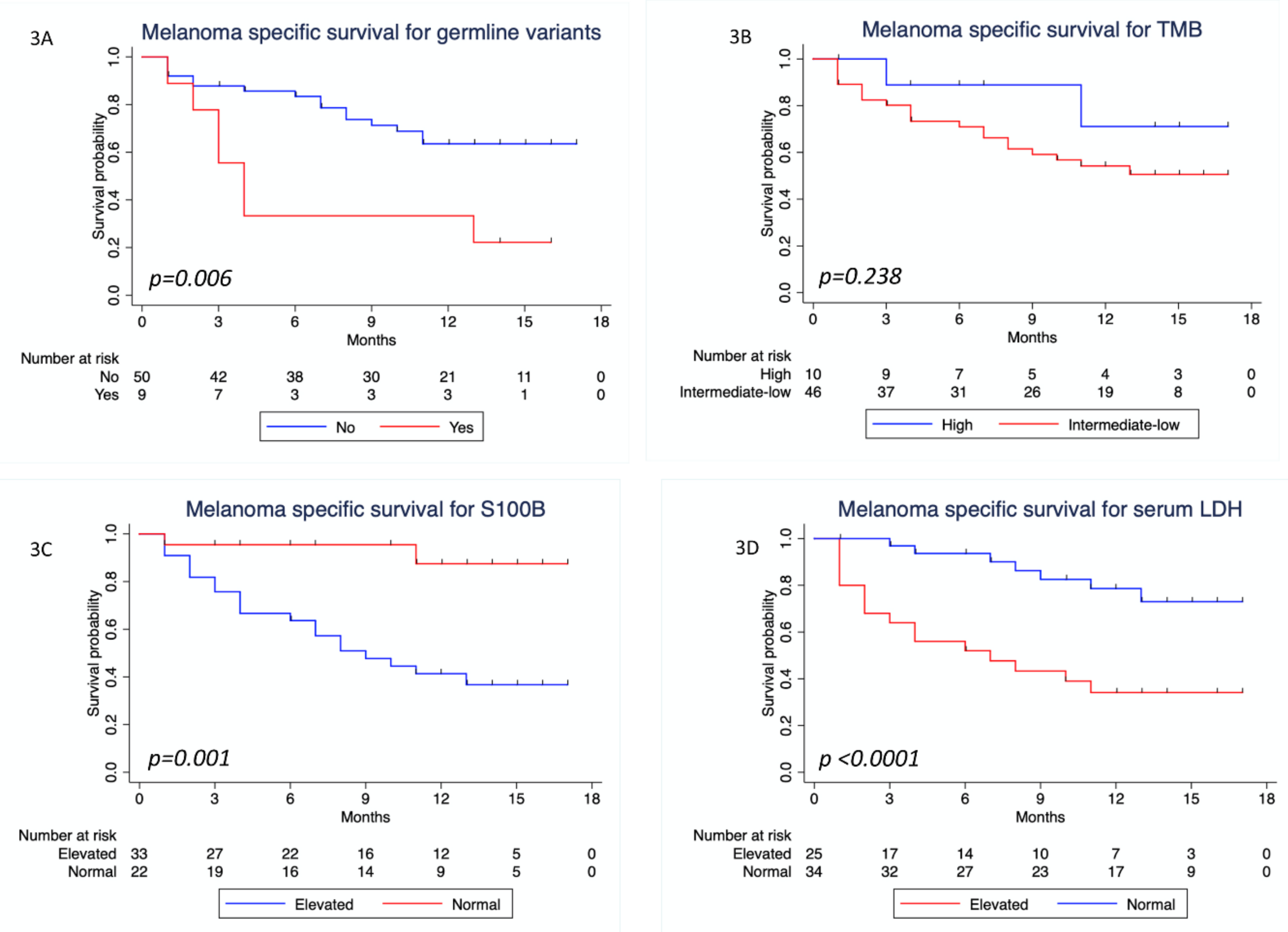
3.4. Survival Analysis

The median follow-up time was 13 months (IQR 11–15). Regarding PFS (Table 4), patients with P/LP germline variants did worse compared to those without P/LP germline variants (HR = 2.16; 95% CI: 1.01–4.64; p = 0.048). A statistically significant favorable outcome was seen in patients with high TMB (HR = 0.348; 95% CI: 0.14–0.89; p = 0.028), but no significant difference was observed when analyzing serum levels of S100B and LDH. Figure 2 shows the Kaplan–Meier PFS curves for these variables. In multivariate Cox regression analysis for PFS, only TMB remained significant (Table 4).
When analyzing MSS, univariate Cox regression analysis showed that there was a statistically significant difference between patients with and without a P/LP germline variant, favoring those without a P/LP germline variant (HR = 3.21; 95% CI: 1.31–7.87; p = 0.011; Table 4). A statistically significant difference was also found when analyzing serum S100B and LDH, favoring the group of patients with normal serum levels (p = 0.007 and p = 0.001, respectively). A trend for better MSS was seen in patients with high TMB (HR = 0.433, 96% CI: 0.10–1.85), but this was not statistically significant (p = 0.258). Figure 3 shows the Kaplan–Meier MSS curves for these variables.
In the multivariate Cox regression analysis we found that presence of a P/LP germline variant, serum S100B, and LDH were independently significant for MSS (p = 0.036, p = 0.044 and p = 0.001, respectively; Table 4).

4. Discussion

In our analysis of stage IV melanoma patients treated with nivolumab plus ipilimumab, we found that 15.2% (9/59) of patients harbored a P/LP germline variant, which is in line with what has been reported in other tumor entities so far [9,41] and thus indicates a high prevalence also in melanoma. More importantly, patients with P/LP germline variants presented a significantly worse progression-free survival when compared to the group of patients in which no P/LP germline variant was detected. The same results were found when analyzing melanoma specific survival, suggesting that patients with P/LP germline variants did not benefit from combined immunotherapy. The presence of a P/LP germline variant remained an independently significant factor for melanoma specific survival in the multivariate Cox regression analysis, along with serum S100B and LDH.
When analyzing survival in relation to TMB subgroups (intermediate-low and high), we found that there was a significant difference in PFS favoring patients with high TMB. This is probably explained by the fact that TMB correlates with disease control and response to immunotherapy, as has been shown by our group and others [3,4,31].
In our patient cohort, the majority of P/LP germline variants were present in genes that are associated with pathways involved in DNA repair mechanisms (Figure 1C). We observed that none of the patients with a P/LP germline variant responded to combined immunotherapy, which is currently considered as the most effective therapy in stage IV melanoma [42,43,44]. The explanation why this happened might be related to two mechanisms:
(A) Double strand breaking induced genes and DNA repair genes are involved in the development of immune cells, such as T cells [45,46,47,48,49,50]. On the one hand, it is well known that non-homologous end-joining is decisive for the V(D)J recombination events in developing B- and T-cells where the light and heavy chains of antibodies and T-cell receptors are recombined as random selections of different ready to use DNA segments. Defects in such processes are causal for severe combined immunodeficiency (SCID) syndromes. On the other hand, there are hints that homologous recombination is also involved in lymphocyte development [51]. For example, quiescent hematopoietic stem cells accumulate DNA damage during aging, which needs to be repaired when entering the cell cycle [52]. Defects in the DNA repair mechanisms would accelerate ageing and the accumulation of DNA damage, including the common lymphatic progenitor cells and hence all forms of lymphocytes. This could ultimately lead to a dampened antitumoral immune response and the failure of immune checkpoint inhibitors. Evidence also shows that standard DNA repair mechanisms are involved in immune responses, and defects in these repair mechanisms could translate into deteriorated immune responses and, therefore, worse responses to immunotherapy [45]. The fact that we did not see any response in patients with P/LP germline variants that are involved in DNA repair or homologous recombination adds to this evidence.
(B) It has recently been shown that higher levels of genomic instability were associated with lower immunogenicity in breast cancer patients, which might explain the fact that immune checkpoint inhibitors are less effective in this population [53]. Moreover, it is known that tumors with high genomic instability represented by elevated numbers of copy number variants (CNVs) and a rather low number of single nucleotide variants (SNVs) respond worse to immune checkpoint blockade [54]. There might be a similar mechanism present in the advanced melanoma patients included in our study, which could explain the absence of response and worse survival of these patients.
In this context, one needs to consider whether some patients with P/LP germline variant might be better served with systemic therapies that include targeted therapies, namely PARPi, and/or cytotoxic chemotherapies and not immunotherapy alone [55]. Platinum compounds that act by targeting the DNA repair mechanisms [56] might be considered a potential combination therapy in future trials including this subgroup of patients, similar to what has been previously shown and proposed in other tumor entities [55,57].
Presently, there is methodological diversity for sequencing DNA from tumor and normal tissue, namely whole genome, whole exome and panel sequencing with the latter showing the highest diagnostic sensitivity with respect to germline and subclonal variants [58,59]. In our study, both tumor and normal DNA have been sequenced deeply, focusing on >700 tumor relevant genes, in order to identify both germline and somatic variants.
Besides the controversy regarding the best sequencing method, there is also no consensus approach for reporting the results, particularly for germline variants. Reporting germline variants is an important aspect when considering the potential implications not only for the therapy of the patients, but also for their relatives. The ACMG classification is, as their authors mentioned, a work in progress [26]. Our understanding is that even if some variants cannot be classified as pathogenic, they should not be excluded from the genetic report, particularly when associated with a potentially increased risk or indicating that a targeted therapy can be offered. This was the concept in the current report.
Our study has a clear limitation regarding the small number of cases included. Therefore, the analyses have a limited statistical power, and should be interpreted with caution. A trend towards a worse prognosis for patients with P/LP germline variants can be seen from our data; none of the patients with a P/LP germline variant responded to combined immunotherapy, but the low number (n = 9) does not allow us to draw further conclusions.

5. Conclusions

The main finding of our study is that the presence of P/LP germline variants appeared to be correlated with primary resistance to combined immunotherapy, shorter progression-free survival and worse melanoma specific survival in melanoma patients treated with combined immunotherapy. The majority of the P/LP germline variants identified concerns genes associated with DNA repair mechanisms. Further, these mutated genes are frequently involved in processes needed for lymphocyte development and an impairment of such processes could lead to a diminished immune response and resistance to immunotherapy by immune checkpoint inhibitors. Our hypothesis needs to be further evaluated in a larger multicentric cohort and ideally in a future clinical trial.

Supplementary Materials

The following are available online at https://0-www-mdpi-com.brum.beds.ac.uk/2072-6694/12/5/1101/s1, Supplement 1: The 710 genes of the tumor panel, Supplement 2: Level of evidence (LOE) existing for each treatment option and used to classify information in table 1.

Author Contributions

A.F., M.S., F.B. and S.B. designed the study. All authors analyzed and interpreted the data. T.A., M.S., T.S., M.N., P.M., F.B., S.B. and A.F. wrote the manuscript. C.G., S.B. and A.F. supervised the study. All authors read and approved the final version of the manuscript.

Funding

The study was supported by the German Federal Ministry of Education and Research (BMBF) within the KMU-innovative initiative. AF was supported by the TÜFF Habilitation Program for Women of the Faculty of Medicine Tuebingen, Germany, grant no 2521-0-0. The authors acknowledge the support of the German Research Foundation (DFG) and the Open Access Publishing Fund of the University of Tuebingen.

Acknowledgments

We thank the whole team of the melanoma outpatient department for their care for our melanoma patients and the patients for their participation in this study.

Conflicts of Interest

T.A. reports travel grants from Novartis, personal fees and travel grants from BMS, outside the submitted work. M.S. is employee of Practice for Human Genetics. T.S. reports grants from Novartis and Pierre-Fabre outside the submitted work. M.N. is employee of Practice for Human Genetics. F.B. is employee of CeGaT. C.G. reports grants and personal fees from BMS, Novartis, Roche and Sanofi, personal fees from Amgen, CeGaT, MSD, NeraCare, Philogen, Pierre-Fabre, outside the submitted work. S.B. is managing director of CeGaT. A.F. served as consultant to Roche, Novartis, MSD, Pierre-Fabre, received travel support from Roche, Novartis, BMS, Pierre-Fabre and speaker fees from Roche, Novartis, BMS, MSD and CeGaT. No competing interests were declared by P.M.

References

  1. Lander, E.S.; Linton, L.M.; Birren, B.; Nusbaum, C.; Zody, M.C.; Baldwin, J.; Devon, K.; Dewar, K.; Doyle, M.; FitzHugh, W.; et al. Initial sequencing and analysis of the human genome. Nature 2001, 409, 860–921. [Google Scholar] [CrossRef] [Green Version]
  2. Venter, J.C.; Adams, M.D.; Myers, E.W.; Li, P.W.; Mural, R.J.; Sutton, G.G.; Smith, H.O.; Yandell, M.; Evans, C.A.; Holt, R.A.; et al. The sequence of the human genome. Science (New York, N.Y.) 2001, 291, 1304–1351. [Google Scholar] [CrossRef] [Green Version]
  3. Samstein, R.M.; Lee, C.-H.; Shoushtari, A.N.; Hellmann, M.D.; Shen, R.; Janjigian, Y.Y.; Barron, D.A.; Zehir, A.; Jordan, E.J.; Omuro, A.; et al. Tumor mutational load predicts survival after immunotherapy across multiple cancer types. Nat. Genet. 2019, 51, 202–206. [Google Scholar] [CrossRef]
  4. Chan, T.A.; Yarchoan, M.; Jaffee, E.; Swanton, C.; Quezada, S.A.; Stenzinger, A.; Peters, S. Development of tumor mutation burden as an immunotherapy biomarker: Utility for the oncology clinic. Ann. Oncol. 2018, 30, 44–56. [Google Scholar] [CrossRef]
  5. Ghazani, A.A.; Oliver, N.M.; St. Pierre, J.P.; Garofalo, A.; Rainville, I.R.; Hiller, E.; Treacy, D.J.; Rojas-Rudilla, V.; Wood, S.; Bair, E.; et al. Assigning clinical meaning to somatic and germ-line whole-exome sequencing data in a prospective cancer precision medicine study. Genet. Med. 2017, 19, 787. [Google Scholar] [CrossRef]
  6. Mandelker, D.; Donoghue, M.; Talukdar, S.; Bandlamudi, C.; Srinivasan, P.; Vivek, M.; Jezdic, S.; Hanson, H.; Snape, K.; Kulkarni, A.; et al. Germline-focussed analysis of tumour-only sequencing: Recommendations from the ESMO Precision Medicine Working Group. Ann. Oncol. 2019, 30, 1221–1231. [Google Scholar] [CrossRef] [Green Version]
  7. Chang, M.T.; Asthana, S.; Gao, S.P.; Lee, B.H.; Chapman, J.S.; Kandoth, C.; Gao, J.; Socci, N.D.; Solit, D.B.; Olshen, A.B.; et al. Identifying recurrent mutations in cancer reveals widespread lineage diversity and mutational specificity. Nat. Biotechnol. 2015, 34, 155. [Google Scholar] [CrossRef]
  8. Parsons, D.W.; Roy, A.; Yang, Y.; Wang, T.; Scollon, S.; Bergstrom, K.; Kerstein, R.A.; Gutierrez, S.; Petersen, A.K.; Bavle, A.; et al. Diagnostic Yield of Clinical Tumor and Germline Whole-Exome Sequencing for Children With Solid TumorsDiagnostic Yield in Genetic Sequencing for Children With Solid TumorsDiagnostic Yield in Genetic Sequencing for Children With Solid Tumors. JAMA Oncol. 2016, 2, 616–624. [Google Scholar] [CrossRef]
  9. Schrader, K.A.; Cheng, D.T.; Joseph, V.; Prasad, M.; Walsh, M.; Zehir, A.; Ni, A.; Thomas, T.; Benayed, R.; Ashraf, A.; et al. Germline Variants in Targeted Tumor Sequencing Using Matched Normal DNAGermline Variants in Tumor Sequencing With Matched Normal DNAGermline Variants in Tumor Sequencing With Matched Normal DNA. JAMA Oncol. 2016, 2, 104–111. [Google Scholar] [CrossRef]
  10. Robert, C.; Ribas, A.; Schachter, J.; Arance, A.; Grob, J.J.; Mortier, L.; Daud, A.; Carlino, M.S.; McNeil, C.M.; Lotem, M.; et al. Pembrolizumab versus ipilimumab in advanced melanoma (KEYNOTE-006): Post-hoc 5-year results from an open-label, multicentre, randomised, controlled, phase 3 study. Lancet Oncol. 2019, 20, 1239–1251. [Google Scholar] [CrossRef]
  11. Hodi, F.S.; Chiarion-Sileni, V.; Gonzalez, R.; Grob, J.J.; Rutkowski, P.; Cowey, C.L.; Lao, C.D.; Schadendorf, D.; Wagstaff, J.; Dummer, R.; et al. Nivolumab plus ipilimumab or nivolumab alone versus ipilimumab alone in advanced melanoma (CheckMate 067): 4-year outcomes of a multicentre, randomised, phase 3 trial. Lancet. Oncol. 2018, 19, 1480–1492. [Google Scholar] [CrossRef]
  12. Robert, C.; Schachter, J.; Long, G.V.; Arance, A.; Grob, J.J.; Mortier, L.; Daud, A.; Carlino, M.S.; McNeil, C.; Lotem, M.; et al. Pembrolizumab versus Ipilimumab in Advanced Melanoma. N. Engl. J. Med. 2015, 372, 2521–2532. [Google Scholar] [CrossRef]
  13. Robert, C.; Long, G.V.; Brady, B.; Dutriaux, C.; Maio, M.; Mortier, L.; Hassel, J.C.; Rutkowski, P.; McNeil, C.; Kalinka-Warzocha, E.; et al. Nivolumab in Previously Untreated Melanoma without BRAF Mutation. N. Engl. J. Med. 2014, 372, 320–330. [Google Scholar] [CrossRef] [Green Version]
  14. Schwaederle, M.; Zhao, M.; Lee, J.J.; Lazar, V.; Leyland-Jones, B.; Schilsky, R.L.; Mendelsohn, J.; Kurzrock, R. Association of Biomarker-Based Treatment Strategies With Response Rates and Progression-Free Survival in Refractory Malignant Neoplasms: A Meta-analysisOutcomes for Biomarker-Based Treatment Strategies in Refractory Malignant NeoplasmsOutcomes for Biomarker-Based Treatment Strategies in Refractory Malignant Neoplasms. JAMA Oncol. 2016, 2, 1452–1459. [Google Scholar] [CrossRef] [Green Version]
  15. Sicklick, J.K.; Kato, S.; Okamura, R.; Schwaederle, M.; Hahn, M.E.; Williams, C.B.; De, P.; Krie, A.; Piccioni, D.E.; Miller, V.A.; et al. Molecular profiling of cancer patients enables personalized combination therapy: The I-PREDICT study. Nat. Med. 2019, 25, 744–750. [Google Scholar] [CrossRef]
  16. Lopez-Chavez, A.; Thomas, A.; Rajan, A.; Raffeld, M.; Morrow, B.; Kelly, R.; Carter, C.A.; Guha, U.; Killian, K.; Lau, C.C.; et al. Molecular profiling and targeted therapy for advanced thoracic malignancies: A biomarker-derived, multiarm, multihistology phase II basket trial. J. Clin. Oncol. 2015, 33, 1000–1007. [Google Scholar] [CrossRef]
  17. Chapman, P.B.; Hauschild, A.; Robert, C.; Haanen, J.B.; Ascierto, P.; Larkin, J.; Dummer, R.; Garbe, C.; Testori, A.; Maio, M.; et al. Improved Survival with Vemurafenib in Melanoma with BRAF V600E Mutation. N. Engl. J. Med. 2011, 364, 2507–2516. [Google Scholar] [CrossRef] [Green Version]
  18. Flaherty, K.T.; Robert, C.; Hersey, P.; Nathan, P.; Garbe, C.; Milhem, M.; Demidov, L.V.; Hassel, J.C.; Rutkowski, P.; Mohr, P.; et al. Improved survival with MEK inhibition in BRAF-mutated melanoma. N. Engl. J. Med. 2012, 367, 107–114. [Google Scholar] [CrossRef] [Green Version]
  19. Benafif, S.; Hall, M. An update on PARP inhibitors for the treatment of cancer. OncoTargets Ther. 2015, 8, 519–528. [Google Scholar] [CrossRef] [Green Version]
  20. Kaufman, B.; Shapira-Frommer, R.; Schmutzler, R.K.; Audeh, M.W.; Friedlander, M.; Balmaña, J.; Mitchell, G.; Fried, G.; Stemmer, S.M.; Hubert, A.; et al. Olaparib Monotherapy in Patients With Advanced Cancer and a Germline BRCA1/2 Mutation. J. Clin. Oncol. 2014, 33, 244–250. [Google Scholar] [CrossRef]
  21. Low, S.-K.; Nakamura, Y. The road map of cancer precision medicine with the innovation of advanced cancer detection technology and personalized immunotherapy. Jpn. J. Clin. Oncol. 2019, 49, 596–603. [Google Scholar] [CrossRef]
  22. Mateo, J.; Chakravarty, D.; Dienstmann, R.; Jezdic, S.; Gonzalez-Perez, A.; Lopez-Bigas, N.; Ng, C.K.Y.; Bedard, P.L.; Tortora, G.; Douillard, J.Y.; et al. A framework to rank genomic alterations as targets for cancer precision medicine: The ESMO Scale for Clinical Actionability of molecular Targets (ESCAT). Ann. Oncol. 2018, 29, 1895–1902. [Google Scholar] [CrossRef]
  23. Thavaneswaran, S.; Rath, E.; Tucker, K.; Joshua, A.M.; Hess, D.; Pinese, M.; Ballinger, M.L.; Thomas, D.M. Therapeutic implications of germline genetic findings in cancer. Nat. Rev. Clin. Oncol. 2019, 16, 386–396. [Google Scholar] [CrossRef]
  24. Meric-Bernstam, F.; Brusco, L.; Daniels, M.; Wathoo, C.; Bailey, A.M.; Strong, L.; Shaw, K.; Lu, K.; Qi, Y.; Zhao, H.; et al. Incidental germline variants in 1000 advanced cancers on a prospective somatic genomic profiling protocol. Ann. Oncol. 2016, 27, 795–800. [Google Scholar] [CrossRef] [Green Version]
  25. Gershenwald, J.E.; Scolyer, R.A. Melanoma Staging: American Joint Committee on Cancer (AJCC) 8th Edition and Beyond. Ann. Surg. Oncol. 2018, 25, 2105–2110. [Google Scholar] [CrossRef]
  26. Richards, S.; Aziz, N.; Bale, S.; Bick, D.; Das, S.; Gastier-Foster, J.; Grody, W.W.; Hegde, M.; Lyon, E.; Spector, E.; et al. Standards and guidelines for the interpretation of sequence variants: A joint consensus recommendation of the American College of Medical Genetics and Genomics and the Association for Molecular Pathology. Genet. Med. 2015, 17, 405–424. [Google Scholar] [CrossRef]
  27. Zhao, Q.; Yang, J.; Li, L.; Cao, D.; Yu, M.; Shen, K. Germline and somatic mutations in homologous recombination genes among Chinese ovarian cancer patients detected using next-generation sequencing. J. Gynecol. Oncol. 2017, 28, e39. [Google Scholar] [CrossRef] [Green Version]
  28. Jiang, H.; Lei, R.; Ding, S.W.; Zhu, S. Skewer: A fast and accurate adapter trimmer for next-generation sequencing paired-end reads. BMC Bioinform. 2014, 15, 182. [Google Scholar] [CrossRef]
  29. Li, H.; Durbin, R. Fast and accurate long-read alignment with Burrows-Wheeler transform. Bioinformatics 2010, 26, 589–595. [Google Scholar] [CrossRef] [Green Version]
  30. Stenson, P.D.; Ball, E.V.; Mort, M.; Phillips, A.D.; Shaw, K.; Cooper, D.N. The Human Gene Mutation Database (HGMD) and its exploitation in the fields of personalized genomics and molecular evolution. Curr. Protoc. Bioinform. 2012. [Google Scholar] [CrossRef]
  31. Forschner, A.; Battke, F.; Hadaschik, D.; Schulze, M.; Weißgraeber, S.; Han, C.-T.; Kopp, M.; Frick, M.; Klumpp, B.; Tietze, N.; et al. Tumor mutation burden and circulating tumor DNA in combined CTLA-4 and PD-1 antibody therapy in metastatic melanoma–results of a prospective biomarker study. J. Immunother. Cancer 2019, 7, 180. [Google Scholar] [CrossRef] [PubMed]
  32. Chalmers, Z.R.; Connelly, C.F.; Fabrizio, D.; Gay, L.; Ali, S.M.; Ennis, R.; Schrock, A.; Campbell, B.; Shlien, A.; Chmielecki, J.; et al. Analysis of 100,000 human cancer genomes reveals the landscape of tumor mutational burden. Genome Med. 2017, 9, 34. [Google Scholar] [CrossRef] [PubMed]
  33. Johnson, D.B.; Frampton, G.M.; Rioth, M.J.; Yusko, E.; Xu, Y.; Guo, X.; Ennis, R.C.; Fabrizio, D.; Chalmers, Z.R.; Greenbowe, J.; et al. Targeted Next Generation Sequencing Identifies Markers of Response to PD-1 Blockade. Cancer Immunol. Res. 2016, 4, 959–967. [Google Scholar] [CrossRef] [PubMed] [Green Version]
  34. Stelzer, G.; Rosen, N.; Plaschkes, I.; Zimmerman, S.; Twik, M.; Fishilevich, S.; Stein, T.I.; Nudel, R.; Lieder, I.; Mazor, Y.; et al. The GeneCards Suite: From Gene Data Mining to Disease Genome Sequence Analyses. Curr. Protoc. Bioinform. 2016, 54, 1–30. [Google Scholar] [CrossRef]
  35. Kanehisa, M.; Goto, S. KEGG: Kyoto encyclopedia of genes and genomes. Nucleic Acids Res. 2000, 28, 27–30. [Google Scholar] [CrossRef]
  36. Kanehisa, M.; Sato, Y.; Furumichi, M.; Morishima, K.; Tanabe, M. New approach for understanding genome variations in KEGG. Nucleic Acids Res. 2019, 47, D590–D595. [Google Scholar] [CrossRef] [Green Version]
  37. Kanehisa, M. Toward understanding the origin and evolution of cellular organisms. Protein Sci 2019, 28, 1947–1951. [Google Scholar] [CrossRef]
  38. Chakravarty, D.; Gao, J.; Phillips, S.M.; Kundra, R.; Zhang, H.; Wang, J.; Rudolph, J.E.; Yaeger, R.; Soumerai, T.; Nissan, M.H.; et al. OncoKB: A Precision Oncology Knowledge Base. JCO Precis. Oncol. 2017, 2017. [Google Scholar] [CrossRef]
  39. STRING. Protein-Protein Interaction Networks Functional Enrichment Analysis. Available online: https://string-db.org (accessed on 20 March 2020).
  40. Schwartz, L.H.; Litière, S.; de Vries, E.; Ford, R.; Gwyther, S.; Mandrekar, S.; Shankar, L.; Bogaerts, J.; Chen, A.; Dancey, J.; et al. RECIST 1.1-Update and clarification: From the RECIST committee. Eur. J. Cancer 2016, 62, 132–137. [Google Scholar] [CrossRef] [Green Version]
  41. Mandelker, D.; Zhang, L.; Kemel, Y.; Stadler, Z.K.; Joseph, V.; Zehir, A.; Pradhan, N.; Arnold, A.; Walsh, M.F.; Li, Y.; et al. Mutation Detection in Patients With Advanced Cancer by Universal Sequencing of Cancer-Related Genes in Tumor and Normal DNA vs Guideline-Based Germline TestingMutation Detection in Patients With Advanced CancerMutation Detection in Patients With Advanced Cancer. JAMA 2017, 318, 825–835. [Google Scholar] [CrossRef]
  42. Larkin, J.; Chiarion-Sileni, V.; Gonzalez, R.; Grob, J.-J.; Rutkowski, P.; Lao, C.D.; Cowey, C.L.; Schadendorf, D.; Wagstaff, J.; Dummer, R.; et al. Five-Year Survival with Combined Nivolumab and Ipilimumab in Advanced Melanoma. N. Engl. J. Med. 2019, 381, 1535–1546. [Google Scholar] [CrossRef] [PubMed] [Green Version]
  43. Rulli, E.; Legramandi, L.; Salvati, L.; Mandala, M. The impact of targeted therapies and immunotherapy in melanoma brain metastases: A systematic review and meta-analysis. Cancer 2019, 125, 3776–3789. [Google Scholar] [CrossRef] [PubMed]
  44. Fizazi, K.; Maillard, A.; Penel, N.; Baciarello, G.; Allouache, D.; Daugaard, G.; Van De Wouw, A.; Soler, G.; Vauleon, E.; Chaigneau, L.; et al. Adjuvant immunotherapy with nivolumab (NIVO) alone or in combination with ipilimumab (IPI) versus placebo in stage IV melanoma patients with no evidence of disease (NED): A randomized, double-blind phase 2 trial (IMMUNED). Ann. Oncol. 2019, 30, v851. [Google Scholar] [CrossRef]
  45. Bednarski, J.J.; Sleckman, B.P. At the intersection of DNA damage and immune responses. Nat. Immunol. 2019, 19, 231–242. [Google Scholar] [CrossRef] [PubMed]
  46. Mak, T.W.; Hakem, A.; McPherson, J.P.; Shehabeldin, A.; Zablocki, E.; Migon, E.; Duncan, G.S.; Bouchard, D.; Wakeham, A.; Cheung, A.; et al. Brca1 required for T cell lineage development but not TCR loci rearrangement. Nat. Immunol. 2000, 1, 77. [Google Scholar] [CrossRef] [PubMed]
  47. Galgano, A.; Barinov, A.; Vasseur, F.; de Villartay, J.-P.; Rocha, B. CD8 Memory Cells Develop Unique DNA Repair Mechanisms Favoring Productive Division. PLoS ONE 2015, 10, e0140849. [Google Scholar] [CrossRef] [Green Version]
  48. Bednarski, J.J.; Sleckman, B.P. Lymphocyte development: Integration of DNA damage response signaling. Adv. Immunol. 2012, 116, 175–204. [Google Scholar] [CrossRef] [Green Version]
  49. Bredemeyer, A.L.; Helmink, B.A.; Innes, C.L.; Calderon, B.; McGinnis, L.M.; Mahowald, G.K.; Gapud, E.J.; Walker, L.M.; Collins, J.B.; Weaver, B.K.; et al. DNA double-strand breaks activate a multi-functional genetic program in developing lymphocytes. Nature 2008, 456, 819–823. [Google Scholar] [CrossRef] [Green Version]
  50. Bredemeyer, A.L.; Sharma, G.G.; Huang, C.Y.; Helmink, B.A.; Walker, L.M.; Khor, K.C.; Nuskey, B.; Sullivan, K.E.; Pandita, T.K.; Bassing, C.H.; et al. ATM stabilizes DNA double-strand-break complexes during V(D)J recombination. Nature 2006, 442, 466–470. [Google Scholar] [CrossRef]
  51. Caddle, L.B.; Hasham, M.G.; Schott, W.H.; Shirley, B.-J.; Mills, K.D. Homologous recombination is necessary for normal lymphocyte development. Mol. Cell. Boil. 2008, 28, 2295–2303. [Google Scholar] [CrossRef] [Green Version]
  52. Beerman, I.; Seita, J.; Inlay, M.A.; Weissman, I.L.; Rossi, D.J. Quiescent hematopoietic stem cells accumulate DNA damage during aging that is repaired upon entry into cell cycle. Cell Stem Cell 2014, 15, 37–50. [Google Scholar] [CrossRef] [PubMed] [Green Version]
  53. Kraya, A.A.; Maxwell, K.N.; Wubbenhorst, B.; Wenz, B.M.; Pluta, J.; Rech, A.J.; Dorfman, L.M.; Lunceford, N.; Barrett, A.; Mitra, N.; et al. Genomic Signatures Predict the Immunogenicity of BRCA-Deficient Breast Cancer. Clin. Cancer Res. 2019, 25, 4363. [Google Scholar] [CrossRef] [PubMed] [Green Version]
  54. Ock, C.-Y.; Hwang, J.-E.; Keam, B.; Kim, S.-B.; Shim, J.-J.; Jang, H.-J.; Park, S.; Sohn, B.H.; Cha, M.; Ajani, J.A.; et al. Genomic landscape associated with potential response to anti-CTLA-4 treatment in cancers. Nat. Commun. 2017, 8, 1050. [Google Scholar] [CrossRef] [PubMed]
  55. Wilson, M.A.; Zhao, F.; Khare, S.; Roszik, J.; Woodman, S.E.; Andrea, K.; Wubbenhorst, B.; Rimm, D.L.; Kirkwood, J.M.; Kluger, H.M.; et al. Copy Number Changes Are Associated with Response to Treatment with Carboplatin, Paclitaxel, and Sorafenib in Melanoma. Clin. Cancer Res. 2016, 22, 374. [Google Scholar] [CrossRef] [PubMed] [Green Version]
  56. Roberts, J.J.; Thomson, A.J. The Mechanism of Action of Antitumor Platinum Compounds. In Progress in Nucleic Acid Research and Molecular Biology; Cohn, W.E., Ed.; Academic Press: Cambridge, MA, USA, 1979; Volume 22, pp. 71–133. [Google Scholar]
  57. Stewart, R.A.; Pilie, P.G.; Yap, T.A. Development of PARP and Immune-Checkpoint Inhibitor Combinations. Cancer Res. 2018, 78, 6717–6725. [Google Scholar] [CrossRef] [Green Version]
  58. Cheng, D.T.; Mitchell, T.N.; Zehir, A.; Shah, R.H.; Benayed, R.; Syed, A.; Chandramohan, R.; Liu, Z.Y.; Won, H.H.; Scott, S.N.; et al. Memorial Sloan Kettering-Integrated Mutation Profiling of Actionable Cancer Targets (MSK-IMPACT): A Hybridization Capture-Based Next-Generation Sequencing Clinical Assay for Solid Tumor Molecular Oncology. J. Mol. Diagn. 2015, 17, 251–264. [Google Scholar] [CrossRef]
  59. FoundationOne CDx™. Available online: https://www.accessdata.fda.gov/cdrh_docs/pdf17/P170019C.pdf (accessed on 15 August 2019).
Figure 1. Pathogenic and likely pathogenic germline variants. (A) Left pie chart: cohort size by type of detected pathogenic and likely pathogenic (P/LP) germline variants. Each patient is counted according to the most pathogenic variant found (e.g., a patient with a pathogenic variant and a second, likely pathogenic variant is counted in the group of pathogenic variants). Germline variants were found in 15% of patients. Right bar diagram: genes in P/LP germline variants. Most genes harbored mutations in only one patient, while BRCA2 and WRN were affected in two patients each. In one patient two different genes were affected by germline variants both categorized as likely pathogenic (WRN + POLE). Red bars indicate pathogenic variants, yellow indicates likely pathogenic variants and blue shows a non-classifiable variant according to ACMG. (B) Left: STRING protein network of the eight different genes with pathogenic and likely pathogenic germline variants showing the known interactions of the corresponding gene products. Right: A table with a functional enrichment analysis of the eight genes with pathogenic and likely pathogenic germline variants using the gene ontology (GO) terms “Biological Process”. The first five top ranked (by the false discovery rate FDR) hits are shown. (C) Overview of DNA damage repair pathways with genes found affected in our cohort (highlighted in pink) as well as additional highly relevant genes (selection). Blue labels show targets for targeted therapies. Most pathogenic and likely pathogenic germline variants reported in our cohort are in genes associated with DNA damage repair pathways.
Cancers 12 01101 g001aCancers 12 01101 g001b
Figure 2. Progression-free survival analysis. (A) Kaplan–Meier curve for progression-free survival, considering the presence of pathogenic and likely pathogenic germline variants (Yes/No); (B) Kaplan–Meier curve for progression-free survival considering TMB intermediate-low (≤23.1 mut/Mb) or high (>23.1 mut/Mb); (C) Kaplan–Meier curve for progression-free survival considering serum S100B levels (normal/elevated); (D) Kaplan–Meier curve for progression-free survival considering serum lactate dehydrogenase (LDH) levels (normal/elevated). p-values refer to two-sided log rank tests.
Cancers 12 01101 g002
Figure 3. Melanoma specific survival analysis. (A) Kaplan–Meier curve for melanoma specific survival, considering the presence of pathogenic and likely pathogenic germline variants (Yes/No); (B) Kaplan–Meier curve for melanoma specific survival considering TMB intermediate-low (≤23.1 mut/Mb) or high (>23.1 mut/Mb); (C) Kaplan–Meier curve for melanoma specific survival considering serum S100B levels (normal/elevated); (D) Kaplan–Meier curve for melanoma specific survival considering serum lactate dehydrogenase (LDH) levels (normal/elevated). p-values refer to two-sided log rank tests.
Cancers 12 01101 g003
Table 1. Pathogenic and likely pathogenic germline variants reported.
Patient No.Melanoma SubtypeGene NameGermline VariantACMG Classifiable [1]RationaleTMB (mut/Mb)Pathway InvolvedPotential TherapyLOE$
1Cutaneous (nodular)BRCA2c.6446_6449delTTAA; p.Ile2149Lysfs*18 (het.), NM_000059.3Class 5Pathogenic germline variant40.9DNA repair, homologous recombinationPARPi2A
2Cutaneous (nodular)POLEc.1270C>T; p.Leu424Phe (het.), NM_006231.3Class 4Likely pathogenic germline variant21.4DNA repair, homologous recombinationPARPi2B
Immunotherapy1B
WRNc.2893_2899del; p.Lys965Cysfs*7 (het.), NM_000553.4Class 4Likely pathogenic germline variantHomologous recombinationPARPi2B
3AcralFANCICNV coding exon 9 (het. Deletion), NM_001113378.1Class 4Likely pathogenic germline variant1.6DNA repair, homologous recombinationPARPi4
4Cutaneous (superficial)WRNc.348delC; p.Met117Cysfs*8 (het.), NM_000553.4Class 4Likely pathogenic germline variant7.9Homologous recombinationPARPi2B
5Cutaneous (polypoid)CDKN2Ac.301G>T; p.Gly101Trp (het.), NM_000077.4Class 5Pathogenic germline variant5.8Cell cycleCDK 4/6 inhibitors2B
6UvealBAP1c.37+1G>T; p.? (het.), NM_004656.3Class 5Pathogenic germline variant1.0Homologous recombination, epigeneticsPARPi2B
EZH2i3
HDACi4
7Cutaneous (superficial)PALB2c.757_758delCT; p.Leu253Ilefs*3 (het.), NM_024675.3Class 5Pathogenic germline variant3.1Homologous recombinationPARPi2B
8Cutaneous (nodular)BRCA2c.1888dupA; p.Thr630Asnfs*6 (het.), NM_000059.3Class 5Pathogenic germline variant12.0DNA repair, homologous recombinationPARPi2A
9Cutaneous (nodular)RAD54Bc.889C>T; p.Arg297* (het.), NM_012415.3NoLikely pathogenic germline variant/ Potentially therapy relevant13.1DNA repair, homologous recombinationPARPi4
ACMG = American College of Medical Genetics and Genomics; LOE= level of evidence—please refer to Supplementary Materials supplement 2; PARPi = adenosine diphosphate (ADP) ribose polymerase (PARP) inhibitors; EZH2i = enhancer of zeste homolog 2 inhibitors; HDACi = histone deacetylase inhibitors; CDK = cyclin-dependent kinases. $ Please refer to Supplementary Materials supplement 2.
Table 2. Patients’ characteristics.
CharacteristicN (%)
Sex
Male36 (61)
Female23 (39)
Age
Primary disease diagnosis (median years; IQR)58; (49–70)
Initiation of combined immunotherapy (median years; IQR)61; (51–74)
Melanoma subtype
Cutaneous melanoma35 (59.3)
Acral melanoma6 (10.2)
Mucosal melanoma4 (6.7)
Melanoma of unknown primary5 (8.5)
Uveal melanoma9 (15.3)
Tumor mutation burden* (n = 56)
TMB (mut/Mb median; IQR) 4.7; (1.7–13.97)
Intermediate-low (≤23.1 mut/Mb)46 (82.1)
High (>23.1 mut/Mb)10 (17.9)
Serum biomarkers
LDH
Normal34 (57.6)
Elevated25 (42.4)
S100B* (n = 55)
Normal22 (40)
Elevated33 (60)
BRAF mutation* (n = 57)
Yes29 (50.9)
No28 (49.1)
Metastasis at start of nivolumab plus ipilimumab
Presence of cerebral metastasis 24 (40.7)
Presence of liver metastasis 17 (28.8)
Presence of lung metastasis 32 (54.2)
Nivolumab plus ipilimumab
First line29 (49.2)
Second line or more30 (50.8)
Response to nivolumab and ipilimumab at first staging*
(n = 55)
Progressive disease34 (61.8)
Disease control 21 (38.2)
* Denotes variables for which patients with missing values were excluded.
Table 3. Response to combined immunotherapy dependent on potential predictors (n = 55).
CategoryDisease Control
N = 21
Progressive Disease
N = 34
pOdds Ratio (95% CI)
N (%)
Pathogenic and likely pathogenic germline variants
Present09 (26.5)0.010NA*
Not present21 (100)25 (73.5)
Tumor mutation burden
(n = 52)
Low /Intermediate11 (57.9)32 (97)<0.000123.27 (2.61–207.7)
High 8 (42.1)1 (3)
Serum biomarkers
S100B (n = 51)
Normal9 (47.4)11 (34.4)0.36
Elevated10 (52.6)21 (65.6) 1.72 (0.54–5.47)
LDH
Normal15 (71.4)17 (50)0.118
Elevated6 (28.6)17 (50) 2.50 (0.78–7.98)
Two-sided exact Pearson Chi-square test (p-value) and logistic regression (odds ratio). * Not available due to zero patients with pathogenic or likely pathogenic germline variants in the disease control group, risk difference: 100% − 54% = 46%.
Table 4. Progression-free survival and melanoma specific survival dependent on potential predictors.
CategoryProgression-Free SurvivalMelanoma Specific Survival
HR (95% CI)1p1HR (95% CI)2p2HR (95% CI)1p1HR (95% CI)2p2
Pathogenic and likely pathogenic germline variants
Present vs. not present 2.16 (1.01–4.64)0.0481.93 (0.89–4.15)0.0953.21 (1.31–7.87)0.0112.93 (1.07–8.0)0.036
Tumor mutation burden
Low/intermediate vs. high2.88 (1.12–7.38)0.0282.75 (1.07–7.09)0.0362.31 (0.54–9.85)0.258NA
S100B
Elevated vs. normal 0.992 (0.55–1.81)0.979NA 7.45 (1.74–31.91)0.0074.65 (1.04–20.76)0.044
LDH
Elevated vs. normal1.26 (0.71–2.26)0.433 NA 4.33 (1.77–10.56)0.0015.16 (1.90–13.96)0.001
1 Univariate Cox regression analysis; 2 multivariate Cox regression analysis for variables significant in the univariate analysis. NA variable was not included in the multivariate Cox regression analysis. p-values refer to the two-sided Wald test.

