Article

Effects of Epstein-Barr Virus Infection on the Risk and Prognosis of Primary Laryngeal Squamous Cell Carcinoma: A Hospital-Based Case-Control Study in Taiwan

1
Department of Otorhinolaryngology—Head and Neck Surgery, Chang Gung Memorial Hospital, Linkou Main Branch, Taoyuan City 33305, Taiwan
2
Faculty of Medicine, Chang Gung University, Taoyuan City 33302, Taiwan
3
Department of Family Medicine, Chang Gung Memorial Hospital, Linkou Main Branch, Taoyuan 33305, Taiwan
4
Department of Industrial Engineering and Management, National Taipei University of Technology, Taipei 10608, Taiwan
5
Department of Pathology, Chang Gung Memorial Hospital, Linkou Main Branch, Taoyuan City 33305, Taiwan
6
Department of Laboratory Medicine, Chang Gung Memorial Hospital, Linkou Main Branch, Taoyuan City 33305, Taiwan
7
Research Center for Emerging Viral Infections, Graduate Institute of Biomedical Sciences, Chang Gung University, Taoyuan City 33302, Taiwan
8
Department of Nuclear Medicine and Molecular Imaging Center, Chang Gung Memorial Hospital, Linkou Main Branch, Taoyuan City 33305, Taiwan
*
Authors to whom correspondence should be addressed.
These authors contributed equally to this work.
Submission received: 16 March 2021 / Revised: 1 April 2021 / Accepted: 1 April 2021 / Published: 6 April 2021
(This article belongs to the Special Issue Epstein-Barr Virus Infection in Cancer)

Abstract

:

Simple Summary

Epstein–Barr virus DNA positivity, age ≥ 55 years, cigarette smoking, and high BCL-2, B2M, and CD161 expression were identified as independent risk factors for primary laryngeal squamous cell carcinoma. A high EBER signal and low CD3 expression significantly and independently predicted local recurrence and disease-free survival within five years. The information obtained in this study improves our understanding of viral infections in laryngeal cancer, and may guide future prevention, treatment, and follow-up strategies.

Abstract

Mounting molecular evidence supports Epstein–Barr virus (EBV) involvement in the pathogenesis of laryngeal squamous cell carcinoma (LSCC); however, the epidemiological data are inconsistent. In this retrospective case-control study, we aimed to determine whether EBV infection underlies the risk and prognosis of LSCC. The prevalence of EBV infection, as analyzed using an EBV DNA polymerase chain reaction assay, was significantly higher in 42 Taiwanese patients with newly diagnosed primary LSCC, compared to 39 age- and sex-matched control patients without cancer (48% vs. 19%). Furthermore, most of the EBER signals detected using in situ hybridization were localized to the nuclei of tumor-infiltrating lymphocytes. In multivariate analysis, EBV DNA positivity, age ≥ 55 years, cigarette smoking, and high BCL-2, B2M, and CD161 expression (assessed using immunohistochemistry) were identified as independent risk factors for LSCC. Furthermore, five-year local recurrence and disease-free survival rates were 34% and 58%, respectively, with a high EBER signal and low CD3 expression independently predicting five-year local recurrence and disease-free survival. Our comprehensive profiling data accurately identified patients at risk for LSCC development, local recurrence, or disease-free survival. The information obtained in this study improves our understanding of EBV infection in LSCC, and may guide precision medicine for patients with LSCC.

1. Introduction

The long-term survival of patients with laryngeal squamous cell carcinoma (LSCC) is excellent when locoregional recurrence is controlled [1]. However, approximately 2.0% of deaths per annum are attributable to LSCC, with survivors frequently experiencing impaired voice quality [2,3]. Therefore, the etiologic risk factors for primary and recurrent LSCC need to be thoroughly studied; potential risk factors include cigarette smoking, alcohol consumption, environmental carcinogens, and gene mutations [4]. According to previous studies, additional factors include human papillomavirus [4,5,6], contrary to our findings [7]; and Epstein–Barr virus (EBV) [8,9,10,11,12], contrary to various reports [5,13,14].
As the upper aerodigestive tract is in constant contact with the environment, it is easily infected by EBV, making it susceptible to local carcinogenesis [9]
In nasopharyngeal carcinoma, local infiltration of EBV-infected leukocytes creates a tumor microenvironment that promotes tumor development [15]. However, the effects of EBV infection on the host’s genetic susceptibility (e.g., expression of BCL-2 (the Akt signaling pathway) [16], MYC (the TGF-β signaling pathway) [17], and p16INK4a (the cell cycle G1/S checkpoint signaling pathway) [7]) and immune response (e.g., expression of B2M [18], CD3 [19], and CD161 [20]) to LSCC have not been reported in detail. Moreover, the relationship between viral infections and LSCC development and prognosis requires further investigation.
In this study, we aim to examine the following hypotheses: (1) EBV infection underlies the risk of primary LSCC, and (2) EBV infection affects the prognosis of LSCC, using a more comprehensive approach. The potential mechanisms of virus-related carcinogenesis will also be discussed.

2. Materials and Methods

2.1. Study Populations

This was a retrospective case-control study. We included patients with laryngeal lesions who had undergone laryngeal surgery in the Department of Otorhinolaryngology-Head and Neck Surgery at Chang Gung Memorial Hospital, Linkou Main Branch (Taoyuan City, Taiwan) between 1 August 2012 and 31 December 2015. These patients had been recruited to a previous study exploring tumor microbiology in laryngeal cancer (funder: Chang Gung Medical Foundation; no. CMRPG3C0661–3). The primary inclusion criteria were as follows: age > 18 years, pathological diagnosis of a laryngeal lesion, and willingness to sign informed consent forms. The exclusion criteria included an unwillingness to undergo tissue examination and/or answer subjective questionnaires. All investigations were conducted in accordance with the World Medical Association Declaration of Helsinki. This study followed the recommendations for prognostic studies of tumor biomarkers (REMARK) [21].
In this study, the case group consisted of patients with newly diagnosed, histologically confirmed LSCC. The control group consisted of patients without a history of cancer who were treated for a non-malignant lesion of the larynx (NMLL), such as polyps and leukoplakia. Patients with initial NMLLs who had malignant transformations during the study period were excluded from this study. Case patients were matched to control patients according to sex.
This study also aimed to investigate the effects of EBV infection on long-term prognoses, including five-year local recurrence (LR), neck recurrence (NR), distant metastasis (DM), disease-free survival (DFS), disease-specific survival (DSS), and overall survival (OS), of primary LSCCs. Medical records obtained from the electronic health information system until 28 February 2021 were reviewed. The study was approved by the Institutional Review Board (no. 202100292B0) of the Chang Gung Medical Foundation (Taoyuan City, Taiwan). Written informed consent for this study was waived.

2.2. Clinicopathologic Evaluations

Data on the following variables were collected at enrollment: age at diagnosis, sex, smoking status, alcohol consumption status, pathological diagnosis, pathological staging (according to the 2009 revision of the American Joint Committee on Cancer tumor-node-metastasis staging system) [22], treatment modality, and LR. LSCC patients underwent transoral laser microsurgery (TLM), radiation therapy (RT), or concurrent chemoradiotherapy (CCRT) for T1–3 tumors, or total laryngectomy with neck dissection for T4a tumors, as previously described [3,23,24]. En bloc resection was attempted for all lesions, and tumor-free margins were confirmed using frozen-section analysis during the operation [3,23,24]. If positive primary excision margins were noted, additional extended resections were performed until all secondary margins were negative for malignancy [25]. Patients for whom complete resection was not possible, as determined by a multidisciplinary tumor board at the hospital, were suggested for adjuvant RT or CCRT [23].
In this study, LR was defined as a positive biopsy in the region of the primary tumor following a complete response to treatment, or in the area of the primary tumor following radical surgery (i.e., a negative post-treatment screen). NR was defined as a positive cytology/biopsy in the cervical lymphatic region following primary surgery. DM was identified through biopsy or imaging, as verified by our tumor board. Therefore, the five-year LR, NR, DM, DFS, DSS, and OS rates were calculated.

2.3. Detection of EBV DNA in Tumor Tissue and Plasma

Formalin-fixed paraffin-embedded (FFPE) tissue sections were prepared from specimens that had been histologically evaluated for lesion type and tissue adequacy (≥10.0% LSCC/NMLL cells) using hematoxylin and eosin staining [26]. We have previously detailed the anti-contamination procedures elsewhere [27]. Three 5-μm-thick FFPE sections per specimen were used for DNA extraction (Lab Turbo Virus Mini Kit; TaiGen Biotechnology Co., Taipei, Taiwan) [7].
Plasma DNA from 10 mL of peripheral blood was extracted using a QIAamp DNA Blood MiniKit (Qiagen Inc., Valencia, CA, USA). Approximately 500 μL of each sample per column was eluted with 80 μL of distilled water and used for DNA detection [28].
Real-time polymerase chain reaction (PCR) assays were performed using an ABI Prism 7700 Sequence Detection Analyzer (Applied Biosystems, Carlsbad, CA, USA) to detect the BamHI W fragment in the EBV genome; we previously detailed the real-time PCR protocol elsewhere [28]. Briefly, we used primers to detect the BamHI W region sequence (forward: 5′CCCAACACTCCACCACACC3′; reverse: 5′TCTTAGGAGCTGTCCGAGGG3′; and a dual fluorescence-labeled oligomer: 5′[FAM]CACACACTACACACACCCACCCG TCTC[TAMRA]3′) [29]. EBV DNA positivity was defined as a detectable EBV-DNA signal after 40 cycles of PCR, whereas EBV DNA negativity was defined as a copy number of zero [30].

2.4. Detection of EBV Antibody in Serum

Serum obtained from 3 mL of peripheral blood was used to determine serum EBV titers. An immunofluorescence assay specific for EBV viral capsid antigen (VCA) IgA (Meridian Bioscience Inc., Cincinnati, OH, USA) was performed in accordance with the manufacturer’s protocol. We have previously outlined the immunofluorescence assay protocol elsewhere [31]. Briefly, acetone-fixed B95.8 cell glass slides were applied to dilutions of patient sera. Appropriate concentrations of fluorescein isothiocyanate-conjugated anti-human IgA were applied following incubation and washing. The antibody titer of the test serum was defined as the reciprocal of the highest dilution that clearly showed apple-green fluorescence in 20% of cells. The cut-off values for EBV VCA IgA were set at 1:40 [31].

2.5. Tissue Microarray Construction

All routine hematoxylin and eosin-stained sections of FFPE tissue blocks that had been histologically evaluated for lesion type and tumor tissue adequacy were used for tissue microarray (TMA) construction, the protocol of which is described elsewhere [7,25]. Briefly, two regions within LSCC or NMLL foci per case were marked on hematoxylin and eosin-stained slides by a pathologist (TCC) blinded to the patient’s clinical information and used to assemble the recipient blocks. Tissue biopsy cores, each 1.0 mm in diameter and 4 µm in thickness, were taken from corresponding donor blocks.

2.6. In Situ Hybridization

For in situ hybridization (ISH) detection of EBV-encoded small RNAs (EBERs), biotin-labeled EBV probe solutions (Bond Ready-to-Use EBER Probe, cat. # PB0589; Leica Biosystems Ltd., Newcastle, UK) were applied to TMA sections using an automated immunostainer (Bond-Max; Leica Microsystems GmbH, Wetzlar, Germany), according to the manufacturer’s instructions. Tumor tissue from EBV-positive nasopharyngeal carcinoma (NPC) was used as a positive control, while LSCC tissue from EBV-negative samples was used as a negative control. As EBERs are expressed in tumor-infiltrating lymphocytes (TILs), neighboring lymphocytes, and tumor cells [32], EBER ISH was considered “positive” when EBER staining was present in tumor cells and/or peritumoral stroma.

2.7. Immunohistochemistry Staining

Immunohistochemistry (IHC) was used to detect the expression of BCL-2 (cat. # M088701; Dako North America, Inc., Carpinteria, CA, USA), MYC (cat. # Z2258; Zeta Corp., Sierra Madre, CA, USA), p16INK4a (cat. # E6H4; Roche Diagnostics GmbH, Heidelberg, Germany), B2M (cat. # NCL-B2MP; Leica Biosystems Ltd., Newcastle, UK), CD3 (cat. #s NCL-L-CD3-565; Leica Biosystems Ltd., Newcastle, UK), and CD161 (cat. # orb5815; Bioorbyt, Cambridge, UK). TMA tissue sections were stained using the same immunostains noted above, according to the manufacturer’s instructions. Detailed IHC procedures have been previously described [7,25]. For optimal detection, antibodies were diluted at the following ratios: 1:10 (p16INK4a), 1:500 (BCL-2), 1:50 (MYC), 1:100 (B2M), 1:200 (CD3), and 1:100 (CD161). Antibody reactions were performed at room temperature for 20 min.

2.8. Computer-Supported Evaluation

Stained slides were digitalized at 40× magnification using an Aperio ScanScope scanner (Leica Biosystems, Richmond, IL, USA). Expression levels of EBER and histological marker proteins were digitally assessed in four specified regions of interest (ROIs), which contained the laryngeal mucosal lesion and submucosal tissue, using semi-quantitative image analysis (Tissue Studio v2.1; Definiens AG, Münich, Germany) [7,25,33]. The image analysis software allowed for the specific mining of staining intensities in the cell nuclei (EBER and MYC) or whole cells (p16INK4a, BCL-2, B2M, CD3, and CD161); nuclei and cells were categorized as “negative” or “positive” (weak, moderate, or high staining intensity). The positivity index (PI; %) ((number of positively stained cells or nuclei/total number of cells or nuclei) × 100) was calculated. The same investigator who was blinded to the clinical outcomes chose the ROIs and performed a computer-supported evaluation.

2.9. Sample Size Calculation

Sample sizes were estimated according to our prior study (Fisher’s exact test, two-tailed α = 0.05, power = 0.80; allocation ratio = 1), which was performed in a previous study investigating EBV infections in patients with LSCC (52%), vocal cord leukoplakia, and polyps (20%). A total of at least 80 patients were required to achieve the aims of the study.

2.10. Statistical Analysis

As most variables were not normally distributed, as assessed using the Kolmogorov–Smirnov test, data were compared using the Mann–Whitney U, Kruskal–Wallis, chi-square, or Fisher’s exact test, as appropriate.
The odds ratio (OR) of each covariate was derived from a simple or multiple logistic regression model as an estimated coefficient with a 95% confidence interval (CI). Spearman correlation coefficients were computed between selected predictors. For translational purposes, an optimal cut-off value was used to further dichotomize selected continuous variables using receiver operating characteristic curves with Youden’s J statistic as the best trade-off between sensitivity and specificity, to predict LSCC [34].
Survival curves were plotted using the Kaplan–Meier method and compared using the log-rank test. Optimal cut-off values were used to further dichotomize selected continuous variables using time-dependent receiver operating characteristic curves [35] with Youden’s J statistic as the best trade-off between sensitivity and specificity, to predict the prognoses. Furthermore, hazard ratios (HRs) and their corresponding 95% CIs were calculated.
Variables with p < 0.05 in univariate analyses were entered into multivariate logistic regression models for risk analysis, or Cox regression models for prognosis analysis. Two-tailed p-values < 0.05 were considered statistically significant.
Statistical analyses were conducted using R (version 3.6.1, R Foundation for Statistical Computing, http://www.r-project.org/, accessed on 28 February 2021) and GraphPad Prism for Windows software (version 9.0, GraphPad Software Inc., San Diego, CA, USA).

3. Results

3.1. Clinical Characteristics of Patients

A total of 98 patients were identified, 17 of whom were excluded due to insufficient tissues (n = 14) and low biomarker quality (n = 3) (Figure 1). Therefore, the overall cohort comprised of 42 patients with LSCC (cases) and 39 patients with NMLL (controls); a total of 81 patients (74 (91%) men and 7 (9%) women) with a median age of 58 years (interquartile age: 51–70 years). Table 1 summarizes the baseline characteristics of the populations in the study. Age ≥ 55 years (unadjusted OR = 8.3; 95% CI: 3.0–22.5; p < 0.001) and cigarette smoking (unadjusted OR = 4.5; 95% CI: 1.1–17.8; p = 0.03) were significantly associated with a risk of LSCC.
Most patients had early stage (Stage I–II) LSCC (81%) and had undergone transoral laser microsurgery (67%) or RT (10%) as the definitive treatment.

3.2. EBV-Related Biomarkers in the Larynx and Peripheral Blood

The overall laryngeal EBV infection rate was 37% (Table 2). The laryngeal EBV DNA positivity rate was significantly higher in case, compared to control, patients (52% vs. 20%; effect size = 0.70; 95% CI 0.24–1.16; p = 0.01); therefore, patients with laryngeal EBV DNA positivity had a greater risk of developing LSCC, compared to those with EBV DNA negativity (unadjusted OR = 4.3; 95% CI: 1.6–11.4; p = 0.004).
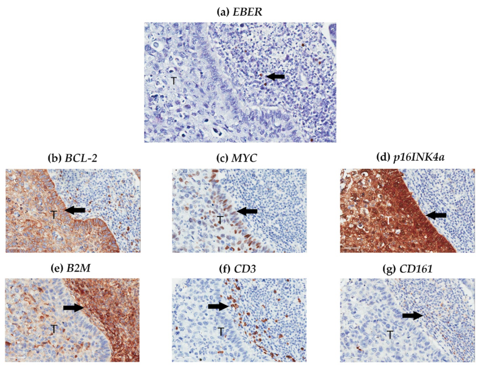
The laryngeal nPI for EBER was relatively low in both case and control groups, and the staining distribution was equal between the groups (Table 2). Most EBER signals were localized to the nuclei of TILs (Figure 2a); however, laryngeal EBV DNA positivity was not related to laryngeal nPI for EBER (r = 0.11, p = 0.34).
Only 30 patients with LSCC had available peripheral blood EBV test data; among these patients, two (7%) and four (13%) had circulating EBV DNA positivity and serum EBV VCA IgA positivity, respectively. Differences in circulating EBV DNA positivity (7% vs. 6%, p > 0.99) and EBV VCA IgA positivity (7% vs. 19%, p = 0.60) between laryngeal EBV DNA-positive and -negative subgroups were not statistically significant. Therefore, the associations between laryngeal EBV DNA positivity, laryngeal nPI for EBER, plasma EBV DNA positivity, and serum EBV VCA IgA positivity were not statistically significant (all p > 0.05).

3.3. Histological Factors of the Larynx

In the IHC analysis, expression of p16INK4A, BCL-2, and MYC occurred mainly in intratumoral cells (Figure 2b–d), whereas B2M, CD3, and CD161 expression occurred mainly in peritumoral (Figure 2e–g) cells. Expression of BCL-2, B2M, CD3, and CD161 was higher, MYC was lower, and p16INK4a was the same in LSCC, compared with NMLL (Table 3).
We further dichotomized these biomarkers using receiver operating characteristic curve analyses, with Youden’s J-point as the best trade-off between sensitivity and specificity, to predict LSCC. Expression of BCL-2 ≥ 68.6% (unadjusted OR = 5.5; 95% CI: 2.0–14.9; p = 0.001), B2M ≥ 84.3% (unadjusted OR = 8.6; 95% CI: 3.1–23.9; p < 0.004), CD3 ≥ 6.9% (unadjusted OR = 7.2; 95% CI: 2.6–20.2; p < 0.001), CD161 ≥ 68.8% (unadjusted OR = 3.5; 95% CI: 1.3–9.4; p = 0.01), MYC ≤ 0.44% (unadjusted OR = 2.9; 95% CI 1.2–7.1; p = 0.02), and p16INK4a ≤ 16.8% (unadjusted OR = 6.0; 95% CI 1.6–22.9; p = 0.01) were significant risk factors for LSCC.

3.4. Relationship between EBV-Related Biomarkers and Clinical and Histological Characteristics in the Overall Cohort

In the overall cohort, primary LSCC was significantly associated with age ≥ 55 years, cigarette smoking, EBV DNA positivity, BCL-2 expression ≥ 68.6%, B2M expression ≥ 84.3%, CD3 expression ≥ 6.9%, CD161 expression ≥ 68.8%, MYC expression ≤ 0.44%, and p16INK4a expression ≤ 16.8% (Table 4).
Additionally, the male sex was significantly associated with cigarette smoking and alcohol consumption. The associations between age ≥ 55 years and MYC expression ≤ 0.44%, B2M expression ≥ 84.3%, and CD3 expression ≥ 6.9% were significant. Furthermore, the relationship between cigarette smoking and alcohol consumption was significant.
EBV DNA positivity was significantly correlated with high BCL-2 expression (≥68.6%), which also significantly correlated with B2M expression ≥ 84.3%, CD3 expression ≥ 6.9%, and CD161 expression ≥ 68.8%. Low p16INK4a expression (≤16.8%) was significantly associated with B2M expression ≥ 84.3% and CD161 expression ≥ 68.8%. B2M expression ≥ 84.3% was significantly associated with CD3 expression ≥ 6.9%, which also significantly associated with CD161 expression ≥ 68.8%.

3.5. Multi-Factor Modeling of Primary LSCC

To improve the precision of the model, we used a “forced simultaneous entry” approach and a “sign-correct” method for multivariate logistic regression analysis [36]. Variables with a p < 0.05 in the univariate analysis were entered into the regression analysis. Age ≥ 55 years (OR = 19.3; 95% CI: 3.2–116.0; p = 0.001), cigarette smoking (OR = 36.4; 95% CI: 2.4–554.2; p = 0.01), EBV DNA positivity (OR = 39.7; 95% CI: 3.3–478.0; p = 0.004), BCL-2 expression ≥ 68.6% (OR = 6.8; 95% CI: 1.2–38.6; p = 0.03), B2M expression ≥ 84.3% (OR = 18.1; 95% CI: 2.9–11.6; p = 0.002), and CD161 expression ≥ 68.8% (OR = 7.5; 95% CI: 1.1–50.7; p = 0.04) were significant independent risk factors for primary LSCC.
Using this six-factor model, we accurately predicted which patients with laryngeal lesions had primary LSCC via the optimal cut-off value of 3 (area under the receiver operating characteristic curve: 0.87 [95% CI: 0.79–0.96]; p < 0.001), with a sensitivity, specificity, positive predictive value, and negative predictive value of 98%, 77%, 82%, and 97%, respectively.

3.6. Five Year Prognoses in Patients with LSCC

As of 28 February 2021, the median follow-up time was 68 months (range: 9–94 months). A total of 14 case patients had an LR within the first 5 years after definitive treatment; thus, the five-year LR rate was 34% (95% CI: 16–53%). Two patients had NR (five-year NR rate = 6%; 95% CI: 0–50%) and one had DM (five-year DM rate = 3%; 95% CI: 0–59%); therefore, five-year DFS was 58% (95% CI: 41–71%). Furthermore, two patients had disease-specific deaths (five-year DSS rate = 94%; 95% CI: 75–98%), while another two died of other causes (five-year OS rate = 89%; 95% CI: 72–96%).
Herein, we further focused on investigating the risk factors associated with five-year LR and DFS rates (Table 5). EBV DNA positivity did not significantly predict five-year LR and DFS rates. However, an EBER signal ≥ 0.04% significantly predicted both the five-year LR (unadjusted HR = 6.2; 95% CI: 2.0–18.6; p = 0.001) and DFS (unadjusted HR = 7.1; 95% CI: 2.6–19.5; p < 0.001) rates using univariate Cox regression models. Furthermore, age ≤ 63 years, TLM, and CD3 expression ≤ 4.9% also predicted the five-year LR rate, whereas TLM, BCL-2 expression ≤ 96.0%, CD3 expression ≤ 4.9%, and EBER signals ≥ 0.04% predicted five-year DFS rates.
Using multivariate Cox regression models, both EBER signals ≥ 0.04% (adjusted HR: 6.0; 95% CI: 1.9–18.6; p = 0.002) and CD3 expression ≤ 4.9% (adjusted HR = 6.9; 95% CI: 1.9–24.6; p = 0.003) independently predicted five-year LR rates; both risk factors significantly predicted five-year LR rates after adjustment for treatment modality (adjusted HR = 4.7 (95% CI: 1.5–15.0) and 5.4 (95% CI: 1.5–19.4), respectively; p = 0.01, and 0.01, respectively). Similarly, both EBER signals ≥ 0.04% (adjusted HR = 8.6; 95% CI: 2.9–25.3; p < 0.001) and CD3 expression ≤ 4.9% (adjusted HR = 6.6; 95% CI: 1.9–23.6; p = 0.004) independently predicted five-year DFS rates; these risk factors remained significant after adjustment for treatment modality (adjusted HR = 7.7 (95% CI: 2.5–24.0) and 5.4 (95% CI: 1.5–19.3), respectively; p < 0.001 and 0.01, respectively).

3.7. Relationship between EBV-Related Biomarkers and Clinical and Histological Characteristics in Patients with LSCC

In patients with primary LSCC, male sex was significantly associated with cigarette smoking and laryngeal EBV DNA positivity (Table 6). Furthermore, an age ≤ 63 years was associated with alcohol consumption and p16INK4a ≤ 81.6%, while cigarette smoking was related to alcohol consumption. As expected, T- and N-stages were associated with LSCC stage. EBV DNA positivity and EBER signal ≥ 0.04% were not associated with other tissue biomarkers. BCL-2 expression ≤ 96.0% correlated significantly with p16INK4a and CD161 expression ≤ 81.6% and ≤ 69.9%, respectively. In addition, p16INK4a expression ≤ 81.6% was associated with CD3 expression ≤ 4.9%.
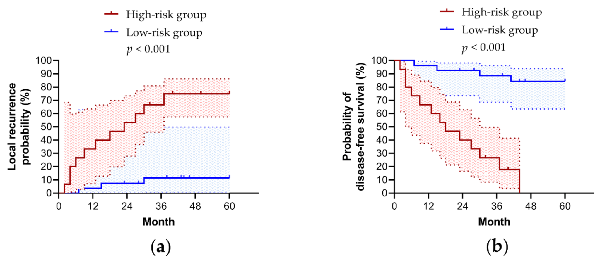
The overall cohort was further dichotomized into a high-risk group (≥1 risk factor) and a low-risk group (<1 risk factor) to predict five-year LR (sensitivity, 79%; specificity, 94%; positive predictive value, 88%; negative predictive value, 90%). This method performed well in predicting five-year DFS (sensitivity, 76%; specificity, 100%; positive predictive value, 100%; negative predictive value, 52%). The five-year LR rate of the high-risk group was significantly higher than that of the low-risk group (75% (95% CI, 57–86%) vs. 11% (95% CI, 0–49%); p < 0.001) (Figure 3a). As expected, the five-year DFS rate of the high-risk group was significantly higher than that of the low-risk group (0% vs. 84% (95% CI, 63–84%); p < 0.001) (Figure 3b). However, this model predicted the five-year DSS (83% (95% CI, 47–96%) vs. 100%; p = 0.05) and OS (76% (95% CI, 43–92%) vs. 96% (95% CI, 72–99%); p = 0.08) rates less efficiently due to relatively low numbers of disease-specific and overall deaths.

4. Discussion

In this study, we found that patients with primary LSCC had a higher prevalence of EBV infection, compared to control patients. In addition to age ≥ 55 years and cigarette smoking [37], EBV DNA positivity, BCL-2 expression ≥ 68.6%, B2M expression ≥ 84.3%, and CD161 expression ≥ 68.8% were independently associated with LSCC. Using this novel model, we accurately predicted the development of LSCC. Moreover, EBER signals ≥ 0.04% and CD3 expression ≤ 4.9% independently predicted five-year LR and DFS rates with or without adjustment for treatment modality. Using this innovative two-factor model, we could also accurately predict the five-year LR and DFS rates of LSCC.
Chronic viral infection is a risk factor for many cancers, such as hepatitis B virus/hepatocellular carcinoma, human papillomavirus/cervical cancer, and EBV/NPC. Quantifying EBV-related biomarker levels in tumor specimens could improve our understanding of the etiology of some laryngeal cancers, and assist in patient selection for treatment and follow-up protocols [38]. However, quantification in FFPE tissue is difficult due to ambiguous uncertainties and the absence of reliable detection methods. Determining the interactions between EBV-related biomarkers and host factors is also challenging. For translation purposes, we developed and validated risk and prognostic models based on EBV-related biomarkers in this case-control study of Taiwanese patients with primary LSCC.
Similar to other studies [8,9,11], we found that EBV infection was significantly more frequent in patients with primary LSCC than in those with NMLL. However, we found that most EBERs were detected in the TILs of LSCC. The absence of latent EBV infection in the tumor tissue of LSCC suggests that its multistep nature of development may include “hit-and-run” carcinogenesis in the laryngeal epithelium. Notably, EBV DNA positivity was positively correlated with BCL-2 expression, an anti-apoptotic protein in tumor cells. Wp, a viral promoter located within BamHI W repeats of the EBV genome, activates the gene encoding the viral BCL-2 homolog BHRF1, thereby increasing apoptosis resistance [39]. Moreover, oncogenic EBV latent membrane protein 1 directly upregulates BCL-2 in NPC [40], potentially also contributing to LSCC [11].
Furthermore, we found that BCL-2 overexpression in LSCC was common (80%). BCL-2 interacts with Hsp90β, and may be involved in the anti-apoptotic progression of LSCC [41]. BCL-2 overexpression appears to be associated with a complete response to induction chemotherapy [16] and worse prognoses due to tumoral radioresistance [42]. Although BCL-2 overexpression might not contribute to the prognostic significance of LSCC development [43], we found that both EBV DNA positivity and BCL-2 overexpression in the larynx were independently associated with a risk of LSCC. Our results also indicate that BCL-2 expression is not related to tumor aggressiveness and prognosis after a single surgical treatment [42].
Regarding therapeutic prognoses, we found that EBER signals ≥ 0.04% and CD3 expression ≤ 4.9% were important prognostic factors for five-year LR and DFS in patients with LSCC. In our literature review, only a few patients with LSCC had positive EBER signals in the larynx [39,44]. Although positive EBER signals in LSCC tissues are considered poorer predictors of head and neck squamous cell carcinoma (HNSCC) [39], a small LSCC sample size (two and six cases of positive and negative EBER signals, respectively) is difficult to draw conclusions from. Therefore, this study was the first to identify a high laryngeal EBER signal as a poorer prognostic factor in LSCC. Since most EBER signals presented the peritumor microenvironment in our patients, we presumed that EBERs could induce the initial transformation of epithelial cells [45], and trigger cancer-related inflammation via the RIG-I pathway to promote tumor development and growth [46]; rather than downregulate B2M expression to evade T cell-mediated cytotoxic immune responses [47], as according to previous NPC studies. Moreover, EBV-infected TILs of residual laryngeal tissue following curative treatment may increase the five-year LR risk via BHRF1-mediated apoptosis resistance [48]. Accordingly, mechanistic studies are warranted to confirm that EBV involves the EBER-mediated process of LSCC recurrence.
Circulating EBV DNA is a robust biomarker for EBV-associated NPC [49] and lymphoma [50]. EBV VCA IgA positivity indicates previous repeated EBV infections or frequent reactivation of latent EBV in B cells [51]. Patients with LSCC might have a higher positive rate of EBV VCA IgA than healthy controls [52]. Therefore, both circulating EBV DNA and VCA IgA could be potential biomarkers for EBV-positive LSCC. In this study, neither circulating EBV DNA positivity nor serum EBV VCA IgA positivity was associated with laryngeal EBV DNA positivity and EBER nPI. Furthermore, the low positive rates of these peripheral blood biomarkers may limit their application in patients with LSCC.
Laryngeal cancer usually develops over many years, and approximately 80% of patients with LSCC are 60 years or older at first discovery [53]. In this study, an age ≥ 55 years was associated with B2M expression ≥ 84.3%, and both were independent risk factors for LSCC development. Recently, B2M was validated as a key reference gene in laryngeal and hypopharyngeal cancers, highlighting its suitability for investigating target gene expression [54]. Interestingly, its expression in patients with EBV-positive cancer was significantly higher than that in patients with EBV-negative cancer [55]. The B2M protein forms the light chain of the class I major histocompatibility complex, and is important for antigen recognition by cytotoxic T cells [56]. Overexpression of B2M is found in solid tumors and blood-borne malignancies, and is associated with advanced disease and poor prognosis [57]. Although B2M expression was not associated with LSCC stage and prognosis, it was an independent risk factor for LSCC development.
Furthermore, cigarette smoking is strongly associated with an increased risk of LSCC in a dose-response and time-response manner [58]. Although significant smoking trend reductions have led to the declined global burden of LSCC in developed countries, smoking continues to trend upward in low socioeconomic countries, potentially inducing an increased burden of LSCC in the future [59]. Notably, smoking prevention programs conducted in health care settings and interventions, including enhancement of interpersonal communication and support strategies, can be effective in preventing smoking behavior for three months to four years in children and adolescents [60]. However, the latest meta-analysis indicated that findings which suggest that reduction-to-quit is more effective than no treatments were inconclusive and of low certainty [61]. Nevertheless, the risk of developing LSCC can be reduced by smoking cessation for ≥ 15 years [58].
CD161 is expressed on natural killer (NK) cells and subpopulations of T lymphocytes, such as invariant natural killer T cells (iNKT), CD4+CD161+ T cells, and CD8+CD161+ T cells [62]. These cells may play significant roles in tumor development [63], and may regulate the immune response in the tumor microenvironment [64]. However, the effect of CD161 expression on tumor development remains unclear. Patients with LSCC had a significantly lower percentage of circulating iNKT cells, compared to healthy controls [65], whereas circulating CD4+CD161+ T cells, representing a memory T cell population, significantly increased in cancer patients [66]. Notably, CD8+CD161+ T cell-specific EBV infections might express high levels of anti-apoptotic molecules to survive hostile inflammatory conditions, also involving the pathogenesis of tumor tissues [67]. Furthermore, we found that CD161 overexpression in LSCC was not uncommon, and correlated with BCL-2 overexpression and low p16INK4a expression. However, both CD161 and BCL-2 overexpression were independent risk factors for LSCC development.
In patients with LSCC, low CD3 expression was associated with a poor prognosis. Similarly, a high infiltration of CD3+ T lymphocytes was associated with a significantly better prognosis in patients with HNSCC [68]. In patients with HNSCC, low CD3 mRNA levels have been found to be worse predictors of 8-year DFS [69]. Therefore, dysfunction of the immune response in removing the oncogenic EBV infection may increase the risk of cancer progression. Although we did not find a significant association between CD3 expression and EBV infection in the larynx, patients with LSCC and EBV infection may have a lower proportion of late activated T lymphocytes in the peripheral blood compared to patients without EBV infection [70]. Therefore, investigating peripheral blood T lymphocytes may help us to comprehensively understand the possible mechanisms of LR and relapse in EBV-related LSCC.
Our study had some limitations. First, when considering high EBV DNA positivity, false positivity is a real risk; PCR, which was used for EBV DNA detection, is extremely sensitive, and the EBER signal was minimal. However, we applied strict anti-contamination measures in the laboratory to reduce the risk of contamination [27]. Second, in some patients, insufficient tissue and low-quality biomarkers limited the sample size; despite the use of TMAs to reduce the lesion volume required for comprehensive comparisons. Nevertheless, larger sample sizes may provide only modest benefits in complex statistical analyses.

5. Conclusions

Taken together, our study is the most comprehensive analysis to date of the clinical and histological manifestations, and etiology of primary LSCCs and five-year prognoses following definitive treatment. We identified EBV DNA positivity, older age, cigarette smoking, and higher expression of BCL-2, B2M, and CD161 as risk factors for primary LSCC. Furthermore, we identified high EBER signals and low CD3 expression as independent predictors of five-year LR and DFS. Early prediction will enable the development of better treatments and follow-up strategies. Furthermore, as understanding the etiology of LSCC and reducing its LR rate is paramount, our data provide significant information that may assist in the accomplishment of these goals.

Author Contributions

Conceptualization: L.-A.L., T.-J.F., H.-Y.L., T.-C.C., C.-G.H., and T.-C.Y.; methodology: L.-A.L., K.-P.C., T.-C.C., and C.-G.H.; software: C.-J.K., C.-T.L., T.-C.C., and C.-G.H.; validation: L.-A.L., K.-P.C., T.-C.C., C.-G.H., and T.-C.Y.; formal analysis: L.-A.L., H.-H.C., and T.-C.Y.; investigation: L.-A.L., T.-J.F., H.-Y.L., C.-J.K., K.-P.C., and C.-T.L.; resources: L.-A.L., C.-G.H., and T.-C.Y.; data curation: C.-T.L. and T.-C.Y.; writing—original draft preparation: L.-A.L., T.-J.F., H.-H.C., K.-P.C., C.-G.H., and T.-C.Y.; writing—review and editing: H.-Y.L., C.-J.K., C.-T.L., and T.-C.C.; visualization: L.-A.L. and H.-H.C.; supervision: H.-Y.L., C.-T.L., T.-C.C., and T.-C.Y.; project administration: C.-J.K. and C.-G.H.; funding acquisition: L.-A.L. All authors have read and agreed to the published version of the manuscript.

Funding

This research was funded by the Chang Gung Medical Foundation, Taoyuan, Taiwan (grant numbers: 3C0661, 3C0662, 3C0663, and 3F1092). The APC was funded by the Chang Gung Medical Foundation, Taoyuan, Taiwan.

Institutional Review Board Statement

The study was conducted according to the guidelines of the Declaration of Helsinki and approved by the Institutional Review Board of the Chang Gung Medical Foundation, Taoyuan, Taiwan (protocol code 202100292B0 and date of approval 12 March 2021).

Informed Consent Statement

Patient consent was waived due to a retrospective medical review.

Data Availability Statement

The data presented in this study are available upon request from the corresponding author. The data are not publicly available due to ethical restrictions.

Acknowledgments

The authors would like to thank Ruo-Chi Wang and Chung-Fang Hsiao (Department of Otorhinolaryngology, Head and Neck Surgery, Linkou Chang Gung Memorial Hospital, Taoyuan City, Taiwan, R.O.C.) for their technical assistance.

Conflicts of Interest

The authors declare no conflict of interest.

References

  1. Tai, P.; Yu, E.; Shiels, R.; Tonita, J. Long-term survival rates of laryngeal cancer patients treated by radiation and surgery, radiation alone, and surgery alone: Studied by lognormal and Kaplan-Meier survival methods. BMC Cancer 2005, 5, 13. [Google Scholar] [CrossRef] [PubMed]
  2. Ferlay, J.; Soerjomataram, I.; Dikshit, R.; Eser, S.; Mathers, C.; Rebelo, M.; Parkin, D.M.; Forman, D.; Bray, F. Cancer incidence and mortality worldwide: Sources, methods and major patterns in GLOBOCAN 2012. Int. J. Cancer 2015, 136, E359–E386. [Google Scholar] [CrossRef] [PubMed]
  3. Chang, J.; Fang, T.J.; Yung, K.; van Zante, A.; Miller, T.; Al-Jurf, S.; Schneider, S.; Courey, M. Clinical and histologic predictors of voice and disease outcome in patients with early glottic cancer. Laryngoscope 2012, 122, 2240–2247. [Google Scholar] [CrossRef] [PubMed]
  4. Bouvard, V.; Baan, R.; Straif, K.; Grosse, Y.; Secretan, B.; El Ghissassi, F.; Benbrahim-Tallaa, L.; Guha, N.; Freeman, C.; Galichet, L.; et al. A review of human carcinogens—Part B: Biological agents. Lancet Oncol. 2009, 10, 321–322. [Google Scholar] [CrossRef]
  5. de Oliveira, D.E.; Bacchi, M.M.; Macarenco, R.S.; Tagliarini, J.V.; Cordeiro, R.C.; Bacchi, C.E. Human papillomavirus and Epstein-Barr virus infection, p53 expression, and cellular proliferation in laryngeal carcinoma. Am. J. Clin. Pathol. 2006, 126, 284–293. [Google Scholar] [CrossRef]
  6. Ndiaye, C.; Mena, M.; Alemany, L.; Arbyn, M.; Castellsagué, X.; Laporte, L.; Bosch, F.X.; de Sanjosé, S.; Trottier, H. HPV DNA, E6/E7 mRNA, and p16INK4a detection in head and neck cancers: A systematic review and meta-analysis. Lancet Oncol. 2014, 15, 1319–1331. [Google Scholar] [CrossRef]
  7. Huang, C.G.; Lee, L.A.; Fang, T.J.; Li, H.Y.; Liao, C.T.; Kang, C.J.; Yen, T.C.; Tsao, K.C.; Chen, T.C.; Shih, S.R. Human papillomavirus infection is not associated with laryngeal squamous cell carcinoma in Taiwan. J. Microbiol. Immunol. Infect 2020, 53, 79–86. [Google Scholar] [CrossRef]
  8. Jeng, K.C.; Hsu, C.Y.; Liu, M.T.; Chung, T.T.; Liu, S.T. Prevalence of Taiwan variant of Epstein-Barr virus in throat washings from patients with head and neck tumors in Taiwan. J. Clin. Microbiol. 1994, 32, 28–31. [Google Scholar] [CrossRef] [Green Version]
  9. Zheng, Y.; Xia, P.; Zheng, H.C.; Takahashi, H.; Masuda, S.; Takano, Y. The screening of viral risk factors in tongue and pharyngolaryngeal squamous carcinoma. Anticancer. Res. 2010, 30, 1233–1238. [Google Scholar]
  10. Brichacek, B.; Hirsch, I.; Sibl, O.; Vilikusova, E.; Vonka, V. Association of some supraglottic laryngeal carcinomas with EB virus. Int. J. Cancer 1983, 32, 193–197. [Google Scholar] [CrossRef]
  11. Foltyn, S.; Strycharz-Dudziak, M.; Drop, B.; Boguszewska, A.; Polz-Dacewicz, M. Serum EBV antibodies and LMP-1 in Polish patients with oropharyngeal and laryngeal cancer. Infect Agent. Cancer 2017, 12, 31. [Google Scholar] [CrossRef]
  12. Callaghan, D.J.; Conner, B.R.; Strauss, M. Epstein-Barr virus antibody titers in cancer of the head and neck. Arch. Otolaryngol. 1983, 109, 781–784. [Google Scholar] [CrossRef]
  13. Yang, H.J.; Cho, Y.J.; Kim, H.S.; Chang, M.S.; Sung, M.W.; Kim, W.H. Association of p53 and BCL-2 expression with Epstein-Barr virus infection in the cancers of head and neck. Head Neck 2001, 23, 629–636. [Google Scholar] [CrossRef]
  14. Muderris, T.; Rota, S.; Muderris, T.; Inal, E.; Fidan, I. Does Epstein-Barr virus infection have an influence on the development of laryngeal carcinoma? Detection of EBV by real-time polymerase chain reaction in tumour tissues of patients with laryngeal carcinoma. Braz. J. Otorhinolaryngol. 2013, 79, 418–423. [Google Scholar] [CrossRef] [Green Version]
  15. Huang, S.C.M.; Tsao, S.W.; Tsang, C.M. Interplay of viral infection, host cell factors and tumor microenvironment in the pathogenesis of nasopharyngeal carcinoma. Cancers 2018, 10, 106. [Google Scholar] [CrossRef] [Green Version]
  16. Trask, D.K.; Wolf, G.T.; Bradford, C.R.; Fisher, S.G.; Devaney, K.; Johnson, M.; Singleton, T.; Wicha, M. Expression of Bcl-2 family proteins in advanced laryngeal squamous cell carcinoma: Correlation with response to chemotherapy and organ preservation. Laryngoscope 2002, 112, 638–644. [Google Scholar] [CrossRef]
  17. Li, J.; Huang, X.; Xie, X.; Wang, J.; Duan, M. Human telomerase reverse transcriptase regulates cyclin D1 and G1/S phase transition in laryngeal squamous carcinoma. Acta Otolaryngol. 2011, 131, 546–551. [Google Scholar] [CrossRef]
  18. Feenstra, M.; Bakema, J.; Verdaasdonk, M.; Rozemuller, E.; van den Tweel, J.; Slootweg, P.; de Weger, R.; Tilanus, M. Detection of a putative HLA-A*31012 processed (intronless) pseudogene in a laryngeal squamous cell carcinoma. Genes Chromosomes Cancer 2000, 27, 26–34. [Google Scholar] [CrossRef]
  19. Lagresle, C.; Gardie, B.; Eyquem, S.; Fasseu, M.; Vieville, J.C.; Pla, M.; Sigaux, F.; Bories, J.C. Transgenic expression of the p16INK4a cyclin-dependent kinase inhibitor leads to enhanced apoptosis and differentiation arrest of CD4CD8 immature thymocytes. J. Immunol. 2002, 168, 2325–2331. [Google Scholar] [CrossRef] [Green Version]
  20. Kesselring, R.; Thiel, A.; Pries, R.; Wollenberg, B. The number of CD161 positive Th17 cells are decreased in head and neck cancer patients. Cell Immunol. 2011, 269, 74–77. [Google Scholar] [CrossRef]
  21. McShane, L.M.; Altman, D.G.; Sauerbrei, W.; Taube, S.E.; Gion, M.; Clark, G.M.; Statistics Subcommittee of the NCI-EORTC Working Group on Cancer Diagnostics. Reporting recommendations for tumor marker prognostic studies. J. Clin. Oncol. 2005, 23, 9067–9072. [Google Scholar] [CrossRef] [Green Version]
  22. Edge, S.B.; Compton, C.C. The American Joint Committee on Cancer: The 7th edition of the AJCC cancer staging manual and the future of TNM. Ann. Surg. Oncol. 2010, 17, 1471–1474. [Google Scholar] [CrossRef]
  23. Fang, T.J.; Courey, M.S.; Liao, C.T.; Yen, T.C.; Li, H.Y. Frozen margin analysis as a prognosis predictor in early glottic cancer by laser cordectomy. Laryngoscope 2013, 123, 1490–1495. [Google Scholar] [CrossRef]
  24. Hsin, L.J.; Fang, T.J.; Tsang, N.M.; Chin, S.C.; Yen, T.C.; Li, H.Y.; Liao, C.T.; Chen, I.H. Tumor volumetry as a prognostic factor in the management of T4a laryngeal cancer. Laryngoscope 2014, 124, 1134–1140. [Google Scholar] [CrossRef]
  25. Lee, L.A.; Fang, T.J.; Li, H.Y.; Huang, C.G.; Chen, T.C.; Liao, C.T.; Kang, C.J.; Chang, K.P.; Yen, T.C. Low expression of pRB predicts disease relapse in early glottic cancer treated with transoral laser microsurgery. Laryngoscope 2018, 129, E220–E226. [Google Scholar] [CrossRef]
  26. Fountzilas, E.; Kotoula, V.; Angouridakis, N.; Karasmanis, I.; Wirtz, R.M.; Eleftheraki, A.G.; Veltrup, E.; Markou, K.; Nikolaou, A.; Pectasides, D.; et al. Identification and validation of a multigene predictor of recurrence in primary laryngeal cancer. PLoS ONE 2013, 8, e70429. [Google Scholar] [CrossRef]
  27. Huang, C.G.; Lee, L.A.; Tsao, K.C.; Liao, C.T.; Yang, L.Y.; Kang, C.J.; Chang, K.P.; Huang, S.F.; Chen, I.H.; Yang, S.L.; et al. Human papillomavirus 16/18 E7 viral loads predict distant metastasis in oral cavity squamous cell carcinoma. J. Clin. Virol. 2014, 61, 230–236. [Google Scholar] [CrossRef]
  28. Hsu, C.L.; Chang, K.P.; Lin, C.Y.; Chang, H.K.; Wang, C.H.; Lin, T.L.; Liao, C.T.; Tsang, N.M.; Lee, L.Y.; Chan, S.C.; et al. Plasma Epstein-Barr virus DNA concentration and clearance rate as novel prognostic factors for metastatic nasopharyngeal carcinoma. Head Neck 2012, 34, 1064–1070. [Google Scholar] [CrossRef]
  29. Lo, Y.M.; Chan, L.Y.; Lo, K.W.; Leung, S.F.; Zhang, J.; Chan, A.T.; Lee, J.C.; Hjelm, N.M.; Johnson, P.J.; Huang, D.P. Quantitative analysis of cell-free Epstein-Barr virus DNA in plasma of patients with nasopharyngeal carcinoma. Cancer Res. 1999, 59, 1188–1191. [Google Scholar]
  30. Hsu, C.L.; Chan, S.C.; Chang, K.P.; Lin, T.L.; Lin, C.Y.; Hsieh, C.H.; Huang, S.F.; Tsang, N.M.; Lee, L.Y.; Ng, S.H.; et al. Clinical scenario of EBV DNA follow-up in patients of treated localized nasopharyngeal carcinoma. Oral Oncol. 2013, 49, 620–625. [Google Scholar] [CrossRef]
  31. Chang, K.P.; Hsu, C.L.; Chang, Y.L.; Tsang, N.M.; Chen, C.K.; Lee, T.J.; Tsao, K.C.; Huang, C.G.; Chang, Y.S.; Yu, J.S.; et al. Complementary serum test of antibodies to Epstein-Barr virus nuclear antigen-1 and early antigen: A possible alternative for primary screening of nasopharyngeal carcinoma. Oral Oncol. 2008, 44, 784–792. [Google Scholar] [CrossRef] [PubMed]
  32. Cho, Y.J.; Chang, M.S.; Park, S.H.; Kim, H.S.; Kim, W.H. In situ hybridization of Epstein-Barr virus in tumor cells and tumor-infiltrating lymphocytes of the gastrointestinal tract. Hum. Pathol. 2001, 32, 297–301. [Google Scholar] [CrossRef] [PubMed]
  33. Braun, M.; Kirsten, R.; Rupp, N.J.; Moch, H.; Fend, F.; Wernert, N.; Kristiansen, G.; Perner, S. Quantification of protein expression in cells and cellular subcompartments on immunohistochemical sections using a computer supported image analysis system. Histol. Histopathol. 2013, 28, 605–610. [Google Scholar] [CrossRef] [PubMed]
  34. Youden, W.J. Index for rating diagnostic tests. Cancer 1950, 3, 32–35. [Google Scholar] [CrossRef]
  35. Heagerty, P.J.; Lumley, T.; Pepe, M.S. Time-dependent ROC curves for censored survival data and a diagnostic marker. Biometrics 2000, 56, 337–344. [Google Scholar] [CrossRef] [PubMed]
  36. Steyerberg, E.W.; Eijkemans, M.J.; Harrell, F.E., Jr.; Habbema, J.D. Prognostic modeling with logistic regression analysis: In search of a sensible strategy in small data sets. Med. Decis. Mak. 2001, 21, 45–56. [Google Scholar] [CrossRef]
  37. Maier, H.; Gewelke, U.; Dietz, A.; Heller, W.D. Risk factors of cancer of the larynx: Results of the Heidelberg case-control study. Otolaryngol. Head Neck Surg. 1992, 107, 577–582. [Google Scholar] [CrossRef]
  38. Turunen, A.; Rautava, J.; Grenman, R.; Syrjanen, K.; Syrjanen, S. Epstein-Barr virus (EBV)-encoded small RNAs (EBERs) associated with poor prognosis of head and neck carcinomas. Oncotarget 2017, 8, 27328–27338. [Google Scholar] [CrossRef] [Green Version]
  39. Kelly, G.L.; Long, H.M.; Stylianou, J.; Thomas, W.A.; Leese, A.; Bell, A.I.; Bornkamm, G.W.; Mautner, J.; Rickinson, A.B.; Rowe, M. An Epstein-Barr virus anti-apoptotic protein constitutively expressed in transformed cells and implicated in burkitt lymphomagenesis: The Wp/BHRF1 link. PLoS Pathog. 2009, 5, e1000341. [Google Scholar] [CrossRef]
  40. Young, L.S.; Rickinson, A.B. Epstein-Barr virus: 40 years on. Nat. Rev. Cancer 2004, 4, 757–768. [Google Scholar] [CrossRef]
  41. Li, S.; Li, J.; Hu, T.; Zhang, C.; Lv, X.; He, S.; Yan, H.; Tan, Y.; Wen, M.; Lei, M.; et al. Bcl-2 overexpression contributes to laryngeal carcinoma cell survival by forming a complex with Hsp90β. Oncol. Rep. 2017, 37, 849–856. [Google Scholar] [CrossRef] [Green Version]
  42. Gioacchini, F.M.; Alicandri-Ciufelli, M.; Rubini, C.; Magliulo, G.; Re, M. Prognostic value of Bcl-2 expression in squamous cell carcinoma of the larynx: A systematic review. Int. J. Biol. Markers 2015, 30, e155–e160. [Google Scholar] [CrossRef]
  43. Douglas, C.M.; Bernstein, J.M.; Ormston, V.E.; Hall, R.C.; Merve, A.; Swindell, R.; Valentine, H.R.; Slevin, N.J.; West, C.M.; Homer, J.J. Lack of prognostic effect of carbonic anhydrase-9, hypoxia inducible factor-1alpha and bcl-2 in 286 patients with early squamous cell carcinoma of the glottic larynx treated with radiotherapy. Clin. Oncol. 2013, 25, 59–65. [Google Scholar] [CrossRef]
  44. McLemore, M.S.; Haigentz, M., Jr.; Smith, R.V.; Nuovo, G.J.; Alos, L.; Cardesa, A.; Brandwein-Gensler, M. Head and neck squamous cell carcinomas in HIV-positive patients: A preliminary investigation of viral associations. Head Neck Pathol. 2010, 4, 97–105. [Google Scholar] [CrossRef] [Green Version]
  45. Yoshizaki, T.; Endo, K.; Ren, Q.; Wakisaka, N.; Murono, S.; Kondo, S.; Sato, H.; Furukawa, M. Oncogenic role of Epstein-Barr virus-encoded small RNAs (EBERs) in nasopharyngeal carcinoma. Auris Nasus Larynx 2007, 34, 73–78. [Google Scholar] [CrossRef]
  46. Duan, Y.; Li, Z.; Cheng, S.; Chen, Y.; Zhang, L.; He, J.; Liao, Q.; Yang, L.; Gong, Z.; Sun, L.Q. Nasopharyngeal carcinoma progression is mediated by EBER-triggered inflammation via the RIG-I pathway. Cancer Lett. 2015, 361, 67–74. [Google Scholar] [CrossRef]
  47. Kouvidou, C.; Rontogianni, D.; Tzardi, M.; Datseris, G.; Panayiotides, I.; Darivianaki, K.; Karidi, E.; Delides, G.; Kanavaros, P. Beta 2-microglobulin and HLA-DR expression in relation to the presence of Epstein-Barr virus in nasopharyngeal carcinomas. Pathobiology 1995, 63, 320–327. [Google Scholar] [CrossRef]
  48. Fitzsimmons, L.; Cartlidge, R.; Chang, C.; Sejic, N.; Galbraith, L.C.A.; Suraweera, C.D.; Croom-Carter, D.; Dewson, G.; Tierney, R.J.; Bell, A.I.; et al. EBV BCL-2 homologue BHRF1 drives chemoresistance and lymphomagenesis by inhibiting multiple cellular pro-apoptotic proteins. Cell Death Differ. 2020, 27, 1554–1568. [Google Scholar] [CrossRef]
  49. Lam, W.K.J.; Chan, K.C.A.; Lo, Y.M.D. Plasma Epstein-Barr virus DNA as an archetypal circulating tumour DNA marker. J. Pathol. 2019, 247, 641–649. [Google Scholar] [CrossRef] [Green Version]
  50. Yalcin, B.; Kutluk, T.; Agbaba, S.K.; Demir, C.; Talim, B. Circulating Epstein-Barr virus DNA and cell-free DNA in pediatric lymphomas. Turk. J. Pediatr. 2020, 62, 541–550. [Google Scholar] [CrossRef]
  51. Lo, S.; Ho, W.K.; Wei, W.I. Outcome of patients with positive Epstein-Barr virus serologic status in the absence of nasopharyngeal carcinoma in Hong Kong. Arch. Otolaryngol. Head Neck Surg. 2004, 130, 770–772. [Google Scholar] [CrossRef] [Green Version]
  52. Morshed, K.; Polz-Dacewicz, M.; Szymanski, M.; Rajtar, B.; Ziaja-Soltys, M.; Golabek, W. Epstein-Barr virus antibodies in patients with laryngeal and hypopharyngeal cancer. Ann. Univ. Mariae Curie Sklodowska Med. 2003, 58, 227–231. [Google Scholar]
  53. Halmos, G.B.; Bras, L.; Siesling, S.; van der Laan, B.; Langendijk, J.A.; van Dijk, B.A.C. Age-specific incidence and treatment patterns of head and neck cancer in the Netherlands-A cohort study. Clin. Otolaryngol. 2018, 43, 317–324. [Google Scholar] [CrossRef]
  54. Yin, W.Z.; Yang, Q.W.; Niu, K.; Ren, M.; He, D.; Song, W.Z. Validation of reference genes for the normalization of RT-qPCR expression studies on human laryngeal cancer and hypopharyngeal cancer. Eur. Rev. Med. Pharmacol. Sci. 2019, 23, 4199–4209. [Google Scholar] [CrossRef]
  55. Keane, C.; Tobin, J.; Gunawardana, J.; Francis, S.; Gifford, G.; Gabrielli, S.; Gill, A.; Stevenson, W.; Talaulikar, D.; Gould, C.; et al. The tumour microenvironment is immuno-tolerogenic and a principal determinant of patient outcome in EBV-positive diffuse large B-cell lymphoma. Eur. J. Haematol. 2019, 103, 200–207. [Google Scholar] [CrossRef] [Green Version]
  56. Rammensee, H.G.; Robinson, P.J.; Crisanti, A.; Bevan, M.J. Restricted recognition of beta 2-microglobulin by cytotoxic T lymphocytes. Nature 1986, 319, 502–504. [Google Scholar] [CrossRef]
  57. Liu, C.; Yang, Z.; Li, D.; Liu, Z.; Miao, X.; Yang, L.; Zou, Q.; Yuan, Y. Overexpression of B2M and loss of ALK7 expression are associated with invasion, metastasis, and poor-prognosis of the pancreatic ductal adenocarcinoma. Cancer Biomark. 2015, 15, 735–743. [Google Scholar] [CrossRef]
  58. Zuo, J.J.; Tao, Z.Z.; Chen, C.; Hu, Z.W.; Xu, Y.X.; Zheng, A.Y.; Guo, Y. Characteristics of cigarette smoking without alcohol consumption and laryngeal cancer: Overall and time-risk relation. A meta-analysis of observational studies. Eur. Arch. Otorhinolaryngol. 2017, 274, 1617–1631. [Google Scholar] [CrossRef]
  59. Ramsey, T.; Guo, E.; Svider, P.F.; Lin, H.; Syeda, S.; Raza, S.N.; Fribley, A.M. Laryngeal cancer: Global socioeconomic trends in disease burden and smoking habits. Laryngoscope 2018, 128, 2039–2053. [Google Scholar] [CrossRef]
  60. Duncan, L.R.; Pearson, E.S.; Maddison, R. Smoking prevention in children and adolescents: A systematic review of individualized interventions. Patient Educ. Couns. 2018, 101, 375–388. [Google Scholar] [CrossRef]
  61. Lindson, N.; Klemperer, E.; Hong, B.; Ordonez-Mena, J.M.; Aveyard, P. Smoking reduction interventions for smoking cessation. Cochrane Database Syst. Rev. 2019, 9, CD013183. [Google Scholar] [CrossRef] [PubMed]
  62. Konduri, V.; Oyewole-Said, D.; Vazquez-Perez, J.; Weldon, S.A.; Halpert, M.M.; Levitt, J.M.; Decker, W.K. CD8(+)CD161(+) T-Cells: Cytotoxic me0mory cells with high therapeutic potential. Front. Immunol. 2020, 11, 613204. [Google Scholar] [CrossRef] [PubMed]
  63. Ostroumov, D.; Fekete-Drimusz, N.; Saborowski, M.; Kuhnel, F.; Woller, N. CD4 and CD8 T lymphocyte interplay in controlling tumor growth. Cell Mol. Life Sci. 2018, 75, 689–713. [Google Scholar] [CrossRef] [PubMed] [Green Version]
  64. Iwahori, K. Cytotoxic CD8+ lymphocytes in the tumor microenvironment. Adv. Exp. Med. Biol. 2020, 1224, 53–62. [Google Scholar] [CrossRef] [PubMed]
  65. Klatka, J.; Grywalska, E.; Hymos, A.; Krasowska, E.; Mielnik, M.; Siwicka-Gieroba, D.; Markowicz, J.; Trojanowski, P.; Olszanski, W.; Rolinski, J. Subpopulations of natural killer-T-like cells before and after surgical treatment of laryngeal cancer. Cent. Eur. J. Immunol. 2017, 42, 252–258. [Google Scholar] [CrossRef] [Green Version]
  66. Iliopoulou, E.G.; Karamouzis, M.V.; Missitzis, I.; Ardavanis, A.; Sotiriadou, N.N.; Baxevanis, C.N.; Rigatos, G.; Papamichail, M.; Perez, S.A. Increased frequency of CD4+ cells expressing CD161 in cancer patients. Clin. Cancer Res. 2006, 12, 6901–6909. [Google Scholar] [CrossRef] [Green Version]
  67. Turtle, C.J.; Swanson, H.M.; Fujii, N.; Estey, E.H.; Riddell, S.R. A distinct subset of self-renewing human memory CD8+ T cells survives cytotoxic chemotherapy. Immunity 2009, 31, 834–844. [Google Scholar] [CrossRef] [Green Version]
  68. Schneider, K.; Marbaix, E.; Bouzin, C.; Hamoir, M.; Mahy, P.; Bol, V.; Gregoire, V. Immune cell infiltration in head and neck squamous cell carcinoma and patient outcome: A retrospective study. Acta Oncol. 2018, 57, 1165–1172. [Google Scholar] [CrossRef]
  69. Lecerf, C.; Kamal, M.; Vacher, S.; Chemlali, W.; Schnitzler, A.; Morel, C.; Dubot, C.; Jeannot, E.; Meseure, D.; Klijanienko, J.; et al. Immune gene expression in head and neck squamous cell carcinoma patients. Eur. J. Cancer 2019, 121, 210–223. [Google Scholar] [CrossRef]
  70. Klatka, J.; Hymos, A.; Szkatula-Lupina, A.; Grywalska, E.; Klatka, B.; Terpilowski, M.; Stepulak, A. T-lymphocyte activation is correlated with the presence of anti-EBV in patients with laryngeal squamous cell carcinoma. In Vivo 2019, 33, 2007–2012. [Google Scholar] [CrossRef] [Green Version]
Figure 1. REMARK diagram. There were 94 patients with laryngeal lesions with available formalin-fixed paraffin-embedded (FFPE) tissue blocks. Seventeen cases with inadequate tissue and low-quality biomarkers were excluded. Therefore, 42 patients with laryngeal squamous cell carcinoma (LSCC) and 39 sex-matched control patients with a non-malignant lesion of the larynx (NMLL) were included in the final statistical analyses.
Cancers 13 01741 g001
Figure 2. Evaluation of Epstein–Barr virus-encoding RNA (EBER), BCL-2, MYC, p16INK4a, B2M, CD3, and CD161 in a representative case with laryngeal squamous cell carcinoma. (a) EBER signals (arrow) were detected using in situ hybridization in the nuclei of tumor-infiltrating lymphocytes. Expression of tumor markers, including (b) BCL-2, (c) MYC, and (d) p16INK4a (arrows), occurred predominantly in intratumoral cells, whereas expression of immunological markers, including (e) B2M, (f) CD3, and (g) CD161 (arrows), occurred mainly in peritumoral cells. Original magnification 40×.
Cancers 13 01741 g002
Figure 3. Kaplan-Meier curves of the predictive models of five-year prognoses. Comparison of (a) five-year local recurrence rates and (b) five-year disease-free survival rates between high-risk and low-risk groups using log-rank tests.
Cancers 13 01741 g003
Table 1. Clinicopathological characteristics and treatments.
CharacteristicOverallPatients with LSCCPatients with NMLLp-Value a
Clinical Characteristics
Patientsn = 81n = 42n = 39
Male, n (%)74 (91)38 (91)36 (92)>0.99
Female, n (%)7 (9)4 (9)3 (8)
Age (years), median (range)58 (51–70)64 (58–74)52 (37–58)<0.001
Age ≥ 55 years, n (%) b45 (56)33 (79)12 (31)<0.001
Age < 55 years, n (%) b36 (44)9 (21)27 (69)
Cigarette smoker, n (%)69 (85)39 (93)29 (74)0.03
Never smoker, n (%)12 (15)3 (7)10 (26)
Alcohol drinker, n (%)48 (59)27 (64)21 (54)0.37
Never drinker, n (%)33 (41)15 (36)18 (46)
Pathological Characteristics
Polyps, n (%)18 (46)
Leukoplakia, n (%)21 (54)
T1–2, n (%)34 (81)
T3–4, n (%)8 (19)
N0, n (%)40 (95)
N1–2, n (%)2 (5)
Stage I–II, n (%)34 (81)
Stage III–IV, n (%)8 (19)
Treatment Modalities
Transoral laser microsurgery, n (%)28 (67)
Total laryngectomy, n (%) 3 (7)
RT, n (%)4 (10)
CCRT, n (%)3 (7)
Surgery + RT, n (%)3 (7)
Surgery + CCRT, n (%) 1 (2)
Single-modality treatment, n (%) 31 (74)
Multiple-modality treatment, n (%)11 (26)
Abbreviations: CCRT, concurrent chemoradiotherapy; LSCC, laryngeal squamous cell carcinoma; NMLL, non-malignant laryngeal lesion; RT, radiotherapy. a Data were compared between LSCC and NMLL subgroups using the Mann-Whitney U test or Fisher’s exact test, as appropriate. b The optimal cut-off value using receiver operating characteristic curves with Youden’s J-point as the best trade-off between sensitivity and specificity, to predict LSCC. Significant p-values are marked in bold.
Table 2. EBV status in laryngeal tissue and peripheral blood.
CharacteristicsOverallPatients with LSCCPatients with NMLLp-Value a
Laryngeal Tissue
Patientsn = 81n = 42n = 39
EBV DNA positivity, n (%)30 (37)22 (52)8 (20)0.01
EBV DNA negativity, n (%)51 (63)20 (48)31 (80)
nPI for EBER, (%), median (range)0.02 (0.01–0.06)0.03 (0.01–0.05)0.02 (0.01–0.07)<0.001
EBER signal ≥ 0.02%, n (%) b50 (62)28 (67)22 (56)0.37
EBER signal < 0.02%, n (%) b31 (62)14 (33)17 (44)
Peripheral Blood
Patientsn = 30
Circulating EBV DNA positivity, n (%)2 (7)
Circulating EBV DNA negativity, n (%)28 (93)
EBV-VCA IgA positivity, n (%)4 (13)
EBV VCA IgA negativity, n (%)26 (87)
Abbreviations: EBER, Epstein–Barr encoding region; EBV, Epstein–Barr virus; LSCC, laryngeal squamous cell carcinoma; NMLL, non-malignant laryngeal lesion; VCA, viral capsid antigen; PI, positivity index. a Data were compared between LSCC and NMLL subgroups using the Mann-Whitney U test or Fisher’s exact test, as appropriate. b The optimal cut-off value using receiver operating characteristic curves with the Youden’s J-point as the best trade-off between sensitivity and specificity, to predict LSCC. Significant results with their p-values are marked in bold.
Table 3. Histological factors of the larynx.
CharacteristicsOverallPatients with LSCCPatients with NMLLp-Value a
Tumor-Related Biomarkers in the Larynx
Patientsn = 81n = 42n = 39
cPI for BCL-2, (%), median (range)82.9 (43.3–98.2)86.7 (76.8–98.7)61.5 (35.5–94.2)0.01
BCL-2 expression ≥ 68.6%, n (%) b30 (37)34 (81)17 (44)0.001
BCL-2 expression < 68.6%, n (%) b51 (63)8 (19)22 (56)
nPI for MYC, (%), median (range)0.44 (0.24–0.56)0.38 (0.17–0.53)0.50 (0.32–0.67)0.03
MYC expression ≤ 0.44%, n (%) b44 (54)28 (67)16 (41)0.03
MYC expression > 0.44%, n (%) b37 (46)14 (33)23 (59)
cPI for p16INK4a, (%), median (range)49.8 (20.3–93.8)45.3 (11.5–92.4)49.9 (25.0–96.3)0.21
p16INK4a expression ≤ 16.8%, n (%) b17 (21)14 (33)3 (8)0.01
p16INK4a expression > 16.8%, n (%) b64 (79)28 (67)36 (92)
Host Mucosal Immune-Related Biomarkers in the Larynx
cPI for B2M, (%), median (range)82.9 (68.2–96.3)92.5 (79.5–97.6)74.1 (55.9–84.0)0.001
B2M expression ≥ 84.3%, n (%) b37 (46)29 (69)8 (21)<0.001
B2M expression < 84.3%, n (%) b44 (54)13 (31)31 (79)
cPI for CD3, (%), median (range)11.3 (3.8–27.3)20.5 (9.1–32.8)6.3 (2.0–15.4)0.001
CD3 expression ≥ 6.9%, n (%) b51 (63)35 (83)16 (41)<0.001
CD3 expression < 6.9%, n (%) b30 (37)7 (17)23 (59)
cPI for CD161, (%), median (range)47.9 (14.7–76.9)59.8 (23.5–80.9)32.0 (6.7–67.3)0.004
CD161 expression ≥ 68.8%, n (%) b28 (35)20 (48)8 (21)0.02
CD161 expression < 68.8%, n (%) b53 (65)22 (52)31 (80)
Abbreviations: EBER, Epstein–Barr encoding region; EBV, Epstein–Barr virus; LSCC, laryngeal squamous cell carcinoma; NMLL, non-malignant laryngeal lesion; VCA, viral capsid antigen; PI, positivity index. a Data were compared between LSCC and NMLL subgroups using the Mann-Whitney U test or Fisher’s exact test, as appropriate. b The optimal cut-off value using receiver operating characteristic curves with Youden’s J-point as the best trade-off between sensitivity and specificity, to predict LSCC. Significant results with their p-values are marked in bold.
Table 4. Spearman’s correlations between clinical characteristics, laryngeal EBV-related biomarkers, and laryngeal histological characteristics in the overall cohort.
CharacteristicsLSCCMale SexAge ≥ 55 YearsCigarette SmokingAlcohol ConsumptionEBV DNA PositivityEBER Signal ≥ 0.02%BCL-2 Expression ≥ 6 8.6%MYC Expression ≤ 0.44%p16INK4a Expression ≤ 16.8%B2M Expression ≥ 84.3%CD3 Expression ≥ 6.9%CD161 Expression ≥ 68.8%
LSCC
Male sex−0.03
Age ≥ 55 years0.48 c−0.01
Cigarette smoking0.25 a0.46 c0.08
Alcohol consumption0.110.28 a−0.080.32 b
EBV DNA positivity0.44 c−0.200.180.05−0.04
EBER signal ≥ 0.02%0.110.120.220.01−0.03−0.12
BCL-2 expression ≥ 68.6%0.39 c−0.050.09−0.060.040.26 a0.03
MYC expression ≤ 0.44%0.26 a−0.190.33 b0.14−0.160.140.150.07
p16INK4a expression ≤ 16.8%0.32 b−0.06−0.090.140.120.08−0.030.140.05
B2M expression ≥ 84.3%0.49 c0.020.22 a0.060.110.140.060.24 a0.140.32 b
CD3 expression ≥ 6.9%0.44 c−0.050.24 a0.080.150.200.130.52 c0.170.210.40 c
CD161 expression ≥ 68.8%0.29 a−0.050.020.11−0.030.060.200.24 a0.200.39 c0.170.29 b
Abbreviations: EBER, Epstein–Barr encoding region; EBV, Epstein–Barr virus; LSCC, laryngeal squamous cell carcinoma. Data are summarized as r-vales. Significant results are marked in bold: a p ≥ 0.01 to < 0.05, b p ≥ 0.001 to < 0.01, and c p < 0.001.
Table 5. Cox regression models of variables of interest for predicting five-year prognosis in patients with LSCC.
Characteristics5-Year Local Recurrence5-Year Disease-Free Survival
Cut-Off ValueUnivariate ModelsMultivariate ModelsCut-Off ValueUnivariate ModelsMultivariate Models
HR (95% CI)p-ValueHR (95% CI)p-ValueHR (95% CI)p-ValueHR (95% CI)p-Value
Male sexyes0.5 (0.1–2.5)0.43 yes0.7 (0.2–3.1)0.65
Age, years≤633.2 (1.0–10.3)0.048NS≤631.8 (0.7–4.8)0.23
Cigarette smokingyes23.2 (0.1–82375.3)0.45 yes23.6 (0.1–29976.8)0.39
Alcohol drinkingyes2.2 (0.6–7.9)0.23 yes2.0 (0.6–6.1)0.23
T-stage≤230.7 (0.2–6190.3)0.21 ≤332.8 (0.3–3521.6)0.14
N-stage022.0 (0–4.4 × 10⁵)0.54 ≤022.3 (0.1–1.3×10⁵)0.48
pStage≤230.7 (0.2–6190.3)0.21 ≤232.8 (0.3–3521.6)0.14
TreatmentTLM8.3 (1.1–63.8)0.04NSTLM5.6 (1.3–24.9)0.02NS
EBV DNA positivityyes0.56 (0.2–1.64)0.28 yes0.4 (0.1–1.1)0.08
EBER signal, %≥0.046.2 (2.0–18.6)0.0016.0 (1.9–18.6)0.002≥0.047.1 (2.6–19.5)<0.0018.6 (2.9–25.3)<0.001
BCL-2 expression, %≤96.02.9 (0.8–10.6)0.10 ≤96.03.9 (1.1–13.6)0.03NS
MYC expression, %≤0.503.1 (0.7–13.8)0.14 ≤0.504.2 (1.0–18.6)0.06
p16INK4a expression, %≤81.61.1 (0.4–3.4)0.83 ≤81.62.1 (0.7–6.0)0.16
B2M expression, %≤92.73.5 (1.0–12.7)0.05 ≤93.82.0 (0.7–5.7)0.19
CD3 expression, %≤4.97.2 (2.2–1209)0.0016.9 (1.9–24.6)0.003≤4.96.7 (2.1–21.8)0.0016.6 (1.9–23.6)0.004
CD161 expression, %≤69.92.4 (0.7–7.5)0.15 ≤73.11.8 (0.6–5.2)0.26
Abbreviations: EBER, Epstein–Barr encoding region; EBV, Epstein–Barr virus; HR, hazard ratio; LSCC, laryngeal squamous cell carcinoma; NS, not significant. Significant results with their p-values are marked in bold.
Table 6. Spearman’s correlations between clinical characteristics, laryngeal EBV-related biomarkers, and laryngeal histological characteristics in patients with LSCC.
CharacteristicsMale SexAge ≤ 63 yearsCigarette SmokingAlcohol ConsumptionT-Stage ≤ 2N-Stage = 0Stage ≤ 2Laryngeal EBV DNA PositivityEBER Signal ≥ 0.04%BCL-2 Expression ≤ 96.0%MYC Expression ≤ 0.50%p16INK4a Expression ≤ 81.6%B2M Expression ≤ 92.7%CD3 Expression ≤ 4.9%CD161 Expression ≤ 69.9%
Male sex
Age ≤63 years−0.02
Cigarette smoking0.54 a0.08
Alcohol consumption0.270.31 b0.37 b
T-stage ≤ 20.13−0.100.25−0.02
N-stage = 0−0.07−0.01−0.060.070.20
Stage ≤ 20.02−0.150.100.020.44 c0.46 c
Laryngeal EBV DNA positivity−0.34 b−0.15−0.110.01−0.29−0.24−0.27
EBER signal ≥ 0.04%0.030.240.180.140.060.140.31 b−0.18
BCL-2 expression ≤ 96.0%0.060.200.150.200.07−0.180.22−0.090.20
MYC expression ≤ 0.50%−0.22−0.290.01−0.180.200.090.200.020.200.08
p16INK4a expression ≤ 81.6%0.050.44 c0.130.160.110.030.07−0.230.120.36 b0.25
B2M expression ≤ 92.7%0.030.01−0.070.120.060.020.050.200.150.130.12−0.21
CD3 expression ≤ 4.9%0.120.090.10−0.030.110.080.18−0.200.090.300.090.32 b0.19
CD161 expression ≤ 69.9%0.030.01−0.070.02−0.040.020.050.010.150.32 b−0.20−0.300.42 c0.19
Abbreviations: EBER, Epstein–Barr encoding region; EBV, Epstein–Barr virus; LSCC, laryngeal squamous cell carcinoma. Data are summarized as r-vales. Significant results are marked in bold: a p ≥ 0.01 to <0.05, b p ≥ 0.001 to < 0.01, and c p < 0.001.
Publisher’s Note: MDPI stays neutral with regard to jurisdictional claims in published maps and institutional affiliations.

