Article

Genetic Determinants for Prediction of Outcome of Patients with Papillary Thyroid Carcinoma

by
Antónia Afonso Póvoa
1,2,3,4,*,†,
Elisabete Teixeira
2,3,4,†,
Maria Rosa Bella-Cueto
5,
Rui Batista
2,3,4,
Ana Pestana
2,3,4,
Miguel Melo
2,3,6,
Thalita Alves
7,
Mafalda Pinto
2,3,
Manuel Sobrinho-Simões
2,3,4,
Jorge Maciel
1,8 and
Paula Soares
2,3,4,*
1
Department of General Surgery, Centro Hospitalar de Vila Nova de Gaia/Espinho (CHVNG/E), 4434-502 Vila Nova de Gaia, Portugal
2
IPATIMUP—Instituto de Patologia e Imunologia Molecular da Universidade do Porto, 4200-135 Porto, Portugal
3
Cancer Signaling and Metabolism, i3S—Instituto de Investigação e Inovação em Saúde, 4200-135 Porto, Portugal
4
Departament of Pathology, Faculdade de Medicina da Universidade do Porto, 4200-319, Porto, Portugal
5
Department of Pathology, Parc Taulí Sabadell Hospital Universitari—Institut d’Investigació i Innovació Parc Taulí—I3PT—Universitat Autònoma de Barcelona, 08208 Barcelona, Spain
6
Department of Endocrinology, Centro Hospitalar Universitário de Coimbra,3000-075 Coimbra, Portugal
7
Laboratório de Endocrinologia Molecular e Translacional—Departamento de Medicina, Universidade Federal de São Paulo, São Paulo 04039-032, Brazil
8
Faculdade de Ciências da Saúde, Universidade Fernando Pessoa, 4200-253 Porto, Portugal
*
Authors to whom correspondence should be addressed.
Both authors contributed equally for the work.
Submission received: 12 March 2021 / Revised: 13 April 2021 / Accepted: 18 April 2021 / Published: 23 April 2021
(This article belongs to the Special Issue Advances in Thyroid Carcinoma)

Abstract

:

Simple Summary

Aggressive metastatic disease is rare in papillary thyroid carcinoma (PTC), a neoplasia that usually carries an excellent prognosis. BRAF, RAS, and TERT promoter (TERTp) genes are altered in PTC, and their impact on patient outcomes remains controversial. We performed Sanger sequencing on a series of 241 PTCs to determine the role of genetic mutations (BRAF, RAS, and TERTp) in PTC patient outcomes. The implication of RASmut tumors remain uncertain in clinical terms. BRAFmut/TERTpwt tumors were prone to be associated with local aggressiveness (recurrent, persistent/disease), whereas TERTpmut tumors were predisposed to recurrent/persistent structural disease, and disease-specific mortality. Our results indicate that different molecular markers play a distinct role in predicting PTC patient outcomes.

Abstract

Papillary thyroid carcinoma (PTC) usually presents an excellent prognosis, but some patients present with aggressive metastatic disease. BRAF, RAS, and TERT promoter (TERTp) genes are altered in PTC, and their impact on patient outcomes remains controversial. We aimed to determine the role of genetic alterations in PTC patient outcomes (recurrent/persistent disease, structural disease, and disease-specific mortality (DSM)). The series included 241 PTC patients submitted to surgery, between 2002–2015, in a single hospital. DNA was extracted from tissue samples of 287 lesions (primary tumors and metastases). Molecular alterations were detected by Sanger sequencing. Primary tumors presented 143 BRAF, 16 TERTp, and 13 RAS mutations. Isolated TERTpmut showed increased risk of structural disease (HR = 7.0, p < 0.001) and DSM (HR = 10.1, p = 0.001). Combined genotypes, BRAFwt/TERTpmut (HR = 6.8, p = 0.003), BRAFmut/TERTpmut (HR = 3.2, p = 0.056) and BRAFmut/TERTpwt (HR = 2.2, p = 0.023) showed increased risk of recurrent/persistent disease. Patients with tumors BRAFwt/TERTpmut (HR = 24.2, p < 0.001) and BRAFmut/TERTpmut (HR = 11.5, p = 0.002) showed increased risk of structural disease. DSM was significantly increased in patients with TERTpmut regardless of BRAF status (BRAFmut/TERTpmut, log-rank p < 0.001; BRAFwt/TERTpmut, log-rank p < 0.001). Our results indicate that molecular markers may have a role in predicting PTC patients’ outcome. BRAFmut/TERTpwt tumors were prone to associate with local aggressiveness (recurrent/persistent disease), whereas TERTpmut tumors were predisposed to recurrent structural disease and DSM.

Graphical Abstract

1. Introduction

Thyroid cancer (TC) is the most common endocrine malignancy worldwide, and its incidence has been remarkably increasing over the past three decades, particularly in developed countries [1,2,3,4]. According to GLOBOCAN 2018, TC accounts for 3.1% of all diagnosed cancer worldwide [5]. TC incidence was 18.6/100,000 in women and 4.1/100,000 in men in the same period [6], mainly due to one TC histotype, papillary thyroid carcinoma (PTC) [7,8]. The overall mortality rate for both genders was about 0.30/100,000 [6] (less than 5% at 10-year follow-up [9]), which remains low despite increasing TC incidence. While usually presenting an excellent prognosis, some PTC patients experience persistence/recurrence or metastatic disease [10,11], and patients with distant metastases (DM) display 45% disease-specific survival (DSS) at 10-year follow-up [12]. Presurgical prognostication is practically nonexistent [13]. Fine-needle aspiration biopsy (FNAB) is an important tool for TC diagnosis [13]. FNAB presents limitations, with 10–40% of cases being classified as indeterminate without providing any prognostic indication [14,15]. Several factors (such as gender and age, tumor invasion, namely angioinvasion and extrathyroidal extension, presence of metastases, response to treatment) have proven prognostic value in PTC [16]. Over the last years, initiation and progression of TC have been steadily associated with genetic/epigenetic events that lead to the activation of cellular signaling pathways, namely MAPK and PI3K-AKT [17,18,19]. Several studies have advanced the detection of alterations of genes or gene panels for PTC diagnosis and/or prognosis evaluation and patient management [20]. Mutations in BRAF, RAS, and TERT promoter (TERTp) genes have been described as altered in PTC by our group [10,21,22] and by others [20,23,24]. Yet, discordance exists on how these genes impact tumor behavior and patient outcome. A BRAFV600E point mutation on exon 15 is frequently detected in PTC, accounting for more than 90% of all BRAFmut in TC. BRAFmut is present in about 45% PTCs [25] and has been associated, in some studies, with features of clinical aggressiveness such as older age, larger tumors, extrathyroidal extension (ETE), lymph node metastases (LNM), higher stage, and poorer prognosis [26,27]. The impact of BRAFmut in PTC DM, lack of response to radioiodine (RAI) therapy, and mortality are still controversial [10,26,28,29,30]. The genetic significance of RASmut in TC remains an open question since point mutations are detected in all subtypes of thyroid nodules, from benign to anaplastic lesions [31,32]. Studies on RASmut and TC prognostication have shown controversial results so far. Some studies showed no association of RASmut with poorer prognosis [33], while others have reported an association with DM and poorer survival [34,35]. TERTpmut was found in approximately 10% of PTC cases [4], being more frequent in aggressive PTC variants [22]. In TC, TERTpmut has been associated with older age, larger tumors, ETE, higher tumor stage, DM, RAI therapy resistance, and patient mortality, by our group [10,21,22] and by others [36,37,38].
Beyond their utility for diagnostic purposes, further data are needed in order to use molecular markers as prognostic tools in TC. In this study, we decided to evaluate BRAF, RAS (NRAS, HRAS, and KRAS), and TERTp molecular status in a consecutive series of PTC patients in an attempt to assess the impact of gene status on patients’ outcome, namely recurrent/persistent disease, structural disease, and patient survival.

2. Materials and Methods

2.1. Patient Samples

The study was performed in a consecutive series of patients submitted to thyroid surgery at a single hospital (Centro Hospitalar de Vila Nova de Gaia e Espinho (CHVNG/E)), from January 2002 to December 2015 and diagnosed as PTC. Formalin-fixed, paraffin-embedded (FFPE) tissues were collected from institutional files. All tumor samples were reviewed by a single pathologist (MRB), according to the fourth edition of the World Health Organization classification of tumors of endocrine organs [39]. Inclusion criteria were PTC diagnosis in patients older than 18 years, followed for a minimum of two years (unless recurrence or disease-specific mortality (DSM) has occurred earlier), in which there were thyroid samples for histological re-evaluation. Following these criteria, 241 patients were included in the study. A total of 287 lesions were evaluated: besides 238 primary tumors, we reviewed 31 LNM at diagnosis, 16 locoregional recurrences (LR), and two DM, along with clinical information that was revised by a single physician (AAP).
The histological diagnosis of the 241 patients was the following: classical PTC (CPTC), follicular variant PTC (FVPTC), oncocytic variant PTC (OVPTC: Warthin-like PTC, classical oncocytic PTC, and follicular patterned oncocytic PTC) and aggressive variants of PTC (APTC: tall cell, hobnail, diffuse sclerosing and solid trabecular).
All procedures described in this study were in accordance with national and institutional ethical standards. The study was conducted in accordance with the Declaration of Helsinki, and the protocol was approved by the Ethics Committee of CHVNG/E (Project investigation 30/2016, 28 January 2016, Comissão de Ética do Centro Hospitalar de Vila Nova de Gaia/Espinho). According to Portuguese law, informed consent is not required for retrospective studies.

2.2. Patient Follow-Up and Risk Stratification

Patients were staged using the eighth edition of the American Joint Cancer Committee/Union for International Cancer Control (AJCC/UICC) staging system [40]. Risk stratification at the second year and at the end of follow-up was evaluated according to the 2015 American Thyroid Association (ATA) guidelines [11] for patients submitted to total thyroidectomy followed by RAI therapy. For patients that were not submitted to RAI or less than total thyroidectomy, risk stratification was performed using the system published by Momesso et al. [41].
According to 2015 ATA guidelines, no clinical evidence of disease (NED) at final follow-up was established if patients had thyroglobulin (Tg) levels that fit excellent response [11], no detectable Tg antibodies (TgAb), and no structural evidence of disease. Patients were classified as persistent disease whenever Tg values fit indeterminate or incomplete response (elevated basal or stimulated Tg values alone, without structural correlation), or there was any evidence of disease on cross-sectional imaging (ultrasonography (US), computed tomography (CT) scan), functional imaging (RAI scintigraphy or 2-[18F]fluoro-2-deoxy-d-glucose positron-emission tomography (2-18F-FDG-PET) scan), or biopsy-proven disease (cytology or histology). Several Tg assays were used with different functional sensitivities, reflecting the long study period. For the sake of simplification, the two patients who had positive TgAb, were included in the group of incomplete biochemical responses. Recurrence was defined if new biochemical, structural, and/or functional evidence of disease was detected following any period of NED.
Patients were considered to have a positive structural disease status if any of the following conditions were met: (1) positive cytology/histology, (2) highly suspicious lymph nodes or thyroid bed nodules in the neck US (hypervascularity, cystic areas, heterogeneous content, rounded shape, or enlargement on the follow-up), (3) findings in RAI scintigraphy, 18F-FDG-PET scans, or other cross-sectional imaging highly suspicious for metastatic disease. Recurrent/persistent disease (n = 57) was considered if patients had an indeterminate or incomplete response to treatment, whether it was only biochemical disease (n = 32) or biochemical and structural disease (n = 25). DSM was also an endpoint (patients dying of unrelated conditions had the final status determined based on data available before their demise).

2.3. DNA Extraction

DNA from FFPE tissues was retrieved from 10 µm sections after H&E guided careful microdissection. DNA extraction was performed using the GRS Genomic DNA Kit BroadRange (GRiSP Research Solutions, Porto, Portugal) following the manufacturer’s instructions. Quantitative and qualitative analysis of all samples was then performed by spectrophotometry using Nanodrop N-1000 Spectrophotometer for microvolume UV-Vis measurements (Thermo Scientific, Waltham, MA, USA).

2.4. PCR and Sanger Sequencing Analysis

Genetic characterization of the series of tumors regarding BRAF, RAS (NRAS, HRAS, and KRAS), and TERTp mutations was performed as previously reported [9,19,27]. Primer design was performed accounting for the most frequent regions mutated in PTC, namely, BRAF codon 600, NRAS codon 61, HRAS and KRAS codons 12, 13, and 61, and TERTp-124, and −146 regions.
Amplification of genomic DNA (25–50 ng) was achieved using the QIAGEN multiplex PCR kit (QIAGEN, Hilden, Germany) following the manufacturer’s instructions. The annealing temperature of 61 °C was established after protocol optimization for BRAF, NRAS, and TERTp segments amplification in the same reaction. HRAS and KRAS were screened separately by a touchdown PCR using the MyTaq HS Mix 2× Bioline PCR Kit (Meridian Bioscience, Cincinnati, OH, USA) following the manufacturer’s instructions. PCR amplification was confirmed in 1–2% agarose gel electrophoresis (GRS Agarose LE, GRiSP, Oporto, Portugal) and followed by PCR product purification. All tested hotspot mutations were sequenced by Sanger sequencing using the ABI Prism Big Dye Terminator kit v3.1 Cycle Sequencing (Fisher Scientific Applied Biosystems®, Portsmouth, NH, USA). After sequencing product precipitation, fragments were analyzed by capillary electrophoresis using the Applied Biosystems 3130/3130 ×l Genetic Analyzers (Foster city, CA, USA). For all genes, all detected mutations were validated by performing a new and independent analysis.

2.5. Statistical Analysis

Statistical analysis was performed with IBM SPSS Statistics version 25 (IBM, New York, NY, USA). Results were expressed in absolute frequency, percentage, mean ± standard deviation (Std), and median ± interquartile range (IR). Distribution analyses were performed using crosstabs analyses. Unpaired t-test and Mann–Whitney tests were applied whenever adequate. Disease-free survival (DFS) and DSM were accessed by Kaplan–Meier and log-rank tests. Age- and gender-adjusted models were created to evaluate the mutational status impact of evaluated genes in the outcomes: recurrent/persistent disease, structural disease, and DSM. Hazard ratios (HR) were assessed by Cox proportional hazard models. Statistical significance was accepted with a two-tailed p-value < 0.05.

3. Results

3.1. Description of the Patients

Out of 241 patients, 202 (83.8%) were female. The mean age at diagnosis was 52 ± 15.2 years (18–86 years), with 56.0% of the patients being <55 years. Median tumor size was 12 ± 10.0 mm (2–70 mm), with 97 (40.2%) primary tumors presenting a size ≤10 mm (microcarcinomas). The mean follow-up time was 7 ± 2.9 years (0.2–16.8 years). In three patients, the primary tumors were not available, and just their LNM was analyzed.
Characteristics of the patients and tumors are summarized in Table 1.
Two hundred twenty-five patients (93.4%) were at stage I, 12 patients (5.0%) were at stage II, and four patients (1.7%) were at stage IV.
During follow-up, 57 patients (23.7%) had recurrent/persistent disease, 25 patients (10.4%) had structural disease (LNM and/or DM) and nine patients (3.7%) had DSM.

3.2. Primary Tumors’ Genetic Characterization

The study of primary tumors demonstrated the presence of BRAFmut in 143 (62.7%), TERTpmut in 16 (7.3%), and RASmut in 13 (5.6%) patients.
All BRAFmut cases presented with p.Val600Glu, which was present in all four histological subtypes, but more frequent in CPTC (Table 2). Three different TERTpmut were found, −124 G > A mutation was detected in 12 cases (5.5%) from all four histological variants (Table 2), −146 G > A mutation was found in three PTC (1.4%), all CPTC, and one tandem −124/−125 G > A mutation was detected in an FVPTC. Of the 16 TERTpmut tumors, 11 (5.1%) were concomitantly mutated for BRAF (BRAFmut/TERTpmut), and four (1.9%) were only TERTpmut (BRAFwt/TERTpmut). One hundred and twenty (56.1%) tumors were BRAFmut but not TERTp (BRAFmut/TERTpwt). A total of 13 (5.6%) RASmut were detected in primary tumors. p.Gln61Arg mutation was detected in all RAS genes (NRAS, HRAS, and KRAS) with a frequency of 3.5%, 2.3%, and 1.7%, respectively, mostly in follicular patterned tumors. One KRAS p.Gln61Arg mutation was concomitantly present with a −124 G > A TERTpmut.
The detailed univariate analysis of clinicopathological features in relation to combined gene mutations is presented in Supplementary Table S1.

3.3. Metastatic Lesions’ Genetic Characterization and Molecular Profile Concordance

The study of 31 LNM that were present at diagnosis demonstrated the presence of BRAFmut in 17 LNM (54.8%) and TERTpmut in three LNM (10.0%). Of those, two LNM (6.7%) were concomitantly BRAFmut/TERTpmut. The study of 16 cervical recurrences (14 recurrent LNM and two thyroid bed recurrences) revealed BRAFmut in 12 lesions (75.0%), TERTpmut in five lesions (33.3%), and being in three lesions concomitant BRAFmut/TERTpmut (20.0%). The study of two DM demonstrated the presence of NRASwt in one lesion and one concomitant BRAFmut/TERTpmut in the other lesion. Detailed information of metastatic lesions molecular profile is presented in Supplementary Table S2.

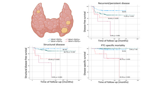
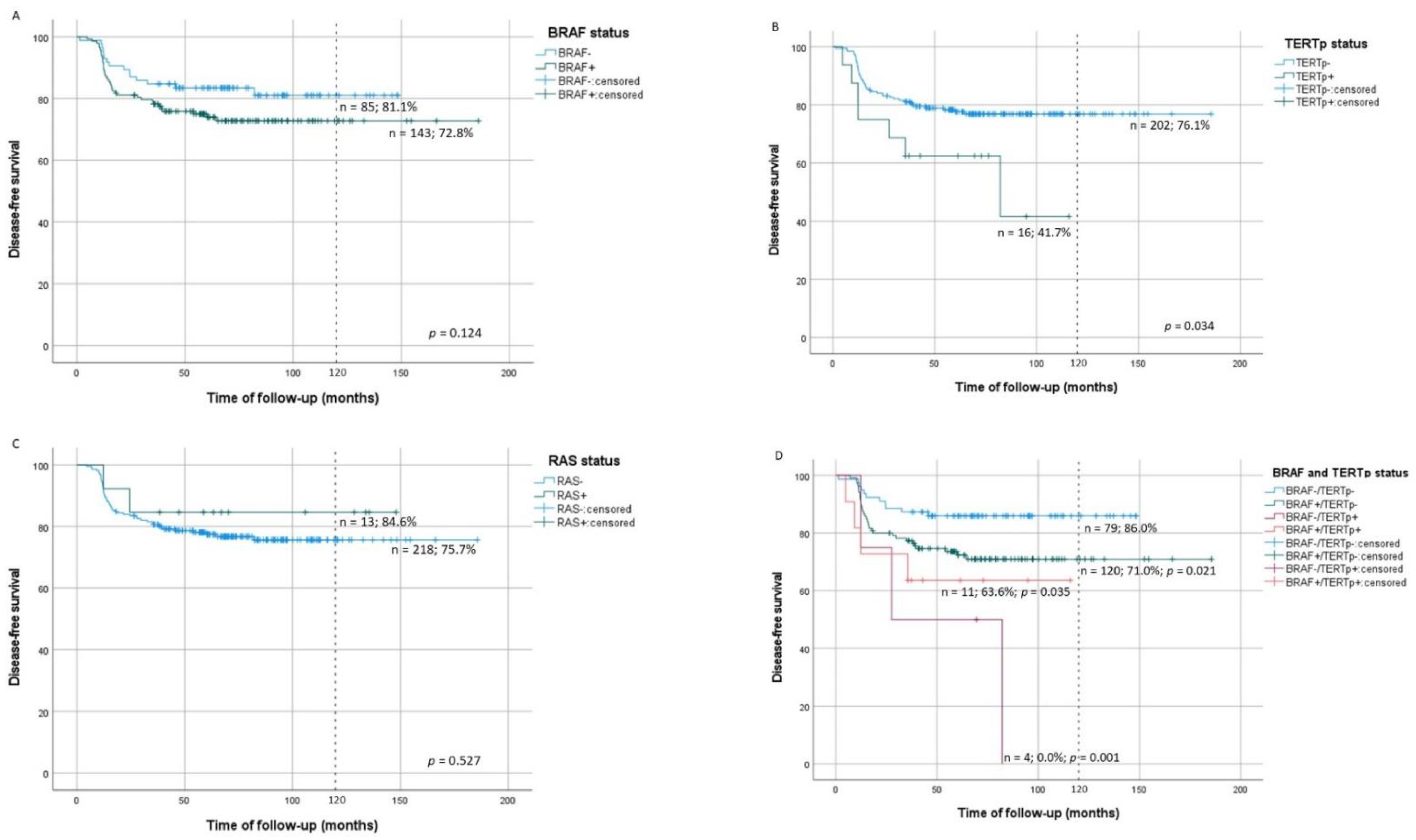
3.4. Molecular Alterations in Recurrent/Persistent Disease

At 10-year evaluation, recurrent/persistent DFS was significantly lower in patients with TERTpmut tumors (41.7%, p = 0.034). Patients with TERTpmut tumors presented 2.3 times increased risk of recurrent/persistent disease (p = 0.04), but this association was lost when the model was adjusted for age and gender (p = 0.062) (Table 3). Recurrent/persistent DFS was not significantly different in patients with BRAFmut or RASmut tumors from patients with BRAFwt or RASwt tumors (Figure 1A–C; Table 2). Considering concomitant mutations, at 10-year evaluation, recurrent/persistent DFS was significantly lower in patients with BRAFmut/TERTpwt (log-rank p = 0.021), BRAFmut/TERTpmut (log-rank p = 0.035) and BRAFwt/TERTpmut (log-rank p = 0.001) tumors than in patients with BRAFwt/TERTpwt tumors (Figure 1D). The aforementioned concomitant mutations significantly increased the risk of recurrent/persistent disease in comparison to BRAFwt/TERTpwt tumors (Table 3).

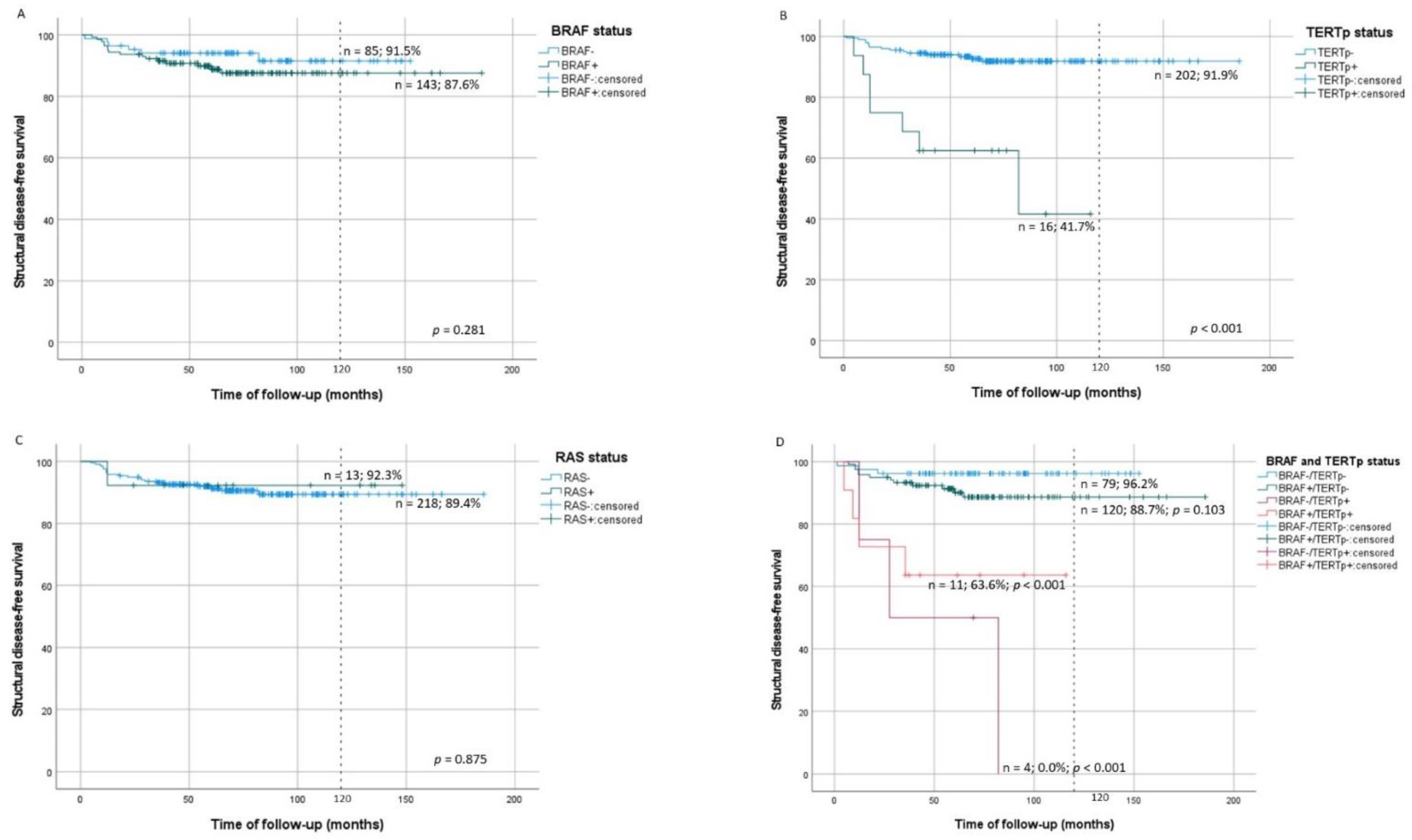
3.5. Molecular Alterations in Structural Disease

Patients with TERTpmut tumors had significantly lower structural disease-free survival (SDFS) (log-rank p < 0.001) than patients without TERTpmut (Figure 2B) and presented significantly increased risk of structural disease, even when adjusted for age and gender (HR = 7.0, p < 0.001) (Table 4). SDFS was not significantly influenced by BRAFmut or RASmut tumors (Figure 2A,C). Considering concomitant mutations, at 10-year evaluation, SDFS was significantly lower in patients with BRAFmut/TERTpmut (log-rank p < 0.001) and BRAFwt/TERTpmut (log-rank p < 0.001) tumors than in patients with BRAFwt/TERTpwt tumors (Figure 2D). The aforementioned concomitant mutations significantly increased the risk of structural disease status both in unadjusted and adjusted models (Table 4). Patients with BRAFmut/TERTpwt tumors did not display a significantly lower SDFS than BRAFwt/TERTpwt tumors (log-rank p = 0.103) (Figure 2D).

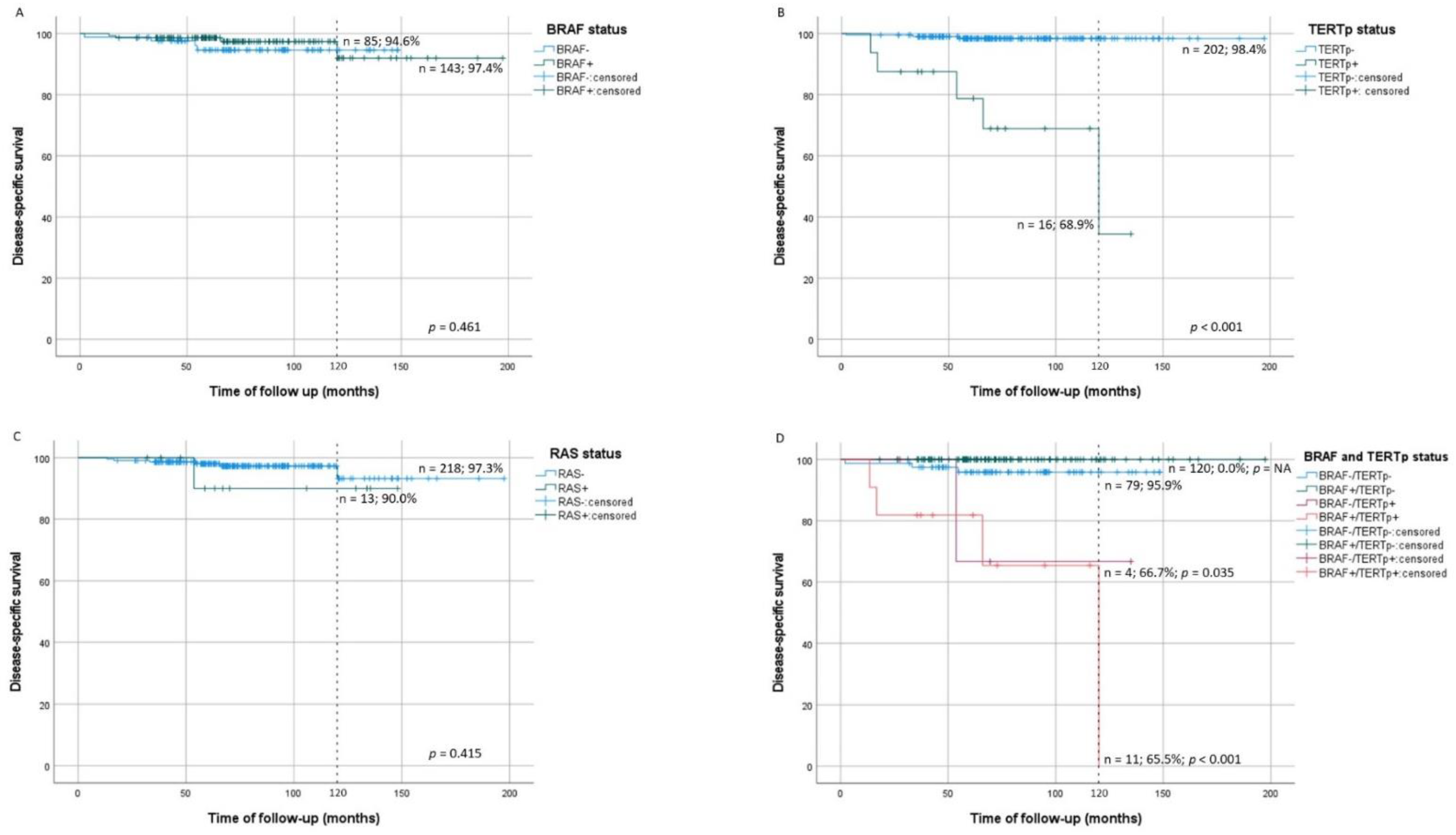
3.6. Molecular Alterations in Disease-Specific Mortality

DSM was significantly increased in patients with TERTpmut tumors (log-rank p < 0.001) in comparison with patients without TERTpmut (Figure 3B). When adjusted for gender and age at diagnosis, TERTpmut significantly increased the risk of DSM (HR = 10.1, p = 0.010) (Table 5). DSM was not significantly increased in patients with BRAFmut or RASmut tumors (Figure 3A,C). DSM was increased in patients with BRAFmut/TERTpmut (log-rank p < 0.001) and BRAFwt/TERTpmut (log-rank p = 0.035) tumors in comparison with patients with BRAFwt/TERTpwt tumors (Figure 3D). There were no DSM events in patients with BRAFmut/TERTpwt tumors (Table 5).

4. Discussion

In the present study, we aimed to investigate the role played by point mutations of BRAF, TERTp, and RAS (NRAS, HRAS, and KRAS) in the outcome of PTC patients (recurrent/persistent disease, structural disease status, and DSM).
The most frequent alteration in our PTC series was, as expected, mutation of BRAF gene. BRAFmut was also frequent in LNM at diagnosis and often detected in locoregional recurrences. All three RAS genes were found mutated in primary tumors, being overall the second most common alteration in PTC. The occurrence of TERTpmut, although less common than alterations of the other genes, was higher in recurrent and metastatic lesions, namely distant metastases, than in primary tumors, as hitherto shown by our group [10].
The role of BRAFmut in the outcome of patients is still an open question. In our study, patients whose tumors had BRAFmut, regardless of the status of the other mutations, did not present a higher risk for any of the outcomes evaluated (recurrent/persistent disease, structural disease status, or DSM). In 2015, George et al. [42] reported a 92% frequency of BRAFmut in a series of recurrent/persistent tumors. The increased incidence of BRAFmut and the lack of a control group in this study did not allow to establish any association with recurrent/persistent disease [42]. In 2019, de Castro et al. [43] also did not find an association between BRAFmut and recurrent/persistent disease, although their series contained 48% BRAFmut tumors. This lack of association was corroborated by other studies [44,45,46]. Some studies have shown that older age and male gender are strong and independent risk factors for PTC—specific mortality in patients with BRAFmut tumors, but not in patients with BRAFwt tumors [27,47]. We did not find similar results in our series. The strength of BRAFmut on patient outcomes may be overestimated in the aforementioned studies [27,47] since in them, no other molecular alterations, namely TERTpmut, were tested. As discussed below, our results indicate that the presence of TERTpmut in PTC, and not of BRAFmut, indeed plays a major role in the outcome.
Several studies have demonstrated BRAFmut association with PTC local invasiveness, recurrent/persistent disease, and LNM [30,48,49,50,51]; detection of BRAFmut has led to advancing the idea that it may predict recurrence in low-risk CPTC patients [52]. Other studies suggested that BRAFmut was associated with poorer outcomes, RAI treatment resistance, and DSM [26,29]. In contrast with this, it was suggested that isolated BRAF status has a limited role in guiding patient management [11], and other studies did not validate BRAFmut impact on the outcome of patients [28,44,53,54]. In our analysis, isolated evaluation of BRAFmut, regardless of the status of the other mutations, converged with the limited role of this mutation in ascertain recurrent/persistent disease, structural disease, or DSM.
We detected mutations in all RAS genes. The most commonly described p.Gln61Arg mutation [55] was present in 13 primary tumors. It was shown that RASmut is present in all stages of thyroid neoplasia [55] ruling out the utilization of RASmut as a marker of malignancy. In a few studies, RASmut has been correlated with DM and poorer outcomes [34,56]. In our series, RASmut was not associated with a poorer prognosis; patients with RASmut had a stage I diagnosis, usually presenting excellent responses to therapy and were free of disease at the end of follow-up, except for one patient who had concomitant TERTpmut and died from PTC. We can hypothesize that, in this patient, TERTpmut was determinant in the outcome rather than the presence of KRASmut. Of note, no RASmut was detected in locoregional metastases nor DM (Supplementary Table S2).
TERTpmut has been consistently associated with poorer outcomes in PTC patients [57]. Some authors showed that recurrent/persistent disease was four times more frequent in patients with TERTpmut than in patients with TERTpwt tumors [58,59]. Whereas in another study, no significant increased risk of recurrent/persistent disease in patients with TERTpmut tumors was reported [60]. The former finding fits with our own results; in the unadjusted analysis, we observed that patients with TERTpmut tumors had twice the risk of recurrent/persistent disease; upon age and gender adjustment, there is still a suggested association (p = 0.062; Table 3).
In our study, TERTpmut tumors were associated with significantly lower SDFS and significantly increased DSM. Furthermore, patients with TERTpmut tumors presented a significantly increased risk of structural disease and of DSM, after adjustment for age and gender, suggesting that TERTpmut may contribute to worse prognosis in PTC patients, as previously shown by our group [21]. Several studies have consistently associated TERTpmut with DSM [21,22,42,46,57,58,59,61,62].
Given previous studies showing a frequent concomitant BRAFmut and TERTpmut in thyroid cancer [13,37,46,63,64,65], we tested the possible relationship of molecular status combinations and different outcomes. This analysis also aimed to evaluate a more trustworthy effect of each mutation separately.
Comparing with BRAFwt/TERTpwt tumors, we observed that BRAFmut/TERTpwt tumors were associated with an increased risk of recurrent/persistent disease. BRAFwt/TERTpmut tumors were associated with an increased risk of recurrent/persistent disease, structural disease, and DSM. We are aware that the latter group (DSM) had few cases, and therefore these results should be interpreted with caution. Nevertheless, taking into account that TERTpmut is far less common than BRAFmut in PTC (less than 10% vs. 45%) [4,25] and that TERTpmut is consistently associated with more aggressive forms of PTC and with increased risk of disease [22,53], we think that our results support the determinant role TERTpmut in the outcome of PTC patients.
In our series, 11/16 TERTpmut cases presented concomitant BRAFmut. We observed that BRAFmut/TERTpmut significantly increased the risk of recurrent/persistent disease, structural disease, and DSM in comparison to BRAFwt/TERTpwt tumors. In accordance with this, Kim et al. [61] showed that the presence of TERTpmut was associated with increased mortality in PTC patients and further demonstrated that BRAFmut/TERTpmut tumors were associated with worsened DSM in PTC patients in comparison to patients with isolated BRAFmut tumors.
It was suggested that BRAFmut/TERTpmut tumors presented a higher risk of worse outcomes in comparison with BRAFwt/TERTpmut tumors [24,66]. In our study, we observed that DSM was similar in cases with BRAFwt/TERTpmut tumors and BRAFmut/TERTpmut tumors. Our results differ from those reported by Vuong et al. [67]; in the latter metanalysis, tumor aggressiveness varies according to isolated BRAFmut, isolated TERTpmut, or concomitant BRAFmut/TERTpmut. Vuong et al. [67] stratified PTC tumors’ aggressiveness in four groups with decreasing aggressiveness: BRAFmut/TERTpmut > BRAFwt/TERTpmut = BRAFmut/TERTpwt > BRAFwt/TERTpwt. Xing et al. [24] proposed a synergistic role of concomitant BRAFmut/TERTpmut on patients’ prognosis; in their series, BRAFmut/TERTpmut tumors were associated with increased disease recurrence, higher than the sum of the two mutations alone, even when performing multivariable adjustments for the classical clinicopathologic risk factors. Xing et al. [24] hypothesized that a possible synergistic effect between the two mutations might occur, advancing that coexistence of the two mutations might be associated with increased expression of the TERT mRNA in PTC [24,64,65,66,68].
We observed that BRAFwt/TERTpmut tumors had an increased risk of recurrent/persistent disease and structural disease in comparison with BRAFmut/TERTpmut tumors. These results may indicate that TERTpmut, rather than BRAFmut, is the molecular alteration that confers higher aggressiveness to PTC. These results fit with our previous studies, in which we showed that PTC patients with TERTpmut tumors had a significantly lower survival [10,21] at variance with the results obtained regarding BRAFmut. Similar findings were reported by Vuong et al. [53], who concluded that TERTpmut was associated with unfavorable DSS and DFS, whereas BRAFmut revealed an association with increased risk of recurrence but not with mortality, thus concluding that BRAFmut usefulness to evaluate patient prognosis must be cautiously considered [53]. Gandolfi et al. [37] reported decreased survival in tumors TERTpmut and BRAFmut/TERTpmut, but no association between BRAFmut and DM or decreased survival was detected. BRAFmut appears associated with local aggressiveness, while TERTpmut was associated with distant metastasis that confers a dismal prognosis [10]. In accordance with our series, no significant differences in recurrent/persistent disease, structural disease, or DSM were observed when comparing patients with BRAFmut/TERTpmut tumors and patients whose tumors were BRAFwt/TERTpmut (Supplementary Figures S1–S3). The low number of tumors with BRAFwt/TERTpmut genotype in our series indicates that a larger series is necessary to confirm our results.

5. Conclusions

We realize that our series has advantages and disadvantages; the positive aspect resides in the fact that it is a consecutive real-life series obtained from a single hospital and not a selected series. On the other hand, we are aware of the consequent limitations: few patients with DM and few patients with PTC-related death. As an example, having only nine patients dying from PTC disease makes the multivariate analysis for such output rather limited. These results should be evaluated with caution also due to the low number of patients with some genotypes.
Summing up, our results indicate that molecular markers can play a role in predicting the outcome of PTC patients. We have data supporting the importance of searching BRAFmut/TERTpwt in terms of putative association with local aggressiveness (recurrent/persistent disease). Furthermore, we have observed a much more important result regarding the prognosis of patients with tumors presenting TERTp point mutations, given their increased risk to develop structural disease and DSM.

Supplementary Materials

The following are available online at https://0-www-mdpi-com.brum.beds.ac.uk/article/10.3390/cancers13092048/s1, Figure S1: Kaplan–Meier curves of recurrent/persistent disease-free survival by concomitant BRAF and TERT promoter mutations in comparison to tumors mutated only for BRAF (E) and only for TERTp (F). Figure S2: Kaplan–Meier curves of structural disease-free survival by concomitant BRAF and TERT promoter mutations in comparison to tumors mutated only for BRAF (E) and only for TERTp (F). Figure S3: Kaplan–Meier curves of disease-specific survival by concomitant BRAF and TERT promoter mutations in comparison to tumors mutated only for BRAF (E) and only for TERTp (F). Table S1: Univariate analysis of clinicopathological features and combined gene mutations. Table S2: BRAF, TERTp, and RAS molecular status in lymph node metastases at diagnosis, locoregional recurrences, and distant metastases and distribution in the different PTC variants.

Author Contributions

Conceptualization, A.A.P. and P.S.; methodology, A.A.P., E.T., and M.R.B.-C.; software, A.A.P. and E.T.; validation, A.A.P., E.T., and P.S.; formal analysis, A.A.P., M.R.B.-C., and E.T.; investigation, A.A.P., E.T., R.B., A.P., T.A., and M.P.; resources, P.S.; data curation, A.A.P. and E.T.; writing—original draft preparation, A.A.P. and E.T.; writing—review and editing, M.M., M.S.-S., J.M., and P.S.; supervision, P.S. and J.M.; project administration, P.S.; funding acquisition, P.S. All authors have read and agreed to the published version of the manuscript.

Funding

This study was supported by FCT, the Portuguese Foundation for Science and Technology, through a PhD grant to E.T. SFRH/BD/143458/2019. This work was financed by FEDER—Fundo Europeu de Desenvolvimento Regional funds through the COMPETE 2020—Operational Programme for Competitiveness and Internationalization (POCI), Portugal 2020. Additional funding by the European Regional Development Fund (ERDF) through the Operational Programme for Competitiveness and Internationalization—COMPETE2020, and Portuguese national funds via FCT, under project POCI-01-0145-FEDER-016390: CANCEL STEM and from the FCT under the project POCI-01-0145-FEDER-031438: The other faces of telomerase: Looking beyond tumor immortalization (PDTC/MED_ONC/31438/2017). Additional funding through the Sociedade Portuguesa de Endocrinologia, Diabetes e Metabolismo-BOLSA SPEDM PARA PROJECTO DE INVESTIGAÇÃO-2017.

Institutional Review Board Statement

The study was conducted in accordance with the Declaration of Helsinki, and the protocol was approved by the Ethics Committee of CHVNG/E (Project investigation 30/2016, 28 January 2016, Comissão de Ética do Centro Hospitalar de Vila Nova de Gaia/Espinho).

Informed Consent Statement

It was an anonymized retrospective study that was exempted from the informed consent according to DR n.º 18/2005, Série I-A de 2005-01-26.

Data Availability Statement

The data presented in this study are available on request from the corresponding author.

Conflicts of Interest

The authors declare no conflict of interest. The funders had no role in the design of the study; in the collection, analyses, or interpretation of data; in the writing of the manuscript, or in the decision to publish the results.

References

  1. Raposo, L.; Morais, S.; Oliveira, M.J.; Marques, A.P.; Bento, M.J.; Lunet, N. Trends in thyroid cancer incidence and mortality in Portugal. Eur. J. Cancer Prev. 2017, 26, 135–143. [Google Scholar] [CrossRef]
  2. Olson, E.; Wintheiser, G.; Wolfe, K.M.; Droessler, J.; Silberstein, P.T. Epidemiology of Thyroid Cancer: A Review of the National Cancer Database, 2000-2013. Cureus 2019, 11, e4127. [Google Scholar] [CrossRef] [Green Version]
  3. Maso, L.D.; Tavilla, A.; Pacini, F.; Serraino, D.; Van Dijk, B.; Chirlaque, M.; Capocaccia, R.; Larrañaga, N.; Colonna, M.; Agius, D.; et al. Survival of 86,690 patients with thyroid cancer: A population-based study in 29 European countries from EUROCARE-5. Eur. J. Cancer 2017, 77, 140–152. [Google Scholar] [CrossRef]
  4. Agrawal, N.; Akbani, R.; Aksoy, B.A.; Ally, A.; Arachchi, H.; Asa, S.L.; Auman, J.T.; Balasundaram, M.; Balu, S.; Baylin, S.B.; et al. Integrated Genomic Characterization of Papillary Thyroid Carcinoma. Cell 2014, 159, 676–690. [Google Scholar] [CrossRef] [Green Version]
  5. Bray, F.; Ferlay, J.; Soerjomataram, I.; Siegel, R.L.; Torre, L.A.; Jemal, A. Global cancer statistics 2018: GLOBOCAN estimates of incidence and mortality worldwide for 36 cancers in 185 countries. Ca Cancer J. Clin. 2018, 68, 394–424. [Google Scholar] [CrossRef] [PubMed] [Green Version]
  6. Global Cancer Observatory. Available online: https://gco.iarc.fr (accessed on 30 December 2020).
  7. Enewold, L.; Zhu, K.; Ron, E.; Marrogi, A.J.; Stojadinovic, A.; Peoples, G.E.; Devesa, S.S. Rising Thyroid Cancer Incidence in the United States by Demographic and Tumor Characteristics, 1980-2005. Cancer Epidemiol. Biomark. Prev. 2009, 18, 784–791. [Google Scholar] [CrossRef] [Green Version]
  8. Kitahara, C.M.; Sosa, J.A. The changing incidence of thyroid cancer. Nat. Rev. Endocrinol. 2016, 12, 646–653. [Google Scholar] [CrossRef] [PubMed]
  9. Ito, Y.; Miyauchi, A.; Kihara, M.; Fukushima, M.; Higashiyama, T.; Miya, A. Overall Survival of Papillary Thyroid Carcinoma Patients: A Single-Institution Long-Term Follow-Up of 5897 Patients. World J. Surg. 2018, 42, 615–622. [Google Scholar] [CrossRef] [PubMed] [Green Version]
  10. Melo, M.; Da Rocha, A.G.; Batista, R.; Vinagre, J.; Martins, M.J.; Costa, G.; Ribeiro, C.; Carrilho, F.; Leite, V.; Lobo, C.; et al. TERT, BRAF, and NRAS in Primary Thyroid Cancer and Metastatic Disease. J. Clin. Endocrinol. Metab. 2017, 102, 1898–1907. [Google Scholar] [CrossRef] [PubMed]
  11. Haugen, B.R.; Alexander, E.K.; Bible, K.C.; Doherty, G.M.; Mandel, S.J.; Nikiforov, Y.E.; Pacini, F.; Randolph, G.W.; Sawka, A.M.; Schlumberger, M.; et al. 2015 American Thyroid Association Management Guidelines for Adult Patients with Thyroid Nodules and Differentiated Thyroid Cancer: The American Thyroid Association Guidelines Task Force on Thyroid Nodules and Differentiated Thyroid Cancer. Thyroid 2016, 26, 1–133. [Google Scholar] [CrossRef] [Green Version]
  12. Sugitani, I.; Fujimoto, Y.; Yamamoto, N. Papillary thyroid carcinoma with distant metastases: Survival predictors and the importance of local control. Surgery 2008, 143, 35–42. [Google Scholar] [CrossRef]
  13. Giorgenon, T.M.V.; Carrijo, F.T.; Arruda, M.A.; Cerqueira, T.L.O.; Barreto, H.R.; Cabral, J.B.; Da Silva, T.M.; Magalhães, P.K.R.; Maciel, L.M.Z.; Ramos, H.E. Preoperative detection of TERT promoter and BRAFV600E mutations in papillary thyroid carcinoma in high-risk thyroid nodules. Arch. Endocrinol. Metab. 2019, 63, 107–112. [Google Scholar] [CrossRef]
  14. Nikiforov, Y.E.; Steward, D.L.; Robinson-Smith, T.M.; Haugen, B.R.; Klopper, J.P.; Zhu, Z.; Fagin, J.A.; Falciglia, M.; Weber, K.; Nikiforova, M.N. Molecular Testing for Mutations in Improving the Fine-Needle Aspiration Diagnosis of Thyroid Nodules. J. Clin. Endocrinol. Metab. 2009, 94, 2092–2098. [Google Scholar] [CrossRef] [PubMed]
  15. Baloch, Z.W.; Sack, M.J.; Yu, G.H.; Livolsi, V.A.; Gupta, P.K. Fine-Needle Aspiration of Thyroid: An Institutional Experience. Thyroid 1998, 8, 565–569. [Google Scholar] [CrossRef] [PubMed]
  16. Sipos, J.; Mazzaferri, E. Thyroid Cancer Epidemiology and Prognostic Variables. Clin. Oncol. 2010, 22, 395–404. [Google Scholar] [CrossRef] [PubMed]
  17. Castro, L.; Alves, S.; Chaves, S.R.; Costa, J.L.; Soares, P.; Preto, A. RAF-1 promotes survival of thyroid cancer cells harboring RET/PTC1 rearrangement independently of ERK activation. Mol. Cell. Endocrinol. 2015, 415, 64–75. [Google Scholar] [CrossRef] [PubMed]
  18. Sobrinho-Simões, M.; Máximo, V.; Rocha, A.S.; Trovisco, V.; Castro, P.; Preto, A.; Lima, J.; Soares, P. Intragenic Mutations in Thyroid Cancer. Endocrinol. Metab. Clin. N. Am. 2008, 37, 333–362. [Google Scholar] [CrossRef]
  19. Nikiforov, Y.E. Thyroid carcinoma: Molecular pathways and therapeutic targets. Mod. Pathol. 2008, 21, S37–S43. [Google Scholar] [CrossRef] [Green Version]
  20. Panebianco, F.; Mazzanti, C.; Tomei, S.; Aretini, P.; Franceschi, S.; Lessi, F.; Di Coscio, G.; Bevilacqua, G.; Marchetti, I. The combination of four molecular markers improves thyroid cancer cytologic diagnosis and patient management. Bmc Cancer 2015, 15, 918. [Google Scholar] [CrossRef] [Green Version]
  21. Melo, M.; Da Rocha, A.G.; Vinagre, J.; Batista, R.; Peixoto, J.; Tavares, C.; Celestino, R.; Almeida, A.; Salgado, C.; Eloy, C.; et al. TERT Promoter Mutations Are a Major Indicator of Poor Outcome in Differentiated Thyroid Carcinomas. J. Clin. Endocrinol. Metab. 2014, 99, E754–E765. [Google Scholar] [CrossRef] [Green Version]
  22. Penna, G.C.; Pestana, A.; Cameselle, J.M.; Momesso, D.; De Andrade, F.A.; Vidal, A.P.A.; Junior, M.L.A.; Melo, M.; Fernandes, P.V.; Corbo, R.; et al. TERTp mutation is associated with a shorter progression free survival in patients with aggressive histology subtypes of follicular-cell derived thyroid carcinoma. Endocrine 2018, 61, 489–498. [Google Scholar] [CrossRef]
  23. Censi, S.; Cavedon, E.; Bertazza, L.; Galuppini, F.; Watutantrige-Fernando, S.; De Lazzari, P.; Nacamulli, D.; Pennelli, G.; Fassina, A.; Iacobone, M.; et al. Frequency and Significance of Ras, Tert Promoter, and Braf Mutations in Cytologically Indeterminate Thyroid Nodules: A Monocentric Case Series at a Tertiary-Level Endocrinology Unit. Front. Endocrinol. 2017, 8, 273. [Google Scholar] [CrossRef]
  24. Xing, M.; Liu, R.; Liu, X.; Murugan, A.K.; Zhu, G.; Zeiger, M.A.; Pai, S.; Bishop, J. BRAF V600E and TERT Promoter Mutations Cooperatively Identify the Most Aggressive Papillary Thyroid Cancer With Highest Recurrence. J. Clin. Oncol. 2014, 32, 2718–2726. [Google Scholar] [CrossRef] [PubMed] [Green Version]
  25. Cantwell-Dorris, E.R.; O’Leary, J.J.; Sheils, O.M. BRAFV600E: Implications for Carcinogenesis and Molecular Therapy. Mol. Cancer Ther. 2011, 10, 385–394. [Google Scholar] [CrossRef] [PubMed] [Green Version]
  26. Xing, M.; Westra, W.H.; Tufano, R.P.; Cohen, Y.; Rosenbaum, E.; Rhoden, K.J.; Carson, K.A.; Vasko, V.; Larin, A.; Tallini, G.; et al. BRAF Mutation Predicts a Poorer Clinical Prognosis for Papillary Thyroid Cancer. J. Clin. Endocrinol. Metab. 2005, 90, 6373–6379. [Google Scholar] [CrossRef] [PubMed] [Green Version]
  27. Shen, X.; Zhu, G.; Liu, R.; Viola, D.; Elisei, R.; Puxeddu, E.; Fugazzola, L.; Colombo, C.; Jarzab, B.; Czarniecka, A.; et al. Patient Age–Associated Mortality Risk Is Differentiated by BRAF V600E Status in Papillary Thyroid Cancer. J. Clin. Oncol. 2018, 36, 438–445. [Google Scholar] [CrossRef] [Green Version]
  28. Trovisco, V.; Soares, P.; Preto, A.; De Castro, I.V.; Lima, J.; Castro, P.; Máximo, V.; Botelho, T.; Moreira, S.; Meireles, A.M.; et al. Type and prevalence of BRAF mutations are closely associated with papillary thyroid carcinoma histotype and patients’ age but not with tumour aggressiveness. Virchows Arch. 2005, 446, 589–595. [Google Scholar] [CrossRef]
  29. Xing, M.; Alzahrani, A.S.; Carson, K.A.; Viola, D.; Elisei, R.; Bendlova, B.; Yip, L.; Mian, C.; Vianello, F.; Tuttle, R.M.; et al. Association Between BRAF V600E Mutation and Mortality in Patients With Papillary Thyroid Cancer. JAMA 2013, 309, 1493–1501. [Google Scholar] [CrossRef] [Green Version]
  30. Xing, M.; Alzahrani, A.S.; Carson, K.A.; Shong, Y.K.; Kim, T.Y.; Viola, D.; Elisei, R.; Bendlová, B.; Yip, L.; Mian, C. Association between BRAF V600E mutation and recurrence of papillary thyroid cancer. J. Clin. Oncol. 2015, 33, 42. [Google Scholar] [CrossRef] [Green Version]
  31. Lemoine, N.R.M.E.; Wyllie, F.S.; Williams, E.D.; Goyns, M.; Stringer, B.; Wynford-Thomas, D. High frequency of ras oncogene activation in all stages of human thyroid tumorigenesis. Oncogene 1989, 4, 149–164. [Google Scholar]
  32. Schulten, H.-J.; Salama, S.; Al-Ahmadi, A.; Al-Mansouri, Z.; Mirza, Z.; Al-Ghamdi, K.; Al-Hamour, O.A.; Huwait, E.; Gari, M.; Al-Qahtani, M.H.; et al. Comprehensive survey of HRAS, KRAS, and NRAS mutations in proliferative thyroid lesions from an ethnically diverse population. Anticancer. Res. 2013, 33, 4779–4784. [Google Scholar] [PubMed]
  33. Medici, M.; Kwong, N.; Angell, T.E.; Marqusee, E.; Kim, M.I.; Frates, M.C.; Benson, C.B.; Cibas, E.S.; Barletta, J.A.; Krane, J.F.; et al. The variable phenotype and low-risk nature of RAS-positive thyroid nodules. BMC Med. 2015, 13, 184. [Google Scholar] [CrossRef] [PubMed] [Green Version]
  34. Fukahori, M.; Yoshida, A.; Hayashi, H.; Yoshihara, M.; Matsukuma, S.; Sakuma, Y.; Koizume, S.; Okamoto, N.; Kondo, T.; Masuda, M.; et al. The association between RAS gene mutations and clinical characteristics in follicular thyroid tumors: New insights from a single center and a large patient cohort. Thyroid 2012, 22, 683–689. [Google Scholar] [CrossRef] [PubMed]
  35. Jang, E.K.; Song, D.E.; Sim, S.Y.; Kwon, H.; Choi, Y.M.; Jeon, M.J.; Han, J.M.; Kim, W.G.; Kim, T.Y.; Shong, Y.K.; et al. NRAS Codon 61 Mutation Is Associated with Distant Metastasis in Patients with Follicular Thyroid Carcinoma. Thyroid 2014, 24, 1275–1281. [Google Scholar] [CrossRef] [PubMed] [Green Version]
  36. Jin, A.; Xu, J.; Wang, Y. The role of TERT promoter mutations in postoperative and preoperative diagnosis and prognosis in thyroid cancer. Medicine 2018, 97, e11548. [Google Scholar] [CrossRef] [PubMed]
  37. Gandolfi, G.; Ragazzi, M.; Frasoldati, A.; Piana, S.; Ciarrocchi, A.; Sancisi, V. TERT promoter mutations are associated with distant metastases in papillary thyroid carcinoma. Eur. J. Endocrinol. 2015, 172, 403–413. [Google Scholar] [CrossRef] [PubMed]
  38. Liu, R.; Xing, M. TERT promoter mutations in thyroid cancer. Endocr. Relat. Cancer 2016, 23, R143–R155. [Google Scholar] [CrossRef] [PubMed] [Green Version]
  39. Lloyd, R.V.; Osamura, R.Y.; Klöppel, G.; Rosai, J. WHO Classification of Tumours of Endocrine Organs, 4th ed.; International Agency for Research on Cancer (IARC): Lyon, France, 2017. [Google Scholar]
  40. Tuttle, R.M.; Haugen, B.; Perrier, N.D. Updated American Joint Committee on Cancer/Tumor-Node-Metastasis Staging System for Differentiated and Anaplastic Thyroid Cancer (Eighth Edition): What Changed and Why? Thyroid 2017, 27, 751–756. [Google Scholar] [CrossRef]
  41. Momesso, D.P.; Vaisman, F.; Yang, S.P.; Bulzico, D.A.; Corbo, R.; Vaisman, M.; Tuttle, R.M. Dynamic Risk Stratification in Patients with Differentiated Thyroid Cancer Treated Without Radioactive Iodine. J. Clin. Endocrinol. Metab. 2016, 101, 2692–2700. [Google Scholar] [CrossRef] [PubMed] [Green Version]
  42. George, J.R.; Henderson, Y.C.; Williams, M.D.; Roberts, D.B.; Hei, H.; Lai, S.Y.; Clayman, G.L. Association of TERT Promoter Mutation, But Not BRAF Mutation, with Increased Mortality in PTC. J. Clin. Endocrinol. Metab. 2015, 100, E1550–E1559. [Google Scholar] [CrossRef] [Green Version]
  43. De Castro, T.P.; Penha, R.C.C.; Buexm, L.A.; De Carvalho, F.N.; Oliveira, R.D.V.C.; Agarez, F.V.; Pinto, L.W.; Carvalho, D.P. Molecular Predictors for Advanced Papillary Thyroid Carcinoma Recurrence. Front. Endocrinol. 2019, 10, 839. [Google Scholar] [CrossRef] [PubMed] [Green Version]
  44. Yan, C.; Huang, M.L.; Li, X.; Wang, T.; Ling, R. Relationship between BRAFV600E and clinical features in papillary thyroid carcinoma. Endocr. Connect. 2019, 8, 988–996. [Google Scholar] [CrossRef] [PubMed] [Green Version]
  45. Garre, M.C.; Casares, M.L.D.L.T.; Massare, P.B.; Nevot, M.; Ángel, L.; Del-Moral, J.V.; Pérez, N.M.; Joya, R.V.; Ramírez, R.M.; Elvira, J.M.L. Presencia de la mutación BRAFT1799A en el tumor primario como indicador de riesgo, recidiva o persistencia de carcinoma papilar de tiroides. Endocrinol. Y Nutr. 2011, 58, 175–184. [Google Scholar] [CrossRef]
  46. Shen, X.; Liu, R.; Xing, M. A six-genotype genetic prognostic model for papillary thyroid cancer. Endocr. Relat. Cancer 2017, 24, 41–52. [Google Scholar] [CrossRef] [Green Version]
  47. Wang, F.; Zhao, S.; Shen, X.; Zhu, G.; Liu, R.; Viola, D.; Elisei, R.; Puxeddu, E.; Fugazzola, L.; Colombo, C.; et al. BRAF V600E Confers Male Sex Disease-Specific Mortality Risk in Patients With Papillary Thyroid Cancer. J. Clin. Oncol. 2018, 36, 2787–2795. [Google Scholar] [CrossRef] [Green Version]
  48. Howell, G.M.; Nikiforova, M.N.; Carty, S.E.; Armstrong, M.J.; Hodak, S.P.; Stang, M.T.; McCoy, K.L.; Nikiforov, Y.E.; Yip, L. BRAF V600E Mutation Independently Predicts Central Compartment Lymph Node Metastasis in Patients with Papillary Thyroid Cancer. Ann. Surg. Oncol. 2012, 20, 47–52. [Google Scholar] [CrossRef] [PubMed]
  49. Kim, S.-J.; Lee, K.E.; Myong, J.P.; Park, J.-H.; Jeon, Y.K.; Min, H.S.; Park, S.Y.; Jung, K.C.; Koo, D.H.; Youn, Y.-K. BRAFV600E Mutation is Associated with Tumor Aggressiveness in Papillary Thyroid Cancer. World J. Surg. 2012, 36, 310–317. [Google Scholar] [CrossRef]
  50. O’Neill, C.J.; Bullock, M.; Chou, A.; Sidhu, S.B.; Delbridge, L.W.; Robinson, B.G.; Gill, A.J.; Learoyd, D.L.; Clifton-Bligh, R.; Sywak, M.S. BRAFV600E mutation is associated with an increased risk of nodal recurrence requiring reoperative surgery in patients with papillary thyroid cancer. Surgery 2010, 148, 1139–1146. [Google Scholar] [CrossRef]
  51. Tufano, R.P.; Bishop, J.; Wu, G. Reoperative central compartment dissection for patients with recurrent/persistent papillary thyroid cancer: Efficacy, safety, and the association of the BRAF mutation. Laryngoscope 2012, 122, 1634–1640. [Google Scholar] [CrossRef]
  52. Kim, T.Y.; Kim, W.B.; Rhee, Y.S.; Song, J.Y.; Kim, J.M.; Gong, G.; Lee, S.; Kim, S.Y.; Kim, S.C.; Hong, S.J.; et al. The BRAF mutation is useful for prediction of clinical recurrence in low-risk patients with conventional papillary thyroid carcinoma. Clin. Endocrinol. 2006, 65, 364–368. [Google Scholar] [CrossRef]
  53. Vuong, H.G.; Duong, U.N.; Altibi, A.M.; Ngo, H.T.; Pham, T.Q.; Tran, H.M.; Gandolfi, G.; Hassell, L. A meta-analysis of prognostic roles of molecular markers in papillary thyroid carcinoma. Endocr. Connect. 2017, 6, R8–R17. [Google Scholar] [CrossRef] [PubMed] [Green Version]
  54. Zheng, X.; Wei, S.; Han, Y.; Li, Y.; Yu, Y.; Yun, X.; Ren, X.; Gao, M. Papillary Microcarcinoma of the Thyroid: Clinical Characteristics and BRAFV600E Mutational Status of 977 Cases. Ann. Surg. Oncol. 2013, 20, 2266–2273. [Google Scholar] [CrossRef] [PubMed]
  55. Howell, G.M.; Hodak, S.P.; Yip, L. RAS Mutations in Thyroid Cancer. Oncology 2013, 18, 926–932. [Google Scholar] [CrossRef] [PubMed] [Green Version]
  56. Garcia-Rostan, G.; Zhao, H.; Camp, R.L.; Pollan, M.; Herrero, A.; Pardo, J.; Wu, R.; Carcangiu, M.L.; Costa, J.; Tallini, G. ras Mutations Are Associated With Aggressive Tumor Phenotypes and Poor Prognosis in Thyroid Cancer. J. Clin. Oncol. 2003, 21, 3226–3235. [Google Scholar] [CrossRef] [PubMed]
  57. Bullock, M.; Ren, Y.; Oneill, C.J.; Gill, A.J.; Aniss, A.; Sywak, M.S.; Sidhu, S.B.; Delbridge, L.; Learoyd, D.L.; De Vathaire, F.; et al. TERT promoter mutations are a major indicator of recurrence and death due to papillary thyroid carcinomas. Clin. Endocrinol. 2016, 85, 283–290. [Google Scholar] [CrossRef] [PubMed] [Green Version]
  58. Liu, C.; Liu, Z.; Chen, T.; Zeng, W.; Guo, Y.; Huang, T. TERT promoter Mutation and Its Association with Clinicopathological Features and Prognosis of Papillary Thyroid Cancer: A Meta-analysis. Sci. Rep. 2016, 6, 36990. [Google Scholar] [CrossRef]
  59. Yin, D.-T.; Mengyuan, L.; Lu, R.-Q.; Li, X.; Xu, J.; Lei, M.; Li, H.; Wang, Y.; Liu, Z. Clinicopathological significance of TERT promoter mutation in papillary thyroid carcinomas: A systematic review and meta-analysis. Clin. Endocrinol. 2016, 85, 299–305. [Google Scholar] [CrossRef]
  60. Myung, J.K.; Kwak, B.K.; Lim, J.A.; Lee, M.-C.; Kim, M.J. TERT Promoter Mutations and Tumor Persistence/Recurrence in Papillary Thyroid Cancer. Cancer Res. Treat. 2016, 48, 942–947. [Google Scholar] [CrossRef] [Green Version]
  61. Kim, T.H.; Kim, Y.-E.; Ahn, S.; Kim, J.-Y.; Ki, C.-S.; Oh, Y.L.; Kim, K.; Yun, J.W.; Park, W.-Y.; Choe, J.-H.; et al. TERT promoter mutations and long-term survival in patients with thyroid cancer. Endocr. Relat. Cancer 2016, 23, 813–823. [Google Scholar] [CrossRef] [Green Version]
  62. Yang, J.; Gong, Y.; Yan, S.; Chen, H.; Qin, S.; Gong, R. Association between TERT promoter mutations and clinical behaviors in differentiated thyroid carcinoma: A systematic review and meta-analysis. Endocrine 2019, 67, 44–57. [Google Scholar] [CrossRef] [PubMed] [Green Version]
  63. Rusinek, D.; Pfeifer, A.; Krajewska, J.; Oczko-Wojciechowska, M.; Handkiewicz-Junak, D.; Pawlaczek, A.; Zebracka-Gala, J.; Kowalska, M.; Cyplinska, R.; Zembala-Nozynska, E.; et al. Coexistence of TERT Promoter Mutations and the BRAF V600E Alteration and Its Impact on Histopathological Features of Papillary Thyroid Carcinoma in a Selected Series of Polish Patients. Int. J. Mol. Sci. 2018, 19, 2647. [Google Scholar] [CrossRef] [Green Version]
  64. Jin, L.; Chen, E.; Dong, S.; Cai, Y.; Zhang, X.; Zhou, Y.; Zeng, R.; Yang, F.; Pan, C.; Liu, Y.; et al. BRAF and TERT promoter mutations in the aggressiveness of papillary thyroid carcinoma: A study of 653 patients. Oncotarget 2016, 7, 18346–18355. [Google Scholar] [CrossRef] [Green Version]
  65. Vinagre, J.; Almeida, A.; Pópulo, H.; Batista, R.; Lyra, J.; Pinto, V.; Coelho, R.; Celestino, R.; Prazeres, H.; Lima, L.; et al. Frequency of TERT promoter mutations in human cancers. Nat. Commun. 2013, 4, 2185. [Google Scholar] [CrossRef] [PubMed] [Green Version]
  66. Trybek, T.; Walczyk, A.; Gąsior-Perczak, D.; Pałyga, I.; Mikina, E.; Kowalik, A.; Hińcza, K.; Kopczyński, J.; Chrapek, M.; Góźdź, S.; et al. Impact of BRAF V600E and TERT Promoter Mutations on Response to Therapy in Papillary Thyroid Cancer. Endocrinology 2019, 160, 2328–2338. [Google Scholar] [CrossRef]
  67. Vuong, H.G.; Altibi, A.M.; Duong, U.N.; Hassell, L. Prognostic implication of BRAF and TERT promoter mutation combination in papillary thyroid carcinoma-A meta-analysis. Clin. Endocrinol. 2017, 87, 411–417. [Google Scholar] [CrossRef] [PubMed] [Green Version]
  68. Liu, R.; Bishop, J.; Zhu, G.; Zhang, T.; Ladenson, P.W.; Xing, M. Mortality Risk Stratification by Combining BRAF V600E and TERT Promoter Mutations in Papillary Thyroid Cancer. Jama Oncol. 2017, 3, 202–208. [Google Scholar] [CrossRef] [PubMed]
Figure 1. Kaplan–Meier curves of PTC of recurrent/persistent disease-free survival by tumor molecular status in comparison with wild type tumors; (A) BRAF status, (B) TERTp status, (C) RAS status, (D) combined BRAF and TERTp status in comparison to wild-type tumors; wt: wild-type; mut: mutated.
Cancers 13 02048 g001
Figure 2. Kaplan–Meier survival curves of PTC structural disease-free survival (SDFS) by tumor molecular status in comparison with wild type tumors; (A) BRAF status, (B) TERTp status, (C) RAS status, (D) combined BRAF and TERTp status in comparison to wild-type tumors; wt: wild-type; mut: mutated.
Cancers 13 02048 g002
Figure 3. Kaplan–Meier survival curves of PTC disease-specific survival (DSS) by tumor molecular status in comparison with wild type tumors; (A) BRAF status, (B) TERTp status, (C) RAS status, (D) combined BRAF and TERTp status in comparison to wild-type tumors; wt: wild-type; mut: mutated.
Cancers 13 02048 g003
Table 1. Series description: patient characteristics, tumor histology, staging, lymph node metastases, and patient outcome.
Patient Characteristics n(%)n (Total)Staging n(%)n (Total)
Age 241T stage 241
<55 years135(56.0%) T1a96(39.8%)
≥55 years106(44.0%) T1b97(40.2%)
Gender 241 T231(12.9%)
Female202(83.8%) T3a7(2.9%)
Male39(16.2%) T3b9(3.7%)
Histology n(%)n (total) T4a1(0.4%)
Nodule sizeMedian ± IR12.0 ± 10.00 238 aN stage 241
Histological subtype 238 a N0207(85.9%)
CPTC156(65.5%) N134(14.1%)
FVPTC55(23.1%) M stage 241
OVPTC18(7.6%) M0237(98.3%)
AVPTC9(3.8%) M14(1.7%)
Necrosis 237Stage 241
Absent232(97.9%) SI225(93.4%)
Present5(2.1%) SII12(5.0%)
Psammomas 237 SIV4(1.7%)
Absent155(65.4%) Lymph node invasion and extranodal extension n(%)n (total)
Present82(34.6%) Lateral compartment 241
Focality 241Lymph node invasionAbsent225(93.4%)
Unifocal151(62.7%) Present16(6.6%)
Multifocal90(37.3%) Lateral compartment 241
Extrathyroidal extension 240Extranodal extensionAbsent236(97.9%)
Absent/minimal230(95.8%) Present5(2.1%)
Major10(4.2%) Patient outcome n(%)n (total)
Lymphatic invasion 237Recurrent/persistent 241
Absent190(80.2%) diseaseAbsent184(76.3%)
Present47(19.8%) Present57(23.7%)
Venous invasion 239Structural disease 241
Absent223(93.3%) Absent216(89.6%)
Present16(6.7%) Present25(10.4%)
Resection margins 240PTC related death 241
R0209(87.1%) Alive or deceased by other causes232(96.3%)
R1 31(12.9%) Deceased of PTC9(3.7%)
CPTC: classical PTC; FVPTC: follicular variant PTC; OVPTC: oncocytic variant PTC (includes Warthin-like, classical oncocytic PTC, and follicular patterned oncocytic PTC); AVPTC: aggressive variants of PTC (includes tall cell, hobnail, diffuse sclerosing, and solid/trabecular variants). a Three primary tumors were not available, only lymph node metastases.
Table 2. BRAF, TERTp, and RAS molecular status in primary tumors and distribution in the different PTC variants.
Gene(s)StatusAll n (%)Histological Variants of Primary Tumors
CPTCFVPTCOVPTCAVPTC
n = 238 an = 156n = 55n = 18n = 9
BRAFwt85393394
p.Val600Glu143 (62.7%)110 (73.8%)20 (37.7%)8 (47.1%)5 (55.6%)
TERTpwt20213347148
−124 G > A12 (5.5%)8 (5.6%)2 (4.0%)1 (6.7%)1 (11.1%)
−146 G > A3 (1.4%)3 (2.1%)0 (0.0%)0 (0.0%)0 (0.0%)
−124/-125 G > A1 (0.5%)0 (0.0%)1 (2.0%)0 (0.0%)0 (0.0%)
BRAF/TERTpBRAFwt/TERTpwt79363184
BRAFmut/TERTpwt120 (56.1%)94 (67.1%)16 (32.0%)6 (40.0%)4 (44.4%)
BRAFwt/TERTpmut4 (1.9%)2 (1.4%)1 (2.0%)1 (6.7%)0 (0.0%)
BRAFmut/TERTpmut11 (5.1%)8 (5.7%)2 (4.0%)0 (0.0%)1 (11.1%)
NRASwt22315247159
p.Gln61Arg8 (3.5%)1 (0.7%)5 (9.6%)2 (11.8%)0 (0.0%)
HRASwt1277536106
p.Gln61Arg3 (2.3%)0 (0.0%)3 (7.7%)0 (0.0%)0 (0.0%)
KRASwt1176735105
p.Gln61Arg2 (1.7%)1 (1.5%)1 (2.8%)0 (0.0%)0 (0.0%)
RASbwt21815143159
RASmut13 (5.6%)2 (1.3%)9 (17.3%)2 (11.8%)0 (0.0%)
CPTC: classical PTC; FVPTC: follicular variant PTC; OVPTC: oncocytic variant PTC (includes Warthin-like, classical oncocytic PTC, and follicular patterned oncocytic PTC); AVPTC: aggressive variants of PTC (includes tall cell, hobnail, diffuse sclerosing, and solid/trabecular variants). a Three primary tumors were not available, only lymph node metastases. b Represents RAS status if at least one of the RAS genes was sequenced; wt: wild-type; mut: mutated.
Table 3. Analysis of molecular status in the risk of recurrent/persistent disease.
Molecular StatusnRecurrent/Persistent Disease AbsentRecurrent/Persistent Disease PresentUnadjusted HR (95% CI)p-ValueAdjusted HR
(95% CI)
p-Value *
BRAF status228
BRAFwt8570 (39.8%)15 (28.8%)1 (Reference) 1 (Reference)
BRAFmut143106 (60.2%)37 (71.2%)1.6 (0.88–2.91)0.128
TERTp status218
TERTpwt202157 (94.6%)45 (86.5%)1 1
TERTpmut169 (5.4%)7 (13.5%)2.3 (1.04–5.12)0.0402.2 (0.96–5.01)0.062
RAS status231
RASwt218168 (93.9%)50 (96.2%)1 1
RASmut1311 (6.1%)2 (3.8%)0.6 (0.16–2.61)0.530
BRAF and TERTp status 214
BRAFwt/TERTpwt7968 (41.7%)11 (21.6%)1 1
BRAFmut/TERTpwt12087 (53.4%)33 (64.7%)2.2 (1.11–4.34)0.0242.2 (1.11–4.36)0.023
BRAFwt/TERTpmut41 (0.6%)3 (5.9%)6.5 (1.82–23.42)0.0046.8 (1.89–24.55)0.003
BRAFmut/TERTpmut117 (4.3%)4 (7.8%)3.4 (1.10–10.82)0.0433.2 (0.97–10.33)0.056
n: number of cases; HR: Hazard ratio calculated by Cox Regression analysis; CI: Confidence interval; * Age- and gender-adjusted; p-value; wt: wild-type; mut: mutated.
Table 4. Analysis of molecular status in the risk of structural disease.
Molecular StatusnStructural Disease
Absent
Structural
Disease
Present
Unadjusted HR (95% CI)p-ValueAdjusted HR
(95% CI)
p-Value *
BRAF status228
BRAFwt8579 (38.3%)6 (27.3%)1 (Reference) 1 (Reference)
BRAFmut143127 (61.7%)16 (72.7%)1.7 (0.65–4.26)0.286
TERTp status218
TERTpwt202187 (95.4%)15 (68.2%)1 1
TERTpmut169 (4.6%)7 (31.8%)7.4 (3.01–18.23)<0.0017.0 (2.67–18.54)<0.001
RAS status231
RASwt218198 (94.3%)20 (95.2%)1 1
RASmut1312 (5.7%)1 (4.8%)0.9 (0.11–6.35)0.875
BRAF and TERTp status214
BRAFwt/TERTpwt7976 (39.6%)3 (13.6%)1 1
BRAFmut/TERTpwt120108 (56.3%)12 (54.5%)2.8 (0.79–9.90)0.112
BRAFwt/TERTpmut41 (0.5%)3 (13.6%)24.3 (4.89–120.80)<0.00124.2 (4.80–122.05)<0.001
BRAFmut/TERTpmut117 (3.6%)4 (18.2%)13.2 (2.93–59.05)0.00111.5 (2.40–55.60)0.002
n: number of cases; HR: Hazard ratio calculated by Cox Regression analysis; CI: Confidence interval; * Age- and gender-adjusted; p-value; wt: wild-type; mut: mutated.
Table 5. Analysis of molecular status in the risk of disease-specific mortality.
Molecular Status nDisease-Specific Mortality AbsentDisease-Specific Mortality PresentUnadjusted HR (95% CI)p-ValueAdjusted HR
(95% CI)
p-Value *
BRAF status228
BRAFwt8581 (36.8%)4 (50.0%)1 (Reference) 1 (Reference)
BRAFmut143139 (63.2%)4 (50.0%)0.6 (0.15–2.39)0.466--
TERTp status218
TERTpwt202199 (94.8%)3 (37.5%)1 1
TERTpmut1611 (5.2%)5 (62.5%)23.9 (5.70–100.23)<0.00110.1 (1.76–58.41)0.010
RAS status231
RASwt218212 (94.6%)6 (85.7%)1 1
RASmut1312 (5.4%)1 (14.3%)2.4 (0.28–20.36)0.429--
BRAF and TERTp status 214
BRAFwt/TERTpwt7976 (36.9%)3 (37.5%)1 1
BRAFmut/TERTpwt120120 (58.3%)0 (0.0%)n.a.n.a.--
BRAFwt/TERTpmut43 (1.5%)1 (25.0%)6.5 (0.67–63.31)0.109--
BRAFmut/TERTpmut117 (3.4%)4 (50.0%)11.6 (2.59–52.09)0.001n.s.n.s.
n: number of cases; HR (Hazard ratio) calculated by Cox Regression analysis; CI: Confidence interval; * Age- and gender-adjusted; p-value; wt: wild-type; mut: mutated.
Publisher’s Note: MDPI stays neutral with regard to jurisdictional claims in published maps and institutional affiliations.

