
Targeting Apoptosis in AML: Where Do We Stand?
by
 , ,
, ,
Kinga Krawiec
1,2 ,
,
Piotr Strzałka
1,2,
Magdalena Czemerska
1,2,
Aneta Wiśnik
2,
Izabela Zawlik
3,4,
Agnieszka Wierzbowska
1,2 and
Agnieszka Pluta
1,2,* 1
Department of Hematology, Medical University of Lodz, 93-513 Lodz, Poland
2
Copernicus Multi-Specialist Oncology and Traumatology Center, 93-513 Lodz, Poland
3
Institute of Medical Sciences, College of Medical Sciences, University of Rzeszow, 35-310 Rzeszow, Poland
4
Laboratory of Molecular Biology, Centre for Innovative Research in Medical and Natural Sciences, College of Medical Sciences, University of Rzeszow, 35-310 Rzeszow, Poland
*
Author to whom correspondence should be addressed.
Cancers 2022, 14(20), 4995; https://0-doi-org.brum.beds.ac.uk/10.3390/cancers14204995
Submission received: 2 September 2022
/
Revised: 1 October 2022
/
Accepted: 8 October 2022
/
Published: 12 October 2022
(This article belongs to the Special Issue New Targets and Therapies of Acute Myeloid Leukemia)
Abstract
:Simple Summary
In patients with acute myeloid leukemia (AML), genetic mutations can cause cells to evade regulated cell death (RCD), resulting in excessive cell proliferation. The best-known form of RCD is apoptosis, which prevents the emergence of cancer cells; disturbances in this process are an important factor in the development and progression of AML. Clearly, it is essential to understand the mechanisms of apoptosis to establish a personalized, patient-specific approach in AML therapy. Therefore, this paper comprehensively reviews the current range of AML treatment approaches related to apoptosis and highlights other promising concepts such as neddylation.
Abstract
More than 97% of patients with acute myeloid leukemia (AML) demonstrate genetic mutations leading to excessive proliferation combined with the evasion of regulated cell death (RCD). The most prominent and well-defined form of RCD is apoptosis, which serves as a defense mechanism against the emergence of cancer cells. Apoptosis is regulated in part by the BCL-2 family of pro- and anti-apoptotic proteins, whose balance can significantly determine cell survival. Apoptosis evasion plays a key role in tumorigenesis and drug resistance, and thus in the development and progression of AML. Research on the structural and biochemical aspects of apoptosis proteins and their regulators offers promise for new classes of targeted therapies and strategies for therapeutic intervention. This review provides a comprehensive overview of current AML treatment options related to the mechanism of apoptosis, particularly its mitochondrial pathway, and other promising concepts such as neddylation. It pays particular attention to clinically-relevant aspects of current and future AML treatment approaches, highlighting the molecular basis of individual therapies.
1. Introduction
Acute myeloid leukemia (AML) is a molecularly and clinically-heterogeneous disease characterized by uncontrolled clonal proliferation of aberrantly differentiated myeloid progenitor cells in the bone marrow. This differentiation, and the resulting genomic instability associated with numerous genetic alterations, including both mutational and epigenetic changes, results in the accumulation of abnormal myeloid cells and the suppression of regular hematopoiesis [1,2]. Such mutations are present in more than 97% of AML patients and result in unrestrained proliferation via the evasion of regulated cell death (RCD) [3].
RCD, or the cell suicide pathway, plays a critical role in organismal development and homeostasis. The most prominent and well-defined form of RCD is apoptosis, which serves as a defense mechanism against the emergence of cancer cells [4] and serves as a homeostatic mechanism to maintain cell populations in tissues [5,6].
Apoptosis dysregulation facilitates the development and progression of AML by permitting the continued survival of cells with activated oncogenes. However, much has been learned about the structural and biochemical details of apoptosis proteins and their upstream regulators, and this knowledge has opened possibilities for the design of new classes of personalized therapies. Indeed, several components of the apoptotic pathway in leukemic cells have already been highlighted as potential therapeutic targets [7].
Despite these advances, the treatment of AML remains a challenge. Hence, new therapeutic concepts are needed for treating acute leukemia in daily clinical practice. Such development should certainly include an individual, tailored approach to treatment that takes into account complex molecular diagnostics. Therefore, it is critical to have a thorough understanding of apoptosis and its mechanisms before effective strategies can be designed for improving AML treatment.
The aim of this review is to provide a comprehensive overview of current AML treatment approaches related to the mechanism of apoptosis, specifically its mitochondrial pathway, and other promising concepts such as neddylation. This review examines clinically-relevant aspects of current and future AML treatments, highlighting the molecular background of individual therapies.
2. Apoptosis
Apoptosis (from Ancient Greek “falling off”) is a physiological process of cellular suicide required for controlling tissue homeostasis, particularly in rapidly-renewing tissues such as hematopoietic tissue [8]. It is characterized by distinct morphological changes including nuclear condensation, cell shrinkage, membrane blebbing, DNA fragmentation, and the formation of apoptotic bodies [9]. This elimination occurs naturally in the course of organ development or following cellular stress [10]. However, such stress signals can be overcome by cancer cells overexpressing anti-apoptotic proteins, especially those of the BCL-2 (B-cell lymphoma-2) family [10]. The BCL2 gene acts as an oncogene that prevents hemopoietic cell death [11]. It has been proposed that the blockage of apoptotic signaling promotes oncogenesis, and this has been demonstrated in several model systems [12,13].
3. Pathways of Apoptosis
Apoptosis can occur by the intrinsic (or mitochondrial) and extrinsic (or death receptor) pathways [14]. The intrinsic pathway serves to activate apoptosis as an appropriate response to various internal traumas, such as metabolic stress, hypoxia, checkpoint violations, growth factor withdrawal, activation of oncogenes, and irreparable genomic damage. The extrinsic pathway is distinctly triggered by the ligation of proapoptotic transmembrane death receptors from the tumor necrosis factor (TNF) family [15]. A common feature of apoptosis is the involvement of caspases, a family of intracellular cysteine proteases, which are present as inactive zymogens in all animal cells, but can be triggered to assume an active state [8,16]. Caspases can be broadly categorized based on their role in apoptosis (caspase-2, -3, -6, -7, -8, -9, and 10) then further subdivided into two groups: the initiator caspases (caspase-2, -8, -9, and -10) and the effector caspases (caspase-3, -6, and -7) [11].
4. The Mechanism of the Mitochondrial Apoptosis Pathway
The mitochondrial apoptosis pathway is the dominant form of cell death, leading to the death of over 60 billion cells each day [17]. The process is initiated by stress signals and is triggered by mitochondrial outer membrane permeabilization (MOMP), caused by inter alia BCL-2 family proteins.
MOMP leads to the release of apoptogenic proteins from the intermembrane space. It results in the secretion of various cell death modulators such as cytochrome c, apoptosis-inducing factor (AIF), endonuclease G (ENDOG), direct IAP-binding protein with low pI (DIABLO, also known as second mitochondria-derived activator of caspases, SMAC) or Omi/HtrA2, and BCL-2 family proteins; it also prevents mitochondrial ATP synthesis, inhibits the respiratory chain and increases reactive oxygen species (ROS) production [18]. These events promote the activation of the initiator caspase 9 and executioner caspases (caspases 3, 6, and 7) in order to destroy the cell [19]. A key element in the regulation and induction of intrinsic apoptosis is the BCL-2 protein family, whose pro- and anti-apoptotic members, and the balance between them, play significant roles in determining the fate of cells [17,20] (Figure 1).
5. BCL-2 Family
The BCL-2 family, which tightly regulates MOMP, consists of three subfamilies: the first is the pro-apoptotic BH3-only members (BIM [BCL-2-like protein 11], BID [BH3 Interacting Domain Death Agonist], PUMA [p53 upregulated modulator of apoptosis], NOXA [Phorbol-12-myristate-13-acetate-induced protein 1], HRK [Activator of apoptosis harakiri], BMF [BCL-2 modifying factor], and BAD [BCL-2 associated agonist of cell death]); the second is the pro-apoptotic effector molecules (BAX [BCL-2 associated X] and BAK [BCL-2 homologous antagonist killer]); and the third is the anti-apoptotic BCL-2 family proteins (BCL-2, BCL-xL [B-Cell Lymphoma-extra-large], BCL-W [BCL-2 like protein 2], MCL-1 [myeloid cell leukemia-1], A1 [BCL-2 related protein A1], and BCL-B [BCL-2 like protein 10]).
The receipt of an apoptotic stimulus upregulates the transcription of the pro-apoptotic BH3-only members. These proteins bind anti-apoptotic members of the BCL-2 family and inhibit their activity. Some direct activators (for example, BID and BIM) can also bind and activate the effectors BAK and BAX, which induces MOMP, resulting in the release of cytochrome c and SMAC from mitochondria and the subsequent activation of initiator caspases [21,22].
Another protein regulating apoptosis is the p53 tumor suppressor, known as the ‘guardian of the genome’. It induces cell-cycle arrest with DNA repair or apoptosis by activating the BCL-2 family. The activation of p53 triggers pro-apoptotic proteins such as PUMA, NOXA, BIM, and BAX to counteract MCL-1, thus overcoming MCL-1 mediated resistance at multiple levels. Moreover, p53 stimulates extrinsic pathway activation by the upregulation of death receptors [7,23,24] (Figure 1).
6. Evasion of Apoptosis in AML
The expression of programmed cell death genes is commonly dysregulated in hematological malignancies, promoting cell accumulation and creating favorable conditions for oncogene activation, genetic instability, and metastasis. Such defects enable the survival of genetically unstable cells, leading to the selection of progressively aggressive clones [25]. Multiple elements of the apoptotic pathway are known to be disturbed in AML, thus facilitating the evasion of apoptosis.
The most common pathway used by cancer cells to evade apoptosis is based on the upregulation of antiapoptotic BCL-2 proteins and the loss of BAX/BAK proteins [26]; indeed, BCL-2 gene overexpression is present in over half of all cancers [18]. As many traditional anti-cancer drugs act on the BCL-2/BAX pathway, any disruption of this target may increase resistance to chemotherapy and radiotherapy by elevating the threshold needed for cell death [26].
TP53 gene mutation prevents apoptotic pathway function [26]. Such mutations are observed in 5–10% of de novo AML and 30–40% of therapy-related cases and are considered important indicators of poor outcome [27,28]. In a study conducted on 500 AML patients, Hou et al. found TP53 mutation to be associated with an inferior response rate (complete remission [CR] rate 28.6% vs. 80.2%, p < 0.0001) and shorter overall survival (OS) (median, 5 vs. 35 months, p < 0.001) compared to unmutated patients [29].
Unlike solid tumors, in most cases of non-complex karyotype de novo AML, the TP53 locus is found to be the wild-type. Most TP53 changes result in missense mutations, i.e., resulting in changes in the amino acid sequence of the DNA-binding domain (encoded by exons 5–8). These are more common in complex karyotypes, relapsed, and elderly AML patients, as well as therapy-related AML [28,30,31]. Where the mutation is absent, as noted in the majority of AML cases, it is possible that apoptosis is blocked by alternate mechanisms [32]. Intriguingly, patients with a TP53 mutation have a lower-than-expected frequency of mutations in other myeloid-related genes, involving splicing, epigenetics, and signal transduction [28,33]. However, irrespective of TP53 mutational status, many AML cases are characterized by p53 dysfunction, presumably through the alteration of p53-regulatory proteins, resulting in the disruption of apoptosis [7,34].
The wild-type TP53 may be significantly inhibited by high levels of mouse double minute 2 homolog (MDM2) or inhibitor of apoptosis proteins (IAP); this presents an appealing targeted strategy for AML treatment [35].
IAPs consist of several proteins which inhibit apoptosis pathway at different levels, namely X-linked inhibitor of apoptosis (XIAP), cellular (cIAP1, cIAP2), neuronal (NIAP), testis-specific (Ts-IAP), Bir-ubiquitin conjugating enzyme (BRUCE), livin, and survivin [36]. IAPs take part in modulating autophagy, necroptosis, and immune regulation, and can inhibit both intrinsic and extrinsic apoptosis; they also stimulate cell survival and increase tumor growth and metastasis when present at elevated levels [37,38]. Moreover, IAP overexpression reduces CR rate in AML patients and increases chemoresistance in several types of cancer. In addition, low IAP expression has also been associated with longer OS in AML patients [36,39,40]. Lack of IAP expression is also associated with higher sensitivity to standard chemotherapy [7,41,42]. Several molecules antagonize IAP activity, with the most prominent ones being SMAC/DIABLO, Omi/HtrA2 (HTRA serine peptidase 2), and XAF-1 (XIAP-associated factor 1) [43,44].
7. An Overview of the Impact of Dysregulation in the Expression of Apoptosis Genes on AML
One of the malfunctions in AML is the overexpression of anti-apoptotic protein BCL-2, which increases the threshold level of proapoptotic signaling required for initiating apoptosis [26,45]. High BCL-2 mRNA expression has been noted in approximately 65% of 119 de novo AML patients and was connected with a significantly worse three-year OS than in those with little or no expression (10% vs. 49%, respectively) [46,47]. A similar trend was observed by Campos et al. and Tóthová et al. [32,48].
Another study conducted on 198 AML patients by Kornblau et al. showed that high BCL-2 protein levels may result in a lowered median survival in the case of favorable/intermediate cytogenetics, but a higher survival in the case of poor cytogenetics [49]. However, Zhou et al. did not find that BCL-2 overexpression had any negative effect on clinical outcomes, and it was significantly up-regulated in newly-diagnosed AML patients [50].
In addition, disturbances in apoptosis may be particularly characteristic of some AML subtypes. AML1-ETO fusion protein related to AML with t (8; 21) has been shown to trigger the transcription of BCL-2 [27]. In addition, BCL-2 expression is likely to be induced by transcription factor CCAAT/enhancer binding protein α, encoded by the CEBPA gene, which is mutated in about 10% of AML patients [30,51,52].
In a study conducted on 235 AML patients, Schaich et al. reported that neither BCL-2 nor BAX nor BCL-XL expression had a significant influence on OS or on disease-free survival (DFS). However, enhanced BCL-XL expression was found in 38% of AML patients, and surprisingly, an elevated BAX mRNA expression was found in 74%. Moreover, BCL-XL overexpression was found to be an independent negative prognostic factor for response to induction therapy in patients with intermediate risk cytogenetics, and the BAX gene was found to be less frequently expressed in the high-risk group than in the intermediate risk group [53].
In addition, higher expression of BAX has been associated with a higher incidence of CR rate [54]. However, surprisingly, another study which assessed 232 samples from AML patients found that a higher BAX and BAD expression correlated with poor outcome [55].
BCL-XL upregulation has also been detected in patients with mutations in fms-like tyrosine kinase 3 (FLT3) and c-KIT, resulting in the activation of a signal transducer and activator of transcription (STAT) [56,57]. It also caused the constitutive activation of the phosphoinositide 3-kinase (PI3K)-AKT signaling pathway in myeloblasts, resulting in the eventual suppression of apoptosis via phosphorylation of pro-apoptotic BAD by inhibiting its interaction with BCL-2 [58]. In FLT3-internal tandem duplication (ITD) mutant AML cells, similar anti-apoptotic effects of BAD phosphorylation have been identified [46,59,60].
Another important member of the anti-apoptotic protein family is MCL1. It has prevented apoptosis by binding to the pro-apoptotic BCL-2 proteins and is thought to be vital for the development and maintenance of AML [5]. MCL-1 overexpression has been associated with tumorigenesis, poor prognosis, and drug resistance [61]. High MCL-1 expression has also been associated with relapse in AML patients [48,49]. This has been particularly evident in the presence of the FLT3-ITD mutation, which upregulated MCL1 through STAT5 activation to prolong the survival of leukemic stem cells [7,49,61]. Li et al. reported the presence of high MCL-1 mRNA expression in 50% of the studied AML patients and noted that this was connected with a shorter OS than in patients with lower MCL-1 expression [62]. In addition, MCL-1, BCL-XL, and BAK were found to be expressed at lower levels in the high-risk AML group based on cytogenetic/molecular characteristics [63].
Mutations in the TP53 gene have been evaluated in AML many times [64], but little is known about the influence of abnormalities in its expression. It has been found that TP53 gene expression may be lowered in nearly 90% of AML patients, indicating that gene expression levels may be more important in AML than gene mutation [65].
The SMAC/DIABLO gene has been shown to be expressed in nearly 90% of AML patients, and a high SMAC/DIABLO expression has also been associated with increased DFS and OS rate [66]. Pluta et al. reported that SMAC/DIABLO proteins were found in 98% of AML patients and that this high level is a good predictor of CR achievement and longer OS [67].
8. Targeting BCL-2 in AML
Apoptosis-based therapies for AML continue to be a significant objective of research. One current therapy is the BCL-2 inhibitor venetoclax (ABT-199), which unlike other targeted treatments in AML, like FLT3 or IDH1 inhibitors, can be used in a molecularly heterogeneous group of patients [68].
AML monotherapy with the BCL-2 inhibitor has not been entirely successful [69]. However, the results of a multicenter phase Ib study evaluating the efficacy and safety of venetoclax in combination with decitabine or azacitidine has offered hope [70]. The study was conducted on a population of 145 patients aged ≥ 65 years who were not eligible for intensive chemotherapy. The median OS for all patients was 17.5 months, with a median follow-up time of 15.1 months. Overall response rates (CR + CR with incomplete blood count recovery [CRi]+ partial remission [PR]) were similar in the venetoclax 400 mg + azacitidine group and the venetoclax 400 mg + decitabine group (76% and 71%, respectively) [70]. The findings indicate that the combination of venetoclax and decitabine/azacytidine demonstrated good tolerability and a favorable remission rate (CR + CRi: 67%) [70]. In addition, the combination therapy demonstrated beneficial effects in the presence of high-risk factors such as age ≥ 75 years, high cytogenetic risk, or the presence of secondary AML [70]. This was the first study to achieve such promising therapeutic success among AML patients not eligible for intensive treatment. It should be emphasized that achieving a CR and improving the general condition of the patient offers the possibility of requalification for other types of treatment with hematopoietic stem cell transplantation as the only curative option.
A multicenter phase Ib/II study evaluated the efficacy of venetoclax in combination with low-dose cytarabine (LDAC) in 82 patients aged ≥60 years ineligible for intensive chemotherapy [71]. In total, 49% of the patients were characterized by secondary AML with an extremely unfavorable prognosis. Of the patients, 54% achieved CR/CRi, of which 26% were CR and 28% achieved CRi. The median OS in the analyzed cohort was 10.1 months [71].
In the VIALE-A prospective, randomized phase III study (NCT02993523) [72], 431 patients with AML who were ineligible for intensive chemotherapy were randomly allocated to receive venetoclax with azacitidine (n = 286) or placebo with azacitidine (n = 145). The analysis revealed a significantly longer OS in the venetoclax + azacitidine group (14.7 months) compared to the venetoclax + placebo group (9.6 months) and a significantly higher rate of achieving CR/CRi (66.4% vs. 28.3%) [72]. The results confirm previous observations that venetoclax in combination with DNA hypomethylating agent (HMA) is an excellent option for first-line treatment in patients with AML who are not eligible for intensive chemotherapy [72].
The VIALE-C randomized phase III trial (NCT03069352) assessed whether venetoclax in combination with low-dose cytarabine (LDAC) improves OS in previously-untreated AML patients ineligible for intensive chemotherapy compared with LDAC plus placebo [73]. A total of 211 patients were randomised to either venetoclax 600 mg/d in combination with LDAC (n = 143) or to placebo + LDAC (n = 68). In total, 38% of participants had secondary AML and 20% had previously been treated with HMA. At the primary analysis, the study did not meet its primary endpoint of a statistically significant improvement in OS. However, in an additional 6-month follow-up, venetoclax + LDAC resulted in a longer OS than LDAC monotherapy (8.4 vs. 4.1 months). CR/CRi rates were 48% for combination treatment vs. 13% for LDAC monotherapy [73]. Hence, venetoclax with LDAC yielded clinically significant improvements in remission rate and OS compared to LDAC monotherapy, with an acceptable safety profile [73].
Data from prospective, randomized phase III trials, as well as results from earlier studies, confirm the effectiveness of venetoclax when used in combination with a hypomethylating agent or low-dose cytarabine as a backbone therapy. This is especially true for older patients with newly-diagnosed AML who are not candidates for intensive induction chemotherapy [74].
The possibility of combining venetoclax treatment with intensive chemotherapy is currently being investigated (NCT03709758). There are high hopes for this therapeutic option, especially for groups of patients with unfavorable cytogenetic-molecular risk and with relapsed or the initially refractory disease. A prospective single-center phase Ib/II study evaluating fludarabine, cytarabine, G-CSF, and idarubicin combined with venetoclax (FLAG-IDA + VEN) in patients with newly-diagnosed or relapsed/refractory (r/r) AML demonstrated a 98% overall response rate ([ORR]: CR + CR with partial hematologic recovery [CRh] + CRi + morphologic leukemia free state [MLFS] + PR) and an 89% composite CR (CRc + CRh + CRi) [75]. In a phase II trial investigating the activity and safety of 3 + 7 daunorubicin and cytarabine plus venetoclax (DAV) chemotherapy in adults with AML, a high CR + CRi rate (91%) was achieved after one cycle of DAV, with a high rate of measurable residual disease (MRD) negativity (97%), as well as manageable adverse effects [76].
Promising results may also be obtained in the ongoing VIALE-T study evaluating the effectiveness of venetoclax in combination with azacitidine versus the standard of care in AML patients as a maintenance after allogeneic hematopoietic stem cell transplantation (allo-HSCT) (NCT04161885). Although venetoclax is currently used most widely in combination with HMA or LDAC, it is hoped that therapy will soon be optimized by replacing azacitidine with molecularly targeted therapy (dual molecularly targeted therapy) or adding molecularly targeted therapy to the venetoclax-HMA backbone (triple therapy) [77].
There are several ongoing clinical trials in AML patients investigating various combinations of regimens containing venetoclax, as well as other drugs inhibiting the BCL-2 family. More detailed information is provided in Table 1.
9. In the Grip of Apoptosis—The Desired Future after Venetoclax
9.1. Targeting BCL-XL
Among the agents targeting the BCL-2 family, only venetoclax has FDA approval. There is a need to identify new alternatives targeting the intrinsic pathway of apoptosis to overcome resistance to venetoclax [20,57,78], which often occurs due to the upregulation of other anti-apoptotic proteins such as MCL and BCL-XL. In addition, venetoclax has limited utility for the treatment of solid tumors, which are generally not dependent on BCL-2 for survival; however, BCL-XL can be overexpressed in many solid tumors, as well as in a subset of leukemia cells, and its expression is highly correlated with chemoresistance, independent of TP53 mutational status [79].
To find treatments that can eliminate resistance to standard BCL-2 inhibitors, possible new approaches involving the induction of alternative antiapoptotic BCL-2 family members, such as BCL-XL and MCL-1, are under investigation [56,58]. This has resulted in the discovery of promising anticancer drug candidates including ABT-263 (navitoclax), as well as several BCL-XL and MCL-1 monoselective inhibitors [79,80].
Navitoclax, which inhibits BCL-2/BCL-XL/BCL-W, has been extensively tested in lymphoid malignancies and myeloproliferative neoplasms; however, its applicability in AML has proven to be limited due to significant thrombocytopenia, since platelets solely depend on BCL-XL for survival [81,82]. Therefore, despite being a key cancer target, BCL-XL is currently without any safe or effective therapy [79,83]. NCT05222984 is a new study evaluating navitoclax with venetoclax and decitabine in treating patients with r/r AML after previous treatment with venetoclax.
Nevertheless, two more advanced molecules targeting BCL-XL (PROTAC DT2216) and BCL-XL/2 (PROTAC 753B) have recently appeared; these constitute a new emerging technology for the development of novel and safe targeted anticancer therapeutics. PROTACs are small molecules, superior to conventional small molecule inhibitors (SMIs), that utilize the ubiquitin proteasome system (UPS) to degrade proteins of interest (POI) [84]. What makes them unique is their improved target selectivity and ability to target “undruggable” and mutant proteins [85]. Upon binding to the POI, the PROTAC can recruit E3 ligase for POI ubiquitination, which is subjected to proteasome-mediated degradation [86].
DT2216 is a first-generation BCL-XL PROTAC that is more potent against BCL-XL-dependent tumor cells and demonstrates reduced platelet toxicity than navitoclax. DT2216 targets the von Hippel–Lindau (VHL) E3 ligase at BCL-XL for degradation; this provides an advantage over navitoclax by protecting platelets, where VHL is poorly expressed [84,87].
753B PROTAC, which causes BCL-XL/BCL-2 ubiquitination and selective degradation of cells expressing VHL, is an innovative approach to inducing apoptosis activation and eliminating chemotherapy-induced senescent leukemia cells [88]. It has been found that 753B effectively eliminates AML cells and enhances the efficacy of chemotherapy by targeting senescent cells; this study also confirmed recent findings that standard chemotherapy with cytosine arabinose (Ara-C) induced cellular senescence (SnCs) in AML cells, manifested by increased cell size, the induction of senescence-associated β-galactosidase activity, the upregulation of cell cycle regulator proteins (p16, p21, p53), and the expression of the senescence-associated secretory phenotype factors (IL-6, IL-18, IL-1β), leading to chemoresistance in AML [89]. 753B has been found to reverse chemotherapy-induced SnCs phenotype and induce death in senescent AML cells by increasing BCL-XL/BCL-2 expression, and to trigger time and dose-dependent BCL-XL degradation at concentrations lower than that of DT2216 [90,91]. 753B also showed potency in nine venetoclax-resistant samples, of which six demonstrated BCL-2 degradation [88,92].
Another drug targeting BCL-2/BCL-XL, which is currently under research in the NIMBLE study in patients with relapsed/refractory AML, is AZD0466 [93]. AZD0466 is a drug-dendrimer conjugate consisting of AZD4320 (BCL2/XL dual inhibitor) covalently conjugated to a pegylated poly-L-lysine dendrimer; the drug is gradually released by hydrolysis, allowing for more efficient delivery [94].
9.2. Targeting MCL-1
MCL-1 overexpression can be identified in patients with relapsed AML. It was also found to be a major factor in resistance to the dual BCL-2/BCL-XL inhibitors in AML-derived cell lines and blast cells from AML patients [95]. MCL-1 not only contributed to leukemic stem cell survival and MRD, but also facilitated AML progression. Contrary to the clinical success of venetoclax, MCL-1 inhibitor clinical trials have been more challenging due to MCL-1 demonstrating a less flexible binding site than BCL-2 or BCL-XL. However, S64315/MIK665 (NCT04629443, NCT03672695, NCT04702425), demonstrating activity against MCL-1, entered phase I/II clinical trials in AML, while AMG 176 (NCT02675452) and PRT1419 (NCT04543305, NCT05107856) have entered phase I. An interesting approach is to combine S64315/MIK665 with S65487 (VOB560); the latter is a selective BCL-2 inhibitor, which is also active against BCL-2 mutations such as G101V and D103Y, and is known to induce robust anti-tumor activity (NCT04702425).
Unfortunately, many trials on anti-MCL-1 agents have been halted due to cardiac toxicity, which may be caused by deficiencies in the respiratory chain and ATP production, accompanied by increased ROS production [96]. Some, like that evaluating AZD5991 (NCT03218683), have been put on hold to allow further evaluation of safety-related information. Nevertheless, the development of MCL-1 PROTACs raises hopes for reducing on-target cardiotoxicity [84,97].
Moreover, like BCL-XL, MCL-1 expression is also upregulated in both hematologic malignancies and some solid tumors, providing another advantage over BCL-2 inhibition [84,98].
A promising approach is based on the use of indirect MCL blockade using cyclin-dependent kinase 9 (CDK9) inhibitors. These have demonstrated preclinical activity in AML, which has been primarily attributed to the depletion of MCL-1 protein with subsequent apoptosis. Several clinical trials with CDK9 inhibitors like alvocidib (NCT03298984, NCT03441555, NCT01349972), dinaciclib (NCT03484520), CYC065 (NCT04017546) and AZD4573 (NCT03263637) have been underway to evaluate their effectiveness in combination with other therapeutics [99,100,101].
9.3. Targeting IAPs
IAP overexpression induces chemotherapeutic drug resistance in many cancer types. Exploring the apoptosis-inducing activity of IAP inhibitors in AML provides new opportunities to sensitize drug-resistant AML cells to other therapeutics. These antagonists have been partially tested in both pre-clinical and clinical studies, mainly in combination therapies [102,103,104].
One new drug is ASTX660 (a dual antagonist of both cIAP1/2 and XIAP). It is currently in a clinical trial in combination with ASTX727 (decitabine/cedazuridine, hypomethylating agent), in patients with relapsed/refractory AML (NCT04155580) [105].
Upregulation of the smallest IAP, survivin, correlates with cancer progression and poor prognosis in AML [106]. A unique strategy for downregulating survivin expression involves the use of antisense oligonucleotide (ASO) technology, investigated as LY2181308 in a phase II study (NCT00620321). In this trial, the molecule demonstrated a good safety profile and some clinical benefit in r/r AML patients when used in combination with cytarabine and idarubicin [107,108]. However, despite initially promising results, phase II of the study examining the second-generation bivalent SMAC mimetic birinapant (NCT01486784) in r/r AML patients was never initiated [109,110].
9.4. Targeting p53—Dependent Apoptosis Pathways
Mutations in the TP53 gene result in impaired apoptosis due to innate resistance to conventional chemotherapy. The effect of HMA may appear initially satisfactory, but as with allo-HSCT in this group of patients, it does not provide a long-term response. Consequently, all patients with TP53 mutations are a source of great interest in clinical trials using new therapeutic approaches [28,111].
Eprenetapopt (APR-246) is a novel, first-in-class, small molecule that selectively induces apoptosis in TP53-mutant cancer cells. APR-246 is spontaneously converted into methylene quinuclidinone, which covalently binds to cysteine residues in mutant p53, thus stabilizing and shifting the equilibrium toward a functional conformation. The restoration of wild-type TP53 expression triggers cell death by inducing apoptosis in tumor cells [112]. Eprenetapopt can also induce p53-independent cell death in the course of ferroptosis, a recently described cell death process [113].
Two recent phase II studies (NCT03072043 and NCT03588078) have found that eprenetapopt in combination with azacitidine demonstrated a promising safety profile and response rates [112,114,115]. However, a phase III study (NCT03745716) has also compared eprenetapopt plus azacitidine with azacitidine alone in the treatment of patients with myelodysplastic syndrome (MDS), and the coexisting TP53 mutation failed to meet its primary endpoint concerning CR rate.
Currently, there is an ongoing phase I trial evaluating a three-drug combination comprising azacitidine, APR-246, and venetoclax in TP53 mutated treatment-naive AML patients (NCT04214860); in addition, another multi-center, open-label, phase II clinical trial is assessing APR-246 in combination with azacitidine as maintenance therapy after allo-HSCT for patients with TP53 mutant AML or MDS (NCT03931291) [116,117,118].
Moreover, Maslah et al. found that the synergistic effects of APR-246 with azacitidine were mediated by downregulation of the FLT3 pathway in drug-treated cells [118]. Therefore, in these patients, especially those with FLT3 mutations and high blast rates, combining treatment with FLT3 inhibitors may be a valuable option [119].
Although TP53 mutations are more common in some AML subgroups, p53 inactivation is more likely to be caused by the overexpression of its endogenous inhibitors, like mouse double minute 4 homolog (MDMX) or mouse double minute 2 homolog (MDM2), which frequently occur in TP53 wild-type AML [120].
Both the amplification and overexpression of MDM2 and MDMX have been associated with low p53 protein levels and unfavorable clinical outcome in AML patients [7,121]. They have also been found to be a main driver of malignancy, mainly when accompanied by wild-type TP53 status. Han et al. reported that MDMX protein and mRNA are overexpressed in up to 92% of AML [122].
The MDM2 and MDMX homologs are important essential negative regulators of p53, acting as its gatekeepers. They are known to bind together and act most effectively as a complex. MDM2 largely inhibits and degrades p53 through ubiquitination, while MDMX interacts with p53 mainly by inhibiting transactivation activity [119,123,124]. Therefore, inhibition of the p53-MDM2/MDMX interaction represents an attractive therapeutic target, allowing reactivating of p53 function.
Idasanutlin (RG7388) is a representative of second-generation selective MDM2 antagonists, or nutlins, which block the interaction between the MDM2 protein and p53, thus preventing the enzymatic degradation of p53 and causing the transcriptional restoration of p53, leading to apoptosis [124]. However, the effective action of MDM2 antagonists is significantly limited to TP53 wild-type AML patients.
Several phase I studies have reported that idasanutlin is a potentially safe option with promising clinical activity. In a multicenter phase I/Ib trial (NCT01773408) in 122 TP53 wild-type AML patients, idasanutlin yielded a 10.8% CR as a single drug and 32.2% in combination with cytarabine. The composite clinical remission (CRc: CR + CRp [CR with incomplete platelet recovery] + CRi) rate in patients with TP53 mutations was only 4.0% (n = 1/25). Median MDM2 expression was higher in patients who responded to idasanutlin than in non-responders [125].
Idasanutlin + cytarabine (1 g/m2) was compared with placebo + cytarabine in r/r AML in the MIRROS randomized phase III trial (NCT02545283); however, further analysis had to be stopped due to a failure to achieve study endpoints. The same was noted for the NCT03850535 trial with idasanutlin + cytarabine + daunorubicin [126]. In addition, the results of the NCT02670044 study evaluating the effect of idasanutlin in combination with venetoclax are awaited.
Combination targeted therapies may be particularly desirable, where the additional use of venetoclax has been shown to delay the development of resistance to MDM2 inhibitors. A phase Ib study (NCT02670044) found the combination of venetoclax + idasanutlin to demonstrate encouraging safety and efficacy in elderly patients with r/r AML who were ineligible for intensive chemotherapy. The anti-leukemic response rate, defined as CR + CRp, CRi + PR, and MLFS, was 41% with a median OS of 4.4 months [119,127].
Although MDM2 inhibitors have now advanced to clinical trials, those associated with MDMX have not yet been subjected to extensive clinical testing. The resistance and toxicity associated with MDM2 inhibitor treatment has driven the development of new therapies such as dual MDMX/MDM2 directed agents, which offer a more complete activity against p53 [128].
ALRN-6924 is a stabilized peptide and first-in-class MDM2/MDMX dual inhibitor, designed to activate p53. It demonstrates antileukemic activity through activation of p53-dependent transcription and induction of apoptosis [129]. In preclinical studies, ALRN-6924 was observed to inhibit the proliferation of AML cell lines and primary human AML cells, either alone or in combination with cytarabine, achieving CR in approximately 40% of mice in xenotransplantation studies [130]. Currently, ALRN-6924 is being tested in an early phase clinical trial involving patients with r/r AML and MDS [130,131].
Interestingly, ALRN-6924 does not induce deep levels of apoptosis in healthy tissues, even at the highest doses tested; however, it can induce reversible cell cycle arrest in properly proliferating cells. This concept, called cyclotherapy, offers the chance to selectively protect tissues, such as bone marrow, from cytotoxic chemotherapeutics, thus enabling cancer treatment with fewer side effects [128,132].
Novel anticancer therapies that reactivate p53 are a promising option; however, they require further research. It is likely that new therapeutic combinations are needed to truly improve long-term outcome and to have a reasonable possibility of introducing this group of drugs into routine clinical practice [28,119].
10. Ferroptosis—A New Type of Regulated Cell Death
Ferroptosis is an iron-dependent form of RCD. There is growing evidence that the boundary between ferroptosis and other types of RCDs appears to be blurred. This process is triggered by impaired redox and antioxidant mechanisms and is primarily featured by changes in iron homeostasis, iron-dependent lipid peroxidation, and accumulation of glutamate toxicity, which can be specifically reversed by ferroptosis inducers or inhibitors. Ferroptosis, similar to apoptosis, can be induced by both endogenous and exogenous pathways. The endogenous pathway is activated primarily by reducing the expression or activity of intracellular antioxidant enzymes. Meanwhile, the exogenous pathway is triggered by inhibition of cell membrane transporters (e.g., the cysteine/glutamate reverse transporter) or by induction of transferrin and ferroportin [133].
Ferroptosis is mediated by intercellular iron overload. After conjugation with transferrin, extracellular ferric iron (Fe3+) is imported into cells through the membrane protein transferrin receptor 1 (TfR1). As a result of the ferroptosis mechanism, cells lose the integrity of the plasma membrane, the cytoplasm becomes swollen, the mitochondria become smaller, the outer mitochondrial membrane breaks down, and the membrane density increases [134].
Cancer cells have a stronger need for iron compared to non-malignant cells. In recent years, the possibility has been presented that cancer cells that are resistant to conventional therapies may be particularly susceptible to ferroptosis. Moreover, ferroptosis has recently been shown to be associated with cancer immunotherapy, where T cells and INFγ promote tumor cell sensitivity to this type of RCD. It has now been established that AML is sensitive to ferroptosis-promoting compounds. Current research on the role of ferroptosis in AML is based on cell lines and generally involves the development of new therapeutic reagents that produce interference to a particular link in the mechanism of ferroptosis [135].
11. Future Directions in Apoptosis-Related Neddylation
Another aspect worth attention in targeted treatment connected with apoptosis is the inhibition of neddylation. Neddylation is a post-translational modification closely related to ubiquitination and apoptosis which conjugates a ubiquitin-like protein NEDD8 to substrate proteins. The conjugation of NEDD8 to its substrates is catalyzed by NEDD8-activating enzyme E1 (NAE), two NEDD8-conjugating enzyme E2s (UBC12/UBE2M and UBE2F), and several substrate-specific NEDD8-E3 ligases [136]. The best-characterized substrate of the NEDD8 pathway is the cullin family of proteins, Cul-1–3, 4A, 4B, 5, 7, and 9, also known as PARC (p53-associated Parkin-like Cytoplasmic Protein), which provide support for ubiquitin ligases (E3). Other NEDD8 pathway substrates include p53, p73, EGFR, caspases/IAPs/RIP1, pVHL, and Parkin/PINK1 [137] (Figure 2).
Neddylation has been associated with multiple cellular processes, including cell cycle progression, metabolism, immunity, and carcinogenesis. Moreover, it contributes to tumorigenesis and resistance to apoptosis by promoting ubiquitin/proteasome-dependent degradation of proteins involved in inhibiting cell growth. In addition, as neddylation upregulation is found in many human cancers, the NEDD8 pathway may be considered a novel therapeutic target [137,138].
12. Targeting Neddylation in AML
12.1. Pevonedistat
Pevonedistat (TAK-924/MLN4924) is an inhibitor of the NEDD8 activating enzyme (NAE), which suppresses neddylation and induces cell cycle arrest and apoptosis [139]. Moreover, it increases the accumulation of the pro-apoptotic protein NOXA, as well as the upregulation of PUMA, BIK, and BIM [140,141,142]. However, the phase III PANTHER study (NCT03268954) of pevonedistat plus azacitidine (n = 227) vs azacitidine (n = 227) in patients with newly diagnosed higher-risk MDS, higher-risk chronic myelomonocytic leukemia, or AML with 20–30% blasts, failed to reach pre-defined statistical significance for the primary endpoint of event-free survival (EFS) [143]. More detailed information on clinical trials concerning the neddylation pathway in AML is provided in Table 4.
12.2. TAS4464
The most potent and selective NAE inhibitor is TAS4464, which has demonstrated widespread antiproliferative activity against AML cell lines. It activates both the intrinsic and extrinsic apoptotic pathways and triggers caspases (especially caspase-8 and caspase-9) by increasing the NOXA level and decreasing the antiapoptotic factor c-FLIP. TAS4464 demonstrates a long-acting NAE inhibition effect in hematological malignancies. Unfortunately, its trial (NCT02978235) was terminated before Phase II due to cases of drug-induced liver injury [136].
13. Conclusions
Our knowledge of the mechanisms of apoptosis regulation continues to grow and this translates into new therapeutic possibilities. Despite this, not all new drugs in phase II and III clinical trials show satisfactory efficacy with acceptable toxicity. Nevertheless, the introduction of new personalized targeted therapies may effectively translate into higher response rates and longer survival among AML patients.
Author Contributions
Conceptualization, K.K. and A.P.; methodology, K.K. and A.P.; formal analysis, A.W. (Aneta Wiśnik) and I.Z.; writing—original draft preparation, K.K. and P.S.; writing—review and editing, K.K. and M.C.; visualization, K.K. and A.P.; supervision, A.P. and A.W. (Agnieszka Wierzbowska). All authors have read and agreed to the published version of the manuscript.
Funding
This research received no external funding.
Institutional Review Board Statement
Not applicable.
Informed Consent Statement
Not applicable.
Data Availability Statement
Not applicable.
Conflicts of Interest
The authors declare no conflict of interest.
References
- Halik, A.; Arends, C.M.; Bullinger, L.; Damm, F.; Frick, M. Refining AML Treatment: The Role of Genetics in Response and Resistance Evaluation to New Agents. Cancers 2022, 14, 1689. [Google Scholar] [CrossRef]
- Short, N.J.; Rytting, M.E.; Cortes, J.E. Acute myeloid leukaemia. Lancet 2018, 392, 593–606. [Google Scholar] [CrossRef]
- Patel, J.P.; Gönen, M.; Figueroa, M.E.; Fernandez, H.; Sun, Z.; Racevskis, J.; Van Vlierberghe, P.; Dolgalev, I.; Thomas, S.; Aminova, O.; et al. Prognostic Relevance of Integrated Genetic Profiling in Acute Myeloid Leukemia. N. Eng. J. Med. 2012, 366, 1079–1089. [Google Scholar] [CrossRef] [Green Version]
- Hu, X.-M.; Li, Z.-X.; Lin, R.-H.; Shan, J.-Q.; Yu, Q.-W.; Wang, R.-X.; Liao, L.-S.; Yan, W.-T.; Wang, Z.; Shang, L.; et al. Guidelines for Regulated Cell Death Assays: A Systematic Summary, A Categorical Comparison, A Prospective. Front. Cell Dev. Biol. 2021, 9, 634690. [Google Scholar] [CrossRef] [PubMed]
- Peng, F.; Liao, M.; Qin, R.; Zhu, S.; Peng, C.; Fu, L.; Chen, Y.; Han, B. Regulated cell death (RCD) in cancer: Key pathways and targeted therapies. Signal Transduct. Target. Ther. 2022, 7, 286. [Google Scholar] [CrossRef] [PubMed]
- Portt, L.; Norman, G.; Clapp, C.; Greenwood, M.; Greenwood, M.T. Anti-apoptosis and cell survival: A review. Biochim. Biophys. Acta 2011, 1813, 238–259. [Google Scholar] [CrossRef] [Green Version]
- Valentin, R.; Grabow, S.; Davids, M.S. The rise of apoptosis: Targeting apoptosis in hematologic malignancies. Blood 2018, 132, 1248–1264. [Google Scholar] [CrossRef] [Green Version]
- Testa, U.; Riccioni, R. Deregulation of apoptosis in acute myeloid leukemia. Haematologica 2007, 92, 81–94. [Google Scholar] [CrossRef] [Green Version]
- Li, B.; Dou, S.-X.; Yuan, J.-W.; Liu, Y.-R.; Li, W.; Ye, F.; Wang, P.-Y.; Li, H. Intracellular transport is accelerated in early apoptotic cells. Proc. Natl. Acad. Sci. USA 2018, 115, 12118–12123. [Google Scholar] [CrossRef] [PubMed] [Green Version]
- Fernald, K.; Kurokawa, M. Evading apoptosis in cancer. Trends Cell Biol. 2013, 23, 620–633. [Google Scholar] [CrossRef]
- Kontomanolis, E.N.; Koutras, A.; Syllaios, A. Role of Oncogenes and Tumor-suppressor Genes in Carcinogenesis: A Review. Anticancer Res. 2020, 40, 6009–6015. [Google Scholar] [CrossRef]
- Hanahan, D.; Weinberg, R.A. Hallmarks of cancer: The next generation. Cell 2011, 144, 646–674. [Google Scholar] [CrossRef] [PubMed] [Green Version]
- Nechiporuk, T.; Kurtz, S.E.; Nikolova, O.; Liu, T.; Jones, C.L.; D’Alessandro, A.; Culp-Hill, R.; D’Almeida, A.; Joshi, S.K.; Rosenberg, M.; et al. The TP53 Apoptotic Network Is a Primary Mediator of Resistance to BCL2 Inhibition in AML Cells. Cancer Discov. 2019, 9, 910–925. [Google Scholar] [CrossRef]
- Letai, A. Apoptosis and Cancer. Annu. Rev. Cancer Biol. 2017, 1, 275–294. [Google Scholar] [CrossRef]
- Koren, E.; Fuchs, Y. Modes of Regulated Cell Death in Cancer. Cancer Discov. 2021, 11, 245–265. [Google Scholar] [CrossRef] [PubMed]
- MacKenzie, S.H.; Clark, A.C. Death by Caspase Dimerization. In Protein Dimerization and Oligomerization in Biology; Springer: Berlin/Heidelberg, Germany, 2012; Volume 747, pp. 55–73. [Google Scholar] [CrossRef] [Green Version]
- Singh, R.; Letai, A.; Sarosiek, K. Regulation of apoptosis in health and disease: The balancing act of BCL-2 family proteins. Nat. Rev. Mol. Cell Biol. 2019, 20, 175–193. [Google Scholar] [CrossRef]
- Galluzzi, L.; Vitale, I.; Abrams, J.M.; Alnemri, E.S.; Baehrecke, E.H.; Blagosklonny, M.V.; Dawson, T.M.; Dawson, V.L.; El-Deiry, W.S.; Fulda, S.; et al. Molecular definitions of cell death subroutines: Recommendations of the Nomenclature Committee on Cell Death 2012. Cell Death Differ. 2012, 19, 107–120. [Google Scholar] [CrossRef] [Green Version]
- Boice, A.; Bouchier-Hayes, L. Targeting apoptotic caspases in cancer. Biochim. Biophys. Acta 2020, 1867, 118688. [Google Scholar] [CrossRef] [PubMed]
- Hatok, J.; Racay, P. Bcl-2 family proteins: Master regulators of cell survival. Biomol. Concepts 2016, 7, 259–270. [Google Scholar] [CrossRef] [PubMed]
- Nagata, S. Apoptosis and Clearance of Apoptotic Cells. Annu. Rev. Immunol. 2018, 36, 489–517. [Google Scholar] [CrossRef]
- Zhang, L.-N.; Li, J.-Y.; Xu, W. A review of the role of Puma, Noxa and Bim in the tumorigenesis, therapy and drug resistance of chronic lymphocytic leukemia. Cancer Gene Ther. 2012, 20, 1–7. [Google Scholar] [CrossRef] [PubMed] [Green Version]
- Cassier, P.A.; Castets, M.; Belhabri, A.; Vey, N. Targeting apoptosis in acute myeloid leukaemia. Br. J. Cancer 2017, 117, 1089–1098. [Google Scholar] [CrossRef] [PubMed] [Green Version]
- Toufektchan, E.; Toledo, F. The Guardian of the Genome Revisited: P53 Downregulates Genes Required for Telomere Maintenance, DNA Repair, and Centromere Structure. Cancers 2018, 10, 135. [Google Scholar] [CrossRef] [Green Version]
- Kitada, S.; Pedersen, I.M.; Schimmer, A.D.; Reed, J.C. Dysregulation of apoptosis genes in hematopoietic malignancies. Oncogene 2002, 21, 3459–3474. [Google Scholar] [CrossRef] [PubMed] [Green Version]
- Pfeffer, C.M.; Singh, A.T.K. Apoptosis: A Target for Anticancer Therapy. Int. J. Mol. Sci. 2018, 19, 448. [Google Scholar] [CrossRef] [Green Version]
- Hunter, A.M.; Sallman, D.A. Current status and new treatment approaches in TP53 mutated AML. Best Pract. Res. Clin. Haematol. 2019, 32, 134–144. [Google Scholar] [CrossRef]
- Wang, C.; Sallman, D.A. What Are the Prospects for Treating TP53 Mutated Myelodysplastic Syndromes and Acute Myeloid Leukemia? Cancer J. 2022, 28, 51–61. [Google Scholar] [CrossRef]
- Hou, H.-A.; Chou, W.-C.; Kuo, Y.-Y.; Liu, C.-Y.; Lin, L.-I.; Tseng, M.-H.; Chiang, Y.-C.; Liu, M.-C.; Liu, C.W.; Tang, J.-L.; et al. TP53 mutations in De Novo acute myeloid leukemia patients: Longitudinal follow-ups show the mutation is stable during disease evolution. Blood Cancer J. 2015, 5, e331. [Google Scholar] [CrossRef] [PubMed] [Green Version]
- Barbosa, K.; Li, S.; Adams, P.D.; Deshpande, A.J. The role of TP53 in acute myeloid leukemia: Challenges and opportunities. Genes Chromosom. Cancer 2019, 58, 875–888. [Google Scholar] [CrossRef]
- Shallis, R.M.; Bewersdorf, J.P.; Stahl, M.F.; Halene, S.; Zeidan, A.M. Are We Moving the Needle for Patients with TP53-Mutated Acute Myeloid Leukemia? Cancers 2022, 14, 2434. [Google Scholar] [CrossRef] [PubMed]
- Campos, L.; Rouault, J.P.; Sabido, O.; Oriol, P.; Roubi, N.; Vasselon, C.; Archimbaud, E.; Magaud, J.P.; Guyotat, D. High expression of bcl-2 protein in acute myeloid leukemia cells is associated with poor response to chemotherapy. Blood 1993, 81, 3091–3096. [Google Scholar] [CrossRef] [PubMed] [Green Version]
- Niparuck, P.; Police, P.; Noikongdee, P.; Siriputtanapong, K.; Limsuwanachot, N.; Rerkamnuaychoke, B.; Chuncharunee, S.; Siriboonpiputtana, T. TP53 mutation in newly diagnosed acute myeloid leukemia and myelodysplastic syndrome. Diagn. Pathol. 2021, 16, 100. [Google Scholar] [CrossRef] [PubMed]
- Quintás-Cardama, A.; Hu, C.; Qutub, A.; Qiu, Y.H.; Zhang, X.; Post, S.M.; Zhang, N.; Coombes, K.; Kornblau, S.M. P53 pathway dysfunction is highly prevalent in acute myeloid leukemia independent of TP53 mutational status. Leukemia 2016, 31, 1296–1305. [Google Scholar] [CrossRef]
- Nag, S.; Qin, J.; Srivenugopal, K.S.; Wang, M.; Zhang, R. The MDM2-p53 pathway revisited. J. Biomed. Res. 2013, 27, 254–271. [Google Scholar] [CrossRef]
- Jan, R.; Chaudhry, G.-E. Understanding Apoptosis and Apoptotic Pathways Targeted Cancer Therapeutics. Adv. Pharm. Bull. 2019, 9, 205–218. [Google Scholar] [CrossRef] [Green Version]
- Hrdinka, M.; Yabal, M. Inhibitor of apoptosis proteins in human health and disease. Genes Immun. 2019, 20, 641–650. [Google Scholar] [CrossRef]
- Cheung, C.H.A.; Chang, Y.-C.; Lin, T.-Y.; Cheng, S.M.; Leung, E. Anti-apoptotic proteins in the autophagic world: An update on functions of XIAP, Survivin and BRUCE. J. Biomed. Sci. 2020, 27, 31. [Google Scholar] [CrossRef]
- Oberoi-Khanuja, T.K.; Murali, A.; Rajalingam, K. IAPs on the move: Role of inhibitors of apoptosis proteins in cell migration. Cell Death Dis. 2013, 4, e784. [Google Scholar] [CrossRef] [PubMed] [Green Version]
- Pluta, A.; Wierzbowska, A.; Cebula-Obrzut, B.; Pluta, P.; Stępka, K.; Szmigielska-Kapłon, A.; Grzybowska-Izydorczyk, O.; Czemerska, M.; Smolewski, P.; Wrzesien-Kus, A.; et al. Prognostic value of inhibitor of apoptosis protein family expression in patients with acute myeloid leukemia. Leuk. Lymphoma 2015, 56, 2529–2535. [Google Scholar] [CrossRef] [PubMed]
- Rathore, R.; McCallum, J.E.; Varghese, E.; Florea, A.-M.; Büsselberg, D. Overcoming chemotherapy drug resistance by targeting inhibitors of apoptosis proteins (IAPs). Apoptosis 2017, 22, 898–919. [Google Scholar] [CrossRef] [Green Version]
- Zhang, Y.; Huang, F.; Luo, Q.; Wu, X.; Liu, Z.; Chen, H.; Huang, Y. Inhibition of XIAP increases carboplatin sensitivity in ovarian cancer. Onco Targets Ther. 2018, 11, 8751–8759. [Google Scholar] [CrossRef] [Green Version]
- Martinez-Ruiz, G.; Maldonado, V.; Ceballos-Cancino, G.; Grajeda, J.P.R.; Melendez-Zajgla, J. Role of Smac/DIABLO in cancer progression. J. Exp. Clin. Cancer Res. 2008, 27, 48. [Google Scholar] [CrossRef] [PubMed] [Green Version]
- Mastrangelo, E.; Vachette, P.; Cossu, F.; Malvezzi, F.; Bolognesi, M.; Milani, M. The Activator of Apoptosis Smac-DIABLO Acts as a Tetramer in Solution. Biophys. J. 2015, 108, 714–723. [Google Scholar] [CrossRef] [Green Version]
- Campbell, K.J.; Tait, S.W.G. Targeting BCL-2 regulated apoptosis in cancer. Open Biol. 2018, 8, 180002. [Google Scholar] [CrossRef] [PubMed] [Green Version]
- Karakas, T.; Maurer, U.; Weidmann, E.; Miething, C.C.; Hoelzer, D.; Bergmann, L. High expression of bcl-2 mRNA as a determinant of poor prognosis in acute myeloid leukemia. Ann. Oncol. 1998, 9, 159–165. [Google Scholar] [CrossRef] [PubMed]
- Sahu, G.; Jena, R.K. Clinical Significance of P53 and Bcl-2 in Acute Myeloid Leukemia Patients of Eastern India. Hematol. Rep. 2011, 3, e28. [Google Scholar] [CrossRef] [PubMed] [Green Version]
- Tóthová, E.; Fricova, M.; Stecová, N.; Kafková, A.; Elbertová, A. High expression of Bcl-2 protein in acute myeloid leukemia cells is associated with poor response to chemotherapy. Neoplasma 2002, 49, 141–144. [Google Scholar] [PubMed]
- Kornblau, S.M.; Thall, P.F.; Estrov, Z.; Walterscheid, M.; Patel, S.; Theriault, A.; Keating, M.J.; Kantarjian, H.; Estey, E.; Andreeff, M. The prognostic impact of BCL2 protein expression in acute myelogenous leukemia varies with cytogenetics. Clin. Cancer Res. 1999, 5, 1758–1766. [Google Scholar]
- Zhou, J.-D.; Zhang, T.-J.; Xu, Z.-J.; Gu, Y.; Ma, J.-C.; Li, X.-X.; Guo, H.; Wen, X.-M.; Zhang, W.; Yang, L.; et al. BCL2 overexpression: Clinical implication and biological insights in acute myeloid leukemia. Diagn. Pathol. 2019, 14, 68. [Google Scholar] [CrossRef] [Green Version]
- Fuchs, O.; Kostecka, A.; Provazníková, D.; Krásná, B.; Kotlín, R.; Staňková, M.; Kobylka, P.; Dostálová, G.; Zeman, M.; Chochola, M. CCAAT/enhancer-binding protein alpha (CEBPA) polymorphisms and mutations in healthy individuals and in patients with peripheral artery disease, ischaemic heart disease and hyperlipidaemia. Folia Biol. (Praha) 2010, 56, 51–57. [Google Scholar]
- Lange, A.; Almeida, L.; Silva, C.A.; Scheucher, P.; Chahud, F.; Krause, A.; Bohlander, S.; Rego, E. CCAAT/enhancer-binding protein alpha (CEBPA) gene haploinsufficiency does not alter hematopoiesis or induce leukemia in Lck-CALM/AF10 transgenic mice. Braz. J. Med. Biol. Res. 2019, 52, e8424. [Google Scholar] [CrossRef]
- Schaich, M.; Illmer, T.; Seitz, G.; Mohr, B.; Schäkel, U.; Beck, J.F.; Ehninger, G. The prognostic value of Bcl-XL gene expression for remission induction is influenced by cytogenetics in adult acute myeloid leukemia. Haematologica 2001, 86, 470–477. [Google Scholar]
- Bekhet, M.M.; Fouad, N.T.A.; Fathey, H.; El-Sayed, E.; Hendawy, L.M.; Khattab, D. Prognostic value of Bcl2-associated X protein (Bax) expression in adult Egyptian patients with acute myeloid leukemia. Indian J. Med. Res. Pharm. Sci. 2016, 3, 1–7. [Google Scholar]
- Köhler, T.; Schill, C.; Deininger, M.; Krahl, R.; Borchert, S.; Hasenclever, D.; Leiblein, S.; Wagner, O.; Niederwieser, D. High Bad and Bax mRNA expression correlate with negative outcome in acute myeloid leukemia (AML). Leukemia 2002, 16, 22–29. [Google Scholar] [CrossRef] [Green Version]
- Zhu, R.; Li, L.; Nguyen, B.; Seo, J.; Wu, M.; Seale, T.; Levis, M.; Duffield, A.; Hu, Y.; Small, D. FLT3 tyrosine kinase inhibitors synergize with BCL-2 inhibition to eliminate FLT3/ITD acute leukemia cells through BIM activation. Signal Transduct. Target. Ther. 2021, 6, 186. [Google Scholar] [CrossRef]
- Lin, V.S.; Lew, T.E.; Handunnetti, S.M.; Blombery, P.; Nguyen, T.; Westerman, D.A.; Kuss, B.J.; Tam, C.S.; Roberts, A.W.; Seymour, J.F.; et al. BTK inhibitor therapy is effective in patients with CLL resistant to venetoclax. Blood 2020, 135, 2266–2270. [Google Scholar] [CrossRef]
- Wei, Y.; Cao, Y.; Sun, R.; Cheng, L.; Xiong, X.; Jin, X.; He, X.; Lu, W.; Zhao, M. Targeting Bcl-2 Proteins in Acute Myeloid Leukemia. Front. Oncol. 2020, 10, 584974. [Google Scholar] [CrossRef] [PubMed]
- Yang, X.; Liu, L.; Sternberg, D.; Tang, L.; Galinsky, I.; DeAngelo, D.; Stone, R. The FLT3 Internal Tandem Duplication Mutation Prevents Apoptosis in Interleukin-3-Deprived BaF3 Cells Due to Protein Kinase A and Ribosomal S6 Kinase 1–Mediated BAD Phosphorylation at Serine 112. Cancer Res. 2005, 65, 7338–7347. [Google Scholar] [CrossRef] [Green Version]
- Kim, K.-T.; Levis, M.; Small, D. Constitutively activated FLT3 phosphorylates BAD partially through Pim-1. Br. J. Haematol. 2006, 134, 500–509. [Google Scholar] [CrossRef] [PubMed]
- Yoshimoto, G.; Miyamoto, T.; Jabbarzadeh-Tabrizi, S.; Iino, T.; Rocnik, J.L.; Kikushige, Y.; Mori, Y.; Shima, T.; Iwasaki, H.; Takenaka, K.; et al. FLT3-ITD up-regulates MCL-1 to promote survival of stem cells in acute myeloid leukemia via FLT3-ITD–specific STAT5 activation. Blood 2009, 114, 5034–5043. [Google Scholar] [CrossRef] [Green Version]
- Li, X.-X.; Zhou, J.-D.; Wen, X.-M.; Zhang, T.-J.; Wu, D.-H.; Deng, Z.-Q.; Zhang, Z.-H.; Lian, X.-Y.; He, P.-F.; Yao, X.-Y.; et al. Increased MCL-1 expression predicts poor prognosis and disease recurrence in acute myeloid leukemia. Onco Targets Ther. 2019, 12, 3295–3304. [Google Scholar] [CrossRef] [PubMed] [Green Version]
- Jiang, H.; Li, C.; Liu, Z.; Hospital, S.; Hu, R. Expression and Relationship of SAMHD1 with Other Apoptotic and Autophagic Genes in Acute Myeloid Leukemia Patients. Acta Haematol. 2019, 143, 51–59. [Google Scholar] [CrossRef] [PubMed]
- Molica, M.; Mazzone, C.; Niscola, P.; de Fabritiis, P. TP53 Mutations in Acute Myeloid Leukemia: Still a Daunting Challenge? Front. Oncol. 2021, 10, 610820. [Google Scholar] [CrossRef]
- Ahmadzadeh, A.; Mohammadi, M.H.; Mezginezhad, F.; Nezhad, H.A.; Parkhideh, S.; Khosravi, M.; Khazaei, Z.; Adineh, H.A.; Farsani, M.A. The Expression of the Tp53 Gene in Various Classes of Acute Myeloid Leukemia. World Cancer Res. J. 2018, 5, e1178. [Google Scholar]
- Suliman, G.A.; Mabrouk, M.M.; Rabee, E.S.; Gawaly, A. SMAC/DIABLO gene expression in acute myeloid leukemia patients. Egypt. J. Haematol. 2013, 38, 80–83. [Google Scholar] [CrossRef]
- Pluta, A.; Robak, T.; Cebula, B.; Majchrzak, A.; Pluta, P.; Brzozowski, K.; Stępka, K.; Szmigielska-Kapłon, A.; Grzybowska-Izydorczyk, O.; Czemerska, M.; et al. The role of NF-κB and Smac/DIABLO proteins in the treatment response and survival of acute myeloid leukemia patients. Arch. Med. Sci. 2021, 17, 700–707. [Google Scholar] [CrossRef]
- Saxena, K.; DiNardo, C.; Daver, N.; Konopleva, M. Harnessing Apoptosis in AML. Clin. Lymphoma Myeloma Leuk. 2020, 20, S61–S64. [Google Scholar] [CrossRef]
- Konopleva, M.; Pollyea, D.A.; Potluri, J.; Chyla, B.; Hogdal, L.; Busman, T.; McKeegan, E.; Salem, A.H.; Zhu, M.; Ricker, J.L.; et al. Efficacy and Biological Correlates of Response in a Phase II Study of Venetoclax Monotherapy in Patients with Acute Myelogenous Leukemia. Cancer Discov. 2016, 6, 1106–1117. [Google Scholar] [CrossRef] [Green Version]
- Dinardo, C.D.; Pratz, K.; Pullarkat, V.; Jonas, B.A.; Arellano, M.; Becker, P.S.; Frankfurt, O.; Konopleva, M.; Wei, A.H.; Kantarjian, H.M.; et al. Venetoclax combined with decitabine or azacitidine in treatment-naive, elderly patients with acute myeloid leukemia. Blood 2018, 133, 7–17. [Google Scholar] [CrossRef] [PubMed] [Green Version]
- Wei, A.H.; Strickland, S.A., Jr.; Hou, J.-Z.; Fiedler, W.; Lin, T.; Walter, R.B.; Enjeti, A.; Tiong, I.S.; Savona, M.; Lee, S.; et al. Venetoclax combined with low-dose cytarabine for previously untreated patients with acute myeloid leukemia: Results from a phase Ib/II study. J. Clin. Oncol. 2019, 37, 1277–1284. [Google Scholar] [CrossRef]
- Dinardo, C.D.; Jonas, B.A.; Pullarkat, V.; Thirman, M.J.; Garcia, J.S.; Wei, A.H.; Konopleva, M.; Döhner, H.; Letai, A.; Fenaux, P.; et al. Azacitidine and Venetoclax in Previously Untreated Acute Myeloid Leukemia. N. Eng. J. Med. 2020, 383, 617–629. [Google Scholar] [CrossRef]
- Wei, A.H.; Panayiotidis, P.; Montesinos, P.; Laribi, K.; Ivanov, V.; Kim, I.; Novak, J.; Stevens, D.A.; Fiedler, W.; Pagoni, M.; et al. Author Correction: 6-month follow-up of VIALE-C demonstrates improved and durable efficacy in patients with untreated AML ineligible for intensive chemotherapy. Blood Cancer J. 2021, 11, 163. [Google Scholar] [CrossRef] [PubMed]
- Pollyea, D.A.; Amaya, M.; Strati, P.; Konopleva, M.Y. Venetoclax for AML: Changing the treatment paradigm. Blood Adv. 2019, 3, 4326–4335. [Google Scholar] [CrossRef] [Green Version]
- DiNardo, C.D.; Lachowiez, C.A.; Takahashi, K.; Loghavi, S.; Kadia, T.; Daver, N.; Xiao, L.; Adeoti, M.; Short, N.J.; Sasaki, K.; et al. Venetoclax combined with FLAG-IDA induction and consolidation in newly diagnosed acute myeloid leukemia. Am. J. Hematol. 2022, 97, 1035–1043. [Google Scholar] [CrossRef]
- Wang, H.; Mao, L.; Yang, M.; Qian, P.; Lu, H.; Tong, H.; Xie, W.; Zhou, D.; Huang, X.; Wang, Y.; et al. Venetoclax plus 3+7 daunorubicin and cytarabine chemotherapy as first-line treatment for adults with acute myeloid leukaemia: A multicentre, single-arm, phase 2 trial. Lancet Haematol. 2022, 9, e415–e424. [Google Scholar] [CrossRef]
- Daver, N.; Wei, A.H.; Pollyea, D.A.; Fathi, A.T.; Vyas, P.; DiNardo, C.D. New directions for emerging therapies in acute myeloid leukemia: The next chapter. Blood Cancer J. 2020, 10, 107. [Google Scholar] [CrossRef] [PubMed]
- Tausch, E.; Close, W.; Dolnik, A.; Bloehdorn, J.; Chyla, B.; Bullinger, L.; Döhner, H.; Mertens, D.; Stilgenbauer, S. Venetoclax resistance and acquired BCL2 mutations in chronic lymphocytic leukemia. Haematologica 2019, 104, e434–e437. [Google Scholar] [CrossRef] [Green Version]
- Khan, S.; Zhang, X.; Lv, D.; Zhang, Q.; He, Y.; Zhang, P.; Liu, X.; Thummuri, D.; Yuan, Y.; Wiegand, J.S.; et al. A selective BCL-XL PROTAC degrader achieves safe and potent antitumor activity. Nat. Med. 2019, 25, 1938–1947. [Google Scholar] [CrossRef] [PubMed]
- Leverson, J.D.; Zhang, H.; Chen, J.; Tahir, S.K.; Phillips, D.C.; Xue, J.; Nimmer, P.; Jin, S.; Smith, M.T.; Xiao, Y.; et al. Potent and selective small-molecule MCL-1 inhibitors demonstrate on-target cancer cell killing activity as single agents and in combination with ABT-263 (navitoclax). Cell Death Dis. 2015, 6, e1590. [Google Scholar] [CrossRef] [Green Version]
- Parry, N.; Wheadon, H.; Copland, M. The application of BH3 mimetics in myeloid leukemias. Cell Death Dis. 2021, 12, 222. [Google Scholar] [CrossRef]
- Anuar, N.N.M.; Hisam, N.S.N.; Liew, S.L.; Ugusman, A. Clinical Review: Navitoclax as a Pro-Apoptotic and Anti-Fibrotic Agent. Front. Pharmacol. 2020, 11, 564108. [Google Scholar] [CrossRef] [PubMed]
- Arulananda, S.; O’Brien, M.; Evangelista, M.; Harris, T.J.; Steinohrt, N.S.; Jenkins, L.J.; Walkiewicz, M.; O’Donoghue, R.J.J.; Poh, A.R.; Thapa, B.; et al. BCL-XL is an actionable target for treatment of malignant pleural mesothelioma. Cell Death Discov. 2020, 6, 114. [Google Scholar] [CrossRef]
- He, Y.; Khan, S.; Huo, Z.; Lv, D.; Zhang, X.; Liu, X.; Yuan, Y.; Hromas, R.; Xu, M.; Zheng, G.; et al. Proteolysis targeting chimeras (PROTACs) are emerging therapeutics for hematologic malignancies. J. Hematol. Oncol. 2020, 13, 103. [Google Scholar] [CrossRef] [PubMed]
- Fuchs, O.; Bokorova, R. Preclinical Studies of PROTACs in Hematological Malignancies. Cardiovasc. Hematol. Disord. Targets 2021, 21, 7–22. [Google Scholar] [CrossRef] [PubMed]
- Li, X.; Song, Y. Proteolysis-targeting chimera (PROTAC) for targeted protein degradation and cancer therapy. J. Hematol. Oncol. 2020, 13, 50. [Google Scholar] [CrossRef] [PubMed]
- Lv, D.; Pal, P.; Liu, X.; Jia, Y.; Thummuri, D.; Zhang, P.; Hu, W.; Pei, J.; Zhang, Q.; Zhou, S.; et al. Development of a BCL-xL and BCL-2 dual degrader with improved anti-leukemic activity. Nat. Commun. 2021, 12, 6896. [Google Scholar] [CrossRef] [PubMed]
- Jia, Y.; Zhang, Q.; Zhang, W.; Andreeff, M.; Jain, N.; Zhang, P.; Zheng, G.; Zhou, D.; Konopleva, M. Targeting BCL-XL and BCL-2 by Protac 753B Effectively Eliminates AML Cells and Enhances Efficacy of Chemotherapy by Targeting Senescent Cells. Blood 2021, 138, 2230. [Google Scholar] [CrossRef]
- Duy, C.; Li, M.; Teater, M.; Meydan, C.; Garrett-Bakelman, F.E.; Lee, T.C.; Chin, C.R.; Durmaz, C.; Kawabata, K.C.; Dhimolea, E.; et al. Chemotherapy Induces Senescence-Like Resilient Cells Capable of Initiating AML Recurrence. Cancer Discov. 2021, 11, 1542–1561. [Google Scholar] [CrossRef] [PubMed]
- Cao, S.; Ma, L.; Liu, Y.; Wei, M.; Yao, Y.; Li, C.; Wang, R.; Liu, N.; Dong, Z.; Li, X.; et al. Proteolysis-Targeting Chimera (PROTAC) Modification of Dovitinib Enhances the Antiproliferative Effect against FLT3-ITD-Positive Acute Myeloid Leukemia Cells. J. Med. Chem. 2021, 64, 16497–16511. [Google Scholar] [CrossRef] [PubMed]
- Gilioli, D.; Fusco, S.; Giannetti, K. Therapy-Induced Senescence as an Anti-Cancer and Immune-Stimulatory Strategy. Blood 2021, 138, 4419. [Google Scholar] [CrossRef]
- Bond, M.J.; Crews, C.M. Proteolysis targeting chimeras (PROTACs) come of age: Entering the third decade of targeted protein degradation. RSC Chem. Biol. 2021, 2, 725–742. [Google Scholar] [CrossRef]
- Konopleva, M.; Jain, N.; Andersen, C.L.; Francisco, N.C.; Elgeioushi, N.; Hobson, R.; Scott, M.; Stone, J.; Sharma, S.; Gutierrez, P.M.; et al. NIMBLE: A Phase I/II Study of AZD0466 Monotherapy or in Combination in Patients with Advanced Hematological Malignancies. Blood 2021, 138, 2353. [Google Scholar] [CrossRef]
- Patterson, C.M.; Balachander, S.B.; Grant, I.; Pop-Damkov, P.; Kelly, B.; McCoull, W.; Parker, J.; Giannis, M.; Hill, K.J.; Gibbons, F.D.; et al. Design and optimisation of dendrimer-conjugated Bcl-2/xL inhibitor, AZD0466, with improved therapeutic index for cancer therapy. Commun. Biol. 2021, 4, 112. [Google Scholar] [CrossRef] [PubMed]
- Wang, H.; Guo, M.; Wei, H.; Chen, Y. Targeting MCL-1 in cancer: Current status and perspectives. J. Hematol. Oncol. 2021, 14, 67. [Google Scholar] [CrossRef]
- Sancho, M.; Leiva, D.; Lucendo, E.; Orzáez, M. Understanding MCL1: From cellular function and regulation to pharmacological inhibition. FEBS J. 2021. [Google Scholar] [CrossRef]
- Tron, A.E.; Belmonte, M.A.; Adam, A.; Aquila, B.M.; Boise, L.; Chiarparin, E.; Cidado, J.; Embrey, K.J.; Gangl, E.; Gibbons, F.D.; et al. Discovery of Mcl-1-specific inhibitor AZD5991 and preclinical activity in multiple myeloma and acute myeloid leukemia. Nat. Commun. 2018, 9, 5341. [Google Scholar] [CrossRef] [Green Version]
- Quinn, B.A.; Dash, R.; Azab, B.; Sarkar, S.; Das, S.K.; Kumar, S.; Oyesanya, R.A.; Dasgupta, S.; Dent, P.; Grant, S.; et al. Targeting Mcl-1 for the therapy of cancer. Expert Opin. Investig. Drugs 2011, 20, 1397–1411. [Google Scholar] [CrossRef] [Green Version]
- Zeidner, J.F.; Foster, M.C.; Blackford, A.L.; Litzow, M.R.; Morris, L.E.; Strickland, S.A.; Lancet, J.E.; Bose, P.; Levy, M.Y.; Tibes, R.; et al. Randomized multicenter phase II study of flavopiridol (alvocidib), cytarabine and mitoxantrone (FLAM) versus cytarabine/daunorubicin (7+3) in newly diagnosed acute myeloid leukemia. Haematologica 2015, 100, 1172–1179. [Google Scholar] [CrossRef] [Green Version]
- Zeidner, J.F.; Lee, D.J.; Frattini, M.G.; Fine, G.D.; Costas, J.; Kolibaba, K.; Anthony, S.P.; Bearss, D.J.; Smith, B.D. Phase I Study of Alvocidib Followed by 7+3 (Cytarabine + Daunorubicin) in Newly Diagnosed Acute Myeloid Leukemia. Clin. Cancer Res. 2021, 27, 60–69. [Google Scholar] [CrossRef]
- Cidado, J.; Boiko, S.; Proia, T.; Ferguson, D.; Criscione, S.W.; Martin, M.S.; Pop-Damkov, P.; Su, N.; Franklin, V.N.R.; Chilamakuri, C.S.R.; et al. AZD4573 Is a Highly Selective CDK9 Inhibitor That Suppresses MCL-1 and Induces Apoptosis in Hematologic Cancer Cells. Clin. Cancer Res. 2020, 26, 922–934. [Google Scholar] [CrossRef] [Green Version]
- Cetraro, P.; Plaza-Diaz, J.; MacKenzie, A.; Abadía-Molina, F. A Review of the Current Impact of Inhibitors of Apoptosis Proteins and Their Repression in Cancer. Cancers 2022, 14, 1671. [Google Scholar] [CrossRef]
- Ball, S.; Borthakur, G. Apoptosis targeted therapies in acute myeloid leukemia: An update. Expert Rev. Hematol. 2020, 13, 1373–1386. [Google Scholar] [CrossRef]
- Schimmer, A.D.; Herr, W.; Hänel, M.; Borthakur, G.; Frankel, A.; Horst, H.-A.; Martin, S.; Kassis, J.; Desjardins, P.; Seiter, K.; et al. Addition of AEG35156 XIAP Antisense Oligonucleotide in Reinduction Chemotherapy Does Not Improve Remission Rates in Patients with Primary Refractory Acute Myeloid Leukemia in a Randomized Phase II Study. Clin. Lymphoma Myeloma Leuk. 2011, 11, 433–438. [Google Scholar] [CrossRef]
- Mita, M.M.; LoRusso, P.M.; Papadopoulos, K.P.; Gordon, M.S.; Mita, A.C.; Ferraldeschi, R.; Keer, H.; Oganesian, A.; Su, X.Y.; Jueliger, S.; et al. A Phase I Study of ASTX660, an Antagonist of Inhibitors of Apoptosis Proteins, in Adults with Advanced Cancers or Lymphoma. Clin. Cancer Res. 2020, 26, 2819–2826. [Google Scholar] [CrossRef]
- Garg, H.; Suri, P.; Gupta, J.C.; Talwar, G.P.; Dubey, S. Survivin: A unique target for tumor therapy. Cancer Cell Int. 2016, 16, 49. [Google Scholar] [CrossRef] [PubMed] [Green Version]
- Erba, H.P.; Sayar, H.; Juckett, M.; Lahn, M.; André, V.; Callies, S.; Schmidt, S.; Kadam, S.; Brandt, J.T.; Van Bockstaele, D.; et al. Safety and Pharmacokinetics of the Antisense Oligonucleotide (ASO) LY2181308 as a Single-Agent or in Combination with Idarubicin and Cytarabine in Patients with Refractory or Relapsed Acute Myeloid Leukemia (AML). Investig. N. Drugs 2013, 31, 1023–1034. [Google Scholar] [CrossRef] [Green Version]
- Li, Y.; Lu, W.; Yang, J.; Edwards, M.; Jiang, S. Survivin as a biological biomarker for diagnosis and therapy. Expert Opin. Biol. Ther. 2021, 21, 1429–1441. [Google Scholar] [CrossRef]
- Frey, M.N.V.; Luger, S.; Mangan, J.; Zebrowski, B.A.; Loren, M.A.W.; Minderman, H.; Baird, J.; Porter, D.L.; Hexner, E.O.; Kumar, M.A.J.; et al. A Phase I Study Using Single Agent Birinapant in Patients with Relapsed Myelodysplastic Syndrome and Acute Myelogenous Leukemia. Blood 2014, 124, 3758. [Google Scholar] [CrossRef]
- Chang, Y.-C.; Cheung, C.H.A. An Updated Review of Smac Mimetics, LCL161, Birinapant, and GDC-0152 in Cancer Treatment. Appl. Sci. 2020, 11, 335. [Google Scholar] [CrossRef]
- George, B.; Kantarjian, H.; Baran, N.; Krocker, J.D.; Rios, A. TP53 in Acute Myeloid Leukemia: Molecular Aspects and Patterns of Mutation. Int. J. Mol. Sci. 2021, 22, 10782. [Google Scholar] [CrossRef]
- Sallman, D.A.; DeZern, A.E.; Garcia-Manero, G.; Steensma, D.P.; Roboz, G.J.; Sekeres, M.A.; Cluzeau, T.; Sweet, K.L.; McLemore, A.; McGraw, K.L.; et al. Eprenetapopt (APR-246) and Azacitidine in TP53-Mutant Myelodysplastic Syndromes. J. Clin. Oncol. 2021, 39, 1584–1594. [Google Scholar] [CrossRef]
- Fujihara, K.M.; Zhang, B.Z.; Jackson, T.D.; Ogunkola, M.O.; Nijagal, B.; Milne, J.V.; Sallman, D.A.; Ang, C.S.; Nikolic, I.; Kearney, C.J. Eprenetapopt triggers ferroptosis, inhibits NFS1 cysteine desulfurase, and synergizes with serine and glycine dietary restriction. Sci. Adv. 2022, 8, 9427. [Google Scholar] [CrossRef] [PubMed]
- Cluzeau, T.; Sebert, M.; Rahmé, R.; Cuzzubbo, S.; Lehmann-Che, J.; Madelaine, I.; Peterlin, P.; Bève, B.; Attalah, H.; Chermat, F.; et al. Eprenetapopt Plus Azacitidine in TP53-Mutated Myelodysplastic Syndromes and Acute Myeloid Leukemia: A Phase II Study by the Groupe Francophone des Myélodysplasies (GFM). J. Clin. Oncol. 2021, 39, 1575–1583. [Google Scholar] [CrossRef]
- Mishra, A.; Tamari, R.; DeZern, A.E.; Byrne, M.T.; Gooptu, M.; Chen, Y.-B.; Deeg, H.J.; Gallacher, P.; Wennborg, A.; Hickman, D.K.; et al. Phase II Trial of Eprenetapopt (APR-246) in Combination with Azacitidine (AZA) as Maintenance Therapy for TP53 Mutated AML or MDS Following Allogeneic Stem Cell Transplantation (SCT). Blood 2021, 138, 409. [Google Scholar] [CrossRef]
- Garcia-Manero, G.; Goldberg, A.D.; Winer, E.S.; Altman, J.K.; Fathi, A.T.; Odenike, O.; Roboz, G.J.; Gallacher, P.; Wennborg, A.; Hickman, D.K.; et al. Phase I and Expansion Study of Eprenetapopt (APR-246) in Combination with Venetoclax (VEN) and Azacitidine (AZA) in TP53-Mutant Acute Myeloid Leukemia (AML). Blood 2021, 138, 3409. [Google Scholar] [CrossRef]
- Birsen, R.; Larrue, C.; Decroocq, J.; Johnson, N.; Guiraud, N.; Gotanegre, M.; Cantero-Aguilar, L.; Grignano, E.; Huynh, T.; Fontenay, M.; et al. APR-246 induces early cell death by ferroptosis in acute myeloid leukemia. Haematologica 2021, 107, 403–416. [Google Scholar] [CrossRef]
- Maslah, N.; Salomao, N.; Drevon, L.; Verger, E.; Partouche, N.; Ly, P.; Aubin, P.; Naoui, N.; Schlageter, M.-H.; Bally, C.; et al. Synergistic effects of PRIMA-1Met (APR-246) and 5-azacitidine in TP53-mutated myelodysplastic syndromes and acute myeloid leukemia. Haematologica 2019, 105, 1539–1551. [Google Scholar] [CrossRef] [Green Version]
- Grob, T.; Al Hinai, A.S.A.; Sanders, M.A.; Kavelaars, F.G.; Rijken, M.; Gradowska, P.L.; Biemond, B.J.; Breems, D.A.; Maertens, J.; Kooy, M.V.M.; et al. Molecular characterization of mutant TP53 acute myeloid leukemia and high-risk myelodysplastic syndrome. Blood 2022, 139, 2347–2354. [Google Scholar] [CrossRef]
- Munisamy, M.; Mukherjee, N.; Thomas, L.; Pham, A.T.; Shakeri, A.; Zhao, Y.; Kolesar, J.; Rao, P.P.N.; Rangnekar, V.M.; Rao, M. Therapeutic opportunities in cancer therapy: Targeting the p53-MDM2/MDMX interactions. Am. J. Cancer Res. 2021, 11, 5762–5781. [Google Scholar]
- Tisato, V.; Voltan, R.; Gonelli, A.; Secchiero, P.; Zauli, G. MDM2/X inhibitors under clinical evaluation: Perspectives for the management of hematological malignancies and pediatric cancer. J. Hematol. Oncol. 2017, 10, 133. [Google Scholar] [CrossRef] [Green Version]
- Han, X.; Medeiros, L.J.; Zhang, Y.H.; You, M.J.; Andreeff, M.; Konopleva, M.; Bueso-Ramos, C.E. High Expression of Human Homologue of Murine Double Minute 4 and the Short Splicing Variant, HDM4-S, in Bone Marrow in Patients with Acute Myeloid Leukemia or Myelodysplastic Syndrome. Clin. Lymphoma Myeloma Leuk. 2016, 16, S30–S38. [Google Scholar] [CrossRef]
- Shadfan, M.; Lopez-Pajares, V.; Yuan, Z.-M. MDM2 and MDMX: Alone and together in regulation of p53. Transl. Cancer Res. 2012, 1, 88–89. [Google Scholar]
- García-Cano, J.; Sánchez-Tena, S.; Sala-Gaston, J.; Figueras, A.; Viñals, F.; Bartrons, R.; Ventura, F.; Rosa, J.L.; Sala-Gaston, J. Regulation of the MDM2-p53 pathway by the ubiquitin ligase HERC2. Mol. Oncol. 2019, 14, 69–86. [Google Scholar] [CrossRef]
- Yee, K.; Papayannidis, C.; Vey, N.; Dickinson, M.J.; Kelly, K.R.; Assouline, S.; Kasner, M.; Seiter, K.; Drummond, M.W.; Yoon, S.-S.; et al. Murine double minute 2 inhibition alone or with cytarabine in acute myeloid leukemia: Results from an idasanutlin phase 1/1b study. Leuk. Res. 2020, 100, 106489. [Google Scholar] [CrossRef] [PubMed]
- Montesinos, P.; Beckermann, B.M.; Catalani, O.; Esteve, J.; Gamel, K.; Konopleva, M.Y.; Martinelli, G.; Monnet, A.; Papayannidis, C.; Park, A.; et al. MIRROS: A randomized, placebo-controlled, Phase III trial of cytarabine ± idasanutlin in relapsed or refractory acute myeloid leukemia. Future Oncol. 2020, 16, 807–815. [Google Scholar] [CrossRef]
- Daver, N.; Garcia, J.; Jonas, B.; Kevin Kelly, R.; Assouline, S.; Brandwein, J.M.; Fenaux, P.; Olin, R.L.; Martinelli, G.; Paolini, S.; et al. Updated Results from the Venetoclax (Ven) in Combination with Idasanutlin (Idasa) Arm of a Phase 1b Trial in Elderly Patients (Pts) with Relapsed or Refractory (R/R) AML Ineligible for Cytotoxic Chemotherapy. Blood 2019, 134, 229. [Google Scholar] [CrossRef]
- Saleh, M.N.; Patel, M.R.; Bauer, T.M.; Goel, S.; Falchook, G.S.; Shapiro, G.I.; Chung, K.Y.; Infante, J.R.; Conry, R.M.; Rabinowits, G.; et al. Phase 1 Trial of ALRN-6924, a Dual Inhibitor of MDMX and MDM2, in Patients with Solid Tumors and Lymphomas Bearing Wild-type TP53. Clin. Cancer Res. 2021, 27, 5236–5247. [Google Scholar] [CrossRef] [PubMed]
- Carvajal, L.A.; Ben Neriah, D.; Senecal, A.; Benard, L.; Thiruthuvanathan, V.; Yatsenko, T.; Narayanagari, S.-R.; Wheat, J.C.; Todorova, T.I.; Mitchell, K.; et al. Dual inhibition of MDMX and MDM2 as a therapeutic strategy in leukemia. Sci. Transl. Med. 2018, 10, eaao3003. [Google Scholar] [CrossRef] [Green Version]
- Sallman, D.A.; Borate, U.; Cull, E.H.; Donnellan, W.B.; Komrokji, R.S.; Steidl, U.G.; Corvez, M.M.; Payton, M.; Annis, D.A.; Pinchasik, D.; et al. Phase 1/1b Study of the Stapled Peptide ALRN-6924, a Dual Inhibitor of MDMX and MDM2, as Monotherapy or in Combination with Cytarabine for the Treatment of Relapsed/Refractory AML and Advanced MDS with TP53 Wild-Type. Blood 2018, 132, 4066. [Google Scholar] [CrossRef]
- Andreozzi, F.; Massaro, F.; Wittnebel, S.; Spilleboudt, C.; Lewalle, P.; Salaroli, A. New Perspectives in Treating Acute Myeloid Leukemia: Driving towards a Patient-Tailored Strategy. Int. J. Mol. Sci. 2022, 23, 3887. [Google Scholar] [CrossRef] [PubMed]
- Zhu, H.; Gao, H.; Ji, Y.; Zhou, Q.; Du, Z.; Tian, L.; Jiang, Y.; Yao, K.; Zhou, Z. Targeting p53–MDM2 interaction by small-molecule inhibitors: Learning from MDM2 inhibitors in clinical trials. J. Hematol. Oncol. 2022, 15, 91. [Google Scholar] [CrossRef] [PubMed]
- Zhao, Y.; Huang, Z.; Peng, H. Molecular Mechanisms of Ferroptosis and Its Roles in Hematologic Malignancies. Front. Oncol. 2021, 11, 743006. [Google Scholar] [CrossRef]
- Ju, J.; Song, Y.-N.; Wang, K. Mechanism of Ferroptosis: A Potential Target for Cardiovascular Diseases Treatment. Aging Dis. 2021, 12, 261–276. [Google Scholar] [CrossRef]
- Nie, Q.; Hu, Y.; Yu, X.; Li, X.; Fang, X. Induction and application of ferroptosis in cancer therapy. Cancer Cell Int. 2022, 22, 12. [Google Scholar] [CrossRef]
- Zhou, L.; Zhang, W.; Sun, Y.; Jia, L. Protein neddylation and its alterations in human cancers for targeted therapy. Cell Signal. 2018, 44, 92–102. [Google Scholar] [CrossRef]
- Zhao, Y.; Morgan, M.A.; Sun, Y. Targeting Neddylation Pathways to Inactivate Cullin-RING Ligases for Anticancer Therapy. Antioxid. Redox Signal. 2014, 21, 2383–2400. [Google Scholar] [CrossRef]
- Zhang, Y.; Shi, C.-C.; Zhang, H.-P.; Li, G.-Q.; Li, S.-S. MLN4924 suppresses neddylation and induces cell cycle arrest, senescence and apoptosis in human osteosarcoma. Oncotarget 2016, 7, 45263–45274. [Google Scholar] [CrossRef] [Green Version]
- Sumi, H.; Inazuka, M.; Morimoto, M.; Hibino, R.; Hashimoto, K.; Ishikawa, T.; Kuida, K.; Smith, P.G.; Yoshida, S.; Yabuki, M. An inhibitor of apoptosis protein antagonist T-3256336 potentiates the antitumor efficacy of the Nedd8-activating enzyme inhibitor pevonedistat (TAK-924/MLN4924). Biochem. Biophys. Res. Commun. 2016, 480, 380–386. [Google Scholar] [CrossRef]
- Cojocari, D.; Smith, B.N.; Purkal, J.J.; Arrate, M.P.; Huska, J.D.; Xiao, Y.; Gorska, A.; Hogdal, L.J.; Ramsey, H.E.; Boghaert, E.R.; et al. Pevonedistat and azacitidine upregulate NOXA (PMAIP1) to increase sensitivity to venetoclax in preclinical models of acute myeloid leukemia. Haematologica 2021, 107, 825–835. [Google Scholar] [CrossRef]
- Snow, A.; Zeidner, J.F. The development of pevonedistat in myelodysplastic syndrome (MDS) and acute myeloid leukemia (AML): Hope or hype? Ther. Adv. Hematol. 2022, 13, 20406207221112899. [Google Scholar] [CrossRef] [PubMed]
- Zhou, Q.; Zheng, Y.; Sun, Y. Neddylation regulation of mitochondrial structure and functions. Cell Biosci. 2021, 11, 55. [Google Scholar] [CrossRef] [PubMed]
- Adès, L.; Girshova, L.; Doronin, V.A.; Díez-Campelo, M.; Valcárcel, D.; Kambhampati, S.; Viniou, N.-A.; Woszczyk, D.; Arias, R.D.P.; Symeonidis, A.; et al. Pevonedistat plus azacitidine v.s. azacitidine alone in higher-risk MDS/chronic myelomonocytic leukemia or low-blast-percentage AML. Blood Adv. 2022, 6, 5132–5145. [Google Scholar] [CrossRef] [PubMed]
Figure 1.
The pathways of apoptosis. Arrows represent activation and T bars represent inhibition.

Figure 2.
Schematic representation of the main steps of the neddylation pathway. Arrows represent activation and T bars represent inhibition.
Figure 2.
Schematic representation of the main steps of the neddylation pathway. Arrows represent activation and T bars represent inhibition.


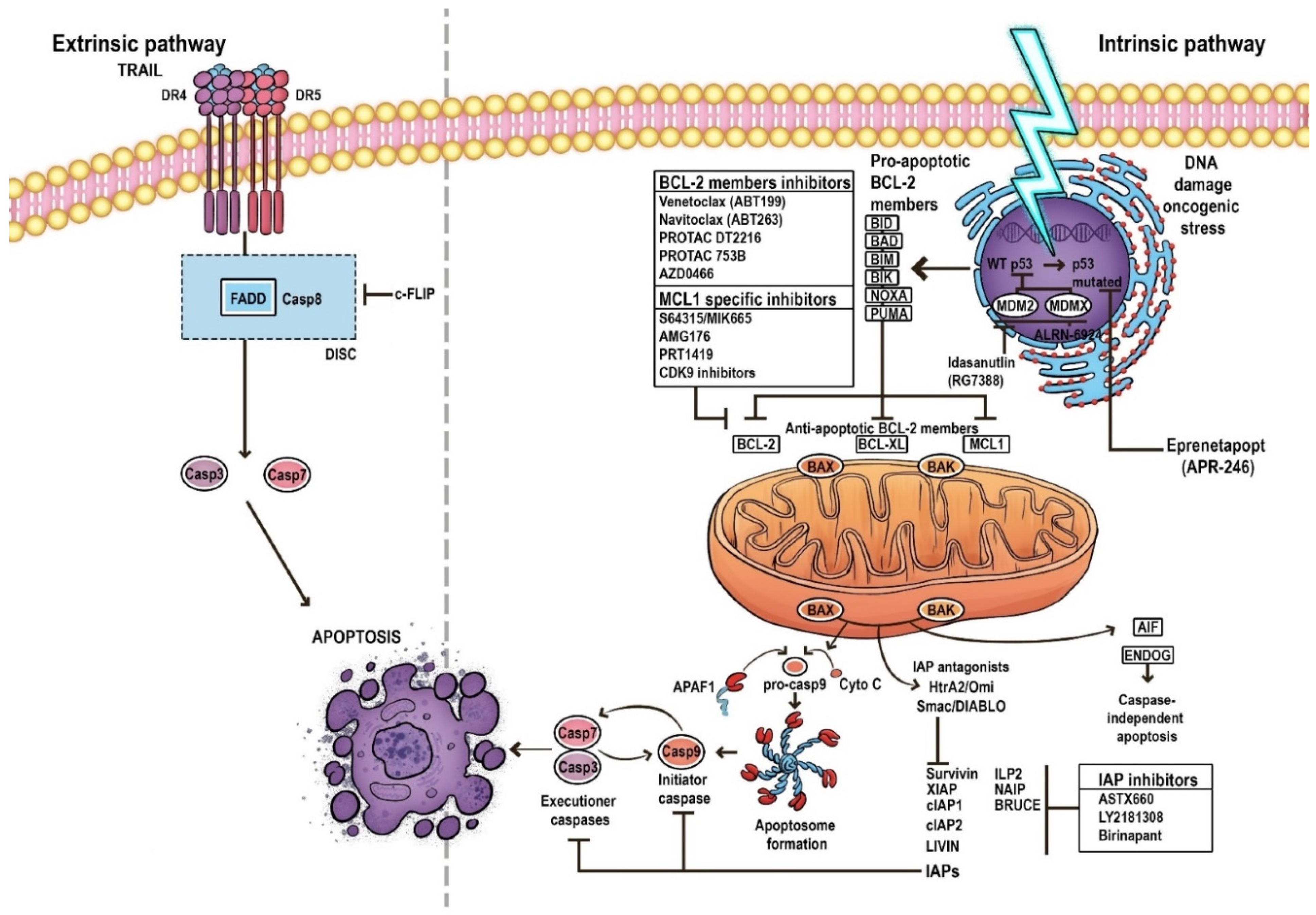
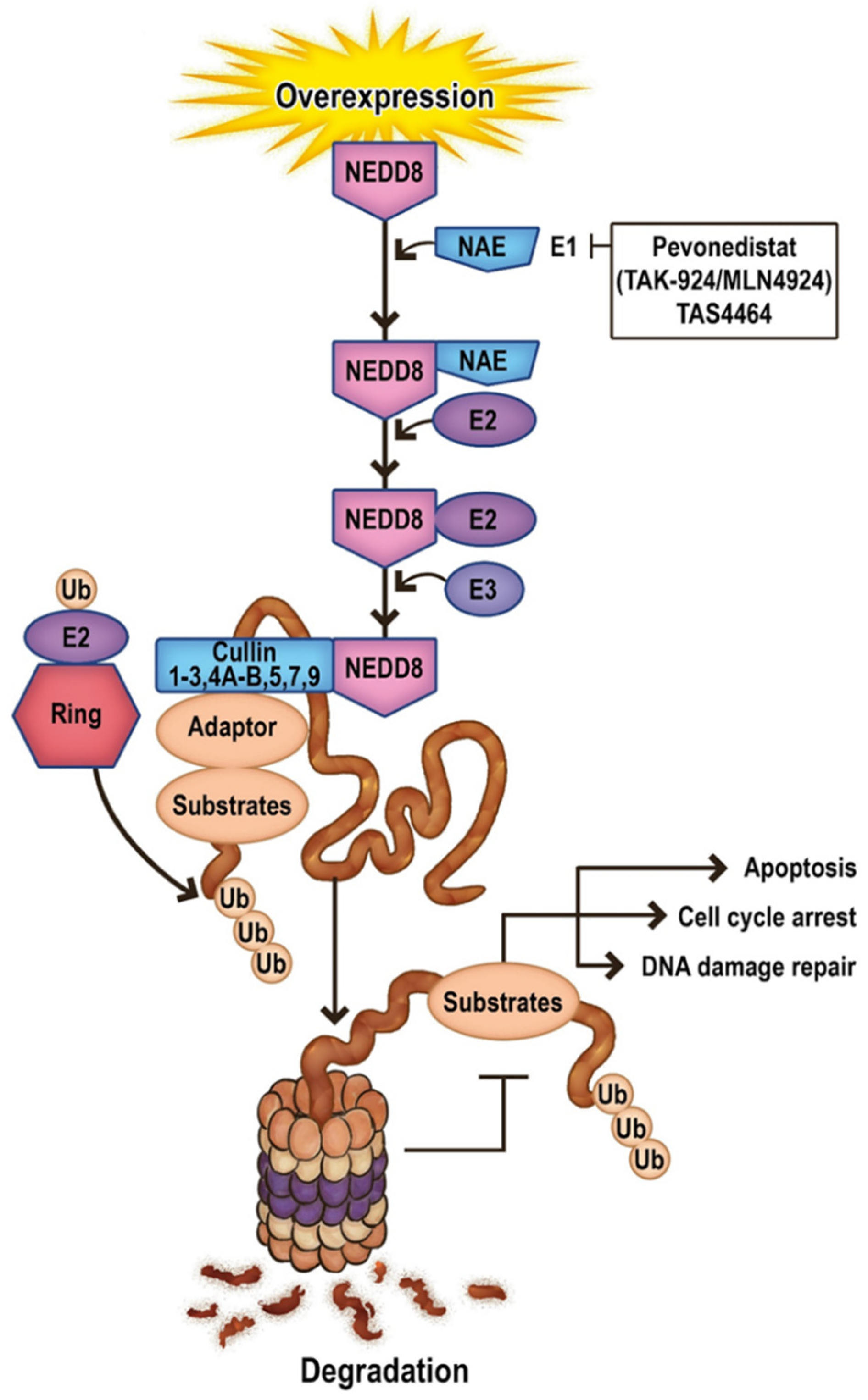
Table 1.
List of ongoing clinical trials in phase II-III in AML testing venetoclax (ABT-199).
| Ongoing Trial | Intervention/Treatment | Clinical Trial Phase | Serial NCT Number | Current Status |
|---|---|---|---|---|
| CC-90011 given concurrently with Venetoclax and Azacitidine in r/r AML and treatment-naive subjects with AML who are not eligible for intensive induction chemotherapy. | CC-90011, Venetoclax, Azacitidine | Phase I/II | NCT04748848 | Completed |
| Milademetan tosylate with cytarabine with or without ventoclax in treating participants with r/r AML. | Cytarabine, Milademetan Tosylate, Venetoclax | Phase I/II | NCT03634228 | Completed |
| Low-dose cytarabine or azacitidine plus venetoclax and quizartinib in newly diagnosed AML patients aged equal or more than 60 years old. | Azacitidine, Venetoclax, Quizartinib, Cytarabine | Phase I/II | NCT04687761 | Recruiting |
| CA-4948 monotherapy and in combination with azacitidine or venetoclax in AML patients. | Emavusertib, Azacitidine, Venetoclax | Phase I/IIa | NCT04278768 | Active, not recruiting |
| Anti-OX40 antibody PF-04518600 (OX40) with or without venetoclax, avelumab, glasdegib, gemtuzumab ozogamicin, and azacitidine in treating r/r AML. | PF-04518600, Avelumab, Azacitidine, Gemtuzumab Ozogamicin, Glasdegib, Glasdegib Maleate, Venetoclax | Phase Ib/II | NCT03390296 | Active, not recruiting |
| VEN-OM assessing venetoclax with escalating doses of omacetaxine in patients with r/r AML. | Omacetaxine, Venetoclax | Phase Ib/II | NCT04926285 | Recruiting |
| ENAVEN-AML assessing enasidenib in combination with venetoclax in patients with IDH2-mutated r/r AML. | Enasidenib, Venetoclax | Phase Ib/II | NCT04092179 | Recruiting |
| Omacetaxine with venetoclax in treating patients with r/r AML and have a genetic change RUNX1. | Omacetaxine Mepesuccinate, Venetoclax | Phase Ib/II | NCT04874194 | Recruiting |
| IMGN632 with azacitidine and/or venetoclax in patients with relapsed and frontline CD123-positive AML, and antileukemia activity of IMGN632 when administered as monotherapy in patients with MRD+ AML after frontline treatment. | Azacitidine, IMGN632, Venetoclax | Phase Ib/II | NCT04086264 | Recruiting |
| Decitabine/cedazuridine (ASTX727) and venetoclax in combination with ivosidenib or enasidenib in r/r AML. | Decitabine, Cedazuridine, Enasidenib, Ivosidenib, Venetoclax | Phase Ib/II | NCT04774393 | Recruiting |
| Venetoclax in combination with quizartinib in r/r FLT3 (+) AML. | Quizartinib, Venetoclax | Phase Ib/II | NCT03735875 | Active, not recruiting |
| Venetoclax with ivosidenib with or without azacitidine, in treating patients with IDH1-mutated AML. | Azacitidine, Ivosidenib, Venetoclax | Phase Ib/II | NCT03471260 | Recruiting |
| Quizartinib, decitabine, and venetoclax in treating participants with acute myeloid leukemia that is untreated or has come back (relapsed). | Decitabine, Quizartinib, Venetoclax | Phase I/II | NCT03661307 | Recruiting |
| DS-1594b with or without azacitidine, venetoclax, or mini-HCVD in treating patients with r/r AML. | DS-1594b, Azacitidine, Venetoclax, Mini-HCVD | Phase I/II | NCT04752163 | Active, not recruiting |
| Gilteritinib with azacitidine and venetoclax in treating patients with FLT3-mutation positive r/r acute myeloid leukemia. | Azacitidine, Gilteritinib, Venetoclax | Phase I/II | NCT04140487 | Recruiting |
| Venetoclax with azacitidine and pevonedistat in treating patients with newly diagnosed AML. | Azacitidine, Pevonedistat, Venetoclax | Phase I/II | NCT03862157 | Active, not recruiting |
| Venetoclax and lintuzumab-ac225 in patients with with CD33 positive r/r AML. | Lintuzumab-Ac225, Venetoclax, Spironolactone | Phase I/II | NCT03867682 | Recruiting |
| RELAX assessing venetoclax in combination with increasing cytarabine doses plus mitoxantrone in r/r AML. | Venetoclax, Cytarabine, Mitoxantron | Phase I/II | NCT04330820 | Recruiting |
| Gilteritinib given together with ASTX727 and venetoclax in treating patients with FLT3-mutated AML that is newly diagnosed or r/r. | Decitabine, Cedazuridine, Gilteritinib, Venetoclax | Phase I/II | NCT05010122 | Recruiting |
| Liposome-encapsulated daunorubicin-cytarabine (CPX-351) and venetoclax in r/r or untreated AML patients. | CPX-351, Venetoclax | Phase II | NCT03629171 | Recruiting |
| Venetoclax, cladribine, low dose cytarabine, and azacitidine in treating patients with AML that has previously not been treated. | Azacitidine, Cladribine, Cytarabine, Venetoclax | Phase II | NCT03586609 | Recruiting |
| AML secondary to myeloproliferative neoplasms unfit for intensive chemotherapy investigating a treatment combination including decitabine and venetoclax. | Venetoclax, Decitabine | Phase II | NCT04763928 | Recruiting |
| Azacitidine, venetoclax, and trametinib in r/r AML. | Azacitidine, Trametinib, Venetoclax | Phase II | NCT04487106 | Recruiting |
| Venetoclax together with busulfan, cladribine, and fludarabine in treating patients with high-risk acute myeloid leukemia who are undergoing stem cell transplant. | Busulfan, Cladribine, Fludarabine Phosphate, HSCT, Thiotepa, Venetoclax | Phase II | NCT04708054 | Recruiting |
| STIMULUS-AML1 assessing the efficacy of MBG453 in combination with the HMA azacitidine and venetoclax. | MBG453, Venetoclax, Azacitidine | Phase II | NCT04150029 | Active, not recruiting |
| (V-FIRST) assessing the efficacy of venetoclax in combination with fludarabine, cyratabine, and idarubicine in induction for AML patients with poor prognosis. | Venetoclax, Fludarabine, Cyratabine, Idarubicine | Phase II | NCT03455504 | Recruiting |
| Decitabine, venetoclax, and ponatinib for the treatment of Philadelphia chromosome-positive AML. | Decitabine, Ponatinib, Venetoclax | Phase II | NCT04188405 | Recruiting |
| Venetoclax plus azacitidine in newly diagnosed AML in patients who cannot receive intensive chemotherapy. | Low-dose Venetoclax, Azacitidine | Phase II | NCT05048615 | Recruiting |
| Venetoclax and ASTX727 in relapsed/refractory (r/r) AML or unfit patients and the same combination for higher-risk AML patients without FLT3 (NCT04817241). | Decitabine, Cedazuridine, Venetoclax | Phase II | NCT04746235 | Recruiting |
| Azacytidine + venetoclax versus conventional cytotoxic chemotherapy in induction-eligible AML patients. | Cytarabine, Idarubicin, Daunorubicin, Venetoclax, Azacitidine | Phase II | NCT04801797 | Recruiting |
| Azacitidine and venetoclax with or without pembrolizumab in treating older patients with newly diagnosed acute myeloid leukemia who are ineligible or who refuse intensive chemotherapy. | Azacitidine, Pembrolizumab, Venetoclax | Phase II | NCT04284787 | Recruiting |
| Venetoclax and sequential busulfan, cladribine, and fludarabine phosphate before donor stem cell transplant in treating patients with AML. | Busulfan, Cladribine, Fludarabine, Venetoclax | Phase II | NCT02250937 | Active, not recruiting |
| Venetoclax and azacitidine for non-elderly adult patients with acute myeloid leukemia. | Venetoclax, Azacitidine | Phase II | NCT03573024 | Recruiting |
| BP1001 (a liposomal Grb2 antisense oligonucleotide) in combination with venetoclax plus decitabine in patients with AML who are ineligible for intensive induction therapy. | BP1001, Ventoclax, Decitabine | Phase IIa | NCT02781883 | Recruiting |
| Therapeutic efficacy of (venetoclax and decitabine) versus conventional “7 + 3” chemotherapy in induction young patients with AML. | Venetoclax, Decitabine, Cytarabine, Idarubicin | Phase III | NCT05177731 | Recruiting |
| ENHANCE-3 study comparing the efficacy of magrolimab + venetoclax + azacitidine versus placebo + venetoclax + azacitidine in unfit de novo AML. | Magrolimab, Venetoclax, Azacitidine | Phase III | NCT05079230 | Recruiting |
| ENHANCE-2 comparing the efficacy of magrolimab + azacitidine versus venetoclax + azacitidine in adults with previously untreated TP53 mutant acute myeloid leukemia (AML) who are appropriate for non-intensive therapy as measured by overall survival (OS). | Magrolimab, Venetoclax, Azacitidine, Cytarabine, Daunorubicin, Idarubicin | Phase III | NCT04778397 | Recruiting |
| VIALE-T assessing the efficacy of venetoclax in combination with azacitidine to improve Relapse Free Survival (RFS) in AML participants compared to Best Supportive Care (BSC) when given as maintenance therapy following allo-HSCT. | Venetoclax, Azacitidine | Phase III | NCT04161885 | Recruiting |
Abbreviations: AML, acute myeloid leukemia; MRD, measurable residual disease; FLT3, fms-like tyrosine kinase 3; mini-HCVD, hyperfractionated cyclophosphamide, vincristine, doxorubicin, and dexamethasone; HMA, DNA hypomethylating agent.
Table 2.
Characteristics of apoptosis-targeting drug trials in AML.
| Author; Year | Target Disease | Intervention Arm | Control Arm | Number of Patients | Important Findings |
|---|---|---|---|---|---|
| BCL2 inhibitor (Venetoclax) | |||||
| Konopleva et al. 2016 | High-risk r/r AML or patients unfit for intensive chemotherapy | Ven | - | 32 |
|
| |||||
| |||||
| DiNardo et al. 2019 | Naïve AML or patients unfit for intensive chemotherapy | Ven + HMA (AZA/DEC) | - | 145 |
|
| |||||
| |||||
| Wei et al. 2019 | Naïve AML or patients unfit for intensive chemotherapy | Ven + LDAC | - | 82 |
|
| |||||
| DiNardo et al. 2020 VIALE-A | Naïve AML, ineligible for intensive chemotherapy | Ven + AZA | Placebo + AZA | 431 |
|
| |||||
| |||||
| Wei et al. 2021 VIALE-C | Naïve AML, ineligible for intensive chemotherapy | Ven + LDAC | Placebo + LDAC | 211 |
|
| |||||
| |||||
| IAP Antagonists | |||||
| Schimmer et al. 2011 | r/r AML | AEG35156 + high-dose cytarabine and idarubicin | High-dose Ara-C + Idarubicin | 40 |
|
| |||||
| Erba et al. 2013 | r/r AML | LY2181308 alone or with Idarubicin + Ara-C | - | 24 |
|
| |||||
| |||||
| |||||
| Frey et al. 2014 | r/r AML or high risk MDS refractory to HMA | Birinapant | - | 20 |
|
| CDK9 inhibitors—Indirect MCL1 Inhibitors | |||||
| Zeidner et al. 2021 | Naïve AML ≤ 65 years | Alvocidib | - | 32 |
|
| Cytarabine |
| ||||
| Daunorubicin |
| ||||
| Zeidner et al. 2015 | Naïve AML with intermediate/adverse-risk cytogenetics | Alvocidib, Cytarabine, Mitoxantrone | Cytarabine | 165 |
|
| (FLAM) | +Daunorubicin |
| |||
| Targeting p53 | |||||
| 1. The restoration of wild-type TP53 | |||||
| Sallman et al. 2021 | TP53-mutant AML with 20–30% marrow blasts or MDS | Eprenetapopt (APR-246) + AZA | - | 55 (n = 11 AML) |
|
| |||||
| NCT03072043 | |||||
| Cluzeau et al. | TP53-mutant AML or MDS | Eprenetapopt (APR-246) + AZA | - | 53 (n = 18 AML) |
|
| 2021 |
| ||||
| |||||
| NCT03588078 | |||||
| 2. MDM2 Inhibitors | |||||
| Yee et al. 2021 | r/r AML, s-AML, t-AML, or naïve AML, ineligible for intensive chemotherapy | Idasanutlin (RG-7388) + Ara-C | Idasanutlin | 122 |
|
| |||||
| Daver et al. 2019 | r/r AML, ineligible for intensive chemotherapy | Idasanutlin + VEN | - | 49 |
|
| |||||
Abbreviations: AML, acute myeloid leukemia; Ven, venetoclax; mOS, median overall survival; CR/CRi, complete remission/CR with incomplete blood count recovery HMA, DNA hypomethylating agent; AZA, azacytidine; DEC, decitabine; LDAC, low-dose cytarabine; CR/CRp, CR/CR with incomplete platelet recovery; SD, stable disease; MDS, myelodysplastic syndrome; ORR, overall response rate; FLAM, fludarabine, cytarabine, and mitoxantrone; NGS, next-generation sequencing; Idasa, idasanutlin; s-AML, secondary AML; t-AML, therapy-related AML; Ara-C, cytosine arabinose.
Table 3.
Ongoing clinical trials in AML involving molecules affecting apoptosis, as a single agent or in combination.
Table 3.
Ongoing clinical trials in AML involving molecules affecting apoptosis, as a single agent or in combination.
| Agents and Ongoing Trials | Target Disease | Intervention Arm | Control Arm | Estimated Enrollment | Design; Clinical Trial Phase | Serial NCT Number | Current Status |
|---|---|---|---|---|---|---|---|
| BCL-2, BCL-XL, BCL-W inhibitors | |||||||
| Navitoclax; BCL-2/BCL-XL/BCL-W inhibitor; | r/r AML Previously Treated with Venetoclax | Navitoclax, Venetoclax, Decitabine | - | 24 | Phase Ib, Open-Label study | NCT05222984 | Recruiting |
| AZD0466 BCL-2/BCL-XL inhibitor NIMBLE | r/r AML | AZD0466 | - | 141 | Phase I/II, Open-Label study | NCT04865419 | Recruiting |
| Direct MCL1 Inhibitors | |||||||
| S64315/MIK665 | r/r AML, ineligible for intensive chemotherapy | S64315 + AZA | - | 180 | Phase I/II, Open-Label study | NCT04629443 | Recruiting |
| S64315/MIK665 | r/r AML, ineligible for intensive chemotherapy | S64315 + VEN | - | 40 | Phase Ib, Open-Label study | NCT03672695 | Recruiting |
| S64315/MIK665 | r/r AML, ineligible for intensive chemotherapy | S64315+ S65487 (VOB560) | - | 170 | Phase Ib, Open-Label study | NCT04702425 | Recruiting |
| AMG176 | r/r AML | AMG176 ± AZA | - | 175 | Phase I, Open-Label study | NCT02675452 | Recruiting |
| PRT1419 | r/r AML | PRT1419 | - | 36 | Phase I, Open-Label study | NCT04543305 | Active, not recruiting |
| PRT1419 | r/r AML | PRT1419 | - | 30 | Phase I, Open-Label study | NCT05107856 | Recruiting |
| AZD5991 | r/r AML, ineligible for intensive chemotherapy | AZD5991 ± VEN | - | 144 | Phase I/II, Open-Label study | NCT03218683 | Terminated |
| CDK9 inhibitors—Indirect MCL1 Inhibitors | |||||||
| Alvocidib | r/r AML | Alvocidib + VEN | - | 36 | Phase Ib, Open-Label study | NCT03441555 | Completed |
| Dinaciclib/MK7965 | r/r AML | Dinaciclib (MK7965) + VEN | - | 48 | Phase Ib, Open-Label study | NCT03484520 | Active, not recruiting |
| CYC065 | r/r AML | CYC065 + VEN | - | 25 | Phase I, Open-Label study | NCT04017546 | Active, not recruiting |
| AZD4573 | r/r AML | AZD4573 | - | 44 | Phase I, Open-Label study | NCT03263637 | Completed |
| IAP Antagonists | |||||||
| ASTX660 | r/r AML | ASTX660 ± ASTX727 | - | 68 | Phase I, Open-Label study | NCT04155580 | Terminated |
| Targeting p53 | |||||||
| 1. The restoration of wild-type TP53 | |||||||
| Eprenetapopt/APR-246 | TP53-mutant AML | Eprenetapopt (APR-246) + AZA + VEN | - | 51 | Phase I, Open-Label study | NCT04214860 | Completed |
| Eprenetapopt/APR-246 | TP53-mutant AML after allo-HSCT | Eprenetapopt (APR-246) + AZA | - | 33 | Phase II, Open-Label study | NCT03931291 | Completed |
| 2. MDM2 Inhibitors | |||||||
| Idasanutlin | Newly diagnosed AML | Idasanutlin+ Cytarabine + Daunorubicin | - | 24 | Phase Ib/II, Open-Label Study | NCT03850535 | Terminated |
| Idasanutlin | r/r AML, ineligible for intensive chemotherapy | Idasanutlin + Cobimetinib + Venetoclax | - | 88 | Phase Ib, Open-Label Study | NCT02670044 | Completed |
| Idasanutlin | r/r AML | Idasanutlin + Ara-C | Placebo + Ara-C | 447 | Phase III, Double-Blind, Randomized Study | NCT02545283 | Terminated |
| 3. MDM2/MDMX inhibitors | |||||||
| ALRN-6924 | r/r AML | ALRN-6924 ± Ara-C | - | 55 | Phase I/Ib Open-Label Study | NCT02909972 | Completed |
Abbreviations: r/r AML, relapsed/refractory AML; AZA, azacitidine; Ven, venetoclax; allo-HSCT, allogeneic hematopoietic stem cell transplantation; Ara-C, cytosine arabinose.
Table 4.
Current ongoing clinical trials in AML involving molecules affecting neddylation.
| Targeting Neddylation (NAE Inhibitors) | |||||||
|---|---|---|---|---|---|---|---|
| Agents and Ongoing Trials | Target Disease | Intervention Arm | Control Arm | Estimated Enrollment | Design; Clinical Trial Phase | Serial NCT Number | Current Status |
| Pevonedistat/TAK-924/MLN4924 | AML, ineligible for intensive chemotherapy | Pevonedistat + VEN + AZA | VEN + AZA | 164 | Phase II, Open-Label study | NCT04266795 | Active, not recruiting |
| Pevonedistat/TAK-924/MLN4924 | r/r AML | Pevonedistat + Belinostat | - | 30 | Phase I, Open-Label study | NCT03772925 | Recruiting |
| Pevonedistat/TAK-924/MLN4924 | Naïve high-risk AML with at least 1: adverse genetic features/t-AML/AML with antecedent MDS/≥55 years and considered fit for chemotherapy/AML with MDS-related changes | Pevonedistat + Ara-C + Idarubicin | - | 53 | Phase Ib/II, Open-Label study | NCT03330821 | Active, not recruiting |
| Pevonedistat/TAK-924/MLN4924 | r/r AML or r/r MDS | Pevonedistat + AZA + Ara-C + Fludarabine Phosphate + Methotrexate | - | 12 | Phase I, Open-Label study | NCT03813147 | Active, not recruiting |
| Pevonedistat/TA K-924/MLN4924 | Newly diagnosed s-AML or newly diagnosed CMML/MDS or post-HMA failure CMML/MDS | Pevonedistat +VEN + AZA | - | 40 | Phase I/II, Open-Label study | NCT03862157 | Active, not recruiting |
| Pevonedistat/TAK-924/MLN4924 | Newly diagnosed AML not eligible for intensive chemotherapy | Pevonedistat + AZA | AZA | 466 | Phase III | NCT04090736 | Active, not recruiting |
| Pevonedistat/TAK-924/MLN4924 | r/r AML (dose-escalation phase), newly diagnosed AML or r/r AML (expansion phase) | Pevonedistat +VEN + HMA | - | 24 | Phase I | NCT04172844 | Active, not recruiting |
| Pevonedistat/TAK-924/MLN4924 | AML or MDS after first CR with intensive chemotherapy or CR after allo-HSCT with MRD | Pevonedistat + AZA | AZA | 102 | Phase II | NCT04712942 | Active, not recruiting |
Abbreviations: Ven, venetoclax; AZA, azacytidine; r/r AML, relapsed/refractory AML; t-AML, therapy-related AML; MDS, myelodysplastic syndrome; Ara-C, cytosine arabinose; s-AML, secondary AML; CMML, chronic myelomonotic leukemia; HMA, hypomethylating agent; CR, complete remission; allo-HSCT, allogeneic hematopoietic stem cell transplantation; MRD, measurable residual disease.
Publisher’s Note: MDPI stays neutral with regard to jurisdictional claims in published maps and institutional affiliations. |
© 2022 by the authors. Licensee MDPI, Basel, Switzerland. This article is an open access article distributed under the terms and conditions of the Creative Commons Attribution (CC BY) license (https://creativecommons.org/licenses/by/4.0/).
Share and Cite
MDPI and ACS Style
Krawiec, K.; Strzałka, P.; Czemerska, M.; Wiśnik, A.; Zawlik, I.; Wierzbowska, A.; Pluta, A. Targeting Apoptosis in AML: Where Do We Stand? Cancers 2022, 14, 4995. https://0-doi-org.brum.beds.ac.uk/10.3390/cancers14204995
AMA Style
Krawiec K, Strzałka P, Czemerska M, Wiśnik A, Zawlik I, Wierzbowska A, Pluta A. Targeting Apoptosis in AML: Where Do We Stand? Cancers. 2022; 14(20):4995. https://0-doi-org.brum.beds.ac.uk/10.3390/cancers14204995
Chicago/Turabian StyleKrawiec, Kinga, Piotr Strzałka, Magdalena Czemerska, Aneta Wiśnik, Izabela Zawlik, Agnieszka Wierzbowska, and Agnieszka Pluta. 2022. "Targeting Apoptosis in AML: Where Do We Stand?" Cancers 14, no. 20: 4995. https://0-doi-org.brum.beds.ac.uk/10.3390/cancers14204995
Note that from the first issue of 2016, this journal uses article numbers instead of page numbers. See further details here.