Article

The Indicative Value of Serum Tumor Markers for Metastasis and Stage of Non-Small Cell Lung Cancer

1
Department of Thoracic Surgery, National Regional Medical Center, Binhai Campus of the First Affiliated Hospital, Fujian Medical University, Fuzhou 350212, China
2
Department of Thoracic Surgery, The First Affiliated Hospital, Fujian Medical University, 20 Chazhong Road, Fuzhou 350005, China
3
Department of Thoracic Surgery, Tianjin Union Medical Center, Nankai University, Tianjin 300121, China
4
Department of Occupational and Environmental Health, School of Public Health, Tongji Medical College, Huazhong University of Science and Technology, Wuhan 430030, China
5
Department of Respiratory, The Second People’s Hospital of Linhai City, Linhai 317000, China
6
Department of Cancer Prevention, Tianjin Medical University Cancer Institute and Hospital, Tianjin 300181, China
7
Baodi District People’s Hospital of Tianjin, Tianjin Baodi Hospital of Tianjin Medical University, Tianjin 301000, China
8
Medical College, Nankai University, Tianjin 300071, China
9
Department of Thoracic Surgery, Tianjin Medical University Cancer Institute and Hospital, Tianjin 300181, China
10
National Clinical Research Center for Cancer, Key Laboratory of Cancer Prevention and Therapy, Tianjin 300181, China
*
Authors to whom correspondence should be addressed.
These authors contributed equally to this work.
Submission received: 27 September 2022 / Revised: 11 October 2022 / Accepted: 13 October 2022 / Published: 16 October 2022
(This article belongs to the Special Issue Biomarkers of Tumor Metastasis and Invasiveness)

Abstract

:

Simple Summary

This study recruited 3272 non-small cell lung cancer (NSCLC) cases to analyze the predictive abilities of serum tumor markers (CEA, SCC-Ag, CYFRA 21-1, NSE, ProGRP, TPSA and CA199) for metastasis and clinical stage, and found that tumor marker levels may be indicative of tumor metastasis (intrapulmonary, lymphatic and distant metastasis) and stage. Increased CEA and CA199 provided an accurate prediction of intrapulmonary and distant metastasis. Increased CEA, CYFRA 21-1 and CA199 provided an accurate prediction of lymphatic metastasis and higher tumor stage. Combined detection of serum tumor markers can indicate tumor metastasis and stage in NSCLC patients.

Abstract

Objective: This study aimed to explore the roles of serum tumor markers for metastasis and stage of non-small cell lung cancer (NSCLC). Methods: This study recruited 3272 NSCLC patients admitted to the Tianjin Union Medical Center and the Tianjin Medical University Cancer Institute and Hospital. The predictive abilities of some serum tumor markers (carcinoembryonic antigen (CEA), squamous cell carcinoma antigen (SCC-Ag), cytokeratin-19 fragment (CYFRA 21-1), neuron-specific enolase (NSE), pro-gastrin-releasing peptide (ProGRP), total prostate-specific antigen (TPSA) and carbohydrate antigen 199 (CA199)) for NSCLC metastasis (intrapulmonary, lymphatic and distant metastasis) and clinical stage were analyzed. Results: Tumor markers exhibited different numerical and proportional distributions in NSCLC patients. Elevated CEA, CYFRA 21-1 and CA199 levels were indicative of tumor metastasis and stage. Increased CEA and CA199 provided an accurate prediction of intrapulmonary and distant metastasis with the area under the receiver operator characteristic curve (AUC) of 0.69 both (p < 0.001); Increased CEA, CYFRA 21-1 and CA199 provided an accurate prediction of lymphatic metastasis with the AUC of 0.62 (p < 0.001). Conclusion: Combined detection of serum tumor markers can indicate tumor metastasis and stage in NSCLC patients.

Graphical Abstract

1. Introduction

Lung cancer has been identified as one of the most common malignant tumors. In recent years, the incidence of lung cancer has gradually increased. Among different malignancies, lung cancer has the fastest-growing incidence and mortality, becoming the biggest threat to people’s health and life [1,2]. Lung cancer is divided into two types: non-small cell lung cancer (NSCLC) and small cell lung cancer (SCLC). NSCLC accounts for about 80–85% of lung cancers, including squamous cell carcinoma, adenocarcinoma, and large cell carcinoma [3]. Compared with SCLC, NSCLC cells grow and divide slowly and metastasize relatively late [4]. The early clinical symptoms of lung cancer are not obvious; thus, lung cancer is usually diagnosed at an advanced stage, and optimal treatment and surgical opportunities are lost. Thus, improving the early detection rate of lung cancer is needed to immediately adopt positive treatment measures to reduce the harm of the disease.
Tumor markers reflect the presence of the tumor, and changes in the presence and level of markers indicate the nature of the tumor. Detecting tumor markers facilitates the early diagnosis and operation of tumor development. In recent years, more serum tumor markers have been identified for the early diagnosis of lung cancer. Due to the low sensitivity and specificity of single serum tumor markers, detecting multiple tumor markers has been used to improve the sensitivity and specificity of clinical diagnosis in lung cancer patients. Therefore, the application of single tumor markers has gradually progressed to the diagnostic use of multiple markers, thus improving the positive rate of diagnosis and monitoring the development of lung cancer [5,6]. Clinically significant serum tumor markers for lung cancer include carcinoembryonic antigen (CEA), cytokeratin-19 fragment (CYFRA 21-1), neuron-specific enolase (NSE), squamous cell carcinoma antigen (SCC-Ag), pro-gastrin-releasing peptide (ProGRP), total prostate-specific antigen (TPSA) and carbohydrate antigen 199 (CA199) [7,8,9,10]. Numerous studies have reported the application of these tumor markers in the diagnosis of lung cancer. Clinical studies have also focused on the use of these markers for monitoring treatment efficacy and prognosis; furthermore, progress has been made in the application of these markers [11,12,13,14,15]. High levels of tumor markers at baseline are correlated with worse survival in stage III-IV NSCLC patients [13]. Tumor markers such as CYFRA21-1, SCC-Ag, NSE, and CEA in the serum of lung cancer patients are significantly increased, and the degree of elevation are significantly correlated with tumor invasion, clinical stage and lymph node metastasis [16].
Serum tumor markers have been used for the early diagnosis of lung cancer and the clinical practice of tumor efficacy monitoring for more than 10 years. However, confirmative studies with large clinical sample size on the consistency of various tumor markers for determining the pathology and tumor progression of lung cancer remain lacking. Thus, the purpose of this study was to explore the effectiveness of serum markers in determining tumor metastasis and stage in lung cancer patients from two clinical centers with a large sample size.

2. Materials and Methods

2.1. Patients and Control Subjects

In total, 4690 lung cancer patients admitted to Tianjin Union Medical Center from September 2016 to September 2019 and 2700 lung cancer patients admitted to Tianjin Medical University Cancer Institute and Hospital from January 2018 to September 2019 were screened as study subjects. All patients were screened using case data, relevant laboratory examination data, imaging data, and pathological examination data. Patients were determined to be all Chinese from north China and northeast China. The inclusion criteria were complete information including age, sex, smoking history, and other basic data of the patients. Patients were excluded from the study if they had other tumors, inflammation in the lungs or areas other than the lungs, or a history of chronic gastritis or ulcer in the digestive system. A total of 3272 patients with NSCLC were included in this study.
Fasting blood samples were taken for determination of lung cancer-related serum tumor markers before surgery, chemotherapy, radiotherapy, or other special treatments at the first admission. The pathological diagnosis was based on lung cancer surgery, lung puncture biopsy or tracheoscopy. Pathological diagnoses of lung cancer included squamous cell carcinoma, adenocarcinoma, adenosquamous cell carcinoma, etc. Data entry for all patients included smoking index, intrapulmonary, lymphatic and distant metastasis, and tumor stage (according to the International Association for the Study of Lung Cancer, IASLC 2015 TNM stage for lung cancer).

2.2. Sample Collection and Measurement

All the patients had an empty stomach the morning after the first admission without any treatment. Venous blood (5 mL) was collected from each patient to detect lung cancer-related tumor markers. The whole blood was separated into serum and cellular fractions within 2 h by centrifugation at 4000 rpm for 10 min. Serum samples were obtained after separation, and serum concentrations of tumor markers were determined. CEA, SCC-Ag and CA199 were determined by chemiluminescent microparticle immunoassay (CMIA) using an Abbott ARCHITECT I2000SR automatic chemical microparticle immune analyzer and its supporting reagents. CYFRA 21-1, NSE, ProGRP, and TPSA were determined using a Roche Elecsys 2010 automatic electrochemiluminescence immune analyzer and its supporting reagents.

2.3. Statistical Analysis

All tumor markers had non-normal distribution, and markers were represented by the median (P25–P75). Nonparametric tests were used to compare the concentrations of tumor markers and the smoking index between the different groups. Chi-square tests were also used to determine the distribution differences of basic information (age, sex and smoking index) and tumor markers among different groups. The Bonferroni method was used for paired comparisons. Binary logistic analysis was used to analyze the influencing factors for lung tumor metastasis, lymphatic metastasis and distant metastasis, while ordinal logistic analysis was used in examining the influencing factors for tumor stage. The two logistic analyses were divided into two steps: (1) univariate factor analysis and logistic analysis for each potential influencing factor was conducted; (2) influencing factors of p < 0.2 [17] in univariate analysis were included in a multivariate logistic analysis. Finally, the prediction probabilities of tumor markers with p < 0.05 were reassessed by logistic analysis using multivariate analysis, and the receiver operating characteristic (ROC) curves were used for joint predictions. SPSS 24.0 (IBM, Chicago, IL, USA) was used for all analyses. For two-sided tests, a p value less than 0.05 was considered significant.

3. Results

3.1. Demographics and Clinical Characteristics

In total, 3272 NSCLC patients were analyzed in this study. Patient characteristics are described in Table 1. The correlations of age, sex, and smoking index with tumor metastasis, and the stage of patients with NSCLC, are presented in Table 2. The percentages of intrapulmonary and lymphatic metastases were higher in male patients than in female patients. Distribution ratios of tumor stage were statistically different; the proportions of patients with stages II–IV were lower than stage I; the proportions of patients with stages II and III were lower than stage IV.
The distribution of tumor stage, intrapulmonary and lymphatic metastases in patients ≤ 61 years old were statistically different from patients > 61 years. In patients > 61 years, the number of patients with stage II and III cancers was lower than stage I. Patients with lung tumor metastasis had a higher smoking index. Smoking indexes were statistically different for patients with different tumor stages.

3.2. Clinical Data and Risk Factor of Tumor Metastasis and Stage in NSCLC Patients

As shown in Table 3 and Table 4, the levels of CEA, CYFRA 21-1 and NSE were significantly higher in patients with intrapulmonary, lymphatic and distant metastases when compared with those in non-metastatic patients. The levels of SCC-Ag, ProGRP and TPSA in patients with lymphatic metastasis were significantly higher than those in non-metastatic patients. The levels of CA199 in patients with lymphatic and distant metastases were significantly higher than those in non-metastatic patients. Moreover, the levels of the six tumor markers CEA, SCC-Ag, CYFRA 21-1, NSE, TPSA and CA199 were significantly different in patients with different tumor stages.
The results of the univariate analysis were summarized in Table 5. Single factors with p < 0.2 were included in the multivariate logistic analysis. The results of the collinearity analysis were presented in Table 6. The results of the multi-factor analysis were presented in Table 7. Binary logistic regression analysis showed that age, smoking index, CEA and CA199 were independent factors for intrapulmonary metastasis; age, CEA, CYFRA 21-1 and CA199 were independent factors for lymphatic metastasis; and age, CEA and CA199 were independent factors for distant metastasis. Ordinal logistic analysis showed that gender, age, adenocarcinoma (vs. squamous carcinoma), CEA, CYFRA 21-1, NSE and CA199 were independent factors for tumor stage.

3.3. The Predictions of Single and Combined Factors for Tumor Metastasis and Stage in NSCLC Patients

Multivariate analysis of tumor markers with p < 0.05 was followed by a logistic analysis of prediction probability. For analysis of the influencing factors of tumor metastasis and clinical stage of NSCLC patients, the area under the ROC curve (AUC) for factors is shown in Table 8. ROC curves were used to predict lung cancer metastasis and stage, and the results were shown in Figure 1. CEA and CA199 provided an accurate prediction of intrapulmonary and distant metastasis with the AUC of 0.69 both (p < 0.001); CEA, CYFRA 21-1 and CA199 provided an accurate prediction of lymphatic metastasis with the AUC of 0.62 (p < 0.001).

4. Discussion

Tumor markers have been widely used in the clinical diagnosis and treatment of malignant tumors as they serve as important indicators of disease outcome monitoring. At present, varied tumor markers, such as CEA, SCC-Ag, CYFRA 21-1, NSE, are applied in diagnosing lung cancer, which can also be used to monitor metastasis and recurrence of NSCLC [18]. Although tumor markers are widely used in clinical practice, the clinical analysis and validation of these markers using a large sample size remain lacking. In this study, a large sample of lung cancer patients from two medical centers was selected to verify the accuracy of tumor markers from multiple perspectives, including predicting tumor metastasis and clinical stage. Our results support the use of these markers in clinical practice.
CEA is widely found in adult cancer tissues and has been used in the auxiliary diagnosis, efficacy observation, prognostic judgment, and recurrence prediction of cancer [19,20]. CEA elevation is common in multisystem tumors, including lung cancer [21]. Due to the non-specificity of this indicator, CEA is often used in combination with other tumor markers in clinical practice [22,23]. SCC-Ag participates in the regulation of protein decomposition during malignant transformation, and it is the preferred tumor marker for cervical squamous cell carcinoma [24,25]. Additionally, this marker is observed to increase in lung squamous cell cancers [26]. CYFRA 21-1 is highly expressed in lung squamous cell carcinoma compared with adenocarcinoma and SCLC [27]. The use of increased serum levels of CYFRA 21-1 for predicting postoperative recurrence in lung cancer patients shows good sensitivity and specificity. CYFRA 21-1 is also a highly sensitive and specific biomarker for the prediction of post-chemotherapy progression [28].
A high concentration of serum NSE is a specific marker of neuroendocrine tumors [29,30]. SCLC regulates the secretion of a variety of related enzymes, active peptides, and hormones [31]. Thus, NSE is a preferred marker for SCLC. NSE is only significantly changed in middle and advanced SCLC. NSE has been found to be related to changes in tumor growth and can be combined with clinical observations and monitoring to predict metastasis and recurrence for NSCLC [32]. ProGRP is a marker of small cell lung cancer. Serum CA199 can be used for pancreatic cancer. Auxiliary diagnostic indicators for malignant tumors such as gallbladder cancer are mainly used as indicators for disease monitoring and predicting recurrence.
In this study, we have analyzed the differences in tumor markers among patients with different metastases and tumor stages. The results showed that the levels of SCC-Ag, ProGRP and CA199 in patients with lymphatic metastasis, and the levels of CEA, CYFRA 21-1 and NSE in patients with intrapulmonary, lymphatic, and distant metastasis, were significantly higher than those patients with non-metastasis. This data indicates that the increased tumor markers significantly correlate with NSCLC metastasis [13]. Lung cancer markers have been also associated with the clinical stage of lung cancer. Tumor markers related to NSCLC, such as CEA, SCC-Ag and CYFRA 21-1, show a clear relationship with tumor stage [12,33]. The results of this study showed that there were statistical differences in the numeric levels and proportion of six tumor markers, including SCC-Ag, CEA, CYFRA 21-1, NSE, CA199 and TPSA, among patients with different tumor stages of NSCLC.
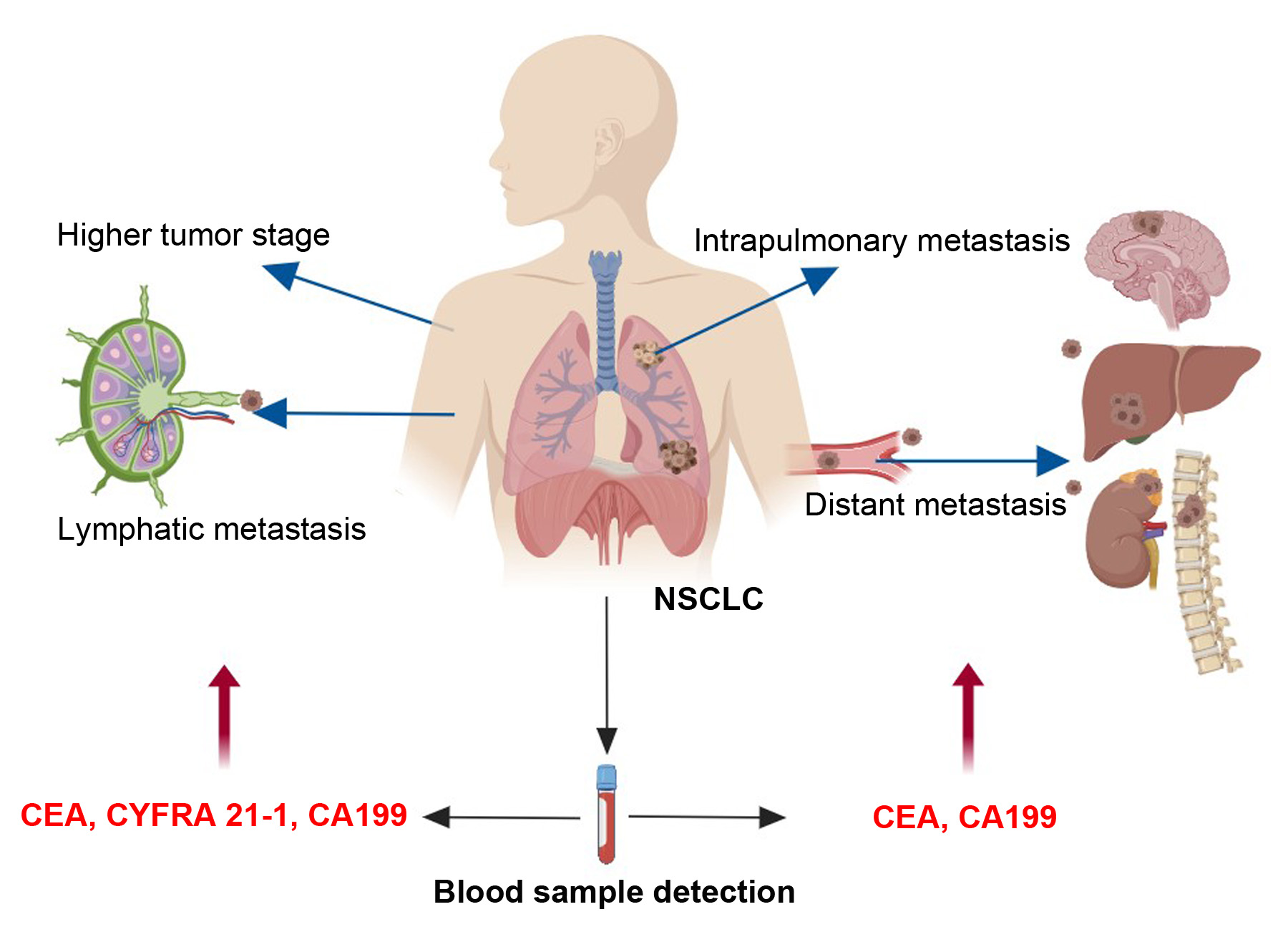
The results of risk factors showed that the patients, with increased levels of CEA, CYFRA 21-1, NSE and CA199, tended to have higher tumor stages. The risk factors for intrapulmonary metastasis were smoking index > 600, and increased levels of CEA and CA199. The risk factors for lymphatic metastasis were higher levels of CEA, CYFRA 21-1 and CA199. The risk factors for distant metastasis were elevated CEA and CA199 levels.
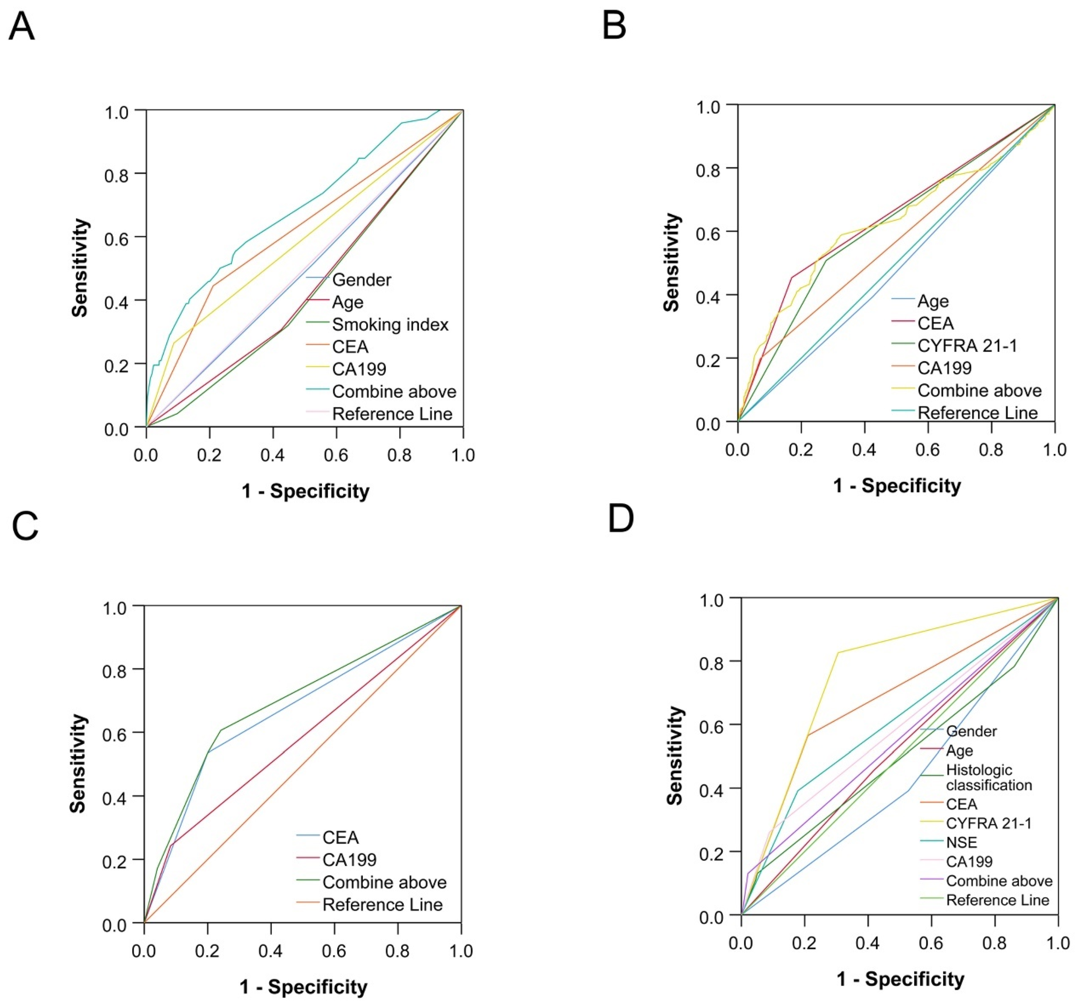
Combined detection of certain serum tumor markers in lung cancer patients can significantly improve diagnostic sensitivity and the roles of monitoring tumor progression [34,35]. At last, joint predictions of combined lung cancer-related tumor markers for tumor metastasis have been performed by ROC curve analyses. The result showed that the combined elevations in CEA and CA199 were also useful for the diagnoses of lymphatic metastasis and distant metastasis, respectively. These results are in accordance with previous reports [36].

5. Conclusions

In summary, our results suggest that the levels of CEA, SCC-Ag, CYFRA 21-1, NSE and CA199 were positively related to tumor metastasis and stage. Elevated CEA and CA199 levels in NSCLC patients are indicative of intrapulmonary and distant metastases; elevated CEA, CYFRA 21-1 and CA199 levels in patients with NSCLC are indicative of lymphatic metastasis. These tumor markers could be useful in predicting tumor metastasis in patients with NSCLC.

Author Contributions

C.J. and S.Z. conceived and designed this study; C.J. and B.S. carried out the data collection; C.J., S.Z. and M.Z. mainly performed data statistical analysis; X.H. and S.H. mainly presented all tables; C.J., B.S., S.Z., M.Z. and X.H. participated in analyzing the analysis results and wrote the paper; all other authors participated in the data collection; C.J., B.S. and S.Z. supervised and directed the project. All authors have read and agreed to the published version of the manuscript.

Funding

This work was supported by the Natural Science Foundation of Tianjin-Municipal Science and Technology Commission (Grant Number: 20JCYBJC01030) and Social Development Science and Technology Project of TaiZhou City (Grant Number: 21ywb99).

Institutional Review Board Statement

This study was approved by the Medical Ethics Committees of Tianjin Union Medical Center (TUMCME20200228, approve date 28 February 2020) and Tianjin Medical University Cancer Institute and Hospital (20200115, approve date 15 January 2020).

Informed Consent Statement

All authors have approved the manuscript and consent for publication.

Data Availability Statement

Data are available upon request.

Conflicts of Interest

The authors declare no conflict of interest.

Abbreviations

CA199, carbohydrate antigens; CEA, carcinoembryonic antigen; CMIA, chemiluminescent microparticle immunoassay; CYFRA 21-1, cytokeratin-19 fragment; DM, Distant metastasis; IM, Intrapulmonary metastasis; LM, Lymphatic metastasis; NSCLC, non-small cell lung cancer; NSE, neuron-specific enolase; ProGRP, pro-gastrin-releasing peptide; ROC, receiver operating characteristic; SCC-Ag, squamous cell carcinoma antigen; SCLC, small cell lung cancer; TPSA, total prostate-specific antigen.

References

  1. Elbeyli, L.; Sanli, M.; Kasap, M.; Gezici, S.; Ozaslan, M.; Akpinar, G. Comparative Proteomics and Bioinformatics Analysis of Tissue from Non-Small Cell Lung Cancer Patients. Curr. Proteom. 2017, 14, 58–77. [Google Scholar]
  2. Siegel, R.L.; Miller, K.D.; Jemal, A. Cancer statistics, 2018. CA Cancer J. Clin. 2018, 60, 277–300. [Google Scholar] [CrossRef]
  3. Zhan, M.; Wen, F.; Liu, L.; Chen, Z.; Wei, H.; Zhou, H. JMJD1A promotes tumorigenesis and forms a feedback loop with EZH2/let-7c in NSCLC cells. Tumor Biol. 2016, 37, 11237–11247. [Google Scholar] [CrossRef] [PubMed]
  4. Funakoshi, T.; Tachibana, I.; Kimura, H.; Takeda, Y.; Kijima, T.; Hoshida, Y.; Nishino, K.; Goto, H.; Yoneda, T.; Kumagai, T.; et al. Expression of tetraspanins in human lung cancer cells: Frequent downregulation of CD9 and its contribution to cell motility in small cell lung cancer. Oncogene 2003, 22, 674–687. [Google Scholar] [CrossRef] [PubMed] [Green Version]
  5. Wu, H.; Wang, Q.; Liu, Q.; Zhang, Q.; Huang, Q.; Yu, Z. The Serum Tumor Markers in Combination for Clinical Diagnosis of Lung Cancer. Clin. Lab. 2020, 66. [Google Scholar] [CrossRef] [PubMed]
  6. Liu, L.; Teng, J.; Zhang, L.; Cong, P.; Yao, Y.; Sun, G.; Liu, Z.; Yu, T.; Liu, M. The Combination of the Tumor Markers Suggests the Histological Diagnosis of Lung Cancer. Biomed. Res. Int. 2017, 2017, 2013989. [Google Scholar] [CrossRef] [PubMed] [Green Version]
  7. Molina, R.; Auge, J.M.; Escudero, J.M.; Marrades, R.; Viñolas, N.; Carcereny, E.; Ramirez, J.; Filella, X. Mucins CA 125, CA 19.9, CA 15.3 and TAG-72.3 as tumor markers in patients with lung cancer: Comparison with CYFRA 21-1, CEA, SCC and NSE. Tumour Biol. 2008, 29, 371–380. [Google Scholar] [CrossRef]
  8. Hu, Q.; Xiao, P.; Li, J.; Yu, P. A retrospective analysis of serum tumor markers found in non-small cell lung cancer. J. Cancer Res. 2016, 12, 117–120. [Google Scholar]
  9. Nisman, B.; Biran, H.; Ramu, N.; Heching, N.; Barak, V.; Peretz, T. The diagnostic and prognostic value of ProGRP in lung cancer. Anticancer. Res. 2009, 29, 4827–4832. [Google Scholar]
  10. Wang, C.F.; Peng, S.J.; Liu, R.Q.; Yu, Y.J.; Ge, Q.M.; Liang, R.B.; Li, Q.Y.; Li, B.; Shao, Y. The Combination of CA125 and NSE Is Useful for Predicting Liver Metastasis of Lung Cancer. Dis. Markers 2020, 2020, 8850873. [Google Scholar] [CrossRef]
  11. Wang, L.; Wang, D.; Zheng, G.; Yang, Y.; Du, L.; Dong, Z.; Zhang, X.; Wang, C. Clinical Evaluation and Therapeutic Monitoring Value of Serum Tumor Markers in Lung Cancer. Int. J. Biol. Markers 2016, 31, 80–87. [Google Scholar] [CrossRef] [PubMed]
  12. Molina, R.; Filella, X.; Augé, J.M.; Fuentes, R.; Bover, I.; Rifa, J.; Moreno, V.; Canals, E.; Viñolas, N.; Marquez, A.; et al. Tumor Markers (CEA, CA 125, CYFRA 21-1, SCC and NSE) in Patients with Non-Small Cell Lung Cancer as an Aid in Histological Diagnosis and Prognosis. Tumor Biol. 2003, 24, 209–218. [Google Scholar] [CrossRef] [PubMed]
  13. Cedrés, S.; Nuñez, I.; Longo, M.; Martinez, P.; Checa, E.; Torrejón, D.; Felip, E. Serum Tumor Markers CEA, CYFRA21-1, and CA-125 Are Associated with Worse Prognosis in Advanced Non–Small-Cell Lung Cancer (NSCLC). Clin. Lung Cancer 2011, 12, 172–179. [Google Scholar] [CrossRef] [PubMed]
  14. Bello, M.G.D.; Filiberti, R.A.; Alama, A.; Orengo, A.M.; Mussap, M.; Coco, S.; Vanni, I.; Boccardo, S.; Rijavec, E.; Genova, C.; et al. The role of CEA, CYFRA21-1 and NSE in monitoring tumor response to Nivolumab in advanced non-small cell lung cancer (NSCLC) patients. J. Transl. Med. 2019, 17, 74. [Google Scholar] [CrossRef] [PubMed] [Green Version]
  15. Shirasu, H.; Ono, A.; Omae, K.; Nakashima, K.; Omori, S.; Wakuda, K.; Kenmotsu, H.; Naito, T.; Murakami, H.; Endo, M.; et al. CYFRA 21-1 predicts the efficacy of nivolumab in patients with advanced lung adenocarcinoma. Tumor Biol. 2018, 40, 101042831876042. [Google Scholar] [CrossRef] [Green Version]
  16. Li, Q.; Sang, S. Diagnostic Value and Clinical Significance of Combined Detection of Serum Markers CYFRA21-1, SCC Ag, NSE, CEA and ProGRP in Non-Small Cell Lung Carcinoma. Clin. Lab. 2020, 66, 11. [Google Scholar] [CrossRef]
  17. Kang, S.J.; Cho, Y.R.; Park, G.M.; Ahn, J.M.; Han, S.B.; Lee, J.Y.; Kim, W.J.; Park, D.W.; Lee, S.W.; Kim, Y.H.; et al. Predictors for functionally significant in-stent restenosis: An integrated analysis using coronary angiography, IVUS, and myocardial perfusion imaging. JACC Cardiovasc. Imaging 2013, 6, 1183–1190. [Google Scholar] [CrossRef] [Green Version]
  18. Vinolas, N.; Molina, R.; Fuentes, R.; Bover, I.; Rifa, J.; Moreno, V.; Canals, E.; Marquez, A.; Barreiro, E.; Borras, J.; et al. Tumor markers (CEA, CA 125, CYFRA 21.1, SCC and NSE) in non small cell lung cancer (NSCLC) patients as an aid in histological diagnosis and prognosis: Comparison with the main clinical and pathological prognostic factors. Lung Cancer 2000, 29, 195. [Google Scholar] [CrossRef]
  19. Zamcheck, N. The present status of carcinoembryonic antigen (CEA) in diagnosis, detection of recurrence, prognosis and evaluation of therapy of colonic and pancreatic cancer. Clin. Gastroenterol. 1976, 5, 625–638. [Google Scholar] [CrossRef]
  20. Grunnet, M.; Sorensen, J.B. Carcinoembryonic antigen (CEA) as tumor marker in lung cancer. Lung Cancer 2012, 76, 138–143. [Google Scholar] [CrossRef] [PubMed]
  21. Kim, K.N.; Joo, N.S.; Je, S.Y.; Kim, K.M.; Kim, B.T.; Park, S.B.; Cho, D.Y.; Park, R.W.; Lee, D.J. Carcinoembryonic Antigen Level Can be Overestimated in Metabolic Syndrome. J. Korean Med. Sci. 2011, 26, 759–764. [Google Scholar] [CrossRef] [Green Version]
  22. Hall, C.; Clarke, L.; Pal, A.; Buchwald, P.; Eglinton, T.; Wakeman, C.; Frizelle, F. A Review of the Role of Carcinoembryonic Antigen in Clinical Practice. Ann. Coloproctol. 2019, 35, 294–305. [Google Scholar] [CrossRef] [PubMed]
  23. Hao, C.; Zhang, G.; Zhang, L. Serum CEA levels in 49 different types of cancer and noncancer diseases. Prog. Mol. Biol. Transl. Sci. 2019, 162, 213–227. [Google Scholar] [PubMed]
  24. Suzuki, Y.; Nakano, T.; Ohno, T.; Abe, A.; Morita, S.; Tsujii, H. Serum CYFRA 21-1 in cervical cancer patients treated with radiation therapy. J. Cancer Res. Clin. Oncol. 2000, 126, 332–336. [Google Scholar] [CrossRef] [PubMed]
  25. Holdenrieder, S.; Molina, R.; Qiu, L.; Zhi, X.; Rutz, S.; Engel, C.; Kasper-Sauer, P.; Dayyani, F.; Mkorse, C. Technical and clinical performance of a new assay to detect squamous cell carcinoma antigen levels for the differential diagnosis of cervical, lung, and head and neck cancer. Tumour Biol. 2018, 40, 1010428318772202. [Google Scholar] [CrossRef] [PubMed] [Green Version]
  26. Schneider, J.; Velcovsky, H.G.; Morr, H.; Katz, N.; Neu, K.; Eigenbrodt, E. Comparison of the tumor markers tumor M2-PK, CEA, CYFRA 21-1, NSE and SCC in the diagnosis of lung cancer. Anticancer. Res. 2000, 20, 5053–5058. [Google Scholar]
  27. Jiang, Z.F.; Wang, M.; Xu, J.L. Thymidine kinase 1 combined with CEA, CYFRA21-1 and NSE improved its diagnostic value for lung cancer. Life Sci. 2018, 194, 1–6. [Google Scholar] [CrossRef]
  28. Zissimopoulos, A.; Stellos, K.; Permenopoulou, V.; Petrakis, G.; Theodorakopoulos, P.; Baziotis, N.; Thalassinos, N. The importance of the tumor marker CYFRA 21-1 in patients with lung cancer after surgery or chemotherapy. Hell. J. Nucl. Med. 2007, 10, 62–66. [Google Scholar]
  29. Sandoval, J.A.; Malkas, L.H.; Hickey, R.J. Clinical significance of serum biomarkers in pediatric solid mediastinal and abdominal tumors. Int. J. Mol. Sci. 2012, 13, 1126–1153. [Google Scholar] [CrossRef]
  30. Kamiya, N.; Suzuki, H.; Kawamura, K.; Imamoto, T.; Naya, Y.; Tochigi, N.; Kakuta, Y.; Yamaguchi, K.; Ishikura, H.; Ichikawa, T. Neuroendocrine differentiation in stage D2 prostate cancers. Int. J. Urol. 2008, 15, 423–428. [Google Scholar] [CrossRef]
  31. Rosati, R.; Adil, M.R.; Ali, M.A.; Eliason, J.; Orosz, A.; Sebestyén, F.; Kalemkerian, G.P. Induction of apoptosis by a short-chain neuropeptide analog in small cell lung cancer. Peptides 1998, 19, 1519–1523. [Google Scholar] [CrossRef]
  32. Tiseo, M.; Ardizzoni, A.; Cafferata, M.A.; Loprevite, M.; Chiaramondia, M.; Filiberti, R.; Marroniet, P.; Grossi, F.; Paganuzzi, M. Predictive and prognostic significance of neuron-specific enolase (NSE) in non-small cell lung cancer. Anticancer. Res. 2008, 28, 507–513. [Google Scholar] [PubMed]
  33. Yang, Q.; Zhang, P.; Wu, R.; Lu, K.; Zhou, H. Identifying the Best Marker Combination in CEA, CA125, CY211, NSE, and SCC for Lung Cancer Screening by Combining ROC Curve and Logistic Regression Analyses: Is It Feasible? Dis. Markers 2018, 2018, 2082840. [Google Scholar] [CrossRef] [PubMed] [Green Version]
  34. Wang, W.J.; Tao, Z.; Gu, W.; Sun, L.H. Clinical observations on the association between diagnosis of lung cancer and serum tumor markers in combination. Asian Pac. J. Cancer Prev. 2013, 14, 4369–4371. [Google Scholar] [CrossRef]
  35. Chu, X.Y.; Hou, X.B.; Song, W.A.; Xue, Z.Q.; Wang, B.; Zhang, L.B. Diagnostic values of SCC, CEA, Cyfra21-1 and NSE for lung cancer in patients with suspicious pulmonary masses: A single center analysis. Cancer Biol. 2011, 11, 995–1000. [Google Scholar] [CrossRef]
  36. Hatate, K.; Yamashita, K.; Hirai, K.; Kumamoto, H.; Sato, T.; Ozawa, H.; Nakamura, T.; Onozato, W.; Kokuba, Y.; Ihara, A.; et al. Liver metastasis of colorectal cancer by protein-tyrosine phosphatase type 4A, 3 (PRL-3) is mediated through lymph node metastasis and elevated serum tumor markers such as CEA and CA19-9. Oncol Rep 2008, 20, 737–743. [Google Scholar]
Figure 1. ROC curves of tumor markers for tumor metastasis and stage. (A) The ROC curve for gender, age, smoking index, CEA and CA199 in the diagnosis of intrapulmonary metastasis; (B) the ROC curve for age, CEA, CYFRA 21-1 and CA199 in the diagnosis of lymphatic metastasis. (C) the ROC curve for CEA and CA199 in the diagnosis of distant metastasis. (D) the ROC curve for gender, age, histologic classification, CEA, CYFRA 21-1, NSE and CA199 in the diagnosis of tumor stage.
Cancers 14 05064 g001
Table 1. Basic information of the patients with NSCLC.
Patient CharacteristicsCase or Median%
Age
median (P25–P75, year)61 (55–67)
Gender
male181255.4
female146044.6
Smoking index
median (P25–P75)1.5 (0.00–600)
non-smoking163349.9
≤600 *100931.2
>60063019.5
Intrapulmonary metastasis
none265683.8
yes51216.2
Lymphatic metastasis
none214767.6
yes103132.4
Distant metastasis
none260781.7
yes58518.3
Histologic classification
Squamous carcinoma73522.5
Adenocarcinoma235472.0
Adenosquamous carcinoma441.3
Others1394.2
Staging
Ι199761.3
ΙΙ50315.4
ΙΙΙ1755.4
ΙV58517.9
Note: * The P75 smoking index among smokers was 600.
Table 2. The correlation of age, sex, and smoking index with tumor metastasis, and stage of patients with NSCLC.
VariablesIMLMDMStaging
NoneYespNoneYespNoneYespΙΙΙΙΙΙΙVp
Gender (case)
Male1417327<0.0011079670<0.00114033600.098966346129360<0.001
Female1239185106836112042251031157 *46 *225 *#&
Age (case)
≤61 years1377221<0.00111114940.04713182900.701106420677290<0.001
>61 years127929110365371289295933297 *98 *295
Smoking index (case)
non1356231<0.0011170423<0.00113072890.003111217852289<0.001
60084914066932183715961617652159
>600451141308287463137268149 *71 *#137 *#&
Note: DM, distant metastasis; IM, intrapulmonary metastasis; LM, lymphatic metastasis. Chi-square test was used to analyze. * Compared with staging Ι, adjustment p < 0.05; # compared with staging ΙΙ, adjustment p < 0.05; & compared with staging ΙΙΙ, adjustment p < 0.05.
Table 3. The difference in tumor metastasis for the tumor markers in the patients with NSCLC (CEA, SCC-Ag, CYFRA 21-1, NSE, ProGRP, TPSA and CA199).
VariablesIMLMDM
NoneYespNoneYespNoneYesp
CEA 3.026.71<0.0012.715.67<0.0012.9410.58<0.001
(1.78–5.86)(3.18–34.55)(1.64–4.98)(2.91–25.93)(1.73–5.47)(3.43–58.54)
SCC-Ag 0.90.90.1290.91<0.0010.90.90.45
(0.70–1.30)(0.60–1.80)(0.70–1.30)(070–1.80)(0.70–1.40)(0.60–1.70)
CYFRA 21-1 2.774.96<0.0012.594.6<0.0012.774.83<0.001
(1.92–4.56)(2.63–10.13)(1.85–3.95)(2.63–10.13)(1.92–4.47)(2.58–12.57)
NSE 13.3216.5<0.00113.415.2<0.00113.3616.09<0.001
(11.30–16.35)(13.00–20.70)(11.30–16.00)(12.33–20.70)(11.30–16.35)(12.47–23.64)
ProGRP 32.7932.370.67432.3734.380.01332.6633.790.44
(26.23–40.64)(26.04–38.82)(26.06–40.28)(27.90–42.08)(26.16–40.66)(27.48–39.34)
TPSA 45.7350.120.1744.4656.28<0.00146.2139.490.58
(26.10–85.73)(35.07–87.54)(25.74–81.91)(29.61–101.00)(26.20–85.83)(25.27–80.74)
CA199 0.390.460.0730.380.45<0.0010.390.48<0.001
(0.26–0.63)(0.26–1.03)(0.25–0.61)(0.31–0.87)(0.25–0.63)(0.31–1.05)
Note: DM, distant metastasis; IM, intrapulmonary metastasis; LM, lymphatic metastasis. Non-normal data are represented by median (P25–P75). Nonparametric test was used to analyze. Comparison of the concentrations of tumor markers between different groups was conducted by a nonparametric test.
Table 4. The difference in clinical stages for the tumor markers in the patients with NSCLC (CEA, SCC-Ag, CYFRA21-1, NSE, ProGRP, TPSA and CA199).
VariablesIIIIIIIVp
CEA 2.59 (1.57–4.64)4.10 (2.45–10.12) *5.39 (3.26–15.75) *#10.58 (3.43–58.54) *#&<0.001
SCC-Ag 0.90 (0.70–1.20)1.00 (0.70–2.10) *1.10 (0.80–2.90) *#0.90 (0.60–1.70) *#&<0.001
CYFRA 21-1 2.50 (1.81–3.68)4.21 (2.48–8.56) *5.72 (3.62–14.22) *#4.83 (2.58–12.57) *#&<0.001
NSE 12.93 (11.07–15.60)14.72 (12.20–18.59) *17.45 (12.82–24.96) *#16.09 (12.47–23.64) *#<0.001
ProGRP 32.64 (26.15–40.76)32.40 (26.01–39.19)34.51 (29.14–41.78)33.79 (27.48–39.34)0.526
TPSA 44.37 (25.82–82.10)56.94 (30.34–118.79) *58.57 (45.11–128.41) *39.49 (25.27–80.74) #&<0.001
CA199 2.31 (1.35–5.60)2.85 (1.36–7.35) *3.01 (1.44–8.29) *#3.25 (1.43–10.72) *<0.001
Note: Non-normal data are represented by median (P25–P75). Nonparametric test was used to analyze. Comparison of the concentrations of tumor markers between different groups was conducted by a nonparametric test. * Compared with staging I, adjustment p < 0.05; # compared with staging II, adjustment p < 0.05; & compared with staging III, adjustment p < 0.05.
Table 5. Univariate analysis of influencing factors for tumor metastasis and clinical stage in patients with NSCLC.
VariablesIMLMDMStaging
OR (95% CI)pOR (95% CI)pOR (95% CI)pOR (95% CI)p
Gender 0.64 (0.53–0.77)<0.0010.54 (0.47–0.63)<0.0010.73 (0.61–0.87)<0.0010.69 (0.64–0.76)<0.001
Age 1.04 (1.03–1.05)<0.0011.02 (1.01–1.03)<0.0011.02 (1.01–1.03)<0.0011.01 (1.01–1.02)<0.001
Smoking index
nonReference Reference Reference Reference
6000.60 (0.48–0.75)<0.0010.82 (0.69–0.97)0.0190.60 (0.48–0.75)<0.0010.85 (0.78–0.94)0.001
>6002.00 (1.64–2.45)<0.0012.39 (2.00–2.86)<0.0011.55 (1.27–1.90)<0.0011.54 (1.39–1.71)<0.001
Histologic classification
Squamous carcinomaReference Reference Reference Reference
Adenocarcinoma0.82 (0.66–1.03)0.0860.63 (0.74–1.62)<0.0010.98 (0.77–1.25)0.8730.63 (0.54–0.73)<0.001
Adenosquamous carcinoma1.33 (0.62–2.87)0.4691.80 (0.96–3.40)0.0681.55 (0.72–3.34)0.2591.31 (0.75–2.29)0.338
Others0.52 (0.29–0.93)0.0270.91 (0.62–1.34)0.9120.95 (0.55–1.64)0.8580.84 (0.60–1.19)0.331
CEA 3.19 (2.66–3.82)<0.0013.60 (3.09–4.19)<0.0014.27 (3.57–5.11)<0.0012.40 (2.20–2.62)<0.001
SCC-Ag 1.82 (1.49–2.22)<0.0012.02 (1.70–2.40)<0.0011.63 (1.34–2.00)<0.0011.52 (1.38–1.67)<0.001
CYFRA 21-1 4.15 (3.38–5.08)<0.0014.28 (3.65–5.02)<0.0013.69 (3.04–4.47)<0.0012.63 (2.41–2.84)<0.001
NSE 3.11 (2.59–3.74)<0.0012.59 (2.21–3.03)<0.0012.88 (2.41–3.44)<0.0011.95 (1.78–2.13)<0.001
ProGRP 0.76 (0.18–3.17)0.7071.84 (1.07–3.16)0.0281.38 (0.55–3.51)0.4951.23 (0.89–1.69)0.205
TPSA 0.92 (0.54–1.57)0.7601.61 (1.26–2.06)<0.0010.89 (0.57–1.40)0.6151.21 (1.05–1.39)0.007
CA199 3.82 (2.23–6.54)<0.0010.32 (0.23–0.44)<0.0014.00 (2.53–6.31)<0.0012.08 (1.73–2.50)<0.001
Note 1: DM, distant metastasis; IM, intrapulmonary metastasis; LM, lymphatic metastasis. Note 2: (1) The influencing factors of IM, LM and DM were analyzed by binary logistic analysis. Assignment of dependent variable: IM, LM and DM are all 0 = without and 1 = with. Independent variable assignment: gender (0 = male, 1 = female); age (0 = ≤61 years, 1 = >61 years); smoking index (0 = non, 1 = 1–600, 2 = ≥600); histologic classification (0 = Squamous carcinoma, 1 = Adenocarcinoma, 3 = Adenosquamous carcinoma, 4 = Others); CEA, SCC, CYFRA 21-1, NSE, ProGRP, TPSA, CA199 (0 = normal, 1 = high). (2) The influencing factors of tumor stage were analyzed by ordered logistics. Assignment of dependent variable: tumor stage (1–4 are stage Ι–ΙV, respectively); independent variable assignment: gender, age, smoking index, histologic classification and seven kinds of tumor markers are all the same as above.
Table 6. Collinearity examination of multifactor analysis for basic condition and tumor markers related with metastasis and stage of patients with NSCLC.
VariablesIMLMDMStaging
TVIFTVIFTVIFTVIF
Gender 0.6351.5760.6331.5800.6361.5720.6331.581
Age 0.9371.0670.9371.0670.9361.0680.9371.067
Smoking index 0.6291.5890.6281.5910.6291.5890.6281.592
Histologic classification 0.9441.0590.9391.064--0.9391.064
CEA 0.8911.1220.8921.1220.8951.1180.8911.122
SCC-Ag 0.9001.1110.8961.1160.9111.0970.8961.116
CYFRA 21-1 0.8631.1590.7371.3560.8721.1470.7371.357
NSE 0.9751.0250.9761.0250.9801.0200.9761.025
TPSA --0.7971.254--0.7971.254
CA199 0.9221.0850.9221.0840.9251.0810.9221.084
Note 1: DM, distant metastasis; IM, intrapulmonary metastasis; LM, lymphatic metastasis. Note 2: T, tolerance; VIF, variance inflation factor.
Table 7. Multivariate analysis of influencing factors for tumor metastasis and clinical stage in patients with NSCLC.
VariablesIMLMDMStaging
OR (95% CI)pOR (95% CI)pOR (95% CI)pOR (95% CI)p
Gender 0.52 (0.29–0.94)0.0310.85 (0.61–1.19)0.3350.66 (0.39–1.13)0.1170.68 (0.49–0.94)0.021
Age 0.58 (0.34–0.99)0.0450.64 (0.48–0.83)0.0010.66 (0.42–1.03)0.0680.76 (0.59–0.98)0.036
Smoking index
nonReference Reference Reference Reference
6000.53 (0.27–1.03)0.0591.00 (0.70–1.42)0.9870.66 (0.37–1.15)0.1440.95 (0.67–1.34)0.775
>6000.28 (0.08–0.99)0.0481.22 (0.75–2.00)0.4130.45 (0.18–1.11)0.0821.03 (0.65–1.64)0.889
Histologic classification
Squamous carcinomaReference Reference Reference Reference
Adenocarcinoma2.69 (0.78–9.31)0.1190.92 (0.62–1.38)0.695--0.68 (0.47–0.97)0.033
Adenosquamous carcinoma--1.58 (0.35–7.15)0.553--1.05 (0.26–4.31)0.948
Others1.28 (0.20–8.12)0.7931.32 (0.72–2.43)0.365--0.87 (0.49–1.54)0.628
CEA 2.66 (1.53–4.63)<0.0013.08 (2.33–4.07)<0.0014.51 (2.86–7.12)<0.0012.85 (2.17–3.75)<0.001
SCC-Ag 0.40 (0.14–1.18)0.0961.35 (0.95–1.93)0.0930.50 (0.24–1.03)0.0601.04 (0.74–1.45)0.842
CYFRA 21-1 1.27 (0.73–2.22)0.3942.00 (1.48–2.69)<0.0011.06 (0.66–1.69)0.8222.56 (1.92–3.42)<0.001
NSE 1.51 (0.86–2.66)0.1551.15 (0.84–1.57)0.3821.26 (0.77–2.08)0.3621.76 (1.33–2.34)<0.001
TPSA --0.98 (0.72–1.32)0.876--0.75 (0.56–1.01)0.055
CA199 2.80 (1.49–5.17)0.0012.04 (1.41–2.96)<0.0012.20 (1.31–3.69)0.0032.20 (1.54–3.14)<0.001
Notes are the same as in Table 5.
Table 8. Receiver operating characteristic (ROC) curves of influencing factors for tumor metastasis and clinical stage in patients with NSCLC.
VariablesIMLMDMStaging
AUC (95% CI)pAUC (95% CI)pAUC (95% CI)pAUC (95% CI)p
Gender 0.49 (0.43–0.56)0.862----0.43 (0.35–0.52)0.116
Age 0.44 (0.38–0.51)0.0850.48 (0.45–0.52)0.362--0.52 (0.43–0.60)0.659
Smoking index 0.43 (0.37–0.49)0.044------
Histologic classification ------0.50 (0.40–0.59)0.949
CEA 0.62 (0.55–0.69)0.0010.64 (0.61–0.68)<0.0010.67 (0.61–0.73)<0.0010.68 (0.59–0.76)<0.001
CYFRA 21-1 --0.62 (0.58–0.65)<0.001--0.76 (0.70–0.83)<0.001
NSE ------0.61 (0.52–0.70)0.013
CA199 0.59 (0.52–0.66)0.0110.56 (0.53–0.60)<0.0010.58 (0.52–0.64)0.0070.59 (0.49–0.68)0.046
Combine 0.69 (0.62–0.75)<0.0010.62 (0.59–0.66)<0.0010.69 (0.63–0.75)<0.0010.55 (0.46–0.65)0.205
Notes: DM, distant metastasis; IM, intrapulmonary metastasis; LM, lymphatic metastasis.
Publisher’s Note: MDPI stays neutral with regard to jurisdictional claims in published maps and institutional affiliations.

