Low-Cost Synthesis of Cu-Modified Immobilized Nanoporous TiO2 for Photocatalytic Degradation of 1H-Benzotriazole

 , ,
, ,  ,
, 
Abstract
:1. Introduction
2. Results and Discussion
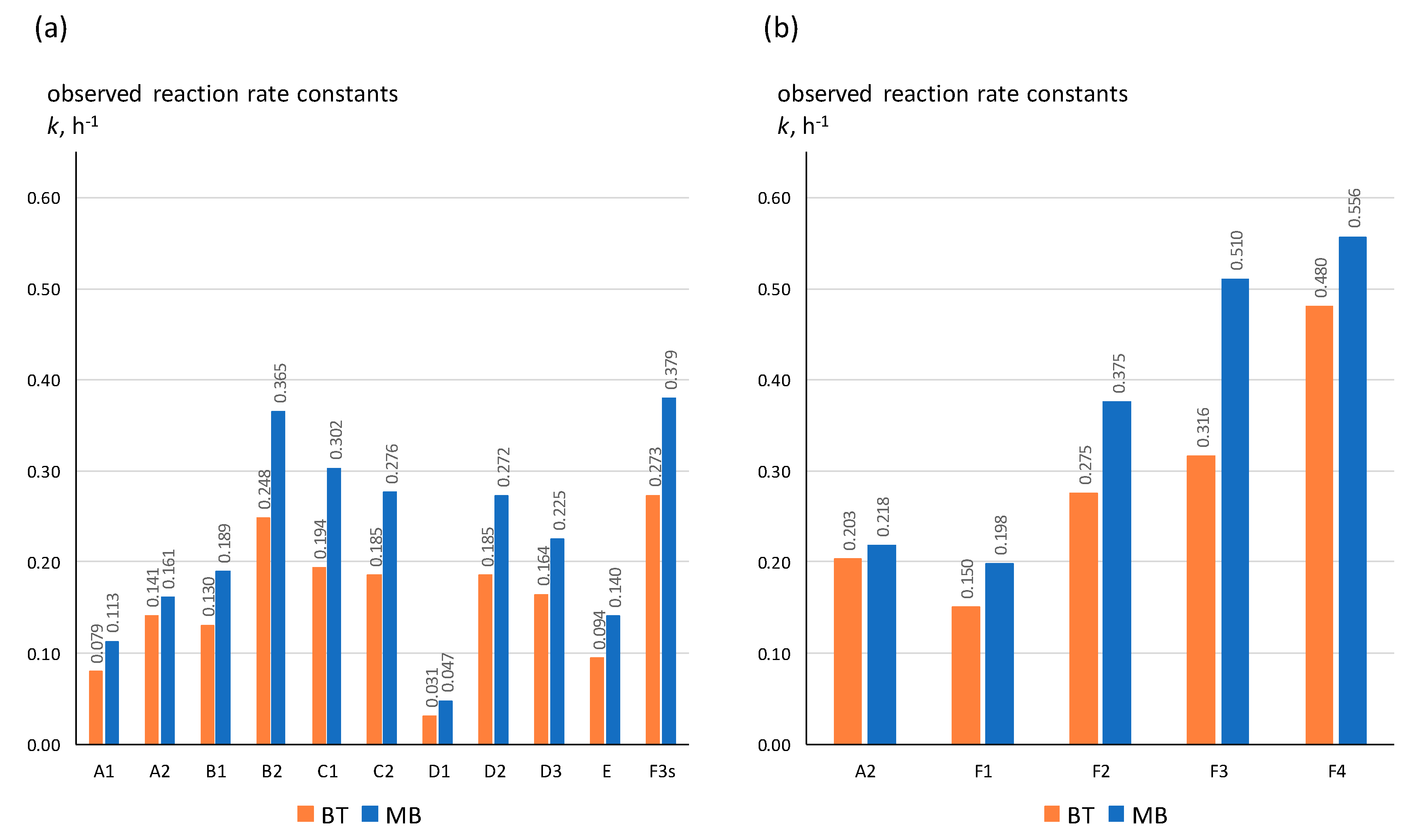
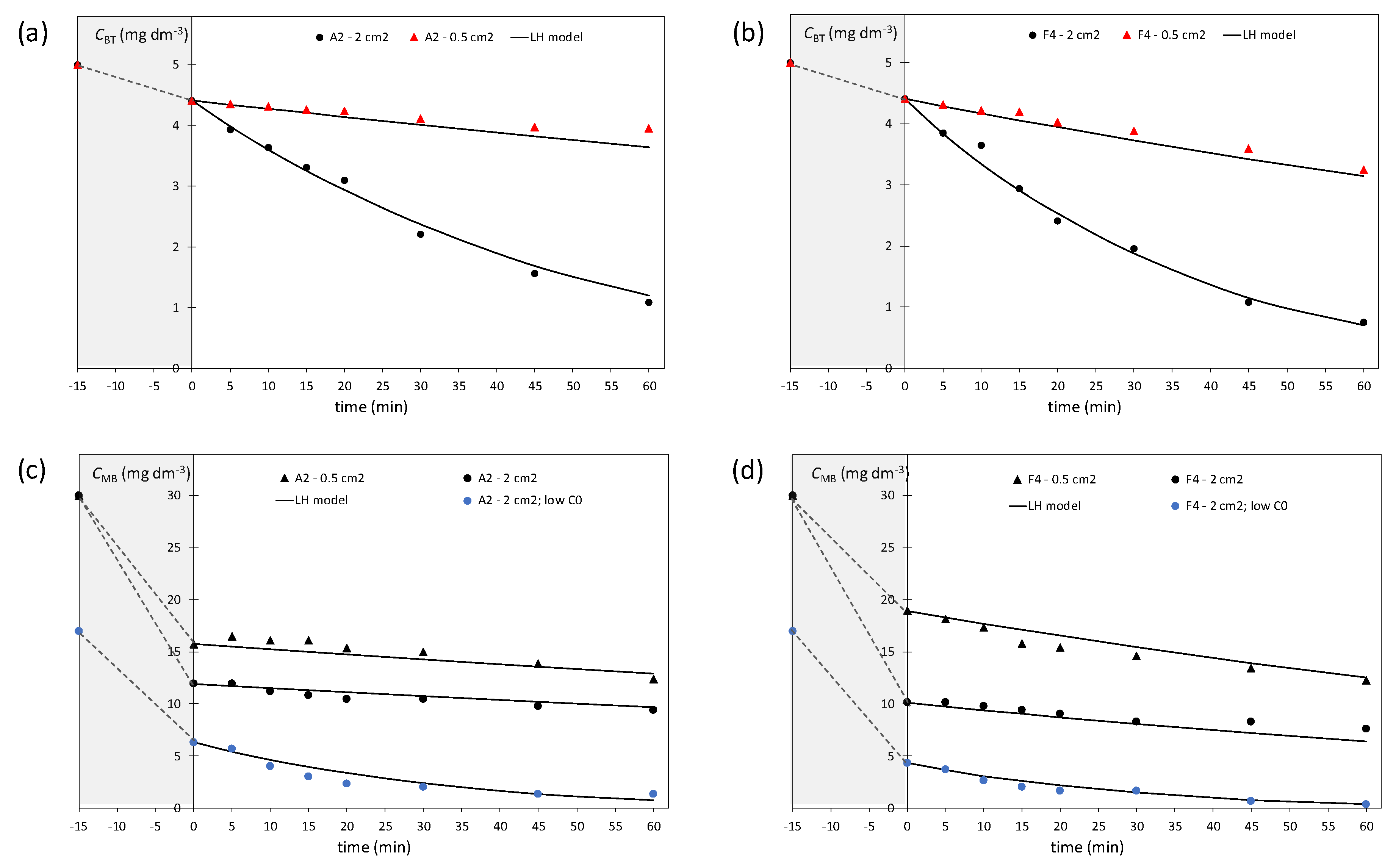
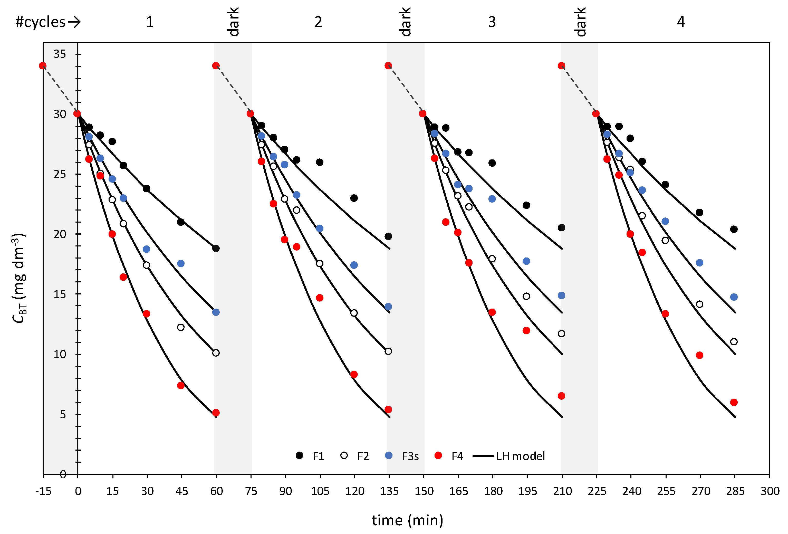
2.1. Photocatalytic Activity
2.2. Structural Properties
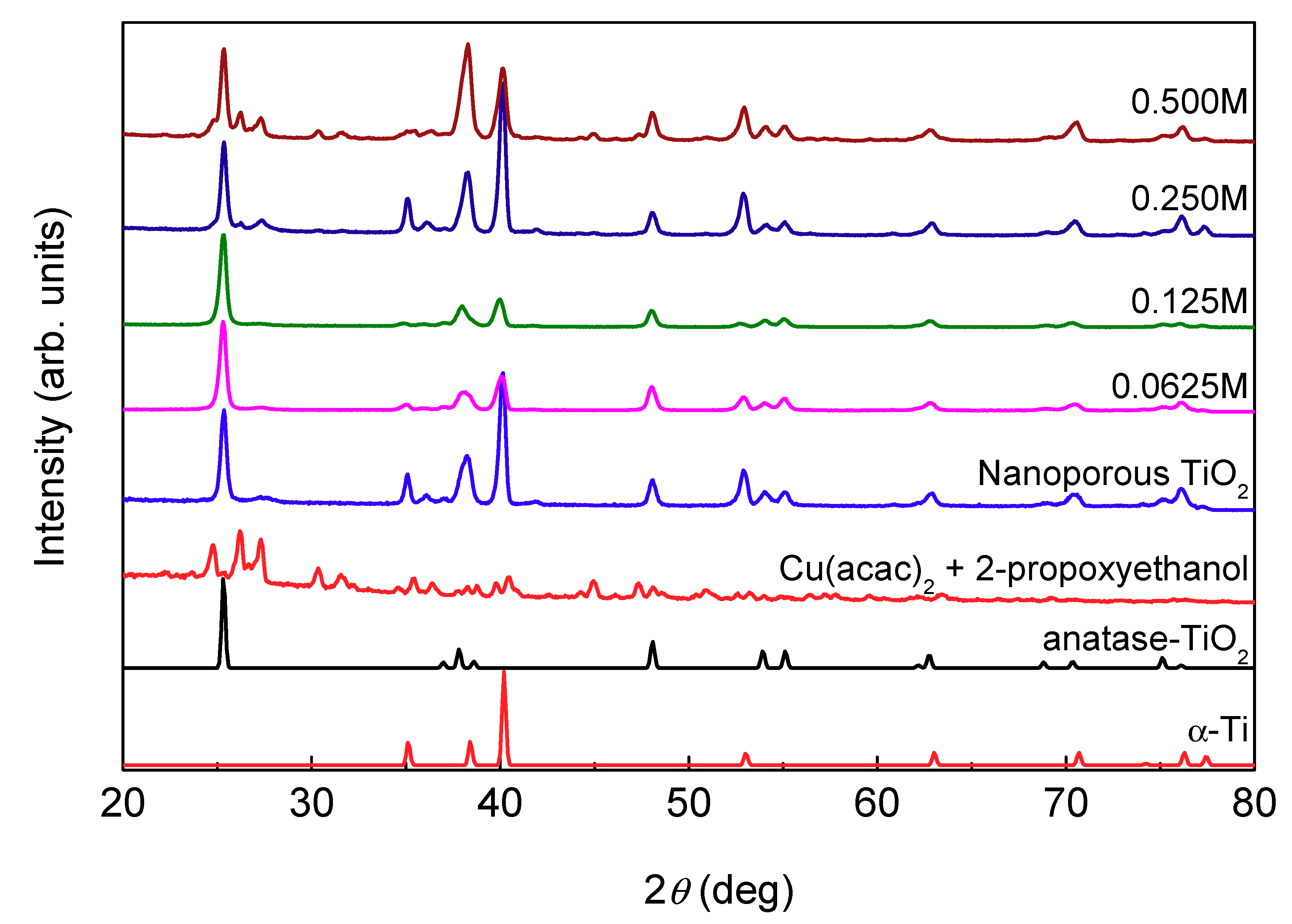
2.2.1. Grazing Incidence X-ray Diffraction (GIXRD) Results
2.2.2. Raman Spectroscopy Results
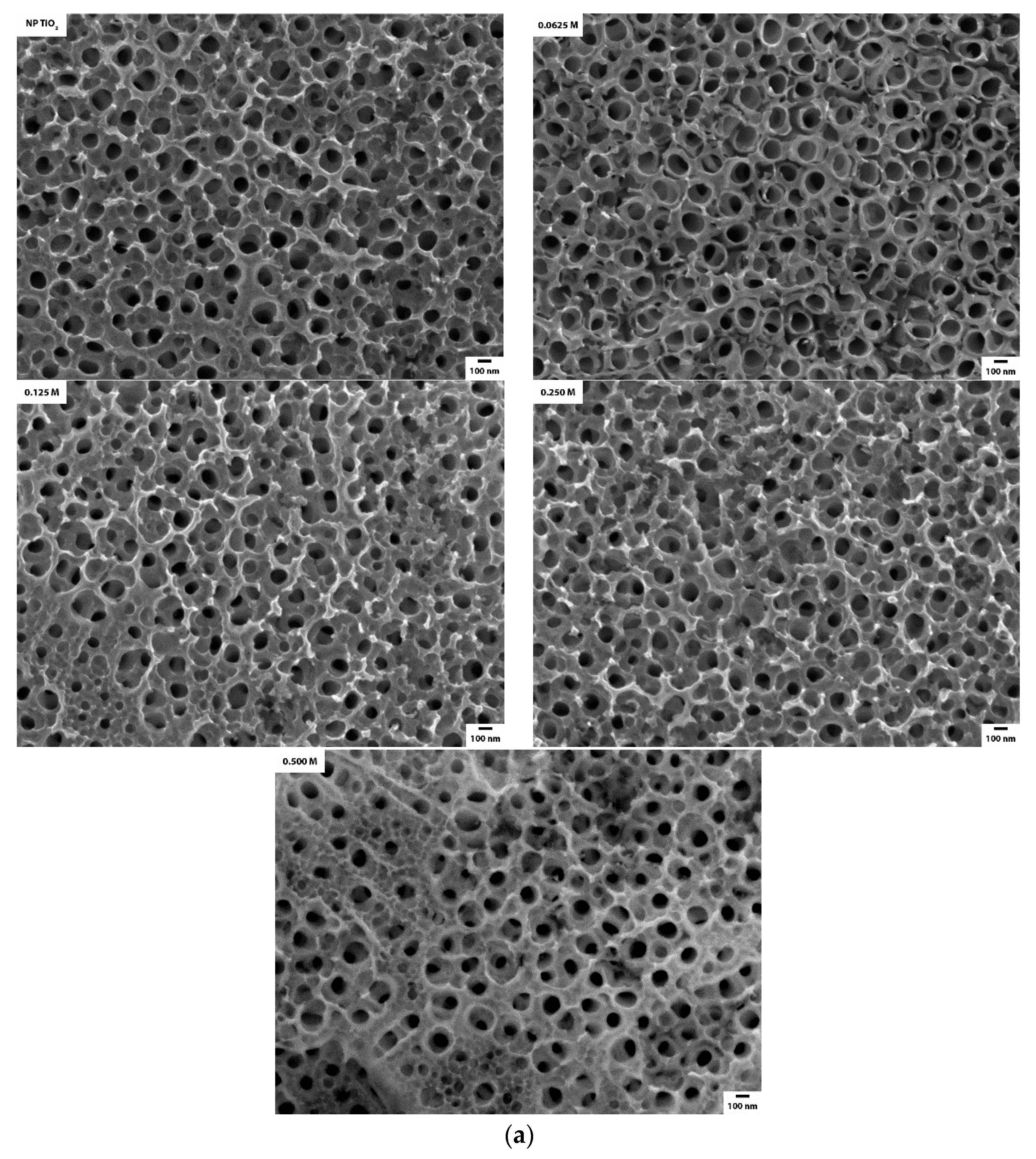
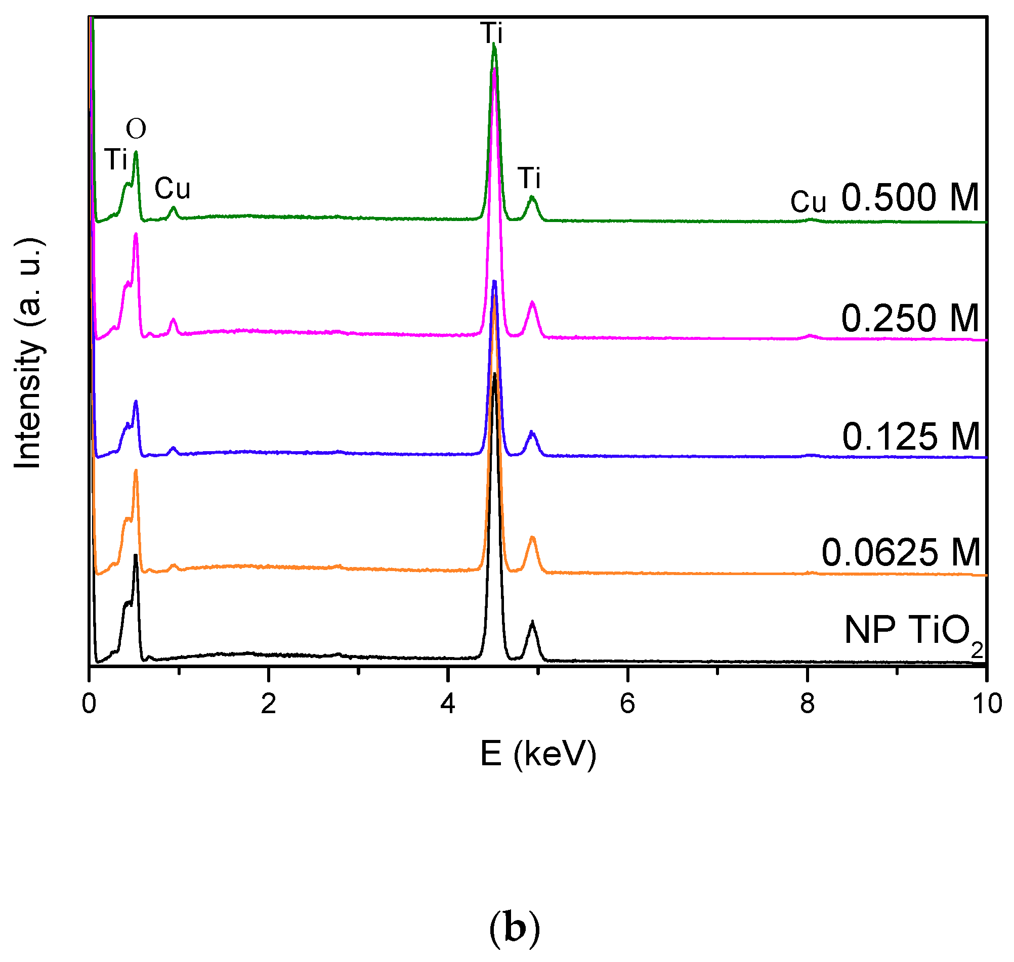
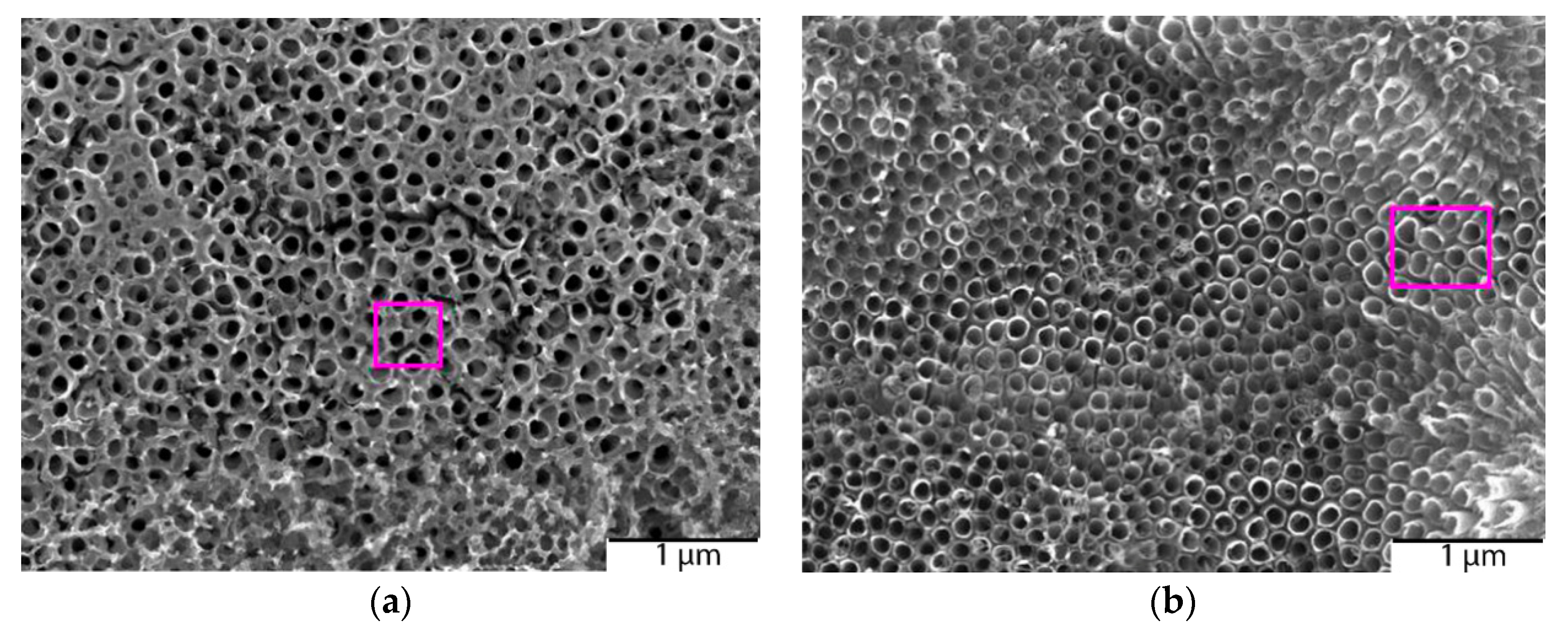
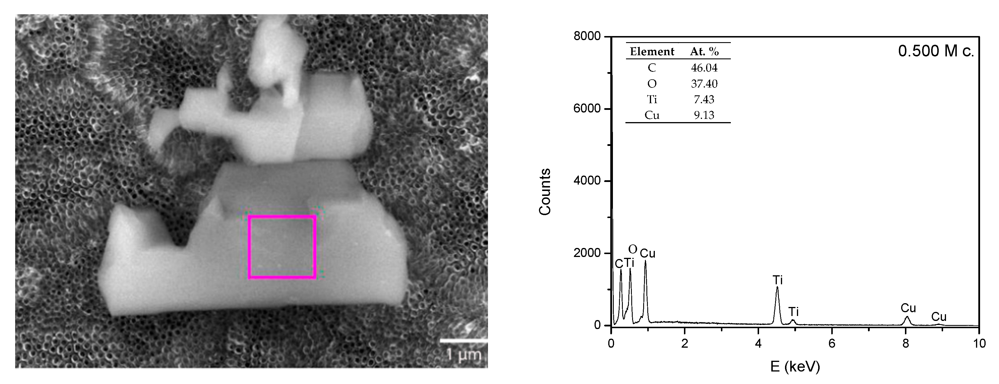
2.2.3. High-Resolution Scanning Electron Microscopy Results
3. Materials and Methods
3.1. Catalyst Preparation
3.2. Catalyst Characterization
3.2.1. Photocatalytic Activity Tests
3.2.1.1. Photoreactor Setup
3.2.1.2. Analyses
3.2.2. Grazing Incidence X-ray Diffraction (GIXRD) Analysis
3.2.3. Raman Spectroscopy
3.2.4. Scanning Electron Microscopy (SEM) and Energy-Dispersive X-ray Spectroscopy (EDS)
4. Conclusions
Author Contributions
Funding
Conflicts of Interest
References
- Fujishima, A.; Rao, T.N.; Tryk, D.A. Titanium dioxide photocatalysis. J. Photochem. Photobiol. C 2000, 1, 1–21. [Google Scholar] [CrossRef]
- Zou, Z.G.; Ye, J.H.; Sayama, K.; Arakawa, H. Direct splitting of water under visible light irradiation with an oxide semiconductor photocatalyst. Nature 2001, 414, 625–627. [Google Scholar] [CrossRef] [PubMed]
- Asahi, R.; Morikawa, T.; Ohwaki, T.; Aoki, K.; Taga, Y. Visible-light photocatalysis in nitrogen-doped titanium oxides. Science 2001, 293, 269–271. [Google Scholar] [CrossRef] [PubMed]
- Malato, S.; Caceres, J.; Aguera, A.; Mezcua, M.; Hernando, D.; Vial, J.; Fernandez-Alba, A.R. Degradation of Imidacloprid in Water by Photo-Fenton and TiO2 Photocatalysis at a Solar Pilot Plant: A Comparative Study. Environ. Sci. Technol. 2001, 35, 4359–4366. [Google Scholar] [CrossRef] [PubMed]
- Nagaveni, K.; Sivalingam, G.; Hegde, M.S.; Madras, G. Photocatalytic Degradation of Organic Compounds over Combustion-Synthesized Nano-TiO2. Environ. Sci. Technol. 2004, 38, 1600–1604. [Google Scholar] [CrossRef]
- Petronellaa, F.; Truppia, A.; Ingrossoa, C.; Placidoa, T.; Striccolia, M.; Curria, M.L. Nanocomposite materials for photocatalytic degradation of pollutants. Catal. Today 2017, 281, 85–100. [Google Scholar] [CrossRef]
- Guan, K. Relationship between photocatalytic activity, hydrophilicity and self-cleaning effect of TiO2/SiO2 films. Surf. Coat. Technol. 2005, 191, 155–160. [Google Scholar] [CrossRef]
- Hoffmann, M.R.; Martin, S.T.; Choi, W.; Bahnemann, D.W. Environmental Applications of Semiconductor Photocatalysis. Chem. Rev. 1995, 95, 69–96. [Google Scholar] [CrossRef]
- Matthews, R.W. Photooxidation of organic impurities in water using thin films of titanium dioxide. J. Phys. Chem. 1987, 91, 3328–3333. [Google Scholar] [CrossRef]
- Zhang, L.; Dillert, R.; Bahnemann, D.; Vormoor, M. Photo-induced hydrophilicity and self-cleaning: Models and reality. Environ. Sci. 2012, 5, 7491–7507. [Google Scholar] [CrossRef]
- Grätzel, M. Recent advances in sensitized mesoscopic solar cells. Acc. Chem. Res. 2009, 42, 1788–1798. [Google Scholar] [CrossRef] [PubMed]
- Manassero, A.; Satuf, M.L.; Alfano, O.M. Photocatalytic reactors with suspended and immobilized TiO2: Comparative efficiency evaluation. Chem. Eng. J. 2017, 326, 29–36. [Google Scholar] [CrossRef]
- Roy, P.; Berger, S.; Schmuki, P. TiO2 nanotubes: Synthesis and applications. Angew. Chem. Int. Ed. 2011, 50, 2904–2939. [Google Scholar] [CrossRef] [PubMed]
- Yu, D.; Zhu, X.; Zhen, X.; Zhong, X.; Gui, Q.; Ye, S.; Zhang, S.; Chen, X.; Li, D. Facile method to enhance the adhesion of TiO2 nanotube arrays to Ti substrate. ACS Appl. Mater. Interfaces 2014, 6, 8001–8005. [Google Scholar] [CrossRef] [PubMed]
- Lee, K.; Mazare, A.; Schmuki, P. One-dimensional titanium dioxide nano-materials: Nanotubes. Chem. Rev. 2014, 114, 9385–9454. [Google Scholar] [CrossRef] [Green Version]
- Paramasivam, I.; Macak, J.M.; Ghicov, A.; Schmuki, P. Enhanced photochromism of Ag loaded self-organized TiO2 nanotube layers. Chem. Phys. Lett. 2007, 445, 233–237. [Google Scholar] [CrossRef]
- Yang, K.; Dai, Y.; Huang, B.J. Understanding photocatalytic activity of S-and P-doped TiO2 under visible light from first-principles. Phys. Chem. C 2007, 111, 18985–18994. [Google Scholar] [CrossRef]
- Choi, W.; Termin, A.; Hoffmann, M.R. The Role of Metal Ion Dopants in Quantum-Sized TiO2: Correlation between Photoreactivity and Charge Carrier Recombination Dynamics. J. Phys. Chem. 1994, 98, 13669–13679. [Google Scholar] [CrossRef]
- Irie, H.; Kamiya, K.; Shibanuma, T.; Miura, S.; Tryk, D.A.; Yokoyama, T.; Hashimoto, K. Visible Light-Sensitive Cu(II)-Grafted TiO2 Photocatalysts: Activities and X-ray Absorption Fine Structure Analyses. J. Phys. Chem. C 2009, 113, 10761–10766. [Google Scholar] [CrossRef]
- Xin, B.; Jing, L.; Ren, Z.; Wang, B.; Fu, H. Effects of simultaneously doped and deposited Ag on the photocatalytic activity and surface states of TiO2. J. Phys. Chem. B 2005, 109, 2805–2809. [Google Scholar] [CrossRef]
- Wang, Z.; Lang, X. Visible light photocatalysis of dye-sensitized TiO2: The selective aerobic oxidation of amines to imines. Appl. Catal. B 2018, 224, 404–409. [Google Scholar] [CrossRef]
- Bahadur Rawal, S.; Bera, S.; Lee, D.; Jang, D.; Lee, W. Design of visible-light photocatalysts by coupling of narrow bandgap semiconductors and TiO2: Effect of their relative energy band positions on the photocatalytic efficiency. Catal. Sci. Technol. 2013, 3, 1822–1830. [Google Scholar] [CrossRef]
- Marschall, R.; Wang, L. Non-metal doping of transition metal oxides for visible-light photocatalysis. Catal. Today 2014, 225, 111–135. [Google Scholar] [CrossRef]
- Sclafani, A.; Herrmann, J.M. Influence of metallic silver and of platinum-silver bimetallic deposits on the photocatalytic activity of titania (anatase and rutile) in organic and aqueous media. J. Photochem. Photobiol. A 1998, 113, 181–188. [Google Scholar] [CrossRef]
- Klosek, S.; Raftery, D. Visible Light Driven V-Doped TiO2 Photocatalyst and Its Photooxidation of Ethanol. J. Phys. Chem. B 2001, 105, 2815–2819. [Google Scholar] [CrossRef]
- Macak, J.M.; Barczuk, P.J.; Tsuchiya, H.; Nowakowska, M.Z.; Ghicov, A.; Chojak, M.; Bauer, S.; Virtanen, S.; Kulesza, P.J.; Schmuki, P. Self-organized nanotubular TiO2 matrix as support for dispersed Pt/Ru nanoparticles: Enhancement of the electrocatalytic oxidation of methanol. Electrochem. Commun. 2005, 7, 1417–1422. [Google Scholar] [CrossRef]
- Haick, H.; Paz, Y. Long-Range Effects of Noble Metals on the Photocatalytic Properties of Titanium Dioxide. J. Phys. Chem. B 2003, 107, 2319–2326. [Google Scholar] [CrossRef]
- Nguyen, N.T.; Ozkan, S.; Tomanec, O.; Zhou, X.; Zboril, R.; Schmuki, P. Nanoporous AuPt and AuPtAg alloy co-catalysts formed by dewetting-dealloying on ordered TiO2 nanotube surface lead to significantly enhanced photocatalytic H2 generation. J. Mater. Chem. A 2018, 6, 13599–13606. [Google Scholar] [CrossRef]
- Plodinec, M.; Grčić, I.; Willinger, M.G.; Hammud, A.; Huang, X.; Panžić, I.; Gajović, A. Black TiO2 nanotube arrays decorated with Ag nanoparticles for enhanced visible-light photocatalytic oxidation of salicylic acid. J. Alloys Compd. 2019, 776, 883–896. [Google Scholar] [CrossRef]
- Chand, R.; Obuchi, E.; Katoh, K.; Luitel, H.N.; Nakano, K. Enhanced photocatalytic activity of TiO2/SiO2 by the influence of Cu-doping under reducing calcination atmosphere. Catal. Commun. 2011, 13, 49–53. [Google Scholar] [CrossRef]
- Momeni, M.M.; Ghayeb, Y.; Ezati, F. Fabrication, characterization and photoelectrochemical activity of tungsten-copper co-sensitized TiO2 nanotube composite photoanodes. J. Colloid Interface Sci. 2018, 514, 70–82. [Google Scholar] [CrossRef] [PubMed]
- Janczarek, M.; Kowalska, E. On the Origin of Enhanced Photocatalytic Activity of Copper-Modified Titania in the Oxidative Reaction Systems. Catalysts 2017, 7, 317. [Google Scholar] [CrossRef] [Green Version]
- Chan, G.H.; Zhao, J.; Hicks, E.M.; Schatz, G.C.; Van Duyne, R.P. Plasmonic properties of copper nanoparticles fabricated by nanosphere lithography. Nano Lett. 2007, 7, 1947–1952. [Google Scholar] [CrossRef]
- Kowalska, E.; Prieto Mahaney, O.O.; Abe, R.; Ohtani, B. Visible-light-induced photocatalysis through surface plasmon excitation of gold on titania surfaces. Phys. Chem. Chem. Phys. 2010, 12, 2344–2355. [Google Scholar] [CrossRef] [PubMed] [Green Version]
- Zhang, S.; Peng, B.; Yang, S.; Wang, H.; Yu, H.; Fang, Y.; Peng, F. Non-noble metal copper nanoparticles-decorated TiO2 nanotube arrays with plasmon-enhanced photocatalytic hydrogen evolution under visible light. Int. J. Hydrogen Energy 2015, 40, 303–310. [Google Scholar] [CrossRef]
- DeSario, P.A.; Pietron, J.J.; Brintlinger, T.H.; McEntee, M.; Parker, J.F.; Baturina, O.; Stroud, R.M.; Rolison, D.R. Oxidation-stable plasmonic copper nanoparticles in photocatalytic TiO2 nanoarchitectures. Nanoscale 2017, 9, 11720–11729. [Google Scholar] [CrossRef] [PubMed] [Green Version]
- Hussein, M.; Assadi, N.; Hanaor, D.A.H. The effects of copper doping on photocatalytic activity at (101) planes of anatase TiO2: A theoretical study. Appl. Surf. Sci. 2016, 387, 682–689. [Google Scholar]
- Li, J.; Zhen, D.; Sui, G.; Zhang, C.; Deng, Q.; Jia, L. Nanocomposite of Cu–TiO2-SiO2 with High Photoactive Performance for Degradation of Rhodamine B Dye in Aqueous Wastewater. J. Nanosci. Nanotechnol. 2012, 12, 6265–6270. [Google Scholar] [CrossRef]
- Ohko, Y.; Noguchi, H.; Nakamura, Y.; Negishi, N.; Takeuchi, K. Highly Selective Photocatalytic Reduction of NO2 in Air to NO Using Cu2+-Loaded TiO2 Thin Films. J. Photochem. Photobiol. A 2009, 206, 27–31. [Google Scholar] [CrossRef]
- Miyauchi, M.; Irie, H.; Liu, M.; Qiu, X.; Yu, H.; Sunada, K.; Hashimoto, K. Visible-Light-Sensitive Photocatalysts: Nanocluster-Grafted Titanium Dioxide for Indoor Environmental Remediation. J. Phys. Chem. Lett. 2016, 7, 75–84. [Google Scholar] [CrossRef]
- Yu, J.; Hai, Y.; Jaroniec, M. Photocatalytic Hydrogen Production over CuO-Modified Titania. J. Colloid Interface Sci. 2011, 357, 223–228. [Google Scholar] [CrossRef] [PubMed]
- Jin, Q.; Fujishima, M.; Iwaszuk, A.; Nolan, M.; Tada, H. Loading Effect in Copper(II) Oxide Cluster-Surface-Modified Titanium(IV) Oxide on Visible-and UV-Light Activities. J. Phys. Chem. C 2013, 117, 23848–23857. [Google Scholar] [CrossRef] [Green Version]
- Etape, E.P.; Ngolui, L.J.; Foba-tendo, J.; Yufanyi, D.M.; Namondo, B.V. Synthesis and Characterization of CuO, TiO2, and CuO-TiO2 Mixed Oxide by a Modified Oxalate Route. J. Appl. Chem. 2017, 2017, 1548654. [Google Scholar]
- Wu, X.; Chou, N.; Lupher, D.; Davis, L.C. Benzotriazoles: Toxicity and degradation. In Proceedings of the 1998 Conference on Hazardous Waste Research, Snowbird, Utah, 18–21 May 1998; pp. 374–382. [Google Scholar]
- Hartwell, S.I.; Jordahl, D.M.; Evans, J.E.; May, E.B. Toxicity of aircraft de-icer and anti-icer solutions to aquatic organisms. Environ. Toxicol. Chem. 1995, 14, 1375–1386. [Google Scholar] [CrossRef]
- Trček, B.; Žigon, D.; Kramarič Zidar, V.; Auersperger, P. Profiles of the benzotriazole pollutant transformation products in an urban intergranular aquifer. Water Res. 2018, 144, 254–264. [Google Scholar] [CrossRef]
- Yang, C.; Dong, W.; Cui, G.; Zhao, Y.; Shi, X.; Xia, X.; Tang, B.; Wang, W. Highly efficient photocatalytic degradation of methylene blue by P2ABSA-modified TiO2 nanocomposite due to the photosensitization synergetic effect of TiO2 and P2ABSA. RSC Adv. 2017, 7, 23699–23708. [Google Scholar] [CrossRef] [Green Version]
- Ohsada, T.; Izumi, F.; Fyiki, Y. Raman Spectrum of Anatase, TiO2. J. Raman Spec. 1978, 7, 321–324. [Google Scholar] [CrossRef]
- Mandić, V.; Plodinec, M.; Kerekovic, I.; Juraic, K.; Janicki, V.; Gracin, D.; Gajović, A.; Mogus-Milankovic, A.; Willinger, M.G. Tailoring anatase nanotubes for the photovoltaic device by the anodization process on behalf of microstructural features of titanium thin film. Sol. Energy Mater. Sol. Cells 2017, 168, 136–145. [Google Scholar] [CrossRef] [Green Version]


| Mark | Full Sample Name | Preparation Method | Copper Source | k (BT)/h−1 | k (MB)/h−1 |
|---|---|---|---|---|---|
| A1 | NEA TiO2 | Anodization | / | 0.079 | 0.113 |
| A2 | NP TiO2 | Anodization and annealing | / | 0.141 | 0.198 |
| B1 | HT_NP_TiO2_oCu_0.025 M | Hydrothermal synthesis | Cu(acac)2 | 0.130 | 0.161 |
| B2 | HT_NP_TiO2_Cu_0.025 M | Hydrothermal synthesis | Cu(NO3)2 | 0.248 | 0.365 |
| C1 | ED_NP_TiO2_oCu_0.05M_5 min | Electrodeposition | Cu(acac)2 | 0.194 | 0.302 |
| C2 | ED_NP_TiO2_Cu_0.05M_5 min | Electrodeposition | Cu(NO3)2 | 0.185 | 0.276 |
| D1 | NP_TiO2_sCu_g_10V_20 min | Anodization with Cu source | Cu(acac)2 | 0.031 | 0.047 |
| D2 | NP_TiO2_sCu_g_20V_20 min | Anodization with Cu source | Cu(NO3)2 | 0.185 | 0.272 |
| D3 | NP_TiO2_sCu_g_30V_20min | Anodization with Cu source | Cu(NO3)2 | 0.164 | 0.225 |
| E | SC_NP_TiO2_Cu_0.250 M | Spin coating | Cu(NO3)2 | 0.094 | 0.140 |
| F1 | 0.0625 M | Spin coating | Cu(acac)2 | 0.150 | 0.198 |
| F2 | 0.125 M | Spin coating | Cu(acac)2 | 0.275 | 0.375 |
| F3s | 0.250 M | Spin coating | Cu(acac)2 | 0.273 | 0.379 |
| F3 | 0.250 M | Spin coating | Cu(acac)2 | 0.316 | 0.510 |
| F4 | 0.500 M | Spin coating | Cu(acac)2 | 0.480 | 0.556 |
| Mark | Sample | Pollutant (i) | ka,i, mg dm−3 min−1 | Average R2 |
|---|---|---|---|---|
| A2 | NP TiO2 | BT | 1.05 ± 0.02 (0.17 ± 0.00 *) | 0.9805 |
| MB | 0.60 ± 0.01 | |||
| MB (low C0) | 5.00 ± 0.03 | |||
| F1 | 0.0625 M | BT | 0.55 ± 0.00 | 0.9645 |
| F2 | 0.125 M | BT | 1.32 ± 0.02 | 0.9913 |
| F3s | 0.250 M | BT | 0.35 ± 0.00 | 0.9543 |
| F3 | 0.250 M | BT | 1.45 ± 0.02 | 0.9778 |
| F4 | 0.500 M | BT | 1.70 ± 0.04 (0.36 ± 0.00 *) | 0.9861 |
| MB | 0.61 ± 0.01 | |||
| MB (low C0) | 2.40 ± 0.01 |
| Sample | Ti K (at.%) | O K (at.%) | Cu K (at.%) |
|---|---|---|---|
| NP TiO2 | 32.53 | 67.47 | 0 |
| 0.0625 M | 32.26 | 66.98 | 0.76 |
| 0.125 M | 35.45 | 62.96 | 1.59 |
| 0.250 M | 31.36 | 66.61 | 2.03 |
| 0.500 M | 31.83 | 65.92 | 2.25 |
© 2019 by the authors. Licensee MDPI, Basel, Switzerland. This article is an open access article distributed under the terms and conditions of the Creative Commons Attribution (CC BY) license (http://creativecommons.org/licenses/by/4.0/).
Share and Cite
Čižmar, T.; Panžić, I.; Salamon, K.; Grčić, I.; Radetić, L.; Marčec, J.; Gajović, A. Low-Cost Synthesis of Cu-Modified Immobilized Nanoporous TiO2 for Photocatalytic Degradation of 1H-Benzotriazole. Catalysts 2020, 10, 19. https://0-doi-org.brum.beds.ac.uk/10.3390/catal10010019
Čižmar T, Panžić I, Salamon K, Grčić I, Radetić L, Marčec J, Gajović A. Low-Cost Synthesis of Cu-Modified Immobilized Nanoporous TiO2 for Photocatalytic Degradation of 1H-Benzotriazole. Catalysts. 2020; 10(1):19. https://0-doi-org.brum.beds.ac.uk/10.3390/catal10010019
Chicago/Turabian StyleČižmar, Tihana, Ivana Panžić, Krešimir Salamon, Ivana Grčić, Lucija Radetić, Jan Marčec, and Andreja Gajović. 2020. "Low-Cost Synthesis of Cu-Modified Immobilized Nanoporous TiO2 for Photocatalytic Degradation of 1H-Benzotriazole" Catalysts 10, no. 1: 19. https://0-doi-org.brum.beds.ac.uk/10.3390/catal10010019