Antiviral Effect of Visible Light-Sensitive CuxO/TiO2 Photocatalyst


1
Department of Materials Science and Engineering, School of Materials and Chemical Technology, Tokyo Institute of Technology, 2-12-1, Ookayama, Meguro-ku, Tokyo 152-8552, Japan
2
Kanagawa Institute of Industrial Science and Technology (KISTEC), 3-25-13, Tonomachi, Kawasaki, Kanagawa 210-0821, Japan
3
National Institute for Materials Science (NIMS), 1-2-1 Sengen, Tsukuba, Ibaraki 305-0047, Japan
*
Authors to whom correspondence should be addressed.
Catalysts 2020, 10(9), 1093; https://0-doi-org.brum.beds.ac.uk/10.3390/catal10091093
Submission received: 1 September 2020
/
Revised: 15 September 2020
/
Accepted: 18 September 2020
/
Published: 21 September 2020
(This article belongs to the Special Issue Nanomaterials for Photocatalytic Degradation of Organic Pollutants and Inactivation of Microorganisms)
Abstract
:Photocatalysis is an effective technology for preventing the spread of pandemic-scale viruses. This review paper presents an overview of the recent progress in the development of an efficient visible light-sensitive photocatalyst, i.e., a copper oxide nanoclusters grafted titanium dioxide (CuxO/TiO2). The antiviral CuxO/TiO2 photocatalyst is functionalised by a different mechanism in addition to the photocatalytic oxidation process. The CuxO nanocluster consists of the valence states of Cu(I) and Cu(II); herein, the Cu(I) species denaturalizes the protein of the virus, thereby resulting in significant antiviral properties even under dark conditions. Moreover, the Cu(II) species in the CuxO nanocluster serves as an electron acceptor through photo-induced interfacial charge transfer, which leads to the formation of an anti-virus Cu(I) species and holes with strong oxidation power in the valence band of TiO2 under visible-light irradiation. The antiviral function of the CuxO/TiO2 photocatalyst is maintained under indoor conditions, where light illumination is enabled during the day but not during the night; this is because the remaining active Cu(I) species works under dark conditions. The CuxO/TiO2 photocatalyst can thus be used to reduce the risk of virus infection by acting as an antiviral coating material.
1. Introduction
Human beings have suffered from numerous kinds of pandemic viruses, such as SARS [1], Ebola virus [2], H1N2/2009 influenza [3], and COVID-19 (SARS-CoV-2) [4]. These viruses spread through direct person-to-person contact and/or indirect contact via virus-containing airborne droplets or contaminated surfaces of objects such as floors, handrails, touch panel/buttons, or furniture [5]. Therefore, antiviral chemicals and/or materials are useful for protecting against the spread of pandemic-scale viruses. For example, alcohol [6], hydrogen peroxide [7], and hypochlorous acid [8] have been widely used to disinfect various objects against bacteria or viruses. These chemicals deactivate viruses by denaturising their proteins [9]. However, the antiviral effect of these chemicals is not sustainable over the long term because of their evaporation and/or dissipation. Conversely, solid-state antiviral metal compounds could be useful because of their robustness and feasibility for use as coating materials. Although the biocidal properties of copper and silver have been reported previously [10], their antiviral effects are insufficient and do not last over the long term. Once their surfaces become contaminated by organic molecules, contact between the active metal and the viruses is inhibited.
Among various antiviral materials, the titanium dioxide (TiO2)-based photocatalysts are promising [11,12,13,14], because their antiviral effect is functioned under ultraviolet (UV) light irradiation [15,16]. Photogenerated holes in the valence band of TiO2 exhibit strong oxidation power for decomposing organic molecules [17,18,19]; thus, virus components such as surface proteins are oxidized under UV irradiation, resulting in virus disinfection [12]. Furthermore, a TiO2 photocatalyst film has a self-cleaning function by the strong oxidation power of holes [20] and its super-hydrophilic function [21,22,23,24,25], which helps the film retain its clean surface under UV light. Thus, surface contaminants are removed to expose antiviral active sites. However, TiO2 can only be activated by UV light, which is hardly contained in normal room light. Because viral infections mainly occur in indoor environments, it is necessary to use a visible light-sensitive antiviral photocatalyst. It is also noted that lighting is usually turned off during the night; thus, the sustained antiviral properties of photocatalysts under dark conditions are also important for their practical use.
Recently, we developed an efficient visible light-sensitive photocatalyst based on Cu(II) oxide nanoclusters grafted onto TiO2 [Cu(II)/TiO2] by using the concept of interfacial charge transfer (IFCT) [26,27,28,29,30,31,32]. Although the Cu(II)/TiO2 photocatalyst exhibited efficient photocatalytic oxidation activity and antiviral properties under visible light irradiation, its antiviral activity under dark conditions was limited. To improve the antiviral activity in the dark, we further developed CuxO (1 < x < 2) nanoclusters, which consisted of Cu(I) and Cu(II) species, and grafted them onto the TiO2 surface (denoted as CuxO/TiO2) [33]. While the Cu(II) species in CuxO nanoclusters is indispensable for the photocatalysis process, the Cu(I) species plays a crucial role in denaturing virus proteins, thereby causing their disinfection under dark conditions [33,34,35].
This review paper explains the role of the Cu(I) and Cu(II) species on TiO2 in terms of efficient antiviral activity. We first introduce the antiviral properties of pristine copper oxides (CuO and Cu2O) under dark conditions in the next section on the basis of our previous reports [34,35] and discuss the role of the Cu(I) species in Cu2O in terms of its antiviral properties. We then show the disadvantage of Cu2O for practical use because its surface can easily be oxidized into the inactive Cu(II) state in ambient humid air. Subsequently, we introduce our recent studies regarding Cu(II)/TiO2 as a visible light-sensitive photocatalyst [26,27,32], and CuxO/TiO2 as a visible light-sensitive as well as an efficient antiviral catalyst even under dark conditions [33]. The characterization, photocatalytic working principle, and sustained antiviral mechanism of these materials have been presented in this paper. We also show the results of the antiviral tests using a pseudo splash-containing bacteriophage Qβ on CuxO/TiO2-coated sheet fabric. This review paper comprehensively introduces the practical advantage of using CuxO/TiO2 as an antiviral coating material to protect against the spread of pandemic-scale viruses.
2. Antiviral Effect of Pristine Copper Oxides (CuO and Cu2O) Under Dark Conditions
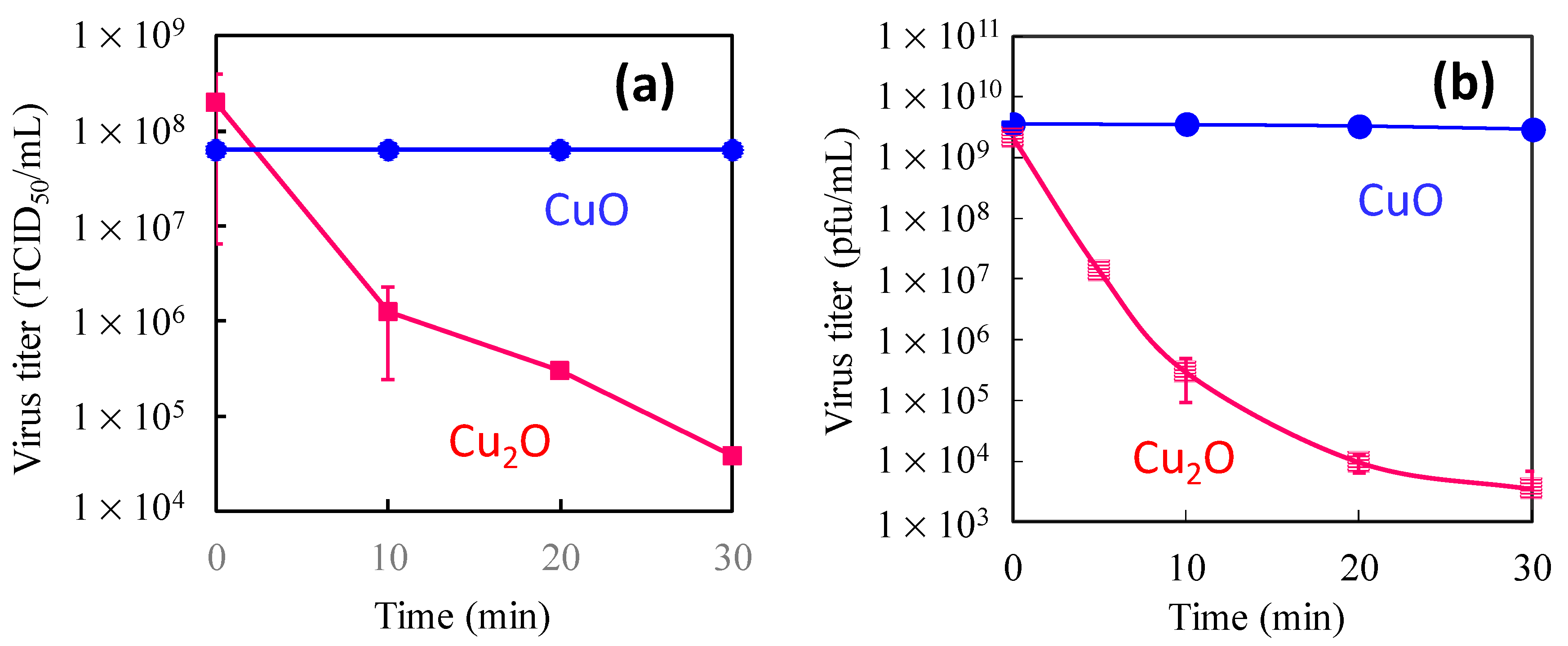
While copper-based compounds are used as a catalyst [36,37], copper oxides have been previously reported to have antimicrobial activity [38,39,40]. In our previous studies, the antiviral effects of CuO and Cu2O coated on glass substrates were reported [34,35]. Figure 1 shows the antiviral properties of CuO and Cu2O films under dark conditions. In this experiment, two types of viruses with different surface structures, the H1N1 influenza A virus (A/PR8/H1N1) and bacteriophage Qβ were examined. The influenza A virus possesses a viral envelope, a cell membrane-like structure that encases its central core, whereas bacteriophages lack an envelope; instead, their surface is composed of protein capsids. As shown in Figure 1, the titers of influenza A and bacteriophage Qβ drastically decreased upon contact with Cu2O by several orders of magnitude even after 30 min, whereas the CuO was not active against either influenza A or bacteriophage Qβ. We also compared the antiviral properties of CuS and Cu2S and found that those of Cu2S were significantly superior to those of CuS [34]. These results strongly indicate that the Cu(I) species plays an important role for efficient antiviral properties.
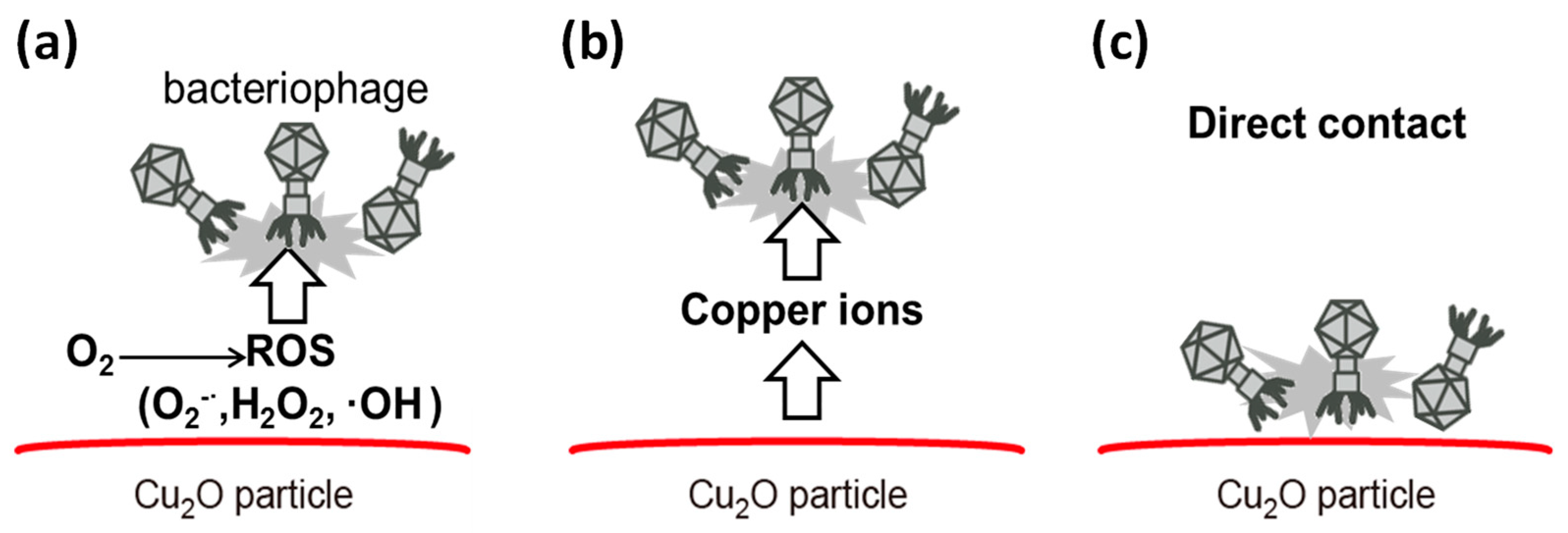
We anticipated three plausible reasons for the efficient antiviral properties of Cu2O, as shown in Figure 2: (a) reactive oxygen species (ROS) [41], (b) leached copper ions [10], and (c) the solid-state compound itself [34,35]. Based on our careful investigation, we excluded ROS by evaluating the antiviral properties under nitrogen atmosphere. The antiviral activity of Cu2O under nitrogen was consistent with that under oxygen atmosphere, indicating that ROS did not contribute to the antiviral activity of Cu2O. It was also found that leached copper ions did not influence the antiviral activity of Cu2O according to a control experiment using a copper ion solution [34]. Therefore, the most plausible reason for the efficient antiviral properties of Cu2O is the solid-state Cu2O compound itself involving Cu(I) species. There are several experimental results that support the importance of direct physical contact between Cu2O and viruses [34]. For example, we inserted a 105 µm thickness of filter paper (pore size = 30 nm) between the Cu2O-coated glass substrate and the viral suspension, which inhibited the antiviral properties of the Cu2O [34]. Furthermore, we chemically modified the Cu2O surface with 1H-benzotriazole (BTA), which strongly coordinates with surface copper atoms via the nitrogen atoms of its triazole ring [42], and the results showed that the antiviral properties of Cu2O treated with BTA were significantly worse than those of untreated Cu2O [34]. These results strongly imply that the surface of Cu2O causes the denaturation or degradation of biomolecules in viruses, which results in their inactivation.
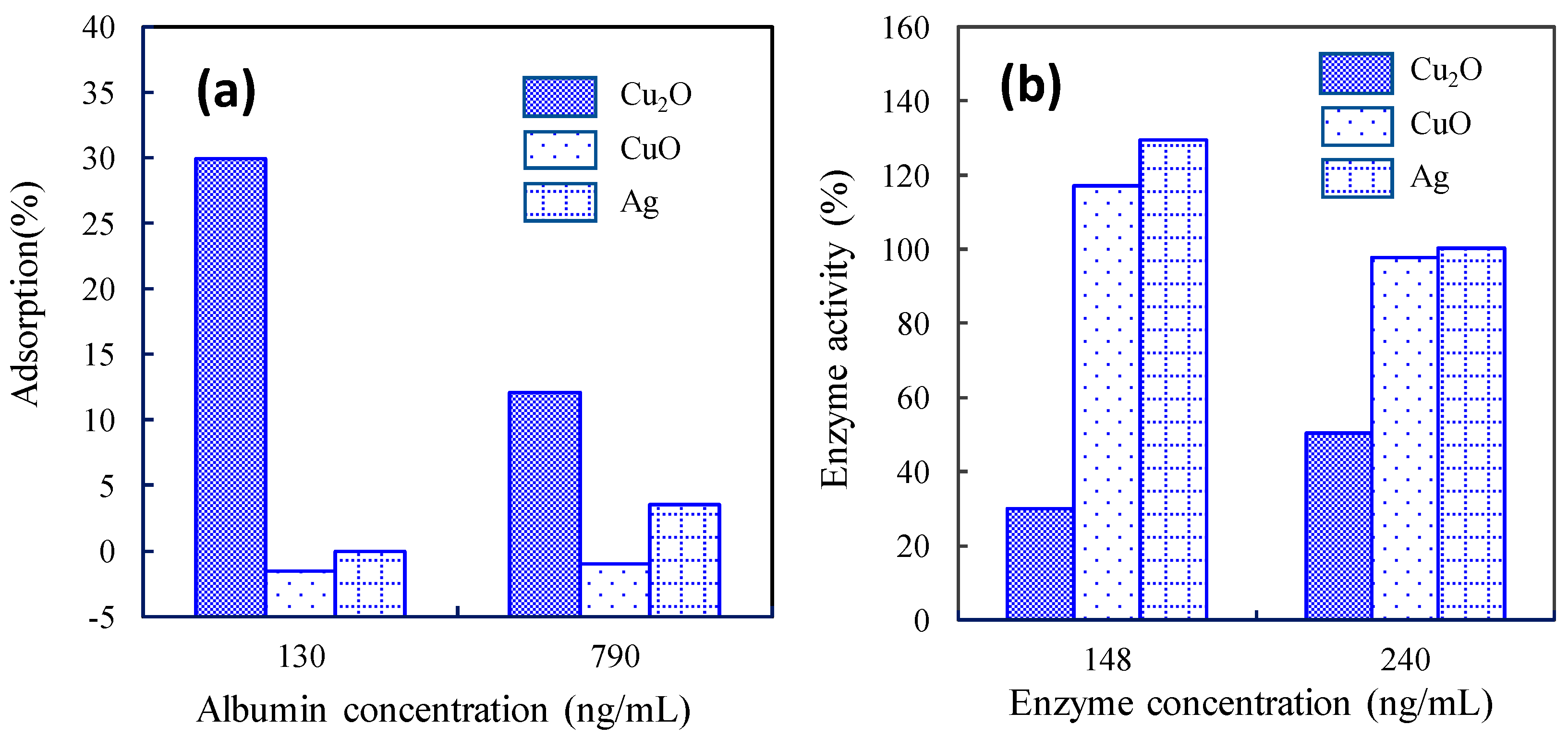
To verify the distinctive antiviral mechanism of Cu2O, we investigated the adsorption properties of model protein molecules [bovine serum albumin (BSA)] on the surface of Cu2O, because the outer capsids of bacteriophage Qβ are composed of protein molecules. Figure 3a shows the adsorption properties of Cu2O in comparison with those of CuO and silver (Ag) as control groups. We used Ag for comparison because metallic Ag compounds have also been reported as effective anti-bacterial materials [43,44,45,46]. As shown in Figure 3a, the incubation of a 130 ng/mL solution of BSA with Cu2O for 8 h resulted in a 30% decrease in the supernatant concentration, revealing strong protein adsorption onto the solid-state Cu2O. Conversely, BSA adsorption onto CuO and Ag was limited. Furthermore, we investigated the protein denaturation by measuring the enzyme activity of alkaline phosphatase as a model enzyme, and the results are shown in Figure 3b. After exposure of the enzyme to Cu2O for 1 h, the enzyme activity decreased to 30% and 50% of the original activity at enzyme concentrations of 148 and 240 ng/mL, respectively. However, after exposure to CuO or Ag, the active enzyme concentration did not decrease from that of its original state. These results strongly imply that the protein adsorption and denaturation abilities of solid-state Cu2O are significantly higher than those of CuO and Ag, resulting in strong deactivation of bacteriophage Qβ.
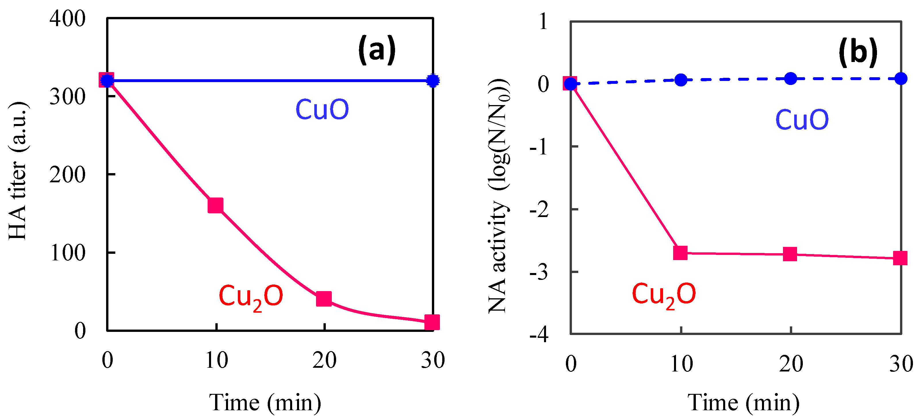
To further verify the disinfection of influenza viruses by Cu2O, we focused on the viral surface proteins that are highly involved in the infection process. Influenza viruses consist of hundreds of haemagglutinin (HA) and neuraminidase (NA) protein groups on the envelope surface. HA is a glycosylated lectin protein that recognizes sialic acid residues on the receptor proteins of the host cells [47]. Once influenza viruses bind through the HA-sialic acid interaction, they can enter the host cells through endocytosis. NA is an endoglycosidase that is necessary for the release of viruses from the surfaces of host cells; it is also involved in the initiation of influenza infection [48]. Both proteins play important roles in the spread of influenza infection. To determine HA activity after exposure to copper oxides, the HA protein was incubated and mixed with chicken red blood cells [49]. To determine NA activity, the 1,2-dioxetane derivative of sialic acid (NA-STAR) was used as a chemiluminescence substrate for highly sensitive detection [50]. Figure 4a,b show the changes in HA and NA activity. After exposure to Cu2O, the HA titer drastically decreased and fell below the detection limit within 30 min. Conversely, the HA titer after exposure to CuO did not change over 30 min. Similarly, NA activity decreased after exposure to Cu2O after 10 min, whereas NA activity was not influenced by exposure to CuO. These results reveal that both the haemagglutination ability of HA and the enzymatic activity of NA are disrupted by exposure to Cu2O. Based on these results, we can conclude that the protein denaturation property of Cu2O yields efficient antiviral function, even under dark conditions.
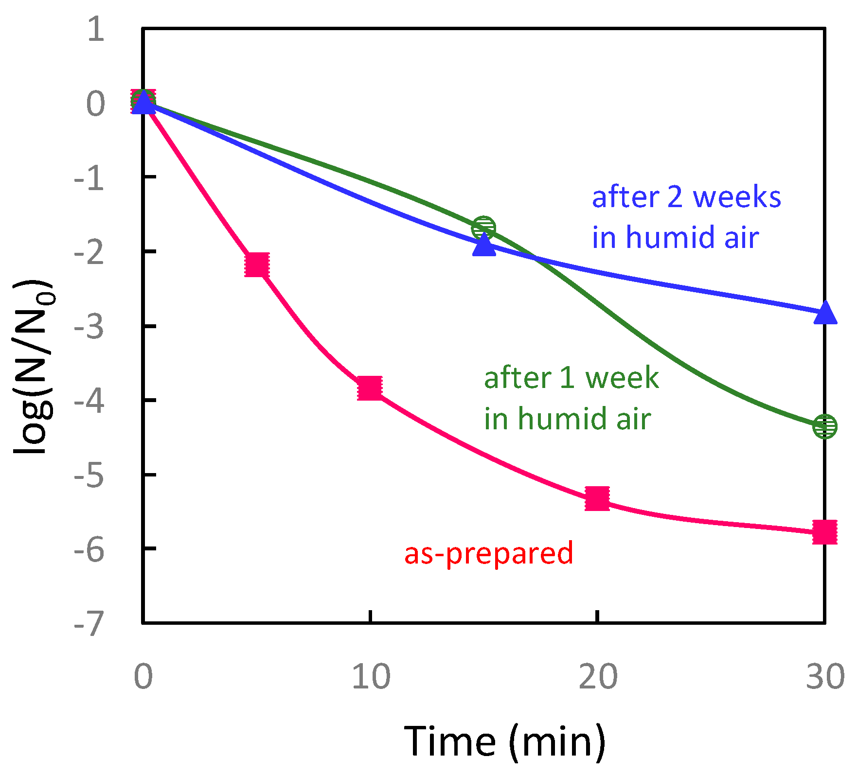
Although Cu2O exhibits strong antiviral properties, Cu(I) is easily oxidized to Cu(II) states under ambient humid atmosphere. In fact, the antiviral properties of Cu2O exposed to humid air (relative humidity 90% at 25 °C) for one week or two weeks significantly worsened compared to those of fresh Cu2O (Figure 5). These results indicate that the antiviral activity of Cu2O is decreased by its self-oxidation [51]. Platzman et al. reported that the Cu2O surface transformed to a copper hydroxide [Cu(OH)2] metastable state with several nanometres in thickness, due to the interactions of Cu ions with hydroxyl groups present at the surface [52]. Further, the metastable Cu(OH)2 phase transformed into a stable CuO layer [51,52]. Therefore, keeping Cu(I) species on the surface of Cu2O under ambient conditions is important for achieving the sustained antiviral activity of Cu2O.
3. Visible Light-Sensitive Cu(II)/TiO2 Photocatalyst
The previous section suggests that maintaining the Cu(I) species is critical for sustaining antiviral properties over the long term. The main goal of this paper is to introduce the combination of a TiO2 photocatalyst with CuxO nanoclusters containing Cu(I) and Cu(II) species to achieve sustained antiviral properties. Before providing a detailed explanation of the CuxO/TiO2 system, we describe the role of the Cu(II) species attached to the TiO2 photocatalyst.
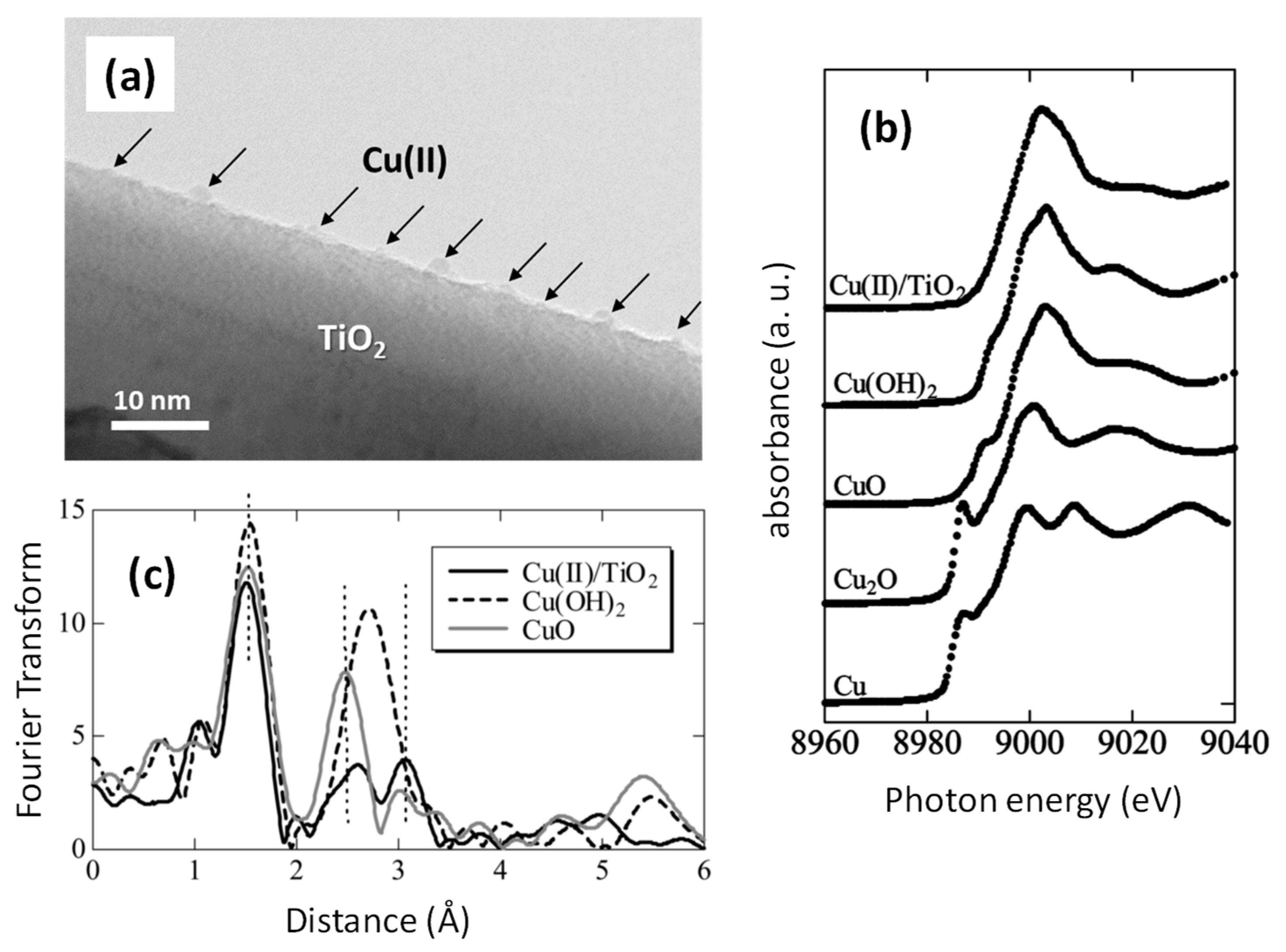
We previously reported Cu(II) nanoclusters grafted onto TiO2 [Cu(II)/TiO2] as an efficient visible light-sensitive photocatalyst for the oxidation of organic molecules [26,27]. Cu(II) nanoclusters could be grafted onto TiO2 (rutile, MT-150A, TAYCA Corporation) by wet chemical impregnation method using copper chloride dissolved aqueous media (0.1 wt % versus TiO2) as reported in our previous studies [26,27]. Figure 6a shows a transmission electron microscope (TEM) image of Cu(II)/TiO2, where Cu(II) clusters a few nanometres in size were grafted onto the TiO2 surface. Although the size of the Cu(II) nanocluster was too small to detect its X-ray diffraction, a previous study determined the local chemical structure of the Cu(II) nanoclusters by X-ray absorption near-edge structure (XANES) and extended X-ray absorption fine structure (EXAFS) [27]. Figure 6b shows the XANES spectra of Cu(II)/TiO2 and commercial reference powders. The spectrum of Cu(II)/TiO2 resembles that of Cu(OH)2, indicating that the valence number of the nanoclusters is in the 2+ state and that the Cu(II) species are likely to be in the five-coordinate square pyramidal form [53,54,55]. Figure 6c shows the EXAFS results of Cu(II)/TiO2 and commercial powder references of Cu(OH)2 and CuO. In contrast to the XANES results, the local chemical environment of the Cu(II) nanoclusters resembles that of CuO. The EXAFS data were carefully analysed using the REX2000 (Rigaku Corporation) and the FEFF program [56], and a one-coordinate Cu–O bond length (2.1–2.2 Å) was observed in Cu(OH)2 and Cu(II)/TiO2. Thus, the grafted Cu(II) nanoclusters are in the five-coordinate environment, which is consistent with the XANES results. In addition, one four-coordinate Cu–Cu and three types of two-coordinate Cu–Cu were observed, and the Cu–Cu bond lengths were similar to those in CuO, and so it can be considered that the grafted Cu(II) nanoclusters resemble the chemical environment of Cu(II) in CuO. That is, the local structure of the Cu(II) nanoclusters is distorted CuO, wherein the apical oxygen approaches Cu(II), forming a five-coordinate square pyramid attached to the TiO2 surface [27].
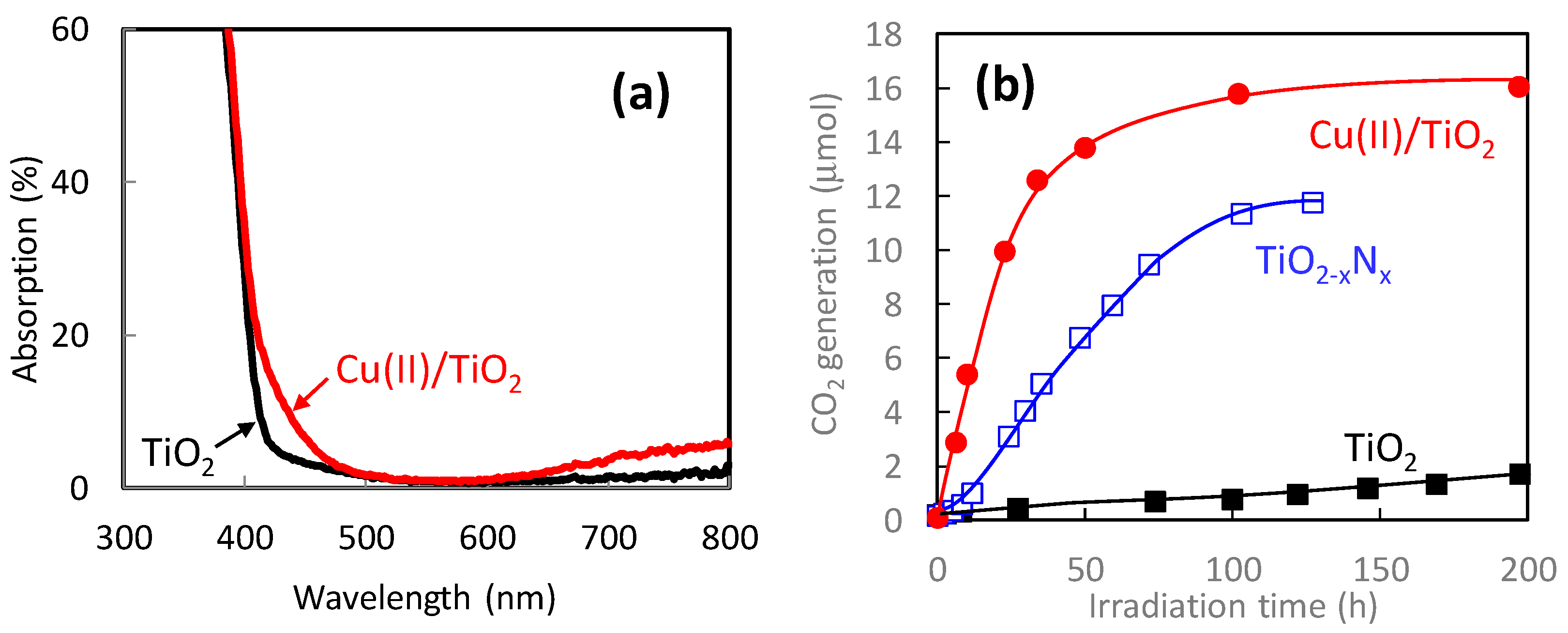
Figure 7a shows the UV-vis absorption spectra of pristine TiO2 and Cu(II)/TiO2. The pristine TiO2 exhibited strong UV light absorption shorter than 400 nm owing to its bandgap excitation. Meanwhile, Cu(II)/TiO2 exhibited additional visible-light absorption around 400–480 nm and over 650 nm. The former absorption is owing to the inter facial charge transfer (IFCT) excitation from the valence band of TiO2 to the Cu(II) nanocluster [26,27], whereas the latter originates in the d–d transition in the Cu(II) species [57]. The IFCT process is theoretically feasible between a semiconductor and ligand under photon irradiation [58], and visible-light absorption through IFCT was experimentally observed in previous studies [59,60,61]. The IFCT transition was also observed in the iron oxide-based Fe(III) nanocluster-grafted TiO2 [31,62].
Figure 7b shows the photocatalytic oxidation activities of gaseous 2-propanol to carbon dioxide (CO2) under visible-light irradiation. As control groups, we also evaluated the photocatalytic activities of bare TiO2 and nitrogen-doped TiO2 (TiO2-xNx). The TiO2-xNx photocatalyst, which is recognized as an efficient visible-light photocatalyst [63], was prepared by a wet chemical method using titanium tetrachloride and ammonia, similar to a previous report [64]. The activity of pristine TiO2 was limited because of the lack of its visible-light absorption. In the case of TiO2-xNx, CO2 molecules were generated by the oxidation of 2-propanol; however, its activity was worse than that of Cu(II)/TiO2 because of the lower oxidation power of the holes excited in the nitrogen orbital [65,66,67]. It is noted that the Cu(II)/TiO2 photocatalyst decomposed 2-propanol with an initial amount of 5 μmol, producing approximately 15 μmol of CO2, showing that complete decomposition was achieved under visible-light irradiation. The quantum efficiency of the Cu(II)/TiO2 system reached over 80% by the optimization of the fabrication process [29], and thus it was significantly superior to that of TiO2-xNx [65,66].
The mechanism of the photocatalytic reaction by Cu(II)/TiO2 was previously investigated by various spectroscopic analyses. For example, Nosaka et al. examined the in situ electron spin resonance (ESR) of Cu(II)/TiO2 under visible-light irradiation [68]. Cu(II) species involve unpaired electrons, thus exhibiting an ESR signal, whereas Cu(I) is ESR-inactive. Furthermore, the photogenerated electrons and holes in TiO2 can be detected by ESR. When the Cu(II)/TiO2 sample was irradiated by visible light under vacuum conditions, the ESR signal of the Cu(II) species decreased and that of photogenerated holes in the valence band of TiO2 appeared. These results strongly suggest that the electron transition occurs from the valence band of TiO2 to the Cu(II) species through their interface under visible-light irradiation to generate Cu(I) species and holes in TiO2. The signal of the photogenerated holes decreased by the introduction of gaseous 2-propanol into the ESR chamber, whereas that of Cu(II) recovered by exposure to oxygen [68]. These results also indicate that the photogenerated holes oxidize 2-propanol, whereas excited electrons in the copper ion species react with oxygen molecules. Formation of Cu(I) species on TiO2 under light irradiation was also reported in the other previous literature [69]. The redox potential of Cu(II)/Cu(I) is approximately 0.16 V [versus a normal hydrogen electrode (NHE)] [26,27], which is more negative than that of the multi-electron reduction reaction of oxygen molecules to hydrogen peroxide (0.68 V vs. NHE) [70,71,72]. Therefore, excited electrons in the Cu(I) species react with oxygen molecules through a multi-electron reduction process under an oxygen-abundant atmosphere. A similar electron transition trend was seen in the XANES results [27]. Furthermore, Osako et al. visualized the reduction and oxidation sites in a Cu(II)/TiO2 system by using an ultrathin CuO film with a well-defined pattern coated onto a TiO2 single crystal prepared by pulsed laser deposition and photolithography [73]. Using an atomic force microscope (AFM), the authors observed the formation of metal Ag particles on the film resulting from the photoreduction of Ag+ ions, and Ag particles were selectively deposited on the edge of a CuO film under visible-light irradiation [74]. These results also suggest that the IFCT transition occurs by visible light and that the Cu(II) species acts as reduction sites. The concept of an IFCT transition for the development of visible light-sensitive photocatalysts has been extended to semiconductor systems other than TiO2, such as ZnO [75,76], SrTiO3 [77,78], SnO2 [79], Nb3O8- [80], Ag3PO4, Bi2O3 [81], BiOCl [82], BiVO4 [83], and Ag-based compounds [84]. The concept of an IFCT transition was also adopted for impurity-doped TiO2, such as Ti(III) self-doped TiO2 [28], Nb(IV)-doped TiO2 [85], and W(IV) and Ga(III)-codoped TiO2 [86].
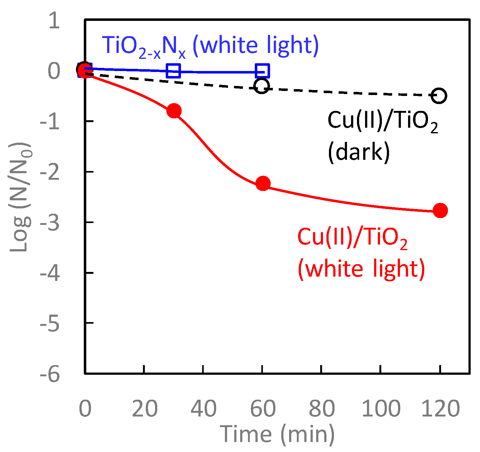
Figure 8 shows the antiviral bacteriophage Qβ activity of TiO2-xNx and Cu(II)/TiO2 under white-light irradiation and dark conditions. Among these samples, the antiviral activity of Cu(II)/TiO2 under white-light irradiation was the most significant. Even though TiO2-xNx exhibited photocatalytic oxidation activity for 2-propanol [Figure 7b], its antiviral activity was negligible, attributed to its limited oxidation power [65,66,67]. In contrast, the number of bacteriophage Qβ on contact with Cu(II)/TiO2 under white-light irradiation decreased more than two orders of magnitude after 60 min of exposure. The antiviral properties of Cu(II)/TiO2 under dark conditions, however, were limited because the Cu(II) species was not as effective for the disinfection of viruses, as described in the previous section.
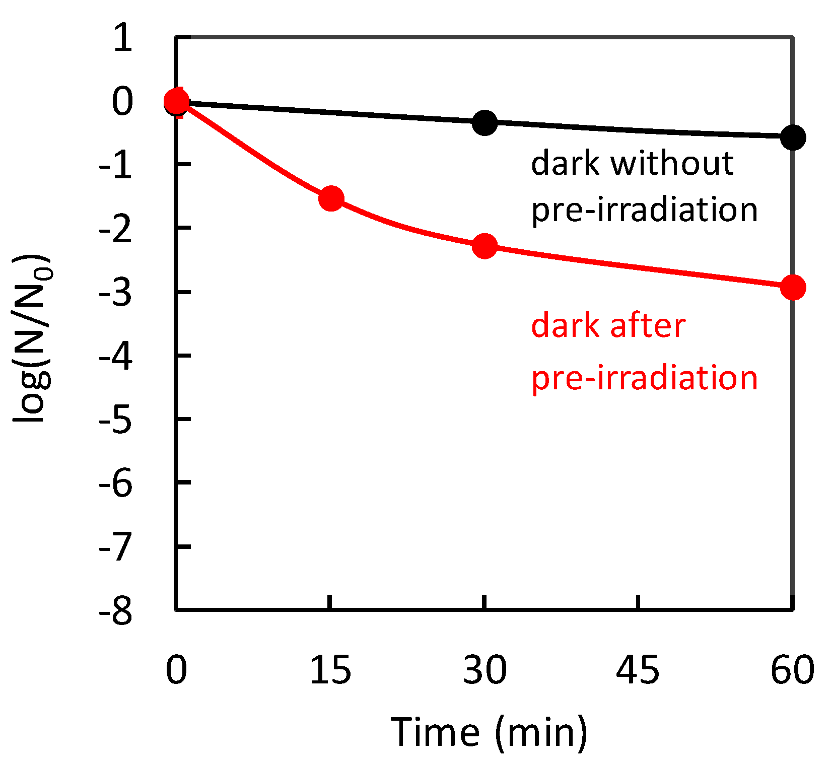
Through the IFCT transition in Cu(II)/TiO2, the Cu(I) species are created, in addition to the generation of holes in the valence band of TiO2. The produced Cu(I) species are effective for protein denaturation, and the holes, which have strong oxidation power, causing protein decomposition, and leading to virus disinfection. The contribution of the Cu(I) species generated by an IFCT transition to the antiviral properties was suggested by a previously reported “pre-irradiation” experiment [32]. Figure 9 shows the antiviral activities of Cu(II)/TiO2 under dark conditions without/with pre-irradiation. As a pre-irradiation treatment, the Cu(II)/TiO2 sample was placed under a white fluorescence lightbulb passed through a UV cut-off film below 400 nm before the evaluation of the antiviral effect. After the pre-irradiation treatment, the Cu(II)/TiO2 film was subjected to antiviral activity testing using bacteriophage Qβ under dark conditions. As shown in Figure 9, the pre-irradiation treatment improved the antiviral activity of Cu(II)/TiO2. This result suggests that pre-irradiation produced the Cu(I) species through the IFCT process, and some of them reacted with oxygen molecules in air, but the others remained even in the dark for a while, causing an antiviral effect. The previous study also showed that pre-irradiation with UV light improved the antiviral activity of Cu(II)/TiO2 [32], indicating that the excited electrons in the conduction band of TiO2 would also be injected into Cu(II) nanoclusters to form Cu(I) species.
4. Antiviral CuxO/TiO2 Photocatalyst
Although Cu(II)/TiO2 exhibited efficient antiviral properties under visible-light irradiation, its antiviral function under dark conditions was limited as shown in Figure 8. Here, we introduce the CuxO (1 < x < 2) nanoclusters grafted TiO2 for efficient antiviral properties even under dark conditions. CuxO nanoclusters were facilely grafted onto TiO2 powder by a method similar to that used for the fabrication of Cu(II)/TiO2. Different from the case of Cu(II)/TiO2 synthesis, we added sodium hydroxide and glucose to the aqueous solution of copper chloride for the grafting process [33]. Glucose dissolved in an alkaline solution acts as a reducing agent of Cu(II) into Cu(I) species; thus, we could control the ratio of Cu(II)/Cu(I) in the CuxO nanoclusters by the concentration of glucose and sodium hydroxide in the aqueous solution [33].
Figure 10a shows the TEM image of CuxO/TiO2. Nanoclusters of CuxO were well dispersed on the surfaces of TiO2. In the X-ray diffraction (XRD) pattern of CuxO/TiO2 [33], no additional peaks other than those of TiO2 were observed, indicating the amorphous nature of the CuxO nanoclusters. Figure 10b shows the XANES spectra of CuxO/TiO2 with the reference data of commercial Cu2O and Cu(OH)2 powders. Peaks I and II are assigned to Cu(I) and Cu(II) species, respectively. The CuxO nanoclusters contained both Cu(I) and Cu(II) species. The ratio of Cu(I)/Cu(II) was estimated by their peak intensities in XANES, and the Cu(I)/Cu(II) ratio of the sample was 1.3, which is the optimum ratio to maintain efficient photocatalytic visible-light activity and sustain antiviral properties, which will be discussed later.
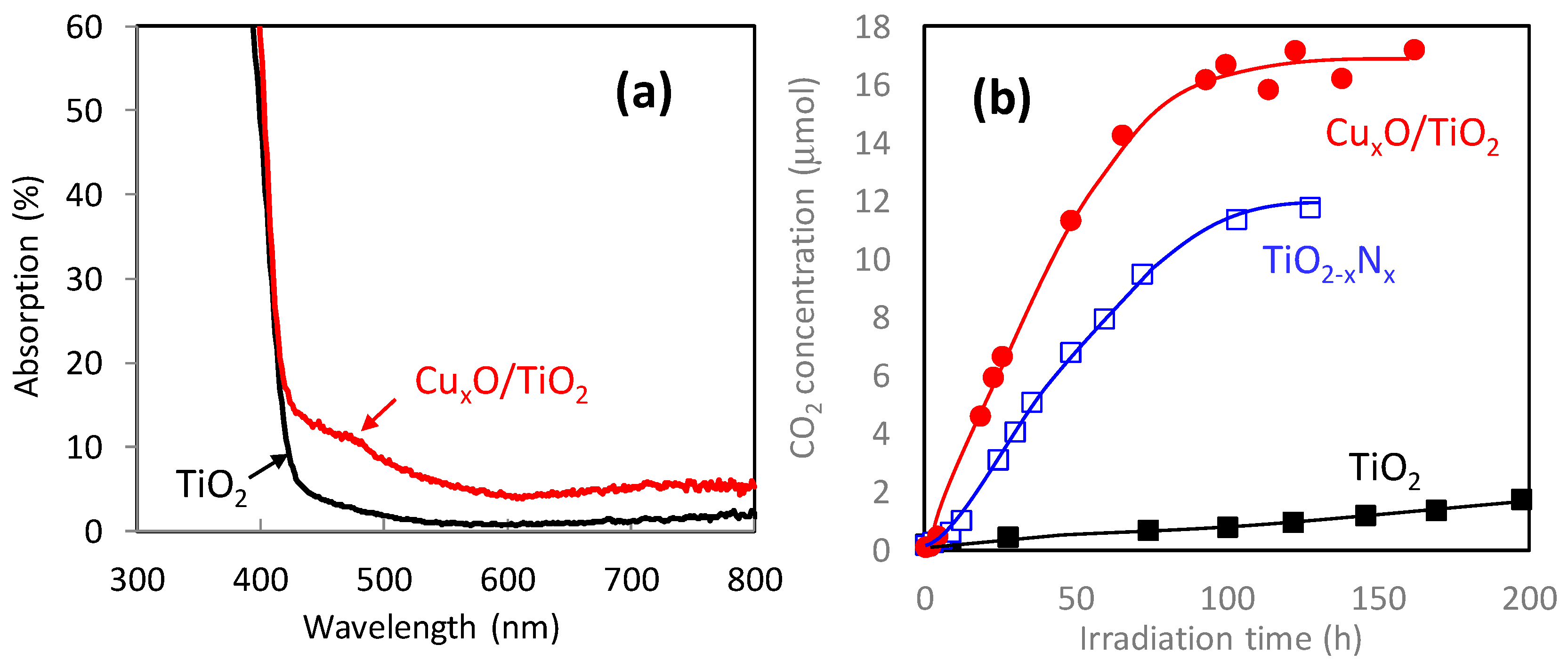
Figure 11a shows the optical absorption spectrum of CuxO/TiO2. In addition to the intrinsic inter-band absorption below 400 nm of TiO2, the absorption band assigned to the IFCT in the range of 400–500 nm [26,27], and the absorption over 650 nm attributable to the d-d transition of the Cu(II) species [57], all of which were observed with Cu(II)/TiO2, as described in the previous section. The CuxO/TiO2 nanocomposites showed an additional absorption band in the range of 500–600 nm, owing to the inter-band transition of Cu2O [87].
Figure 11b shows the photocatalytic oxidation activities of gaseous 2-propanol to carbon dioxide (CO2) under visible-light irradiation. In addition to the CuxO/TiO2 composite, we evaluated the photocatalytic activities of TiO2 and nitrogen-doped TiO2 (TiO2-xNx) as control groups. The photocatalytic oxidation activity of CuxO/TiO2 was superior to those of TiO2 and TiO2-xNx and comparable to the Cu(II)/TiO2 result [Figure 7b]. It is noted that the photocatalytic oxidation activity depends on the ratio of Cu(I)/Cu(II) [33]. A higher content of Cu(II) is better for photocatalytic oxidation activity. The ratio of Cu(I)/Cu(II) in the study sample was 1.3 [33], which optimized to exhibit high photocatalytic activity as well as antiviral activity under dark conditions, which is discussed below.
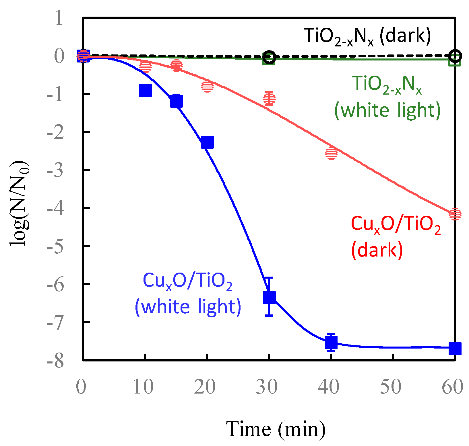
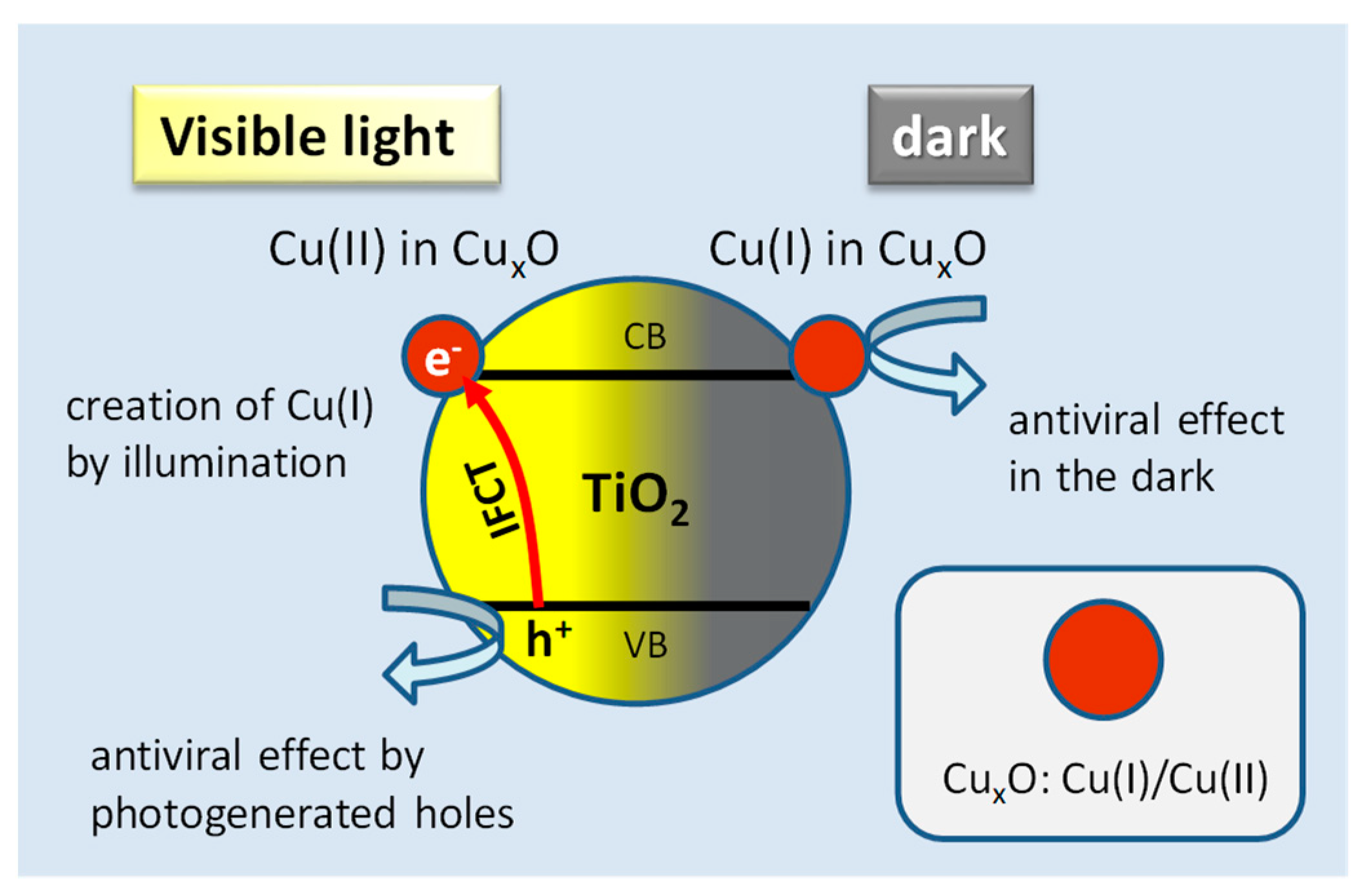
Figure 12 shows the antiviral properties of CuxO/TiO2 under white-light irradiation and dark conditions in comparison with TiO2-xNx. The CuxO/TiO2 displayed a 4-log reduction (i.e., a 99.9 9% reduction of bacteriophage Qβ) after 1 h of contact time under dark conditions, which was significantly superior to the antiviral activity of Cu(II)/TiO2 under dark conditions (Figure 8, black circles). The antiviral activity of CuxO/TiO2 was further improved under visible-light irradiation as a 7.5-log reduction of bacteriophage was achieved after 40 min. The Cu(I) species in CuxO nanoclusters can denature proteins and lose virus activity under dark conditions. Also, the Cu(II) species in the CuxO nanocluster accepts electrons from the valence band of TiO2 to form a Cu(I) species through photo-induced IFCT transition. Therefore, both antiviral active species, i.e., the Cu(I) species and holes in the valence band of TiO2, are simultaneously created in the CuxO/TiO2 system under visible-light irradiation, exhibiting efficient antiviral function under both visible-light irradiation and dark conditions.
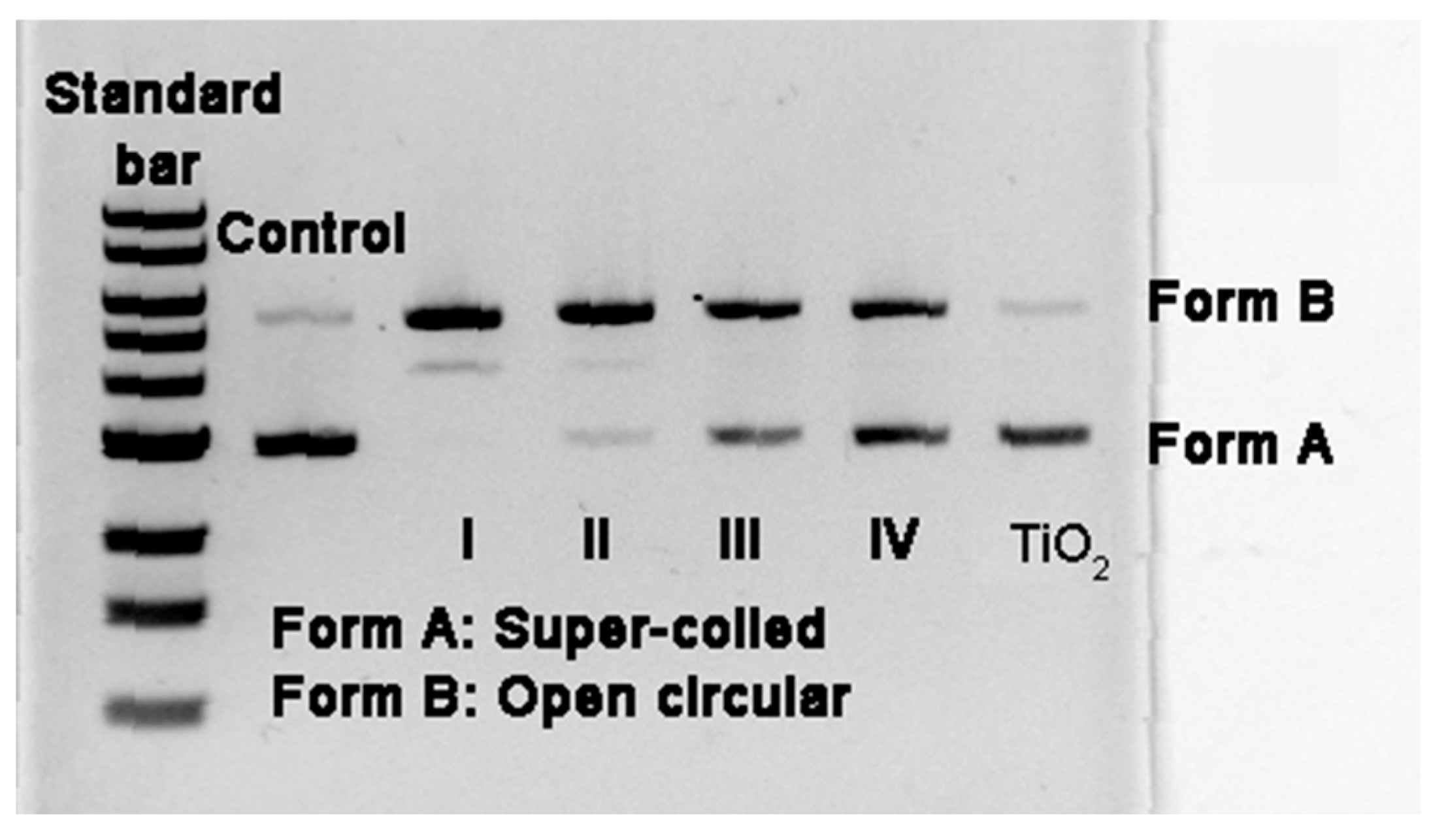
Here, we discuss the optimum ratio of Cu(I)/Cu(II) for both photocatalytic visible light-activity and antiviral properties under dark conditions. We previously evaluated the visible-light activities of CuxO/TiO2 samples with Cu(I)/Cu(II) ratios of 0.13, 0.2, and 1.3 [33], and those activities were comparable to that of Cu(II)/TiO2. We also investigated the degradation activity of DNA, which is an essential component of viruses, for the various CuxO/TiO2 samples with different Cu(I)/Cu(II) ratios and pristine TiO2 as a control group [33]. Figure 13 shows the resulting agarose gel electrophoresis patterns after the exposure of supercoiled plasmid pBR322 DNA to various samples for 2 h under dark conditions. Among the examined samples, bare TiO2 did not cleave the plasmid DNA; however, conversion of the plasmid DNA from the supercoiled to the open circular form was clearly observed in the systems of the hybrid CuxO/TiO2 nanocomposites. Notably, the degradation activity was enhanced as the ratio of Cu(I)/Cu(II) in the hybrid CuxO/TiO2 nanocomposites increased. The complete conversion of supercoiled DNA was achieved using a CuxO/TiO2 [Cu(I)/Cu(II) = 1.3] sample. These results suggest that the hybrid CuxO/TiO2 nanocomposites can destroy the critical biomolecules of viruses, leading to their death and inactivation, even under dark conditions.
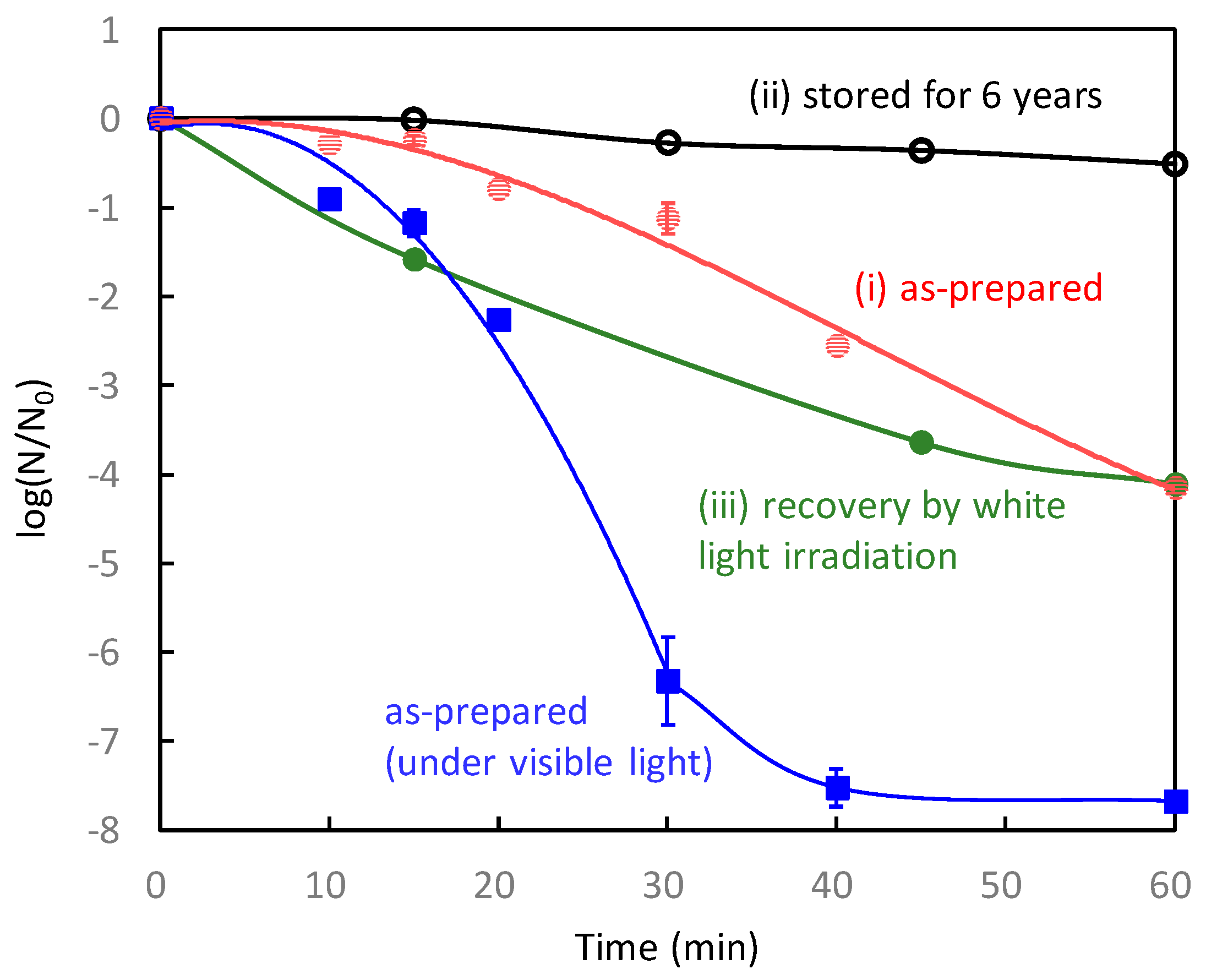
Next, we investigated the long-term antiviral properties of CuxO/TiO2 [Cu(I)/Cu(II) = 1.3] according to the following procedure using bacteriophage Qβ. First, the as-prepared CuxO/TiO2 sample was initially examined under dark conditions [label (i) in Figure 14]. Second, the sample stored under ambient air conditions for more than 6 years was examined [label (ii) in Figure 14]. Third, the stored sample was irradiated with white light for 4 days, and its antiviral properties were evaluated under dark conditions [label (iii) in Figure 14]. The initial activity of CuxO/TiO2 decreased under ambient air exposure by self-oxidation [(i)→(ii)], similar to the results for bare Cu2O shown in Figure 5. However, the deteriorated activity after air exposure was significantly recovered by light irradiation for 4 days. These results imply that the oxidized Cu(II) species in CuxO can be recovered to Cu(I) species by light irradiation. Such a recovery function has never been observed in a pristine Cu2O sample or other solid-state antiviral materials. In contrast to conventional antiviral solid materials, our CuxO/TiO2 maintains its efficient antiviral function, even when light illumination is turned on during the day and off during the night.
Figure 15 shows a schematic illustration of the working principle of the present antiviral CuxO/TiO2 photocatalyst. Cu(I) species disinfect viruses by denaturalizing their protein under dark conditions. Under light irradiation, photogenerated holes oxidize the organic components of the viruses. Further, light irradiation continuously produces Cu(I) species to suppress the self-oxidation of CuxO, resulting in sustained antiviral properties.
Table 1 summarizes the comparison of the antiviral properties of various copper-based compounds. The antiviral activity of pristine CuO is negligible. Conversely, pristine Cu2O exhibits efficient antiviral properties at its initial use; however, its initial red colour turns black by self-oxidation to change into Cu(II) inactive species [51,52]. Further, Cu(II)/TiO2 shows photocatalytic oxidation activity under visible light because of the IFCT transition, but its antiviral activity is limited because of the lack of Cu(I) species. Among these samples, the CuxO/TiO2 composite exhibited good antiviral activity under both light irradiation and dark conditions.
5. Viruses Droplet Splash Test of CuxO/TiO2 Photocatalyst
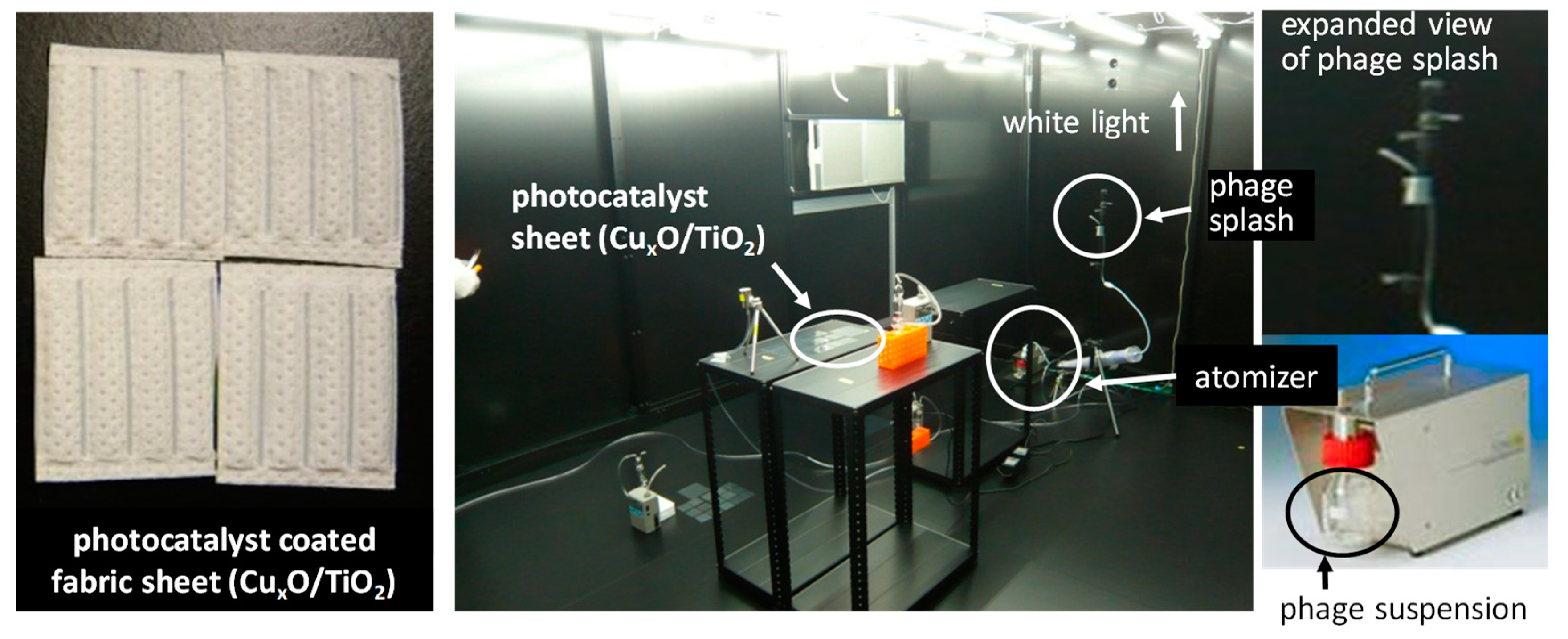
Considering the practical application of the CuxO/TiO2 photocatalyst, we conducted antiviral tests on the CuxO/TiO2-coated sheet fabrics using the pseudo splash-containing bacteriophage Qβ. Figure 16 shows a photograph of the experimental setup for the antiviral splash test. An atomizer generated an aerosol that contained 6 × 10⁷ pfu/h of bacteriophage Qβ, and the particle size of the aerosol was approximately 0.3 μm. The virus aerosol from the atomizer attached to the photocatalyst sheets on a desk of 1 m high from the floor under white fluorescence light at an illuminance of 1000 lux. After 4 h, the number of bacteriophages was counted using the same procedure with the previous studies [33,34,35]. Bacteriophages on a control sheet without CuxO/TiO2 coating were also sampled at 1 h and 2 h.
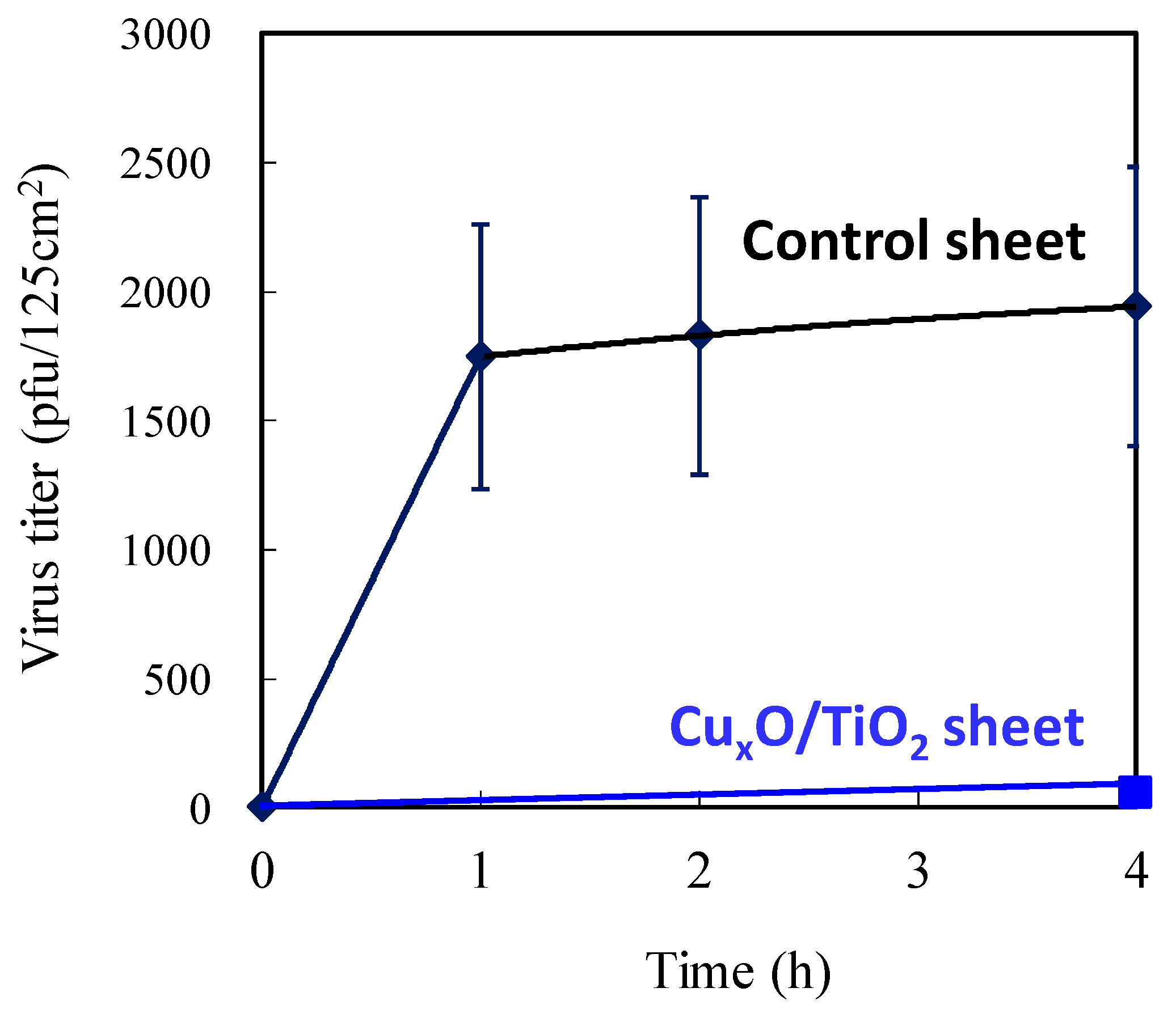
Figure 17 shows the changes in the number of bacteriophages on the photocatalyst sheet and control sheet. It is noteworthy that the number of bacteriophages on the CuxO/TiO2 sheet was negligible, indicating its strong antiviral function against the virus attached to the surface. A CuxO/TiO2-coated material can thus potentially disinfect viruses on any surface derived from droplets and aerosol to protect against viral disease spread by contact infection.
6. Conclusions
This review paper introduces the recent progress in the development of CuxO/TiO2 as an efficient visible light-sensitive photocatalyst for antiviral applications. The CuxO nanocluster consists of the valence states of Cu(I) and Cu(II). Cu(I) species in CuxO nanoclusters can denature viral proteins, resulting in significant antiviral properties even under dark conditions. Unfortunately, the Cu(I) species in CuxO are easily oxidized to inactive Cu(II) in ambient air. However, the combination of CuxO with the TiO2 photocatalyst maintained its antiviral function by visible-light irradiation. In the CuxO/TiO2 photocatalyst, electron transition occurs by visible-light irradiation through the IFCT process; this results in the generation of antiviral Cu(I) species and holes in the valence band of TiO2, which are effective in disinfecting viruses. Once the Cu(I) species in CuxO turn into Cu(II) by self-oxidation, antiviral active Cu(I) species can be regenerated by visible light like a white fluorescence bulb. Therefore, the antiviral function of CuxO/TiO2 can be maintained, even under indoor conditions, where light illumination is turned on during the day and off during the night. It is also noted that the CuxO/TiO2 composite samples have been commercialized (NAKA CORPORATION, Tokyo Japan). We expect the CuxO/TiO2 material to be applied to various antiviral industrial items in indoor circumstances, such as hospitals, airports, metro stations, and schools, as coating materials for air filters, respiratory face masks, and antifungal fabrics to prevent the COVID-19 spread. Furthermore, the present concept contributes to the design of various antiviral materials, such as bimetallic catalysts [88,89,90].
Author Contributions
Conceptualization, K.H.; antiviral investigation, K.S.; photocatalysis investigation, M.M.; writing—original draft preparation, M.M.; writing—review and editing, K.H.; project leader, K.H. All authors have read and agreed to the published version of the manuscript.
Funding
This research was funded by New Energy and Industrial Technology Development Organisation (NEDO) in Japan, project name: Project to Create Photocatalyst Industry for Recycling-Oriented Society. This research was also funded by JSPS Kakenhi, grant No. 18H02055.
Acknowledgments
We appreciate the project members of NEDO, including H. Irie at Yamanashi University, Japan, M. Minoshima at Osaka University, Japan, Y. Kuroda at Showa Denko K.K., Japan, H. Yu at Wuhan University of Technology, China, X. Qiu and M. Liu at Central South University, China, and other collaborators for their great help in the development of the present photocatalyst. Nitrogen-doped TiO2 (TiO2-xNx) was provided by Showa Denko K.K. (HP-N08).
Conflicts of Interest
The authors declare no conflict of interest.
References
- Morens, D.M.; Folkers, G.K.; Fauci, A.S. The challenge of emerging and re-emerging infectious diseases. Nature 2004, 430, 242–249. [Google Scholar] [CrossRef] [PubMed]
- Leroy, E.M.; Kumulungui, B.; Pourrut, X.; Rouquet, P.; Hassanin, A.; Yaba, P.; Délicat, A.; Paweska, J.T.; Gonzalez, J.P.J.; Swanepoel, R. Fruit bats as reservoirs of Ebola virus. Nature 2005, 438, 575–576. [Google Scholar] [CrossRef] [PubMed]
- Vijaykrishna, D.; Poon, L.L.M.; Zhu, H.C.; Ma, S.K.; Li, O.T.W.; Cheung, C.L.; Smith, G.J.D.; Peiris, J.S.M.; Guan, Y. Reassortment of pandemic h1n1/2009 influenza a virus in swine. Science 2010, 328, 1529. [Google Scholar] [CrossRef] [PubMed] [Green Version]
- Wölfel, R.; Corman, V.M.; Guggemos, W.; Seilmaier, M.; Zange, S.; Müller, M.A.; Niemeyer, D.; Jones, T.C.; Vollmar, P.; Rothe, C.; et al. Virological assessment of hospitalized patients with COVID-2019. Nature 2020, 581, 465–469. [Google Scholar] [CrossRef] [PubMed] [Green Version]
- Brankston, G.; Gitterman, L.; Hirji, Z.; Lemieux, C.; Gardam, M. Transmission of influenza A in human beings. Lancet Infect. Dis. 2007, 7, 257–265. [Google Scholar] [CrossRef]
- Kampf, G.; Grotheer, D.; Steinmann, J. Efficacy of three ethanol-based hand rubs against feline calicivirus, a surrogate virus for norovirus. J. Hosp. Infect. 2005, 60, 144–149. [Google Scholar] [CrossRef]
- Tuladhar, E.; Terpstra, P.; Koopmans, M.; Duizer, E. Virucidal efficacy of hydrogen peroxide vapour disinfection. J. Hosp. Infect. 2012, 80, 110–115. [Google Scholar] [CrossRef]
- Barclay, L.; Park, G.W.; Vega, E.; Hall, A.; Parashar, U.; Vinjé, J.; Lopman, B. Infection control for norovirus. Clin. Microbiol. Infect. 2014, 20, 731–740. [Google Scholar] [CrossRef] [Green Version]
- Anson, M. Protein denaturation and the properties of protein groups. In Advances in Protein Chemistry; Anson, M.L., Edsall, J.T., Eds.; Academic Press: Cambridge, MA, USA, 1945; Volume 2, pp. 361–386. [Google Scholar]
- Thurman, R.B.; Gerba, C.P.; Bitton, G. The molecular mechanisms of copper and silver ion disinfection of bacteria and viruses. Crit. Rev. Environ. Control. 1989, 18, 295–315. [Google Scholar] [CrossRef]
- Sunada, K.; Kikuchi, Y.; Hashimoto, K.; Fujishima, A. Bactericidal and detoxification effects of TiO2 thin film Photocatalysts. Environ. Sci. Technol. 1998, 32, 726–728. [Google Scholar] [CrossRef]
- Sunada, K.; Watanabe, T.; Hashimoto, K. Studies on photokilling of bacteria on TiO2 thin film. J. Photochem. Photobiol. A Chem. 2003, 156, 227–233. [Google Scholar] [CrossRef]
- Sunada, K.; Watanabe, T.; Hashimoto, K. Bactericidal activity of copper-deposited TiO2 thin film under weak UV light illumination. Environ. Sci. Technol. 2003, 37, 4785–4789. [Google Scholar] [CrossRef]
- Hajkova, P.; Spatenka, P.; Horsky, J.; Horska, I.; Kolouch, A. Photocatalytic effect of TiO2 films on viruses and bacteria. Plasma Process. Polym. 2007, 4, S397–S401. [Google Scholar] [CrossRef]
- Ishiguro, H.; Nakano, R.; Yao, Y.; Kajioka, J.; Fujishima, A.; Sunada, K.; Minoshima, A.M.; Hashimoto, K.; Kubota, Y. Photocatalytic inactivation of bacteriophages by TiO2-coated glass plates under low-intensity, long-wavelength UV irradiation. Photochem. Photobiol. Sci. 2011, 10, 1825–1829. [Google Scholar] [CrossRef]
- Nakano, R.; Ishiguro, H.; Yao, Y.; Kajioka, J.; Fujishima, A.; Sunada, K.; Minoshima, A.M.; Hashimoto, K.; Kubota, Y. Photocatalytic inactivation of influenza virus by titanium dioxide thin film. Photochem. Photobiol. Sci. 2012, 11, 1293–1298. [Google Scholar] [CrossRef] [PubMed]
- Fujishima, A.; Rao, T.N.; Tryk, D.A. Titanium dioxide photocatalysis. J. Photochem. Photobiol. C Photochem. Rev. 2000, 1, 1–21. [Google Scholar] [CrossRef]
- Mills, A.; Le Hunte, S. An overview of semiconductor photocatalysis. J. Photochem. Photobiol. A Chem. 1997, 108, 1–35. [Google Scholar] [CrossRef]
- Hoffmann, M.R.; Martin, S.T.; Choi, W.; Bahnemann, D.W. Environmental applications of semiconductor photocatalysis. Chem. Rev. 1995, 95, 69–96. [Google Scholar] [CrossRef]
- Paz, Y.; Luo, Z.; Rabenberg, L.; Heller, A. Photooxidative self-cleaning transparent titanium dioxide films on glass. J. Mater. Res. 1995, 10, 2842–2848. [Google Scholar] [CrossRef]
- Wang, R.; Hashimoto, K.; Fujishima, A.; Chikuni, M.; Kojima, E.; Kitamura, A.; Shimohigoshi, M.; Watanabe, T. Light-induced amphiphilic surfaces. Nature 1997, 388, 431–432. [Google Scholar] [CrossRef]
- Wang, R.; Hashimoto, K.; Fujishima, A.; Chikuni, M.; Kojima, E.; Kitamura, A.; Shimohigoshi, M.; Watanabe, T. Photogeneration of highly amphiphilic tio2 surfaces. Adv. Mater. 1998, 10, 135–138. [Google Scholar] [CrossRef]
- Miyauchi, M.; Nakajima, A.; Hashimoto, K.; Watanabe, T. A highly hydrophilic thin film under 1 μW/cm2 UV illumination. Adv. Mater. 2000, 12, 1923–1927. [Google Scholar] [CrossRef]
- Miyauchi, M.; Nakajima, A.; Watanabe, T.; Hashimoto, K. Photocatalysis and photoinduced hydrophilicity of various metal oxide thin films. Chem. Mater. 2002, 14, 2812–2816. [Google Scholar] [CrossRef]
- Miyauchi, M.; Tokudome, H. Highly hydrophilic conversion on oriented TiO2 thin films synthesized by a facile spin-coating method. Appl. Phys. Lett. 2007, 91, 43111. [Google Scholar] [CrossRef]
- Irie, H.; Miura, S.; Kamiya, K.; Hashimoto, K. Efficient visible light-sensitive photocatalysts: Grafting Cu(II) ions onto TiO2 and WO3 photocatalysts. Chem. Phys. Lett. 2008, 457, 202–205. [Google Scholar] [CrossRef]
- Irie, H.; Kamiya, K.; Shibanuma, T.; Miura, S.; Tryk, D.A.; Yokoyama, T.; Hashimoto, K. Visible light-sensitive cu(II)-grafted TiO2 photocatalysts: Activities and X-ray absorption fine structure analyses. J. Phys. Chem. C 2009, 113, 10761–10766. [Google Scholar] [CrossRef]
- Liu, M.; Qiu, X.; Miyauchi, M.; Hashimoto, K. Cu(II) oxide amorphous nanoclusters grafted Ti3+self-doped TiO2: An efficient visible light photocatalyst. Chem. Mater. 2011, 23, 5282–5286. [Google Scholar] [CrossRef]
- Liu, M.; Inde, R.; Nishikawa, M.; Qiu, X.; Atarashi, D.; Sakai, E.; Nosaka, Y.; Hashimoto, K.; Miyauchi, M. Enhanced photoactivity with nanocluster-grafted titanium dioxide photocatalysts. ACS Nano 2014, 8, 7229–7238. [Google Scholar] [CrossRef]
- Miyauchi, M.; Liu, Z.; Zhao, Z.-G.; Anandan, S.; Tokudome, H. Visible-light-driven superhydrophilicity by interfacial charge transfer between metal ions and metal oxide nanostructures. Langmuir 2010, 26, 796–801. [Google Scholar] [CrossRef]
- Miyauchi, M.; Irie, H.; Liu, M.; Qiu, X.; Yu, H.; Sunada, K.; Hashimoto, K. Visible-light-sensitive photocatalysts: Nanocluster-grafted titanium dioxide for indoor environmental remediation. J. Phys. Chem. Lett. 2016, 7, 75–84. [Google Scholar] [CrossRef]
- Liu, M.; Sunada, K.; Hashimoto, K.; Miyauchi, M. Visible-light sensitive Cu(II)–TiO2 with sustained anti-viral activity for efficient indoor environmental remediation. J. Mater. Chem. A 2015, 3, 17312–17319. [Google Scholar] [CrossRef] [Green Version]
- Qiu, X.; Miyauchi, M.; Sunada, K.; Minoshima, A.M.; Liu, M.; Lu, Y.; Li, D.; Shimodaira, Y.; Hosogi, Y.; Kuroda, Y.; et al. Hybrid CuxO/TiO2 nanocomposites as risk-reduction materials in indoor environments. ACS Nano 2012, 6, 1609–1618. [Google Scholar] [CrossRef] [PubMed]
- Sunada, K.; Minoshima, A.M.; Hashimoto, K. Highly efficient antiviral and antibacterial activities of solid-state cuprous compounds. J. Hazard. Mater. 2012, 235–236, 265–270. [Google Scholar] [CrossRef]
- Minoshima, A.M.; Lü, Y.; Kimura, T.; Nakano, R.; Ishiguro, H.; Kubota, Y.; Hashimoto, K.; Sunada, K. Comparison of the antiviral effect of solid-state copper and silver compounds. J. Hazard. Mater. 2016, 312, 1–7. [Google Scholar] [CrossRef]
- Deka, P.; Borah, B.J.; Saikia, H.; Bharali, P. Cu-based nanoparticles as emerging environmental catalysts. Chem. Rec. 2019, 19, 462–473. [Google Scholar] [CrossRef]
- Scotti, N.; Monticelli, D.; Zaccheria, F. Dispersed copper oxide: A multifaceted tool in catalysis. Inorganica Chim. Acta 2012, 380, 194–200. [Google Scholar] [CrossRef]
- Ren, G.; Hu, D.; Cheng, E.W.; Vargas-Reus, M.A.; Reip, P.; Allaker, R.P. Characterisation of copper oxide nanoparticles for antimicrobial applications. Int. J. Antimicrob. Agents 2009, 33, 587–590. [Google Scholar] [CrossRef]
- Pang, H.; Gao, F.; Lu, Q. Morphology effect on antibacterial activity of cuprous oxide. Chem. Commun. 2009, 9, 1076–1078. [Google Scholar] [CrossRef]
- Borkow, G.; Zhou, S.S.; Page, T.; Gabbay, J. A Novel anti-influenza copper oxide containing respiratory face mask. PLoS ONE 2010, 5, e11295. [Google Scholar] [CrossRef] [Green Version]
- Imlay, J.A. Pathways of oxidative damage. Annu. Rev. Microbiol. 2003, 57, 395–418. [Google Scholar] [CrossRef]
- Nilsson, J.O.; Tornkvist, C.; Liedberg, B. Photoelectron and infrared reflection absorption spectroscopy of benzotriazole adsorbed on copper and cuprous oxide surfaces. Appl. Surf. Sci. 1989, 37, 306–326. [Google Scholar] [CrossRef]
- Chernousova, S.; Epple, M. ChemInform abstract: Silver as antibacterial agent: Ion, nanoparticle, and metal. Angew. Chem. Int. 2013, 52, 1636–1653. [Google Scholar] [CrossRef] [PubMed]
- Glover, R.D.; Miller, J.M.; Hutchison, J.E. Generation of metal nanoparticles from silver and copper objects: Nanoparticle dynamics on surfaces and potential sources of nanoparticles in the environment. ACS Nano 2011, 5, 8950–8957. [Google Scholar] [CrossRef] [PubMed]
- Alexander, J.W. History of the medical use of silver. Surg. Infect. 2009, 10, 289–292. [Google Scholar] [CrossRef] [PubMed] [Green Version]
- Panáček, A.; Kvítek, L.; Prucek, R.; Kolář, M.; Večeřová, R.; Pizúrová, N.; Sharma, V.K.; Nevěčná, T.; Zbořil, R. Silver colloid nanoparticles: Synthesis, characterization, and their antibacterial activity. J. Phys. Chem. B 2006, 110, 16248–16253. [Google Scholar] [CrossRef] [PubMed]
- Das, K.; Aramini, J.M.; Ma, L.C.; Krug, R.M.; Arnold, E. Structures of influenza A proteins and insights into antiviral drug targets. Nat. Struct. Mol. Biol. 2010, 17, 530–538. [Google Scholar] [CrossRef] [Green Version]
- Matrosovich, M.N.; Matrosovich, T.Y.; Gray, T.; Roberts, N.A.; Klenk, H.D. Neuraminidase is important for the initiation of influenza virus infection in human airway epithelium. J. Virol. 2004, 78, 12665–12667. [Google Scholar] [CrossRef] [Green Version]
- Donald, H.B.; Isaacs, A. Counts of influenza virus particles. J. Gen. Microbiol. 1954, 10, 457–464. [Google Scholar] [CrossRef] [Green Version]
- Buxton, R.C.; Edwards, B.; Juo, R.R.; Voyta, J.C.; Tisdale, M.; Bethell, R.C. Development of a sensitive chemiluminescent neuraminidase assay for the determination of influenza virus susceptibility to zanamivir. Anal. Biochem. 2000, 280, 291–300. [Google Scholar] [CrossRef]
- Gattinoni, C.; Michaelides, A. Atomistic details of oxide surfaces and surface oxidation: The example of copper and its oxides. Surf. Sci. Rep. 2015, 70, 424–447. [Google Scholar] [CrossRef] [Green Version]
- Platzman, I.; Brener, R.; Haick, H.; Tannenbaum, R. Oxidation of polycrystalline copper thin films at ambient conditions. J. Phys. Chem. C 2008, 112, 1101–1108. [Google Scholar] [CrossRef]
- Nian, J.N.; Chen, S.A.; Tsai, C.C.; Teng, H. Structural feature and catalytic performance of cu species distributed over TiO2 nanotubes. J. Phys. Chem. B 2006, 110, 25817–25824. [Google Scholar] [CrossRef] [PubMed]
- Hsiung, T.L.; Wang, H.P.; Lu, Y.M.; Hsiao, M.C. In situ XANES studies of CuO/TiO2 thin films during photocatalytic degradation of CHCl3. Radiat. Phys. Chem. 2006, 75, 2054–2057. [Google Scholar] [CrossRef]
- Okamoto, Y.; Kubota, T.; Gotoh, H.; Ohto, Y.; Aritani, H.; Tanaka, T.; Yoshida, S. XAFS study of zirconia-supported copper catalysts for the NO–CO reaction: Deactivation, rejuvenation and stabilization of Cu species. J. Chem. Soc. Faraday Trans. 1998, 94, 3743–3752. [Google Scholar] [CrossRef]
- Stern, E.A.; Newville, M.; Ravel, B.; Yacoby, Y.; Haskel, D. The UWXAFS analysis package: Philosophy and details. Phys. B Condens. Matter 1995, 208, 117–120. [Google Scholar] [CrossRef]
- Choudhury, B.; Dey, M.; Choudhury, A. Defect generation, d-d transition, and band gap reduction in Cu-doped TiO2 nanoparticles. Int. Nano Lett. 2013, 3, 25. [Google Scholar] [CrossRef] [Green Version]
- Hush, N. Homogeneous and heterogeneous optical and thermal electron transfer. Electrochim. Acta 1968, 13, 1005–1023. [Google Scholar] [CrossRef]
- Creutz, C.; Brunschwig, B.S.; Sutin, N. Interfacial charge-transfer absorption: Semiclassical treatment. J. Phys. Chem. B 2005, 109, 10251–10260. [Google Scholar] [CrossRef]
- Creutz, C.; Brunschwig, B.S.; Sutin, N. Interfacial charge-transfer absorption: 3. Application to semiconductor−molecule assemblies. J. Phys. Chem. B 2006, 110, 25181–25190. [Google Scholar] [CrossRef]
- Nakamura, R.; Okamoto, A.; Osawa, H.; Irie, H.; Hashimoto, K. Design of all-inorganic molecular-based photocatalysts sensitive to visible light: Ti(iv)−o−ce(iii) bimetallic assemblies on mesoporous silica. J. Am. Chem. Soc. 2007, 129, 9596–9597. [Google Scholar] [CrossRef]
- Yu, H.; Irie, H.; Shimodaira, Y.; Hosogi, Y.; Kuroda, Y.; Miyauchi, M.; Hashimoto, K. An efficient visible-light-sensitive fe(iii)-grafted tio2 photocatalyst. J. Phys. Chem. C 2010, 114, 16481–16487. [Google Scholar] [CrossRef]
- Asahi, R.; Morikawa, T.; Ohwaki, T.; Aoki, K.; Taga, Y. Visible-light photocatalysis in nitrogen-doped titanium oxides. Science 2001, 293, 269–271. [Google Scholar] [CrossRef] [PubMed]
- Sakthivel, S.; Kisch, H. Photocatalytic and photoelectrochemical properties of nitrogen-doped titanium dioxide. ChemPhysChem 2003, 4, 487–490. [Google Scholar] [CrossRef] [PubMed]
- Irie, H.; Watanabe, Y.; Hashimoto, K. Nitrogen-concentration dependence on photocatalytic activity of tio2-xnx powders. J. Phys. Chem. B 2003, 107, 5483–5486. [Google Scholar] [CrossRef]
- Miyauchi, M.; Ikezawa, A.; Tobimatsu, H.; Irie, H.; Hashimoto, K. Zeta potential and photocatalytic activity of nitrogen doped TiO2 thin films. Phys. Chem. Chem. Phys. 2004, 6, 865–870. [Google Scholar] [CrossRef]
- Nakamura, R.; Tanaka, T.; Nakato, Y. Mechanism for visible light responses in anodic photocurrents at N-doped TiO2 film electrodes. J. Phys. Chem. B 2004, 108, 10617–10620. [Google Scholar] [CrossRef]
- Nosaka, Y.; Takahashi, S.; Sakamoto, H.; Nosaka, A.Y. Reaction mechanism of cu(ii)-grafted visible-light responsive TiO2 and WO3 photocatalysts studied by means of ESR spectroscopy and chemiluminescence photometry. J. Phys. Chem. C 2011, 115, 21283–21290. [Google Scholar] [CrossRef]
- Jung, M.; Hart, J.N.; Scott, J.A.; Ng, Y.H.; Jiang, Y.; Amal, R. Exploring Cu oxidation state on TiO2 and its transformation during photocatalytic hydrogen evolution. Appl. Catal. A Gen. 2016, 521, 190–201. [Google Scholar] [CrossRef]
- Yeager, E. Electrocatalysts for O2 reduction. Electrochim. Acta 1984, 29, 1527–1537. [Google Scholar] [CrossRef]
- Wang, Y.; Balbuena, P.B. Ab initio molecular dynamics simulations of the oxygen reduction reaction on a pt(111) surface in the presence of hydrated hydronium (H3O)+(H2O)2: Direct or series pathway? J. Phys. Chem. B 2005, 109, 14896–14907. [Google Scholar] [CrossRef]
- Mustain, W.E.; Prakash, J. A model for the electroreduction of molecular oxygen. J. Electrochem. Soc. 2007, 154, A668–A676. [Google Scholar] [CrossRef]
- Osako, K.; Matsuzaki, K.; Hosono, H.; Yin, G.; Atarashi, D.; Sakai, E.; Susaki, T.; Miyauchi, M. Examination of interfacial charge transfer in photocatalysis using patterned CuO thin film deposited on TiO2. APL Mater. 2015, 3, 104409. [Google Scholar] [CrossRef]
- Osako, K.; Matsuzaki, K.; Susaki, T.; Ueda, S.; Yin, G.; Yamaguchi, A.; Hosono, H.; Miyauchi, M. Direct Observation of interfacial charge transfer between rutile tio2 and ultrathin cuox film by visible-light illumination and its application for efficient photocatalysis. ChemCatChem 2018, 10, 3666–3670. [Google Scholar] [CrossRef]
- Anandan, S.; Ohashi, N.; Miyauchi, M. Zno-based visible-light photocatalyst: Band-gap engineering and multi-electron reduction by co-catalyst. Appl. Catal. B 2010, 100, 502–509. [Google Scholar] [CrossRef]
- Anandan, S.; Miyauchi, M. Ce-doped ZnO (CexZn1−xO) becomes an efficient visible-light-sensitive photocatalyst by co-catalyst (Cu2+) grafting. Phys. Chem. Chem. Phys. 2011, 13, 14937–14945. [Google Scholar] [CrossRef] [PubMed]
- Qiu, X.; Miyauchi, M.; Yu, H.; Irie, H.; Hashimoto, K. Visible-light-driven cu(ii)−(sr1−ynay)(ti1−xmox)o3 photocatalysts based on conduction band control and surface ion modification. J. Am. Chem. Soc. 2010, 132, 15259–15267. [Google Scholar] [CrossRef]
- Nosaka, Y.; Takahashi, S.; Mitani, Y.; Qiu, X.; Miyauchi, M. Reaction mechanism of visible-light responsive Cu(II)-grafted Mo-doped SrTiO3 photocatalyst studied by means of ESR spectroscopy and chemiluminescence photometry. Appl. Catal. B Environ. 2012, 111–112, 636–640. [Google Scholar] [CrossRef]
- Pan, S.; Wang, S.; Zhang, Y.; Xu, S.; Kong, F.; Luo, Y.; Tian, Y.; Teng, X.; Li, G. Surface Fe3+-decorated pristine SnO2 nanoparticles with enhanced ·OH radical generation performance. Catal. Commun. 2012, 24, 96–99. [Google Scholar] [CrossRef]
- Yin, G.; Nishikawa, M.; Nosaka, Y.; Srinivasan, N.; Atarashi, D.; Sakai, E.; Miyauchi, M. Photocatalytic carbon dioxide reduction by copper oxide nanocluster-grafted niobate nanosheets. ACS Nano 2015, 9, 2111–2119. [Google Scholar] [CrossRef]
- Hu, J.; Li, H.; Huang, C.; Liu, M.; Qiu, X. Enhanced photocatalytic activity of Bi2O3 under visible light irradiation by Cu(II) clusters modification. Appl. Catal. B Environ. 2013, 142–143, 598–603. [Google Scholar] [CrossRef]
- Huang, C.; Hu, J.; Cong, S.; Zhao, Z.; Qiu, X. Hierarchical BiOCl microflowers with improved visible-light-driven photocatalytic activity by Fe(III) modification. Appl. Catal. B Environ. 2015, 174, 105–112. [Google Scholar] [CrossRef]
- Yang, Y.; Yamaguchi, A.; Jiang, H.; Van Der Kooy, A.; Okunaka, S.; Hosogai, M.; Tokudome, H.; Miyauchi, M. Green light active photocatalyst for complete oxidation of organic molecules. Chem. Commun. 2020, 56, 9210–9213. [Google Scholar] [CrossRef] [PubMed]
- Wang, P.; Xia, Y.; Wu, P.; Wang, X.; Yu, H.; Yu, J. Cu(II) as a general cocatalyst for improved visible-light photocatalytic performance of photosensitive ag-based compounds. J. Phys. Chem. C 2014, 118, 8891–8898. [Google Scholar] [CrossRef]
- Liu, M.; Qiu, X.; Hashimoto, K.; Miyauchi, M. Cu(ii) nanocluster-grafted, Nb-doped TiO2 as an efficient visible-light-sensitive photocatalyst based on energy-level matching between surface and bulk states. J. Mater. Chem. A 2014, 2, 13571–13579. [Google Scholar] [CrossRef] [Green Version]
- Yu, H.; Irie, H.; Hashimoto, K. Conduction band energy level control of titanium dioxide: Toward an efficient visible-light-sensitive photocatalyst. J. Am. Chem. Soc. 2010, 132, 6898–6899. [Google Scholar] [CrossRef]
- Banerjee, S.; Chakravorty, D. Optical absorption by nanoparticles of Cu2O. EPL Europhys. Lett. 2000, 52, 468–473. [Google Scholar] [CrossRef]
- Han, Y.; Wang, Y.; Ma, T.; Li, W.; Zhang, J.; Zhang, M. Mechanistic understanding of Cu-based bimetallic catalysts. Front. Chem. Sci. Eng. 2020, 14, 689–748. [Google Scholar] [CrossRef]
- Spanu, D.; Recchia, S.; Mohajernia, S.; Tomanec, O.; Kment, S.; Zbořil, R.; Schmuki, P.; Altomare, M. Templated dewetting–Alloying of NiCu bilayers on TiO2 nanotubes enables efficient noble-metal-free photocatalytic H2 evolution. ACS Catal. 2018, 8, 5298–5305. [Google Scholar] [CrossRef] [Green Version]
- Wysocka, I.; Kowalska, E.; Ryl, J.; Nowaczyk, G.; Zielińska-Jurek, A. Morphology, photocatalytic and antimicrobial properties of TiO2 modified with mono- and bimetallic copper, platinum and silver nanoparticles. Nanomaterials 2019, 9, 1129. [Google Scholar] [CrossRef] [Green Version]
Figure 1.
(a) Titer of influenza A virus and (b) bacteriophage Qβ as a function of exposure time to Cu2O (red squares) and CuO films (blue circles) [35]. Error bars indicate standard deviations of two or three replicate experiments. CuO and Cu2O powder were coated on glass substrates and their antiviral test was examined under room temperature. These experimental methods are based on the protocols (ISO 18184:2014 Textiles—Determination of antiviral activity of textile products, and ISO 18071:2016 Fine ceramics—Determination of antiviral activity of semiconducting photocatalytic materials under indoor lighting environment—Test method using bacteriophage Q-beta).

Figure 2.
Possible mechanisms of the antiviral activity of Cu2O: (a) reactive oxygen species (ROS), (b) leached copper ions, and (c) direct contact with the surface [34].
Figure 2.
Possible mechanisms of the antiviral activity of Cu2O: (a) reactive oxygen species (ROS), (b) leached copper ions, and (c) direct contact with the surface [34].

Figure 3.
(a) Adsorption properties of bovine serum albumin (BSA) onto Cu2O, CuO, and Ag after 8 h exposure. Panel (b) shows enzyme activities of these materials after 1 h exposure [34]. These data are based on average of triplicate measurements.
Figure 3.
(a) Adsorption properties of bovine serum albumin (BSA) onto Cu2O, CuO, and Ag after 8 h exposure. Panel (b) shows enzyme activities of these materials after 1 h exposure [34]. These data are based on average of triplicate measurements.

Figure 4.
Hemagglutinin (HA) titer and neuraminidase (NA) activity exposed to Cu2O and CuO suspensions. Effect on (a) HA titer and (b) NA activity of Cu2O (red squares) and CuO (blue circles) as determined by a hemagglutination test and chemiluminescence using the NA-Star method, respectively. N0 in panel (b) is the initial NA amount [35]. These data are based on an average of triplicate measurements.

Figure 5.
Antiviral properties of Cu2O after a week storage in 90% humid air atmosphere (green circles), those after two weeks storage in 90% humid air (blue triangles), and those of as-prepared sample using fresh Cu2O powder (FUJIFILM Wako Pure Chemical Corporation) taken from a commercial bottle (red squares). The data were based on averages of triplicate measurements for as-prepared sample, while duplicate measurements for 1 and 2 weeks after samples.

Figure 6.
(a) TEM image, (b) XANES analyses, and (c) Fourier transforms of EXAFS for Cu(II)/TiO2 [27]. Commercial powder of Cu, Cu2O, CuO, and Cu(OH)2 (Wako Ltd.) were used as references.
Figure 6.
(a) TEM image, (b) XANES analyses, and (c) Fourier transforms of EXAFS for Cu(II)/TiO2 [27]. Commercial powder of Cu, Cu2O, CuO, and Cu(OH)2 (Wako Ltd.) were used as references.

Figure 7.
(a) Optical absorption spectra of TiO2 (black line) and Cu(II)/TiO2 (red line). Amount of Cu(II) was 0.1 wt% versus TiO2 particles. (b) Photocatalytic oxidation activities of 2-propanol under visible-light irradiation for bare TiO2 (black), TiO2-xNx (blue), and Cu(II)/TiO2 (red). Visible-light irradiation was conducted using a xenon lamp passed through optical filters to set the wavelength at 400–530 nm with an illuminance of 1 mW/cm2.

Figure 8.
Antiviral bacteriophage Qβ for Cu(II)/TiO2 under dark (black), TiO2-xNx under white-light irradiation (blue), and Cu(II)/TiO2 under white-light irradiation (red). Light irradiation was conducted using a commercial 10 W cylindrical white fluorescent lightbulb (FL-10, Mitsubishi) with a UV cut-off film shorter than 400 nm at an illuminance of 800 lux, which was measured by photometer (Topcon IM-5).

Figure 9.
Inactivation of bacteriophage Qβ by Cu(II)/TiO2 under dark conditions without pre-irradiation (black) and after pre-irradiation treatment (red) [32]. The pre-irradiation treatment was conducted using a white fluorescence lightbulb passed through a UV cut-off film below 400 nm.
Figure 9.
Inactivation of bacteriophage Qβ by Cu(II)/TiO2 under dark conditions without pre-irradiation (black) and after pre-irradiation treatment (red) [32]. The pre-irradiation treatment was conducted using a white fluorescence lightbulb passed through a UV cut-off film below 400 nm.

Figure 10.
(a) TEM image of CuxO/TiO2 and (b) XANES spectra of Cu2O, Cu(OH)2, and CuxO/TiO2 [33].
Figure 10.
(a) TEM image of CuxO/TiO2 and (b) XANES spectra of Cu2O, Cu(OH)2, and CuxO/TiO2 [33].

Figure 11.
(a) Optical absorption spectra of TiO2 (black line) and CuxO/TiO2 (red line). (b) Photocatalytic oxidation activities of 2-propanol under visible-light irradiation for bare TiO2 (black), TiO2-xNx (blue), and CuxO/TiO2 (red) [33]. Visible-light irradiation was conducted using a xenon lamp passed through optical filters to set the wavelength at 400–530 nm with an illuminance of 1 mW/cm2. The ratio of Cu(I)/Cu(II) in CuxO was 1.3.

Figure 12.
Inactivation of bacteriophage Qβ for various samples. CuxO/TiO2 under white light (blue), CuxO/TiO2 under dark conditions (red), TiO2-xNx under white light (green), and TiO2-xNx under dark conditions (black) [33]. Light irradiation was conducted using a commercial 10 W cylindrical white fluorescent lightbulb with a UV cut-off film at an illuminance of 800 lux.

Figure 13.
The cleavage of supercoiled plasmid pBR322 DNA by different samples under dark conditions for 2 h [33]. Lanes I, II, III, and IV correspond to Cu(I)/Cu(II) = 1.3, 0.2, 0.13 and Cu(II)/TiO2, respectively.
Figure 13.
The cleavage of supercoiled plasmid pBR322 DNA by different samples under dark conditions for 2 h [33]. Lanes I, II, III, and IV correspond to Cu(I)/Cu(II) = 1.3, 0.2, 0.13 and Cu(II)/TiO2, respectively.

Figure 14.
Inactivation of bacteriophage Qβ by CuxO/TiO2 [Cu(I)/Cu(II) = 1.3] under the following sequential conditions: (i) as-prepared sample in the dark (red), (ii) the sample stored under ambient air for more than 6 years (black), (iii) after light irradiation onto the 6-year stored sample for 4 days (green). The antiviral tests of (i)–(iii) were performed under dark conditions. The results of the as-prepared CuxO/TiO2 sample under visible-light irradiation are also shown (blue).

Figure 15.
Schematic illustration of the working principle of the antiviral CuxO/TiO2 photocatalyst.
Figure 15.
Schematic illustration of the working principle of the antiviral CuxO/TiO2 photocatalyst.

Figure 16.
Procedure for evaluating an antiviral CuxO/TiO2-coated fabric sheet using pseudo splash-containing bacteriophage Qβ. Room volume was (4 m × 3 m × 2 m) and the ventilation frequency was 1.8 time/h. White light was irradiated by fluorescent lightbulbs at an illuminance of 1000 lux. Bacteriophage Qβ containing 6 × 10⁷ pfu/h was sprayed for 4 h by an atomizer (ATM-226, KANOMAX JAPAN INC.) to attach it on the sheet surfaces.

Figure 17.
Antiviral properties of the CuxO/TiO2-coated sheet and the control sheet without the photocatalyst using splash-containing bacteriophage Qβ. In the case of CuxO/TiO2 to avoid the overestimation of its antiviral property, the number of experiments was set to 1 time (after 4h) in order to exclude the influence of air flow due to human’s entering into the room for measurement.


Table 1.
The comparison of antiviral properties of various copper based compounds.
| Material | Antiviral Activity Under Dark Condition | Photocatalytic Activity | Antiviral Activity for Long Term in Indoor Condition |
|---|---|---|---|
| CuO | × | × | × |
| Cu2O | ○ | × | △ |
| Cu(II)/TiO2 | × | ○ | △ |
| CuxO/TiO2 | ○ | ○ | ○ |
(○: good, △: fair, ×: poor).
© 2020 by the authors. Licensee MDPI, Basel, Switzerland. This article is an open access article distributed under the terms and conditions of the Creative Commons Attribution (CC BY) license (http://creativecommons.org/licenses/by/4.0/).
Share and Cite
MDPI and ACS Style
Miyauchi, M.; Sunada, K.; Hashimoto, K. Antiviral Effect of Visible Light-Sensitive CuxO/TiO2 Photocatalyst. Catalysts 2020, 10, 1093. https://0-doi-org.brum.beds.ac.uk/10.3390/catal10091093
AMA Style
Miyauchi M, Sunada K, Hashimoto K. Antiviral Effect of Visible Light-Sensitive CuxO/TiO2 Photocatalyst. Catalysts. 2020; 10(9):1093. https://0-doi-org.brum.beds.ac.uk/10.3390/catal10091093
Chicago/Turabian StyleMiyauchi, Masahiro, Kayano Sunada, and Kazuhito Hashimoto. 2020. "Antiviral Effect of Visible Light-Sensitive CuxO/TiO2 Photocatalyst" Catalysts 10, no. 9: 1093. https://0-doi-org.brum.beds.ac.uk/10.3390/catal10091093
Note that from the first issue of 2016, this journal uses article numbers instead of page numbers. See further details here.