Article

Elovl2 Is Required for Robust Visual Function in Zebrafish

1
Viterbi Family Department of Ophthalmology, Shiley Eye Institute, School of Medicine, University of California San Diego, La Jolla, CA 92093, USA
2
Department of Biophysics and Physiology, Center for Translational Vision Research, Gavin Hebert Eye Institute, School of Medicine, University of California Irvine, Irvine, CA 92617, USA
3
The Salk Institute for Biological Studies, Clayton Foundation Laboratories for Peptide Biology, 10010 N. Torrey Pines Rd, La Jolla, CA 92037, USA
*
Authors to whom correspondence should be addressed.
Submission received: 13 October 2020 / Revised: 12 November 2020 / Accepted: 29 November 2020 / Published: 2 December 2020
(This article belongs to the Section Cells of the Nervous System)

Abstract

:
Omega-3 and omega-6 polyunsaturated fatty acids (PUFAs) play critical roles in membrane stability and cell signaling within the retina. ELOVL2 (Elongation of Very Long Chain Fatty Acids-Like 2), an elongase involved in the synthesis of long chain polyunsaturated fatty acids (LC-PUFAs), has recently been implicated in regulating aging in the mammalian retina. In this work, we characterize the expression and function of elovl2 in the retina development in embryonic zebrafish. Whole mount in situ hybridization shows elovl2 is expressed in the Muller glia in embryonic and adult zebrafish. Lipidomics analysis of elovl2 crispants whole embryos at day 2 and eyes at day 7 demonstrated significant changes in lipids composition, especially on the level of lipids containing docosahexaenoic acid (DHA). Histological analysis of zebrafish lacking elovl2 revealed increased retinal thickness compared to controls at day 7 without gross disruptions of the retinal architecture. Finally, elovl2 crispants showed differences in the visual motor reflex light off (VMR-OFF) at day 7 compared to controls. In sum, inactivation of elovl2 in zebrafish embryos caused changes in lipid composition and in visual behavior, further confirming the important role of LC-PUFAs in healthy vision.

1. Introduction

Omega-3 and omega-6 polyunsaturated fatty acids (PUFAs), are critical for diverse biological functions including membrane stability, cell signaling and metabolism, particularly in the brain and retina [1,2]. Synthesis of PUFAs begins with the dietary intake of essential amino acids, linoleic acid, and alpha linoleic acid, which then goes through a series of elongation and desaturation reactions to form longer chain omega-3 and omega-6 fatty acids, including arachidonic acid, eicosapentaenoic acid (EPA), and docosahexaenoic acid (DHA). DHA, the major polyunsaturated fatty acid in the brain and retina, is a critical component of photoreceptor outer segments necessary for photoreceptor function [1,3,4] while very long chain PUFAs (VLC-PUFAs; greater than 22 carbon length fatty acids [5]) are believed to be indispensable in maintaining the curvature of the photoreceptor disk membrane. PUFAs have been implicated in eye diseases, as a decrease of dietary intake of food rich in omega-3 fatty acids, such as fish, have been linked to higher risk of age related eye diseases such as macular degeneration in multiple epidemiologic studies [4,6,7].
ELOVL2 (Elongation Of Very Long Chain Fatty Acids-Like 2) encodes an enzyme involved in the elongation of long-chain omega-3 and omega-6 polyunsaturated fatty acids (LC-PUFAs) [8]. In particular, ELOVL2 in mammals elongates docosapentaenoic acid (DPA—22:5n-3) to 24:5n-3, which in turn is the substrate for the formation of VLC-PUFAs as well as 22:6n-3, i.e., DHA [9]. Interestingly, DNA methylation of the Elovl2 regulatory element has been well established as an epigenetic biomarker of aging, as multiple studies have shown that the DNA methylation of the ELOVL2 promoter across multiple human tissues highly correlates with chronological age [10,11].
We have previously studied the role of Elovl2 in the mammalian retina. We have implicated Elovl2 as a critical molecular regulator of aging, as loss of Elovl2 activity in mice accelerates anatomical and functional surrogates of aging in the retina. Additionally, we observed sub-RPE deposits, which contained multiple proteins found in human drusen, a pathologic hallmark of age related macular degeneration [12]. Pharmacological demethylation of Elovl2 can increase Elovl2 gene expression and prevent the progression of the age-related decline of the electroretinogram response, a functional surrogate of aging [12].
Zebrafish (Danio rerio) represents an excellent model system to study retinal development and biology, given its small size, transparent embryo, and the availability of robust visual behavior assays [13]. The role of elovl2 in zebrafish, particularly within the eye, is still poorly understood. Previous studies have demonstrated that zebrafish elovl2 can elongate C18-C22 PUFAs in a heterologous yeast system, in contrast to human ELOVL2 which has been shown to only elongate C20 and C22 PUFAs [8,14,15]. In addition, zebrafish elovl5, which in humans is specific to the elongation of C18 PUFAs, can also elongate C20 and C22 PUFAs to a lesser extent. In a recent study of elovl2 and elovl5 knockout zebrafish, it was observed that elovl2, but not elovl5, is required for the conversion of C20 EPA to DPA, and thus synthesis of DHA [16]. Finally, zebrafish elovl4 is also able to elongate LC-PUFAs in striking contrast to the mammalian ortholog [17].
Muller glia are macro glia found in the retina whose processes span the entire retina and contact most of the neurons in the retina. They play diverse functions including providing trophic support, removing metabolic waste, as well as regulating ion and water homeostasis, and regulating immune and inflammatory responses [18]. Whether Muller glia can synthesize PUFAs, and what the role of Muller glia derived PUFAs is, is still poorly understood.
Here, we present the investigation of the function of elovl2 in the zebrafish eye. We characterized the expression of elovl2 in embryonic and adult wildtype zebrafish retina. We created zebrafish lacking elovl2 function through the introduction of biallelic mutations in elovl2 using CRISPR-Cas9 technology, termed “crispants” [19]. Using lipidomics, retinal morphology, and visual behavior we showed the unexpected expression pattern and role of elovl2 in zebrafish eye.

2. Materials and Methods

2.1. Zebrafish Husbandry

The AB strain (Zebrafish International Research Center (ZIRC); Eugene, OR, USA) was used for all experiments. Zebrafish were maintained in the fish facility of the University of California, San Diego (UCSD) under a controlled 14/10 h light cycle between 27 and 29 degrees Celsius and fed with a standard brine shrimp diet twice daily with a recirculating aquarium system. Breeding and experimental procedures were approved by the Institute of Animal Care and Use Committee of the University of California, San Diego (S18067).

2.2. Zebrafish Crispant Creation

Potential gRNA sequences for zebrafish elovl2 were searched for using the Chop-chop algorithm (https://chopchop.cbu.uib.no). Two elovl2 gRNA sequences were selected, GACAGCCTATTTGGAGAAAG in exon 2 and TTCCCAGGTAGATTGTTAGG in exon 3. The control gRNA used (TGAGTATTCGCATGCAACTA) does not target any known zebrafish nucleotide sequence.
GRNA oligonucleotides (Integrated DNA Technologies (IDT), Coraville Iowa) were synthesized by and were duplexed individually with tracrRNA (Integrated DNA Technologies (IDT) Coraville, IA, USA). The injection mix containing 250 ng/μL of gRNA duplex complex (both gRNAs), 500 ng/μL rCas9 protein (PNA Bio CP01-20 Thousand Oaks, CA, USA) and duplex buffer (IDT) was injected into one cell stage zebrafish embryos. The embryos were maintained in a 28 °C incubator. The level of DNA editing was determined through DNA Sanger sequencing (Genewiz La Jolla, CA, USA) and analysis using the ICE v2CRISPR Analysis Tool (Synthego (Redwood City, CA, USA), found at https://www.synthego.com/products/bioinformatics/crispr-analysis) [20] Sequencing primers for elovl2 gRNA exon 2 were 5′ TTGAAGCTTGCAATCTGACTGT3′ and 5′ TGGAACGTTCTATTGAGTGTCG 3′. Sequencing primers for elovl2 gRNA exon 3 were 5′ TTTGTTTGATGTCAGATACCCG3′ and 5′ ATGAGCACATGGACTGCTATTG3′.

2.3. Zebrafish In-Situ Hybridization

Whole mount RNA in situ hybridization was performed on fixed 1–3 dpf zebrafish embryos using previously described protocol [21]. The stained embryos were imaged using a stereomicroscope (Zeiss STEMI 508 (Zeiss Oberkocken, Germany)). A 550 bp fragment of elovl2 cDNA was PCR amplified and cloned into a TOPO vector (Invitrogen). (Primers 5′ F AGGCAGTCATTTAGGTGACACTATAGATGG and 3′ R CGTCGTGGACTAATACGACTCACTATAGAC). The plasmid was linearized and an in vitro transcription reaction with DIG labeling mix (Roche, cat. no. 1277073) was performed, as previously described [21]. For staining of retinal sections, the RNAscope protocol was followed as per manufacturers protocol (ACD (Newark, CA, USA); elovl2 probe Cat. No. 550301). The optional step of target retrieval was excluded to allow better retention of the samples on the slides. The stained sections were flooded with ProlongTM Gold Antifade Mountant (Cat. No. P36930) and covered with coverslips (Fisher Scientific. Waltham, MA 12-545F, USA) before imaging.

2.4. Microscopic Imaging

Fluorescent imaging of the fluorescent in situ hybridization (RNAscope) as well as the retinal anatomy analysis were performed using the Keyence microscope (BZ-X710, Keyence, Itasca, IL, USA) using structured illumination and optical sectioning. Ten mm sections were imaged using the CFI PLAN APO 40x LAMBDA Lens (Nikon, Englewood, CO, USA) and analyzed using the BZX Analyzer Software v2.0 (Keyence, USA).

2.5. Zebrafish Visual Motor Response

Larvae at 6 dpf were placed in flat bottom clear 96 well plates (Corning CLS3370 Corning, NY, USA) and the assay was performed in DanioVision observation chamber (Noldus Information Technology, Wageningen, The Netherlands). The light program was designed based on a previously published protocol [22]. The larvae were dark-adapted for three hours, followed by three cycles of light on/off program. In each light cycle, white light at 100% intensity was turned on for 5 min followed by a 30 min dark period. The activity of larvae was measured 30 s before and after light on/off using EthoVision XT software (Noldus Information Technology, Wageningen, The Netherlands). A 5 min five dark period was included in the light program to measure the activity of larvae 30 s before the first light was on. The activity data was exported from the software and analyzed using Microsoft Excel. Three rounds of experiments, each encompassing about 130 embryos were performed, with all data pooled for the analysis.

2.6. Zebrafish Retinal Anatomy Analysis

The embryos were fixed in 4% PFA for 72 h and transferred into 100% ethanol solution. The samples were submitted to HistoWiz (https://home.histowiz.com) for sectioning and hematoxylin and eosin (H&E) staining for histological analysis. For uniformity, only sections close to nerve fibers were selected for the measurements. Measurements were taken at three different regions in each selected section: near optic nerve (middle), anterior to the optic nerve (anterior) and posterior to optic nerve (posterior). Measurements were recorded using Fiji ImageJ software version 1.52p (National Institutes of Health, Maryland, MA, USA).

2.7. Lipid Analysis

For lipidomics sample preparation, two and seven days and post fertilization (dpf) larvae were transferred in an Eppendorf tube and excess water was removed. The tubes were then placed in a dish containing dry ice and ethanol to flash freeze the larvae. Twelve embryos were placed in each tube, with three replicates per timepoint. Lipids were extracted using a modified version of the Bligh-Dyer method [23]. Briefly, zebrafish embryos were homogenized in 1 mL PBS and shaken in a glass vial (VWR International, Radnor, PA, USA) with 1 mL methanol and 2 mL chloroform containing internal standards (13C16-palmitic acid, d7-Cholesterol) for 30 s. The resulting mixture was vortexed for 15 s and centrifuged at 2400× g for 6 min to induce phase separation. The organic (bottom) layer was retrieved using a Pasteur pipette, dried under a gentle stream of nitrogen, and reconstituted in 2:1 chloroform:methanol for LC/MS analysis.
Extracted lipids were resuspended in 200 μL of EtOH, incubated with 0.1 M KOH at room temperature for 24 h for saponification. The reaction was stopped by addition of 0.2 M HCl. Lipids were extracted as described above with d31-palmitic acid as internal standard.
Untargeted lipidomic analysis was performed on a Vanquish HPLC online with a Q-Exactive quadrupole-orbitrap mass spectrometer equipped with an electrospray ion source (Thermo Fisher Scientific, Milan, Italy). A Bio-Bond C4 column (Dikma, 5 μm, 4.6 mm × 50 mm) was used. Solvent A consisted of 95:5 water:methanol, Solvent B was 60:35:5 isopropanol:methanol:water. Solvents A and B contained 5 mM ammonium formate with 0.1% formic acid. The gradient was held at 0% B between 0 and 5 min, raised to 20% B at 5.1 min, increased linearly from 20% to 100% B between 5.1 and 55 min, held at 100% B between 55 min and 63 min, returned to 0% B at 63.1 min, and held at 0% B until 70 min. Flow rate was 0.1 mL/min from 0 to 5 min, 0.4 mL/min between 5.1 min and 55 min, and 0.5 mL/min between 55 min and 70 min. Data was acquired in negative ionization mode. Spray voltage was −2.5 kV. Sheath, auxiliary, and sweep gases were 53, 14, and 3 arbitrary units (a.u.), respectively. Capillary temperature was 275 °C. Data was collected in full MS/dd-MS2 (top 5). Full MS was acquired from 100 to 1500 m/z with a resolution of 70,000, AGC target of 1 × 10⁶ and a maximum injection time of 100 ms. MS2 was acquired with resolution of 17,500, a fixed first mass of 50 m/z, AGC target of 1 × 10⁵ and a maximum injection time of 200 ms. Stepped normalized collision energies were 20, 30, and 40%.
Targeted lipidomic analysis was performed on a Dionex Ultimate 3000 LC system (Thermo Fisher Scientific, Milan, Italy) coupled to a TSQ Quantiva mass spectrometer (Thermo Fisher Scientific, Milan, Italy). A XBridge C8 column (Waters, 5 μm, 4.6 mm × 50 mm) was used. The solvents and gradient were as described above. MS analyses were performed using electrospray ionization in negative mode, with spray voltages of −2.5 kV, ion transfer tube temperature of 325 °C, and vaporizer temperature of 200 °C. Pseudo multiple reaction monitoring (MRM) was performed to detect fatty acids.
Lipid identification was performed with LipidSearch (Thermo Fisher Scientific, Milan, Italy). Mass accuracy, chromatography and peak integration of all LipidSearch-identified lipids were verified with Skyline [24]. Peak integration of targeted fatty acids was also performed with Skyline. Peak areas were used in data reporting, data was normalized using internal standards. The relative abundance of lipid classes were calculated by the percent relative area method with proper normalization using internal standard and considering the sum of all relative areas of the identified lipids.
Statistical analyses were conducted using Prism 7 (GraphPad Prism, La Jolla, CA, USA). All values are expressed as means ± SD (standard deviations). One-way ANOVA was performed to determine significant differences between different groups. Significant calls were made based on P values < 0.05 and the Fold Change (FC) >1.5.

3. Results

3.1. Zebrafish ELOVL2 Is Well Conserved Compared to Other Higher Vertebrates

We first investigated the conservation of zebrafish Elovl2 protein to other vertebrate species (Supplementary Figure S1A). There is a single zebrafish elovl2 ortholog based on BLAST searches. We performed protein sequence alignment between zebrafish and other vertebrate species (Supplementary Figure S1A,B). Multiple sequence alignment of ELOVL2 proteins from these species showed high sequence conservation in the transmembrane regions (Supplementary Figure S1B, red highlight). In total, the zebrafish Elovl2 protein has 65.2% sequence conservation to the human ELOVL2 protein. Of note, critical residues such as human amino acid 234 cysteine is well conserved (Supplementary Figure S1B, green highlight) which provides substrate specificity of the enzyme [9].

3.2. Elovl2 Is Highly Expressed in Zebrafish Retina

Next, we investigated the expression of elovl2 in both the zebrafish embryonic development as well in adults, focusing on the eye. Whole mount in situ hybridization of elovl2 showed only a weak expression at 2 dpf (Figure 1A,B) embryo but a strong expression of elovl2 was observed in the eye as well as in the hindbrain at day 3 (Figure 1C,C’).
Using RNAscope in situ hybridization on fixed retinal sections, elovl2 expression was studied in developing (7 dpf), young adult (3 mpf), and old (14 mpf) zebrafish retinas. The 7 dpf retina showed the significant expression of elovl2 at the ciliary margin region (Figure 1D’’,D’’’, small rectangle and Supplementary Figure S2A’’’). In the other regions of the retina, the punctate RNAscope signal of elovl2.
Probe was detected across all the retinal layers with the particular enrichment in the inner nuclear layer (INL) (Figure 1D’’’, larger boxed area and Supplementary Figure S2B). To determine the changes in elovl2 expression with age, we examined the expression of elovl2 in 3 mpf and 14 mpf adult zebrafish retinas (Supplementary Figure S2). Similarly, to observations at 7 dpf, a higher expression of elovl2 was observed in the INL compared to the other retinal layers at adult stages (Supplementary Figure S2). Interestingly, although the older retina at 14 mpf also showed higher expression of elovl2 in the INL compared to other layers, an overall reduction in elovl2 expression in 14-month zebrafish retina compared to three-month zebrafish retinas was observed (Supplementary Figure S2).

3.3. Elovl2 Crispants Disrupt elovl2 Coding Sequences and Have Functional Effects on Fatty Acid Elongation

Elovl2 is an enzyme involved in elongation of polyunsaturated fatty acids (Figure 2A). To determine the function of elovl2 during zebrafish development, we generated biallelic elovl2 mutants (‘crispants’) using CRISPR-Cas9 technology (Figure 2B). This approach has been validated to be a suitable method to investigate loss of function phenotypes in zebrafish embryos [19,25,26,27]. To knockdown elovl2, we injected gRNAs targeting exon 2 and 3 of the zebrafish elovl2 gene (Supplementary Figure S3). Injection of the duplexed gRNA complexes resulted in minimal lethality, with over 85% of embryos surviving the injection. Sequence analysis of the target region showed high efficiency of both gRNAs (Supplementary Figure S3).
There were no gross morphological phenotypes of zebrafish injected with the elovl2 gRNA complexes. We then assessed whether elovl2 crispants had any changes in fatty acids, particularly the Elovl2 substrates and direct products. Untargeted lipidomic analysis was performed on whole zebrafish embryo (day 2) and on embryo eyes (day 7). Overall, 110 lipid species in 11 lipid classes (Supplementary Figure S4) were identified, including Free Fatty acids (FFA), Ceramides (Cer), Dimethylphosphatidylethanolamine (dMePE), Lysophosphatidylethanolamine (LPE), Phosphatidic acid (PA), Phosphatidylcholine (PC), Phosphatidylethanolamine (PE), Phosphatidylglycerol (PG), phosphatidylinositol (PI), phosphatidylmethanol (PMe), and phosphatidylserine (PS). The relative signal of each lipid class of control and elovl2 crispants was analyzed (Supplementary Figure S5). FAs were the lipids that showed the highest signal in both groups on days 2 and 7. On day 2, significant increases in relative signal were observed for FA (29.8%; p = 0.0010), PC (35.4%, p = 0.0142), and PMe (24.4%, p = 0.0372) in elovl2 crispants, dMePE, PE, PG, PI, PS were decreased by 80.0%, 33.3%, 46.4%, 49.4%, and 52.9%, respectively. Day 7 elovl2 crispants only showed a significant increase in FA, while Cer, dMePE, LPE, PA, PE, PG, PI, PMe, and PS were significantly decreased in the extract. Further analysis showed downregulation of 20 and 32 lipids, respectively (p < 0.05, |log2 (fold change)| > 1.5) (Figure 2C), including dMePE, FA, PE, PI, PS at day 2, and dMePE, FA, PE, PI, PS at day 7. Interestingly, most significant decreases were observed for lipids containing at least one fatty acid tail of 20:4, 20:5, 22:4, 22:5, 22:6, 24:5, and 24:6, which are products of elovl2 in LC-PUFAs pathway. Lastly, targeted analysis of very long chain fatty acids revealed reduced abundance of 24:5 and 24:6 fatty acids (Figure 2D), crucial substrates for VLC-PUFA synthesis. These data revealed that targeted elimination of elovl2 significantly affects the biosynthesis of long and very long chain PUFAs.

3.4. Elovl2 Knockdown Disrupts the Thickness of Retinal Layers

To determine the effect of elovl2, knockdown on the retinal architecture images of H&E stained retinal sections of elovl2 and control crispants were analyzed. No gross changes in the retina morphology were observed (Figure 3A). However, when the thickness of the individual retinal layers of elovl2 and control crispants was plotted (Supplementary Figure S6, Figure 3B), day 7 elovl2 crispants showed an increase in the thickness of several retinal layers (Figure 3B). In particular, we found a significant increase in the thickness of the retinal ganglion cell layer (RGL), inner plexiform layer (IPL), inner nuclear layer (INL), outer nuclear layer (ONL), and photoreceptor layer (PRL). Figure 3 and Supplementary Figure S3B shows the difference in the thickness observed in each of these layers at three different regions measured. Since the thickness of retinal layers vary along the retina, percent change in lengths of each layer showing significant difference in elovl2 mutants compared to controls was calculated (Figure 3 and Figure S3B). The increased thickness of retinal layers in crispants was found to be more profound at the region surrounding the optic nerve. The retinal ganglion cell layer and inner nuclear layer were found to be the most affected by elovl2 knockdown. This data suggests that the elovl2 mutation affects the mechanism responsible for the maintenance of retinal architecture.

3.5. Elovl2 Is Expressed Muller Glia

A previous study in zebrafish correlated an increase in retinal layer thickness with a lack of the Muller glia cells [28]. To verify whether elovl2 is expressed in Muller glia we have performed a RNAscope experiment with a probe against glutamine synthetase (glula), a specific marker for Muller glia, and elovl2 in adult zebrafish. As presented on Figure 4A and Supplementary Figure S7A, elovl2 RNA was clearly detectable and overlapping with glula signal in the inner nuclear layer and inner plexiform layer. To investigate whether lack of elovl2 causes significant changes in Muller glia abundance we have performed RNAscope experiment on day 7 wild-type and elovl2 crispants embryonic eyes. Our data show (Figure 4B and Supplementary Figure S7B) that glula signal was easily detectable on the comparable levels in both genotypes suggesting that Muller glia are still present in crispant eyes.

3.6. Elovl2 Crispants Show Changes in Visual Behavior

To determine the effect of elovl2 knockdown on visual function in zebrafish, we performed the motor response (VMR) assay at 6 dpf. The VMR is a well-studied visual behavior reflex that is a startle response in reaction to bright light (Figure 5A) [22,29,30]. This response begins to manifest on day 3 but becomes more robust by day 5 [30]. There are two elements to the VMR, the light-on reflex (VMR-ON) which is a sharp increase in locomotor activity at the onset of light with a return to baseline activity in 30 s, and the light-off VMR (VMR-OFF), which is a sudden increase in locomotor activity at the light offset, which then gradually returns to baseline over 30 min [31].
The control and elovl2 crispants showed an exponential increase in the activity in response to light-on stimulus (Figure 5B). The activity peaked between four to six seconds after light on stimulus in both groups. However, we did not observe any significant difference between control and elovl2 crispants for VMR-ON assay. Both groups showed a gradual decrease in the mean activity of larvae to baseline between 15 and 30 s. Therefore, we concluded that the elovl2 knockdown does not affect the VMR-ON response in zebrafish at 6 dpf.
We further analyzed the activity of larvae in response to light-off stimulus. We observed the sharp VMR-OFF response in control and elovl2 larvae (Figure 5B). The activity of the larvae first peaked around 1 to 2 s after light-off stimulus followed by a short decline. Unlike light-on stimulus, the activity of larvae peaked again after a short decline (Figure 5B). We observed the VMR-OFF response was ~50% weaker than VMR-ON within same group of larvae (Supplementary Figure S7, control crispants: panel A vs. elovl2 crispants: panel B). Interestingly, the elovl2 crispants showed much weaker activity in response to light-off stimulus as compared to control crispants. We calculated the activity difference between control and elovl2 crispants at one and two seconds after light-off stimulus. The elovl2 crispants showed 48.96% (p = 0.0004) and 51.6% (p = 0.0007) less activity compared to control crispants at one and two seconds after light on, respectively (control crispants, n = 372; elovl2 crispants, n = 336).

4. Discussion

We have recently implicated ELOVL2 as a molecular regulator of aging in the mouse retina [12]. In this work, we investigated the expression and function of elovl2 in the embryonic zebrafish retina. We found that elovl2 is strongly expressed in the zebrafish retina during early embryogenesis as well as in the adult retina, with expression primarily confined to Muller glia. Analysis of elovl2 crispants results in changes in the lipid levels in the zebrafish embryo, in particular in lipids containing polyunsaturated fatty acids longer than 22 carbons. Analysis of elovl2 crispants demonstrates no gross changes in retinal morphology, but significant changes in the VMR-OFF response further underlying importance of the enzyme in a healthy vision.
The elovl2 enzyme is strongly conserved between zebrafish and other species. Interestingly, while work has shown that zebrafish elovl2, elongates C20 and C22 carbons, it also has some elongation activity for C18 fatty acids, which is different from mammalian Elovl2 [11,12]. This is likely due to some overlap of functions between elovl2 and elovl5 in zebrafish (Figure 2A), however recent work suggests that elovl2 is the primary elongase required for the synthesis of DHA [16]. Interestingly, the zebrafish elovl4 enzyme is also able to elongate C20 and C22 fatty acids in addition to the activity towards VLC-PUFAs.
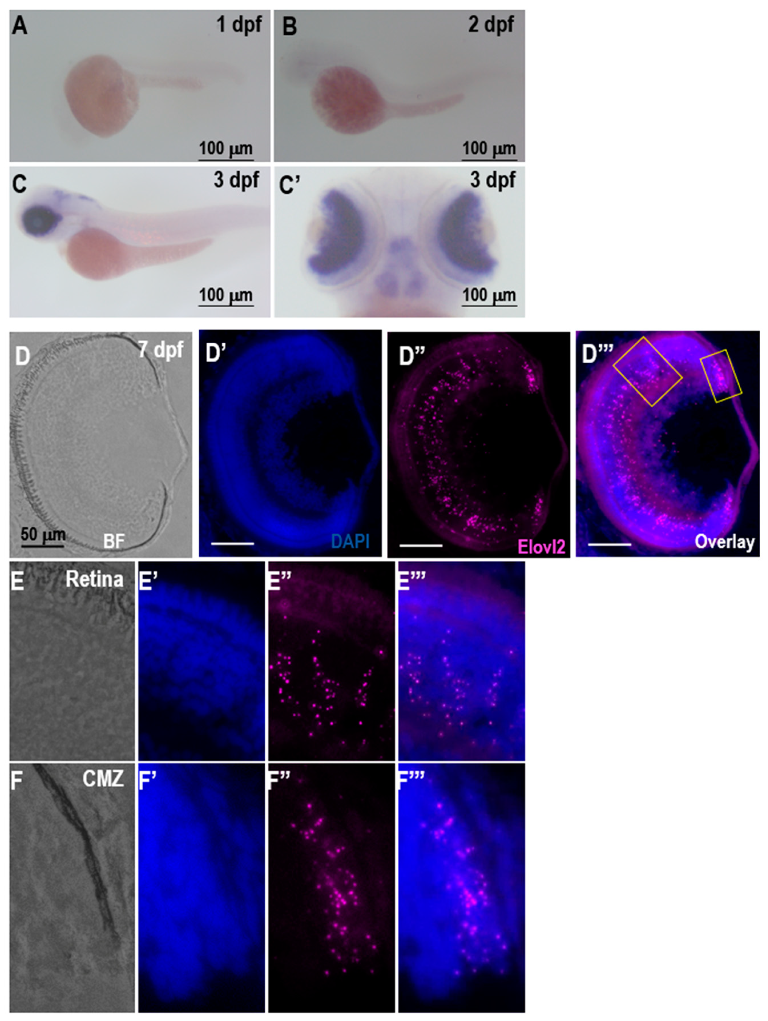
When investigating the expression of the elovl2, we observed a strong retinal expression at 3 dpf. On histological sections at 7 days, clear RNAscope signal can be observed throughout the retina. In situ hybridization of retina sections in adult zebrafish showed a similar pattern of the expression, with no observable signal on negative control slides (Supplementary Figure S8). A lower expression of elovl2 in old zebrafish compared to young adult zebrafish was observed, suggesting that there may also be an age dependent decrease in elovl2 expression that has been seen in human and mouse tissues [10,12]. Several groups analyzed expression patterns of elovl2, 4, and 5 in zebrafish retina [15,17,32]. Our data is in general agreement with published data, although one of the groups detected the first expression of Elovl2 earlier than at 3 dpf [32]. Intriguingly, none of the studies convincingly presented the expression of elovl5 in the eye while elovl4 seems to be expressed mostly in retinal pigmented epithelium cells in striking contrast to the mammalian gene. Taken together, the data suggests that the elovl2 is the sole fatty acid elongase that is able to elongate C22 to C24 in the inner nuclear layer.
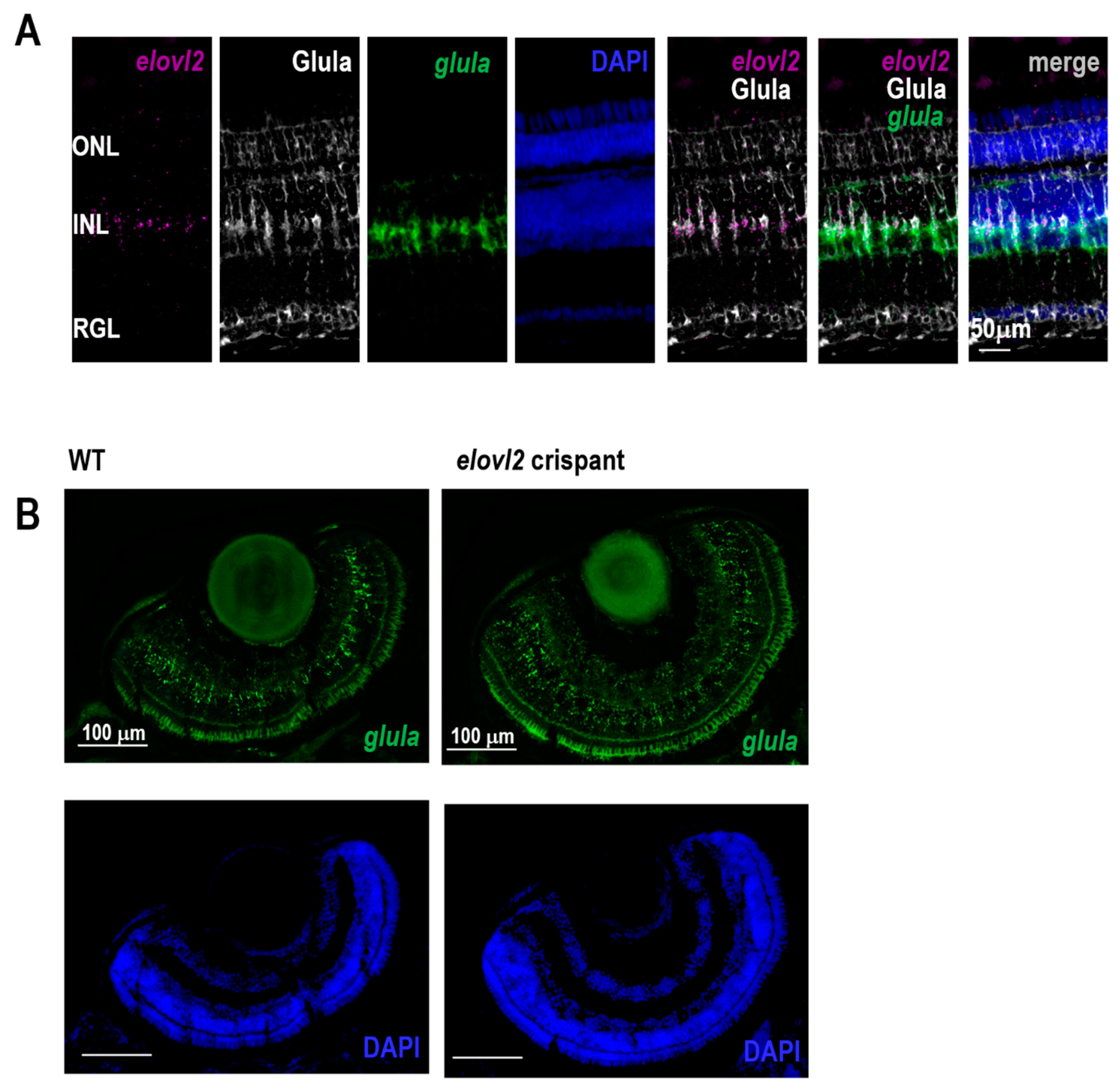
We find expression of elovl2 primarily in the Muller glia, which is in contrast to the mouse data where it is primarily expressed in photoreceptors in the retinal pigment epithelium [12]. The role of the Muller glia synthesized PUFAs is still poorly understood. Interestingly, Muller glia synthesis of 9,20-dihydroxydocosapentaenoic acid (DHDP), a diol of docosahexenoic acid (DHA), has been observed inhibit endothelial Notch signaling and regulate retinal angiogenesis [33]. In the mammalian retina, Elovl2 is expressed in photoreceptors, especially in cones. Since the VLC-PUFAs are the key fatty acids in photoreceptor disks membranes, the striking difference in the expression pattern of mammalian and zebrafish elovl2 gene suggests a potential dependence of photoreceptor cells on Muller glia. This interesting interaction would involve the Muller glia in zebrafish eye to provide VLC-PUFAs for the photoreceptor disks during the daily disk regeneration. Further studies are needed to elucidate the role of Muller glia derived PUFAs in retinal structure and function.
We then assessed the function of elovl2 in zebrafish embryos by creating elovl2 crispants. This was done by injecting 2 gRNAs specific to elovl2 as well as Cas9 protein into single cell embryos. High rates of gene editing were observed at the sites of both gRNAs (Supplementary Figure S3), causing high biallelic changes in the elovl2 gene. One caveat of this approach is that off target effects of CRISPR editing can occur.
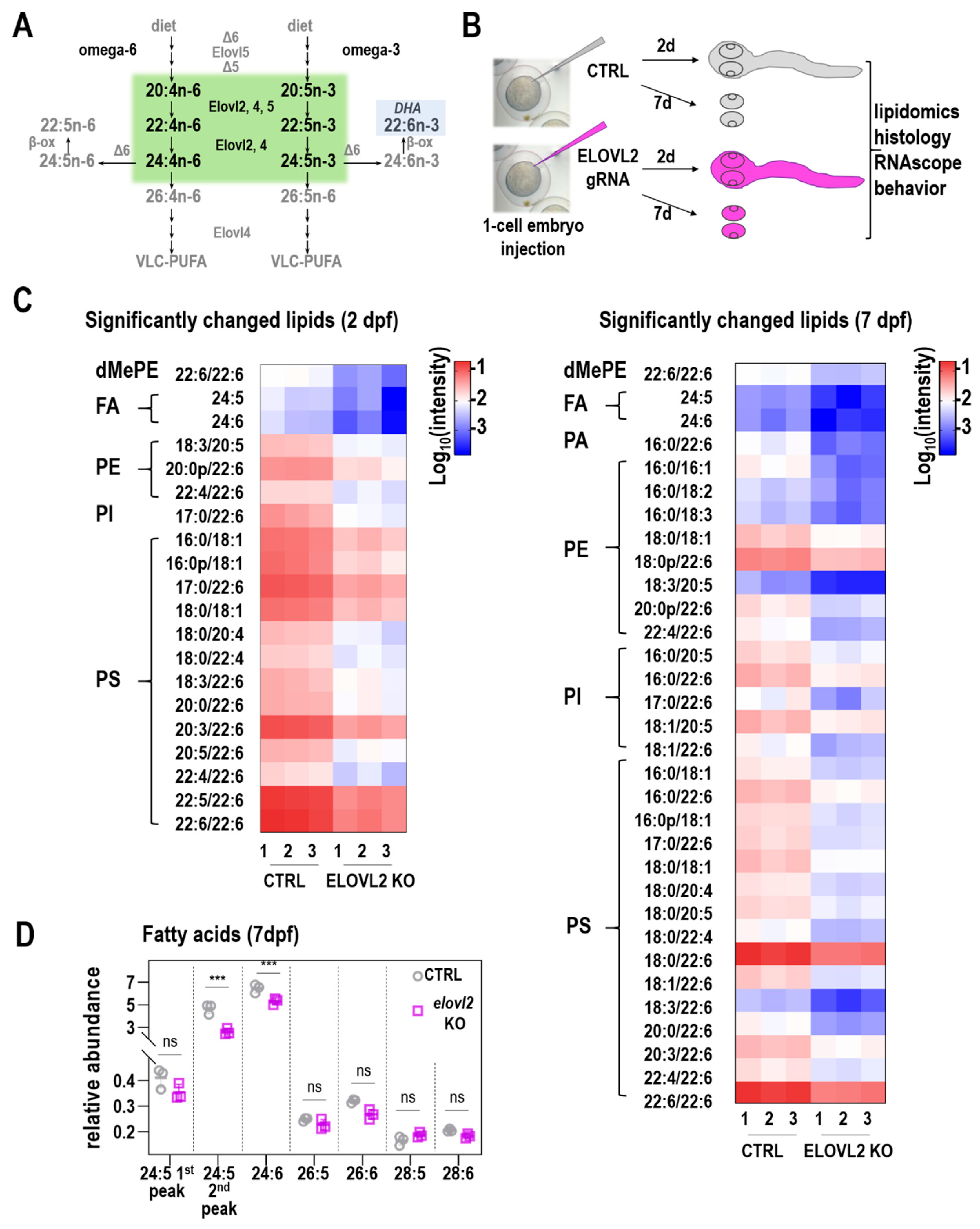
Elovl2 crispants had significant changes in levels of various fatty acids, consistent with loss of Elovl2 function compared to control crispants. Lipidomics performed on whole zebrafish embryo showed a significant decrease of LC-PUFAs in lipids isolated from elovl2 crispants. These FAs, including 20:4, 20:5, 22:4, 22:5, 22:6, 24:5 and 24:6, are potential products of Elovl2 enzymatic activity. Our results are in general agreement with the data presented by Liu et al. [16], in particular regarding the key role of Elovl2 in DHA synthesis. However, we did not observe the accumulation of 20:5n-3 in our extracts. This might be due to the stage difference (2 dpf vs. 3 dpf) or different detection methods; more sensitive GC-MS used in Liu et al., vs. the LC-MS used in our studies. Our approach however allowed us to detect VLC-PUFAs and their lower abundance in 7 dpf retina. In addition, the availability of specific fatty acids affected the overall composition of lipids in the zebrafish embryos. In particular, levels of phospholipids were significantly changed while availability of free fatty acids substantially increased. Importantly, the samples analyzed at day 2 were those of whole zebrafish embryos. Therefore, these changes reflect a diversity of tissue types and not specifically changes in the zebrafish retina. At day 7, we analyzed dissected eyes. Nevertheless, this data shows that lack of specific fatty acids can affect the lipid biosynthesis, and therefore the overall compositions of membranes.
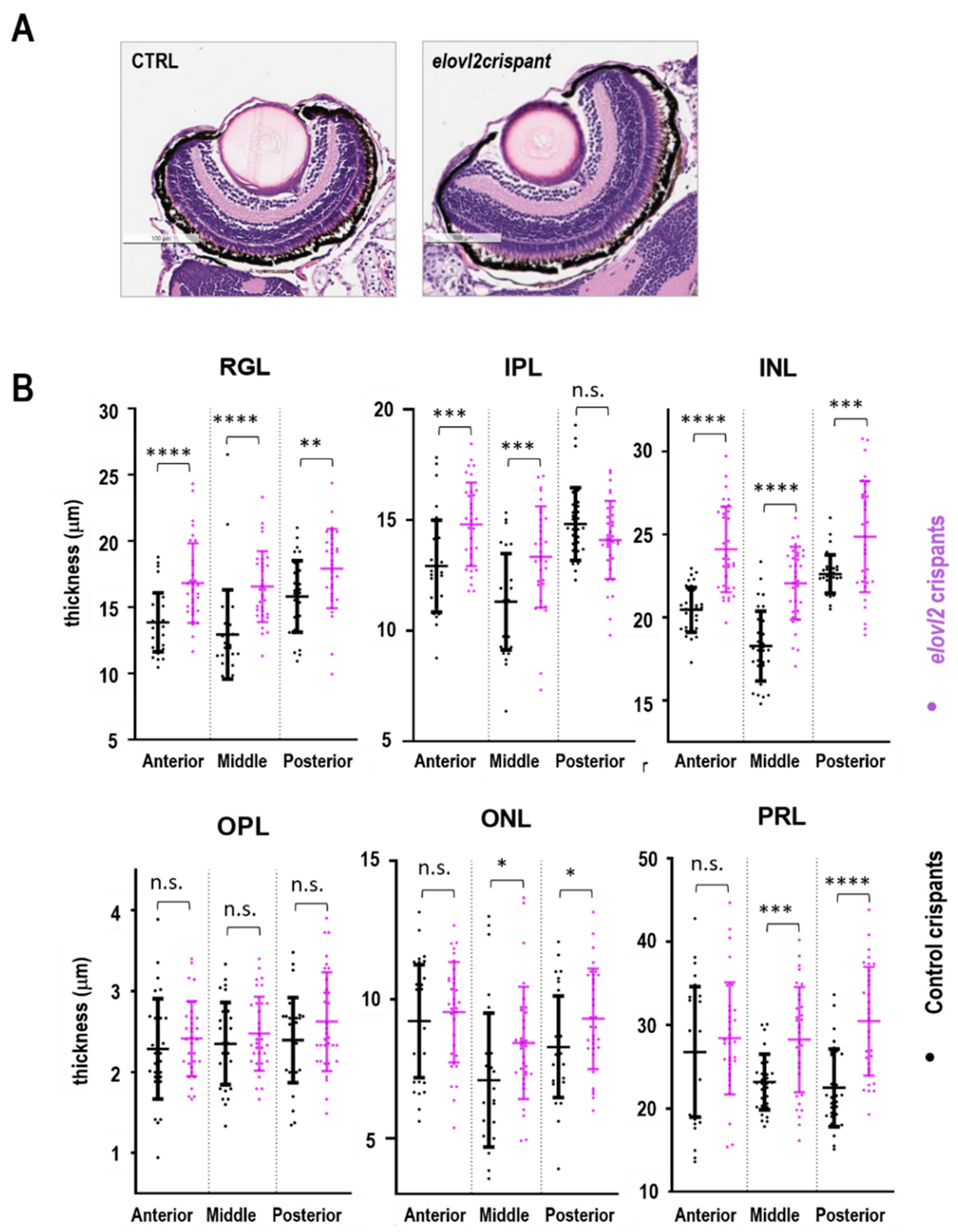
To analyze the impact of the lack of elovl2 enzymatic activity on the retinal morphology thickness of the retinal layers in the 7 dpf control and elovl2 crispants, sections were examined and quantified. No gross disruptions of the retinal architecture were observed, however, the thickening of several retinal layers in elovl2 crispants when compared to controls was detected. Other studies have reported changes in the thickness of retinal layers after the inactivation of Muller glia cells [28,34]. MacDonald et al. showed that inhibition of Muller glia formation during zebrafish development results into wider RGL but no effect was observed in other retinal layers. This suggest that other factors play role in maintenance of retinal layers in the absence of Muller glia. While our observation further supports the indispensable role of Muller glia in maintenance of RGL in zebrafish retina, the factors involved in the maintenance of other retinal layers in the absence of Muller glia cells remain unstudied. Another study targeting the deletion of Dicer1 miRNA specifically in Muller glia cells present in INL of mice retina reported an increase in the thickness of INL. Intriguingly, the group also observed reduction in thickness of ONL. Considering the global change in the membrane lipids composition in elovl2 crispants (Supplementary Figure S5) and therefore different physical properties of the membranes, our studies further supports the observation of the key role of the Muller glia in the maintenance of retinal architecture.
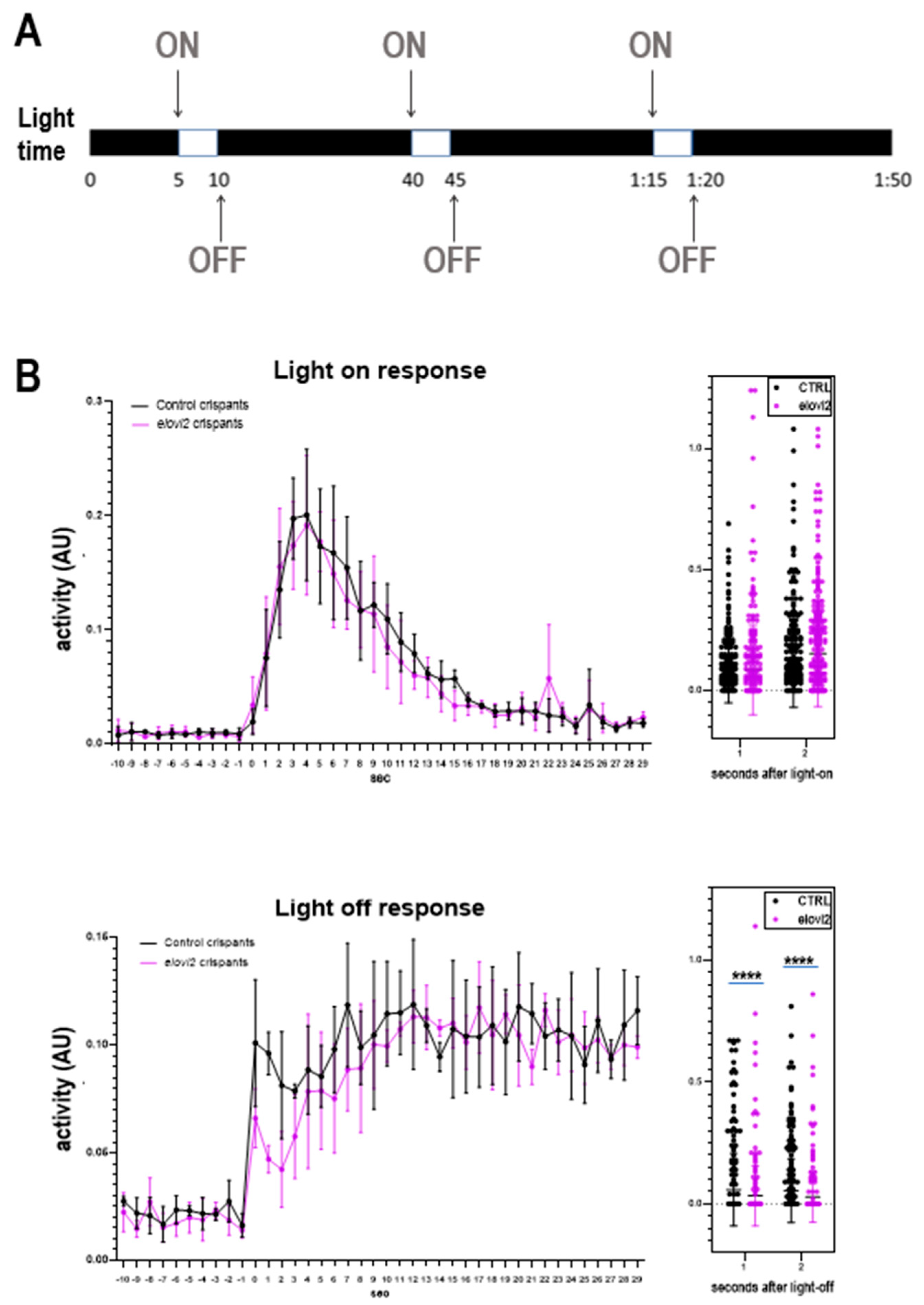
Finally, we examined the changes in visual behavior in zebrafish elovl2 crispants. We performed the VMR assay, a well characterized visual reflex that develops by day 5. We assessed the visual motor response at day 6 and did not find any changes in the VMR-ON response. However, we did see a significantly weaker, albeit still present, VMR-OFF response after elovl2 knockdown. It is still unclear what parts of the visual circuit mediate this VMR-OFF response. Interesting, slc7a14 mutant zebrafish, which affects rod photoreceptors more than cones, shows greater defects in the VMR-OFF response than the VMR-ON response [35]. This is in contrast to Pde6c mutation, which results in cone degeneration, and leads to defects in the VMR-ON response rather than VMR-OFF response 22. This suggests a possibility that VMR-ON and VMR-OFF responses may be primarily influenced by different classes of photoreceptors [35]. Further studies are needed to examine the effect of elovl2 on visual function in the zebrafish retina.
In conclusion, this is the first study to investigate the role of elovl2 in the zebrafish retina. Loss of elovl2 activity results in broad changes across the production of lipids compatible with its function as an elongase of LC-PUFAs as well as in impact on visual function. Elovl2 expression in the Muller glia may suggest additional and unique functions of zebrafish elovl2 compared to mammals. Taken together, our work further advances our understanding of the role of elovl2 and LC-PUFAs in retina structure and function.

Supplementary Materials

The following are available online at https://0-www-mdpi-com.brum.beds.ac.uk/2073-4409/9/12/2583/s1, Supplementary Figure S1. A. Conservation of elovl2 gene and protein sequence across different species. B. Multiple sequence alignment of ELOVL2 protein in different species showing conserved sequences. Amino acids forming transmembrane helices are highlighted in red; * represents identical amino acids; represents amino acids with highly similar properties; represents amino acids with weakly similar properties. Supplementary Figure S2 Expression of elovl2 in adult zebrafish retina. Expression analysis of elovl2 in 3 months (Panel A) and 14 months (Panel B) old zebrafish retina showing brightfield (A, B), DAPI stained (A’,B’), elovl2 stained (A’’,B’’) and overlay (A’’’,B’’’) images. Scale bars indicated. Supplementary Figure S3. Sequence analysis of elovl2 and control crispants at exon 2 and exon 3. A. Schematic of elovl2 gene structure and location of gRNAs with sequence in exon 2 and exon 3 used in this study. B. Representative sanger sequencing traces showing gene editing at the cut site. C. ICE analysis of indel distribution of a representative zebrafish embryo of elovl2 gRNA on exon2. Note cumulative frameshift indel of 91% D. ICE analysis of indel distribution of a representative zebrafish embryo of elovl2 gRNA on exon 3. Note cumulative frameshift indel of 87%. Supplementary Figure S4. Heatmap of all the identified lipids on whole zebrafish embryo (day 2) and on embryos eyes (day 7). Supplementary Figure S5. The relative abundance of each lipid class of control and elovl2 crispants. Supplementary Figure S6. A. Representative H&E staining images of elovl2 and control crispants showing different layers of retina. Scale bars = 100 µm. B. Table with data presented in Figure 3B. Supplementary Figure S7. Expression of Elovl2 in Muller glia. A. Overlap of elovl2 and glula signals detected by RNAscope confirms that elovl2 is expressed in Muller glia. *—autofluorescence B. Expression of elovl2 and Muller glia marker detected in crispens—lower level of elovl2 is noted in crispants. No changes in glula expression can be detected. Scale bars indicated. Supplementary Figure S8 Comparison of staining intensity with elovl2 and control probe. The negative control probe (Panel A) showed light autofluorescence in photoreceptor layer (A’) confirming specific staining of elovl2 (B’) in the three-month-old retina (Panel B). Scale bars = 50 µm.

Author Contributions

M.D.—acquiring experimental data, analysis of data, and writing of the manuscript; F.G., Q.X.—acquiring experimental data, analysis of data, and editing the manuscript; D.V.F., E.Z., A.F.M.P., A.S.—acquiring experimental data and analysis; D.S.-K., D.L.C.—conceptualization of experiments, data analysis, writing and editing of the manuscript. All authors have read and agreed to the published version of the manuscript.

Funding

This work was funded by an unrestricted Research to Prevent Blindness grant to Gavin Herbert Eye Institute at University of California, Irvine, an unrestricted Research to Prevent Blindness grant to University of California, San Diego, Research to Prevent Blindness Special Scholar award to D.S.K. and NEI K08EY030510 to D.L.C., as well as a faculty senate grant from the University of California San Diego to D.L.C.

Acknowledgments

We thank Michal Krawczyk for help with preparing the figures.

Conflicts of Interest

D.L.C. and D.S.K. have intellectual property related to Elovl2 which has been licensed to Visgenx. D.L.C. and D.S.K. are consultants for Visgenx. No funds from Visgenx were received for the project.

References

  1. Bazan, N.G.; Molina, M.F.; Gordon, W.C. Docosahexaenoic acid signalolipidomics in nutrition: Significance in aging, neuroinflammation, macular degeneration, Alzheimer’s, and other neurodegenerative diseases. Annu. Rev. Nutr. 2011, 31, 321–351. [Google Scholar] [CrossRef] [Green Version]
  2. Skowronska-Krawczyk, D.; Budin, I. Aging membranes: Unexplored functions for lipids in the lifespan of the central nervous system. Exp. Gerontol. 2020, 131, 110817. [Google Scholar] [CrossRef] [PubMed]
  3. Echeverria, F.; Valenzuela, R.; Catalina Hernandez-Rodas, M.; Valenzuela, A. Docosahexaenoic acid (DHA), a fundamental fatty acid for the brain: New dietary sources. Prostaglandins Leukot. Essent. Fat. Acids 2017, 124, 1–10. [Google Scholar] [CrossRef]
  4. Skowronska-Krawczyk, D.; Chao, D.L. Long-Chain Polyunsaturated Fatty Acids and Age-Related Macular Degeneration. Adv. Exp. Med. Biol. 2019, 1185, 39–43. [Google Scholar] [CrossRef]
  5. Savary, S.; Trompier, D.; Andreoletti, P.; Le Borgne, F.; Demarquoy, J.; Lizard, G. Fatty Acids - Induced Lipotoxicity and Inflammation. Curr. Drug Metab. 2012, 13, 1358–1370. [Google Scholar] [CrossRef]
  6. Chong, E.W.; Kreis, A.J.; Wong, T.Y.; Simpson, J.A.; Guymer, R.H. Dietary omega-3 fatty acid and fish intake in the primary prevention of age-related macular degeneration: A systematic review and meta-analysis. Arch. Ophthalmol. 2008, 126, 826–833. [Google Scholar] [CrossRef]
  7. van Leeuwen, E.M.; Emri, E.; Merle, B.M.J.; Colijn, J.M.; Kersten, E.; Cougnard-Gregoire, A.; Dammeier, S.; Meester-Smoor, M.; Pool, F.M.; de Jong, E.K.; et al. A new perspective on lipid research in age-related macular degeneration. Prog. Retin. Eye Res. 2018, 67, 56–86. [Google Scholar] [CrossRef]
  8. Leonard, A.E.; Kelder, B.; Bobik, E.G.; Chuang, L.T.; Lewis, C.J.; Kopchick, J.J.; Mukerji, P.; Huang, Y.S. Identification and expression of mammalian long-chain PUFA elongation enzymes. Lipids 2002, 37, 733–740. [Google Scholar] [CrossRef]
  9. Gregory, M.K.; Cleland, L.G.; James, M.J. Molecular basis for differential elongation of omega-3 docosapentaenoic acid by the rat Elovl5 and Elovl2. J. Lipid Res. 2013, 54, 2851–2857. [Google Scholar] [CrossRef] [Green Version]
  10. Hannum, G.; Guinney, J.; Zhao, L.; Zhang, L.; Hughes, G.; Sadda, S.; Klotzle, B.; Bibikova, M.; Fan, J.B.; Gao, Y.; et al. Genome-wide methylation profiles reveal quantitative views of human aging rates. Mol. Cell 2013, 49, 359–367. [Google Scholar] [CrossRef] [Green Version]
  11. Horvath, S. DNA methylation age of human tissues and cell types. Genome Biol. 2013, 14, R115. [Google Scholar] [CrossRef] [Green Version]
  12. Chen, D.; Chao, D.L.; Rocha, L.; Kolar, M.; Nguyen Huu, V.A.; Krawczyk, M.; Dasyani, M.; Wang, T.; Jafari, M.; Jabari, M.; et al. The lipid elongation enzyme ELOVL2 is a molecular regulator of aging in the retina. Aging Cell 2020, 19, e13100. [Google Scholar] [CrossRef] [Green Version]
  13. Niklaus, S.; Neuhauss, S.C.F. Genetic approaches to retinal research in zebrafish. J. Neurogenet. 2017, 31, 70–87. [Google Scholar] [CrossRef] [Green Version]
  14. Agaba, M.; Tocher, D.R.; Dickson, C.A.; Dick, J.R.; Teale, A.J. Zebrafish cDNA encoding multifunctional Fatty Acid elongase involved in production of eicosapentaenoic (20:5n-3) and docosahexaenoic (22:6n-3) acids. Mar. Biotechnol. 2004, 6, 251–261. [Google Scholar] [CrossRef]
  15. Monroig, O.; Rotllant, J.; Sanchez, E.; Cerda-Reverter, J.M.; Tocher, D.R. Expression of long-chain polyunsaturated fatty acid (LC-PUFA) biosynthesis genes during zebrafish Danio rerio early embryogenesis. Biochim. Biophys. Acta 2009, 1791, 1093–1101. [Google Scholar] [CrossRef] [Green Version]
  16. Liu, C.; Ye, D.; Wang, H.; He, M.; Sun, Y. Elovl2 but not Elovl5 is essential for the biosynthesis of docosahexaenoic (DHA) in zebrafish: Insight from a comparative gene knockout study. BioRxiV 2020, 22, 613–619. [Google Scholar] [CrossRef]
  17. Monroig, O.; Rotllant, J.; Cerda-Reverter, J.M.; Dick, J.R.; Figueras, A.; Tocher, D.R. Expression and role of Elovl4 elongases in biosynthesis of very long-chain fatty acids during zebrafish Danio rerio early embryonic development. Biochim. Biophys. Acta 2010, 1801, 1145–1154. [Google Scholar] [CrossRef] [Green Version]
  18. Bringmann, A.; Pannicke, T.; Grosche, J.; Francke, M.; Wiedemann, P.; Skatchkov, S.N.; Osborne, N.N.; Reichenbach, A. Muller cells in the healthy and diseased retina. Prog. Retin. Eye Res. 2006, 25, 397–424. [Google Scholar] [CrossRef]
  19. Burger, A.; Lindsay, H.; Felker, A.; Hess, C.; Anders, C.; Chiavacci, E.; Zaugg, J.; Weber, L.M.; Catena, R.; Jinek, M.; et al. Maximizing mutagenesis with solubilized CRISPR-Cas9 ribonucleoprotein complexes. Development 2016, 143, 2025–2037. [Google Scholar] [CrossRef] [Green Version]
  20. ICE v2 CRISPR Analysis Tool. Available online: https://www.synthego.com/products/bioinformatics/crispr-analysis (accessed on 30 November 2020).
  21. Thisse, C.; Thisse, B. High-resolution in situ hybridization to whole-mount zebrafish embryos. Nat. Protoc. 2008, 3, 59–69. [Google Scholar] [CrossRef]
  22. Zhang, L.; Xiang, L.; Liu, Y.; Venkatraman, P.; Chong, L.; Cho, J.; Bonilla, S.; Jin, Z.B.; Pang, C.P.; Ko, K.M.; et al. A Naturally-Derived Compound Schisandrin B Enhanced Light Sensation in the pde6c Zebrafish Model of Retinal Degeneration. PLoS ONE 2016, 11, e0149663. [Google Scholar] [CrossRef] [Green Version]
  23. Bligh, E.G.; Dyer, W.J. A rapid method of total lipid extraction and purification. Can. J. Biochem. Physiol. 1959, 37, 911–917. [Google Scholar] [CrossRef] [Green Version]
  24. MacLean, B.; Tomazela, D.M.; Shulman, N.; Chambers, M.; Finney, G.L.; Frewen, B.; Kern, R.; Tabb, D.L.; Liebler, D.C.; MacCoss, M.J. Skyline: An open source document editor for creating and analyzing targeted proteomics experiments. Bioinformatics 2010, 26, 966–968. [Google Scholar] [CrossRef] [Green Version]
  25. Buglo, E.; Sarmiento, E.; Martuscelli, N.B.; Sant, D.W.; Danzi, M.C.; Abrams, A.J.; Dallman, J.E.; Zuchner, S. Genetic compensation in a stable slc25a46 mutant zebrafish: A case for using F0 CRISPR mutagenesis to study phenotypes caused by inherited disease. PLoS ONE 2020, 15, e0230566. [Google Scholar] [CrossRef] [Green Version]
  26. Trubiroha, A.; Gillotay, P.; Giusti, N.; Gacquer, D.; Libert, F.; Lefort, A.; Haerlingen, B.; De Deken, X.; Opitz, R.; Costagliola, S. A Rapid CRISPR/Cas-based Mutagenesis Assay in Zebrafish for Identification of Genes Involved in Thyroid Morphogenesis and Function. Sci. Rep. 2018, 8, 5647. [Google Scholar] [CrossRef]
  27. Wong, C.E.D.; Hua, K.; Monis, S.; Saxena, V.; Norazit, A.; Noor, S.M.; Ekker, M. gdnf affects early diencephalic dopaminergic neuron development through regulation of differentiation-associated transcription factors in zebrafish. J. Neurochem. 2020. [Google Scholar] [CrossRef]
  28. MacDonald, R.B.; Randlett, O.; Oswald, J.; Yoshimatsu, T.; Franze, K.; Harris, W.A. Muller glia provide essential tensile strength to the developing retina. J. Cell Biol. 2015, 210, 1075–1083. [Google Scholar] [CrossRef] [Green Version]
  29. Emran, F.; Rihel, J.; Dowling, J.E. A behavioral assay to measure responsiveness of zebrafish to changes in light intensities. J. Vis. Exp. 2008. [Google Scholar] [CrossRef]
  30. Liu, Y.; Ma, P.; Cassidy, P.A.; Carmer, R.; Zhang, G.; Venkatraman, P.; Brown, S.A.; Pang, C.P.; Zhong, W.; Zhang, M.; et al. Statistical Analysis of Zebrafish Locomotor Behaviour by Generalized Linear Mixed Models. Sci. Rep. 2017, 7, 2937. [Google Scholar] [CrossRef]
  31. Ganzen, L.; Venkatraman, P.; Pang, C.P.; Leung, Y.F.; Zhang, M. Utilizing Zebrafish Visual Behaviors in Drug Screening for Retinal Degeneration. Int. J. Mol. Sci. 2017, 18, 1185. [Google Scholar] [CrossRef] [Green Version]
  32. Tan, S.H.; Chung, H.H.; Shu-Chien, A.C. Distinct developmental expression of two elongase family members in zebrafish. Biochem. Biophys. Res. Commun. 2010, 393, 397–403. [Google Scholar] [CrossRef]
  33. Hu, J.; Popp, R.; Fromel, T.; Ehling, M.; Awwad, K.; Adams, R.H.; Hammes, H.P.; Fleming, I. Muller glia cells regulate Notch signaling and retinal angiogenesis via the generation of 19,20-dihydroxydocosapentaenoic acid. J. Exp. Med. 2014, 211, 281–295. [Google Scholar] [CrossRef] [Green Version]
  34. Wohl, S.G.; Jorstad, N.L.; Levine, E.M.; Reh, T.A. Muller glial microRNAs are required for the maintenance of glial homeostasis and retinal architecture. Nat. Commun. 2017, 8, 1603. [Google Scholar] [CrossRef]
  35. Zhuang, Y.Y.; Xiang, L.; Wen, X.R.; Shen, R.J.; Zhao, N.; Zheng, S.S.; Han, R.Y.; Qu, J.; Lu, F.; Jin, Z.B. Slc7a14 Is Indispensable in Zebrafish Retinas. Front. Cell Dev. Biol. 2019, 7, 333. [Google Scholar] [CrossRef] [Green Version]
Figure 1. A–C, expression of elovl2 in zebrafish larvae. Lateral view of elovl2 staining in (A) 1 dpf and (B) 2 dpf zebrafish retinas. Lateral (C) and dorsal (C’) view of elovl2 staining in 3 dpf zebrafish retina, respectively. D–F. RNAscope in situ hybridization with elovl2 probe in 7 dpf zebrafish retina showing brightfield (DF), 4′,6-diamidino-2-phenylindole (DAPI staining (D’F’), elovl2 (D’’F’’) and overlay (D’’’F’’’) images. Scale bars = 50 µm. Magnified view (40×) of boxed regions in Figure 1D’’’. The magnified views of Retina (Panel E) and ciliary marginal zone (CMZ) (Panel F) showing elovl2 staining in 7 dpf zebrafish retina.
Cells 09 02583 g001
Figure 2. Generation and analysis of elovl2 crispants. (A) ω-3 and ω-6 PUFA elongation pathway. (B) Workflow for creating biallelic elovl2 mutants (‘crispants’) in zebrafish using CRISPR/Cas. (C) Significantly changed lipids in elovl2 crispants at days 2 and 7. In particular, all lipids containing DHA are significantly less abundant in elovl2 crispants at both days 2 and 7. (D) Relative abundance of PUFAs in control and elovl2 crispants shows lower abundance of products and elongated products of Elovl2. *** p < 0.005; ns—not significant.
Cells 09 02583 g002
Figure 3. (A) H&E stained retinal sections of elovl2 and control crispants. (B) The thickness of the individual retinal layers of elovl2 and control crispants. Plots showing comparison of lengths of different layers in retina between control and elovl2 crispants. The measurements were taken at three different regions in the retina termed as anterior, posterior, and middle. RGL, Retinal Ganglion Cell Layer; IPL, Inner Plexiform Layer; INL, Inner Nuclear Layer; OPL, Outer Plexiform Layer; ONL, Outer Nuclear Layer; PRL, Photoreceptor Layer. (* p < 0.05; ** p < 0.01; *** p < 0.001; **** p < 0.0001, ns—not significant). Each dot is a single cryosection.
Cells 09 02583 g003
Figure 4. (A) Expression of elovl2 RNA and Muller Glia specific gene glula as detected by RNAscope and Glula immunohistochemistry in normal adult zebrafish. (B) Expression of glula in 7dpf control and elovl2 crispants shows no changes of glula expression upon elovl2 knockout.
Cells 09 02583 g004
Figure 5. Zebrafish elovl2 crispants show deficits in the visual motor response (VMF). OFF response activity show visual phenotype. (A) Diagram of motor response (VMR) assay. (B) Motor response of control and elovl2 crispants to VMR-ON (top) and VMR-OFF (bottom) stimulus. Right Charts: quantification of the results at second 1 and 2 after the light stimulus change. AU, arbitrary unit.
Cells 09 02583 g005
Publisher’s Note: MDPI stays neutral with regard to jurisdictional claims in published maps and institutional affiliations.

