
Au-Decorated WS2 Microflakes Based Sensors for Selective Ammonia Detection at Room Temperature

School of Microelectronics, Dalian University of Technology, Dalian 116024, China
*
Authors to whom correspondence should be addressed.
Chemosensors 2022, 10(1), 9; https://0-doi-org.brum.beds.ac.uk/10.3390/chemosensors10010009
Submission received: 21 November 2021
/
Revised: 17 December 2021
/
Accepted: 22 December 2021
/
Published: 27 December 2021
(This article belongs to the Special Issue The State-of-the-Art Gas Sensor)
Abstract
:Gold nanoparticles decorated WS microflakes (Au/WS) have been synthesized by an in situ chemical reducing process. A chemiresistive-type sensor using as-synthesized Au/WS heterostructures as sensing materials shows an improved response to different concentrations of ammonia compared to pure WS at room temperature. As the concentrations of gold nanoparticles increased in heterostructures, response/recovery speeds of the sensors became faster although the sensitivity of the sensor was compromised compared to the sensitivity of the sensor with lower concentrations of Au. In addition, the Au/WS-based sensor indicated excellent selectivity to formaldehyde, ethanol, benzene and acetone at room temperature. The improved performance of the sensors was attributed to the synergistic effect of electronic sensitization and chemical sensitization between WS and Au.
1. Introduction
Ammonia is a natural gas existing throughout the atmosphere that is used extensively in many areas such as fertilizers, chemical technology and environmental protection [1]. The demand for fuel combustion efficiency and strict regulations on automobile exhaust emissions result in an urgent need for new exhaust gas treatment methods such as ammonia selective catalytic NO reduction (SCR) [2]. Toxic NO could be reduced effectively by NH, which has been used for lowering emissions [3]. However, since ammonia is a toxic gas that is harmful to human health, and the concentration of ammonia injected into the atmosphere should be controlled in order to avoid environmental pollution and damage to human health. Therefore, the concentration of ammonia in the exhaust system needs to be detected to optimize emissions [4]. In these circumstances, there is an increasing need of ammonia sensors with good sensing performances in these fields.
Due to the need of low cost and low energy consumption, ammonia sensors working at low working temperature with high sensitivity, simplicity, and compatibility with miniaturized electronic devices are in high demand. As the core component of the sensor, sensing materials are key factors affecting sensing performance. Therefore, selecting appropriate sensing materials that can work at room temperature is an effective means to reduce energy consumption of the sensor [5,6,7,8]. Two-dimensional (2D) materials such as graphene and other 2D transitional metal dichalcogenides (TMDs) with graphene-like structures have become potential candidates for meeting such requirements for chemical gas sensors [9,10,11]. According to the reported studies, the 2D materials have been widely used in a variety of fields, and they are expected to be used in next-generation nanoelectronic devices [12,13,14,15,16,17]. Based on the sensing mechanism of semiconductor gas sensors, 2D material has highly specific surface areas and can provide sufficient active sites, which is conducive to the adsorption of gas molecules on the surface of sensitive materials so as to realize the improvement of sensing performance [18,19,20,21,22]. Due to its unique electronic transport properties, graphene has become the most concerned 2D material and has been widely studied. However, the band gap of pristine graphene is zero, which is an essential characteristic in many applications, resulting in its application being limited [23,24]. TMDs materials not only have excellent electrical properties similar to graphene but also have semiconductor energy band structure, which have been considered to be a promising sensing material. In the TMDs family, MoS and WS as typical 2D materials have attracted the most attention because of their wider band gap structure (1–3 eV) and excellent physical and chemical properties [25,26,27]. In particular, relative to WS based chemical gas sensors, Li et al. fabricated an ammonia gas sensor used WS nanoflakes as the sensing material, which can work at room temperature and displayed high selectivity to ammonia gas [28]. Gu developed a gas sensor based on WS microflakes and found that the fabricated sensor showed better sensitivity and selectivity to ammonia gas under different light illumination compared to those in the dark [29].
In order to further improve the sensing performance of WS based chemiresistive-type sensor, secondary additives such as metallic nanoparticles was introduced. Chao investigated the gas sensor based on WS nanosheets modified by Pt quantum dots (QDs) and found that the sensor indicated enhanced sensing properties and fast recovery speeds to ammonia, which is attributed to the extra charges transferred from noble metal Pt QDs to WS nanosheets induced by unbalanced Fermi levels at the interfaces [30]. Zhang et al. reported that Cu doped WS displayed good sensing properties for CO and NO gases suitable for NO gas scavenger, while it was not suitable for CO gas sensing [18]. Xu et al. demonstrated a NO sensor that used WS nanosheets/carbon nanofibers (CNFs) composites as the sensing material, which can significantly enhance sensing performances relative to NO at room temperature [31]. More interestingly, Suh et al. assembled WS particles on porous SiO nanorods templates with numerous edge-exposed WS microflakes and the resulting sensor exhibited a high sensitivity relative to NO at room temperature [32]. The introduced secondary phase could enhance both sensitivity and response speeds through mechanisms such as electronic sensitization or chemical sensitization [33,34].
In this study, we synthesized Au/WS nanocomposites via in situ reduction processes and examined the effect of introduced Au nanoparticles on the sensing properties relative to NH at room temperatures. The gas sensor based on the as-prepared Au/WS nanocomposites displayed a higher response value and faster response and recovery properties compared to those of the pristine one, indicating a significantly improved gas sensing performance to ammonia. Moreover, with Au introduction, the sensor could even work in high humidity backgrounds exhibiting a potential application in exhaled breath gas-phase analysis.
2. Experimental Method
2.1. Synthesis of Au/WS Nanocomposites
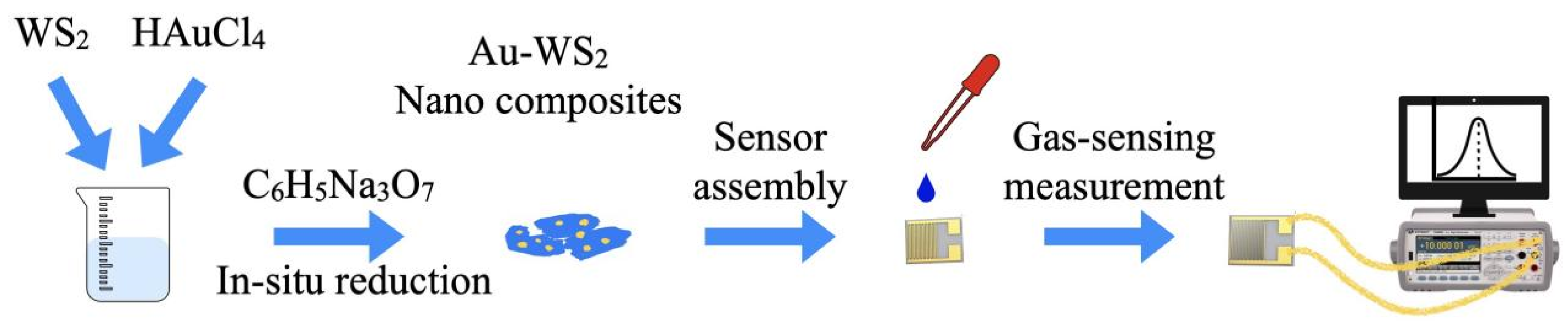
Commercial WS powders were purchased from Aladdin Co., Shanghai, China. In this research study, all chemical reagents are analytically pure and did not undergo further purification. In typical synthesis, 50 mg of WS microflakes was added in 15 mL deionized water followed with ultrasonication for 15 min to form a homogeneous mixed solution. A certain amount (0.1 mL, 0.2 mL, 0.6 mL and 1 mL, respectively) of aqueous solution of chlorauric acid (HAuCl) with concentrations of 0.01 M and a corresponding amount (0.1 mL, 0.2 mL, 0.6 mL and 1 mL, respectively) of L-lysine solution (NO) with concentrations of 0.01 M were added in the as-prepared solution. L-lysine plays an important role in the bond of Au nanoparticles and WS microflakes. After the above process, different amounts (0.01 mL, 0.02 mL, 0.06 mL and 0.1 mL, respectively) of 0.1 M Nacit solution were dropped into the mixture and stirred continuously for 30 min. The obtained precipitate was desiccated at 60 °C for 6 h, which had been washed several times with deionized water and absolute ethanol. For convenience, the samples decorated with different Au concentrations were named as Au/WS-0.56, Au/WS-1.12, Au/WS-3.36 and Au/WS-5.6, respectively. The preparation of the sensors is shown in Figure 1.
2.2. Characterization
In order to investigate the chemical composition and crystallographic structure of WS microflakes and Au/WS nanocomposite-based samples, X-ray diffraction studies were carried out on an X-ray diffractometer (XRD, D8 Advance, Bruker Corporation, Billerica, USA). Field emission scanning electronic microscope (FESEM, Gemini 300, ZEISS Corporation, Jena, Germany) characterization and transmission electron microscope (TEM, Tecnai G2 F20, FEI Corporation, Hillsboro, USA) characterization were used to produce a clear morphology of the WS microflakes and Au/WS nanocomposite-based samples. Energy-dispersive X-ray spectroscopy (EDS) characterization was used to analyze the element’s composition of WS microflakes and Au/WS nanocomposite-based samples. X-ray photoelectron spectrometer (XPS, ESCALAB 250Xi, Thermo Fisher Scientific Corporation, Waltham, USA) was used to analyze the surface chemistry of the as-prepared samples.
2.3. Fabrication and Measurement of the Gas Sensors
Pure WS microflakes and the prepared Au/WS nanocomposites were added in a small amount deionized water and ground into a pastes, which were coated on the AlO substrate preprinted with Au electrodes to form a gas sensor. In the testing procedure, the as-prepared sensors were placed in a test chamber with a volume of 18 L. After the sensor’s resistance stabilized, the target gas obtained by vaporizing the corresponding liquid was injected into the test chamber. The concentration of target gas can be calculated by the following equation.
In this equation, C (ppm), (g/mL), d, M (g/mol), (L) and (L) represent target gas concentration, liquid density, liquid purity, liquid molecular weight, liquid volume and test chamber volume, respectively.
The digital electromultimeter (Keysight Technologies 34465A, Santa Rosa, USA) was used to measure the resistance of the as-prepared samples. The ratio of the sensor resistances measured in target gas () and in air () was used to represent the sensor’s response value (). Response time () and recovery time () were defined as the time required for sensor resistance to change from the initial value to 90% of the full amplitude of resistance change when the target gas is injected and expelled, respectively.
3. Results and Discussion
3.1. Structure and Morphology Characterizations
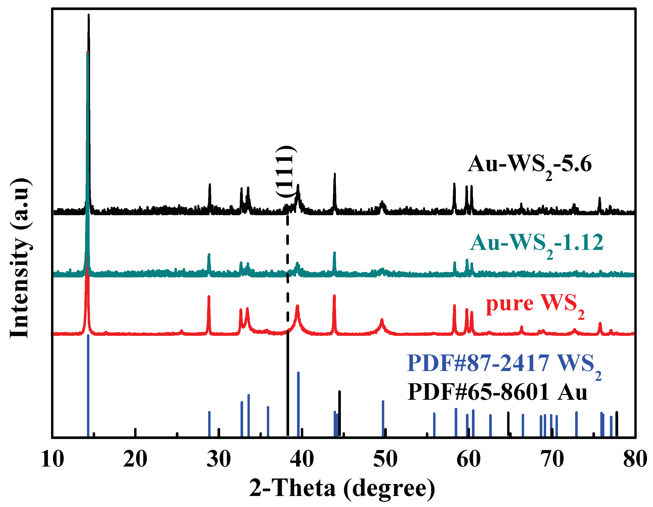
The XRD patterns of pure WS microflakes and Au-decorated WS nanocomposites are shown in Figure 2. Pure WS microflakes could be indexed to 2H-WS according to PDF#87-2417 with lattice constant of a = 3.16 Å. The XRD patterns of Au/WS-1.12 and Au/WS-5.6 samples were very similar to the XRD pattern of the WS sample. Moreover, the weak peak shown in the pattern of Au/WS-5.6 sample can be ascribed to the crystal plane of (111) of the face-centered cubic (fcc) Au according to PDF#65-8601. However, for the decoration amount of Au in WS microflakes was very small, and the peaks that can be ascribed to the crystal planes of (200), (220) and (311) of Au cannot be detected in XRD patterns of Au/WS-5.6 and Au/WS-1.12 with lower Au content.
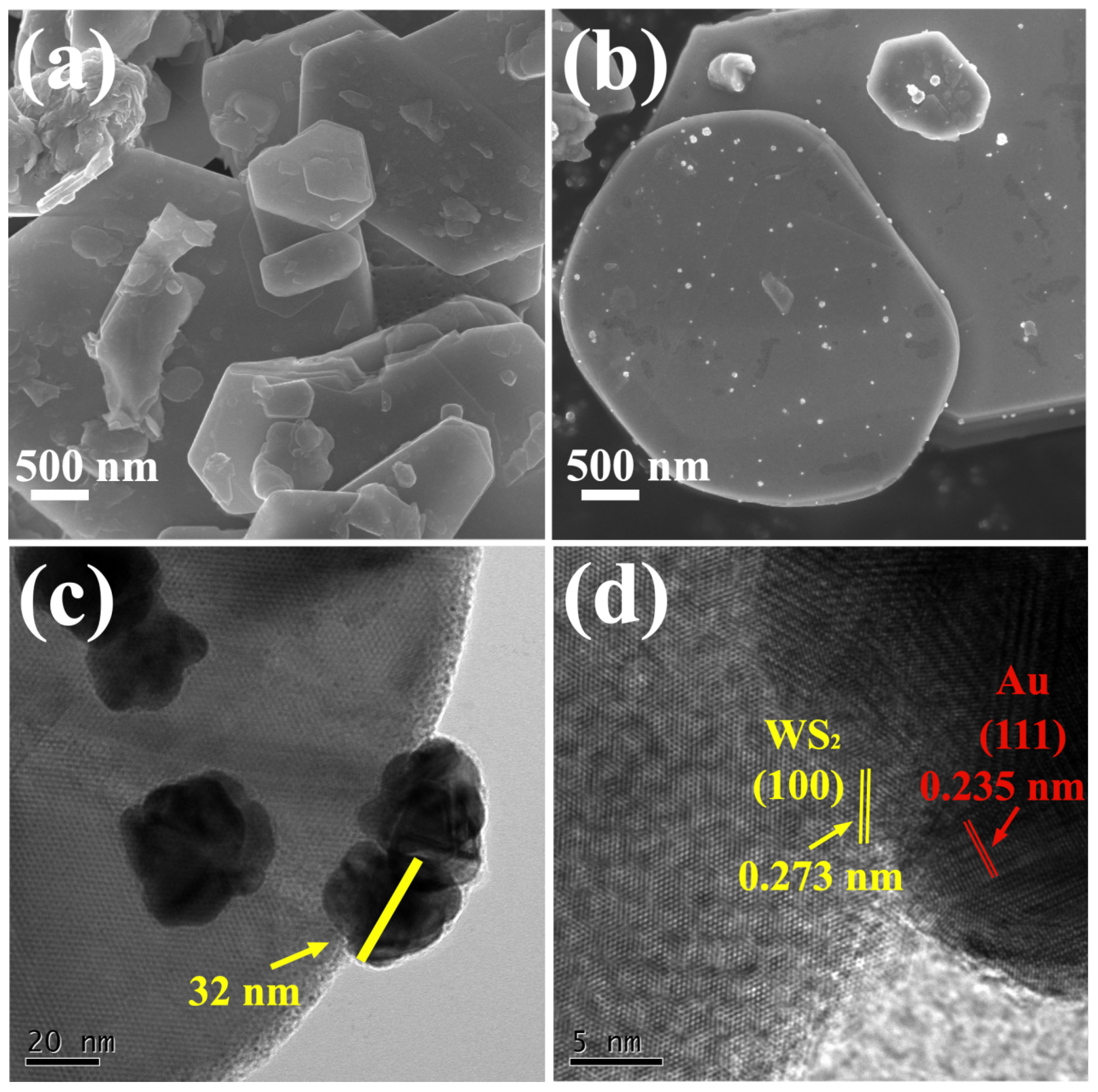
Figure 3a,b show FESEM images of the surface morphology of pure WS microflakes and Au/WS-5.6 nanocomposites. WS and Au/WS-5.6 samples both exhibited layered flake morphology indicating the formation of Au nanoparticles that did not result in morphological change of WS microflakes. After Au decoration, WS microflake structure remained unchanged and the formed isolated crystal Au nanoparticles were found to be attached to the surface of WS microflakes in a discrete state, as shown in Figure 3b. Lower magnification SEM images of WS microflakes and Au/WS-5.6 nanocomposites are shown in Figure S1 in the Supplementary Materials. The diameters of WS microflakes are 0.4–4.3 micrometers, and the diameters of decorated Au are 17.8–48.8 nm according to the statistical size distribution shown in Figure S2. In order to further investigate the microstructure of gold nanoparticles, high resolution transmission electron microscope (HRTEM) of Au/WS-5.6 nanocomposites was conducted and shown in Figure 3c,d. As observed in Figure 3c, the decorated Au nanoparticles present a spherical shape. The HRTEM image in Figure 3d exhibited the lattice fringes of Au/WS-5.6 nanocomposites. The intervals of lattice fringes of 0.235 nm and 0.273 nm correspond to the d-spacing of (111) of Au and the d-spacing of (100) of WS, respectively, indicating that there are obvious heterostructure interfaces between Au and WS. EDS was further obtained to investigate the element composition of Au/WS-5.6 nanocomposites. As shown in Figure S3, the elements of W, S, and Au were all identified. All expected elements including S, W and Au with atomic percentages of 65.98%, 33.12% and 0.9%, respectively, can be found in the EDS spectra. Moreover, EDS mapping analyses are presented in Figure S4. All expected elements are present in the mapping images demonstrating the successful formation of Au nanoparticles on WS microflakes.
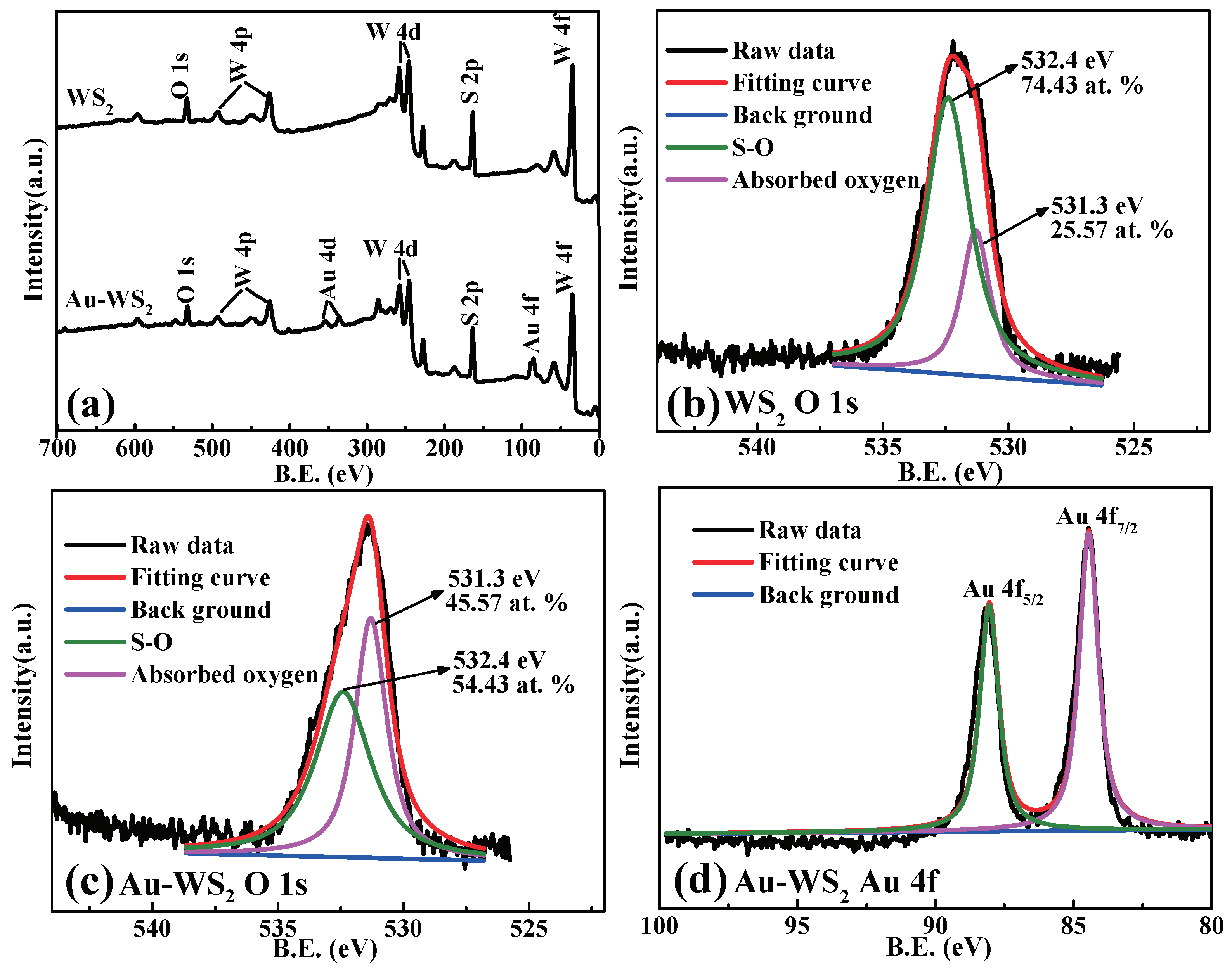
Figure 4 shows the XPS characterizations of pure WS and Au/WS-5.6 samples. As observed from the survey of XPS pattern of the Au/WS-5.6 sample as shown in Figure 4a, the peaks identifying Au 4d and Au 4f can be observed, which demonstrates that Au has been successfully introduced on WS microflakes. Figure 4b,c show the refined O 1s core spectra of pure WS microflakes and Au/WS-5.6 nanocomposites, respectively. For pure WS microflakes, the peak for O 1s can be deconvoluted into two peaks located at 531.3 eV and 532.4 eV, respectively. The peak at 531.3 eV can be assigned to absorbed oxygen [35]. The peak at 532.4 eV can be attributed to SO [29]. Thus, the atomic percentage of absorbed oxygen and SO is 25.57% and 74.43%, respectively. However, for the Au/WS-5.6 sample, the peak for O 1s spectra can also be deconvoluted into two peaks located at 531.3 eV and 532.4 eV attributing to absorbed oxygen and SO, respectively. The relative atomic percentage of absorbed oxygen and SO is 45.57% and 54.43%, respectively. This indicates that there is a larger amount of absorbed oxygen on the surface of Au/WS-5.6 nanocomposites compared to the pristine WS, presumably due to the chemical sensitization effect of Au on the surface of WS. The refined core spectra of Au 4f of Au/WS-5.6 sample in Figure 4d shows that the two peaks located at 84.45 eV and 88.05 eV correspond to Au 4f and Au 4f, respectively. The binding energy of Au has slightly shifted up to higher energy compared to bulk Au, which could be attributed to the stronger electronic interactions between WS microflakes with Au nanoparticles.
Information on other elements such as W and S is provided in Figure S5a–d in the Supplementary Materials. By performing a comparison on the refined core spectra of S 2p of pure WS and Au/WS-5.6 samples, the amount of SO (169.4 eV of S 2p) was reduced in Au/WS-5.6 sample, which confirmed a reduction in the number of SO after Au decoration. This could be explained by the fact that the slight surface oxidation of WS was inhibited by the reduction agent of Nacit used during preparation of the Au/WS nanocomposites. Moreover, as shown in Figure S5a, for the S 2p spectra of WS microflakes, the peaks located at 162.6 eV and 163.8 eV correspond to S 2p and S 2p, respectively. The S 2p spectra of Au/WS-5.6 nanocomposites as shown in Figure S5c displays two peaks located at 162.8 eV and 164 eV, which correspond to S 2p and S 2p, respectively. It indicates that sulfide has the valence of -2 both in pure WS microflakes and Au/WS-5.6 nanocomposites. The binding energy of S 2p has a slight shift to high energy after Au decoration.
According to the refined core spectra of the W 4f in WS sample shown in Figure S5b, the two peaks located at 33 eV and 35.2 eV correspond to W 4f and W 4f, respectively. The W 4f spectrum of Au/WS-5.6 nanocomposites in Figure S5d exhibits two peaks located at 33.25 eV and 35.45 eV corresponding to W 4f and W 4f, respectively. This indicates that tungsten has the valence of +4 both in WS microflakes and Au/WS-5.6 nanocomposites. Therefore, the decoration of Au did not result in a change of chemical oxidation states of S and W in Au/WS-5.6 compared to the pristine WS. However, the binding energy of S 2p and W 4f had a slight shift to high energy after Au decoration. This could be attributed to the stronger electronic interactions between WS microflakes with Au nanoparticles.
3.2. Electrical and Sensing Performance
Figure 5 shows the polarization curves of the sensors using WS and Au/WS as the sensing materials with a bias voltage from −5 V to +5 V. The linear relationship between the measured current and applied voltage of the synthesized gas sensors indicates that the contact between the electrode and the sensing materials is ohmic contact. Furthermore, with the decoration amount of Au nanoparticles increase, the resistance of Au/WS nanocomposite-based sensors decreased, which is related to electrons transfer between WS and Au nanoparticles due to the formation of heterostructures.
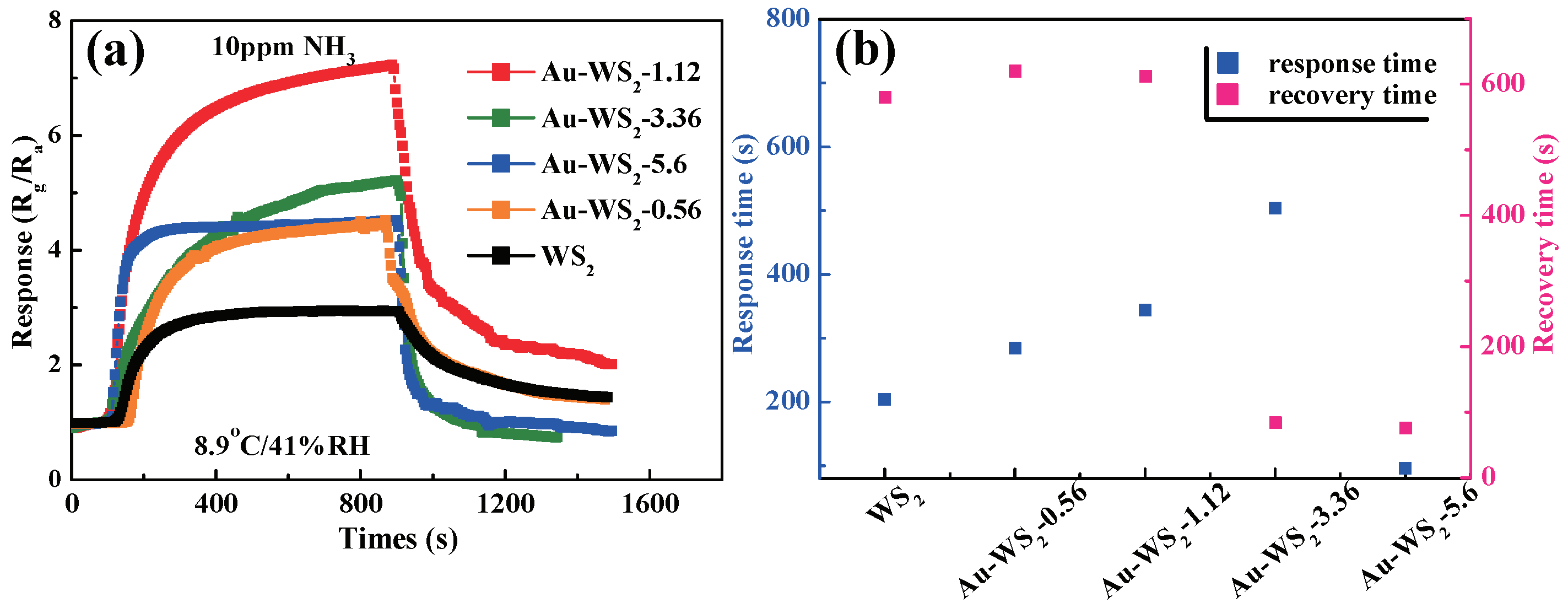
The sensing properties of the fabricated WS and Au/WS sensors were investigated. Figure 6a shows the response of chemiresistive-type sensors to 10 ppm ammonia at room temperature. The sensors show an increase in resistance when exposed to NH, indicating a p-type response. With Au decoration, the WS based sensors indicated a significantly improved response. The sensor using WS microflakes decorated with 1.12 wt% Au achieved the largest response (723%), which is about 2.5 times higher than that of the pure one. Moreover, the sensor using WS microflakes decorated with 5.6 wt% Au achieved a response of 452%, which is about 1.5 times higher than the pure one. The response and recovery times of the sensors to 10 ppm NH are summarized in Figure 6b. This indicates that as the concentration of decorated Au nanoparticles in Au/WS increases further up to 5.6 wt%, the response/recovery speeds of the Au/WS-5.6-based sensor improved, although the response decreased compared to the Au/WS-1.12-based one. However, the fast response/recovery features at room temperature are desirable for applications in some real scenes. For example, when the sensor was used in detecting ammonia in humid human breath, the sensor should have a fast response to ammonia to avoid more errors due to possible sticking of more ammonia molecule to the wall of the gas sampling bag and possible leakage of the sampled gas as sampling time progressed. It also needs a faster recovery speed with respect to the sensor in order to retain a quick measuring rate. Beyond room temperature, the response of the Au/WS-5.6 nanocomposite-based sensor to 10 ppm NH dropped below 150%, which is lower than that of the sensor working at room temperature, as shown in Figure S6. Compared with other WS and TMDs-based sensors, the Au/WS nanocomposite-based sensor exhibited an excellent performance to ammonia, as shown in Table 1.
As a p-type semiconductor, there would be less charge carriers when the surface coverage of phisisorbed oxygen is decreased by the presence of a reducing species such as ammonia [36]. When exposed to NH, the resistance of the sensor based on WS increased indicating a typical p-type character at room temperature. Moreover, the sensing process follows the proposed steps: the surface absorption of gas species and the desorption of reaction products according to the following reactions [38].
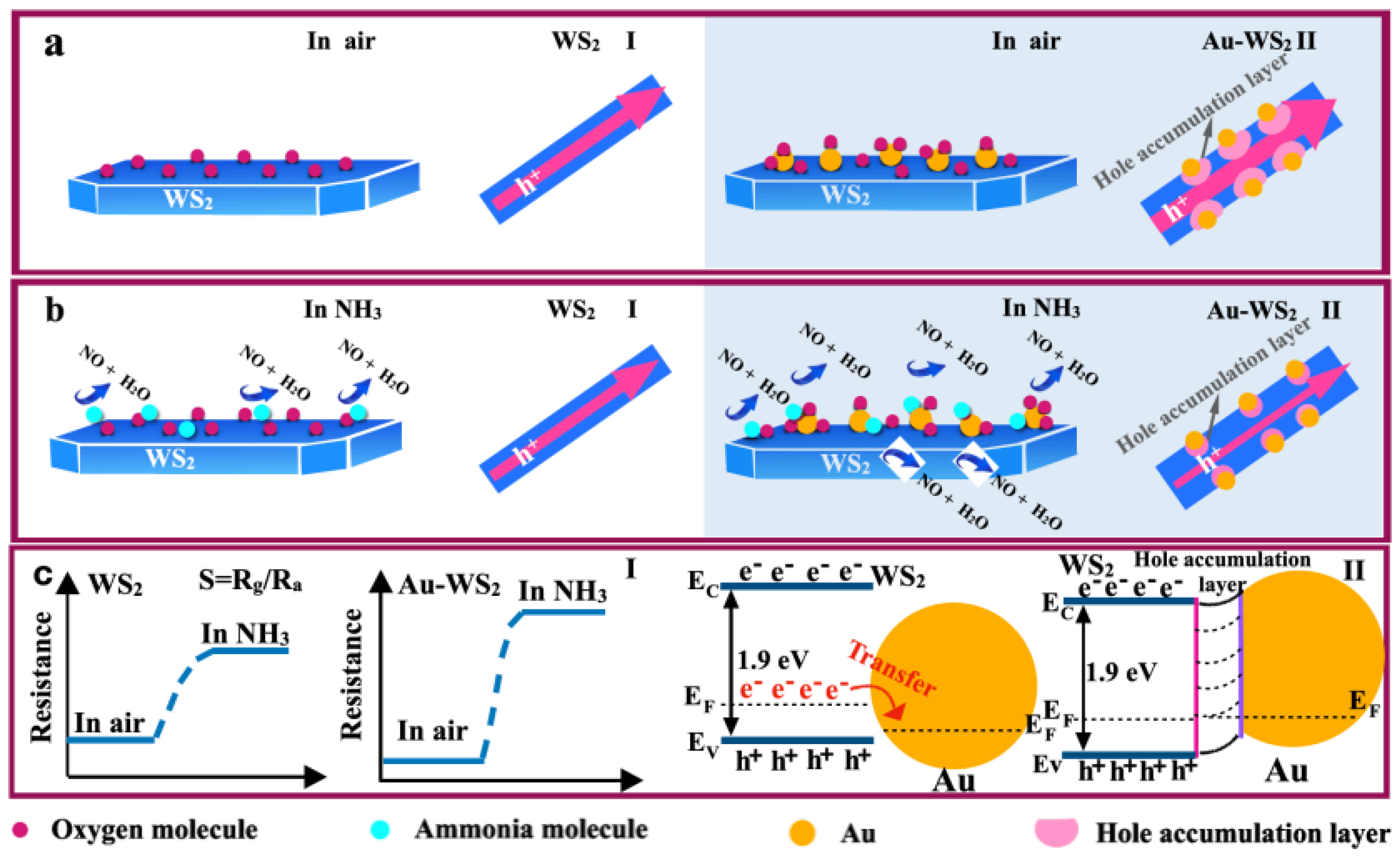
As shown in Figure 7a(I), when the WS-based sensor is exposed in air, oxygen molecules are absorbed on the surface of WS microflakes according to Equation (2). They will capture electrons from the conducting band of WS to form oxygen ions resulting in an increase in the concentration of the majority carriers (holes) according to Equation (3). When the WS-based sensor is presented in NH ambient (Figure 7b(I)), ammonia molecules react with the oxygen ions to form nitric oxide, water and free electrons, according to Equation (4). The produced electrons returned back to the conducting band of WS, which resulted in a decrease in the concentration of holes, resulting in an increase in the resistance of the WS-based sensor (Figure 7c(I)). Compared to pure WS microflakes, the enhanced gas sensing mechanism of WS decorated by gold nanoparticles can be suggested as the following two aspects. Firstly, enhanced sensing performance is related to the “electronic sensitization” effect between Au and WS. As illustrated in Figure 7c(II), as contact formed between Au and WS, the electrons in the conduction band of WS microflakes would transfer to Au nanoparticles due to the higher Fermi level of WS microflakes than that of Au nanoparticles [39,40]. Therefore, the entire accumulation layer (HAL) is created at the interface of Au and WS, resulting in an increase in the concentration of holes in Au/WS nanocomposites. Therefore, the initial resistance of the Au/WS-based sensor will be reduced, which is exhibited in Figure 7c(I), and this is beneficial to the enhancement of sensing responses according to the definition of sensing response (). Secondly, the enhanced sensing performance is also related to the “chemical sensitization” effect of Au [41,42]. Due to the fact that the catalytic effect of Au nanoparticles can lower the enthalpy change of gas decomposition, the sensing reaction at room temperature will be enhanced, and absorption/desorption processes will be accelerated [13]. Furthermore, the formation of heterostructure between WS and Au contributes an abundance of electrons assembled on Au nanoparticles, which will provide more active sites for more oxygen molecules due to the spillover effect of Au (Figure 7a(II)). The additional physisorbed oxygen ions on the surface of Au/WS nanocomposites can be verified in O 1s core spectra in Figure 4c. Moreover, in NH ambient (Figure 7b(II)), the existence of a high number of oxygen ions will facilitate surface sensing reactions. More NH molecules will react with the absorbed oxygen ions, which results in more electrons injected back into WS microflakes. As a result, there will be a thinner HAL at the interface of Au and WS in Au/WS nanocomposites, which will result in a greater increase in resistance of the sensor displayed in Figure 7c(I). In addition, the excellent chemical sensitization effect of Au nanoparticles can accelerate absorption/desorption processes, as shown in Figure 6b. Consequently, under the synergistic effect of electronic sensitization and chemical sensitization between WS and Au, Au/WS-based sensors exhibit a higher response to ammonia and shorter response/recovery time than the pure WS-based sensor.
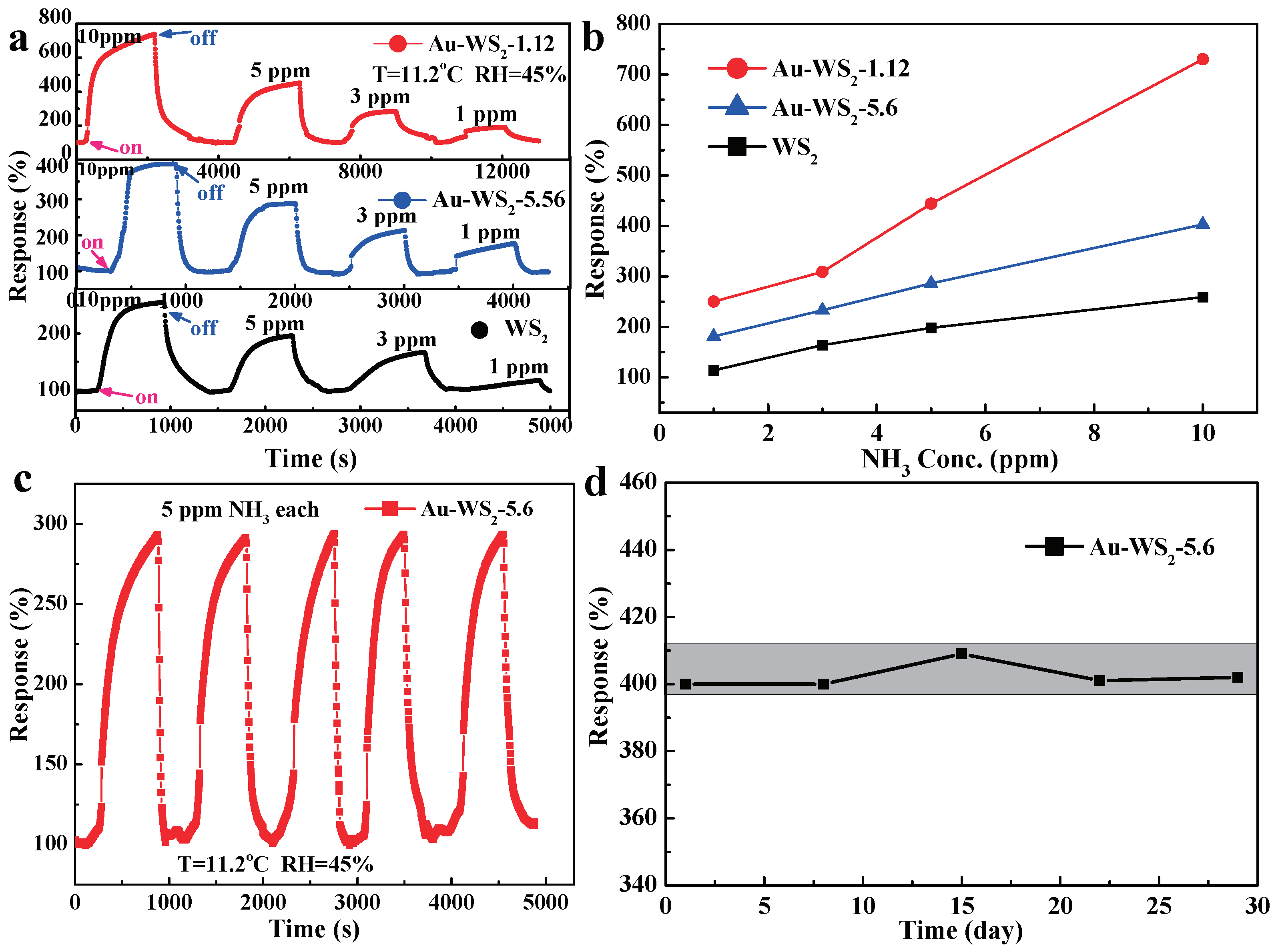
Figure 8a shows the response curves of pure WS, Au/WS-1.12 and Au/WS-5.6-based sensors to different concentrations of NH from 1 ppm to 10 ppm at room temperature. Compared with the pure WS-based sensor, Au/WS-1.12 and Au/WS-5.6-based sensors exhibited higher responses to different concentration of ammonia. The correlations between sensor response and NH concentrations were plotted in Figure 8b. As the ammonia concentration increased, the responses of the sensors increased. The responses of the sensors increased almost linearly with the increase in NH concentrations. However, the slope of the Au/WS-1.12 sensor is higher than the pure WS sensor, indicating better gas detection capability. The lower signals of the Au/WS sensors with higher gold loading (5.6 wt%) to NH may result from “localized” NH consumption without generation of electrons that would transfer to the conduction band of WS. In this case, Au acted as a “good catalyst” and the catalytic oxidation of ammonia would partly occur locally on gold nanoparticles. The induced charge transfer took place only in a localized manner and had no direct impact on the electrical conduction of WS. Similar observations are also found in literature [30,43].
Figure 8c shows the repeatability of the response of Au/WS-5.6 to 5 ppm NH at room temperature. The response of the sensor maintained a stable line shape after 5-cycling measurements, indicating good repeatability of the sensor. A longer period of stability of the sensor for 30 days was studied by measuring the sensing performance of the sensor periodically. The signals remained pretty well within a variation error less than 3% after a 30-day operation, as shown in Figure 8d. Moreover, it shows excellent long term stability.
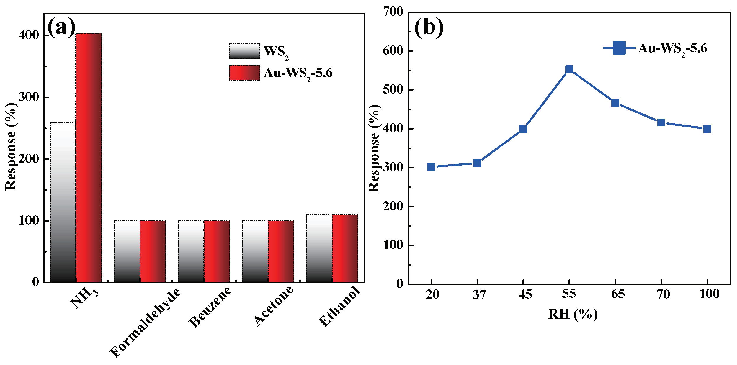
The selectivity of the sensor using Au/WS-5.6 was examined. Figure 9a shows that the developed sensor has a negligible response to several potential interference such as formaldehyde, benzene, acetone and ethanol with a concentration of 10 ppm each, indicating possible excellent selectivity. The dependency of the Au/WS-5.6-based sensor response on humidity was examined, and the results are shown in Figure 9b. The results indicated that the sensor could yield the maximum response when the RH was around 50%. The sensor was resistant to humidity and could work in a highly humid background. The sensor did not show much poisoning when operating at different humidities, as shown in Figure 9b. Sensor response decreased at RH beyond 55%; however, it recovered when RH changed to 55% again.
4. Conclusions
Gold nanoparticles were decorated on WS microflakes using an in situ chemical reduction route. The WS nanoflakes’ diameters are 0.4–4.3 micrometers and the diameters of the decorated Au nanoparticles are 17.8–48.8 nm. It was found that Au/WS nanocomposite-based gas sensors present a higher response and faster response and recovery speeds relative to different concentrations of ammonia from 1 to 10 ppm. Moreover, the Au/WS-5.6 based sensor also displayed excellent selectivity relative to several typical interferents such as formaldehyde, benzene, acetone and ethanol. Gold nanoparticles loaded on the surface of WS microflakes promoted sensing reactions, resulting in the improvement of gas-sensing properties due to the synergistic effect of electronic sensitization and chemical sensitization between WS and Au.
Supplementary Materials
The following are available online at https://0-www-mdpi-com.brum.beds.ac.uk/article/10.3390/chemosensors10010009/s1.
Author Contributions
Conceptualization, Q.G.; methodology, Q.G.; validation, Q.G., B.H. and X.L.; formal analysis, Q.G.; investigation, Q.G.; writing—original draft preparation, Q.G.; writing—review and editing, B.H. and X.L.; supervision, X.L.; project administration, X.L.; funding acquisition, B.H. and X.L. All authors have read and agreed to the published version of the manuscript.
Funding
The authors are thankful to the National Key R&D Program of China (No. 2021YFB3201302), National Natural Science Foundation of China (No. 61971085, 62111530055) and the Fundamental Research Funds for the Central Universities (Nos: DUT19RC(3)054) for financial support.
Institutional Review Board Statement
Not applicable.
Informed Consent Statement
Not applicable.
Data Availability Statement
Not applicable.
Acknowledgments
The authors are thankful to the National Key R&D Program of China (No. 2021YFB3201302), National Natural Science Foundation of China (No. 61971085, 62111530055 ) and the Fundamental Research Funds for the Central Universities (Nos: DUT19RC(3)054) for financial support.
Conflicts of Interest
The authors declare no conflict of interest.
Sample Availability
Samples of compounds are available from the authors.
Abbreviations
The following abbreviations are used in this manuscript:
| SCR | Selective catalytic reduction; |
| 2D | Two-dimensional; |
| TMDs | Transitional metal dichalcogenides; |
| QDs | Quantum dots; |
| rGO | Reduced graphene oxides; |
| CNFs | Carbon nanofibers; |
| XRD | X ray diffractometer; |
| FESEM | Field emission scanning electronic microscope; |
| TEM | Transmission electron microscope; |
| HRTEM | High resolution transmission electron microscope; |
| EDS | Energy dispersive X-ray spectroscopy; |
| XPS | X-ray photoelectron spectra; |
| RT | Room temperature; |
| HAL | Hole accumulation layer. |
References
- Aslam, M.; Chaudhary, V.A.; Mulla, I.S.; Sainkar, S.R.; Mandale, A.B.; Belhekar, A.A.; Vijayamohanan, K. A highly selective ammonia gas sensor using surface-ruthenated zinc oxide. Sens. Actuators A Phys. 1999, 75, 162–167. [Google Scholar] [CrossRef]
- Schönauer, D.; Wiesner, K.; Fleischer, M.; Moos, R. Selective mixed potential ammonia exhaust gas sensor. Sens. Actuators B Chem. 2009, 140, 585–590. [Google Scholar] [CrossRef]
- Buckley, S.G.; Damm, C.J.; Vitovec, W.M.; Sgro, L.A.; Sawyer, R.F.; Koshland, C.P.; Lucas, D. Ammonia detection and monitoring with photofragmentation fluorescence. Appl. Opt. 1998, 37, 8382–8391. [Google Scholar] [CrossRef]
- Timmer, B.; Olthuis, W.; Berg, A.V.D. Ammonia sensors and their applictions-a review. Sens. Actuators B Chem. 2005, 107, 666–677. [Google Scholar] [CrossRef]
- Miller, D.R.; Akbar, S.A.; Morris, P.A. Nanoscale metal oxide-based heterojunctions for gas sensing: A review. Sens. Actuators B Chem. 2014, 204, 250–272. [Google Scholar] [CrossRef]
- Li, X.; Li, X.; Wang, J.; Lin, S. Highly sensitive and selective room-temperature formaldehyde sensors using hollow TiO2 microspheres. Sens. Actuators B Chem. 2015, 219, 158–163. [Google Scholar] [CrossRef]
- Qin, Z.; Zeng, D.; Zhang, J.; Wu, C.; Wen, Y.; Shan, B.; Xie, C. Effect of layer number on recovery rate of WS2 nanosheets for ammonia detection at room temperature. Appl. Surf. Sci. 2017, 414, 244–250. [Google Scholar] [CrossRef]
- Zhou, Y.; Liu, G.; Zhu, X.; Guo, Y. Ultrasensitive NO2 gas sensing based on rGO/MoS2 nanocomposite film at low temperature. Sens. Actuators B Chem. 2017, 251, 280–290. [Google Scholar] [CrossRef]
- Lu, G.; Ocola, L.E.; Chen, J. Reduced graphene oxide for room-temperature gas sensors. Nanotechnology 2009, 20, 445502. [Google Scholar] [CrossRef]
- Geim, A.K.; Novoselov, K.S. The rise of graphene. Nat. Mater. 2007, 6, 183–191. [Google Scholar] [CrossRef]
- Kim, Y.H.; Phan, D.T.; Ahn, S.; Nam, K.H.; Park, C.M.; Jeon, K.J. Two-dimensional SnS2 materials as high-performance NO2 sensors with fast response and high sensitivity. Sens. Actuators B Chem. 2018, 255, 616–621. [Google Scholar] [CrossRef]
- Dai, L.; Huang, K.; Xia, Y.; Xu, Z. Two-dimensional material separation membranes for renewable energy purification, storage, and conversion. Green Energy Environ. 2021, 6, 193–211. [Google Scholar] [CrossRef]
- Huang, B.; Wang, Y.; Hu, Q.; Mu, X.; Zhang, Y.; Bai, J.; Wang, Q.; Sheng, Y.; Zhang, Z.; Xie, E. A low temperature and highly sensitive ethanol sensor based on Au modified In2O3 nanofibers by coaxial electrospinning. J. Mater. Chem. C 2018, 6, 10935–10943. [Google Scholar] [CrossRef]
- Zhang, X.; Liu, Y.; Chen, L.; Li, Z.; Qu, Y.; Wu, W.; Jing, L. Porous two-dimension MnO2-C3N4/titanium phosphate nanocomposites as efficient photocatalsyts for CO oxidation and mechanisms. Appl. Catal. 2021, 282, 119563. [Google Scholar] [CrossRef]
- Cheng, M.; Wu, Z.; Liu, G.; Zhao, L.; Gao, Y.; Zhang, B.; Liu, F.; Yan, X.; Liang, X.; Sun, P.; et al. Highly sensitive sensors based on quasi-2D rGO/SnS2 hybrid for rapid detection of NO2 gas. Sens. Actuators B Chem. 2019, 291, 216–225. [Google Scholar] [CrossRef]
- Perrozzi, F.; Emamjomeh, S.M.; Paolucci, V.; Taglieri, G.; Ottaviano, L.; Cantalini, C. Thermal stability of WS2 flakes and gas sensing properties of WS2/WO3 composite to H2, NH3 and NO2. Sens. Actuators B Chem. 2017, 243, 812–822. [Google Scholar] [CrossRef]
- Chen, Y.; Pei, Y.; Jiang, Z.; Shi, Z.; Xu, J.; Wu, D.; Xu, T.; Tian, Y.; Wang, X.; Li, X. Humidity sensing properties of the hydrothermally synthesized WS2-modified SnO2 hybrid nanocomposite. Appl. Surf. Sci. 2018, 447, 325–330. [Google Scholar] [CrossRef]
- Zhang, X.; Wang, J.; Chen, D.; Liu, L. The adsorption performance of harmful gas on Cu doped WS2: A first-principle study. Mater. Today Commun. 2021, 28, 102488. [Google Scholar] [CrossRef]
- Nguyen, T.K.; Jeong, S.; Youn, J.S.; Ahn, S.; Nam, K.H.; Park, C.M.; Jeon, K.J. Insight into mechanism of temperature-dependent limit of NO2 detection using monolayer MoS2. Sens. Actuators B Chem. 2021, 329, 129138. [Google Scholar] [CrossRef]
- Xiong, Y.; Xu, W.; Ding, D.; Lu, W.; Zhu, L.; Zhu, Z.; Wang, Y.; Xue, Q. Ultra-sensitive NH3 sensor based on flower-shaped SnS2 nanostructures with sub-ppm detection ability. J. Hazard. Mater. 2018, 341, 159–167. [Google Scholar] [CrossRef]
- Qin, Z.; Xu, K.; Yue, H.; Wang, H.; Zhang, J.; Ouyang, C.; Xie, C.; Zeng, D. Enhanced room-temperature NH3 gas sensing by 2D SnS2 with sulfur vacancies synthesized by chemical exfoliation. Sens. Actuators B Chem. 2018, 262, 771–779. [Google Scholar] [CrossRef]
- Wang, L.; Fu, H.; Jin, Q.; Jin, H.; Haick, H.; Wang, S.; Yu, K.; Deng, S.; Wang, Y. Directly transforming SnS2 nanosheets to hierarchical SnO2 nanotubes: Towards sensitive and selective sensing of acetone at relatively low operating temperatures. Sens. Actuators B Chem. 2019, 292, 148–155. [Google Scholar] [CrossRef]
- Radisavljevic, B.; Radenovic, A.; Brivio, J.; Giacometti, V.; Kis, A. Single-layer MoS2 transistors. Nat. Nanotechnol. 2011, 6, 147–150. [Google Scholar] [CrossRef] [PubMed]
- Yan, W.; Worsley, M.A.; Pham, T.; Zettl, A.; Carraro, C.; Maboudian, R. Effects of ambient humidity and temperature on the NO2 sensing characteristics of WS2/graphene aerogel. Appl. Surf. Sci. 2018, 450, 372–379. [Google Scholar] [CrossRef]
- Agrawal, A.V.; Kumar, R.; Yang, G.; Bao, J.; Kumar, M. Enhanced adsorption sites in monolayer MoS2 pyramid structures for highly sensitive and fast hydrogen sensor. Int. J. Hydrogen Energy 2020, 45, 9268–9277. [Google Scholar] [CrossRef]
- Shokri, A.; Salami, N. Gas sensor based on MoS2 monolayer. Sens. Actuators B Chem. 2016, 236, 378–385. [Google Scholar] [CrossRef]
- Cui, S.; Wen, Z.; Huang, X.; Chang, J.; Chen, J. Stabilizing MoS2 Nanosheets through SnO2 Nanocrystal Decoration for High-Performance Gas Sensing in Air. Small 2015, 11, 2305–2313. [Google Scholar]
- Li, X.; Li, X.; Li, Z.; Wang, J.; Zhang, J. WS2 nanoflakes based selective ammonia sensors at room temperature. Sens. Actuators B Chem. 2017, 240, 273–277. [Google Scholar] [CrossRef]
- Gu, D.; Li, X.; Wang, H.; Li, M.; Xi, Y.; Chen, Y.; Wang, J.; Rumyntseva, M.N.; Gaskov, A.M. Light enhanced VOCs sensing of WS2 microflakes based chemiresistive sensors powered by triboelectronic nangenerators. Sens. Actuators B Chem. 2018, 256, 992–1000. [Google Scholar] [CrossRef]
- Ouyang, C.; Chen, Y.; Qin, Z.; Zeng, D.; Zhang, J.; Wang, H.; Xie, C. Two-dimensional WS2-based nanosheets modified by Pt quantum dots for enhanced room-temperature NH3 sensing properties. Appl. Surf. Sci. 2018, 455, 45–52. [Google Scholar] [CrossRef]
- Xu, Y.; Xie, J.; Zhang, Y.; Tian, F.H.; Yang, C.; Zheng, W.; Liu, X.; Zhang, J.; Pinna, N. Edge-enriched WS2 nanosheets on carbon nanofibers boosts NO2 detection at room temperature. J. Hazard. Mater. 2021, 411, 125120. [Google Scholar] [CrossRef] [PubMed]
- Suh, J.M.; Kwon, K.C.; Lee, T.H.; Kim, C.; Lee, C.W.; Song, Y.G.; Choi, M.J.; Choi, S.; Cho, S.H.; Kim, S.; et al. Edge-exposed WS2 on 1D nanostructures for highly selective NO2 sensor at room temperature. Sens. Actuators B Chem. 2021, 333, 129566. [Google Scholar] [CrossRef]
- Liu, W.; Gu, D.; Li, X. AuPt Bimetal-Functionalized SnSe2 Microflower-Based Sensors for Detecting Sub-ppm NO2 at Low Temperatures. ACS Appl. Mater. Interfaces 2021, 13, 20336–20348. [Google Scholar] [CrossRef] [PubMed]
- Liu, W.; Gu, D.; Li, X. Detection of Ppb-level NO2 using mesoporous ZnSe/SnO2 core-shell microspheres based chemical sensors. Sens. Actuators B Chem. 2020, 320, 128365. [Google Scholar] [CrossRef]
- Liu, W.; Gu, D.; Zhang, J.W.; Li, X.G.; Rumyantseva, M.N.; Gaskov, A.M. ZnSe/NiO heterostructure-based chemiresistive-type sensors for low-concentration NO2 detection. Rare Met. 2021, 40, 1632–1641. [Google Scholar] [CrossRef]
- Wang, X.Y.; Gu, D.; Li, X.; Lin, S.; Zhao, S.; Rumyantseva, M.N.; Gaskov, A.M. Reduced graphene oxide hybridized with WS2 nanoflakes based heterojunctions for selective ammonia sensors at room temperature. Sens. Actuators B Chem. 2019, 282, 290–299. [Google Scholar] [CrossRef]
- Meng, F.; Zhu, T.; Yuan, Z.; Qin, W.; Gao, H.; Zhang, H. Investigation of Mixed-Phase WS2 Nanomaterials for Ammonia Gas Sensing. IEEE Sens. J. 2021, 21, 7268–7274. [Google Scholar] [CrossRef]
- Sharma, N.; Sharma, N.; Srinivasan, P.; Kumar, S.; Rayappan, J.B.B.; Kailasam, K. Heptazine based organic framework as a chemiresistive sensor for ammonia detection at room temperature. J. Mater. Chem. A 2018, 6, 18389–18395. [Google Scholar] [CrossRef]
- Kim, J.H.; Mirzaei, A.; Kim, H.W.; Kim, S.S. Realization of Au-decorated WS2 nanosheets as low power-consumption and selective gas sensors. Sens. Actuators B Chem. 2019, 296, 126659. [Google Scholar] [CrossRef]
- Zhang, S.; Song, P.; Zhang, J.; Yan, H.; Li, J.; Yang, Z.; Wang, Q. Highly sensitive detection of acetone using mesoporous In2O3 nanospheres decorated with Au nanoparticles. Sens. Actuators B Chem. 2017, 242, 983–993. [Google Scholar] [CrossRef]
- Zhang, S.; Song, P.; Yan, H.; Wang, Q. Self-assembled hierarchical Au-loaded In2O3 hollow microspheres with superior ethanol sensing properties. Sens. Actuators B Chem. 2016, 231, 245–255. [Google Scholar] [CrossRef]
- Fu, H.; Hou, C.; Gu, F.; Han, D.; Wang, Z. Facile preparation of rod-like Au/In2O3 nanocomposites exhibiting high response to CO at room temperature. Sens. Actuators B Chem. 2017, 243, 516–524. [Google Scholar] [CrossRef]
- Zeb, S.; Peng, X.; Shi, Y.; Su, J.; Sun, J.; Zhang, M.; Sun, G.; Nie, Y.; Cui, Y.; Jiang, X. Bimetal Au-Pd decorated hierarchical WO3 nanowire bundles for gas sensing application. Sens. Actuators B Chem. 2021, 334, 129584. [Google Scholar] [CrossRef]
Figure 1.
Schematic diagram of synthesis and gas sensing measure of Au/WS nanocomposite-based gas sensor.
Figure 1.
Schematic diagram of synthesis and gas sensing measure of Au/WS nanocomposite-based gas sensor.

Figure 2.
XRD patterns of pure WS and Au/WS based samples.

Figure 3.
(a) FESEM image of pure WS microflakes, (b) FESEM image of Au/WS-5.6 nanocomposites, (c) TEM image of Au/WS-5.6 nanocomposites and (d) HRTEM image of Au/WS-5.6 nanocomposites.
Figure 3.
(a) FESEM image of pure WS microflakes, (b) FESEM image of Au/WS-5.6 nanocomposites, (c) TEM image of Au/WS-5.6 nanocomposites and (d) HRTEM image of Au/WS-5.6 nanocomposites.

Figure 4.
(a) XPS spectra of WS microflakes and Au/WS-5.6 nanocomposites, (b) O 1s of WS microflakes, (c) O 1s of Au/WS-5.6 nanocomposites and (d) Au 4f of Au/WS-5.6 nanocomposites.
Figure 4.
(a) XPS spectra of WS microflakes and Au/WS-5.6 nanocomposites, (b) O 1s of WS microflakes, (c) O 1s of Au/WS-5.6 nanocomposites and (d) Au 4f of Au/WS-5.6 nanocomposites.

Figure 5.
The polarization curves of the sensors using WS and Au/WS.

Figure 6.
(a) Pure WS and Au/WS gas sensors response to 10 ppm NH at room temperature; (b) the as-fabricated gas sensors response and recovery times to 10 ppm NH.
Figure 6.
(a) Pure WS and Au/WS gas sensors response to 10 ppm NH at room temperature; (b) the as-fabricated gas sensors response and recovery times to 10 ppm NH.

Figure 7.
Schematic diagram of the pure WS and Au/WS sensors exposed to air and ammonia gas. (a) pure WS sensors in air and ammonia gas (b) Au/WS sensors in air and ammonia gas (c) pure WS and Au/WS sensors in air and ammonia gas
Figure 7.
Schematic diagram of the pure WS and Au/WS sensors exposed to air and ammonia gas. (a) pure WS sensors in air and ammonia gas (b) Au/WS sensors in air and ammonia gas (c) pure WS and Au/WS sensors in air and ammonia gas

Figure 8.
(a,b) Gas sensor responses to different concentrations of ammonia of Au/WS-1.12, Au/WS-5.56 and pure WS; (c) repeatability of gas sensor response to 5 ppm ammonia; (d) long-term stability of Au/WS-5.6-based gas sensor.
Figure 8.
(a,b) Gas sensor responses to different concentrations of ammonia of Au/WS-1.12, Au/WS-5.56 and pure WS; (c) repeatability of gas sensor response to 5 ppm ammonia; (d) long-term stability of Au/WS-5.6-based gas sensor.

Figure 9.
(a) Selectivity of the Au/WS-5.6 nanocomposite-based gas sensor to different gases at RH = 45%; (b) dependency of the sensor response on humidity.
Figure 9.
(a) Selectivity of the Au/WS-5.6 nanocomposite-based gas sensor to different gases at RH = 45%; (b) dependency of the sensor response on humidity.


Table 1.
A comparison of the performance of NH sensors based on WS and TMDs materials.
| Sensitive Material | NH Concentration | Temperature (C) | Response (%) | Response/Recovery (s/s) | Ref. |
|---|---|---|---|---|---|
| WS/WO | 10 ppm | 150 | 400 | ∼150/∼100 | [16] |
| WS nanoflakes | 5 ppm | RT | 217 | 120/150 | [28] |
| Pt QDs/WS | 250 ppm | RT | 9.5 | 200/1200 | [30] |
| rGO/WS | 10 ppm | RT | 71 | 240/600 | [36] |
| Mixed-phase WS | 100 ppm | 150 | 4.72 | 19/33 | [37] |
| Flower-shaped SnS | 100 ppm | 200 | 7.4 | 40.6/624 | [20] |
| 2D SnS | 500 ppm | RT | 4.2 | 16/450 | [21] |
| Au/WS | 10 ppm | RT | 452 | 96/76 | This work |
Data deduced from the reported figure; QDs: Quantum dots; rGO: Reduced graphene oxides; RT: Room temperature.
Publisher’s Note: MDPI stays neutral with regard to jurisdictional claims in published maps and institutional affiliations. |
© 2021 by the authors. Licensee MDPI, Basel, Switzerland. This article is an open access article distributed under the terms and conditions of the Creative Commons Attribution (CC BY) license (https://creativecommons.org/licenses/by/4.0/).
Share and Cite
MDPI and ACS Style
Guang, Q.; Huang, B.; Li, X. Au-Decorated WS2 Microflakes Based Sensors for Selective Ammonia Detection at Room Temperature. Chemosensors 2022, 10, 9. https://0-doi-org.brum.beds.ac.uk/10.3390/chemosensors10010009
AMA Style
Guang Q, Huang B, Li X. Au-Decorated WS2 Microflakes Based Sensors for Selective Ammonia Detection at Room Temperature. Chemosensors. 2022; 10(1):9. https://0-doi-org.brum.beds.ac.uk/10.3390/chemosensors10010009
Chicago/Turabian StyleGuang, Qiyilan, Baoyu Huang, and Xiaogan Li. 2022. "Au-Decorated WS2 Microflakes Based Sensors for Selective Ammonia Detection at Room Temperature" Chemosensors 10, no. 1: 9. https://0-doi-org.brum.beds.ac.uk/10.3390/chemosensors10010009
Note that from the first issue of 2016, this journal uses article numbers instead of page numbers. See further details here.