
Radionuclide Delivery Strategies in Tumor Treatment: A Systematic Review

 , , and
Abstract
:1. Introduction
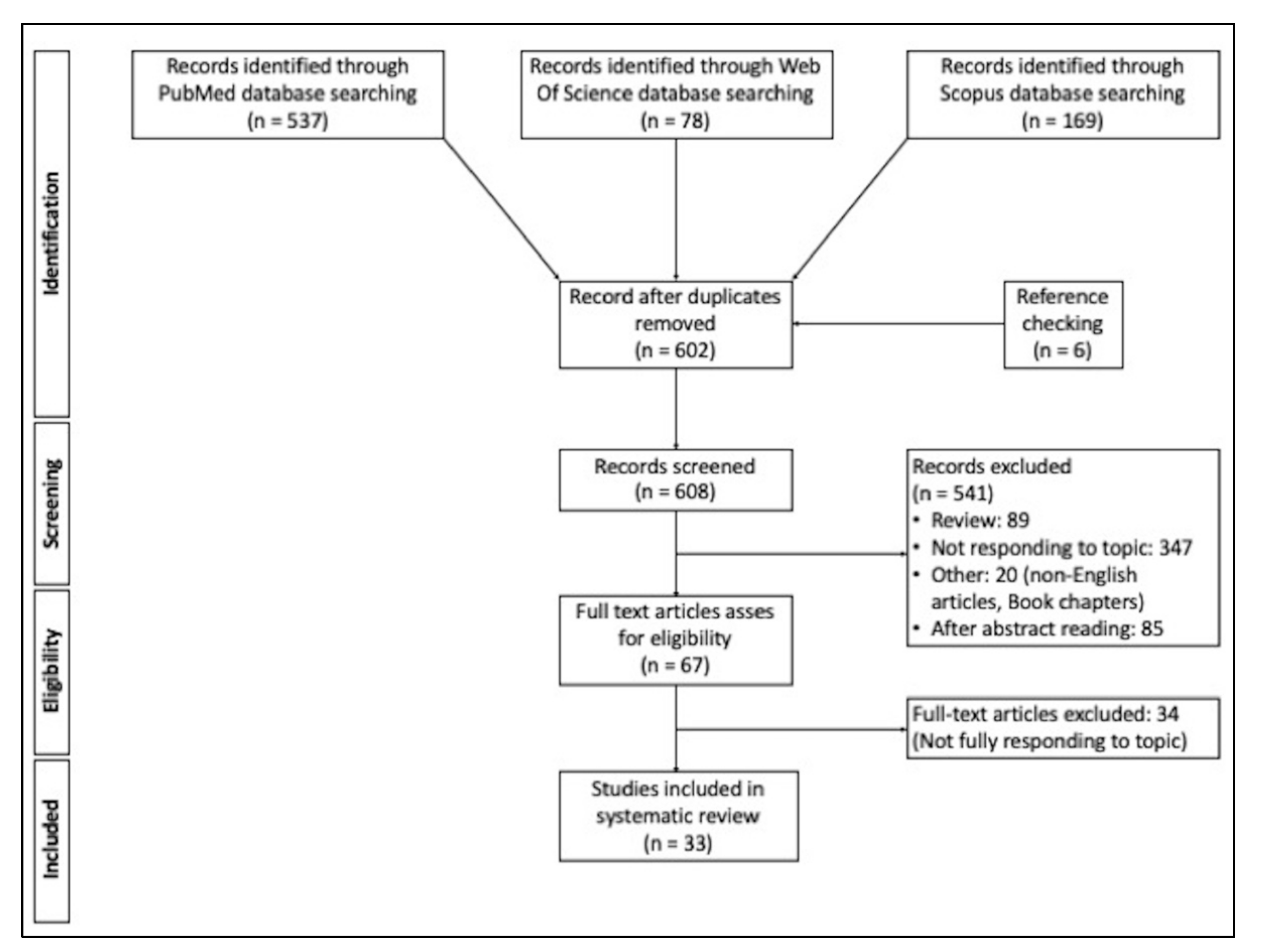
2. Materials and Methods
3. Results
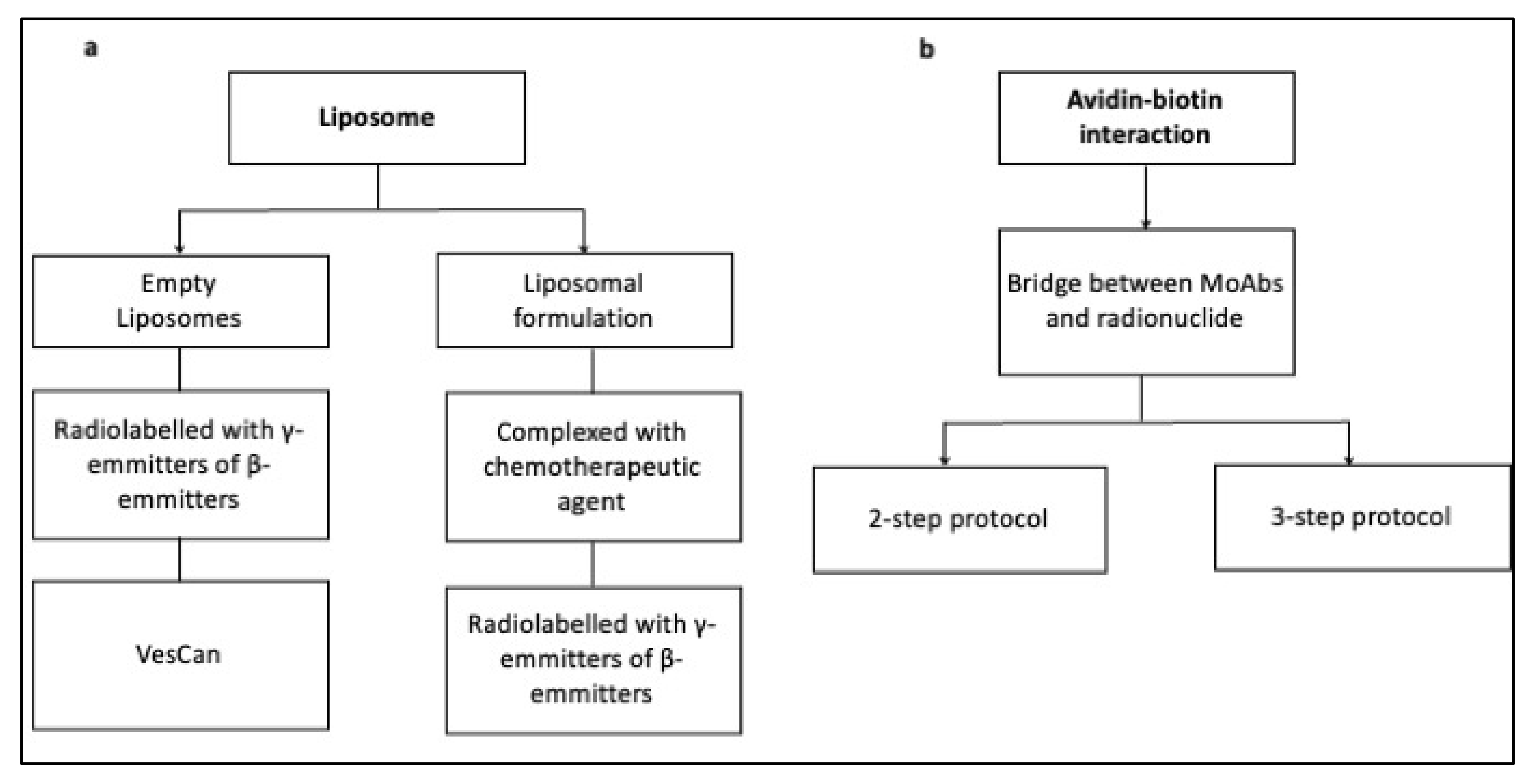
3.1. Liposomes
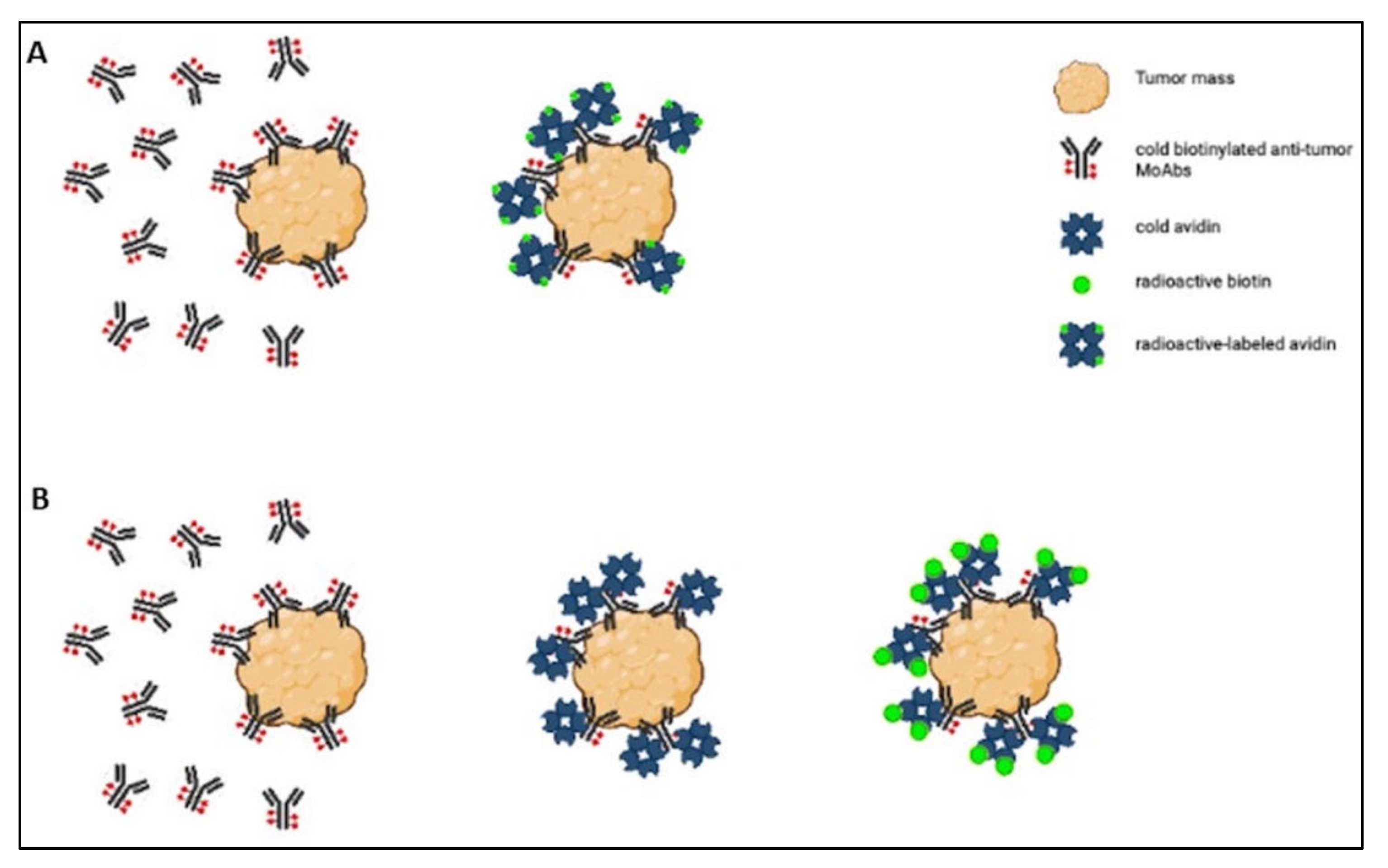
3.2. Avidin–Biotin Interaction
3.3. Docking
4. Discussion
5. Conclusions
Supplementary Materials
Author Contributions
Funding
Institutional Review Board Statement
Informed Consent Statement
Data Availability Statement
Conflicts of Interest
References
- Gill, M.R.; Falzone, N.; Du, Y.; Vallis, K.A. Review targeted radionuclide therapy in combined-modality regimens. Lancet Oncol. 2017, 18, e414–e423. [Google Scholar] [CrossRef]
- Gudkov, S.V.; Shilyagina, N.Y.; Vodeneev, V.A.; Zvyagin, A.V. Targeted radionuclide therapy of human tumors. Int. J. Mol. Sci. 2015, 17, 33. [Google Scholar] [CrossRef] [PubMed]
- Artigas, C.; Mileva, M.; Flamen, P.; Karfis, I. Targeted radionuclide therapy: An emerging field in solid tumours. Curr. Opin. 2021, 33, 493–499. [Google Scholar] [CrossRef] [PubMed]
- Peltek, O.O.; Muslimov, A.R.; Zyuzin, M.V.; Timin, A.S. Current outlook on radionuclide delivery systems: From design consideration to translation into clinics. J. Nanobiotechnol. 2019, 17, 90. [Google Scholar] [CrossRef] [Green Version]
- Malcolm, J.; Falzone, N.; Lee, B.Q.; Vallis, K.A. Targeted radionuclide therapy: New advances for improvement of patient management and response. Cancers 2019, 11, 268. [Google Scholar] [CrossRef] [Green Version]
- Li, S.; Goins, B.; Zhang, L.; Bao, A. Novel multifunctional theranostic liposome drug delivery system: Construction, characterization, and multimodality MR, near-infrared fluorescent, and nuclear imaging. Bioconjug. Chem. 2012, 23, 1322–1332. [Google Scholar] [CrossRef] [Green Version]
- Yang, F.; Wang, H.; Liu, R.; Teng, M.; Li, J.; Lu, M. Pharmacokinetic analysis of 111 In-labeled liposomal doxorubicin in murine glioblastoma after blood-brain barrier disruption by focused ultrasound. PLoS ONE 2012, 7, e45468. [Google Scholar]
- Lin, Y.; Kao, H.; Li, J.; Hwang, J.; Tseng, Y.; Lin, W.; Lin, M.; Ting, G.; Wang, H. Tumor burden talks in cancer treatment with PEGylated liposomal drugs. PLoS ONE 2013, 8, e63078. [Google Scholar] [CrossRef]
- Lee, H.; Zheng, J.; Gaddy, D.; Orcutt, K.D.; Leonard, S.; Geretti, E.; Hesterman, J.; Harwell, C.; Hoppin, J.; Jaffray, D.A.; et al. A gradient-loadable 64Cu-chelator for quantifying tumor deposition kinetics of nanoliposomal therapeutics by positron emission tomography. Nanomed. Nanotechnol. Biol. Med. 2015, 11, 155–165. [Google Scholar] [CrossRef]
- Ito, K.; Hamamichi, S.; Asano, M.; Hori, Y.; Matsui, J.; Iwata, M.; Funahashi, Y.; Umeda, I.O.; Fujii, H. Radiolabeled liposome imaging determines an indication for liposomal anticancer agent in ovarian cancer mouse xenograft models. Cancer Sci. 2015, 107, 60–67. [Google Scholar] [CrossRef] [Green Version]
- Silva, J.O.; Fernandes, R.S.; Lopes, S.C.A.; Cardoso, V.N.; Leite, E.A.; Cassali, G.D.; Marzola, M.C.; Rubello, D.; Oliveira, M.C.; Luis, A.; et al. pH-sensitive, long-circulating liposomes as an alternative tool to deliver doxorubicin into tumors: A feasibility animal study. Mol. Imaging Biol. 2016, 18, 898–904. [Google Scholar] [CrossRef]
- Edmonds, S.; Volpe, A.; Shmeeda, H.; Parente-pereira, A.C.; Radia, R.; Bagun, J.; Szanda, I.; Severin, G.W.; Livieratos, L.; Blower, P.J.; et al. Exploiting the metal-chelating properties of the drug cargo for in vivo positron emission tomography imaging of liposomal nanomedicines. ACS Nano 2016, 10, 10294–10307. [Google Scholar] [CrossRef] [Green Version]
- Du, Y.; Liang, X.; Li, Y.; Sun, T.; Jin, Z.; Xue, H.; Tian, J. Nuclear and fluorescent labeled PD-1-Liposome-DOX-64 Cu/IRDye800CW allows improved breast tumor targeted imaging and therapy. Mol. Pharm. 2017, 14, 3978–3986. [Google Scholar] [CrossRef]
- Lamichhane, N.; Dewkar, G.K.; Sundaresan, G.; Mahon, R.N.; Zweit, J. [18F]-fluorinated carboplatin and [111In]-liposome for image-guided drug delivery. Int. J. Mol. Sci. 2017, 107, 1079. [Google Scholar] [CrossRef] [Green Version]
- Luo, D.; Goel, S.; Liu, H.; Carter, K.A.; Jiang, D.; Geng, J.; Kutyre, C.J.; Engle, J.W.; Huang, W.; Shao, S.; et al. Intrabilayer 64 Cu labeling of photoactivatable, doxorubicin-loaded stealth liposomes. ACS Nano 2017, 11, 12482–12491. [Google Scholar] [CrossRef]
- Monteiro, L.O.F.; Fernandes, R.S.; Oda, C.M.R.; Lopes, S.C.; Townsend, D.M.; Cardoso, V.N.; Oliveira, M.C.; Leite, E.A.; Rubello, D.; Barros, A.L.B. De biomedicine & pharmacotherapy paclitaxel-loaded folate-coated long circulating and pH-sensitive liposomes as a potential drug delivery system: A biodistribution study. Biomed. Pharmacother. 2018, 97, 489–495. [Google Scholar]
- Srimathveeravalli, G.; Abdel-atti, D.; Carlos, P.; Takaki, H.; Solomon, S.B.; Mulder, W.J.M.; Reiner, T. Reversible electroporation—mediated liposomal doxorubicin delivery to tumors can be monitored with 89 Zr-Labeled reporter nanoparticles. Mol. Imaging 2018, 17, 1536012117749726. [Google Scholar] [CrossRef] [Green Version]
- Gawne, P.; Man, F.; Fonslet, J.; Radia, R.; Bordoloi, J.; Long, N.; Rosales, R.T.M. De and liposomal nanomedicine PET imaging using. Dalt. Trans. 2018, 47, 9283–9293. [Google Scholar] [CrossRef] [Green Version]
- Arrieta, Ó.; Medina, L.A.; Estrada-Lobato, E.; Hernández-Pedro, N.; Villanueva-Rodríguez, G.; Martínez-Barrera, L.; MacEdo, E.O.; López-Rodríguez, V.; Motola-Kuba, D.; Corona-Cruz, J.F. First-line chemotherapy with liposomal doxorubicin plus cisplatin for patients with advanced malignant pleural mesothelioma: Phase II trial. Br. J. Cancer 2012, 106, 1027–1032. [Google Scholar] [CrossRef] [Green Version]
- Arrieta, O.; Medina, L.A.; Estrada-Lobato, E.; Ramírez-Tirado, L.A.; Mendoza-García, V.O.; De La Garza-Salazar, J. High liposomal doxorubicin tumour tissue distribution, as determined by radiopharmaceutical labelling with 99mTc-LD, is associated with the response and survival of patients with unresectable pleural mesothelioma treated with a combination of liposomal do. Cancer Chemother. Pharmacol. 2014, 74, 211–215. [Google Scholar] [CrossRef]
- Lee, H.; Shields, A.F.; Siegel, B.A.; Miller, K.D.; Krop, I.; Ma, C.X.; Lorusso, P.M.; Munster, P.N.; Campbell, K.; Gaddy, D.F.; et al. 64Cu-MM-302 positron emission tomography quantifies variability of enhanced permeability and retention of nanoparticles in relation to treatment response in patients with metastatic breast cancer. Clin. Cancer Res. 2017, 23, 4190–4202. [Google Scholar] [CrossRef] [Green Version]
- Paganelli, G.; Malcovati, M.; Siccardi, A.G.; Villa, E.; Sudati, F.; Rossetti, C.; Fazio, F. Three-step monoclonal antibody tumor targeting in carcinoembryonic antigenpositive patients. Cancer Res. 1991, 51, 5960–5966. [Google Scholar]
- Paganelli, G.; Belloni, C.; Magnani, P.; Zito, F.; Pasini, A.; Sassi, I.; Meroni, M.; Mariani, M.; Vignali, M.; Siccardi, A.G.; et al. Two-step tumour targetting in ovarian cancer patients using biotinylated monoclonal antibodies and radioactive streptavidin. Eur. J. Nucl. Med. 1992, 19, 322–329. [Google Scholar] [CrossRef]
- Cremonesi, M.; Ferrari, M.; Chinol, M.; Stabin, M.G.; Grana, C.; Prisco, G.; Robertson, C.; Tosi, G.; Paganelli, G. Original article Three-step radioimmunotherapy with yttrium-90 biotin: Dosimetry and pharmacokinetics in cancer patients. Eur. J. Nucl. Med. 1999, 26, 110–120. [Google Scholar] [CrossRef]
- Paganelli, G.; Grana, C.; Chinol, M.; Cremonesi, M.; De Cicco, C.; De Braud, F.; Robertson, C.; Zurrida, S.; Casadio, C.; Zoboli, S.; et al. Antibody-guided three-step therapy for high grade glioma with yttrium-90 biotin. Eur. J. Nucl. Med. 1999, 26, 348–357. [Google Scholar] [CrossRef]
- Paganelli, G.; Bartolomei, M.; Ferrari, M.; Cremonesi, M.; Broggi, G.; Maira, G.; Sturiale, C.; Grana, C.; Prisco, G.; Gatti, M.; et al. Pre-targeted locoregional radioimmunotheraphy with 90Y-biotin in glioma patients: Phase I study and preliminary therapeutic results. Cancer Biother. Radiopharm. 2001, 16, 227–235. [Google Scholar] [CrossRef]
- Grana, C.; Chinol, M.; Robertson, C.; Mazzetta, C.; Bartolomei, M.; De Cicco, C.; Fiorenza, M.; Gatti, M.; Caliceti, P.; Paganelli, G. Pretargeted adjuvant radioimmunotherapy with Yttrium-90-biotin in malignant glioma patients: A pilot study. Br. J. Cancer 2002, 86, 207–212. [Google Scholar] [CrossRef] [Green Version]
- Paganelli, G.; Ferrari, M.; Cremonesi, M.; De Cicco, C.; Galimberti, V.; Luini, A.; Veronesi, P.; Fiorenza, M.; Carminati, P.; Zanna, C.; et al. IART®: Intraoperative avidination for radionuclide treatment. A new way of partial breast irradiation. Breast 2007, 16, 17–26. [Google Scholar] [CrossRef]
- Paganelli, G.; Ferrari, M.; Ravasi, L.; Cremonesi, M.; De Cicco, C.; Galimberti, V.; Sivolapenko, G.; Luini, A.; De Santis, R.; Travaini, L.L.; et al. Intraoperative avidination for radionuclide therapy: A prospective new development to accelerate radiotherapy in breast cancer. Clin. Cancer Res. 2007, 13, 5646–5652. [Google Scholar] [CrossRef] [Green Version]
- Paganelli, G.; De Cicco, C.; Ferrari, M.E.; Carbone, G.; Pagani, G.; Leonardi, M.C.; Cremonesi, M.; Ferrari, A.; Pacifici, M.; Di Dia, A.; et al. Intraoperative avidination for radionuclide treatment as a radiotherapy boost in breast cancer: Results of a phase II study with 90Y-labeled biotin. Eur. J. Nucl. Med. Mol. Imaging 2010, 37, 203–211. [Google Scholar] [CrossRef]
- Yang, X.; Mease, R.C.; Pullambhatla, M.; Lisok, A.; Chen, Y.; Foss, C.A.; Wang, Y.; Shallal, H.; Edelman, H.; Hoye, A.T.; et al. [18 F]Fluorobenzoyllysinepentanedioic acid carbamates: New scaffolds for positron emission tomography (PET) imaging of prostate-speci fi c membrane antigen (PSMA). J. Med. Chem. 2016, 59, 206–218. [Google Scholar] [CrossRef] [PubMed] [Green Version]
- Kurniawan, F.; Kartasasmita, R.E.; Yoshioka, N.; Mutalib, A.; Tjahjono, D.H. Computational study of imidazolylporphyrin derivatives as a computational study of imidazolylporphyrin derivatives as a radio-pharmaceutical ligand for melanoma. Curr. Comput. Aided. Drug Des. 2018, 14, 191–199. [Google Scholar] [CrossRef] [PubMed]
- Behnammanesh, H.; Jokar, S.; Erfani, M.; Geramifar, P. Design, preparation and biological evaluation of a 177Lu-labeled somatostatin receptor antagonist for targeted therapy of neuroendocrine tumors. Bioorg. Chem. 2020, 94, 103381. [Google Scholar] [CrossRef] [PubMed]
- Sarhan, M.O.; Abdul El-Karmin, S.S.; Anwar, M.M.; Gouda, R.H.; Zaghary, W.A.; Khedr, M.A. Discovery of new coumarin-based lead with potential anticancer, CDK4 inhibition and selective radiotheranostic effect: Synthesis, 2D & 3D QSAR, molecular dynamics, in vitro cytotoxicity, radioiodination, and biodistribution studies. Molecules 2021, 26, 2273. [Google Scholar]
- Matalinska, J.; Kosinska, K.; Halik, P.K.; Kozminski, P.; Lipinski, P.F.J.; Gniazdowska, E.; Misicka, A. Novel NK1R-targeted 68Ga-/177Lu-radioconjugates with potential application against glioblastoma multiforme: Preliminary exploration of structure—activity relationships. Int. J. Mol. Sci. 2022, 23, 1214. [Google Scholar] [CrossRef]
- Jensen, G.M.; Bunch, T.H. Conventional liposome performance and evaluation: Lessons from the development of Vescan. J. Liposome Res. 2007, 17, 121–137. [Google Scholar] [CrossRef]
- Jensen, G.M.; Hodgson, D.F. Opportunities and challenges in commercial pharmaceutical liposome applications. Adv. Drug Deliv. Rev. 2020, 154–155, 2–12. [Google Scholar] [CrossRef]
- Chinol, M.; De Cobelli, O.; Trifirò, G.; Scardino, E.; Bartolomei, M.; Verweij, F.; Papi, S.; Matei, D.V.; Paganelli, G. Localization of avidin in superficial bladder cancer: A potentially new approach for radionuclide therapy. Eur. Urol. 2003, 44, 556–559. [Google Scholar] [CrossRef]
- Li, Y.; Lu, W.; Huang, Q.; Li, C.; Chen, W. Copper sulfide nanoparticles for photothermal ablation of tumor cells research article. Nanomedicine 2010, 5, 1161–1171. [Google Scholar] [CrossRef] [Green Version]
- Chen, W.; Zhang, J. Using nanoparticles to enable simultaneous radiation and photodynamic therapies for cancer treatment. J. Nanosci. Nanotechnol. 2006, 6, 1159–1166. [Google Scholar] [CrossRef]
- Shrestha, S.; Wu, J.; Sah, B.; Vanasse, A.; Cooper, L.N.; Ma, L.; Li, G.; Zheng, H. X-ray induced photodynamic therapy with copper-cysteamine nanoparticles in mice tumors. Proc. Natl. Acad. Sci. USA 2019, 116, 16823–16828. [Google Scholar] [CrossRef] [Green Version]
- Liu, Z.; Xiong, L.; Ouyang, G.; Ma, L.; Sahi, S.; Wang, K. Investigation of copper cysteamine nanoparticles as a new type of radiosensitiers for colorectal carcinoma treatment. Sci. Rep. 2017, 7, 9290. [Google Scholar] [CrossRef] [Green Version]
- Zhang, Q.; Guo, X.; Chen, Y.; Chudal, L.; Pandey, N.K.; Zhang, J.; Ma, L.; Xi, Q.; Yang, G.; Chen, Y.; et al. Use of copper-cysteamine nanoparticles to simultaneously enable radiotherapy, oxidative therapy and immunotherapy for melanoma treatment. Signal Transduct. Target. Ther. 2020, 5, 58. [Google Scholar] [CrossRef]
- Chen, X.; Liu, J.; Li, Y.; Kanatha, N.; Chen, T.; Wang, L.; Horacio, E.; Chen, W.; Liu, F.; Xiao, E.; et al. Bioactive materials study of copper-cysteamine based X-ray induced photodynamic therapy and its effects on cancer cell proliferation and migration in a clinical mimic setting. Bioact. Mater. 2022, 7, 504–514. [Google Scholar] [CrossRef]
- Bao, A.; Goins, B.; Klipper, R.; Negrete, G.; Phillips, W.T. 186Re-Liposomes using 186Re-SNS/S complexes: In Vitro stability, imaging, and biodistribution in rats. J. Nucl. Med. 2003, 44, 1992–1999. [Google Scholar]
- Chang, C.; Chang, M.; Chang, Y.; Chen, L.; Lee, T.; Ting, G. Translating research for the radiotheranostics of nanotaregted 188Re-Liposome. Int. J. Mol. Sci. 2021, 22, 3868. [Google Scholar] [CrossRef]


| (a) | |||||||
|---|---|---|---|---|---|---|---|
| Reference | Year | Tracer | Type of Study | No. of Patients | Disease | Drug | Main Outcomes |
| Shihong Li [6] | 2012 | 99mTc 64Cu | Preclinical | None | Squamous cell carcinoma of head and neck xenograft | None | The simultaneous presence of a radionuclide and a fluorophore improves phamacokinetic studies |
| Feng-Yi Yangh [7] | 2012 | 111In | Preclinical | None | Glioblastoma multiforme animal model | Doxorubicin | The association of focused ultrasound technique to the presence of a targeting agent on the liposome surface enhances their delivery to the brain |
| Yi-Yu Lin [8] | 2013 | 111In | Preclinical | None | Colon carcinoma-bearing mouse model | Vinorelbine | The differences in tumor masses can be translated in different answer of the tumor to the therapy |
| Helen Lee [9] | 2015 | 64Cu | Preclinical | None | Mammary tumor bearing mice | Doxorubicin | There is a heterogeneous distribution of liposomal drugs between different tumors |
| Ken Ito [10] | 2015 | 111In | Preclinical | None | Human ovarian cancer xenograft | Doxorubicin | There is a correlation between the therapeutic effect of Doxil and histological factors associated with the EPR effect |
| Juliana O. Silva [11] | 2016 | 99mTc | Preclinical | None | Breast-tumor bearing mice | Doxorubicin | Long-circulating pH-sensitive liposomes show a higher tumor accumulation and a reduced spleen and liver activity compared to non-pH-sensitive liposomes |
| Scott Edmonds [12] | 2016 | 89Zr 52Mn 64Cu | Preclinical | None | Metastatic mammary carcinoma mouse model | Alendronate Doxorubicin | Liposomes filled with drugs containing metal-binding motifs can be labeled with different isotopes by using metal ionophores like hydroxyquinoline |
| Yang Du [13] | 2017 | 64Cu | Preclinical | None | Mammary tumor | Doxorubicin | The presence of MoAbs against PD-1 on the liposome surface enhance their targeting and therapy abilities. MoAbs against PD-1 work simultaneously as an adjuvant immunotherapy for doxorubicin chemotherapy. |
| Nrottam Lamichhane [14] | 2017 | 18F 111In | Preclinical | None | None | Carboplatin | The labeling of both the liposome and the drug allows obtaining information on the destiny of the drug compared to the one of liposomes |
| Dandan Luo [15] | 2018 | 64Cu | Preclinical | None | Mammary tumor bearing mice | Doxorubicin | The presence of a porphyrin phospholipid on the liposome bilayer may be useful for the development of nanoparticles suitable for imaging |
| Monteiro LOF [16] | 2018 | 99mTc | Preclinical | None | Human breast tumor xenograft | Paclitaxel | The presence of folate on the surface of SpHL leads to a higher tumor-to-muscle ratio than nonfunctionalized liposomes |
| Govindarajan Srimanthveeravalli [17] | 2018 | 89Zr | Preclinical | None | Pancreas tumor xenograft | None | Electroporation enhances the EPR effect and, thus, the tumor deposition |
| Peter Gawne [18] | 2018 | 52Mn | Preclinical | None | None | Doxil (doxorubicin) | 52Mn may be a more suitable radionuclide for pharmacokinetic studies on liposomes due to the half-life compatible with the one of liposomes |
| (b) | |||||||
| Reference | Year | Tracer | Type of Study | No. of Patients | Disease | Drug | Main Outcomes |
| Oscar Arrieta [19] | 2012 | 99mTc | Clinical | 38 | Malignant pleural mesothelioma | Doxorubicin Cisplatin | The combination of liposomal doxorubicin and cisplatin is an active combination for malignant pleural mesothelioma treatment with acceptable toxicity |
| Oscar Arrieta [20] | 2014 | 99mTc | Clinical | 35 | Malignant pleural mesothelioma | Doxorubicin Cisplatin | Patients that showed a 99mTc-LD uptake of 75% or more had a statistically significant better response compared with those having uptake levels less than 75% |
| Helen Lee [21] | 2017 | 64Cu | Clinical | 19 | HER-2-positive metastatic breast cancer | Doxorubicin | A variable 64Cu-MM-302 uptake was observed both across lesions within a patient and across patients; in patients with multiple lesions, not all of them had the same level of uptake |
| Reference | Year | Tracer | Type of Study | No. of Patients | Disease | Main Outcomes |
|---|---|---|---|---|---|---|
| Giovanni Paganelli [22] | 1991 | 111In | Clinical | 20 | Different tumor types with increased circulating CEA | The advantages of the three-step protocol are the drastic reduction in the background radioactivity, the preservation of MoAb immunoreactivity, and the signal amplification The disadvantages are the need of repeated injections and the immunogenicity of avidin |
| Giovanni Paganelli [23] | 1992 | 111In | Clinical | 15 | Ovarian cancer | The major advantage of the two-step protocol is the high tumor-to-nontumor ratio The major drawbacks are the repeated injections and the use of streptavidin |
| Marta Cremonesi [24] | 1999 | 90Y 111In | Clinical | 24 | Different tumor types | Organs receiving the highest doses of radioactivity are the kidneys, the liver, and the urinary bladder |
| Giovanni Paganelli [25] | 1999 | 90Y 111In | Clinical | 48 | Glioma (grade III or IV) | The application of the three-step protocol showed an evident therapeutic effect in most of the patients The only major drawback is the immunogenicity due to streptavidin |
| Giovanni Paganelli [26] | 2001 | 90Y | Clinical | 24 | Anaplastic astrocytoma and glioblastoma | The three-step protocol can be used also for a locoregional treatment of gliomas; the maximum tolerated dose is 1.11 GBq |
| Chiara Grana [27] | 2002 | 90Y | Clinical | 37 | High-grade glioma (grade III glioma and glioblastoma) | The three-step protocol can have an important role as adjuvant treatment in high-grade gliomas, due to its interference with progression, thus prolonging time to relapse and overall survival |
| Giovanni Paganelli [28] | 2007 | 111In | Clinical | 11 | Breast cancer | The uptake of radiolabeled biotin appears fast and stable at the operated tumor site |
| Giovanni Paganelli [29] | 2007 | 111In 90Y | Clinical | 15 | Breast cancer | There is an objective response to IART, with pain remission; IART can be used in any breast cancer amenable to surgery |
| Giovanni Paganelli [30] | 2010 | 111In 90Y | Clinical | 35 | Breast cancer | IART provides a partial irradiation therapy immediately after surgery and shortens conventional EBRT |
| Reference | Year | Tracer | Type of Study | No. of Patients | Disease | Target | Main Outcomes |
|---|---|---|---|---|---|---|---|
| Xing Yang [31] | 2016 | 18F | Preclinical | None | Prostate cancer | PSMA | Lys-OPA-carbamates are more potent ligands for PSMA than the Lys-NPA carbamates 4-Bromo-2-[18F] fluorobenzoyllysine OPA carbamate is the candidate suitable for clinical tests |
| Fransiska Kurniawan [32] | 2018 | None | Preclinical | None | None | FGFR2 | Porphyrin substituted with imidazole and carboxylic acid (3,4-BCP) is the starting point for the development of two new suitable ligand for melanoma therapy |
| Hossein Behanammanesh [33] | 2020 | 177Lu | Preclinical | None | Neuroendocrine tumors | SSTR2 | The authors were able to design a peptide with promising therapeutic properties for NETs |
| Mona O. Sarhan [34] | 2021 | 131I | Preclinical | None | Ehrlich ascites carcinoma | CDK4 | Coumarin can be used as the starting point for the development of a ligand able to bind CDK4 |
| Joanna Matalinsk [35] | 2022 | 177Lu | Preclinical | None | Glioblastoma multiforme | NK1R | Starting from L732,138, the authors were able to develop five ligands with high affinity for NK1R |
| Number of Trial * | Status | Country | Type of Cancer | Type of Tracer |
|---|---|---|---|---|
| NCT01906385 | Recruiting | USA | Glioma | Rhenium-186 liposomes |
| NCT05034497 | Recruiting | USA | Leptomeningeal metastases | Rhenium-186 liposomes |
Publisher’s Note: MDPI stays neutral with regard to jurisdictional claims in published maps and institutional affiliations. |
© 2022 by the authors. Licensee MDPI, Basel, Switzerland. This article is an open access article distributed under the terms and conditions of the Creative Commons Attribution (CC BY) license (https://creativecommons.org/licenses/by/4.0/).
Share and Cite
Poletto, G.; Cecchin, D.; Bartoletti, P.; Venturini, F.; Realdon, N.; Evangelista, L. Radionuclide Delivery Strategies in Tumor Treatment: A Systematic Review. Curr. Issues Mol. Biol. 2022, 44, 3267-3282. https://0-doi-org.brum.beds.ac.uk/10.3390/cimb44080225
Poletto G, Cecchin D, Bartoletti P, Venturini F, Realdon N, Evangelista L. Radionuclide Delivery Strategies in Tumor Treatment: A Systematic Review. Current Issues in Molecular Biology. 2022; 44(8):3267-3282. https://0-doi-org.brum.beds.ac.uk/10.3390/cimb44080225
Chicago/Turabian StylePoletto, Giulia, Diego Cecchin, Paola Bartoletti, Francesca Venturini, Nicola Realdon, and Laura Evangelista. 2022. "Radionuclide Delivery Strategies in Tumor Treatment: A Systematic Review" Current Issues in Molecular Biology 44, no. 8: 3267-3282. https://0-doi-org.brum.beds.ac.uk/10.3390/cimb44080225