Review

Electrodeposited Hydroxyapatite-Based Biocoatings: Recent Progress and Future Challenges

by
Mir Saman Safavi
1,
Frank C. Walsh
2,
Maria A. Surmeneva
3,
Roman A. Surmenev
3,* and
Jafar Khalil-Allafi
1,*
1
Research Center for Advanced Materials, Faculty of Materials Engineering, Sahand University of Technology, Tabriz P.O. Box 51335-1996, Iran
2
Electrochemical Engineering Laboratory, National Centre for Advanced Tribology, Faculty of Engineering and the Environment, University of Southampton, Southampton SO17 1BJ, UK
3
Physical Materials Science and Composite Materials Center, Research School of Chemistry & Applied Biomedical Sciences, National Research Tomsk Polytechnic University, P.O. Box 634050 Tomsk, Russia
*
Authors to whom correspondence should be addressed.
Submission received: 24 December 2020 / Revised: 15 January 2021 / Accepted: 15 January 2021 / Published: 19 January 2021
(This article belongs to the Special Issue Recent Progress in Bioactive Surfaces and Thin Films)

Abstract

:
Hydroxyapatite has become an important coating material for bioimplants, following the introduction of synthetic HAp in the 1950s. The HAp coatings require controlled surface roughness/porosity, adequate corrosion resistance and need to show favorable tribological behavior. The deposition rate must be sufficiently fast and the coating technique needs to be applied at different scales on substrates having a diverse structure, composition, size, and shape. A detailed overview of dry and wet coating methods is given. The benefits of electrodeposition include controlled thickness and morphology, ability to coat a wide range of component size/shape and ease of industrial processing. Pulsed current and potential techniques have provided denser and more uniform coatings on different metallic materials/implants. The mechanism of HAp electrodeposition is considered and the effect of operational variables on deposit properties is highlighted. The most recent progress in the field is critically reviewed. Developments in mineral substituted and included particle, composite HAp coatings, including those reinforced by metallic, ceramic and polymeric particles; carbon nanotubes, modified graphenes, chitosan, and heparin, are considered in detail. Technical challenges which deserve further research are identified and a forward look in the field of the electrodeposited HAp coatings is taken.

1. Introduction

A bioimplant is a material, device, or tissue, which is inserted into the body during a surgical procedure to replace or repair the damaged component. In general, the aim of bioimplantation is to restore or improve the performance of damaged tissue. The implanted material should show high biocompatibility, excellent corrosion resistance, and adequate mechanical durability. The primarily requirement for selection of globally used implants remains biocompatibility. Materials can remain temporarily or permanently in the patient’s body. A conventional classification for implants can be proposed according to the type of constituent material as: (i) Metallic, (ii) ceramic, (iii) polymer, (iv) composite, or (iv) natural. Depending on the targeted application, the shape, and size of an implant can be varied in a wide range, e.g., pin, plate, or screw. Table 1 lists examples of implant materials [1,2,3,4,5].
Metallic components hold a dominant position in the global implant market followed by ceramics and polymers. A significant increase in the market of all types of the implants is forecast by 2022 and the market for metallic implants will extend rapidly. The global implant market is predicted to be $116 billion in 2022. The market value is doubled compared to the preceding 6 years. While there is a broad spectrum of implants classified according to their function, dental, facial, spinal, ophthalmic, stents, and orthopedic implants dominate the global market and their growth is accelerating faster than the others [2].
In spite of attractive properties, including good corrosion resistance, moderate biocompatibility and high mechanical properties, implants, in particular metallic ones, suffer from multiple drawbacks associated with both short/long-term application which limited the successful function of these materials within the service condition such as:
(i)
Inappropriate biocompatibility; releasing metallic ions which are toxic to living cells and tissue. This is the case for NiTi, Ti6Al4V and stainless steel in which release of Ni, Al, V, Mo, and Cr ions can cause a variety of diseases from cancer to Alzheimer’s disease and bronchitis [7,8,9,10,11].
(ii)
Insufficient bioactivity; in which the implanted material may be unable to stimulate bone formation and healing, degrading the implantation process. This is a known problem with stainless steel. Mg alloys such as AZ91 and AZ31, which have received much attraction for biomedical applications within the recent years, commonly suffer from poor corrosion resistance that may degrade their bioactivity [12,13,14,15].
The bone/implant interface plays a vital role in determining durability, integrity, and final success and has become a major criterion in the selection of an implant material. Factors such as surface topography, roughness, morphology, and chemical composition strongly affect osseointegration. Such considerations highlight the need to offer strategies to overcome the challenges. Surface modification of implants is well-accepted as a key solution in addressing these problems [16,17,18,19,20,21].
Many implants demand a biocompatible and bioactive coating layer which is adherent to the substrate. A variety of coating methods is available, depending on economic and technical considerations. Generally, calcium phosphate (CaP) ceramic coatings suits well for this purpose and bears multiple advantages over other coatings that have been proposed for stimulating osseointegration of the implant. The family of CaP encompasses four main members, namely dicalcium dihydrogen phosphate or brushite (DCPD), octacalcium phosphate (OCP), hydroxyapatite (HAp), and tricalcium phosphate (TCP). Among these phases, HAp with Ca/P molar ratio of 1.67 has the highest biocompatibility arise from its similar composition to the natural bone along with favorable surface chemistry supporting the bone development. In addition, it has the highest stability in the physiological condition, while DCPD has the highest solubility. Such desirable characteristics enable synthetic HAp in a vast spectrum of biomedical fields, such as orthopedics and orthodontics [22,23,24,25,26,27,28,29,30,31,32,33,34,35,36,37,38,39,40]. Some of the outstanding advantages of CaP ceramics as a protective coating for a variety of metallic implants can be summarized as:
(i)
Acceptable biological performance embracing biocompatibility, i.e., allowing the human body cell to remain viable, grow, and properly carry out its duties in addition to offering suitable bioactivity, including encouraged formation of apatite, as the main constituent of bone and tooth.
(ii)
Improved corrosion behavior, not only to prolong the service lifetime of the implant through preventing the failure of the protected implant but also suppresses the toxic ions that may be released from the surface of metallic implant [41,42,43].
HAp coatings require controlled porosity, surface roughness, adequate corrosion resistance, and favorable tribological behavior; the deposition rate must be sufficiently fast and the coating technique suited to different scales. The major issue associated with calcium phosphate coatings is their poor adhesion to underlying metallic substrate due to the large difference between their thermal expansion coefficients. Another challenge is natural dissolution of free HAp particles, which may become a third-party agent in deterioration of the femoral head component and implant. The brittle nature of HAp can also restrict the application of this bioactive ceramic in load-bearing applications, such as artificial hip implants [44,45,46]. In conclusion, a hybrid system containing an inner metallic substrate such as Ti coated with an outer layer including HAp holds the promise to solve the problems outlined above since it has both favorable mechanical behavior and advanced biological characteristics [47,48,49]. Advances in the field of bioactive HAp coatings with improved mechanical and corrosion performance do not come easy. An enormous researches have been made since early 1950s to achieve the present millstones. A timeline for the development of electrochemically deposited HAp coatings is illustrated in Figure 1.
In view of the above challenges, the focus of this review is on the formation and detailed analysis of operating factors affecting the final characteristics of the pure HAp coatings. The final performance of mineral-substituted HAp will also be reviewed, highlighting the role of substituted minerals. Finally, the characteristics of the electrodeposited particle-reinforced HAp coatings will be overviewed as a function of included particle. An overview of studies on electrodeposition of HAp coatings is given, while those using other types of calcium phosphates are excluded.

2. Common Deposition Techniques

A variety of laboratory and industrial scale techniques have been proposed for deposition of coatings to improve the mechanical, anti-corrosion, and biological performance of the implants. Each technique has its benefits and limitations, it is important to be aware of the relative merits of these coating techniques and the resultant deposit characteristics. In particular, not all coating techniques are suited to large or production scale processing. Although many papers have discussed the operating principles behind coating techniques, a concise overview provides background.

2.1. Dry Techniques

Dry techniques refer to methods in which the precursor particles are directly coated onto substrates without the need for solvent(s). Thermal spraying including plasma spraying, flame spraying, and high velocity oxygen fuel (HVOF) spraying as well as physical vapor deposition (PVD), such as magnetron sputtering, fall under the category of dry coatings [50].
  • Thermal Spraying
Thermal spraying has found vast applications in multiple fields of surface engineering, especially bioceramic coatings. In general, this process includes the high speed spraying of the molten or semi-molten particles toward a substrate, here implant, to produce the coatings up to 0.2 mm thickness. A wide variety of materials, including pure metals, ceramics, alloys, and composites can be thermally spayed on substrates. Thermal spraying methods can be classified depending on heating sources used for melting the precursor [51,52,53]. A schematic of thermal spraying is shown in Figure 2.
During plasma spraying, which is carried out either in atmosphere or vacuum environment, a direct current (DC) is established between the electrodes using a plasma forming gas such as helium, argon, or hydrogen. This can melt or semi-melt the precursor since the temperature may reach <16,000 °C. These particles enter the gun to be sprayed from a nozzle toward the substrate. Plasma spraying is the most commonly used thermal spraying technique for deposition of HAp coatings [54,55,56]. The thickness of HAp coatings fabricated by this method is on a micron-scale. For instance, plasma sprayed HAp coatings by Vahabzadeh et al. [57] had a thickness of 150 µm, while those produced by Lynn et al. [58] were 5–50 µm thick.
In flame sprayed industrial applications over a century, a partially melted precursor by the flame is blown toward the substrate. The flame containing a mixture of oxygen and fuel, e.g., propane is prepared in front of gun nuzzle. Using this process, it is possible to control the temperature by setting the oxygen/fuel ratio. The coated material may be post-heated by the flame when deposition is finished [59,60,61].
HVOF, a well-developed technique for deposition of HAp, involves combustion of fuel gases such as hydrogen, liquefied petroleum gas (LPG) or paraffin with oxygen to fabricate molten particles, which can reach supersonic speed after passing the combustion chamber to a nozzle. The type of used fuels determines the temperature of the chamber, where it can vary in the range of 2700–3100 °C [62,63,64].
  • Physical Vapor Deposition
PVD encompasses a variety of deposition processes employed to fabricate thin films and protective coatings on electrically conductive substrates. The technique is performed in a high vacuum chamber in which a condensed-phase material (target) transform to vapor-phase via sputtering or evaporation, followed by transferring the resultant vapor-phase at the atomic level through an inert atmosphere. Eventually, a condensed film is deposited on the substrate.
Recently, magnetron sputtering has attracted much attention and developed rapidly for HAp coatings. This method is often included under the umbrella of PVD techniques and enables the fabrication of coatings with a composition almost the same as that of the target, allowing excellent adhesion to the substrate [65,66,67,68,69]. Figure 3 provides a schematic of the PVD process for deposition of HAp coatings.

2.2. Wet Techniques

Wet techniques including sol-gel and electrochemical deposition take the advantages of low production cost and high flexibility, which make them as a promising alternative to dry ones [70].
  • Sol-Gel
Sol-gel is largely used for deposition of HAp coating onto a variety of implants that involves two successive steps including (i) preparation of a sol which is a colloidal suspension containing dissolved precursor in a solvent and (ii) fabrication of gel through polycondensation of the prepared sol. In general, the sol-gel route can be carried out in an aqueous- or alcohol-based medium. In addition, the precursors used in this method are alkoxide or non-alkoxide. Alkoxide precursors are more volatile. To prepare the sol used for deposition of HAp coatings, Ca and P precursors should be added to an appropriate solvent consisted of ethanol and a minute amount of water. The aim behind addition of water is to promote the hydrolysis of the sol. The commonly used precursors for calcium and phosphorous during sol preparation are calcium nitrate (Ca(NO3)2) and phosphorus pentoxide (P4O10) or triethyl phosphite (P(OEt)3; C6H15O3P), respectively [29,71,72,73,74,75].
The prepared sol can be applied on the substrate either by dip-coating or spin-coating approaches. While dip-coating includes three steps of dipping, withdrawing, and drying; spin-coating refers to a method in which the sol is applied on the center of a spinning substrate until it spreads and fully coats the substrate [76,77]. Figure 4 schematically shows a spin-coating process for fabrication of HAp coating.
  • Electrochemical Deposition
Electrochemical deposition can be subdivided into electroless (autocatalytic) deposition, electrophoretic deposition (EPD) and the main theme of this review, electrodeposition (ED). Electrodeposition has become more interesting for development of high performance HAp deposits. Non-metallic substrates, such as polymer mesh or porous ceramics can be metallized by electroless deposition following appropriate sensitization and activation pretreatment [78,79].
EPD is often performed in a two-electrode cell at a constant cell voltage. It is a well-known colloidal technique for fabrication of ceramic coatings in which the charged suspended/dispersed particles move through a liquid medium to deposit onto the conductive substrate. Usually, the suspended particles size should not exceed 30 µm in size [80,81]. EPD makes it possible to deposit both pure HAp and HAp-based composite coatings on metallic implants. The commonly used chemicals for preparation of HAp electrolyte within EPD process encompass HAp particles, a solvent and a dispersant. Commonly, n-butanol and triethanolamine serve as solvent and dispersant, respectively [82,83]. A conventional arrangement for EPD of HAp coatings is shown in Figure 5.
It is to be noted that Figure 5 presents a schematic of cathodic EPD since the positively charged particles move toward the cathode, i.e., negatively charged electrode. EPD of negatively charged particles is known as anodic EPD [81].
Electrodeposition, a widely used surface engineering technique, refers to a process in which the anode material dissolves by applying an electrical current, followed by moving is worth noting that anodes may be sacrificial or inert. At the cathode/electrolyte interface, ions are reduced and a coating with a desired composition is deposited on the surface of cathode. Electrodeposition can also be carried out by anodic oxidation of solution species. The electrolyte provides an electrical circuit between the electrodes in the cell [84,85,86,87,88,89,90,91,92,93]. Electrodeposition offers promising horizons to fabricate HAp coatings as an alternative for dry techniques, especially plasma spraying. During electrodeposition of HAp, calcium-containing and phosphorous-containing salts are dissolved in water to prepare electrolyte. This technique takes the advantages of pH-dependent solubility of calcium phosphate salts. Recently, many attempts have been made to optimize operational parameters. In the next section, the mechanisms governing the deposition of coating during the electrodeposition process are treated comprehensively [94]. Table 2 summarizes the major advantages and limitations of techniques used for fabrication of HAp biocoatings.

3. Pure HAp Biocoatings

3.1. Mechanisms of HAp Electrodeposition

During electrodeposition, the following reactions may be occurred at the surface of the cathode submerged in the salt-containing aqueous electrolyte (without H2O2) [11,100,101]:
O 2 + 2 H 2 O + 4 e 4 OH
2 H 2 O + 2 e H 2 + 2 OH
2 H + + 2 e H 2
H 2 PO 4 + OH HPO 4 2 + H 2
HPO 4 + OH PO 4 3 + H 2
Equation (2) corresponds to the reduction of H2O which results in formation of hydrogen bubbles near the working electrode. As hydrogen is one of the products of Equations (2) and (3), the local pH of the electrolyte solution in the vicinity of the cathode increases. The increased pH level provides a suitable substrate for acid–base reactions, as stated in Equation (4). An increase in concentration of OH ions can lead to the increased number of hydrogen phosphate and phosphate ions through Equations (4) and (5), i.e., reduction reactions of HPO 4 2 and PO 4 3 . It can be stated that the sudden increase in pH is responsible for nucleation and growth of CaP phases. In other words, the spontaneous diffusion of OH ions from surface of the cathode toward bulk electrolyte results in pH change at electrode/electrolyte interface. Electron transfer at the electrode/electrolyte interface contributes to dissociation of the HPO 4 2 ion. The increased pH level may lead to other reactions [102,103,104]:
H 2 PO 4 + OH HPO 4 2 + H 2 O
HPO 4 2 + OH PO 4 3 + H 2 O
During deposition, ions present in the electrolyte, such as Ca2+ and H+, move toward the cathode due to the electric field gradient, while existing HPO 4 2 ions remain in the diffusing layer due to the differential concentration between the surface of cathode and the bulk electrolyte. As the distance from the surface of electrode increases, the concentration of these ions may increase. Differential concentration can act as driving force for HPO 4 2 ions to diffuse through bulk electrolyte toward diffusion layer to achieve the surface of cathode. Figure 6 schematically illustrates the movement of ions toward the cathode during electrodeposition [105,106,107].
Calcium ions react with HPO 4 2 and PO 4 3 ions to form various calcium phosphates phases, including DCPD, OCP, HAp, and TCP [108,109].
Ca 2 + + HPO 4 2 + 2 H 2 O CaHPO 4 · 2 H 2 O   ( DCPD )
4 Ca 2 + + HPO 4 2 + 2 PO 4 3 + 2.5 H 2 O Ca 4 H ( PO 4 ) 3 · 2.5 H 2 O   ( OCP )
5 Ca 2 + + 3 PO 4 3 + OH Ca 5 ( PO 4 ) 3 ( OH )   ( HAp )
3 Ca 2 + + 2 PO 4 3 Ca 3 ( PO 4 ) 2   ( TCP )
DCPD, OCP, and HAp are found to be the main calcium phosphates in the structure of as-deposited films, while TCP exists in the form of an inclusion. At the beginning of the precipitation process, DCPD formation prevails over the HAp because of two major factors: (i) Absence of sufficient OH ions for generation of HAp. Based on Equations (4) and (5) only the reduction process of H 2 PO 4 to HPO 4 2 can be completed in the presence of a low hydroxyl ion concentration in the electrolyte. There is too low hydroxyl ion concentration to reduce HPO 4 2 to PO 4 3 ions. HPO 4 2 ions can react with Ca2+ ions to produce DCPD (see Equation (8)). PO 4 3 and OH ions are the needed to react with Ca2+ ions for precipitation of HAp. Thus, a high concentration of hydroxyl ions is needed to complete this stage; and (ii) DCPD is stable at pH < 5 and its precipitation promotes in the presence of a low amount of OH ions (acidic condition). Hence, the second factor is a higher kinetic stability of DCPD in acidic condition which exists at the start of deposition [107,110].
The HAp crystal can be considered as a regular hexagonal prism assigned to the p63/m hexagonal space group. According to a ball-and-stick model, phosphate and calcium ions are placed at the lower and upper planes of the prism, respectively, with hydroxyl ions located on the right and left sides of the prism. While hydroxyl ions connect to the HAp unit cell via hydrogen bonds, calcium phosphate group makes a connection between upper and lower HAp unit cells [111,112,113].
A practical approach to achieve a HAp precipitate is to apply a pulse reversal current during the electrodeposition. The application of this type of current control favors the formation of PO 4 3 from HPO 4 . Within the reverse current stage, double layer charge leads to the adsorption of OH ions at the expense of proton desorption. This can result in formation of a higher content of the HAp phase [114,115].
As mentioned above, the Ca/P molar ratio in HAp is 1.67. Calcium-deficient HAp (CDHA) is a member of the CaP family in which the Ca/P molar ratio is in the range 1.5–1.67, close to that of HAp. The various pH stability in the aqueous solutions is the major difference between HAp and CDHA, wherein HAp stabilises at higher pH. The formation of CDHA can be written as [101,116]:
9.5 Ca 2 + + 5.5 PO 4 3 + 0.5 HPO 4 2 + 0.5 OH Ca 9.5 ( HPO 4 ) 0.5 ( PO 4 ) 5.5 ( OH ) 0.5 .
In some studies, the formation of carbonated HAp (CHAp) can be observed in the FTIR spectra of the as-deposited coatings due to the dissolution of atmospheric CO2 in the aqueous electrolyte. Nevertheless, the volume fraction of formed CHAp is so small that it cannot be detected via XRD. The reactions involved in the formation of carbonate from dissolved CO2 in the water are [113]:
CO 2 + H 2 O H 2 CO 3
H 2 CO 3 + OH HCO 3 + H 2 O
HCO 3 + OH CO 3 2 + H 2 O
Since HAp has the highest stability and bioactivity together with the lowest biodegradability among the other calcium phosphates, it is desirable to control the composition of electrolyte and/or operating factors to achieve a single phase HAp film after electrodeposition [117,118]. To meet this requirement, the following strategies have been proposed:
(i)
Addition of H2O2 to the electrolyte
As seen in Equation (10), the formation of HAp in particular needs excess hydroxyl (OH) ions which can be supplied from hydrolysis of water molecules (see Equation (1)). The addition of H2O2 to the electrolyte provides an additional electrochemical source for generation of OH ions to meet this requirement through the reaction [10]:
H 2 O 2 + 2 e 2 OH
The required potential for H2O2 reduction is lower than that needed for water reduction. The OH ions initially react with H 2 PO 4 , followed by HPO 4 2 to produce PO 4 3 . The OH ions produced via reduction of H2O2 as well as those generated as a result of water reduction enable sufficient hydroxyl ions to be produced. Consequently, the major role of H2O2 can be summarized as an additional source for supplying hydroxyl ions at the surface of working electrode. The presence of adequate phosphate ions along with excessive concentration of hydroxyl ions gives rise to the possibility of the HAp phase formation. Or the HAp phase can be obtained after electrodeposition from a H2O2-containing electrolyte without post-treatment. The proposed mechanism for generation of HAp among the other calcium phosphates is as follows: Electrogenerated OH ions accumulate around the cathode surface. Calcium and phosphate ions are present in the bulk electrolyte. All of the reactants required for HAp formation (Equation (10)) are available at the cathode/electrolyte interface. A high hydroxyl ions concentration in the electrolyte contributes to the formation of a higher volume fraction of HAp phase in the deposit [119,120,121,122].
(ii)
Electrolyte pH
The possibility whether pure HAp phase is formed on the surface cathode greatly depends on the pH level. Herein, the term “pH level” refers to the pH in the vicinity of cathode, rather than pH of bulk electrolyte. Since electrolyte pH varies the thermodynamics and kinetics of calcium phosphate precipitation, it is expected that a specific calcium phosphate phase can be formed in a given pH range. All reported results confirm that alkaline electrolyte conditions favor HAp precipitation but actual pH values differ considerably. For instance, Dorozhkin [116] have indicated that the desirable pH range for HAp precipitation is 9.5–12, while Lin et al. [123] have shown that HAp precipitated when the pH exceeded 6.2. A higher pH value favors precipitation of HAp and results in a higher HAp content in the as-deposited coating. It is well established that there are three successive steps involved in crystallization process, as: (i) Supersaturation generation, (ii) nucleation, and (iii) growth. The initial step is thermodynamic. Supersaturation can be defined via a saturation index (SI) [121,124,125]:
S I = log ( I A P K S P )
where, IAP and KSP are the ion activity product and thermodynamic solubility product, respectively. The supersaturation condition achieves if SI > 0. The relation between SI and Gibbs free energy (∆G) can be expressed by the following equation [126]:
Δ G = R T ln ( I A P K S P n ) = 2.203 R T ( S I ) n
where R, T, and n are the ideal gas constant, temperature, and number of ions, respectively. It is well-known that a reaction can initiate and proceed if the free energy change is negative, i.e., ∆G < 0. Based on Equations (17) and (18), positive value of SI, i.e., supersaturation, meets this requirement.
The way that pH determines which phase of calcium phosphate precipitates on the cathode depends on the influence of pH on the SI. It is reported that when pH exceeds 6.2, the SI of HAp precipitation becomes positive and it can be formed spontaneously. The pH range in which SI condition is satisfied for OCP formation is 8.3–11.7. From a thermodynamic perspective, an increase in pH favors the formation of HAp [109,123].
Regarding the kinetics of HAp formation, phases are likely to be in the following sequence: DCPD, HAp, and OCP. The increased pH level may result in a higher nucleation rate of both HAp and OCP phases, which consequently lead to a decrease in difference between formation kinetics of HAp, OCP, and DCPD [127]. Metoki et al. [128] have confirmed that the activity of PO 4 3 and OH species remarkably increases with increase in pH of electrolyte solution. In contrast, a rise in the electrolyte temperature from 37 to 90 °C favors the formation of OCP.
Galvanostatic control of electrodeposition allows precise control of electrolyte pH by minimizing pH variations. While there is a decrease in electrolyte pH during the first few minutes of deposition under potentiostatic control, due to the consumption of OH ions during HAp formation, galvanostatic control enables the compensation of the consumed hydroxyl ions as voltage can be increased [121].
(iii)
Post-alkaline treatment
According to the published results, DCPD is the main constituent phase of the electrodeposited CaP coatings at acidic condition. Nevertheless, DCPD can be transformed to HAp at high pH levels since HAp is the most stable CaP phase under alkaline conditions. Yuan et al. [121] have illustrated that at high pH levels, DCPD and OCP are intermediates for HAp formation. The as-deposited coatings were immersed into an alkaline solution containing NaOH [129,130,131]. The proposed mechanism is described below:
The nucleation and growth process of HAp crystals greatly depends on providing supersaturation as well as sufficient OH ions, i.e., alkaline conditions. Post-alkaline treatment satisfies the thermodynamic requirements along with supplying a sufficient concentration of OH ions. On the other hand, some Ca2+ and PO 4 3 ions may be released from a DCPD-containing coating when it is immersed into the alkaline solution. There is no need for incorporation of Ca2+, PO 4 3 , and ions into the bath. The combination of Ca2+ and PO 4 3 ions together with the presence of a sufficient concentration of OH in the alkaline media promotes the formation of HAp from DCPD according to the following reaction [105,132,133,134,135]:
10 CaHPO 4 · 2 H 2 O + 12 OH Ca 10 ( PO 4 ) 6 ( OH ) 2 + 4 PO 4 3 + 3 H 2 O
OCP-HAp transformation in an alkaline solution can be written [132]:
10 Ca 8 H 2 ( PO 4 ) 6 · 5 H 2 O 8 Ca 10 ( PO 4 ) 6 ( OH ) 2 + 12 H 3 PO 4 + 34 H 2 O
In this section, an overview of the formation mechanism of HAp during the cathodic deposition from CaP-containing electrolytes has been given. The influence of process variables favoring the formation of bioactive HAp phase among the other CaP phases has also been addressed in a more comprehensive manner than in previous work [28]. The influence of these processing parameters on physicochemical and biological properties of the electrodeposited HAp coatings will be comprehensively discussed in the following sections.

3.2. The Effect of Operational Parameters

3.2.1. Type of Current/Potential Control

The electrodeposition of HAp can be carried out under constant cell voltage, galvanostatic, or potentiostatic control. In the case of pure HAp deposits, the literature shows interest in both types of control. In a simple two-electrode cell, the cell voltage between working and auxiliary electrodes immersed in the electrochemical cell can be controlled, allowing the current to vary. The controlled and measured variables using a potentiostat are working electrode potential and cell current, respectively. Using a three-electrode configuration, the potential of the working electrode is controlled with respect to a reference electrode close to it. Under galvanostatic control, cell current is set while electrode potential and cell voltage can be measured. While potentiostatic control is often limited to laboratory use, galvanostatic control is common in industry and can allows precise control over electrolyte pH through minimization of pH variations (see Figure 7) [136,137,138].
Regarding the type of electrical control applied during electrodeposition, HAp coatings can be deposited via direct current (DC), pulsed current (PC) or potential, and pulsed reverse current (PRC) or potential. A constant current or cell voltage is applied during DC electrodeposition, while the current/potential can be varied between two values, an anodic current/positive potential during ton and zero cell current, i.e., open-circuit conditions, within the toff interval. The difference between PC and PRC is related to the possibility of the application of negative current/potential value during the toff interval in PRC [139].
The type of applied current density or potential can remarkably change the resulting morphological, microstructural, mechanical, and corrosion properties of electrodeposited HAp coatings [106,110,140,141].
The surface morphology of the deposits is changed from flakes to clusters of nanosized particles on varying the current control from DC to PC [140]. The relaxation time (toff), in which the formed nuclei cannot grow is responsible for the generation of nanostructures. The current type can also affect the uniformity and porosity of the coating. For instance, Saremi et al. [141] have indicated that the HAp coatings fabricated under pulsed potential showed more compact structure than those electrodeposited by constant potential, while the morphology of particles remained unchanged.
The phase structure of the coatings deposited by pulsed potential is composed of a HAp phase, while DCPD phase can also be emerged in the XRD patterns of those deposited via direct voltage. The HAp peak intensity is the strongest when pulsed potential is applied, demonstrating an enhancement in thickness and crystallinity of the electrodeposited coatings. The relaxation time promotes the growth of HAp crystals since it enables the existing ions in the bulk electrolyte to diffuse toward the surface of the working electrode, thereby diminishing the concentration polarization in the next ton period. Mass transfer restrictions may limit the supersaturation required for HAp precipitation [106,141].
An improvement of approx. 80%, in adhesion strength of HAp/metallic implant with changing the applied potential from direct to pulsed has been reported by Xing-Yu et al. [106] but the mechanism governing such an enhancement is not clearly understood.
A better corrosion performance is shown for HAp coatings electrodeposited under pulsed potential control, without explanation of the reasons [141].
In summary, more comprehensive assessments are needed to establish a relationship between the current type and final characteristics of the electrodeposited HAp coatings, with putting emphasis on providing mechanisms and studying the biological aspects.

3.2.2. Current Density and Electrode Potential

Current density and electrode potential are of prime importance in electrodeposition since they markedly affect the final characteristics and performance of the prepared coating. Diverse practical studies have been made to precisely evaluate the influences of current density and potential magnitude on the final properties of the electrodeposited HAp coatings. Table 3 lists the applied current density/potential range, current/potential type, and deposition mode during the electrodeposition of pure HAp coatings.
Many studies have addressed the influence of current density/potential on the morphology-related properties of electrodeposited HAp coatings. Almost all of the published results confirm that there is an optimum current density or potential to attain the required surface finish. In other words, the optimum current density or potential is an important parameter in control of morphology and adhesion to natural bone with nanosized particles, favorable homogeneity, and appropriate volume fraction of porosity. The appropriate volume fraction of porosity not only allows the body fluid to move thorough the growing cells and tissues after the osseointegration phase, but also gives rise to the surface area required for cell adhesion. The latter provides a suitable condition for bone healing since an accelerated material transfer occurs between an implant and tissue. The frequently reported morphologies in the case of electrodeposited HAp coatings are needle-like, plate-like, flake-like, rod-like, and bread-like [11,145,146,147]. The effect of current density or potential variations on morphology-related properties can be classified into two groups:
(i)
Changing the coating morphology; at low current density/potential, there would be some un-coated regions on the surface of the implant arise from the lack of sufficient deposition rate. Apart from the role of applied current density/potential in fully coverage of the implant surface, an increase in current density/potential may change the morphology of the formed particles due to the high HAp deposition rate, which restricts the generation of hydrogen bubbles over the surface. The formed bubbles may separate the microstructure into multiple islands [123,132,144]. Seyedraoufi et al. [132] have shown that surface morphology of HAp coatings electrodeposited on Mg-Zn scaffold varied from plate-like to needle-like with increase in current density by 20 mA cm−2.
(ii)
Varying the grain size and porosity; most published studies have shown that a change in current density/potential magnitude greatly alters the grain size and porosity of the electrodeposited HAp coatings. Increasing current density/potential, not for any amounts, is highly beneficial for deposition of a thick and nanostructured coating with a controlled porosity content. Increased current density induced overpotential favors the nucleation process and provides a nanostructured coating. A contradictory result was reported by Marashi-Najafi et al. [145], where the increased current density leads to the formation of larger plates. Excessive volume fraction of pores or an un-uniform microstructure may be obtained if an optimum current density/potential is not applied. The increased current density/potential not only lead to the formation of increased volume fraction of micro-cracks, but also varies the type of the formed cracks, where an increase in current density from 10 to 20 mA cm−2 changed the surface cracks to those formed along the cross-section of the deposited layer [11,110,122,123,142,143]. To avoid over-porosity, it is better to keep the cell voltage below 1.23 V, to avoid electrolysis of water. Too high electrode potential may result in formation of the excessive evolution of oxygen at the surface of the working electrode, leading to a weaker bonding between the coating and implant [11].
Apart from other minor phases such as DCPD and OCP, XRD patterns of electrodeposited HAp coatings contains four main peaks at 2θ ≈ 26°, 31.8°, 32.2°, 34°, 49.5°, and 53.4° assigned to (002), (211), (112), (202), (213), and (004) crystallographic planes, respectively. A change in applied current density/potential significantly affect the phase structure of electrodeposited HAp coatings. The weight percent of HAp phase, crystallinity, crystallite size, and growth plane are of the characteristics that can be controlled via this factor. The increased current density yields in emerging HAp peaks with a higher intensity. A noticeable increase in the weight percent of HAp was obtained. The XRD pattern of the electrodeposited coating can be only composed of HAp phase if current density increases sufficiently. Lin et al. [123] have reported that the OCP+HAp containing structure of HAp coatings electrodeposited at −1.4 V changes to a HAp structure with increase in applied potential to −1.8 V. As an exception, Xue et al. [144] have indicated that the intensity of HAp peaks increases with increase in potential from −1.3 to −1.5 V, followed by a decrease with further increasing the potential to −1.7 V. The reasons for low intensity of HAp peaks at low and high potentials attributed to low deposition rate and interference of produced hydrogen bubbles at high potential, respectively. The peak width corresponding to the crystallite size widened as the current density/potential was increased. This is a sign that the mean crystallite size is likely to decrease. In addition, a slight improvement in crystallinity of HAp coatings has been reported with current density increment. According to the results, high energy <101> plane only be formed at high current densities owing to the accelerated nucleation rate [11,110,123,142].
Mechanical properties of the HAp coatings as a function of current density/potential is less frequency addressed. Although the adhesion strength between coating/implant is of prime significance in service condition, and considering that poor adhesion strength is one of major disadvantages of electrodeposited HAp coatings, it is surprising to see that few studies have addressed this issue. The tensile strength of electrodeposited coatings decreases with increase in current density due to the formation of a porous structure originated from hydrogen evolution. At low current densities, cohesive strength is higher than bond strength as the failure occurs at coating/implant interface. It is shown that coatings with higher tensile residual stress are electrodeposited at higher current densities without addressing the mechanism involved [142,148].
Although there is a lack of detailed studies, establishing a relationship between the applied current density or potential and corrosion resistance of the coatings, published data reveal that this factor has a considerable impact on electrochemical behavior of the electrodeposited coatings. Overall, the corrosion performance of electrodeposited HAp coatings improves with increase in current density, followed by a remarkable decrement at higher densities. The porosity content, surface roughness, and residual stress are of prominent factors governing the electrochemical performance of the coatings. The higher porosity in the deposit at high current density results in poor corrosion resistance [11,143]. For instance, Chakraborty et al. [11] have shown that the polarization resistance (Rp) of electrodeposited HAp coating slightly improves with increasing current density from 5 to 10 mA cm−2, followed by a pronounced fall with further current density increases up to 20 mA cm−2. The reason why coatings electrodeposited at low and high current density show poor corrosion behavior is ascribed to high surface roughness and residual stress, respectively. A coating with favorable corrosion performance not only protects the underlying implant from aggressive physiological medium, but also provides a barrier to suppress the toxic ions that may be released from the metallic implants.
A deep insight into the effect of current density/potential on morphological and microstructural characteristics of electrodeposited HAp coatings is provided, information on the influence of this parameter on mechanical, corrosion, and biological aspects is scarce. Since a common concern with electrodeposited HAp coating lies in the rapid and successful osseointegration, more attention should be paid to improve the biological performance of these coatings through low-cost and feasible strategies such as controlling current density/potential.

3.2.3. Scanning Potential Range and Scan Number

As a less frequency concerned processing factor, it is found that scanning potential range and scan number may alter the microstructure-related features of the electrodeposited HAp coatings. While a change in the rate of potential sweep is not expected to influence phase structure and surface morphology of the coatings under steady state conditions, larger crystallites can be formed at higher potential sweep rates due to the generation of a higher hydroxyl and phosphate ion concentrations. This, in turn, leads to a decrement in thickness of the coatings since existing a higher concentration of ions at the surface of the working electrode encourages diffusion to the bulk electrolyte and combination with the Ca2+ ions present to form HAp. The number of potential scans can significantly affect the surface morphology as hydroxyl and phosphate ions may form in various ways [149].

3.2.4. Duty Cycle

Duty cycle, (d) an important factor in pulsed electrodeposition, is defined by:
d = t o n t o n + t o f f
Although there is no obvious change in the particles morphology of the electrodeposited HAp coatings with a variation in duty cycle, it is found that increased duty cycle may result in the formation of smaller particles. During the ton period, deposition process initiates and the concentration of ions and species in the vicinity of working electrode is diminished. toff in which the deposition is paused for a given period, enables the present ions in the bulk electrolyte to move toward the electrode. This may recover the concentration of the electrolyte. Hence, at higher duty cycles, there is a limited time and chance for recovery of the electrolyte concentration so that the nuclei cannot grow sufficiently. A more compact structure is expected at lower duty cycles. An overview on phase structure of electrodeposited HAp coatings at various duty cycles reveals that the intensity of HAp peaks markedly decreases as duty cycle increases [106,132,150].
It is believed that an improvement in final performance of the electrodeposited HAp coatings requires precise knowledge of the influence of processing parameters, which highlights the importance of future works in this field. For instance, addressing the effect of duty cycle on the mechanical properties, corrosion resistance, and biocompatibility of these coatings can shed light on attaining improved service performance or prolonged service lifetime.

3.2.5. Electrolyte Composition and Electrode/Cell Geometry

The dependence of the phase composition of the electrodeposited coating on electrolyte composition makes the type/concentration of the used precursors a matter of pronounced importance. In general, it is possible to deposit the HAp coatings using various calcium and phosphate containing precursors. In the simple condition, only two precursors such as calcium nitrate and ammonium dihydrogen phosphate are incorporated into the distilled water to prepare appropriate electrolyte solution. Besides, some other additives, chasing a specific purpose, can be added to the electrolyte. H2O2, NaOH, and NaNO3 are of the commonly embedded additives during the electrodeposition process of the HAp biocoatings. For example, NaNO3 may be added to (i) improve ionic strength via electrochemical reduction of NO 3 ions, (ii) contributing to formation of OH ions, and (iii) increasing electrolyte conductivity. Table 4 lists the electrolyte composition and role of the incorporated chemicals in the electrodeposition of HAp coatings [101,103].
Apart from the above-mentioned additives, bubbling an inert gas, such as nitrogen, through the electrolyte and maintaining a N2 blanket above the electrolyte is found to be effective in avoiding the formation of CaCO3 contamination [110,158].
Many practical studies have been devoted to analyzing the influence of electrolyte composition, i.e., concentration of the embedded precursors and additives, on the physicochemical properties of the coatings. The concentration of precursors in the electrolyte decides the amount of formed HAp, phase structure, and particle morphology of the electrodeposited coatings. The increase in concentration of calcium nitrate and ammonium dihydrogen phosphate to an optimum value can considerably promote the HAp mass in the coating. A change in morphology of the HAp coatings from needle-like to plate-like is also reported with increase in the concentration of the precursors in the electrolyte. In addition, the intensity of peaks corresponded to HAp is varied for coatings electrodeposited from electrolytes with different concentration of the precursors [159,160]. Depending on the concentration of Ca and phosphate precursors in the electrolyte, some studies have labeled electrolytes as (i) concentrated electrolyte containing only calcium and phosphate precursors at high concentrations and (ii) diluted electrolyte in which a low concentration of the precursors, about of one fifth of those used for preparation of concentrated electrolyte, along with NaNO3 are used. Results proved that the coating synthesized from diluted bath enables a better substrate for apatite to form on the coating upon immersion in simulated body fluid (SBF), together with exhibiting superior biocompatibility arise from a higher density of viable cells on its surface after MMT assay [145]. Coskun et al. [108] have attempted to optimize the electrolyte composition, i.e., concentration of Ca, PO4, and H2O2, through response surface methodology (RSM) and central composite design (CCD). They have reported that the optimized concentrations for Ca, PO4, and H2O2 are 0.05 M, 0.04M, and 22.11 mL/L respectively. However, there is no study to answer the question “can the type of precursor affect the biological performance of the electrodeposited HAp?”. For instance, a comparative study between the overall performance of the coatings deposited from CaCl2·2H2O and Ca(NO3)2·4H2O precursors could set the scene.
Regarding additives, many studies have emphasized the importance of H2O2 to the final properties of the coatings. The inclusion of H2O2 to the electrolyte varies the surface morphology of the deposited coatings [106,154,160]. Also, the morphology may further change with the increase in H2O2 concentration. For instance, Xing-yu et al. [106] have demonstrated that the needle-like morphology of the coatings changed to a compact network-like one with addition of H2O2. Changing the surface morphology from spindle-like to porous flake-like by inclusion of H2O2, followed by a change to a wrinkle-like finish at a higher concentration of H2O2 has been reported by Ling et al. [154]. The surface roughness of the coatings fell dramatically on addition of a high concentration of hydrogen peroxide [154].
The increase in hydrogen peroxide concentration in the electrolyte favors the formation of a CaP coating with a single HAp phase. Besides, the more the hydrogen peroxide, the higher the crystallinity of the coatings. It is related to the increased amount of OH. radicals arose from the hydrogen peroxide electrolysis [110,154].
A comparison between the mechanical properties of HAp coating electrodeposited with/without addition of hydrogen peroxide demonstrated the adverse influence of the additive on nano-hardness and elastic modulus. In addition, the results of scratch test revealed that a larger penetration has been obtained for the coatings deposited from H2O2-containing bath owing to the poor microstructural characteristics, e.g., large porosity content and needle-like morphology [107].
As another bath additive, NaOH may alter the morphology and mechanical properties of the coatings, where needle-like appearance of particles changes to plate-like one after incorporation of NaOH. A partial improvement in nano-hardness and elastic modulus of HAp coating is obtained by introduction of NaOH into the bath [107].
Although addition of ethanol in the range of 0–15% has no marked influence on the crystallization of hydrothermally electrodeposited HAp coatings, a noticeable improvement in density, homogeneity, and bonding strength is achieved [109].
Cathodic electrodeposition process of HAp coatings can be carried out either in a two-electrode or three-electrode cells, where the commonly used counter electrodes (anode) are platinum, graphite, and stainless steel in both systems. Ti and its alloys, stainless steels, Mg alloys, and CoCr alloys in forms of bone, surgical 3-D scaffold including 3D-printed meshes and woven textiles, rod, and plate are utilized as cathode substrate. The types of reference electrode in three-electrode system are Ag/AgCl and saturated calomel electrode (SCE). Nowadays, an Ag/AgCl electrode is often favored on a health and safety/environmental basis [118,123,127,144,159]. A comparative study on the final characteristics of HAp coatings electrodeposited from two-electrode and three-electrode cell can be considered as an important topic for future research.

3.2.6. Electrolyte Temperature

Control of electrolyte temperature is important in determining the final characteristics of the electrodeposited HAp coatings. According to the literature, HAp coatings can be electrodeposited at a big variety of temperatures ranging from 25 °C to 100 °C. A higher electrolyte temperature leads to the formation of a microstructure with favorable features such as less porosity and smaller particles with needle-like morphology. The amount of hydrogen bubbles attached to the surface of the growing film on the working electrode diminishes at higher temperatures since the increased temperature degrades the tension force that held these bubbles on the surface. This phenomenon not only gives rise to the generation of a dense structure, but also improves the coating adherence to the substrate. The diminished solubility of HAp with rise in electrolyte temperature is responsible for faster nucleation rate, resulting in a finer microstructure. The higher diffusion rate originated from higher temperature may contribute to formation of HAp films [102,128,161,162,163]. Seyedraoufi et al. [132] have exhibited that the plate-like morphology of the HAp coating electrodeposited at room temperature varies to needle-like through increasing electrolyte temperature above 55 °C. It is well-accepted that the needle-like morphology favors the bone growth during in-vitro bioactivity assay as it provides larger area for Ca and P deposition, together with its resemblance to the morphology of the natural bone. Also, a compact microstructure can provide an improved corrosion resistance of the coatings [162]. Further empirical investigations are needed to approve the positive influence of higher deposition temperature on the corrosion behavior of the coatings.
A higher HAp mass can be obtained when the electrolyte temperature is increased to a certain value; a further increase in temperature may reduce HAp content. The reported descending trend is ascribed to transferring the phosphate and hydroxyl ions from surface of the working electrode toward the bulk electrolyte at higher temperature, i.e., above 70 °C, as reported by Thanh et al. [160]. The electrolyte temperature has no pronounced influence on the phase composition of the coatings so that the increased temperature only enhances the crystallinity of the coatings since sharper HAp peaks were seen in XRD patterns. During electrodeposition of the CaP coatings, HAp crystals grow perpendicularly to the surface of the cathode, along the c-axis. This is one of the likely reasons why the intensity of HAp peak appeared at 2θ ≈ 26° assigned to (002) plane is stronger than the others [102,121,160,162,164].
Fabrication of a hydrophilic surface is a primary requirement for ensuring a favorable osseointegration without allergic reactions from the host tissue. During cell attachment process, it is believed that the water molecules are located at the surface of coated-implant at initial moments. HAp coatings electrodeposited at elevated temperatures show higher hydrophilicity due to their rougher surface. This may facilitates the following protein adherence, which has a tremendous affinity to hydrophilic surfaces. Protein adsorption enables the cells to attach to the implant surface. It is to be mentioned that the surface wettability decides the cell spreading and differentiation so that a higher wettability favors these processes along with prevention of macrophage fusion, cytokine secretion, and leukocyte adhesion [162,165,166,167].

3.2.7. Electrolyte pH

A precise control over the electrolyte pH offers opportunity for attaining favorable surface and microstructure-related properties. In Section 3.1, a reasonable discussion is provided on the ways that electrolyte pH may govern the HAp precipitation mechanism. An overview on the influence of electrolyte pH on SI reveals that HAp has the highest supersaturation over the pH range 4–12 among the other calcium phosphates. A progressive increase in supersaturation is seen for HAp with a rise in electrolyte pH. HAp can be spontaneously deposited over the mentioned pH range [128]. Lin et al. [123] carefully measured the pH level at various regions of the electrolyte, and reported that a substantial difference between the pH level of bulk solution and cathode vicinity emerges at initial minutes of deposition process. Nevertheless, such a difference slightly decreased in the latter half of deposition time.
While there is no change in the type of the formed phases with an increase in pH from 4.2 to 6.0, an improvement in crystallinity of the deposits is obtained. The in vitro cell viability tests using mouse osteogenic cell line MBA-15 demonstrated that there are fewer rounded cells on the surface of the coating electrodeposited from electrolyte with pH of 6.0 than that of 4.2, however, the cells are highly stretched over the surface of the former through large numbers of focal adhesions [168].
It is clear that the quality and quantity of studies addressing the role of electrolyte pH within the electrodeposition of HAp are not sufficient, and an extensive efforts should be made are required to fully exploit the benefits of using appropriate pH value.

3.2.8. Deposition Time

The main focus of the researches within this realm is put on assessing the influence of deposition time on the physicochemical characteristics of the electrodeposited coatings. Lin et al. [123] have evaluated the morphological evolution and growth mechanism of the HAp films electrodeposited under potentiostat mode for 10–60 min, and reported that the multiple submicron-sized plate-like crystals is formed over the surface of the working electrode within the first 10 min of the process. A uniform layer precipitates over the substrate after aggregation of these crystals. After 20 min, an extra layer containing isolated CaP islands is formed on the previously formed plate-like crystals, which encompasses particles with rod-like morphology. A denser layer which uniformly covered the substrate can be formed just after 60 min electrodeposition. Depending on the way that deposition time alters the morphological properties of the coatings, the published results can be divided into two groups, as follows:
(i)
Changes in uniformity of the coating. During the early stages the electrodeposited layer may not completely cover the substrate surface. It may take an extended period to form a homogenous film on the substrate. This may also cause an enlargement in the size of the generated needle-like or plate-like particles. The prolonged deposition time gives rise to the possibility of pore formation throughout the microstructure of the layer [25,102,113,157,159].
(ii)
Variation in particles morphology. Plate-like crystals formed within the first 15 min of the electrodeposition process changes to hexagonal single ones, which elongated merely in the c direction, i.e., vertically oriented to the cathode plane [169].
The surface roughness of the growing deposits over the electrodeposition time do not follow a regular trend, where it significantly diminishes in the range of 60–90 min, followed by a partial increment over the next 30 min [113]. Figure 8 shows the 3-D surface topography images of the HAp layers electrodeposited at various times.
All of the reported results confirmed that more crystalline HAp layer is formed with increase in electrodeposition time. It also increases the volume fraction of the HAp in the coating. There is no other change in the phase composition of the coatings over the time [107,113,127,169]. During galvanostatic electrodeposition, a decrease in potential occurred due to precipitation of CaP layer, leading to a large hydroxyl ion concentration, favoring the formation of HAp [107]. Table 5 outlines the reported electrodeposition time range and the optimum time for obtaining favorable morphological, microstructural, and mechanical features in the HAp coatings.
A progressive improvement in corrosion protection performance of the electrodeposited HAp layer has been shown by Roncevic et al. [135] due to the formation of denser structure as time increased.

3.2.9. Magnetic or Ultrasonic Field Effects

Two important parameters characterizing a magnetic field are (i) the magnitude of the applied magnetic field and (ii) its direction (B) with respect to the current density direction (J), which can be parallel or perpendicular. While some studies merely addressed the role of applied magnetic field, others passed this point and evaluated the influence of magnetic field magnitude and direction. Surface-related characteristics of the electrodeposited HAp coatings under magnetic field are highly stressed. The morphology of the constituent particles is significantly changed with application of an external static magnetic field. The applied magnetic field may alter the characteristics of the electrodeposited layers through mass transport, morphology of the coating, and electrode kinetics. For instance, Xu et al. [170] have exhibited that the broccoli-like morphology of the particles changes to the spherical ones with application of the magnetic field. The main effect of the magnitude of the applied field can be summarized as changing the growth pattern of HAp crystals. While surface morphology remains unchanged, a noticeable decrement in particles size is obtained as the magnetic field value increased. Besides, a turn in directions of magnetic field and current density may vary the surface morphology due to the generation of various Lorentz force. A perpendicular direction between B and J leads to magnetohydrodynamic (MHD) convention which may alter the diffusion of calcium and phosphate ions. Such a convention generated as a result of Lorentz force can result in a change in the direction of the growing particles along a specific direction. When B is parallel to J, Lorentz force is zero because the electric and magnetic fields are homogenous. It is to be inferred that when the size of working electrode is much smaller than that of electrochemical cell (this is common case in laboratory-scale experiments), Lorentz force will generate all over the electrolyte except the surface of the electrode. In addition, the convention of the electrolyte can tune the moving direction of some charged ions to vertical direction with respect to the applied magnetic field. Therefore, the Lorentz force of various directions may be emerged in such a condition. Overall, it can be concluded that the Lorentz force is generated during the electrodeposition process of HAp coatings under magnetic field irrespective of the direction between B and J. There is no change in the phase structure of the coatings with application of magnetic field regardless of its magnitude and direction [157,158,171,172].
The application of a magnetic field greatly improves the adhesion strength between the electrodeposited HAp layer and underlying substrate. To achieve the best outcome, the parameters involve in applied magnetic field should be optimized, where a higher magnetic field intensity along with perpendicular direction to current density can result in the highest adhesion strength. While the mechanism addressing how higher magnetic field intensity affect such a property is not mentioned in the literature, the formation of denser layer along with superior mechanical interlocking with the bottom layer is responsible for bonding strength improvement when B⊥J [158,171]. Unlike the positive contribution to mechanical properties, Xu et al. [170] have demonstrated the adverse influence of application of the static magnetic field on the corrosion behavior and in vitro biocompatibility (human osteoblasts) of the coatings. Finer structure of the coatings electrodeposited without external magnetic field, which likely to be amorphous, accelerates the dissolution rate of these coatings in the physiological medium, they can supply a higher concentration of extracellular calcium ions. The increased concentration of calcium ions promotes the cell viability and proliferation through up-regulating the expressions of the proteins relevant to bone genes including osteopontin and bone sialoprotein. Calcium ions can trigger an adjustment of the signaling route.
Ultrasonication of an electrolyte is often applied to improve the overall characteristics of the electrodeposited HAp coatings. Ultrasonic cavitation, including ultrasonic shock and airflow injection, can be took place during the application of ultrasonic waves on a liquid solution. Ultrasonic cavitation is beneficial for diffusion of the present ions in the electrolyte, refilling the consumed ions in the vicinity of the working electrode, and removing the produced hydrogen bubbles [154,173].
Although there may only be a modest change in surface morphology of the coatings on application of ultrasonics, a considerable decrease in the crystal size of HAp as well as more uniform microstructure is obtained under ultrasonication. Surface condition is also more uniform and nuclei can be formed faster. The ultrasonication of an aqueous medium results in the generation, and subsequently implosive collapse of the cavitation bubbles, increasing the local temperature and pressure within an electrolyte. This can lead to the formation of free hydroxyl radicals through the reaction stated below [174]:
H 2 O + ) ) ) * O H   +   * H
Since these radicals possess an unpaired electron in their outermost orbital, they have a dramatic tendency to be paired with other electrons. These highly reactive radicals provide a suitable condition for nucleation of the new crystals, which is responsible for decreasing the size of the crystals. The synergistic influence of generated impact waves and acoustic cavitation during the implosive collapse of the bubbles can limit the further growth of the nuclei and results in the formation of a homogenous microstructure. The increased ultrasonic power to 60 W refines the size of the crystals, but higher ultrasonic powers may cause crystal enlargement. It is to be stated that a smoother surface can be attained for the coatings produced under ultrasonication. In addition, the more the concentration of hydroxyl radicals guarantees the precipitation of the HAp crystals with a higher crystallinity [174,175,176]. Figure 9 shows the top-view SEM images of HAp electrodeposited over a range of ultrasonic power.
The generated hydrogen bubbles during the electrodeposition process is one of the key factors that degrades the interfacial strength between HAp coating/implant. The excessive volume fraction of hydroxyl group associated with the cavitation generated by ultrasonication may enhance the bonding strength. Such a cavitation may promote ejection of the produced bubbles from the surface of the cathode [174,177,178].
A higher content of hydroxyl ions originated from electrodeposition under ultrasonication can facilitate the adsorption of existing calcium and phosphate ions in SBF solution into the surface of the HAp coating, thereby promoting the formation of apatite, as a sign of enhanced in vitro bioactivity [174,179].
The advanced cell viability and cell propagation is registered for the coatings produced under ultrasonic power due to the presence of CHAp in their structure, higher crystallinity degree, and presence of hydroxyl radicals which facilitates the proteins adsorption [174,180,181].
The generation of a higher hydroxyl ion concentration near the electrode surface due to improved mass transport during the electrodeposition of the HAp coatings under ultrasonication is a major factor leading to coatings with advanced mechanical and biological properties. Unfortunately, the literature on HAp electrodeposition pays insufficient attention to fluid flow, hence mass transport of species, electrode/cell geometry, ultrasonic probe type, shape, location, frequency, and power density (W cm−2) at the electrode surface. Such factors can critically affect the nature of the, often complex, ultrasonic field in an electrolyte and near the electrode surface.

3.2.10. Pre-Treatment

There is a broad spectrum of possible pre-treatments before electrodeposition of HAp, including anodizing, heat treatment, alkali treatment, grinding, etching, passivation, high energy low current DC electron beam (HELCDEB) irradiation, which have been employed for attaining enhanced properties such as improving adhesion strength between the coating and implant, suppressing the toxic ion release from the implant, and promoting the corrosion protection efficiency. It is also possible to utilize two or more pretreatments before electrodeposition to reach the desired goals. As mentioned in Section 3.2.2, electrodeposited HAp coatings can suffer from limited adhesion to the substrate, which can arise from the difference in thermal expansion coefficients of HAp and substrate. For example, the thermal expansion coefficients of HAp and Ti are 15 × 10−6 K−1 and 8.9 × 10⁻⁶ K−1, respectively. Suitable pretreatment is often considered as a solution to such a restriction, while negative effects of pretreatment on the overall characteristics of the coatings have also been reported. There is a clear need for critical research by experienced coating technologists in this area. Here, only the effect of applied pre-treatment on the final properties of HAp-coated substrates will be addressed and the influence of these treatments on the characteristics of the bare substrates is ignored [10,182,183,184,185].
Etching pre-treatment can be employed to enhance surface roughness accompanied by removing the inherent oxide layers. No obvious changes in surface morphology of HAp coating electrodeposited on piranha etched substrate have been observed. There are a variety of etchants such HNO3, H2SO4, HF, and HCl that are commonly utilized for pre-treating the substrates. The increased concentration of used etchant, in the range of 50–75%, is found to have a positive effect on the adhesion strength at the coating/implant interface. The combination of etching and alkali pre-treatments results in a superior interfacial strength due to the increased value of the surface roughness [151,163,183,186,187]. On the other hand, the etching process may have an adverse influence on the corrosion behavior of the HAp coatings, where a slight increase in corrosion current (Jcorr) of the HAp coating precipitated on the etched substrates has been addressed in comparison with those deposited on abraded substrates [184].
The application of alkali pre-treatment in a NaOH solution generally aims to enhance the hydrophilicity of the substrate, promoting interfacial strength, and favoring the formation HAp crystals. When Ti substrate is immersed into a NaOH solution, a Na2TiO3 thin layer is formed over the surface. Since Na2TiO3 thin layer enables continuous calcium ions exchange with the bulk electrolyte, it can be converted to CaTiO3. It is believed that CaTiO3 provides more desirable platform for precipitation of HAp rather than other CaP phases. This layer can also serve as a binding layer enhancing the interfacial strength of HAp/Ti. The alkali pre-treatment of Ti-40Nb alloy results in formation of a sodium titanate/niobate layer, which increases the possibility to generate rosette-like agglomerates throughout the microstructure with needle-like morphology. The rougher surface originated from the alkali pre-treatment is another factor increasing the bonding strength. A higher number of living cells, as a sign of superior biocompatibility, is found on the surface of HAp-coated substrate that underwent alkali-treated in comparison to that deposited on grinded substrate. It is ascribed to higher hydrophilicity arise from alkali pre-treatment [163,186,187,188,189].
HELCDEB pre-treatment severely affects the surface condition, adhesion strength, and electrochemical performance of the top HAp layer. More compact and uniform HAp coating without agglomerated particles can be formed on the HELCDEB-treated substrate, while there is several agglomerations randomly dispersed over the surface of HAp-coated untreated sample. Such a pre-treatment can crucially enhance the surface wettability of the HAp coating that, in turn, yields a better biological characteristics. Adhesion strength and corrosion resistance improvement of HAp layer are of other pronounced benefits of HELCDEB pre-treatment. The enhanced corrosion performance of HAp coatings electrodeposited on HELCDEB-treated samples is attributed to the formation of a homogenous layer as a result of HELCDEB treatment capability to eliminate the impurities [150,156,190]. Since the most published papers have addressed the role of anodizing pre-treatment on ultimate performance of electrodeposited HAp coatings, the subject is concisely summarized below.
  • Anodizing
A remarkable difference between thermal expansion coefficients of Ti alloys and HAp layer can degrade the interfacial strength, thereby limiting the successful and long-term application of HAp-coated Ti implants within the human’s body. Such a hurdle has been the major driving force behind the development of anodizing pre-treatment for Ti implants, which consequently brought a substantial enhancements in obtaining strong bonding strength between HAp/Ti implants. The anodizing process is usually accomplished in a cell containing acidic solution, such as (NH4)2SO4, H2SO4, NH4F, and a mixture of H3PO4 and NaF, with two-electrode configuration, in which Ti serves as anode. To address environmental issues, it is better to use NH4F solution. This process can be carried out either potensiostatically or galvanostatically under direct or pulsed current/potential. The overall properties of the anodized film deeply depends on the operational factors including time, electrolyte composition, and current/potential intensity and control. In some studies, a complementary heat treatment has been performed to enhance the crystallinity of the formed layer as well as phase transformation of titania from rutile to anatase. In some other cases, the formed titania as a result of anodizing is alkali-treated (by soaking in a NaOH solution) to promote the bioactivity of the coatings through formation of sodium titanate, which acts as an intermediate for nucleation of CaP in SBF [10,182,191,192,193,194].
The morphological assays exhibit that it is possible to achieve more compact and uniform HAp top layer with utilizing anodized substrate. While the initial morphology of HAp layer remains unchanged, the porous titania strongly anchor HAp top layer, contributing to enhanced adhesion strength [10,192]. The phase composition of HAp top layer does not depend on the anodization of the Ti substrate so that only an increase in intensity of the emerged HAp peaks can be observed for the HAp-coated/anodized Ti [191].
A crucial improvement in adhesion strength of HAp-coated Ti implant is reported by anodizing the Ti substrate. Four parameters are involved in such an enhancement, as follows: (i) Physicomechanical interlocking; (ii) HAp top layer/titania anchoring; (iii) higher surface area provided by porous titania; and (iv) strengthened bonding at the interface of HAp/titania. The anodizing duration greatly affects the adhesion strength, where it is demonstrated that the highest bonding strength between HAp-coated/anodized Ti can be obtained for substrates anodized for 90 min since it provides titania nanotubes with a desired height and diameter that enable superior locking between HAp/titania [10,191,195,196]. Ahmadi et al. [10] have reported that titania interlayer formed during anodizing process has stronger effect on adhesion strength than that of titania reinforcing nanoparticles.
The HAp-coated/anodized Ti implant exhibit superior corrosion protection than that of HAp-coated/Ti since a compact anodic titania layer restricts the diffusion of corrosive media encompassing electrons and ions to the coating/implant interface [10,192,197]. The electrodeposition of HAp on anodized implant enables superior biocompatibility as stronger adhesion of HAp to underlying titania leads to a higher number of living cells and their subsequent growth [191,198]. Table 6 lists the types and practical protocols used for application of various pre-treatments as well as outstanding achievement(s) obtained in light of these pre-treatments.

3.2.11. Post-Treatment

Together, there are a variety of post-treatments including alkali treatment, heat-treatment, and electron beam irradiation performed on electrodeposited CaP layers mainly to achieve single-phase coatings containing only HAp. However, there are much more efforts have been made for treating the electrodeposited coatings via alkali treatment. The way that alkali post-treatment induces HAp precipitation is comprehensively discussed in Section 3.1. Post-treatment can lead to a significant improvement in deposit properties [101,108,129,132,168,183].
Many empirical investigations have been devoted to assess the role of post-treatments on surface condition of the electrodeposited HAp layers. According to the results, post-treatment may variously alter the surface-related features of the coatings, as below:
(i)
Partial/no change in surface condition. Post-treatment can results in formation of a denser HAp layer, which fully covers the entire surface of the substrate. The irregular plate-like surface is changed to uniform plate-like with post-alkali treatment. These changes fall under the partial changes in surface condition [25,129]. Fornell et al. [107] have indicated that the alkali treatment has no influence on surface condition of the HAp layers. Both alkali/heat-post treatments can degrade the surface-related properties of the HAp layers. While heat-treatment increases the width of present cracks over the surface owing to the vaporization of the present moisture on surface/lattice that shrinks the a-axis of HAp crystal, the alkali treatment causes micro-crack generation throughout the microstructure due to the molar change occurred during the DCPD to HAp conversion [155,183].
(ii)
Considerable change in surface condition. The “considerable change” term is strictly corresponded to a change in surface morphology rather than variation in porosity content or surface cracks. The plate-like morphology of as-deposited HAp layer is varied to needle-like one upon soaking in NaOH solution. The existence of plate-like areas over the microstructure of the as-deposited layers is related to the presence of small amount of DCPD in its structure. It is also reported that a surface comprising needle-like morphology with plate-like particles in some regions can be changed to merely needle-like surface with post-treatment. The needles formed after alkali treatment generally grow in a perpendicular direction to the substrate and may incorporate into the curled needles at a joint to form a macroporous structure [105,132,155]. Whether the post-heat treatment changes the morphology of the particles profoundly depends on temperature. While heat-treating up to 600 °C causes no change in surface morphology, a change from needle-like to spherical growth is seen in samples treated at 800–1000 °C due to the agglomeration of needles [199].
While published results in association with the influence of post-alkali treatment majorly focus on the phase transformation from DCPD or OCP+DCPD to pure HAp, it should also be considered that such a treatment can lead to some other changes as follows: (i) Dehydrogenizing the coated-substrates (for example eliminating TiH2 phase formed HAp-coated Ti implant) and (ii) slight decrease in the crystallite size [105,129,155,183]. The application of post-heat treatment markedly enhances the crystallinity of as-deposited HAp layers, where there is a direct relationship between the heat-treatment temperature and crystallinity level [199].
A partial enhancement is obtained in nano-hardness and elastic modulus of as-deposited HAp coatings after soaking in an alkali solution. In addition, the post-alkali treatment leads to the decreased friction force. The thickness and microstructure condition of the coatings are two predominant parameters affecting their mechanical properties. The thickness of as-deposited coatings is noticeably decreased after post-alkali treatment. Hence, it can be deduced that the increase in nano-hardness is attributed to the positive contribution of the substrate as the thickness of the coating decreases after post-alkali treatment [107,129].
The post-alkali treatment substantially improves the corrosion protection performance of the as-deposited HAp coatings. This may be related to the higher stability and superior structural characteristics of the coatings with application of the post-treatment [25,132].
The contradictory results have been reported for addressing the influence of post-alkali treatment on biological behavior of the as-deposited layers. While it is demonstrated that both in vitro bioactivity and biocompatibility of the as-deposited coatings are increased with alkali treatment owing to higher stability and formation of needle-like particles that provide larger surface area for contacting with SBF [25,105,155], Eliaz et al. [168] have indicated that the biocompatibility of the layers degrades with post-treatment due to the increased hydrophobicity obtained after immersion in NaOH. Table 7 summarizes the various types of post-treatments, corresponded protocol, and their major objectives/outcomes.

3.2.12. Novel Strategies for Pure HAp Deposition

The conventional electrodeposition of HAp coatings without incorporating additives or the application of pre/post-treatments usually yields insufficient supersaturation for HAp precipitation, and the deposited layer has a low degree of crystallinity, which adversely affect the biological performance of the coatings [201]. Recently, dropwise addition of one precursor to the other has been proposed for electrodeposition of HAp coatings with a higher crystallinity and better surface condition. The philosophy of this concept is based on a dropwise addition of one precursor to another which already presents in the electrolyte under application of a fixed potential. The added precursor can be either calcium or phosphorous. The deposited coatings under such an approach are comprised of two layers, i.e., a uniform, continuous, and high-density inner layer and a low-density outer layer which can be uniform if prolonged electrodeposition time is employed. When shorter deposition time is used in this route, a high supersaturation level is obtained that prevents the growth of nuclei. The prolonged deposition time results in lower supersaturation that leads to the growth of the existing nuclei. Unlike conventional electrodeposition in which the HAp precipitation takes place prior to application of the potential, in this strategy the precipitation process is initiated simultaneously with the potential. In this approach, once new compounds are formed in the electrolyte, they can readily move toward the working electrode due to relatively small number of species in the electrolyte. It is to be mentioned that prolonged electrodeposition time accompanied by the incorporation of P precursor over the existing Ca in the solution leads to the highest adhesion strength between HAp/implant, addressing one of the major drawbacks of electrodeposition technique [153].
Another new approach developed to enhance in vitro corrosion resistance of the HAp layers includes a three-step process, i.e., electrodeposition of HAp layer, followed by sintering, and then electrodeposition of HAp layer. The produced coating is called “double HAp coating”. This approach results in the formation of a dense base layer covered with a porous outer one, which considerably increases the corrosion resistance of the coating compared with that produced through conventional electrodeposition [202].
The key factor in both strategies is deposition of a dense base layer covered by a porous outer one offering simultaneous improvement in mechanical, corrosion, and biological performance.

4. Mineral Substituted HAp Biocoatings

4.1. Background

The synthetic HAp coatings suit well for a variety of clinical applications, but a high dissolution rate in physiological medium, slow rate of biological interactions, weak adhesion strength with an implant, and poor antibacterial performance still bring technical related constraints, which challenges its long-term stability and application. While fast dissolution rate in body may result in disintegration of the coating before complete interaction with host tissue, poor antibacterial activity causes post-implantation infections arise from biofilms generation, which show a high resistance against antibiotics [186,188,189]. The natural bone encompasses trace mineral ions including Mg2+, Na+, F, Sr+, Zn2+, Mn2+, Ce3+, K+, Si4+, Cl, Fe3+, and Cu2+, which can govern physiological reactions and bone metabolism. It is to be noted that some of these ions are present at ppm level. Therefore, synthetic HAp has the flexibility to be reinforced by trace amounts of one or more of the aforementioned ions to convert the facing challenges to opportunities through modifying its crystal structure and electrical charge [203,204,205,206,207,208,209,210,211,212,213]. In some cases, it is possible to dope a suitable ion into HAp to address a specific problem, for instance Cu2+ is substituted for Ca2+ to increase the antibacterial activity of the HAp; if a higher dosage of Cu2+ is released in vivo, however, it may lead to cell toxicity. Thus, a favorable secondary dopant, e.g., Sr2+, is highly required for alleviating the potential adverse influence of high Cu2+ dosage [214]. Table 8 lists the potential minerals that can be doped into HAp as well as their precursors and benefits.
The substituted minerals can be often incorporated into the structure of HAp through addition of their precursor to the CaP-containing electrolyte. Apart from added precursors to electrolyte, Zn and Ag can also be doped into the HAp coating through soaking the as-deposited layer into the Zn(NO3)2 and AgNO3 solutions, respectively. The operational parameters, including duty cycle and application of pre or post-treatment has at least as important as the incorporation of suitable type of dopant with favored concentration. It is now possible to put forward the hypothesis that the electrodeposited mineral(s)-substituted HAp coating under optimized condition is a prospective answer in order to cater for the escalating demand for biocompatible, bioactive, antibacterial coatings [203,204,209]. In the following sections, overall properties of the electrodeposited “mineral(s)-substituted HAp” layers, sometimes referred to as “mineral-doped HAp” coatings will be deeply overviewed.

4.2. Physicomechanical Properties

  • Effect of doped minerals
The incorporated minerals may lead to some changes in phase composition of the HAp layers, which highly depends on the chemical composition of the embedded dopant as well as its concentration. Together, four major changes in XRD patterns of pure HAp deposits are observed with inclusion of minerals to the structure of HAp, as follows:
(i)
Shifting the HAp main peak toward lower/higher diffraction angles demonstrating the inclusion of the mineral(s) to the structure of HAp that can expand or contract its unit cell. Whether the substituted ion expand or contract the unit cell of HAp depends on its ionic radius. For instance, Cu2+ or Zn2+ has smaller radius than that of Ca2+, therefore substitution of these ions for Ca2+ can contract the cell parameters of HAp. Unlike, the substitution of Ag+ for Ca2+ enlarges the HAp lattice [188,207,210,212];
(ii)
changing the intensity and width of the emerged HAp peaks. The broader HAp peak demonstrating the crystallite size increment that frequently observed for mineral substituted-HAp. On the other hand, the contradictory results have been reported on the influence of dopants on peak intensity. While some studies showed an increase in HAp peak intensity with introduction of an optimum concentration of mineral ions as a marker of increased crystallinity [189,208,212], the others reported on reduced intensity due to the ion substitution into HAp structure or development of a thinner coating [186,209,214];
(iii)
transformation of DCPD to HAp. In the case of Sr and F co-substituted HAp coating, the inclusion of F enhances the solubility of Sr in HAp structure leading to direct precipitation of HAp without other CaP [211]; and
(iv)
change in crystal growth direction, where the incorporation of mineral ions and increase in their content may favor the growth of crystals in the (002) crystal face, perpendicular to the substrate surface (along the c-axis) [208,211].
The codeposition of ionic minerals with HAp almost follows the same pathway with that mentioned for pure HAp. In other words, all of reactions are same until HPO 4 2 or PO 4 3 are formed. After this stage, the present ionic minerals near the cathode can be incorporated into the structure of HAp simultaneous with its deposition on the cathode. The precipitation mechanisms of Mn-substituted HAp (Equation (23)) and F-substituted HAp (Equations (24)–(26)) are described as follows [189,208,209]:
xMn 2 + + ( 1 x ) Ca 2 + + HPO 4 2 Mn x Ca ( 1 x ) HPO 4 · 2 H 2 O   ( 1 > x > 0 )
Ca 2 + + 2 F CaF 2
5 CaF 2 + ( 3 m ) H 2 PO 4 + mHPO 4 2 + ( 1 x ) H 2 O Ca 5 ( PO 4 ) 3 ( OH ) 1 x F x + ( 10 x ) F + ( 7 x m ) H + ( 0 < x < 1 ,   0 < m < 3 )
Ca 5 ( PO 4 ) 3 OH + xF + xH + Ca 5 ( PO 4 ) 3 F x ( OH ) 1 x + xH 2 O
The embedded minerals in the structure of HAp can affect the surface-related properties through potential changes in the morphology and size of the constituent particles, as well as surface uniformity and density. Whether the substituted minerals alter the particles morphology and size depends in some cases on their concentration in the electrolyte. The needle or flake-like crystals of HAp coating fused together to form spherical particles when an ionic dopant is incorporated into the structure of HAp [188,208,209]. The size of the formed crystals diminishes with substitution of mineral(s) in the HAp structure due to the limited growth of HAp originated from positions of embedded minerals [188,208,209]. Although most investigations have indicated that the incorporation of ionic minerals leads to a smoother surface [189,208,214], Vranceanu et al. [212] proved that the roughness of HAp coatings crucially increases with substitution of Mg2+ for Ca2+ in HAp structure. More compact coatings without delamination and less number of cracks can be obtained via doping a mineral into the structure of HAp, regardless of its chemical composition and concentration [188,209,211,214]. Albeit a vast majority of the studies confirmed the positive contribution of inclusion and increased amount of minerals in HAp to achieve denser structures [186,189,208,211,214], Yan et al. [207] reported the enhanced porosity content with incorporation of Ag into the HAp structure. The added ionic mineral to the electrolyte can determine the nucleation and growth rate of HAp crystals, therefore it may change the coating thickness [212]. The appropriate coating thickness for bone osteogenesis is ≈10 µm [240]. Si-substituted HAp has a lower thickness than that of HAp due to accumulation of Si 4 4 ions on the surface of the cathode, which restricts the accessibility of calcium and phosphate ions to the growing layer [206]. Careful attention should be paid to the incorporation of suitable amounts of substituent, as too low a dopant concentration may lead to incomplete surface coverage [210].
The high adhesion strength between HAp/implant ensures the efficient function of this hybrid structure in vivo. According to the international standard, i.e., ISO 13779-2, the minimum value of adhesion strength for biocoatings should be 15 MPa. A large majority of published results confirmed the positive role of incorporated minerals on adhesion strength of the HAp coatings owing to (i) decreased mismatch between the mechanical properties and coefficient of thermal expansion of metallic substrate and HAp ceramic coating originated from the enhanced density and (ii) strengthening influence of some ionic minerals including Sr2+ and Mn2+ on grain boundary cohesion [188,189,214]. The ascending trend of adhesion strength improvement may be followed by a slight decrease with further increase in mineral concentration beyond a threshold [208]. In contrast, some studies have reported an adverse influence of doped minerals, e.g., Ag and Mg, on the interfacial strength of the coating/implant bonding due to the formation of a loose microstructure using mineral-substituted HAp [207,212].
  • Effect of operational factors
To date, there are few experimental works dealt with assessment of operational factors influence on the physicomechanical properties of the mineral-substituted HAp coatings. The type of current control affects the crystallinity of the mineral-substituted coatings, where the layer deposited under DC shows higher crystallinity than that of PRC due to the smaller size of particles produced during PRC electrodeposition. Besides, a decrease in duty cycle decreases the intensity of emerged HAp peaks while they get broader. Thus, a more refined microstructure is obtained if a lower duty cycle is employed. The mineral-doped HAp coatings deposited on HELCDEB-treated substrates show higher crystallinity than those precipitated on untreated substrates [203,205,213].
While change in duty cycle or the application of pre-treatment can vary the surface morphology of the ion-substituted HAp coatings, alteration of the current type may only affect the porosity content and uniformity of the coatings. The coatings electrodeposited under PRC exhibits dense and uniform structure that fully covered the entire implant surface. Unlike, those produced by DC possess surface-related defects, such as pores and cracks. Moreover, it is found that the coating deposited on HELCDEB-treated substrates has better uniformity than that produced on untreated ones irrespective of the magnitude of input energy during such a treatment [203,205,213].
The bonding strength between mineral-substituted HAp/implant depends on operational parameters including duty cycle and pre-treatment. The lower duty cycle as well as application of HELCDEB pre-treatment at higher energies yields slightly better adhesion strength. There is a need for in-depth studies addressing the mechanisms governing such an enhancement in adhesion strength [203,205]. Table 9 summarizes the reported mechanical properties of mineral-substituted HAp coatings as a function of type and concentration of doped minerals as well as operational factors.

4.3. Electrochemical Behavior

Since the body fluid contains corrosive ions and species such as Cl and H2O, it can significantly corrode the implanted material. Metallic implants that suffer from poor corrosion resistance, including Mg and its alloys, e.g., AZ31 and AZ91, together with those release toxic ions when corroded (e.g., NiTi, Ti6Al4V) and stainless steels should be well-protected to minimize the risk of corrosion. A protective film can satisfactorily diminish the electrochemical contact of corrosive medium with metallic substrate. Usually, in vitro corrosion assays are carried out to make sure whether the deposited layer possesses sufficient long-term protection and stability in the given medium. There are a variety of mediums that mimic the human physiological condition such as SBF, Ringer’s solution, Dulbecco’s modified Eagle’s medium (DMEM), Hank’s solution, and 0.9% NaCl. Electrochemical impedance spectroscopy (EIS) and potentiostatic polarization are widely employed to analyze the corrosion performance of biocoatings. While the former claims advantages over the latter, e.g., higher sensitivity to the structure of the coatings, it has been less used in studies of HAp coatings [241,242,243].
  • Effect of substituted minerals
In most cases, the deposited HAp layer noticeably contributes to corrosion resistance of metallic implants. However, there are highly contradictory results associated with the effect of doped minerals on electrochemical behavior of the HAp layers. The studies demonstrated the positive role of doped minerals have related this improvement to two factors, as follows: (i) Increase in density and uniformity of the coatings which strongly inhibits the ionic diffusion toward substrate; and (ii) decrease in grain size, which enhances the electron activity at grain boundaries. This can improve the passivation ability of the surface leading to the faster generation of a stable and mechanically potent passive film [186,188,189,209,211,214]. The co-substitution of dual minerals into HAp layer yields more advanced corrosion protection performance compared to the substitution of a single mineral [210]. Also, it is shown that the corrosion resistance of Si-substituted HAp drastically enhanced as the concentration of Na2SiO3·9H2O (Si precursor) increased to an optimum value [206]. Unlike, there are some experimental works that offered the adverse impact of mineral dopants on the corrosion behavior of the pure HAp mainly due to the increased porosity content generated as a result of incorporation of dopant [204,207,212].
  • Effect of operational factors
The operational factors have also a meager influence on corrosion resistance of mineral-substituted HAp layers. The lower duty cycle as well as PRC current control leads to superior corrosion behavior. It is attributed to the favorable uniformity in the formed microstructure [203,213]. The corrosion parameters of electrodeposited mineral(s)-doped HAp coatings are summarized in Table 10.
Unfortunately, many studies regarding the degradation of HAp (and other biocompatible coatings) are difficult to interpret and compare as they are limited by a failure to clearly:
(a)
Identify the location and type of corrosion (general or localized),
(b)
consider the thickness, uniformity and through-porosity of the coating,
(c)
state the half-cell electrode reactions involved, consider their reactants/products
(d)
recognize the type of rate control controlling the half-cell reactions (charge-, mixed- or mass transfer control);
(e)
account for, minimize, or compensate for, ohmic drop between the WE and RE
(f)
validate short term, software controlled, computer-driven studies on miniature samples by longer term practical studies of weight loss or solution analysis;
(g)
consider active-passive transitions on the surface of the substrate or
(h)
adequately take electrode/cell geometry and electrolyte flow into account; and
In the case of d.c. polarization studies:
(i)
The potential range, and expected value, of Tafel regions is rarely considered; and
(j)
steady state conditions are not always involved by suitable choice of potential sweep rate.
In the case of EIS measurements:
(k)
The equivalent electrical circuit components are not justified; or
(l)
EEC components sometimes have no physical identity.
When reporting essential corrosion data:
(m)
Confusion is often shown between resistance (or impedance), which has units of ohm, and area resistance or impedance, having units of ohm cm2; or
(n)
corrosion current density and corresponding weight loss or mean penetration rate are not always reported to facilitate a comparison.

4.4. Biological Performance

The assessment of the safety of a coated-bioimplant hybrid system is of a prime importance since a favorable system should not cause toxicity for cells and tissues, enables osseointegration that ensures new bone formation on the surface or in the interior of an implant without interposition of fibrous tissue, and resists bacterial invaders. The initial step that decide whether the system possesses these features is in vitro assays. While in vitro tests can predict the above criteria, they are unable to provide full information on intercellular interactions and mechanisms. For in vitro assays, there is a need for a tissue extract or purified protein to be incubated with a hormone. Monitoring the incubated cell/hormone behavior during the incubation period gives primary information on biological performance of the studied sample. The systems that can pass this qualification assay can be further examined in vivo. Hemocompatibility assays study the influence of the medical devices and materials, which come into contact with blood on blood and/or blood components. Herein, the biological performance directly refers to biocompatibility, bioactivity, and antibacterial characteristics. A material can be considered “biocompatible” if it enables the incubated cells to attach, be viable, grow, and proliferate when coming into contact with cell culture medium. There are two types of redox indicators that broadly utilized to measure the reducing capability of viable cells, as follows: (i) Tetrazolium salts, e.g., MTT (3-(4,5-dimetyl-2-tiazolyl)-2,5-diphenyl-2Htetrazolium bromide) assay and (ii) resazurin-containing formulations, such as alamar blue. The colorimetric or fluorescent signals produced by these dyes illustrate whether the cells are viable. When the hybrid system is implanted into the body, a rapid interaction between the system and physiological environment occurs, followed by formation of the cytoskeleton network. This is why a biocompatibility assay is essential. A “bioactive” system is one that induces apatite formation when immersed in a physiological solution, such as SBF [244,245,246,247].
  • Effect of substituted minerals
The hydrophilic nature of HAp can considerably enhance the cell attachment and growth, therefore the HAp-coated bioimplants show desirable biocompatibility. Cell attachment is a primary event for bone bonding which is followed by cell differentiation. However, the inclusion of ionic minerals may offer insights into development of the next generation of biocompatible coatings [248,249,250]. The mineral(s)-substituted HAp coatings show superior biocompatibility than pure HAp in terms of number of viable cells and ability to stimulate cell proliferation. The morphology of grown cells over the surface of these coatings is also partially different. Albeit the polygonal shape cells grow with multiple filopodia and lamellipodia extensions on surface of HAp layer, the same shape cells propagate more flattened and uniformly over the surface of mineral(s)-substituted HAp [203]. It can be interpreted as superior capability of mineral-substituted HAp for providing a suitable platform for cell adhesion. Mn2+, Sr2+, Cu2+, and F ions are of the ions that appreciably contribute to enhanced biocompatibility [188,189,204,207,209,211,214]. The substituted minerals may facilitate the adsorption of cell components including proteins. Cell adhesion proteins which encompasses protein adhesion molecules are placed over the cell surface. The cell adhesion molecules have a duty to bind with extracellular matrix (ECM) and/or other existing cells. The integrins are a family of cell adhesion proteins that serve as cell adhesion receptor, which bind to extracellular, soluble, and cell-surface ligands. They can affect pathological processes. The positive role of other minerals in improving the biocompatibility of HAp layers is already mentioned in Table 8. The other way that doped-minerals affect the biocompatibility of HAp coatings is related to their impact on surface wettability. The surface wettability highly contributes to enhanced biocompatibility, where the more the hydrophilicity promotes the protein adsorption, cell spreading and differentiation. Therefore, the included mineral(s) impressively promote cell adhesion. The enhanced proliferation induced by incorporation of minerals, such as Sr can be correlated with the ability of mineral to interact with Ca sensing receptors located in osteoblast cells. This can successfully prompt the mitogenic signals in protein kinase CD antigen signaling pathways, promoting cell division. The ALP activity adjusts the organic or inorganic metabolism via hydrolysis of phosphate esters, and can be served as plasma membrane carrier for inorganic phosphates so that it can be considered as an initial marker for evaluating the osteoblastic cells differentiation. The results of this assay proved that the mineral(s)-substituted HAp coatings exhibited higher activity than that of pure ones. Differences in the biocompatibility of these coatings become more pronounced at longer cell culture times [188,251,252,253,254]. When various concentrations of mineral (s) are incorporated into HAp, the positive role of mineral on biocompatibility cannot be interpreted with assurance. While the increased content of NaF in the electrolyte beyond a threshold may result in improved biocompatibility, a descending trend in number of viable cells with increase in Ce(NO3)·6H2O concentration is obtained for Ce3+-substituted HAp coating [208,210]. Interestingly, Si-substituted HAp coating showed no toxicity within the first day of cell culture, while a grade II toxicity arise form occurrence of corrosion with prolonged culture duration is reported. The high pH level and ions content as a result of severe corrosion leads to osmolality shock to cells [206,255].
A bioactive material that favors the compact apatite (bone) formation facilitates osseointegration, in which the implant completely integrated with the bone. It is well-accepted that the deposition of a bioactive HAp layer on the surface of metallic bioimplant favors its osseointegration. The immersion of coated-implant in SBF solution is a valid and cost-effective route to determine whether the system is a bioactive and qualifies for in vivo assays [256,257]. The ion exchange process between the surface and bulk solution is immediately initiated upon the system is immersed into the SBF. It is shown that the spherical apatite particles can be formed over the surface of both pure HAp and mineral(s)-substituted HAp layers due to the presence of bone-like HAp and Ca deficiency on these coatings. Although the reported results highlighted the importance of incorporation of the minerals, the way addressing how these minerals determine the bioactivity of pure HAp remains to be elucidated [189,207,214].
The antibacterial activity of a bioimplant is often measured for the microorganisms that may lead to the post-implantation infections, which majorly observed for knee and hip joints. The common microorganisms causing various infections are S.aureus and Staphylococci. The poor antibacterial characteristic of pure HAp layer highlights requirement for proposing strategies to address this challenge. The incorporation of suitable mineral(s) into HAp structure is a feasible approach, since oral and injectable antibiotics fail to completely remove the implantation infections. The substituted mineral(s) greatly improves the antibacterial activity of pure HAp. Ag+, Ce3+, Eu3+, and Cu2+ are of beneficial ions for this purpose [207,210,214,258]. The antibacterial efficiency of the substituted ions is not equal and profoundly depends on their chemical composition. For instance, the Ce-doped HAp exhibits stronger antibacterial performance than that of Eu-HAp irrespective of the loadings of minerals in the electrolyte. In addition, the higher the dopants concentration leads to superior antibacterial behavior [210]. The co-substitution of minerals with appropriate antibacterial efficiency can result in improved antibacterial performance compared to single mineral-substituted HAp. The released ions with potential antibacterial activity, e.g., Ag+ or Cu2+, from the coating as a result of corrosion phenomenon are responsible for enhanced antibacterial performance. Among various dopants, Ag has attracted tremendous research and practical interest due to its favorable antibacterial activity. A common concern with the incorporation of Ag is the mechanism of its antibacterial activity. Four potential pathways/sites have been suggested for interaction between Ag and bacteria: (i) Bacterial DNA, (ii) bacterial cell wall, (iii) enzymes and membrane proteins, and (iv) interactions with reactive oxygen species (ROS). A difference in membrane configuration of gram negative and gram positive bacteria determines the efficiency an antibacterial agent when fighting against bacteria. There is a critical threshold for each ionic mineral in human body, where the inclusion of excessive amounts can cause cytotoxicity. Care should therefore be taken to incorporate appropriate amount of mineral that has a duty to meet antibacterial activity since excessive concentration of released ion, e.g., Cu2+ can cause cytotoxicity. In this case, co-substitution of a secondary mineral, such as Sr2+ seems to be essential to alleviate this problem [231,259,260].
  • Effect of operational factors
While there are few studies on the role of operational factors in controlling the biological properties of the mineral(s)-substituted HAp layers, considerable influence of the already studied factors highlights the necessity for future in-depth works in this field. The application of lower duty cycle leads to an advanced biocompatibility and antibacterial performance. The reason is correlated with the higher content of mineral incorporated into the structure of HAp when lower duty cycle is applied [203]. Besides, the PRC-electrodeposited mineral-substituted HAp layer showed superior bioactivity and osteoconductivity than DC-electrodeposited one since it provided higher specific surface area for adsorption of calcium and phosphate ions upon immersing in SBF [213]. Table 11 outlines the biological characteristics of the electrodeposited mineral(s)-substituted HAp coatings.

5. HAp-Based Composite Biocoatings

5.1. Overview

There is a high need for development of biocompatible and bioactive HAp coatings with strong antibacterial activity and mechano-corrosion performance. To meet this growing requirement, a wide variety of technical solutions including substitution of ionic mineral(s) for calcium or phosphate ions in HAp structure and incorporation of secondary reinforcing agents have been developed to tailor the required properties. The concept of mineral-substituted HAp coatings is fully interpreted in the prior section. A generally accepted definition of composite materials is as follows: “The composite materials are those produced through combination of constituent materials having different properties and shapes to realize new properties which each constituent does not have individually” [261,262]. This strategy serves as fundamental platform not only to meet the common challenges facing the electrodeposition of pure HAp coatings, but also to further improve their strengths, which makes the clinical applications of HAp-coated bioimplants more feasible. There are a broad spectrum of reinforcing agents from ceramic to polymer materials, which have been satisfactorily incorporated into the structure of pure HAp coatings. Until the present days, the practical works have focused more on evaluation of polymer-reinforced HAp coatings than the others. There are also some studies dealt with the influence of processing parameters including current density, potential and type of current control, deposition technique, pre/post-treatment, and ultrasonication on the overall characteristics of HAp-based biocoatings. A precise control over these parameters can further enhance the benefits of composite coatings and promises to revolutionize electrodeposited HAp-based biocoatings [263,264,265,266,267,268,269].

5.2. Ceramic-Reinforced Composite Biocoatings

5.2.1. HAp-ZrO2

ZrO2 is a bioinert ceramic possessing a high resistance to both corrosion and wear, appropriate mechanical properties, fracture toughness, chemical stability, and suitable biocompatibility [270,271,272].
The codeposition of ZrO2 with HAp noticeably enhances the crystallinity of the coatings irrespective of the content of ZrO2. An observable change in surface morphology from flake-like to flower-like is obtained with the inclusion of ZrO2 into the HAp coating. Also, since the incorporated ZrO2 particles placed among the HAp crystals, a notable increase in density of the coatings can be achieved. Although the introduction of ZrO2 enhances the corrosion resistance and bonding strength of HAp layers, the level that ZrO2 codeposition enhances these properties vastly depends on their concentration. Shojaee et al. [273] have reported that the addition of 10 g/L ZrO2 nanoparticles to the bath yields the most favorable outcomes in terms of mechano-corrosion characteristics due to the increased crystallinity and superior microstructural features such as less interfacial cracks. These nanoparticles have no adverse influence on bioactivity of the pure HAp deposits.
Since common concerns with HAp-coated metallic implants lies in their poor bonding strength and moderate corrosion resistance of HAp top layer, addition of ZrO2 reinforcing agent to the HAp opens the door to a host of new developments in biomedical applications of electrodeposited HAp layers.

5.2.2. HAp-TiO2

More recently, TiO2 has occupied its place among ceramic reinforcing agents used in biomedical applications thanks to its proper corrosion behavior, stability, mechanical performance, and excellent biocompatibility [274].
A considerable improvement in the crystallinity and density of pure HAp can be obtained through codeposition of TiO2 nanoparticles with HAp. This can tremendously enhance the adhesion strength between HAp/metallic implants and the corrosion protection performance of HAp top layer. A striking increase in adhesion strength of HAp/implant by ≈77% and 50% have been reported by various researchers. Besides, the jcorr of pure HAp layers remarkably reduce up to 97% with inclusion of TiO2 nanoparticles [10,266].
Therefore, the incorporation of TiO2 nanoparticles into the HAp deposits hold the promise to meet the expectations regarding the enhancement of the interfacial strength and corrosion behavior of these biocoatings.

5.3. Metallic-Reinforced Composite Biocoatings

HAp-Ag

Ag ions and nanoparticles have become more and more interesting for clinical applications highly due to their profound antimicrobial activity against more than 650 pathogens [275]. Ag can also be considered as a biocompatible material if it loaded at a controlled dosage. Apart from concentration, the mean particle size of Ag particles affects their cytotoxicity. Therefore, a composite system comprising a bone-like HAp and controlled amount of Ag can successfully fulfill the promise of biocompatibility, bioactivity, and antimicrobial activity. Apart from concentration, the mean particle size of Ag particles affects their cytotoxicity. Unlike ceramic reinforcements, a silver salt, i.e., AgNO3, is incorporated into already prepared CaP-containing electrolyte for electrodeposition of HAp-Ag composite coating where Ag+ ions reacts with present phosphate ions to form Ag3PO4 rather than substitution for Ca2+ ions in the structure of HAp may be due to a difference between the ionic radius of Ag+ and Ca2+ and higher reaction intensity of silver ions with phosphate ones. This statement challenges the formation mechanism of Ag-substituted HAp coatings as mentioned in Section 4, so there is a need for in-depth studies that clarify whether the incorporation of silver salt into the CaP electrolyte leads to the formation of an Ag-HAp composite coating or Ag-substituted HAp [275,276,277,278].
The results proved that the co-deposited Ag particles can be homogenously dispersed over the HAp matrix. Although the influence of Ag addition on mechano-corrosion performance of pure HAp has not been specifically addressed, the biological performance of HAp-Ag composite is comprehensively treated. Overall, the general antimicrobial mechanisms of Ag-reinforced coatings can be listed as follows: (i) Interaction of bacteria with released silver ions. In this case, silver ions released as a result of reaction between metal silver and water can be connected to sulfhydryl groups of the respiratory enzyme or nucleic acids of bacteria, thereby killing the bacteria; (ii) direct contact between Ag and bacteria; and (iii) the activated oxygen formed as a result of dissolved oxygen in water and silver is able to break down the bacteria. In contrast with the drastic improvement achieved by codeposition of Ag with HAp, the cell viability and growth are found to be degraded in this case [275,276]. Mokabber et al. [275] have suggested a new strategy to develop HAp-Ag composite layer in which AgNO3 is added to a NaCl-containing bath. The potentiostatic electrodeposition of Ag nanoparticles, which are obtained as a result of Ag+ reduction to Ag0 on the surface of CaP electrode leads to the formation Ag-HAp composite coating. The composite layer formed from such a strategy shows a higher antimicrobial activity and biocompatibility compared to that generated from addition of AgNO3 to CaP-containing electrolyte. The post-immersion of both types of composite coatings into phosphate-buffered saline (PBS) can further enhance their antimicrobial performance due to the formation of AgClx(x−1)− over the Ag nanoparticles and AgPO4.

5.4. Polymer-Reinforced Composite Biocoatings

5.4.1. HAp-CNT

Carbon nanotubes (CNTs) are one of the carbon allotropes that received enormous attention due to their excellent mechanical properties, high specific surface area, low density, desirable tribocorrosion performance, and suitable biocompatibility. However, the main property of CNTs that makes them as a promising candidate for reinforcing the HAp is their high mechanical properties enabling the proper toughness for HAp. It is well-known that the brittle nature of ceramic HAp restricts its mechanical strength and bending capability. Therefore, it is a strong need to composite the HAp with a reinforcing agent that meet the demand for high mechanical behavior, biocompatibility, and a high surface area. In natural bone, organic collagen serves as an appropriate reinforcement for brittle inorganic HAp, which fulfills mechanical strength and favorable bonding. Turning again to the properties of CNTs, it can be easily concluded that CNTs can play a role similar to that of collagen in natural bone for synthetic HAp layers and enhance the long-term stability of HAp-coated implants [263]. In composite systems, the addition of reinforcing agents may result in improved properties if an appropriate content of agents with homogenous dispersion is incorporated into the matrix. The low solubility of CNTs, presence of Van der Waals forces between CNTs, and insufficient interaction with HAp matrix enhance the possibility of agglomeration and bundle generation limiting the exploitation of full benefits of CNTs. Hence, the sidewalls of tubes should be functionalized by various groups including –COOH and –OH to overcome this challenge. Such a surface modification induces negative charge on the surface of CNTs so that the generated repulsion inhibits agglomeration phenomenon [279,280,281,282,283,284,285].
  • Influence of CNTs incorporation
The crystallinity of HAp layers is profoundly increased with inclusion of CNTs. Also, the increased concentration of CNTs in electrolyte yields increased crystallinity as well as low crystallite size. The inclusion of CNTs noticeably alters the morphological features of HAp since they can affect the nucleation and growth processes of HAp precipitates where the Ca2+ may be absorbed on negatively-charged carboxyl groups of CNTs that subsequently enables the adsorption of phosphate ions, thereby forming HAp. The codeposition of CNTs affects the growth direction of the HAp crystals, where the flake-like crystals are transformed to plate-like ones. In some cases, CNTs can be located on already precipitated HAp particles without changing their morphology. A noticeable improvement in density of pure HAp coatings can also be reported through incorporation of CNTs in the HAp. This can be correlated well with bridging the CNTs between the HAp crystals, which impressively contributes to filling the spaces between the crystals. The bridging phenomenon may also diminish the thickness of the composite coating. Overall, the increase in the concentration of CNTs to an optimum content in which a homogenous dispersion of CNTs is obtained without aggregations leads to the superior microstructural conditions in CNTs-reinforced HAp coatings [279,280,281,286].
Since the main objective behind the addition of CNTs to HAp coatings is enhancement of their mechanical behavior, a variety of assays, such as Vickers microhardness, nano-indentation, and adhesion tests (e.g., pull-out and scratch tests) have been carried out to carefully analyze the influences of CNTs. The microhardness, nano-hardness, elastic modulus, and fracture toughness of HAp layers are crucially enhanced with introduction of CNTs. The higher the amount of CNTs results in further increment in the mentioned properties [279]. (i) The generation of more compact structure, (ii) high elastic modulus of the incorporated CNTs, (iii) uniform distribution of the CNTs through the matrix, and (iv) favorable load transfer at HAp/CNTs interface are the main reasons leaded to such an improvement [280,286]. On the other hand, there is an optimum threshold for CNTs content to reach the highest adhesion strength, where over-dosed CNTs may slightly degrade the bonding strength. In general, the bonding strength of deposits mainly depends on two factors, namely (i) adhesive and (ii) cohesive strength. While surface roughness and characteristics of deposit govern the former, the latter is affected by microstructure and crystallinity of the coatings. At relatively low concentration of the embedded CNTs, the adhesion strength can be improved due to the (i) generation of hydrogen bonding between –OH (hydroxyl) groups of HAp and –COOH (carboxyl) groups of functionalized CNTs, (ii) increased crystallinity of the coatings, (iii) close contact between the embedded CNTs and HAp matrix that enhances the mechanical interlocking, and (iv) generation of metal hydroxides when the substrate is highly active in aqueous medium, e.g., Mg alloys. The decreased crystallinity and non-uniform dispersion of reinforcement at higher concentrations of CNTs are responsible for decrement in adhesion strength of the HAp-CNTs composite coatings [281,286,287,288,289]. The most recent advances in the field of CNTs-HAp-coated bioimplants have witnessed the application of these hybrid system for load-bearing applications. Figure 10 presents the elastic modulus, hardness, and adhesion strength of the HAp-CNTs composite coatings as a function of the concentration of CNTs in the electrolyte.
It can be inferred that the codeposition of CNTs with HAp considerably enhances the corrosion-related parameters of pure HAp layers, including open circuit potential (OCP) value, jcorr, Ecorr, and Rp. However, the best outcome can be achieved provided that an optimum amount of CNTs is added. The increased corrosion protection performance of CNTs-reinforced HAp deposits can be associated with the following reasons: (i) Formation of more compact and dense microstructure by bridging the CNTs between HAp crystals and/or placing CNTs in the existing voids that inhibits the diffusion of corrosive medium, and (ii) enhanced crystallinity of the deposits [279,280,290]. Figure 11 exhibits the corrosion parameters of HAp-CNTs composite coatings as a function of CNTs loadings in the electrolyte.
The biocompatibility and bioactivity of pure HAp coatings are notably improved with inclusion of CNTs [279,280,290]. Besides, the increased amount of CNTs further promotes the mentioned properties. There are two successive steps involved in formation of new apatite layer upon immersing in SBF, including (i) dissolution of the as-deposited layer and (ii) nucleation and growth of new apatite layer as the concentration of calcium and phosphate ions increased in the vicinity of the cathode, where the present calcium ions in SBF electrostatically adsorb on a negatively charged surface, i.e., hydroxyl ions in the as-deposited HAp layer, to form a positively charged layer. Then, the exiting phosphate ions in SBF precipitate over the cationic calcium layer resulting in generation of a new apatite layer. The way that embedded functionalized CNTs enhance the bioactivity of pure HAp coatings seriously depends on their hydroxyl group, which act as an extra nucleation site and/or the ability of functionalized CNTs in compensating the low amount of calcium and phosphate ions. The latter may diminish the energy needed for nuclei formation. The higher the CNTs loading, the more the hydroxyl ions that provides nucleation sites for apatite formation. In addition, a marked increase in number of viable L929 mouse fibroblast cells, about 30%, is reported by introduction of CNTs to HAp owing to the favorable characteristics of CNTs [279,280,290].
  • Influence of processing parameters
To date, the role of current density, type of potential control on the characteristics of electrodeposited CNTs-reinforced HAp composite coatings has been identified. While the application of pulsed potential yields a higher crystallinity and more uniform microstructure, the composite layers electrodeposited under direct potential are thicker. The presence of relaxation time during pulsed potential electrodeposition allows formation of a higher number of nuclei leading to the superior microstructural characteristics. Another factor is less concentration of the formed hydrogen bubbles during the pulsed potential electrodeposition compared to that of direct potential. The favorable microstructural characteristics of composite coatings electrodeposited under pulsed potential can guarantee their superior mechanical and corrosion behavior. However, there is no profound change in biological aspects of the coatings fabricated by various potential controls. The increase in current density to a moderate value, i.e., 10 mA cm−2, can lead to the formation of a coating with optimum crystallinity degree, the highest content of HAp phase, and uniform structure with the lowest roughness compared to the others. Faster nucleation rate, which limits the time for conversion of DCPD to HAp is the main reason explaining why the application of higher current densities decrease the HAp phase content in the coating. While the composite coatings electrodeposited at a low current density underwent localized corrosion upon immersing in SBF, those fabricated at a moderate current density are composed of inter-connected spherical particles with nanopores, which facilitate the tissue growth. The application of moderate current density also enables the highest biocompatibility may be due to the presence of Mg and Na over their nanopores. The superior microstructural features as well as higher content of co-deposited CNTs obtained for composite coatings deposited at 10 mA cm−2 governed their high mechanical and corrosion resistance [263,286,290].

5.4.2. HAp-Graphene Oxide

Carbon-derived nanomaterials, such as CNTs and graphene have received significant attention during the recent years. The positive contribution of CNTs to overall properties of electrodeposited HAp is fully addressed in the previous section. Graphene and its deviates, such as graphene oxide (GO), reduced graphene oxide (rGO), graphene microsheets, and multi-layer graphene exhibit excellent mechanical behavior, desirable anti-corrosion performance, large surface-to-volume aspect, high biocompatibility, bioactivity, and antibacterial activity. Graphene and its derivatives bears several advantages over CNTs, including less cytotoxicity to cells and low-cost [251,252,253,254,255,256,257,258,259,260,261,262,263,264,265,266,267,268,269,270,271,272,273,274,275,276,277,278,279,280,281,282,283,284,285,286,287,288,289,290,291]. The lower cytotoxicity of graphene originated from its relatively pure environment, while CNTs are synthesized in the presence of harmful metallic catalysts. The common problems associated with the successful use of graphene as a reinforcing agent are poor dispersion of graphene throughout the matrix and faint interfacial adhesion between graphene/matrix. To avoid this challenge, and to get high quality coatings, the GO is used, which is a oxygenated derivative of graphene encompassing multiple functional groups, such as carboxyl and hydroxyl on the edge and plane, respectively [175,264,292].
  • Influence of GO incorporation
The influence of GO inclusion on the microstructural characteristics of HAp coating can be summarized as follows: (i) Increase in crystallinity, where the higher the GO concentration leads to the higher crystallinity degree; (ii) decrease in size of HAp crystals due to the presence of higher number of nucleation sites; (iii) providing more compact and denser coatings since the calcium cations are adsorbed on the negatively charged oxygen-containing functional groups of GO, e.g., carboxyl and hydroxyl, via electrostatic interactions of C-O-C and OH groups or ion exchange with hydrogen ion of carboxyl groups. The electrovalent bonding of phosphate and calcium ions may take place on incorporated sheets leading to in-situ nucleation of HAp; and (iv) smoothing the microstructure of the coatings [175,251,264,293,294].
The empirical results demonstrate that the codeposition of GO with HAp appreciably improves its mechanical properties, such as nano-hardness, elastic modulus, and adhesion strength. Although much work is needed to clarify the mechanism leading to such an improvement, those proposed so far are as below: (i) Providing a larger interface-to-volume ratio, which facilitates efficient load-transfer between GO and HAp matrix. This strong interconnection also prevent the crack propagation; (ii) enhanced surface contact between implant and top layer due to the formation of HAp on embedded GO; (iii) diminished thermal expansion coefficient mismatch between implant/HAp coating by inclusion of GO; (iv) decreased mechanical mismatch between implant/HAp due to the strengthening effect of incorporated GO; (v) promoted crystallinity arise from GO addition; (vi) compensating for missed hydroxyl groups of Hap; and (vii) formation of in-situ chemical bonds, such as TiC [175,251,291,295,296].
In spite of few number of studies concerning the electrochemical behavior of the HAp-GO composite coatings, the published results demonstrate the positive role of GO codeposition on the corrosion performance of pure coatings. It is attributed to the enhanced microstructural properties where presence of a strong bond between HAp crystals and embedded GO may prevent the corrosive ions to reach the implant [175,291].
The biocompatibility of HAp layers, including cell adhesion, viability, spreading, and differentiation are noticeably improved with introduction of GO. The increased concentration of GO directly affects this property. The presence of hydrophilic functional groups in the structure of GO are responsible for such an advanced biocompatibility. It is to be mentioned that there is an optimum wettability value for obtaining the highest biological performance, where more hydrophilic surfaces can adversely affect cell attachment. In addition, the embedded GO sheets and presence of finer HAp crystals as a results of GO inclusion enable better cell attachment and proliferation. The HAp-GO composite layers show superior bioactivity than pure HAp since the formed apatite layer is fully covered its surface, while a discontinuous apatite layer is precipitated over the surface of HAp. This is attributed to a higher stability of the composite coatings compared to pure ones. In addition, the constituent apatite crystals over the surface of the composite layer are larger than those formed over pure HAp deposit [175,251,264]. Figure 12 exhibits the surface morphology of formed apatite layer over the surface of HAp and HAp-GO layers during immersion in SBF for 7 days.
  • Influence of processing parameters
The electrodeposition of HAp-GO composite layers under ultrasonication results in the formation of more refined crystals since the generated microtubules during the ultrasonication give rise to the diffusion of calcium and phosphate ions, which enhance the number of nucleation sites. The cavitation effect and micro-streams generated of ultrasound may interrupt the HAp crystals growth. The increase in ultrasonic power to a threshold cause further fall in HAp crystal size. The positive contribution of ultrasonic wave to codeposition of GO with HAp leads to an exceptional improvement in the mechanical properties of the composite deposits [175,264].

5.4.3. HAp-Chitosan

Chitosan is a biodegradable natural polysaccharide, which fabricated through alkaline N-deacetylation of chitin possessing high biocompatibility, excellent osteoconductivity, desirable mechanical performance, and favorable antimicrobial performance. Originated from the structural similarity of chitosan to the present glycosaminoglycan in natural bone, it can effectively stimulate the tissue repair, as glycosaminoglycan does. To co-deposit chitosan in a growing HAp film, it should be protonated via weak acids. For instance, 1 g chitosan dissolves in 1% acetic acid under stirring [200,297,298,299]. The reaction mechanism of protonated chitosan with hydroxide, which results in codeposition of neutralized chitosan with calcium phosphate is described as follows:Coatings 11 00110 i001
The influence of chitosan inclusion on surface morphology and microstructure of the HAp coatings is not clearly addressed. However, the role of post-alkali treatment and deposition cycles are well treated. The post-alkali treatment has no significant effect on the surface morphology of the composite coatings but it can enhance the density of the coatings. Also, such a post-treatment may convert some existing DCPD phase to HAp. The increased number of deposition cycles can change the morphology of the constituent particles from spherical to needle-like, and then to aggregated globular-like. While there is no published results on identifying the influence of chitosan on the corrosion behavior of HAp, it is established that the HAp-chitosan composite layer deposited at lower cycles has the highest corrosion behavior due to the formation of thicker passive layer over its surface. In this regard, the impedance magnitude of composite layer deposited falls by 10 kΩ by increasing the deposition cycle from 5 to 20. It is obvious that there is a tremendous need for complementary studies regarding the physicomechanical, corrosion, and biological properties of HAp-chitosan composite deposits with putting stress on both content of chitosan and operational factors [200,297].

5.4.4. HAp-Heparin

Overall, heparin falls under the umbrella of polyanionic polysaccharides offers fascinating anticoagulant activity that decidedly contributes to increase the level of blood compatibility of HAp [300,301,302]. The sodic heparin acts as the precursor of heparin during the electrodeposition of HAp-heparin composite coatings. Also, the electrodeposition of composite layer is carried out in a boiling aqueous electrolyte to eliminate the need for external deaeration. The incorporation of heparin into HAp results in the generation of nanowires, which transforms to globular-like morphology with increase in the concentration of heparin. Further increase in heparin concentration leads to the formation of un-desired structure. Although inclusion of heparin to HAp layer can obviously degrade its adhesion strength, increase in heparin concentration up to 5 g/L moderately improves this property of pure HAp. In addition, the HAp-heparin composite coating shows better cell attachment and viability than that of pure HAp due to the codeposition of heparin [265].

5.5. HAp Reinforced with Duplex Particles

The codeposition of duplex particles with HAp matrix is a feasible strategy to efficiently exploit the benefits of reinforcing agents, and fabricate a composite layer with tailored characteristics. Titania and zirconia can be codeposited with HAp to improve the physicochemical, corrosion and biological performance of pure HAp coatings [303,304].
To prepare the composite plating electrolyte, zirconia and titania powders are added to the aqueous electrolyte, with magnetic stirring and ultrasonic solution agitation. The zirconia and titania-containing suspension was added to the HAp electrolyte. The co-deposited particles eliminate the OCP trace phase. Changes in the surface morphology of HAp depend on the concentration of codeposited particles. While the inclusion of particles at a low concentration only decreases the size of formed plates, addition of a higher concentration of particles changes the morphology from plate-like to microrod features. The duplex particles-reinforce HAp composite coatings show superior corrosion protection performance than that of pure HAp due to the enhanced crystallinity and reduced porosity content. The higher the concentration of the particles, the lower the porosity content, therefore better corrosion resistance. The formation of more compact apatite layer over the surface duplex composite layers is a marker of their better bioactivity than pure HAp [305].

5.6. HAp-Reinforced by both Minerals and Inclusions/Fibres

The concept of “simultaneous incorporation of minerals and reinforcing particles” seems to be a terrific approach to overcome the disadvantages associated with long-term clinical use of electrodeposited pure HAp deposits. It is also possible to add an antibiotic drug, such as vancomycin (Va) along with these minerals and particles. In these systems, each component is incorporated to meet a specific objective. For instance, Sr-substituted HAp/ZnO coating simultaneously exploits the high bioactivity and biocompatibility of Sr2+ ions accompanied by excellent corrosion performance of ZnO nanoparticles. The benefits of various dopants and reinforcing particles are comprehensively listed in the previous sections [267,282,306,307].
The synergistic effects of mineral and reinforcing particles on microstructural properties of electrodeposited pure HAp can be summarized as: (i) A smoother surface, (ii) a greater density, (iii) a grain refined microstructure and (iv) a modified morphology [267,282,306,307,308,309]. The doped mineral initially causes increment in a property, e.g., coating density, then the incorporated particles further improve that property. For example, the surface roughness of pure HAp falls from 832 to 395 nm on incorporation of Sr2+ and vancomycin into the structure of HAp, followed by further falling to 136 nm with codeposition of GO particles [267]. The way that Sr2+ ions and ZnO inclusions enhance the density of HAp layers is correlated with formation of finer crystals due to the restraining the precipitation and providing numerous potential nucleation sites, respectively [306]. The embedded GO particles in the SrHAp/Va layer play a similar role to ZnO with different mechanism of action, where COO– and OH– groups on the surface of GO can adsorb present calcium and phosphate ions via electrovalence bonds and provide nuclei for HAp precipitation. The surface morphology of the HAp coatings change from flake-like to spherical-like with incorporation of Sr, Zn, and Sr substituents into its structure and the present spherical particles size get smaller with codeposition of CNTs [267,282,306,307,308,309].
The nano-hardness, elastic modulus, and adhesion strength of pure HAp markedly increase with incorporation of both minerals and inclusions due to the increased density of the coatings, which diminishes the mechanical mismatch between metallic implant and ceramic coating. In addition, the co-deposited ceramic reinforcing particles, such as GO and CNTs plays a key role in such an improvement [267,282,306]. Gopi et al. [282] have reported that the bonding strength of HAp coatings increase from 17.7 to 20.5 MPa with co-substitution of minerals, and then to 28.2 MPa via introduction of CNTs.
A similar trend to mechanical properties improvement with inclusion of minerals and particles has been illustrated for corrosion performance of the HAp deposits. The refinement of constituent crystals, which enables faster generation of a stable passive film over the coatings, and formation of a denser structure are potential reasons governed the corrosion resistance enhancement. It is clear that co-substation of multiple minerals rather than a single mineral shows better electrochemical behavior but the governing mechanism is not addressed [306,307].
Regarding biological properties of the coatings, it can be inferred that the codeposition of minerals, reinforcing particles, and even antibiotics cause no toxicity to the cells and tissues. Put another way, the biocompatibility of these coatings are at least equal to pure HAp, and in some cases, the codeposition of minerals and reinforcing particles with HAp leads to the improved biocompatibility than mineral-substituted HAp and pure HAp. The biocompatibility of these systems can be enhanced when more than one mineral is co-substituted into the HAp structure. The codeposition of controlled concentration Va along with Sr and CNTs extraordinary increases the antibacterial activity of the coatings without obvious drop in biocompatibility. In vivo results demonstrated the best outcome in terms of osseointegration for HAp layers reinforced by both minerals and particles, where there are higher number of osteoblast-like cells over its surface without inducing necrosis or inflammatory reactions [282]. The simultaneous presence of minerals, such as Sr, Mg, Zn in structure of HAp and CNTs particles can stimulate osteoblast proliferation and accelerate bone growth, respectively. This is why this hybrid system shows better biological performance than both pure HAp and mineral-substituted HAp layers [267,282,306,307,310].
Although studies in this field have attempted to address the deficiencies of pure HAp, some works have not compared the properties of HAp reinforced by both minerals and inclusions with those of pure HAp to show the efficiency of the strategy. Some investigations suffer from a lack of systematic analysis, where the individual role of each dopant/particle on deposit properties is not clear. As a suggested roadmap for future studies in this field, it is recommended that studies should encompass a systematic comparison between the properties of pure HAp, mineral-substituted HAp, particle-reinforced HAp and HAp reinforced by both minerals and inclusions to illustrate the exact role of embedded dopants and reinforcements.
In the electrodeposition of composite coatings containing included particles, several aspects may be highlighted (although the majority of published studies concern a metal matrix for engineering applications):
(a)
It is important to appreciate that both convective-diffusion and electrophoresis are important modes of particle transport to the workpiece [311];
(b)
the rate of convective-diffusion depends on the electrode motion and electrolyte agitation, a variety of agitation techniques being available [312];
(c)
electrophoretic deposition depends on the particle charge and its zeta potential [313];
(d)
the maintenance of a stable suspension of particles in the electrolyte is important to the achievement of a uniform distribution of particles in the electrodeposit [314]; and
(e)
electrolyte additives are generally important in controlling electrocrystallisation [315] of the electrodeposit to achieve the desired surface finish, uniformity and physical properties. In the case of biocoatings, the use of electrolyte additives is restricted by their tendency to leave trace residues in the deposit, possibly leading to a loss of biocompatibility.

5.7. Novel Approach to Composite Plating of HAp Coatings

More recently, a promising strategy has been developed by Geuli et al. [316] to bypass the limitations associated with conventional electrochemical deposition of the HAp-based composites. The suggested approach for fabrication of HAp-ZnO composite layer involves two independent and simultaneous indirect electrochemical deposition processes, where one process contains indirect electrochemical deposition of reinforcing nanoparticle (e.g., ZnO) that stabilized by a pH-sensitive polymer (e.g., polyethylenimine); the other study concerns the indirect electrochemical deposition of metal hydroxide driven by a local pH rise as a result of water reduction. In general, the indirect electrochemical deposition includes the redox reduction of electrolyte, which begins the deposition. The interesting point of this approach so-called “nano-to-nano” is the absence of ionic precursors for HAp precipitation, such as Ca2+ and HPO 4 2 . The HAp nanoparticles can be synthesized by precipitation as calcium and phosphate precursors react in an alkaline medium. The synthesized HAp nanoparticles are added to an aqueous solution, followed by adding polyethylenimine to form a stable suspension. More interestingly, ZnO ceramic particles are not incorporated into the suspension and they form in-situ within the electrolyte through inclusion of polyethylenimine to Zn2+, which results in chemical precipitation as the pH rises. The electrolyte used for ZnO precipitation contained Zn(NO3)2, KNO3 and polyethylenimine at pH 7.

6. Conclusions and Future Horizons

The surface characteristics of a biomaterial decide its service durability, mechano-corrosion performance, and biological response so that a precise control over the surface can ensure the successful long-term in vivo use of a bioimplant. Electrodeposition is a simple and cost-effective technique with a wide acceptance to give a superior properties and advanced functionalities. The present review endeavors to bring together a comprehensive overview on principles of dry and wet techniques used for deposition of HAp layer in particular electrodeposition, deposition mechanism of HAp precipitates, effective parameters determining the final properties of pure HAp coatings, the role of mineral-substituent(s) and reinforcing particle(s) on characteristics of HAp layer. The paper also encompasses the answers to question how operational factors affect the characteristics and functionalities of HAp-based composite layers. Although pure HAp deposits offer noticeable potential for clinical applications, but they lag behind mineral(s)-substituted and particle-reinforced HAp coatings in terms of mechano-corrosion and biological performance. Overall, we believe that the following suggestions can shed new lights on future researches in the field of HAp, enabling them to be more fruitful for biomedical application:
A detailed overview of the present literature, however, demonstrates that clinical milestones in the case of HAp coatings remain. The role of operating parameters should not be neglected when surveying this way. Albeit there are already some findings on effects of operating parameters on morphological and microstructural properties of the HAp layers, more attention should be paid to explain the influence of current/potential density, type of current control, electrolyte condition, pre- and post-treatment on mechano-corrosion and biological aspects of the layers. The future roadmap of electrodeposited pure HAp films requires more effort to determine optimum values of operational parameters after evaluating their effect over a wide range. For example, the role of electrolyte temperature or pH is better investigated over a wide-range to identify an optimum result. Comparative studies on the influence of various pre/post-treatments on bonding strength, corrosion resistance, and antibacterial characteristics of the HAp layers, as a possible coating weakness, would be welcomed. Together, the determination of the optimum processing parameters has a very high priority to bypass obstacles facing the clinical use of electrodeposited pure HAp coatings. Ongoing studies should reveal which type of current/potential control, precursors, electrolyte conditions, and pre-/post treatment yield favorable biocoatings.
When assessing the role of incorporation of substituent mineral (s) into the HAp structure on the overall characteristics of the HAp coatings, it is important to appreciate that there are two additional factors must be taken into account to achieve improved properties, namely the concentration of the doped mineral (s) and the processing conditions. Information on the importance of process conditions is scarce in the published literature.
The field of electrodeposited HAp-based composite coatings requires additional efforts which outline the influence of both type/concentration of reinforcing agent and operational factors. There are no comprehensive studies on the effect of inclusion concentration on the final properties of the HAp layers except those reported for CNTs-reinforced HAp layers. Controlling the processing parameters involved in electrodeposition of particles-reinforced HAp composite layers opens up new opportunities to develop coatings with excellent in vitro and in vivo capability, beyond that already available. The incorporation of duplex reinforcing particles and/or mineral (s)/particles hybrid will create new opportunities to develop highly durable, adherent to implant, bioactive, and antibacterial HAp coatings.
In the field of electrodeposited HAp-based layers, it is important to document more in vivo assessments of the coatings, which results in further clinical applications of the implants coated with electrodeposited coatings.

Author Contributions

Conceptualization, M.S.S.; investigation, M.S.S.; resources, M.S.S.; writing—original draft preparation, M.S.S.; writing—review and editing, F.C.W., M.A.S., R.A.S., and J.K.-A.; supervision, F.C.W., M.A.S., R.A.S., and J.K.-A.; project administration, M.S.S. All authors have read and agreed to the published version of the manuscript.

Funding

J.K.-A. and M.S.S. would like to acknowledge the financial support from the vice chancellor for research of Sahand University of Technology. Some parts of this research was performed at Tomsk Polytechnic University within the framework of the Tomsk Polytechnic University Competitiveness Enhancement Program grant and Ministry of Science and Higher Education of the Russian Federation (State Project “Science” No. WSWW-2020-0011).

Institutional Review Board Statement

Not applicable.

Informed Consent Statement

Not applicable.

Data Availability Statement

The data presented in this review are available in Refs. [10,11,25,100,101,102,105,107,108,110,113,122,123,127,129,130,132,135,140,141,142,143,144,145,151,152,153,154,155,156,157,158,159,168,169,175,182,183,184,185,186,187,188,189,191,192,199,200,203,204,205,206,207,208,209,210,211,212,213,214,264].

Conflicts of Interest

The authors declare no conflict of interest.

List of Symbols

SymbolMeaningUnits
dDuty cycle for pulsed currents−1
EcorrCorrosion potentialV
IAPIon activity product dimensionless
jcorrCorrosion current densityA cm−2
KSPThermodynamic solubility productdimensionless
RctCharge transfer resistanceohm
RpPolarization resistance ohm
tTimes
toffRelaxation times
tonOn times
GreekMeaningUnits
ΔGGibbs free energy change J

Abbreviations

CaPCalcium phosphate
CCDCentral composite design
CDHACalcium-deficient hap
CECounter electrode
CHApCarbonated hap
CNTsCarbon nanotubes
CVDChemical vapour deposition
DCDirect current
DCPDDicalcium dihydrogen phosphate (brushite)
DMEMDulbecco’s modified Eagle’s medium
DVM Digital voltmeter
ECMExtracellular matrix
ED Electrodeposition
EECEquivalent electrical circuit
EISElectrochemical impedance spectroscopy
EPDElectrophoretic deposition
GOGraphene oxide
HApHydroxyapatite
HVOFHigh velocity oxygen fuel
IAPIon activity product
LPG Liquefied petroleum gas
MHDMagnetohydrodynamic
OCPOpen circuit potential
OCPOctacalcium phosphate
PBSPhosphate-buffered saline
PCPulsed current
PEEKPolyetheretherketone
PETPolyethylene terephthalate
PRCPeriodic reversed current
PTFE Polytetrafluoroethylene
PVDPhysical vapor deposition
REReference electrode
rGOReduced graphene oxide
ROSReactive oxygen species
RSM Response surface methodology
SBFSimulated body fluid
SCESaturated calomel electrode
SI Saturation index
TCPTricalcium phosphate
WEWorking electrode
XRDX-ray diffraction

References

  1. Wilhelm, L.; Zippel, R.; von Woedtke, T.; Kenk, H.; Hoene, A.; Patrzyk, M.; Schlosser, M. Immune response against polyester implants is influenced by the coating substances. J. Biomed. Mater. Res. A 2007, 83, 104–113. [Google Scholar] [CrossRef] [PubMed]
  2. Prakasam, M.; Locs, J.; Salma-Ancane, K.; Loca, D.; Largeteau, A.; Berzina-Cimdina, L. Biodegradable materials and metallic implants—A review. J. Funct. Biomater. 2017, 8, 44. [Google Scholar] [CrossRef] [PubMed] [Green Version]
  3. Pobloth, A.M.; Mersiowsky, M.J.; Kliemt, L.; Schell, H.; Dienelt, A.; Pfitzner, B.M.; Burgkart, R.; Detsch, R.; Wulsten, D.; Boccaccini, A.R.; et al. Bioactive coating of zirconia toughened alumina ceramic implants improves cancellous osseointegration. Sci. Rep. 2019, 9, 1–16. [Google Scholar] [CrossRef] [PubMed]
  4. Du, C.; Cui, F.Z.; Feng, Q.L.; Zhu, X.D.; de Groot, K.A. Tissue response to nano-hydroxyapatite/collagen composite implants in marrow cavity. J. Biomed. Mater. Res. 1998, 42, 540–548. [Google Scholar] [CrossRef]
  5. Eslamian, M. Inorganic and organic solution-processed thin film devices. Nanomicro Lett. 2017, 9, 3. [Google Scholar] [CrossRef] [Green Version]
  6. Guglielmotti, M.B.; Olmedo, D.G.; Cabrini, R.L. Research on implants and osseointegration. Periodontol 2000 2019, 79, 178–189. [Google Scholar] [CrossRef]
  7. Jaishankar, M.; Tseten, T.; Anbalagan, N.; Mathew, B.B.; Beeregowda, K.N. Toxicity, mechanism and health effects of some heavy metals. Interdiscip. Toxicol. 2014, 7, 60–72. [Google Scholar] [CrossRef] [Green Version]
  8. Asghari, R.; Safavi, M.S.; Khalil-Allafi, J. A facile and cost-effective practical approach to develop clinical applications of NiTi: Fenton oxidation process. Trans. IMF 2020, 98, 250–257. [Google Scholar] [CrossRef]
  9. Xu, W.; Yu, F.; Yang, L.; Zhang, B.; Hou, B.; Li, Y. Accelerated corrosion of 316L stainless steel in simulated body fluids in the presence of H2O2 and albumin. Mater. Sci. Eng. C 2018, 92, 11–19. [Google Scholar] [CrossRef]
  10. Ahmadi, S.; Mohammadi, I.; Sadrnezhaad, S. Hydroxyapatite based and anodic Titania nanotube biocomposite coatings: Fabrication, characterization and electrochemical behavior. Surf. Coat. Technol. 2016, 287, 67–75. [Google Scholar] [CrossRef]
  11. Chakraborty, R.; Sengupta, S.; Saha, P.; Das, K.; Das, S. Synthesis of calcium hydrogen phosphate and hydroxyapatite coating on SS316 substrate through pulsed electrodeposition. Mater. Sci. Eng. C 2016, 69, 875–883. [Google Scholar] [CrossRef] [PubMed]
  12. Kim, W.G.; Choe, H.C. Surface characteristics of hydroxyapatite/titanium composite layer on the Ti-35Ta-xZr surface by RF and DC sputtering. Thin Solid Films 2011, 519, 7045–7049. [Google Scholar] [CrossRef]
  13. López-Valverde, N.; Flores-Fraile, J.; Ramírez, J.M.; Sousa, B.M.; Herrero-Hernández, S.; López-Valverde, A. Bioactive Surfaces vs. Conventional Surfaces in Titanium Dental Implants: A Comparative Systematic Review. J. Clin. Med. 2020, 9, 2047. [Google Scholar] [CrossRef] [PubMed]
  14. Kumar, K.; Gill, R.; Batra, U. Challenges and opportunities for biodegradable magnesium alloy implants. Mater. Technol. 2018, 33, 153–172. [Google Scholar] [CrossRef]
  15. Radha, R.; Sreekanth, D. Insight of magnesium alloys and composites for orthopedic implant applications—A review. J. Magnes. Alloys 2017, 5, 286–312. [Google Scholar] [CrossRef]
  16. Sheykholeslami, S.O.; Khalil-Allafi, J.; Fathyunes, L. Preparation, characterization, and corrosion behavior of calcium phosphate coating electrodeposited on the modified nanoporous surface of NiTi alloy for biomedical applications. Metall. Mater. Trans. A 2018, 49, 5878–5887. [Google Scholar] [CrossRef]
  17. Shah, F.A.; Thomsen, P.; Palmquist, A. Osseointegration and current interpretations of the bone-implant interface. Acta Biomater. 2019, 84, 1–15. [Google Scholar] [CrossRef]
  18. Etminanfar, M.R.; Sheykholeslami, S.O.; Khalili, V.; Mahdavi, S. Biocompatibility and drug delivery efficiency of PEG-b-PCL/hydroxyapatite bilayer coatings on Nitinol superelastic alloy. Ceram. Int. 2020, 46, 12711–12717. [Google Scholar] [CrossRef]
  19. Khalili, V.; Khalil-Allafi, J.; Sengstock, C.; Motemani, Y.; Paulsen, A.; Frenzel, J.; Eggeler, G.; Köller, M. Characterization of mechanical properties of hydroxyapatite–silicon–multi walled carbon nano tubes composite coatings synthesized by EPD on NiTi alloys for biomedical application. J. Mech. Behav. Biomed. Mater. 2016, 59, 337–352. [Google Scholar] [CrossRef]
  20. Kittur, N.; Oak, R.; Dekate, D.; Jadhav, S.; Dhatrak, P. Dental implant stability and its measurements to improve osseointegration at the bone-implant interface: A review. Mater. Today 2020. [Google Scholar] [CrossRef]
  21. Azarniya, A.; Azarniya, A.; Safavi, M.S.; Farshbaf Ahmadipour, M.; Esmaeeli Seraji, M.; Sovizi, S.; Saqaei, M.; Yamanoglu, R.; Soltaninejad, M.; Madaah Hosseini, H.R.; et al. Physicomechanical properties of porous materials by spark plasma sintering. Crit. Rev. Solid State Mater. Sci. 2020, 45, 22–65. [Google Scholar] [CrossRef]
  22. Yoshinari, M.; Oda, Y.; Inoue, T.; Matsuzaka, K.; Shimono, M. Bone response to calcium phosphate-coated and bisphosphonate-immobilized titanium implants. Biomaterials 2002, 23, 2879–2885. [Google Scholar] [CrossRef]
  23. Darimont, G.L.; Cloots, R.; Heinen, E.; Seidel, L.; Legrand, R. In vivo behaviour of hydroxyapatite coatings on titanium implants: A quantitative study in the rabbit. Biomaterials 2002, 23, 2569–2575. [Google Scholar] [CrossRef]
  24. Shepperd, J. The early biological history of calcium phosphates. In Fifteen Years of Clinical Experience with Hydroxyapatite Coatings in Joint Arthroplasty; Springer: Paris, France, 2004; pp. 3–8. [Google Scholar] [CrossRef]
  25. Su, Y.; Li, D.; Su, Y.; Lu, C.; Niu, L.; Lian, J.; Li, G. Improvement of the biodegradation property and biomineralization ability of magnesium–hydroxyapatite composites with dicalcium phosphate dihydrate and hydroxyapatite coatings. ACS Biomater. Sci. Eng. 2016, 2, 818–828. [Google Scholar] [CrossRef] [PubMed]
  26. Eliaz, N.; Metoki, N. Calcium phosphate bioceramics: A review of their history, structure, properties, coating technologies and biomedical applications. Materials 2017, 10, 334. [Google Scholar] [CrossRef] [PubMed] [Green Version]
  27. Wang, J.; Zhu, Y.; Wang, M.; Liu, D.; Chen, X.; Zhu, X.; Yang, X.; Zhang, K.; Fan, Y.; Zhang, X. Fabrication and preliminary biological evaluation of a highly porous biphasic calcium phosphate scaffold with nano-hydroxyapatite surface coating. Ceram. Int. 2018, 44, 1304–1311. [Google Scholar] [CrossRef]
  28. Li, T.T.; Ling, L.; Lin, M.C.; Peng, H.K.; Ren, H.T.; Lou, C.W.; Lin, J.H. Recent advances in multifunctional hydroxyapatite coating by electrochemical deposition. J. Mater. Sci. 2020, 55, 6352–6374. [Google Scholar] [CrossRef]
  29. Asri, R.I.; Harun, W.S.; Hassan, M.A.; Ghani, S.A.; Buyong, Z. A review of hydroxyapatite-based coating techniques: Sol–gel and electrochemical depositions on biocompatible metals. J. Mech. Behav. Biomed. Mater. 2016, 57, 95–108. [Google Scholar] [CrossRef] [Green Version]
  30. Köster, K.; Karbe, E.; Kramer, H.; Heide, H.; König, R. Experimental bone replacement with resorbable calcium phosphate ceramic (author’s transl). Langenbecks Archiv fur Chirurgie 1976, 341, 77. [Google Scholar] [CrossRef]
  31. Epinette, J.A.; Manley, M.T. Fifteen Years of Clinical Experience with Hydroxyapatite Coatings in Joint Arthroplasty; Springer: Paris, France, 2013. [Google Scholar]
  32. Jarcho, M. Calcium phosphate ceramics as hard tissue prosthetics. Clin. Orthop. Relat. Res. 1981, 157, 259–278. [Google Scholar] [CrossRef]
  33. de Groot, K. Ceramics of Calcium Phosphate: Preparation and Properties; CRC Press: Boca Raton, FL, USA, 1983. [Google Scholar]
  34. Aoki, H.; Kato, K.; Ogiso, M.; Tabata, T. Studies on the application of apatite to dental materials. J. Dent. Eng. 1977, 18, 86–89. [Google Scholar]
  35. Layrolle, P.; Lebugle, A. Characterization and reactivity of nanosized calcium phosphates prepared in anhydrous ethanol. Chem. Mater. 1994, 6, 1996–2004. [Google Scholar] [CrossRef]
  36. Yubao, L.; De Wijn, J.; Klein, C.P.; Van de Meer, S.; De Groot, K. Preparation and characterization of nanograde osteoapatite-like rod crystals. J. Mater. Sci. Mater. Med. 1994, 5, 252–255. [Google Scholar] [CrossRef]
  37. Furlong, R.; Osborn, J. Fixation of hip prostheses by hydroxyapatite ceramic coatings. J. Bone Surg. 1991, 73, 741–745. [Google Scholar] [CrossRef] [PubMed] [Green Version]
  38. Haider, A.; Haider, S.; Han, S.S.; Kang, I.K. Recent advances in the synthesis, functionalization and biomedical applications of hydroxyapatite: A review. RSC Adv. 2017, 7, 7442–7458. [Google Scholar] [CrossRef] [Green Version]
  39. Ferreira, B.J.; Barroca, N.B.; Lopes, P.P.; Daniel-da-Silva, A.L.; Helena, M.; Fernandes, V.; Correia, R.N. Properties of novel PMMA-co-EHA bone cements filled with hydroxyapatite. Polym. Compos. 2014, 35, 759–767. [Google Scholar] [CrossRef]
  40. Sheykholeslami, S.O.; Fathyunes, L.; Etminanfar, M.; Khalil-Allafi, J. In-vitro biological behavior of calcium phosphate coating applied on nanostructure surface of anodized Nitinol alloy. Mater. Res. Express 2019, 6, 095407. [Google Scholar] [CrossRef]
  41. Guo, Y.; Su, Y.; Gu, R.; Zhang, Z.; Li, G.; Lian, J.; Ren, L. Enhanced corrosion resistance and biocompatibility of biodegradable magnesium alloy modified by calcium phosphate/collagen coating. Surf. Coat. Technol. 2020, 401, 126318. [Google Scholar] [CrossRef]
  42. Gu, D.; Feng, G.; Kang, G.; Zheng, X.; Bi, Y.; Wang, S.; Fan, J.; Xia, J.; Wang, Z.; Huo, Z.; et al. Improved biocompatibility of novel biodegradable scaffold composed of poly-L-lactic acid and amorphous calcium phosphate nanoparticles in porcine coronary artery. J. Nanomater. 2016. [Google Scholar] [CrossRef] [Green Version]
  43. Zaporozhets, T.S.; Sinebryukhov, S.L.; Gnedenkov, S.V.; Smolina, T.P.; Besednova, N.N. Biocompatibility of modified osteoinductive calcium-phosphate coatings of metal implants. Bull. Exp. Biol. Med. 2017, 162, 366–369. [Google Scholar] [CrossRef]
  44. Fielding, G.A.; Roy, M.; Bandyopadhyay, A.; Bose, S. Antibacterial and biological characteristics of silver containing and strontium doped plasma sprayed hydroxyapatite coatings. Acta Biomater. 2012, 8, 3144–3152. [Google Scholar] [CrossRef] [PubMed] [Green Version]
  45. Thian, E.S.; Huang, J.; Best, S.M.; Barber, Z.H.; Bonfield, W. Magnetron co-sputtered silicon-containing hydroxyapatite thin films—An in vitro study. Biomaterials 2005, 26, 2947–2956. [Google Scholar] [CrossRef] [PubMed]
  46. Ranjan, N.; Singh, R.; Ahuja, I. Material processing of PLA-HAp-CS-based thermoplastic composite through fused deposition modeling for biomedical applications. In Biomanufacturing; Springer: Cham, Switzerland, 2019; pp. 123–136. [Google Scholar] [CrossRef]
  47. Jansen, J.A.; Leon, B. Thin Calcium Phosphate Coatings for Medical Implants; Springer: New York, NY, USA, 2009. [Google Scholar] [CrossRef]
  48. Oliveira, A.; Mano, J.; Reis, R. Nature-inspired calcium phosphate coatings: Present status and novel advances in the science of mimicry. Curr. Opin. Solid State Mater. Sci. 2003, 7, 309–318. [Google Scholar] [CrossRef] [Green Version]
  49. Yan, C.; Hao, L.; Hussein, A.; Wei, Q.; Shi, Y. Microstructural and surface modifications and hydroxyapatite coating of Ti-6Al-4V triply periodic minimal surface lattices fabricated by selective laser melting. Mater. Sci. Eng. C 2017, 75, 1515–1524. [Google Scholar] [CrossRef] [PubMed]
  50. Luo, Y.; Zhu, J.; Ma, Y.; Zhang, H. Dry coating, a novel coating technology for solid pharmaceutical dosage forms. Int. J. Pharm. 2008, 358, 16–22. [Google Scholar] [CrossRef]
  51. Li, P.; Chu, P. Thin film deposition technologies and processing of biomaterials. In Thin Film Coatings for Biomaterials and Biomedical Applications; Elsevier: Cambridge, UK, 2016; pp. 3–28. [Google Scholar] [CrossRef]
  52. Huang, P.K.; Yeh, J.W.; Shun, T.T.; Chen, S.K. Multi-principal-element alloys with improved oxidation and wear resistance for thermal spray coating. Adv. Eng. Mater. 2004, 6, 74–78. [Google Scholar] [CrossRef]
  53. Pierlot, C.; Pawlowski, L.; Bigan, M.; Chagnon, P. Design of experiments in thermal spraying: A review. Surf. Coat. Technol. 2008, 202, 4483–4490. [Google Scholar] [CrossRef]
  54. Harun, W.S.; Asri, R.I.; Alias, J.; Zulkifli, F.H.; Kadirgama, K.; Ghani, S.A.; Shariffuddin, J.H. A comprehensive review of hydroxyapatite-based coatings adhesion on metallic biomaterials. Ceram. Int. 2018, 44, 1250–1268. [Google Scholar] [CrossRef]
  55. Morks, M. Fabrication and characterization of plasma-sprayed HA/SiO2 coatings for biomedical application. J. Mech. Behav. Biomed. Mater. 2008, 1, 105–111. [Google Scholar] [CrossRef]
  56. Fauchais, P.; Vardelle, A. Innovative and emerging processes in plasma spraying: From micro-to nano-structured coatings. J. Phys. D Appl. Phys. 2011, 44, 194011. [Google Scholar] [CrossRef]
  57. Vahabzadeh, S.; Roy, M.; Bandyopadhyay, A.; Bose, S. Phase stability and biological property evaluation of plasma sprayed hydroxyapatite coatings for orthopedic and dental applications. Acta Biomater. 2015, 17, 47–55. [Google Scholar] [CrossRef] [PubMed] [Green Version]
  58. Lynn, A.; DuQuesnay, D. Hydroxyapatite-coated Ti–6Al–4V: Part 1: The effect of coating thickness on mechanical fatigue behaviour. Biomaterials 2002, 23, 1937–1946. [Google Scholar] [CrossRef]
  59. Rosato, D.V.; Rosato, D.V.; v Rosato, M. Plastic Product Material and Process Selection Handbook; Elsevier: Oxford, UK, 2004. [Google Scholar]
  60. Mardali, M.; SalimiJazi, H.R.; Karimzadeh, F.; Luthringer, B.; Blawert, C.; Labbaf, S. Comparative study on microstructure and corrosion behavior of nanostructured hydroxyapatite coatings deposited by high velocity oxygen fuel and flame spraying on AZ61 magnesium based substrates. Appl. Surf. Sci. 2019, 465, 614–624. [Google Scholar] [CrossRef]
  61. Bose, S. High Temperature Coatings; Butterworth-Heinemann: Oxford, UK, 2017. [Google Scholar]
  62. Weman, K. Welding Processes Handbook; Elsevier: Cambridge, UK, 2011. [Google Scholar]
  63. Gadow, R.; Killinger, A.; Stiegler, N. Hydroxyapatite coatings for biomedical applications deposited by different thermal spray techniques. Surf. Coat. Technol. 2010, 205, 1157–1164. [Google Scholar] [CrossRef]
  64. Melero, H.; Torrell, M.; Fernández, J.; Gomes, J.R.; Guilemany, J.M. Tribological characterization of biocompatible HAp-TiO2 coatings obtained by high velocity oxy-fuel spray. Wear 2013, 305, 8–13. [Google Scholar] [CrossRef]
  65. Rane, A.V.; Kanny, K.; Abitha, V.K.; Thomas, S. Methods for synthesis of nanoparticles and fabrication of nanocomposites. In Synthesis of Inorganic Nanomaterials; Elsevier: Cambridge, UK, 2018; pp. 121–139. [Google Scholar] [CrossRef]
  66. Pichugin, V.F.; Surmenev, R.A.; Shesterikov, E.V.; Ryabtseva, M.A.; Eshenko, E.V.; Tverdokhlebov, S.I.; Prymak, O.; Epple, M. The preparation of calcium phosphate coatings on titanium and nickel–titanium by rf-magnetron-sputtered deposition: Composition, structure and micromechanical properties. Surf. Coat. Technol. 2008, 202, 3913–3920. [Google Scholar] [CrossRef]
  67. Surmenev, R.A.; Surmeneva, M.A.; Evdokimov, K.E.; Pichugin, V.F.; Peitsch, T.; Epple, M. The influence of the deposition parameters on the properties of an rf-magnetron-deposited nanostructured calcium phosphate coating and a possible growth mechanism. Surf. Coat. Technol. 2011, 205, 3600–3606. [Google Scholar] [CrossRef]
  68. Surmeneva, M.A.; Chaikina, M.V.; Zaikovskiy, V.I.; Pichugin, V.F.; Buck, V.; Prymak, O.; Epple, M.; Surmenev, R.A. The structure of an RF-magnetron sputter-deposited silicate-containing hydroxyapatite-based coating investigated by high-resolution techniques. Surf. Coat. Technol. 2013, 218, 39–46. [Google Scholar] [CrossRef]
  69. Safavi, M.S.; Surmeneva, M.A.; Surmenev, R.A.; Khalil-Allafi, J. RF-magnetron sputter deposited hydroxyapatite-based composite & multilayer coatings: A systematic review from mechanical, corrosion, and biological points of view. Ceram. Int. 2020, 47, 3031–3053. [Google Scholar] [CrossRef]
  70. Fotovvati, B.; Namdari, N.; Dehghanghadikolaei, A. On coating techniques for surface protection: A review. J. Manuf. Mater. Process. 2019, 3, 28. [Google Scholar] [CrossRef] [Green Version]
  71. Choi, G.; Choi, A.H.; Evans, L.A.; Akyol, S.; Ben-Nissan, B. A review: Recent advances in sol-gel-derived hydroxyapatite nanocoatings for clinical applications. J. Am. Ceram. Soc. 2020, 103, 5442–5453. [Google Scholar] [CrossRef]
  72. Liu, D.M.; Yang, Q.; Troczynski, T. Sol–gel hydroxyapatite coatings on stainless steel substrates. Biomaterials 2002, 23, 691–698. [Google Scholar] [CrossRef]
  73. Robertson, S.F.; Bandyopadhyay, A.; Bose, S. Titania nanotube interface to increase adhesion strength of hydroxyapatite sol-gel coatings on Ti-6Al-4V for orthopedic applications. Surf. Coat. Technol. 2019, 372, 140–147. [Google Scholar] [CrossRef]
  74. Shokri, N.; Safavi, M.S.; Etminanfar, M.; Walsh, F.C.; Khalil-Allafi, J. Enhanced corrosion protection of NiTi orthopedic implants by highly crystalline hydroxyapatite deposited by spin coating: The importance of pre-treatment. Mater. Chem. Phys. 2020, 259, 124041. [Google Scholar] [CrossRef]
  75. Safavi, M.S.; Etminanfar, M. A review on the prevalent fabrication methods, microstructural, mechanical properties, and corrosion resistance of nanostructured hydroxyapatite containing bilayer and multilayer coatings used in biomedical applications. J. Ultrafine Grained Nanostruct. Mater. 2019, 52, 1–17. [Google Scholar]
  76. Kumar, S.; Saralch, S.; Jabeen, U.; Pathak, D. Metal oxides for energy applications. In Colloidal Metal Oxide Nanoparticles; Elsevier: Amsterdam, The Netherlands, 2020; pp. 471–504. [Google Scholar] [CrossRef]
  77. Kılınç, A.Ç.; Köktaş, S.; Göktaş, A.A. Characterization of eggshell-derived hydroxyapatite on Ti6Al4V metal substrate coated by sol–gel method. J. Aust. Ceram. Soc. 2020, 1–7. [Google Scholar] [CrossRef]
  78. Walsh, F.C.; Larson, C. Towards improved electroplating of metal-particle composite coatings. Trans. IMF 2020, 98, 288–299. [Google Scholar] [CrossRef]
  79. Dai, W.; Wang, W. Selective metallization of cured SU-8 microstructures using electroless plating method. Sens. Actuators A 2007, 135, 300–307. [Google Scholar] [CrossRef]
  80. Ma, Y.; Han, J.; Wang, M.; Chen, X.; Jia, S. Electrophoretic deposition of graphene-based materials: A review of materials and their applications. J. Mater. 2018, 4, 108–120. [Google Scholar] [CrossRef]
  81. Besra, L.; Liu, M. A review on fundamentals and applications of electrophoretic deposition (EPD). Prog. Mater. Sci. 2007, 52, 1–61. [Google Scholar] [CrossRef]
  82. Horandghadim, N.; Khalil-Allafi, J.; Urgen, M. Influence of tantalum pentoxide secondary phase on surface features and mechanical properties of hydroxyapatite coating on NiTi alloy produced by electrophoretic deposition. Surf. Coat. Technol. 2020, 386, 125458. [Google Scholar] [CrossRef]
  83. Horandghadim, N.; Khalil-Allafi, J. Characterization of hydroxyapatite-tantalum pentoxide nanocomposite coating applied by electrophoretic deposition on Nitinol superelastic alloy. Ceram. Int. 2019, 45, 10448–10460. [Google Scholar] [CrossRef]
  84. Rasooli, A.; Safavi, M.S.; Hokmabad, M.K. Cr2O3 nanoparticles: A promising candidate to improve the mechanical properties and corrosion resistance of Ni-Co alloy coatings. Ceram. Int. 2018, 44, 6466–6473. [Google Scholar] [CrossRef]
  85. Safavi, M.S.; Rasooli, A. Ni-P-TiO2 nanocomposite coatings with uniformly dispersed Ni3Ti intermetallics: Effects of TiO2 nanoparticles concentration. Surf. Eng. 2019, 35, 1070–1080. [Google Scholar] [CrossRef]
  86. Safavi, M.S.; Rasooli, A. Ni-P-TiO2 nanocomposite coatings with uniformly dispersed Ni3Ti intermetallics: Effects of current density and post heat treatment. Surf. Coat. Technol. 2019, 372, 252–259. [Google Scholar] [CrossRef]
  87. Safavi, M.S.; Babaei, F.; Ansarian, A.; Ahadzadeh, I. Incorporation of Y2O3 nanoparticles and glycerol as an appropriate approach for corrosion resistance improvement of Ni-Fe alloy coatings. Ceram. Int. 2019, 45, 10951–10960. [Google Scholar] [CrossRef]
  88. Rasooli, A.; Safavi, M.S.; Babaei, F.; Ansarian, A. Electrodeposited Ni-Fe-Cr2O3 nanocomposite coatings: A survey of influences of Cr2O3 nanoparticles loadings in the electrolyte. J. Alloys Compd. 2020, 822, 153725. [Google Scholar] [CrossRef]
  89. Fathi, M.; Safavi, M.S.; Mirzazadeh, S.; Ansariyan, A.; Ahadzadeh, I. A promising horizon in mechanical and corrosion properties improvement of Ni-Mo coatings through incorporation of Y2O3 nanoparticles. Metall. Mater. Trans. A 2020, 51, 897–908. [Google Scholar] [CrossRef]
  90. Safavi, M.S.; Fathi, M.; Mirzazadeh, S.; Ansarian, A.; Ahadzadeh, I. Perspectives in corrosion-performance of Ni–Cu coatings by adding Y2O3 nanoparticles. Surf. Eng. 2020, 1–10. [Google Scholar] [CrossRef]
  91. Safavi, M.S.; Rasooli, A.; Sorkhabi, F. Electrodeposition of Ni-P/Ni-Co-Al2O3 duplex nanocomposite coatings: Towards improved mechanical and corrosion properties. Trans. IMF 2020, 1–9. [Google Scholar] [CrossRef]
  92. Ebrahimi, F.; Ahmed, Z. The effect of current density on properties of electrodeposited nanocrystalline nickel. J. Appl. Electrochem. 2003, 33, 733–739. [Google Scholar] [CrossRef]
  93. Safavi, M.S.; Fathi, M.; Charkhesht, V.; Jafarpour, M.; Ahadzadeh, I. Electrodeposition of Co-P Coatings Reinforced by MoS2+Y2O3 Hybrid Ceramic Nanoparticles for Corrosion-Resistant Applications: Influences of Operational Parameters. Metall. Mater. Trans. A 2020, 51, 6740–6758. [Google Scholar] [CrossRef]
  94. Ducheyne, P. Comprehensive Biomaterials; Elsevier: Amsterdam, The Netherlands, 2015. [Google Scholar]
  95. Maleki-Ghaleh, H.; Khalil-Allafi, J. Characterization, mechanical and in vitro biological behavior of hydroxyapatite-titanium-carbon nanotube composite coatings deposited on NiTi alloy by electrophoretic deposition. Surf. Coat. Technol. 2019, 363, 179–190. [Google Scholar] [CrossRef]
  96. Mohseni, E.; Zalnezhad, E.; Bushroa, A.R. Comparative investigation on the adhesion of hydroxyapatite coating on Ti-6Al-4V implant: A review paper. Int. J. Adhes. Adhes. 2014, 48, 238–257. [Google Scholar] [CrossRef]
  97. Sun, L.; Berndt, C.C.; Gross, K.A.; Kucuk, A. Material fundamentals and clinical performance of plasma-sprayed hydroxyapatite coatings: A review. J. Biomed. Mater. Res. 2001, 58, 570–592. [Google Scholar] [CrossRef] [PubMed]
  98. Yang, Y.; Kim, K.H.; Ong, J.L. A review on calcium phosphate coatings produced using a sputtering process—An alternative to plasma spraying. Biomaterials 2005, 26, 327–337. [Google Scholar] [CrossRef] [PubMed]
  99. Priyadarshini, B. Bioactive coating as a surface modification technique for biocompatible metallic implants: A review. J. Asian Ceram. Soc. 2019, 7, 397–406. [Google Scholar] [CrossRef] [Green Version]
  100. Wang, H.X.; Guan, S.K.; Wang, X.; Ren, C.X.; Wang, L.G. In vitro degradation and mechanical integrity of Mg–Zn–Ca alloy coated with Ca-deficient hydroxyapatite by the pulse electrodeposition process. Acta Biomater. 2010, 6, 1743–1748. [Google Scholar] [CrossRef]
  101. Drevet, R.; Velard, F.; Potiron, S.; Laurent-Maquin, D.; Benhayoune, H. In vitro dissolution and corrosion study of calcium phosphate coatings elaborated by pulsed electrodeposition current on Ti6Al4V substrate. J. Mater. Sci. Mater. Med. 2011, 22, 753–761. [Google Scholar] [CrossRef]
  102. Lobo, A.O.; Marciano, F.R.; Regiani, I.; Matsushima, J.T.; Ramos, S.C.; Corat, E.J. Influence of temperature and time for direct hydroxyapatite electrodeposition on superhydrophilic vertically aligned carbon nanotube films. J. Nanomedic. Nanotechnol. S 2011, 8, 2. [Google Scholar]
  103. Manso, M.; Jimenez, C.; Morant, C.; Herrero, P.; Martınez-Duart, J.M. Electrodeposition of hydroxyapatite coatings in basic conditions. Biomaterials 2000, 21, 1755–1761. [Google Scholar] [CrossRef]
  104. Pezzatini, S.; Solito, R.; Morbidelli, L.; Lamponi, S.; Boanini, E.; Bigi, A.; Ziche, M. The effect of hydroxyapatite nanocrystals on microvascular endothelial cell viability and functions. J. Biomed. Mater. Res. A 2006, 76, 656–663. [Google Scholar] [CrossRef] [PubMed]
  105. Seyedraoufi, Z.; Mirdamadi, S. In vitro biodegradability and biocompatibility of porous Mg-Zn scaffolds coated with nano hydroxyapatite via pulse electrodeposition. Trans. Nonferrous Met. Soc. 2015, 25, 4018–4027. [Google Scholar] [CrossRef]
  106. Chen, X.Y.; Zhao, Z.W.; Chen, A.L.; Li, H.G. Pulsed electrodeposition of hydroxyapatite on titanium substrate in solution containing hydrogen peroxide. Trans. Nonferrous Met. Soc. 2007, 17, 617–621. [Google Scholar] [CrossRef]
  107. Fornell, J.; Feng, Y.P.; Pellicer, E.; Suriñach, S.; Baró, M.D.; Sort, J. Mechanical behaviour of brushite and hydroxyapatite coatings electrodeposited on newly developed FeMnSiPd alloys. J. Alloys Compd. 2017, 729, 231–239. [Google Scholar] [CrossRef] [Green Version]
  108. Ibrahim Coşkun, M.; Karahan, İ.H.; Yücel, Y. Optimized electrodeposition concentrations for hydroxyapatite coatings on CoCrMo biomedical alloys by computational techniques. Electrochim. Acta 2014, 150, 46–54. [Google Scholar] [CrossRef]
  109. Li, Y.; Huang, J.; Zhu, G.; Cao, L.; Zeng, X. Influence of ethanol on the HAp coatings prepared by hydrothermal electrodeposition on C/C composites. J. Coat. Technol. Res. 2010, 7, 67. [Google Scholar] [CrossRef]
  110. Gopi, D.; Indira, J.; Kavitha, L. A comparative study on the direct and pulsed current electrodeposition of hydroxyapatite coatings on surgical grade stainless steel. Surf. Coat. Technol. 2012, 206, 2859–2869. [Google Scholar] [CrossRef]
  111. Allenstein, U.; Selle, S.; Tadsen, M.; Patzig, C.; Hoche, T.; Zink, M.; Mayr, S.G. Coupling of metals and biominerals: Characterizing the interface between ferromagnetic shape-memory alloys and hydroxyapatite. ACS Appl. Mater. Interfaces 2015, 7, 15331–15338. [Google Scholar] [CrossRef]
  112. Chang, S.Y.; Chen, J.Y.; Wen, S.Y.; Jian, T.Y. Growth Mechanism and Structural Correlations of Hydroxyapatites on Surface Functionalized Carbon Fibers. J. Electrochem. Soc. 2011, 159, D31. [Google Scholar] [CrossRef]
  113. Li, T.T.; Ling, L.; Lin, M.C.; Jiang, Q.; Lin, Q.; Lin, J.H.; Lou, C.W. Properties and mechanism of hydroxyapatite coating prepared by electrodeposition on a braid for biodegradable bone scaffolds. Nanomaterials 2019, 9, 679. [Google Scholar] [CrossRef] [PubMed] [Green Version]
  114. Etminanfar, M.; Khalil-Allafi, J.; Parsa, A. On the electrocrystallization of pure hydroxyapatite nanowalls on Nitinol alloy using a bipolar pulsed current. J. Alloys Compd. 2016, 678, 549–555. [Google Scholar] [CrossRef] [Green Version]
  115. Landolt, D.; Marlot, A. Microstructure and composition of pulse-plated metals and alloys. Surf. Coat. Technol. 2003, 169, 8–13. [Google Scholar] [CrossRef]
  116. Dorozhkin, S. History of calcium phosphates in regenerative medicine. In Advances in Calcium Phosphate Biomaterials; Springer: Berlin/Heidelberg, Germany, 2014; pp. 435–483. [Google Scholar] [CrossRef]
  117. Shadanbaz, S.; Dias, G.J. Calcium phosphate coatings on magnesium alloys for biomedical applications: A review. Acta Biomater. 2012, 8, 20–30. [Google Scholar] [CrossRef]
  118. Mohajernia, S.; Hejazi, S.; Eslami, A.; Saremi, M. Modified nanostructured hydroxyapatite coating to control the degradation of magnesium alloy AZ31 in simulated body fluid. Surf. Coat. Technol. 2015, 263, 54–60. [Google Scholar] [CrossRef]
  119. áVishnu Kamath, P. Novel electrosynthetic route to calcium phosphate coatings. J. Mater. Chem. 1998, 8, 405–408. [Google Scholar]
  120. Zhu, P.; Masuda, Y.; Koumoto, K. The effect of surface charge on hydroxyapatite nucleation. Biomaterials 2004, 25, 3915–3921. [Google Scholar] [CrossRef]
  121. Yuan, Q.; Golden, T.D. Electrochemical study of hydroxyapatite coatings on stainless steel substrates. Thin Solid Films 2009, 518, 55–60. [Google Scholar] [CrossRef]
  122. Marashi-Najafi, F.; Khalil-Allafi, J.; Etminanfar, M.R.; Faezi-Alivand, R. Corrosion resistance and in vitro evaluation of the pulsed current electrodeposited hydroxyapatite coatings on Nitinol shape memory alloy. Mater. Corros. 2017, 68, 1237–1245. [Google Scholar] [CrossRef]
  123. Lin, D.Y.; Wang, X.X. Electrodeposition of hydroxyapatite coating on CoNiCrMo substrate in dilute solution. Surf. Coat. Technol. 2010, 204, 3205–3213. [Google Scholar] [CrossRef]
  124. McDowell, H.; Gregory, T.; Brown, W. Solubility of Ca5 (PO4) 3OH in the System Ca(OH) 2-H3PO4-H2O at 5, 15, 25, and 37 C. J. Res. Natl. Bur. Stand. Sec. A 1977, 81, 273–781. [Google Scholar] [CrossRef]
  125. Tung, M.S.; Eidelman, N.; Sieck, B.; Brown, W.E. Octacalcium phosphate solubility product from 4 to 37 C. J. Res. Natl. Bur. Stand. 1988, 93, 613–624. [Google Scholar] [CrossRef]
  126. Nielsen, A.E. Electrolyte crystal growth mechanisms. J. Cryst. Growth 1984, 67, 289–310. [Google Scholar] [CrossRef]
  127. Wang, H.; Zhu, S.; Wang, L.; Feng, Y.; Ma, X.; Guan, S. Formation mechanism of Ca-deficient hydroxyapatite coating on Mg–Zn–Ca alloy for orthopaedic implant. Appl. Surf. Sci. 2014, 307, 92–100. [Google Scholar] [CrossRef]
  128. Metoki, N.; Leifenberg-Kuznits, L.; Kopelovich, W.; Burstein, L.; Gozin, M.; Eliaz, N. Hydroxyapatite coatings electrodeposited at near-physiological conditions. Mater. Lett. 2014, 119, 24–27. [Google Scholar] [CrossRef]
  129. Jamesh, M.; Kumar, S.; Narayanan, T.S. Electrodeposition of hydroxyapatite coating on magnesium for biomedical applications. J. Coat. Technol. Res. 2012, 9, 495–502. [Google Scholar] [CrossRef]
  130. Song, Y.; Shan, D.; Han, E. Electrodeposition of hydroxyapatite coating on AZ91D magnesium alloy for biomaterial application. Mater. Lett. 2008, 62, 3276–3279. [Google Scholar] [CrossRef]
  131. Asl, S.K.F.; Nemeth, S.; Tan, M. Progress in Crystal Growth and Characterization of Materials. Prog. Cryst. Growth Charact. Mater. 2014, 60, 74–79. [Google Scholar]
  132. Seyedraoufi, Z.; Mirdamadi, S. Effects of pulse electrodeposition parameters and alkali treatment on the properties of nano hydroxyapatite coating on porous Mg–Zn scaffold for bone tissue engineering application. Mater. Chem. Phys. 2014, 148, 519–527. [Google Scholar] [CrossRef]
  133. Zhang, X.J.; Lin, D.Y.; Yan, X.H.; Wang, X.X. Evolution of the magnesium incorporated amorphous calcium phosphate to nano-crystallized hydroxyapatite in alkaline solution. J. Cryst. Growth 2011, 336, 60–66. [Google Scholar] [CrossRef]
  134. Chen, X.B.; Birbilis, N.; Abbott, T. A simple route towards a hydroxyapatite–Mg (OH) 2 conversion coating for magnesium. Corros. Sci. 2011, 53, 2263–2268. [Google Scholar] [CrossRef]
  135. Rončević, I.Š.; Grubač, Z.; Metikoš-Huković, M. Electrodeposition of hydroxyapatite coating on AZ91D alloy for biodegradable implant application. Int. J. Electrochem. Sci. 2014, 9, 5907–5923. [Google Scholar]
  136. Dobbelaere, T.; Vereecken, P.M.; Detavernier, C. A USB-controlled potentiostat/galvanostat for thin-film battery characterization. HardwareX 2017, 2, 34–49. [Google Scholar] [CrossRef]
  137. Rasooli, A.; Safavi, M.S.; Ahmadiyeh, S.; Jalali, A. Evaluation of TiO2 Nanoparticles Concentration and Applied Current Density Role in Determination of Microstructural, Mechanical, and Corrosion Properties of Ni-Co Alloy Coatings. Prot. Met. Phys. Chem. Surf. 2020, 56, 320–327. [Google Scholar] [CrossRef]
  138. Scharnweber, D.; Bierbaum, S. Electrochemically assisted deposition of thin CaP coatings. In Thin Calcium Phosphate Coatings for Medical Implants; Springer: New York, NY, USA, 2009; pp. 215–261. [Google Scholar] [CrossRef]
  139. Safavi, M.S.; Tanhaei, M.; Ahmadipour, M.F.; Adli, R.G.; Mahdavi, S.; Walsh, F.C. Electrodeposited Ni-Co alloy-particle composite coatings: A comprehensive review. Surf. Coat. Technol. 2020, 382, 125153. [Google Scholar] [CrossRef]
  140. Saremi, M.; Golshan, B. Microstructural study of nano hydroxyapatite coating obtained by pulse electrodeposition process on Ti–6Al–4V. Trans. IMF 2007, 85, 99–102. [Google Scholar] [CrossRef]
  141. Saremi, M.; Mohajernia, S.; Hejazi, S. Controlling the degradation rate of AZ31 Magnesium alloy and purity of nano-hydroxyapatite coating by pulse electrodeposition. Mater. Lett. 2014, 129, 111–113. [Google Scholar] [CrossRef]
  142. Han, Y.; Fu, T.; Lu, J.; Xu, K. Characterization and stability of hydroxyapatite coatings prepared by an electrodeposition and alkaline-treatment process. J. Biomed. Mater. Res. 2001, 54, 96–101. [Google Scholar] [CrossRef]
  143. Gopi, D.; Prakash, V.C.; Kavitha, L.; Kannan, S.; Bhalaji, P.R.; Shinyjoy, E.; Ferreira, J.M. A facile electrodeposition of hydroxyapatite onto borate passivated surgical grade stainless steel. Corros. Sci. 2011, 53, 2328–2334. [Google Scholar] [CrossRef]
  144. Xue, J.; Farris, A.; Wang, Y.; Yeh, W.; Romany, C.; Guest, J.K.; Grayson, W.L.; Hall, A.S.; Weihs, T.P. Electrodeposition of Hydroxyapatite on a Metallic 3D-Woven Bioscaffold. Coatings 2020, 10, 715. [Google Scholar] [CrossRef]
  145. Marashi-Najafi, F.; Khalil-Allafi, J.; Etminanfar, M. Biocompatibility of hydroxyapatite coatings deposited by pulse electrodeposition technique on the Nitinol superelastic alloy. Mater. Sci. Eng. C 2017, 76, 278–286. [Google Scholar] [CrossRef] [PubMed]
  146. Park, J.H.; Lee, D.Y.; Oh, K.T.; Lee, Y.K.; Kim, K.M.; Kim, K.N. Bioactivity of calcium phosphate coatings prepared by electrodeposition in a modified simulated body fluid. Mater. Lett. 2006, 60, 2573–2577. [Google Scholar] [CrossRef]
  147. Yamada, M.; Ueno, T.; Tsukimura, N.; Ikeda, T.; Nakagawa, K.; Hori, N.; Suzuki, T.; Ogawa, T. Bone integration capability of nanopolymorphic crystalline hydroxyapatite coated on titanium implants. Int. J. Nanomed. 2012, 7, 859. [Google Scholar]
  148. Vladescu, A.; Birlik, I.; Braic, V.; Toparli, M.; Celik, E.; Azem, F.A. Enhancement of the mechanical properties of hydroxyapatite by SiC addition. J. Mech. Behav. Biomed. Mater. 2014, 40, 362–368. [Google Scholar] [CrossRef] [PubMed]
  149. Dinh, T.M.; Nguyen, T.T.; Pham, T.N.; Nguyen, T.P.; Nguyen, T.T.; Hoang, T.; Grossin, D.; Bertrand, G.; Drouet, C. Electrodeposition of HAp coatings on Ti6Al4V alloy and its electrochemical behavior in simulated body fluid solution. Adv. Nat. Sci. Nanosci. Nanotechnol. 2016, 7, 025008. [Google Scholar] [CrossRef]
  150. Gopi, D.; Karthika, A.; Sekar, M.; Kavitha, L.; Pramod, R.; Dwivedi, J. Development of lotus-like hydroxyapatite coating on HELCDEB treated titanium by pulsed electrodeposition. Mater. Lett. 2013, 105, 216–219. [Google Scholar] [CrossRef]
  151. Isa, N.N.C.; Mohd, Y.; Yury, N. Electrochemical deposition and characterization of hydroxyapatite (HAp) on titanium substrate. APCBEE Procedia 2012, 3, 46–52. [Google Scholar] [CrossRef] [Green Version]
  152. Lobo, A.O.; Otubo, J.; Matsushima, J.T.; Corat, E.J. Rapid obtaining of nano-hydroxyapatite bioactive films on NiTi shape memory alloy by electrodeposition process. J. Mater. Eng. Perform. 2011, 20, 793–797. [Google Scholar] [CrossRef]
  153. Bucur, A.I.; Linul, E.; Taranu, B.O. Hydroxyapatite coatings on Ti substrates by simultaneous precipitation and electrodeposition. Appl. Surf. Sci. 2020, 527, 146820. [Google Scholar] [CrossRef]
  154. Ling, L.; Li, T.T.; Lin, M.C.; Jiang, Q.; Ren, H.T.; Lou, C.W.; Lin, J.H. Effect of hydrogen peroxide concentration on the nanostructure of hydroxyapatite coatings via ultrasonic-assisted electrodeposition. Mater. Lett. 2020, 261, 126989. [Google Scholar] [CrossRef]
  155. Wen, C.; Guan, S.; Peng, L.; Ren, C.; Wang, X.; Hu, Z. Characterization and degradation behavior of AZ31 alloy surface modified by bone-like hydroxyapatite for implant applications. Appl. Surf. Sci. 2009, 255, 6433–6438. [Google Scholar] [CrossRef]
  156. Gopi, D.; Sherif, E.S.; Rajeswari, D.; Kavitha, L.; Pramod, R.; Dwivedi, J.; Polaki, S.R. Evaluation of the mechanical and corrosion protection performance of electrodeposited hydroxyapatite on the high energy electron beam treated titanium alloy. J. Alloys Compd. 2014, 616, 498–504. [Google Scholar] [CrossRef]
  157. Tohidi, P.M.; Safavi, M.S.; Etminanfar, M.; Khalil-Allafi, J. Pulsed electrodeposition of compact, corrosion resistant, and bioactive HAp coatings by application of optimized magnetic field. Mater. Chem. Phys. 2020, 254, 123511. [Google Scholar] [CrossRef]
  158. Liu, C.; Tian, A.; Yang, H.; Xu, Q.; Xue, X. Electrodeposited hydroxyapatite coatings on the TiO2 nanotube in static magnetic field. Appl. Surf. Sci. 2013, 287, 218–222. [Google Scholar] [CrossRef]
  159. Zhuravleva, K.; Chivu, A.; Teresiak, A.; Scudino, S.; Calin, M.; Schultz, L.; Eckert, J.; Gebert, A. Porous low modulus Ti40Nb compacts with electrodeposited hydroxyapatite coating for biomedical applications. Mater. Sci. Eng. C 2013, 33, 2280–2287. [Google Scholar] [CrossRef]
  160. Thanh, D.T.; Nam, P.T.; Phuong, N.T.; Van Anh, N.; Hoang, T.; Dai Lam, T. Controlling the electrodeposition, morphology and structure of hydroxyapatite coating on 316L stainless steel. Mater. Sci. Eng. C 2013, 33, 2037–2045. [Google Scholar] [CrossRef]
  161. Lu, X.; Zhao, Z.; Leng, Y. Calcium phosphate crystal growth under controlled atmosphere in electrochemical deposition. J. Cryst. Growth 2005, 284, 506–516. [Google Scholar] [CrossRef]
  162. Cotrut, C.M.; Vladescu, A.; Dinu, M.; Vranceanu, D.M. Influence of deposition temperature on the properties of hydroxyapatite obtained by electrochemical assisted deposition. Ceram. Int. 2018, 44, 669–677. [Google Scholar] [CrossRef]
  163. Schmidt, R.; Hoffmann, V.; Helth, A.; Gostin, P.F.; Calin, M.; Eckert, J.; Gebert, A. Electrochemical deposition of hydroxyapatite on beta-Ti-40Nb. Surf. Coat. Technol. 2016, 294, 186–193. [Google Scholar] [CrossRef]
  164. Da Silva, M.P.; Lima, J.H.; Soares, G.A.; Elias, C.N.; De Andrade, M.C.; Best, S.M.; Gibson, I.R. Transformation of monetite to hydroxyapatite in bioactive coatings on titanium. Surf. Coat. Technol. 2001, 137, 270–276. [Google Scholar] [CrossRef]
  165. Roach, P.; Eglin, D.; Rohde, K.; Perry, C.C. Modern biomaterials: A review—bulk properties and implications of surface modifications. J. Mater. Sci. Mater. Med. 2007, 18, 1263–1277. [Google Scholar] [CrossRef] [PubMed]
  166. Rosales-Leal, J.I.; Rodríguez-Valverde, M.A.; Mazzaglia, G.; Ramón-Torregrosa, P.J.; Díaz-Rodríguez, L.; García-Martínez, O.; Vallecillo-Capilla, M.; Ruiz, C.; Cabrerizo-Vílchez, M.A. Effect of roughness, wettability and morphology of engineered titanium surfaces on osteoblast-like cell adhesion. Colloid. Surf. A 2010, 365, 222–229. [Google Scholar] [CrossRef]
  167. Scotchford, C.A.; Gilmore, C.P.; Cooper, E.; Leggett, G.J.; Downes, S. Protein adsorption and human osteoblast-like cell attachment and growth on alkylthiol on gold self-assembled monolayers. J. Biomed. Mater. Res. 2002, 59, 84–99. [Google Scholar] [CrossRef] [PubMed]
  168. Eliaz, N.; Shmueli, S.; Shur, I.; Benayahu, D.; Aronov, D.; Rosenman, G. The effect of surface treatment on the surface texture and contact angle of electrochemically deposited hydroxyapatite coating and on its interaction with bone-forming cells. Acta Biomater. 2009, 5, 3178–3191. [Google Scholar] [CrossRef] [PubMed]
  169. Dos Santos, E.A.; Moldovan, M.S.; Jacomine, L.; Mateescu, M.; Werckmann, J.; Anselme, K.; Mille, P.; Pelletier, H. Oriented hydroxyapatite single crystals produced by the electrodeposition method. Mater. Sci. Eng. B 2010, 169, 138–144. [Google Scholar] [CrossRef]
  170. Xu, Q.; Liu, Y.; Liu, C.; Tian, A.; Shi, X.; Dong, C.; Zhou, Y.; Zhou, H. Performance of hydroxyapatite coatings electrodeposited on micro-arc oxidized magnesium alloys using a static magnetic field. RSC Adv. 2015, 5, 14458–14464. [Google Scholar] [CrossRef]
  171. Zhao, X.N.; Li, H.J.; Li, K.Z.; Lu, J.H.; Zhang, L.L.; Cao, S. Electrodeposition of nanostructured calcium phosphate coating under magnetic field. Mater. Technol. 2012, 27, 371–374. [Google Scholar] [CrossRef]
  172. Tian, A.; Xue, X.; Liu, C.; He, J.; Yang, Z. Electrodeposited hydroxyapatite coatings in static magnetic field. Mater. Lett. 2010, 64, 1197–1199. [Google Scholar] [CrossRef]
  173. Nevers, A.; Hallez, L.; Touyeras, F.; Hihn, J.Y. Effect of ultrasound on silver electrodeposition: Crystalline structure modification. Ultrason. Sonochem. 2018, 40, 60–71. [Google Scholar] [CrossRef]
  174. Fathyunes, L.; Khalil-Allafi, J. Effect of employing ultrasonic waves during pulse electrochemical deposition on the characteristics and biocompatibility of calcium phosphate coatings. Ultrason. Sonochem. 2018, 42, 293–302. [Google Scholar] [CrossRef]
  175. Fathyunes, L.; Khalil-Allafi, J. Characterization and corrosion behavior of graphene oxide-hydroxyapatite composite coating applied by ultrasound-assisted pulse electrodeposition. Ceram. Int. 2017, 43, 13885–13894. [Google Scholar] [CrossRef]
  176. Fehér, J.; Csomós, G.; Vereckei, A. The chemistry of free radical reactions. In Free Radical Reactions in Medicine; Springer: Berlin, Germany, 1987; pp. 2–10. [Google Scholar]
  177. Tudela, I.; Zhang, Y.; Pal, M.; Kerr, I.; Mason, T.J.; Cobley, A.J. Ultrasound-assisted electrodeposition of nickel: Effect of ultrasonic power on the characteristics of thin coatings. Surf. Coat. Technol. 2015, 264, 49–59. [Google Scholar] [CrossRef]
  178. Pollet, B.G. Power Ultrasound in Electrochemistry; Wiley Online Library: Hoboken, NJ, USA, 2012. [Google Scholar]
  179. Sidane, D.; Chicot, D.; Yala, S.; Ziani, S.; Khireddine, H.; Iost, A.; Decoopman, X. Study of the mechanical behavior and corrosion resistance of hydroxyapatite sol–gel thin coatings on 316 L stainless steel pre-coated with titania film. Thin Solid Films 2015, 593, 71–80. [Google Scholar] [CrossRef] [Green Version]
  180. Liu, X.; Zhao, M.; Lu, J.; Ma, J.; Wei, J.; Wei, S. Cell responses to two kinds of nanohydroxyapatite with different sizes and crystallinities. Int. J. Nanomed. 2012, 7, 1239. [Google Scholar] [CrossRef] [PubMed] [Green Version]
  181. Arima, Y.; Iwata, H. Effect of wettability and surface functional groups on protein adsorption and cell adhesion using well-defined mixed self-assembled monolayers. Biomaterials 2007, 28, 3074–3082. [Google Scholar] [CrossRef]
  182. Benea, L.; Mardare-Danaila, E.; Celis, J.-P. Increasing the tribological performances of Ti–6Al–4V alloy by forming a thin nanoporous TiO2 layer and hydroxyapatite electrodeposition under lubricated conditions. Tribol. Int. 2014, 78, 168–175. [Google Scholar] [CrossRef]
  183. Hayakawa, T.; Kawashita, M.; Takaoaka, G.H. Coating of hydroxyapatite films on titanium substrates by electrodeposition under pulse current. J. Ceram. Soc. Jpn. 2008, 116, 68–73. [Google Scholar] [CrossRef] [Green Version]
  184. Etminanfar, M.; Khalil-Allafi, J.; Sheykholeslami, S. The effect of hydroxyapatite coatings on the passivation behavior of oxidized and unoxidized superelastic nitinol alloys. J. Mater. Eng. Perform. 2018, 27, 501–509. [Google Scholar] [CrossRef]
  185. Huang, S.; Zhou, K.; Huang, B.; Li, Z.; Zhu, S.; Wang, G. Preparation of an electrodeposited hydroxyapatite coating on titanium substrate suitable for in-vivo applications. J. Mater. Sci. Mater. Med. 2008, 19, 437–442. [Google Scholar] [CrossRef]
  186. Huang, Y.; Zhang, X.; Mao, H.; Li, T.; Zhao, R.; Yan, Y.; Pang, X. Osteoblastic cell responses and antibacterial efficacy of Cu/Zn co-substituted hydroxyapatite coatings on pure titanium using electrodeposition method. RSC Adv. 2015, 5, 17076–17086. [Google Scholar] [CrossRef]
  187. Eliaz, N.; Ritman-Hertz, O.; Aronov, D.; Weinberg, E.; Shenhar, Y.; Rosenman, G.; Weinreb, M.; Ron, E. The effect of surface treatments on the adhesion of electrochemically deposited hydroxyapatite coating to titanium and on its interaction with cells and bacteria. J. Mater. Sci. Mater. Med. 2011, 22, 1741–1752. [Google Scholar] [CrossRef] [PubMed]
  188. Huang, Y.; Qiao, H.; Nian, X.; Zhang, X.; Zhang, X.; Song, G.; Xu, Z.; Zhang, H.; Han, S. Improving the bioactivity and corrosion resistance properties of electrodeposited hydroxyapatite coating by dual doping of bivalent strontium and manganese ion. Surf. Coat. Technol. 2016, 291, 205–215. [Google Scholar] [CrossRef]
  189. Huang, Y.; Ding, Q.; Han, S.; Yan, Y.; Pang, X. Characterisation, corrosion resistance and in vitro bioactivity of manganese-doped hydroxyapatite films electrodeposited on titanium. J. Mater. Sci. Mater. Med. 2013, 24, 1853–1864. [Google Scholar] [CrossRef] [PubMed]
  190. Zhang, K.; Zou, J.; Grosdidier, T.; Dong, C.; Yang, D. Improved pitting corrosion resistance of AISI 316L stainless steel treated by high current pulsed electron beam. Surf. Coat. Technol. 2006, 201, 1393–1400. [Google Scholar] [CrossRef]
  191. Parcharoen, Y.; Kajitvichyanukul, P.; Sirivisoot, S.; Termsuksawad, P. Hydroxyapatite electrodeposition on anodized titanium nanotubes for orthopedic applications. Appl. Surf. Sci. 2014, 311, 54–61. [Google Scholar] [CrossRef]
  192. Benea, L.; Danaila, E.; Ponthiaux, P. Effect of titania anodic formation and hydroxyapatite electrodeposition on electrochemical behaviour of Ti–6Al–4V alloy under fretting conditions for biomedical applications. Corros. Sci. 2015, 91, 262–271. [Google Scholar] [CrossRef]
  193. Ghicov, A.; Tsuchiya, H.; Macak, J.M.; Schmuki, P. Annealing effects on the photoresponse of TiO2 nanotubes. Phys. Status Solidi 2006, 203, R28–R30. [Google Scholar] [CrossRef]
  194. Xuhui, Z.H.; Lingfang, Y.A.; Yu, Z.U.; XIONG, J. Hydroxyapatite coatings on titanium prepared by electrodeposition in a modified simulated body fluid. Chin. J. Chem. Eng. 2009, 17, 667–671. [Google Scholar]
  195. Kwok, C.T.; Wong, P.K.; Cheng, F.T.; Man, H.C. Characterization and corrosion behavior of hydroxyapatite coatings on Ti6Al4V fabricated by electrophoretic deposition. Appl. Surf. Sci. 2009, 255, 6736–6744. [Google Scholar] [CrossRef]
  196. Lee, C.K. Fabrication, characterization and wear corrosion testing of bioactive hydroxyapatite/nano-TiO2 composite coatings on anodic Ti-6Al-4V substrate for biomedical applications. Mater. Sci. Eng. B 2012, 177, 810–818. [Google Scholar] [CrossRef]
  197. Bhola, R.; Mishra, B. Characterization of a Biomedical Titanium Alloy using Various Surface Modifications to Enhance its Corrosion Resistance and Biocompatibility. Mater. Sci. Forum 2012, 105–112. [Google Scholar] [CrossRef]
  198. Webster, T.J.; Schadler, L.S.; Siegel, R.W.; Bizios, R. Mechanisms of enhanced osteoblast adhesion on nanophase alumina involve vitronectin. Tissue Eng. 2001, 7, 291–301. [Google Scholar] [CrossRef] [PubMed]
  199. Drevet, R.; Fauré, J.; Benhayoune, H. Thermal treatment optimization of electrodeposited hydroxyapatite coatings on Ti6Al4V substrate. Adv. Eng. Mater. 2012, 14, 377–382. [Google Scholar] [CrossRef]
  200. Park, K.H.; Kim, S.J.; Hwang, M.J.; Song, H.J.; Park, Y.J. Pulse electrodeposition of hydroxyapatite/chitosan coatings on titanium substrate for dental implant. Colloid Polym. Sci. 2017, 295, 1843–1849. [Google Scholar] [CrossRef]
  201. Garcia, C.; Ceré, S.; Duran, A. Bioactive coatings deposited on titanium alloys. J. Non-Cryst. Solids 2006, 352, 3488–3495. [Google Scholar] [CrossRef]
  202. Taranu, B.O.; Bucur, A.I.; Sebarchievici, I. Three-step procedure for the deposition of hydroxyapatite coatings. J. Coat. Technol. Res. 2020, 17, 1075–1082. [Google Scholar] [CrossRef]
  203. Gopi, D.; Karthika, A.; Nithiya, S.; Kavitha, L. In vitro biological performance of minerals substituted hydroxyapatite coating by pulsed electrodeposition method. Mater. Chem. Phys. 2014, 144, 75–85. [Google Scholar] [CrossRef]
  204. Furko, M.; May, Z.; Havasi, V.; Kónya, Z.; Grünewald, A.; Detsch, R.; Boccaccini, A.R.; Balázsi, C. Pulse electrodeposition and characterization of non-continuous, multi-element-doped hydroxyapatite bioceramic coatings. J. Solid State Electrochem. 2018, 22, 555–566. [Google Scholar] [CrossRef]
  205. Gopi, D.; Karthika, A.; Rajeswari, D.; Kavitha, L.; Pramod, R.; Dwivedi, J. Investigation on corrosion protection and mechanical performance of minerals substituted hydroxyapatite coating on HELCDEB-treated titanium using pulsed electrodeposition method. RSC Adv. 2014, 4, 34751–34759. [Google Scholar] [CrossRef]
  206. Dehghanian, C.; Aboudzadeh, N.; Shokrgozar, M.A. Characterization of silicon-substituted nano hydroxyapatite coating on magnesium alloy for biomaterial application. Mater. Chem. Phys. 2018, 203, 27–33. [Google Scholar] [CrossRef]
  207. Yan, Y.; Zhang, X.; Huang, Y.; Ding, Q.; Pang, X. Antibacterial and bioactivity of silver substituted hydroxyapatite/TiO2 nanotube composite coatings on titanium. Appl. Surf. Sci. 2014, 314, 348–357. [Google Scholar] [CrossRef]
  208. Wang, J.; Chao, Y.; Wan, Q.; Zhu, Z.; Yu, H. Fluoridated hydroxyapatite coatings on titanium obtained by electrochemical deposition. Acta Biomater. 2009, 5, 1798–1807. [Google Scholar] [CrossRef] [PubMed]
  209. Yu, W.; Wang, H.; Sun, R.; Chen, K. Electrodeposition of micro-nano hierarchically structured fluoridated hydroxyapatite coating on AZ31B alloy. Surf. Eng. 2020. [Google Scholar] [CrossRef]
  210. Gopi, D.; Sathishkumar, S.; Karthika, A.; Kavitha, L. Development of Ce3+/Eu3+ dual-substituted hydroxyapatite coating on surgical grade stainless steel for improved antimicrobial and bioactive properties. Ind. Eng. Chem. Res. 2014, 53, 20145–20153. [Google Scholar] [CrossRef]
  211. Huang, Y.; Ding, Q.; Pang, X.; Han, S.; Yan, Y. Corrosion behavior and biocompatibility of strontium and fluorine co-doped electrodeposited hydroxyapatite coatings. Appl. Surf. Sci. 2013, 282, 456–462. [Google Scholar] [CrossRef]
  212. Vranceanu, D.M.; Ionescu, I.C.; Ungureanu, E.; Cojocaru, M.O.; Vladescu, A.; Cotrut, C.M. Magnesium doped hydroxyapatite-based coatings obtained by pulsed galvanostatic electrochemical deposition with adjustable electrochemical behavior. Coatings 2020, 10, 727. [Google Scholar] [CrossRef]
  213. Meng, E.C.; Guan, S.K.; Wang, H.X.; Wang, L.G.; Zhu, S.J.; Hu, J.H.; Ren, C.X.; Gao, J.H.; Feng, Y.S. Effect of electrodeposition modes on surface characteristics and corrosion properties of fluorine-doped hydroxyapatite coatings on Mg–Zn–Ca alloy. Appl. Surf. Sci. 2011, 257, 4811–4816. [Google Scholar] [CrossRef]
  214. Huang, Y.; Hao, M.; Nian, X.; Qiao, H.; Zhang, X.; Zhang, X.; Song, G.; Guo, J.; Pang, X.; Zhang, H. Strontium and copper co-substituted hydroxyapatite-based coatings with improved antibacterial activity and cytocompatibility fabricated by electrodeposition. Ceram. Int. 2016, 42, 11876–11888. [Google Scholar] [CrossRef]
  215. Li, Z.Y.; Lam, W.M.; Yang, C.; Xu, B.; Ni, G.X.; Abbah, S.A.; Cheung, K.M.; Luk, K.D.; Lu, W.W. Chemical composition, crystal size and lattice structural changes after incorporation of strontium into biomimetic apatite. Biomaterials 2007, 28, 1452–1460. [Google Scholar] [CrossRef]
  216. Oliveira, A.; Reis, R.; Li, P. Strontium-substituted apatite coating grown on Ti6Al4V substrate through biomimetic synthesis. J. Biomed. Mater. Res. B 2007, 83, 258–265. [Google Scholar] [CrossRef] [Green Version]
  217. Yan, J.; Sun, J.F.; Chu, P.K.; Han, Y.; Zhang, Y.M. Bone integration capability of a series of strontium-containing hydroxyapatite coatings formed by micro-arc oxidation. J. Biomed. Mater. Res. A 2013, 101, 2465–2480. [Google Scholar] [CrossRef] [PubMed]
  218. Šupová, M. Substituted hydroxyapatites for biomedical applications: A review. Ceram. Int. 2015, 41, 9203–9231. [Google Scholar] [CrossRef]
  219. Cacciotti, I.; Bianco, A.; Lombardi, M.; Montanaro, L. Mg-substituted hydroxyapatite nanopowders: Synthesis, thermal stability and sintering behaviour. J. Eur. Ceram. Soc. 2009, 29, 2969–2978. [Google Scholar] [CrossRef]
  220. Zreiqat, H.; Howlett, C.R.; Zannettino, A.; Evans, P.; Schulze-Tanzil, G.; Knabe, C.; Shakibaei, M. Mechanisms of magnesium-stimulated adhesion of osteoblastic cells to commonly used orthopaedic implants. J. Biomed. Mater. Res. 2002, 62, 175–184. [Google Scholar] [CrossRef]
  221. Ren, F.; Xin, R.; Ge, X.; Leng, Y. Characterization and structural analysis of zinc-substituted hydroxyapatites. Acta Biomater. 2009, 5, 3141–3149. [Google Scholar] [CrossRef]
  222. Tang, Y.; Chappell, H.F.; Dove, M.T.; Reeder, R.J.; Lee, Y.J. Zinc incorporation into hydroxylapatite. Biomaterials 2009, 30, 2864–2872. [Google Scholar] [CrossRef]
  223. Sutha, S.; Karunakaran, G.; Rajendran, V. Enhancement of antimicrobial and long-term biostability of the zinc-incorporated hydroxyapatite coated 316L stainless steel implant for biomedical application. Ceram. Int. 2013, 39, 5205–5212. [Google Scholar] [CrossRef]
  224. Kim, J.T.; Baek, S.H.; Lee, S.H.; Park, E.K.; Kim, E.C.; Kwun, I.S.; Shin, H.I. Zinc-deficient diet decreases fetal long bone growth through decreased bone matrix formation in mice. J. Med. Food 2009, 12, 118–123. [Google Scholar] [CrossRef]
  225. Sopyan, I.; Ramesh, S.; Nawawi, N.A.; Tampieri, A.; Sprio, S. Effects of manganese doping on properties of sol–gel derived biphasic calcium phosphate ceramics. Ceram. Int. 2011, 37, 3703–3715. [Google Scholar] [CrossRef]
  226. Li, Y.; Widodo, J.; Lim, S.; Ooi, C.P. Synthesis and cytocompatibility of manganese (II) and iron (III) substituted hydroxyapatite nanoparticles. J. Mater. Sci. 2012, 47, 754–763. [Google Scholar] [CrossRef]
  227. Takada, Y.; Ye, X.; Simon, S. The integrins. Genom. Biol. 2007, 8, 215. [Google Scholar] [CrossRef] [PubMed] [Green Version]
  228. Sima, F.; Socol, G.; Axente, E.; Mihailescu, I.N.; Zdrentu, L.; Petrescu, S.M.; Mayer, I. Biocompatible and bioactive coatings of Mn2+-doped β-tricalcium phosphate synthesized by pulsed laser deposition. Appl. Surf. Sci. 2007, 254, 1155–1159. [Google Scholar] [CrossRef]
  229. Iqbal, N.; Kadir, M.R.; Mahmood, N.H.; Salim, N.; Froemming, G.R.; Balaji, H.R.; Kamarul, T. Characterization, antibacterial and in vitro compatibility of zinc-silver doped hydroxyapatite nanoparticles prepared through microwave synthesis. Ceram. Int. 2014, 40, 4507–4513. [Google Scholar] [CrossRef]
  230. Ruan, H.; Fan, C.; Zheng, X.; Zhang, Y.; Chen, Y. In vitro antibacterial and osteogenic properties of plasma sprayed silver-containing hydroxyapatite coating. Chin. Sci. Bull. 2009, 54, 4438–4445. [Google Scholar] [CrossRef] [Green Version]
  231. Eckhardt, S.; Brunetto, P.S.; Gagnon, J.; Priebe, M.; Giese, B.; Fromm, K.M. Nanobio silver: Its interactions with peptides and bacteria, and its uses in medicine. Chem. Rev. 2013, 113, 4708–4754. [Google Scholar] [CrossRef] [Green Version]
  232. Gibson, I.R.; Huang, J.; Best, S.M.; Bonfield, W. Enhanced in vitro cell activity and surface apatite layer formation on novel silicon-substiuted hydroxyaptites. Bioceramics 1999, 12, 191–194. [Google Scholar] [CrossRef]
  233. Sun, J.; Wu, T.; Fan, Q.; Hu, Q.; Shi, B. Comparative study of hydroxyapatite, fluor-hydroxyapatite and Si-substituted hydroxyapatite nanoparticles on osteogenic, osteoclastic and antibacterial ability. RSC Adv. 2019, 9, 16106–16118. [Google Scholar] [CrossRef] [Green Version]
  234. Li, J.; Song, Y.; Zhang, S.; Zhao, C.; Zhang, F.; Zhang, X.; Cao, L.; Fan, Q.; Tang, T. In vitro responses of human bone marrow stromal cells to a fluoridated hydroxyapatite coated biodegradable Mg–Zn alloy. Biomaterials 2010, 31, 5782–5788. [Google Scholar] [CrossRef]
  235. Wang, Y.; Zhang, S.; Zeng, X.; Cheng, K.; Qian, M.; Weng, W. In vitro behavior of fluoridated hydroxyapatite coatings in organic-containing simulated body fluid. Mater. Sci. Eng. C 2007, 27, 244–250. [Google Scholar] [CrossRef]
  236. Radovanović, Ž.; Jokić, B.; Veljović, D.; Dimitrijević, S.; Kojić, V.; Petrović, R.; Janaćković, D. Antimicrobial activity and biocompatibility of Ag+-and Cu2+-doped biphasic hydroxyapatite/α-tricalcium phosphate obtained from hydrothermally synthesized Ag+-and Cu2+-doped hydroxyapatite. Appl. Surf. Sci. 2014, 307, 513–519. [Google Scholar] [CrossRef]
  237. Habibovic, P.; Barralet, J. Bioinorganics and biomaterials: Bone repair. Acta Biomater. 2011, 7, 3013–3026. [Google Scholar] [CrossRef] [PubMed]
  238. Lin, Y.; Yang, Z.; Cheng, J. Preparation, characterization and antibacterial property of cerium substituted hydroxyapatite nanoparticles. J. Rare Earths 2007, 25, 452–456. [Google Scholar] [CrossRef]
  239. Chen, F.; Huang, P.; Zhu, Y.J.; Wu, J.; Zhang, C.L.; Cui, D.X. The photoluminescence, drug delivery and imaging properties of multifunctional Eu3+/Gd3+ dual-doped hydroxyapatite nanorods. Biomaterials 2011, 32, 9031–9039. [Google Scholar] [CrossRef] [PubMed]
  240. Narayanan, R.; Seshadri, S.K.; Kwon, T.Y.; Kim, K.H. Calcium phosphate-based coatings on titanium and its alloys. J. Biomed. Mater. Res. B 2008, 85, 279–299. [Google Scholar] [CrossRef] [PubMed]
  241. Ducheyne, P.; Radin, S.; King, L. The effect of calcium phosphate ceramic composition and structure on in vitro behavior. I. Dissolution. J. Biomed. Mater. Res. 1993, 27, 25–34. [Google Scholar] [CrossRef] [PubMed]
  242. Vranceanu, D.M.; Parau, A.C.; Cotrut, C.M.; Kiss, A.E.; Constantin, L.R.; Braic, V.; Vladescu, A. In vitro evaluation of Ag doped hydroxyapatite coatings in acellular media. Ceram. Int. 2019, 45, 11050–11061. [Google Scholar] [CrossRef]
  243. Vlădescu, A.; Pârâu, A.; Pană, I.; Cotruț, C.M.; Constantin, L.R.; Braic, V.; Vrânceanu, D.M. In vitro activity assays of sputtered HAp coatings with SiC addition in various simulated biological fluids. Coatings 2019, 9, 389. [Google Scholar] [CrossRef] [Green Version]
  244. Srivastava, L.M. Plant Growth and Development: Hormones and Environment; Elsevier: New York, NY, USA, 2002. [Google Scholar]
  245. Santamaria, A.B.; Sayes, C.M. Toxicological studies with nanoscale materials. In Nanotechnology Environmental Health and Safety; Elsevier: New York, NY, USA, 2010; pp. 3–47. [Google Scholar] [CrossRef]
  246. Peijnenburg, W. Phthalates; Elsevier: Amsterdam, The Netherlands, 2008; pp. 2733–2738. [Google Scholar]
  247. Tserepi, A.; Gogolides, E.; Bourkoula, A.; Kanioura, A.; Kokkoris, G.; Petrou, P.S.; Kakabakos, S.E. Plasma nanotextured polymeric surfaces for controlling cell attachment and proliferation: A short review. Plasma Chem. Plasma Process. 2016, 36, 107–120. [Google Scholar] [CrossRef]
  248. Surmeneva, M.A.; Kleinhans, C.; Vacun, G.; Kluger, P.J.; Schönhaar, V.; Müller, M.; Hein, S.B.; Wittmar, A.; Ulbricht, M.; Prymak, O.; et al. Nano-hydroxyapatite-coated metal-ceramic composite of iron-tricalcium phosphate: Improving the surface wettability, adhesion and proliferation of mesenchymal stem cells in vitro. Colloids Surf. B 2015, 135, 386–393. [Google Scholar] [CrossRef]
  249. Thian, E.S.; Ahmad, Z.; Huang, J.; Edirisinghe, M.J.; Jayasinghe, S.N.; Ireland, D.C.; Brooks, R.A.; Rushton, N.; Bonfield, W.; Best, S.M. The role of surface wettability and surface charge of electrosprayed nanoapatites on the behaviour of osteoblasts. Acta Biomater. 2010, 6, 750–755. [Google Scholar] [CrossRef]
  250. Kilpadi, K.L.; Chang, P.L.; Bellis, S.L. Hydroxylapatite binds more serum proteins, purified integrins, and osteoblast precursor cells than titanium or steel. J. Biomed. Mater. Res. 2001, 57, 258–267. [Google Scholar] [CrossRef]
  251. Zeng, Y.; Pei, X.; Yang, S.; Qin, H.; Cai, H.; Hu, S.; Sui, L.; Wan, Q.; Wang, J. Graphene oxide/hydroxyapatite composite coatings fabricated by electrochemical deposition. Surf. Coat. Technol. 2016, 286, 72–79. [Google Scholar] [CrossRef]
  252. Li, Y.; Li, Q.; Zhu, S.; Luo, E.; Li, J.; Feng, G.; Liao, Y.; Hu, J. The effect of strontium-substituted hydroxyapatite coating on implant fixation in ovariectomized rats. Biomaterials 2010, 31, 9006–9014. [Google Scholar] [CrossRef] [PubMed]
  253. Caverzasio, J. Strontium ranelate promotes osteoblastic cell replication through at least two different mechanisms. Bone 2008, 42, 1131–1136. [Google Scholar] [CrossRef]
  254. Mayrhofer, P.H.; Kunc, F.; Musil, J.; Mitterer, C. A comparative study on reactive and non-reactive unbalanced magnetron sputter deposition of TiN coatings. Thin Solid Films 2002, 415, 151–159. [Google Scholar] [CrossRef]
  255. Witte, F.; Hort, N.; Vogt, C.; Cohen, S.; Kainer, K.U.; Willumeit, R.; Feyerabend, F. Degradable biomaterials based on magnesium corrosion. Curr. Opin. Solid State Mater. Sci. 2008, 12, 63–72. [Google Scholar] [CrossRef] [Green Version]
  256. Hudecki, A.; Kiryczyński, G.; Łos, M.J. Biomaterials, Definition, Overview. In Stem Cells and Biomaterials for Regenerative Medicine; Elsevier: Amsterdam, The Netherlands, 2019; pp. 85–98. [Google Scholar] [CrossRef]
  257. Kokubo, T.; Takadama, H. How useful is SBF in predicting in vivo bone bioactivity? Biomaterials 2006, 27, 2907–2915. [Google Scholar] [CrossRef] [PubMed]
  258. Limongi, T.; Susa, F.; Allione, M.; Di Fabrizio, E. Drug Delivery Applications of Three-Dimensional Printed (3DP) Mesoporous Scaffolds. Pharmaceutics 2020, 12, 851. [Google Scholar] [CrossRef] [PubMed]
  259. Marambio-Jones, C.; Hoek, E.M. A review of the antibacterial effects of silver nanomaterials and potential implications for human health and the environment. J. Nanopart. Res. 2010, 12, 1531–1551. [Google Scholar] [CrossRef]
  260. Bir, F.; Khireddine, H.; Touati, A.; Sidane, D.; Yala, S.; Oudadesse, H. Electrochemical depositions of fluorohydroxyapatite doped by Cu2+, Zn2+, Ag+ on stainless steel substrates. Appl. Surf. Sci. 2012, 258, 7021–7030. [Google Scholar] [CrossRef]
  261. Nishida, Y. Introduction to Metal Matrix Composites: Fabrication and Recycling; Springer: Nagoya, Japan, 2013. [Google Scholar]
  262. Safavi, M.S.; Azarniya, A.; Farshbaf Ahmadipour, M.; Reddy, M.V. New-emerging approach for fabrication of near net shape aluminum matrix composites/nanocomposites: Ultrasonic additive manufacturing. J. Ultrafine Grained Nanostruct. Mater. 2019, 52, 188–196. [Google Scholar]
  263. Chakraborty, R.; Seesala, V.S.; Sen, M.; Sengupta, S.; Dhara, S.; Saha, P.; Das, K.; Das, S. MWCNT reinforced bone like calcium phosphate—Hydroxyapatite composite coating developed through pulsed electrodeposition with varying amount of apatite phase and crystallinity to promote superior osteoconduction, cytocompatibility and corrosion protection performance compared to bare metallic implant surface. Surf. Coat. Technol. 2017, 325, 496–514. [Google Scholar]
  264. Fathyunes, L.; Khalil-Allafi, J.; Sheykholeslami, S.O.; Moosavifar, M. Biocompatibility assessment of graphene oxide-hydroxyapatite coating applied on TiO2 nanotubes by ultrasound-assisted pulse electrodeposition. Mater. Sci. Eng. C 2018, 87, 10–21. [Google Scholar] [CrossRef] [PubMed]
  265. Bozzini, B.; Barca, A.; Bogani, F.; Boniardi, M.; Carlino, P.; Mele, C.; Verri, T.; Romano, A. Electrodeposition of nanostructured bioactive hydroxyapatite-heparin composite coatings on titanium for dental implant applications. J. Mater. Sci. Mater. Med. 2014, 25, 1425–1434. [Google Scholar] [CrossRef]
  266. Qiu, D.; Yang, L.; Yin, Y.; Wang, A. Preparation and characterization of hydroxyapatite/titania composite coating on NiTi alloy by electrochemical deposition. Surf. Coat. Technol. 2011, 205, 3280–3284. [Google Scholar] [CrossRef]
  267. Zhang, X.; Song, G.; Qiao, H.; Lan, J.; Wang, B.; Yang, H.; Ma, L.; Wang, S.; Wang, Z.; Lin, H.; et al. Novel ternary vancomycin/strontium doped hydroxyapatite/graphene oxide bioactive composite coatings electrodeposited on titanium substrate for orthopedic applications. Colloid. Surf. A Physicochem. Eng. Asp. 2020, 603, 125223. [Google Scholar] [CrossRef]
  268. Vasilescu, C.; Drob, P.; Vasilescu, E.; Demetrescu, I.; Ionita, D.; Prodana, M.; Drob, S.I. Characterisation and corrosion resistance of the electrodeposited hydroxyapatite and bovine serum albumin/hydroxyapatite films on Ti-6Al-4V-1Zr alloy surface. Corros. Sci. 2011, 53, 992–999. [Google Scholar] [CrossRef]
  269. Azarniya, A.; Safavi, M.S.; Sovizi, S.; Azarniya, A.; Chen, B.; Madaah Hosseini, H.R.; Ramakrishna, S. Metallurgical challenges in carbon nanotube-reinforced metal matrix nanocomposites. Metals 2017, 7, 384. [Google Scholar] [CrossRef] [Green Version]
  270. Kong, D.J.; Dan, L.O.; WU, Y.Z.; ZHOU, C.Z. Mechanical properties of hydroxyapatite-zirconia coatings prepared by magnetron sputtering. Trans. Nonferrous Met. Soc. 2012, 22, 104–110. [Google Scholar] [CrossRef]
  271. Thaveedeetrakul, A.; Witit-Anun, N.; Boonamnuayvitaya, V. Effect of sputtering power on in vitro bioactivity of zirconia thin films obtained by DC unbalanced magnetron sputtering. J. Chem. Eng. Jpn. 2013, 46, 79–86. [Google Scholar] [CrossRef]
  272. Sánchez-Hernández, Z.E.; Dominguez-Crespo, M.A.; Torres-Huerta, A.M.; Onofre-Bustamante, E.; Adame, J.A.; Dorantes-Rosales, H. Improvement of adhesion and barrier properties of biomedical stainless steel by deposition of YSZ coatings using RF magnetron sputtering. Mater. Charact. 2014, 91, 50–57. [Google Scholar] [CrossRef]
  273. Shojaee, P.; Afshar, A. Effects of zirconia content on characteristics and corrosion behavior of hydroxyapatite/ZrO2 biocomposite coatings codeposited by electrodeposition. Surf. Coat. Technol. 2015, 262, 166–172. [Google Scholar] [CrossRef]
  274. Kaliaraj, G.S.; Ramadoss, A.; Sundaram, M.; Balasubramanian, S.; Muthirulandi, J. Studies of calcium-precipitating oral bacterial adhesion on TiN, TiO2 single layer, and TiN/TiO2 multilayer-coated 316L SS. J. Mater. Sci. 2014, 49, 7172–7180. [Google Scholar] [CrossRef]
  275. Mokabber, T.; Cao, H.T.; Norouzi, N.; van Rijn, P.; Pei, Y.T. Antimicrobial electrodeposited silver-containing calcium phosphate coatings. ACS Appl. Mater. Interfaces 2020, 12, 5531–5541. [Google Scholar] [CrossRef] [PubMed]
  276. Ionita, D.; Dilea, M.; Titorencu, I.; Demetrescu, I. Merit and demerit effects of silver nanoparticles in the bioperformance of an electrodeposited hydroxyapatite: Nanosilver composite coating. J. Nanopart. Res. 2012, 14, 1152. [Google Scholar] [CrossRef]
  277. Sygnatowicz, M.; Keyshar, K.; Tiwari, A. Antimicrobial properties of silver-doped hydroxyapatite nano-powders and thin films. JOM 2010, 62, 65–70. [Google Scholar] [CrossRef]
  278. Rameshbabu, N.; Sampath Kumar, T.S.; Prabhakar, T.G.; Sastry, V.S.; Murty, K.V.; Prasad Rao, K. Antibacterial nanosized silver substituted hydroxyapatite: Synthesis and characterization. J. Biomed. Mater. Res. A 2007, 80, 581–591. [Google Scholar] [CrossRef]
  279. Gopi, D.; Shinyjoy, E.; Sekar, M.; Surendiran, M.; Kavitha, L.; Kumar, T.S. Development of carbon nanotubes reinforced hydroxyapatite composite coatings on titanium by electrodeposition method. Corros. Sci. 2013, 73, 321–330. [Google Scholar] [CrossRef]
  280. Xavier, S.A.; Vijayalakshmi, U. Electrochemically grown functionalized-Multi-walled carbon nanotubes/hydroxyapatite hybrids on surgical grade 316L SS with enhanced corrosion resistance and bioactivity. Colloid. Surf. B 2018, 171, 186–196. [Google Scholar] [CrossRef]
  281. Thom, N.T.; Nam, P.T.; Phuong, N.T.; Hong, C.T.; Van Trang, N.; Xuyen, N.T.; Thanh, D.T. Electrodeposition of hydroxyapatite/functionalized carbon nanotubes (HAp/fCNTs) coatings on the surface of 316L stainless steel. Vietnam J. Sci. Technol. 2017, 55, 706. [Google Scholar] [CrossRef] [Green Version]
  282. Gopi, D.; Shinyjoy, E.; Karthika, A.; Nithiya, S.; Kavitha, L.; Rajeswari, D.; Tang, T. Single walled carbon nanotubes reinforced mineralized hydroxyapatite composite coatings on titanium for improved biocompatible implant applications. RSC Adv. 2015, 5, 36766–36778. [Google Scholar] [CrossRef]
  283. Shin, U.S.; Yoon, I.K.; Lee, G.S.; Jang, W.C.; Knowles, J.C.; Kim, H.W. Carbon nanotubes in nanocomposites and hybrids with hydroxyapatite for bone replacements. J. Tissue Eng. 2011, 2011, 674287. [Google Scholar] [CrossRef]
  284. Khanal, S.P.; Mahfuz, H.; Rondinone, A.J.; Leventouri, T. Improvement of the fracture toughness of hydroxyapatite (HAp) by incorporation of carboxyl functionalized single walled carbon nanotubes (CfSWCNTs) and nylon. Mater. Sci. Eng. C 2016, 60, 204–210. [Google Scholar] [CrossRef] [PubMed] [Green Version]
  285. Barabás, R.; Katona, G.; Bogya, E.S.; Diudea, M.V.; Szentes, A.; Zsirka, B.; Kovács, J.; Kékedy-Nagy, L.; Czikó, M. Preparation and characterization of carboxyl functionalized multiwall carbon nanotubes–hydroxyapatite composites. Ceram. Int. 2015, 41, 12717–12727. [Google Scholar] [CrossRef]
  286. Khazeni, D.; Saremi, M.; Soltani, R. Development of HA-CNTs composite coating on AZ31 magnesium alloy by cathodic electrodeposition. Part 1: Microstructural and mechanical characterization. Ceram. Int. 2019, 45, 11174–11185. [Google Scholar] [CrossRef]
  287. Esmaily, M.; Svensson, J.E.; Fajardo, S.; Birbilis, N.; Frankel, G.S.; Virtanen, S.; Arrabal, R.; Thomas, S.; Johansson, L.G. Fundamentals and advances in magnesium alloy corrosion. Prog. Mater. Sci. 2017, 89, 92–193. [Google Scholar] [CrossRef]
  288. Baradaran, S.; Moghaddam, E.; Basirun, W.J.; Mehrali, M.; Sookhakian, M.; Hamdi, M.; Moghaddam, M.N.; Alias, Y. Mechanical properties and biomedical applications of a nanotube hydroxyapatite-reduced graphene oxide composite. Carbon 2014, 69, 32–45. [Google Scholar] [CrossRef]
  289. Lahiri, D.; Singh, V.; Keshri, A.K.; Seal, S.; Agarwal, A. Carbon nanotube toughened hydroxyapatite by spark plasma sintering: Microstructural evolution and multiscale tribological properties. Carbon 2010, 48, 3103–3120. [Google Scholar] [CrossRef]
  290. Khazeni, D.; Saremi, M.; Soltani, R. Development of HA-CNTs composite coating on AZ31 Magnesium alloy by cathodic electrodeposition. Part 2: Electrochemical and in-vitro behavior. Ceram. Int. 2019, 45, 11186–11194. [Google Scholar] [CrossRef]
  291. Fathyunes, L.; Khalil-Allafi, J.; Moosavifar, M. Development of graphene oxide/calcium phosphate coating by pulse electrodeposition on anodized titanium: Biocorrosion and mechanical behavior. J. Mech. Behav. Biomed. Mater. 2019, 90, 575–586. [Google Scholar] [CrossRef]
  292. Fathyunes, L.; Khalil-Allafi, J. The effect of graphene oxide on surface features, biological performance and bio-stability of calcium phosphate coating applied by pulse electrochemical deposition. Appl. Surf. Sci. 2018, 437, 122–135. [Google Scholar] [CrossRef]
  293. Liu, H.; Xi, P.; Xie, G.; Shi, Y.; Hou, F.; Huang, L.; Chen, F.; Zeng, Z.; Shao, C.; Wang, J. Simultaneous reduction and surface functionalization of graphene oxide for hydroxyapatite mineralization. J. Phys. Chem. C 2012, 116, 3334–3341. [Google Scholar] [CrossRef]
  294. Wang, J.; Ouyang, Z.; Ren, Z.; Li, J.; Zhang, P.; Wei, G.; Su, Z. Self-assembled peptide nanofibers on graphene oxide as a novel nanohybrid for biomimetic mineralization of hydroxyapatite. Carbon 2015, 89, 20–30. [Google Scholar] [CrossRef]
  295. Liu, Y.; Dang, Z.; Wang, Y.; Huang, J.; Li, H. Hydroxyapatite/graphene-nanosheet composite coatings deposited by vacuum cold spraying for biomedical applications: Inherited nanostructures and enhanced properties. Carbon 2014, 67, 250–259. [Google Scholar] [CrossRef]
  296. Saber-Samandari, S.; Gross, K.A. Nanoindentation on the surface of thermally sprayed coatings. Surf. Coat. Technol. 2009, 203, 3516–3520. [Google Scholar] [CrossRef]
  297. Redepenning, J.; Venkataraman, G.; Chen, J.; Stafford, N. Electrochmical preparation of chitosan/hydroxyapatite composite coatings on titanium substrates. J. Biomed. Mater. Res. A 2003, 66, 411–416. [Google Scholar] [CrossRef] [Green Version]
  298. Park, K.H.; Kim, S.J.; Hwang, M.J.; Song, H.J.; Park, Y.J. Biomimetic fabrication of calcium phosphate/chitosan nanohybrid composite in modified simulated body fluids. Express Polym. Lett. 2017, 11, 14–20. [Google Scholar] [CrossRef]
  299. Muzzarelli, R.; Baldassarre, V.; Conti, F.; Ferrara, P.; Biagini, G.; Gazzanelli, G.; Vasi, V. Biological activity of chitosan: Ultrastructural study. Biomaterials 1988, 9, 247–252. [Google Scholar] [CrossRef]
  300. Zhao, D.M.; Wang, Y.X.; Chen, Z.Y.; Xu, R.W.; Wu, G.; Yu, D.S. Preparation and characterization of modified hydroxyapatite particles by heparin. Biomed. Mater. 2008, 3, 025016. [Google Scholar] [CrossRef]
  301. Sun, F.; Pang, X.; Zhitomirsky, I. Electrophoretic deposition of composite hydroxyapatite–chitosan–heparin coatings. J. Mater. Process. Technol. 2009, 209, 1597–1606. [Google Scholar] [CrossRef]
  302. Young, E. The anti-inflammatory effects of heparin and related compounds. Thromb. Res. 2008, 122, 743–752. [Google Scholar] [CrossRef] [PubMed]
  303. Kim, H.W.; Lee, S.Y.; Bae, C.J.; Noh, Y.J.; Kim, H.E.; Kim, H.M.; Ko, J.S. Porous ZrO2 bone scaffold coated with hydroxyapatite with fluorapatite intermediate layer. Biomaterials 2003, 24, 3277–3284. [Google Scholar] [CrossRef]
  304. Goudarzi, M.; Batmanghelich, F.; Afshar, A.; Dolati, A.; Mortazavi, G. Development of electrophoretically deposited hydroxyapatite coatings on anodized nanotubular TiO2 structures: Corrosion and sintering temperature. Appl. Surf. Sci. 2014, 301, 250–257. [Google Scholar] [CrossRef]
  305. Poorraeisi, M.; Afshar, A. The study of electrodeposition of hydroxyapatite-ZrO2-TiO2 nanocomposite coatings on 316 stainless steel. Surf. Coat. Technol. 2018, 339, 199–207. [Google Scholar] [CrossRef]
  306. Huang, Y.; Zeng, H.; Wang, X.; Wang, D. Corrosion resistance and biocompatibility of SrHAp/ZnO composite implant coating on titanium. Appl. Surf. Sci. 2014, 290, 353–358. [Google Scholar] [CrossRef]
  307. Gopi, D.; Shinyjoy, E.; Kavitha, L. Influence of ionic substitution in improving the biological property of carbon nanotubes reinforced hydroxyapatite composite coating on titanium for orthopedic applications. Ceram. Int. 2015, 41, 5454–5463. [Google Scholar] [CrossRef]
  308. Rokidi, S.; Koutsoukos, P.G. Crystal growth of calcium phosphates from aqueous solutions in the presence of strontium. Chem. Eng. Sci. 2012, 77, 157–164. [Google Scholar] [CrossRef]
  309. Wang, Q.; Chu, Y.; He, J.; Shao, W.; Zhou, Y.; Qi, K.; Wang, L.; Cui, S. A graded graphene oxide-hydroxyapatite/silk fibroin biomimetic scaffold for bone tissue engineering. Mater. Sci. Eng. C 2017, 80, 232–242. [Google Scholar] [CrossRef]
  310. Zhang, H.; Sun, Y.; Tian, A.; Xue, X.X.; Wang, L.; Alquhali, A.; Bai, X. Improved antibacterial activity and biocompatibility on vancomycin-loaded TiO2 nanotubes: In vivo and in vitro studies. Int. J. Nanomed. 2013, 8, 4379. [Google Scholar] [CrossRef] [Green Version]
  311. Walsh, F.; Ponce de Leon, C.A. review of the electrodeposition of metal matrix composite coatings by inclusion of particles in a metal layer: An established and diversifying technology. Trans. IMF 2014, 92, 83–98. [Google Scholar] [CrossRef] [Green Version]
  312. Low, C.; Wills, R.; Walsh, F. Electrodeposition of composite coatings containing nanoparticles in a metal deposit. Surf. Coat. Technol. 2006, 201, 371–383. [Google Scholar] [CrossRef]
  313. Corni, I.; Ryan, M.P.; Boccaccini, A.R. Electrophoretic deposition: From traditional ceramics to nanotechnology. J. Eur. Ceram. Soc. 2008, 28, 1353–1367. [Google Scholar] [CrossRef]
  314. Walsh, F.C.; Wang, S.; Zhou, N. The electrodeposition of composite coatings: Diversity, applications and challenges. Curr. Opin. Electrochem. 2020, 20, 8–19. [Google Scholar] [CrossRef]
  315. Walsh, F.C.; Herron, M.E. Electrocrystallization and electrochemical control of crystal growth: Fundamental considerations and electrodeposition of metals. J. Phys. D Appl. Phys. 1991, 24, 217. [Google Scholar] [CrossRef]
  316. Geuli, O.; Mandler, D. Overcoming the barrier of conventional electrochemical deposition of inorganic composites. Chem. Comm. 2020, 56, 379–382. [Google Scholar] [CrossRef]
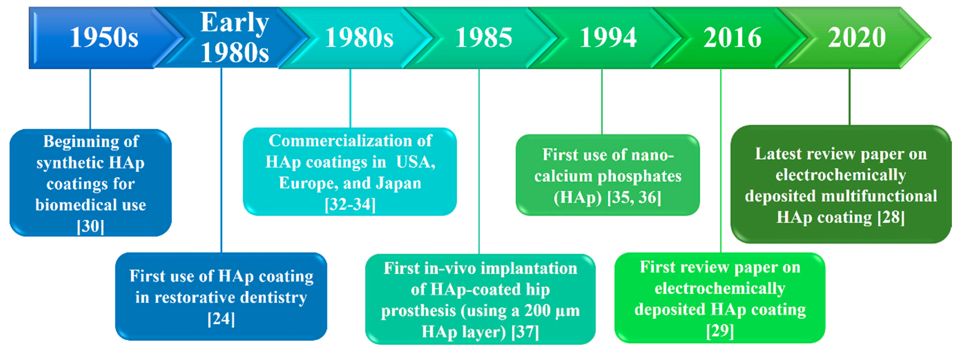
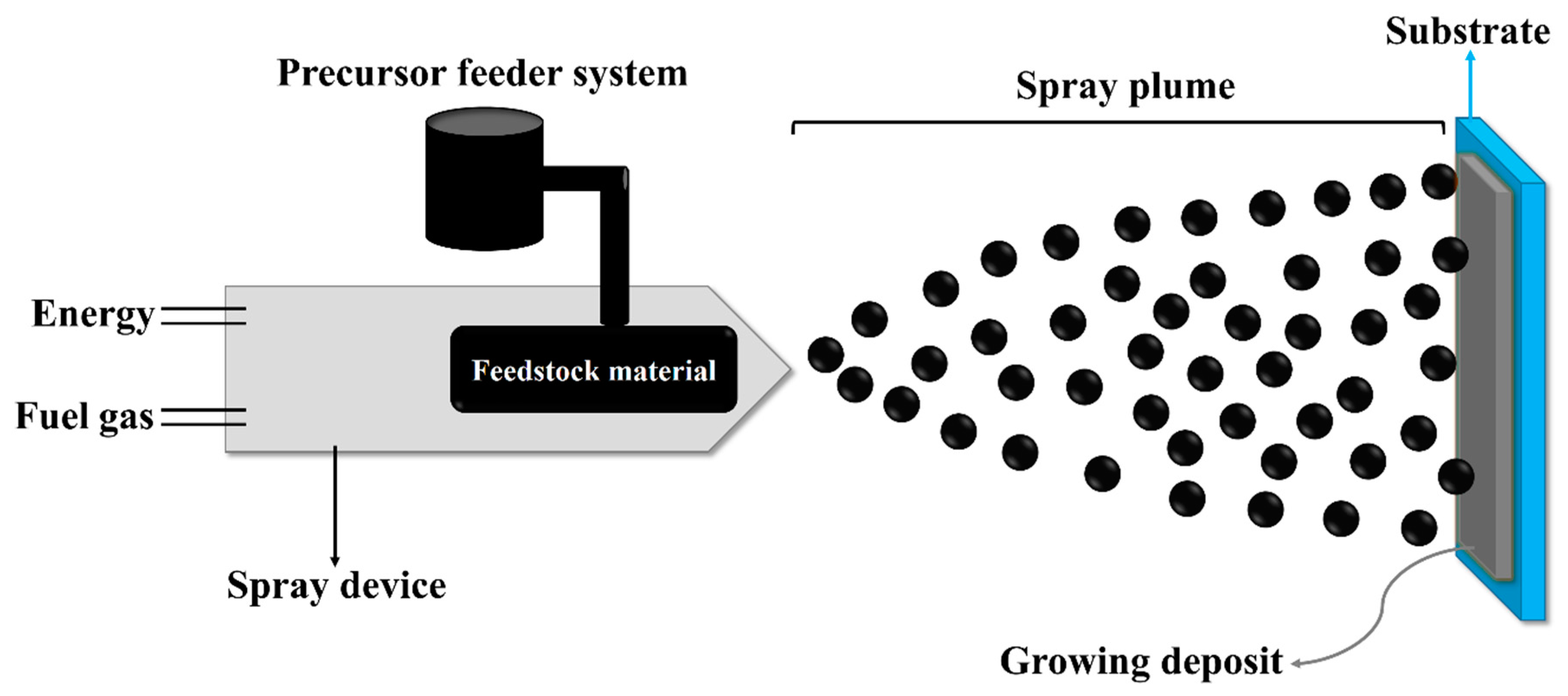
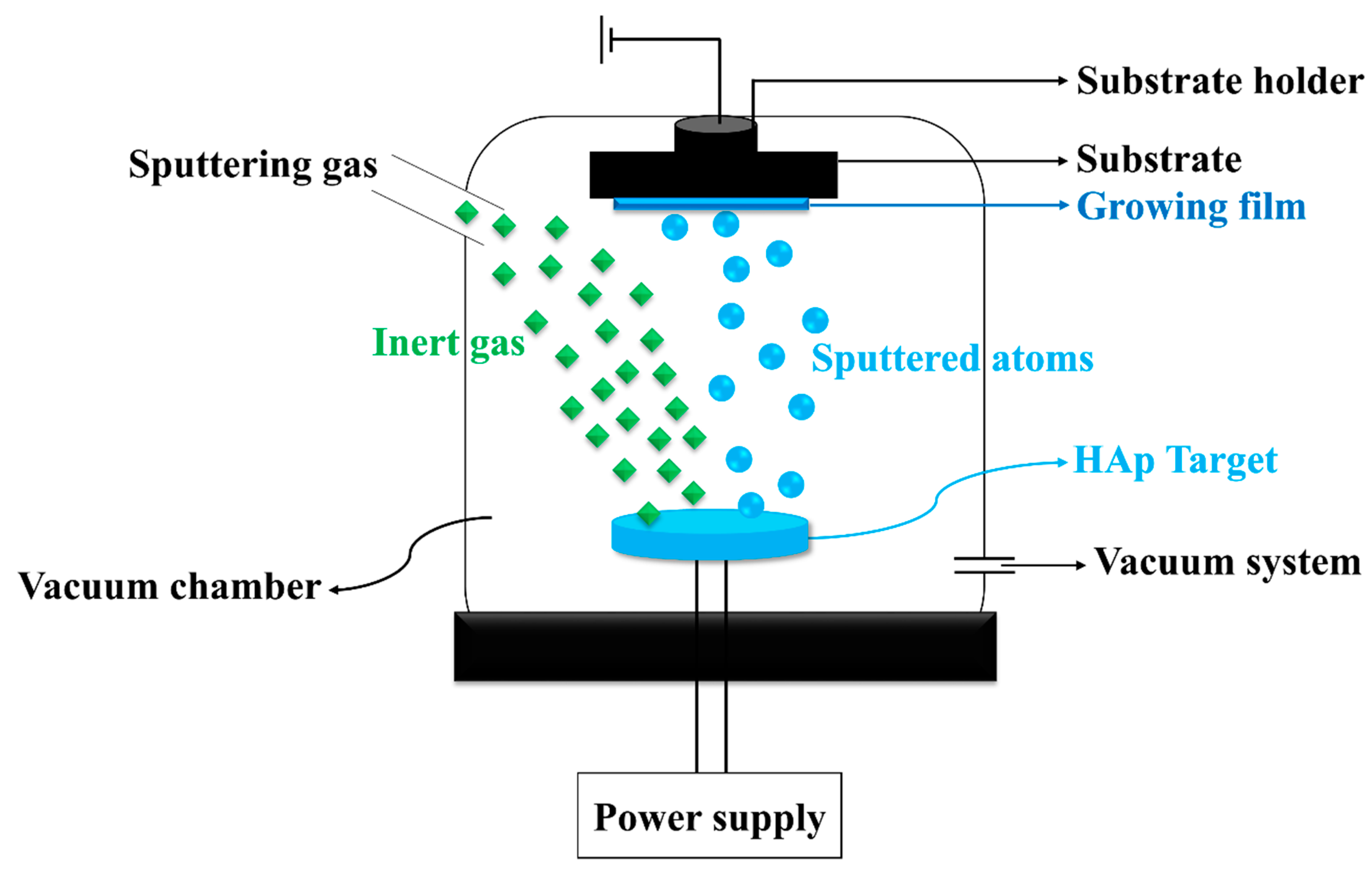
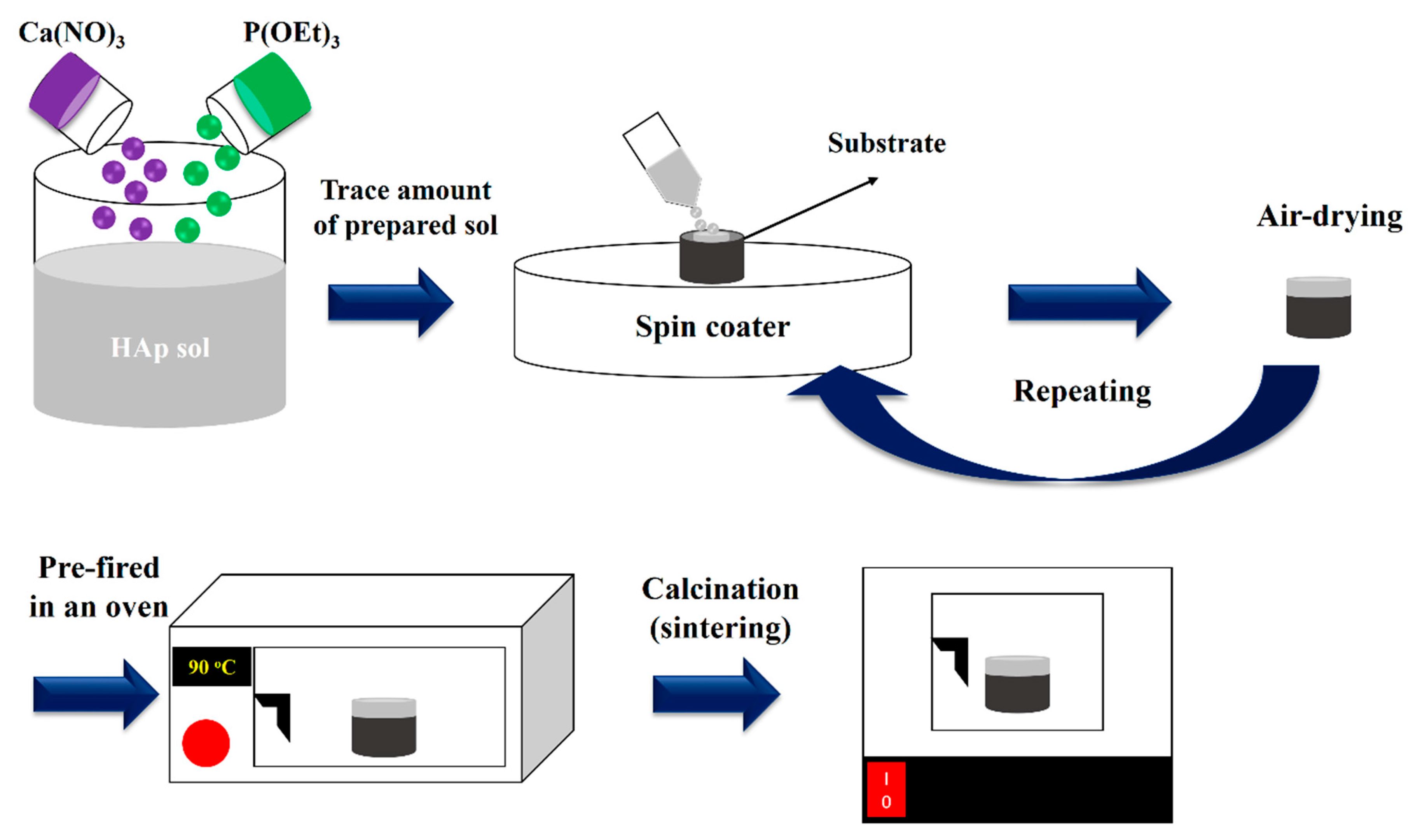
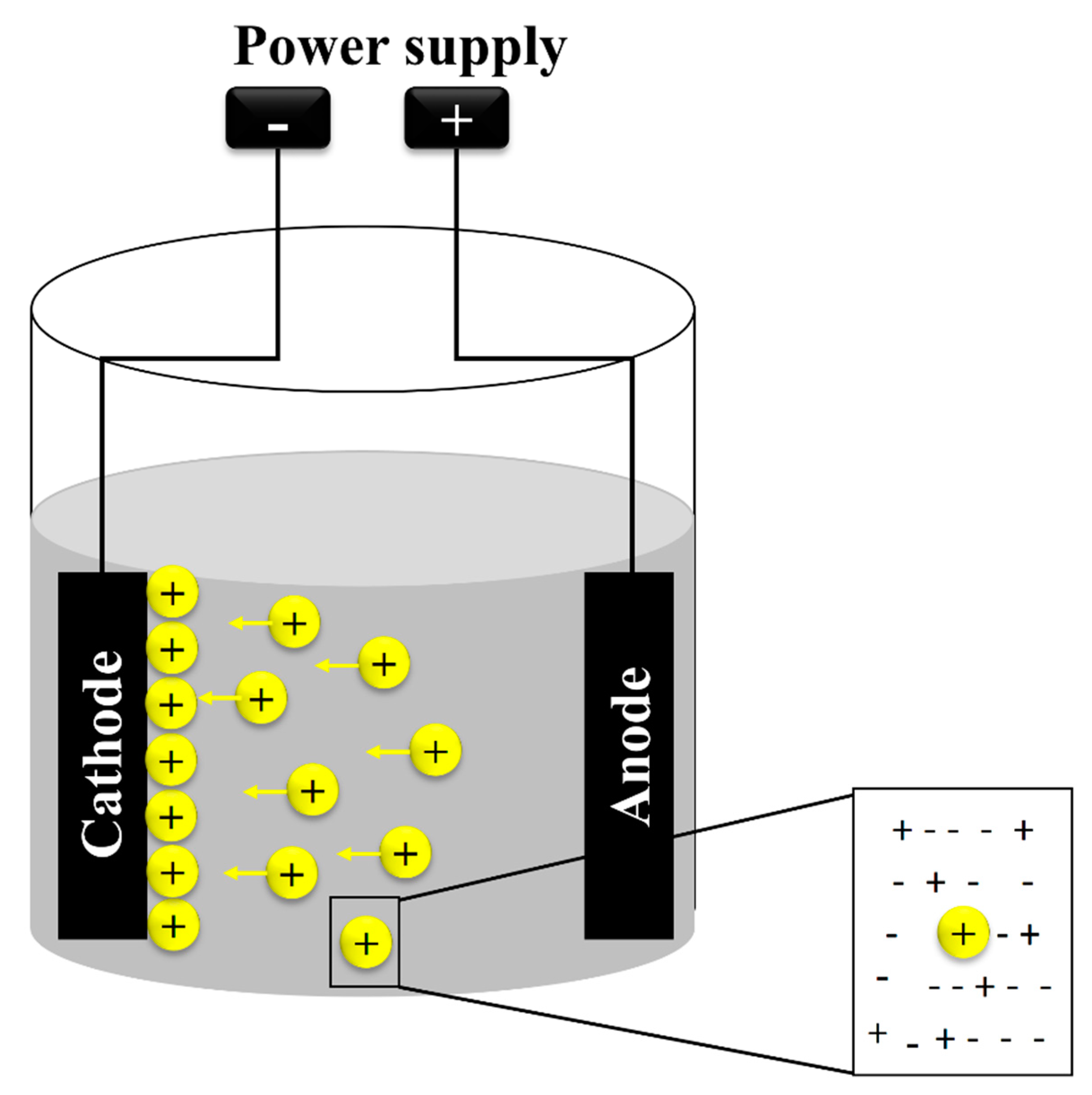
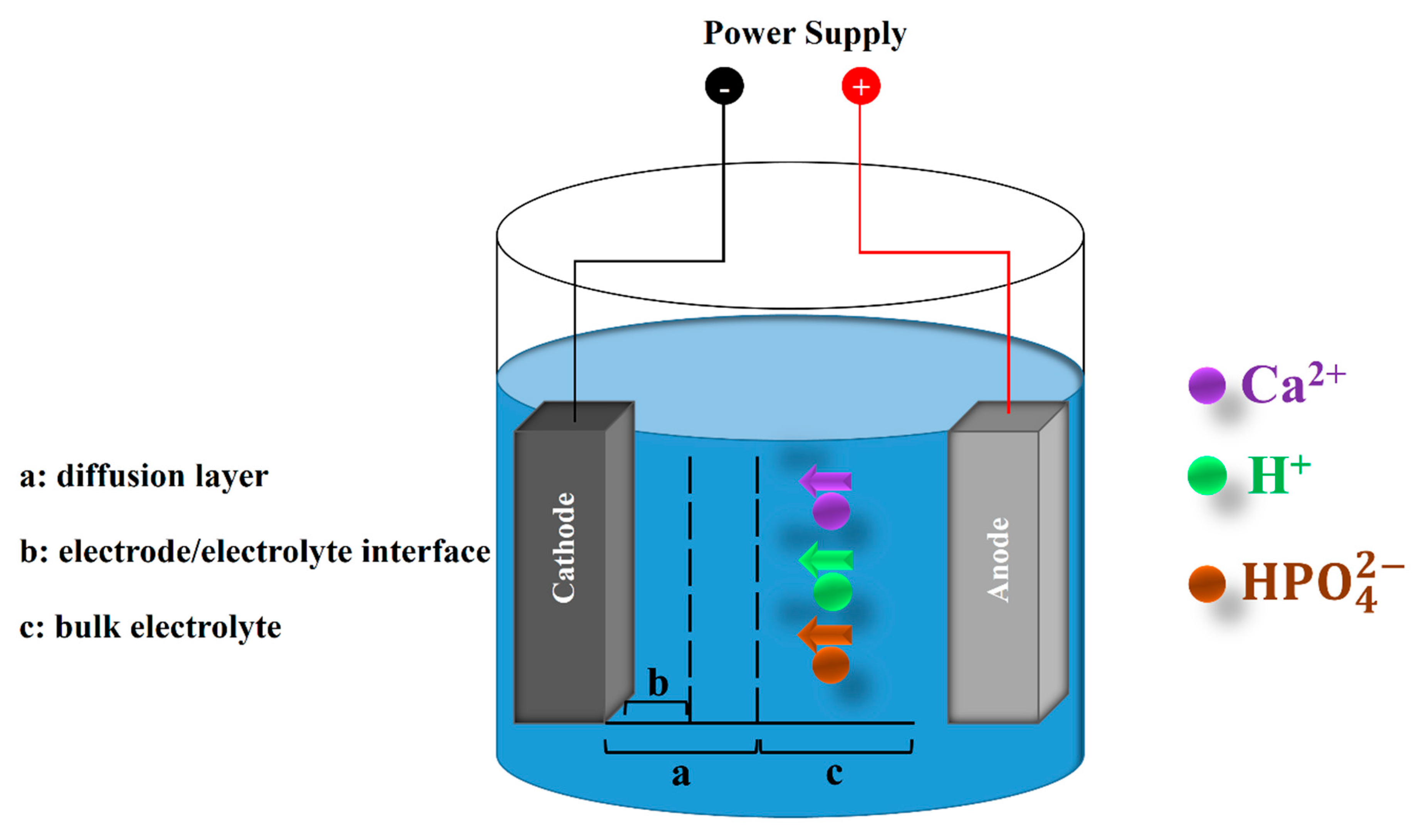
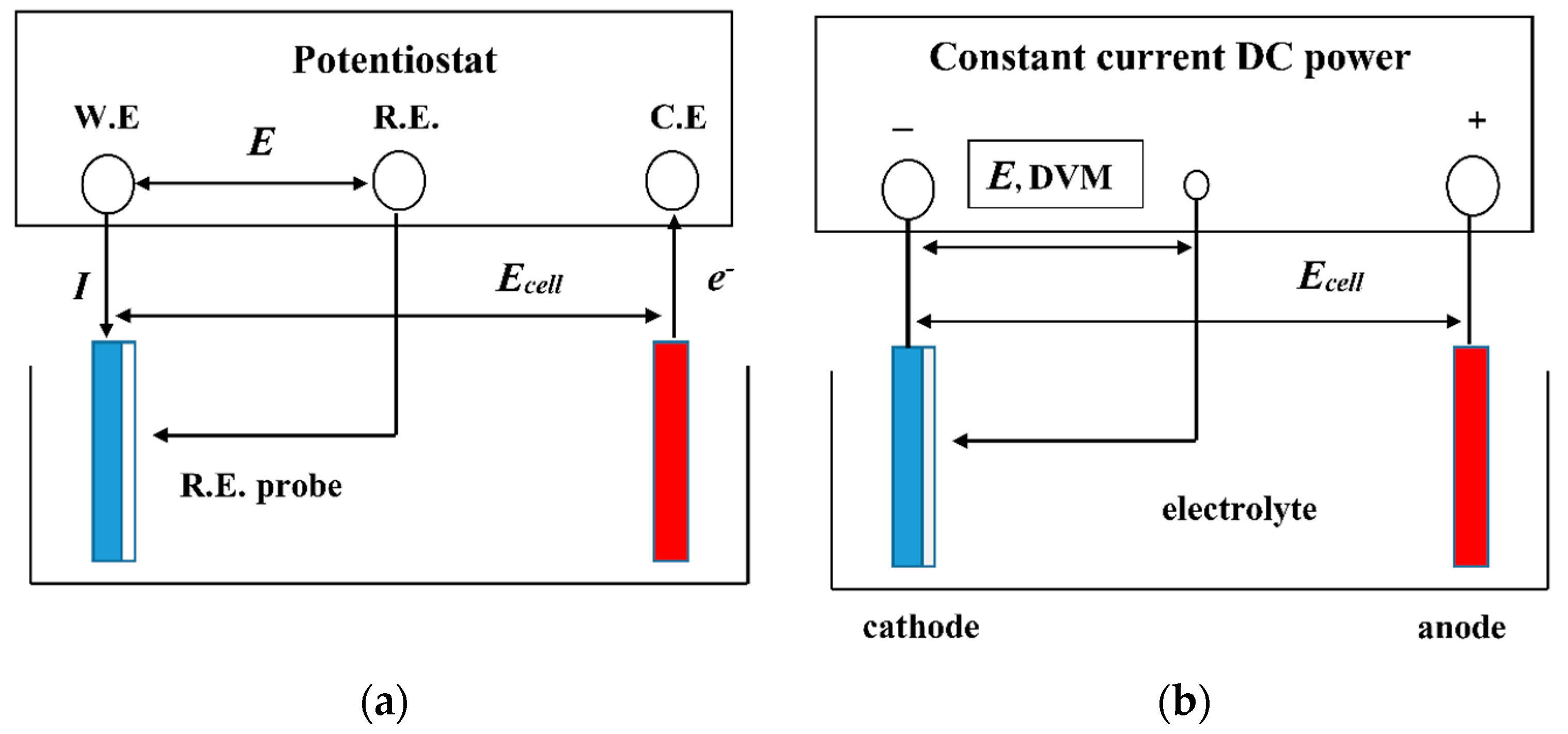
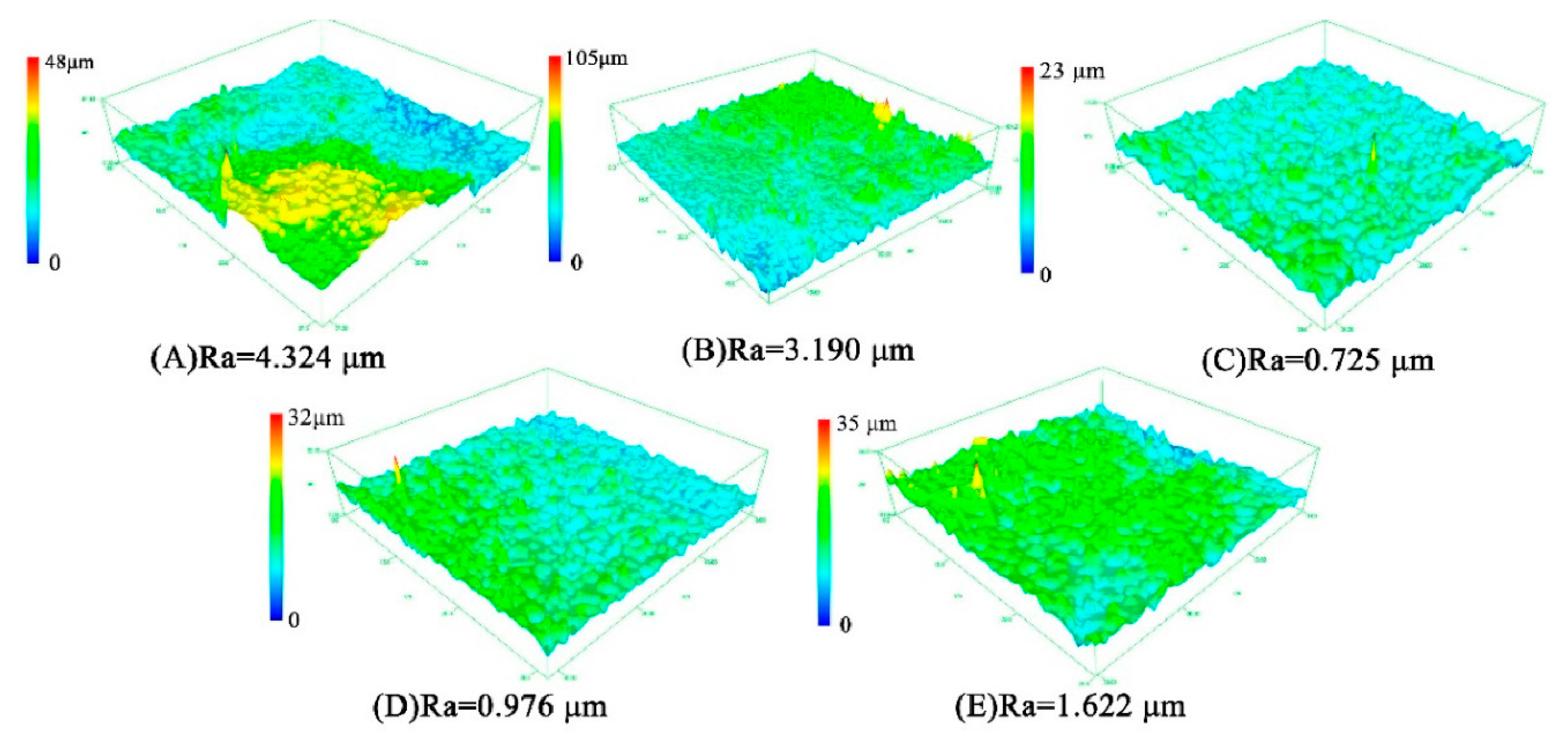
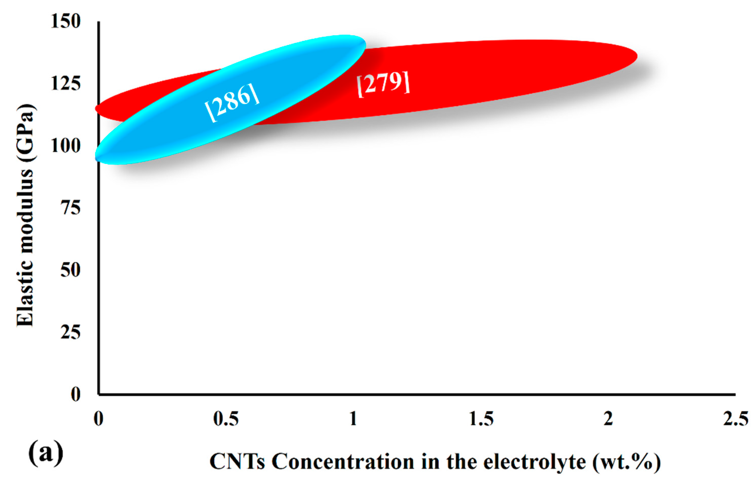
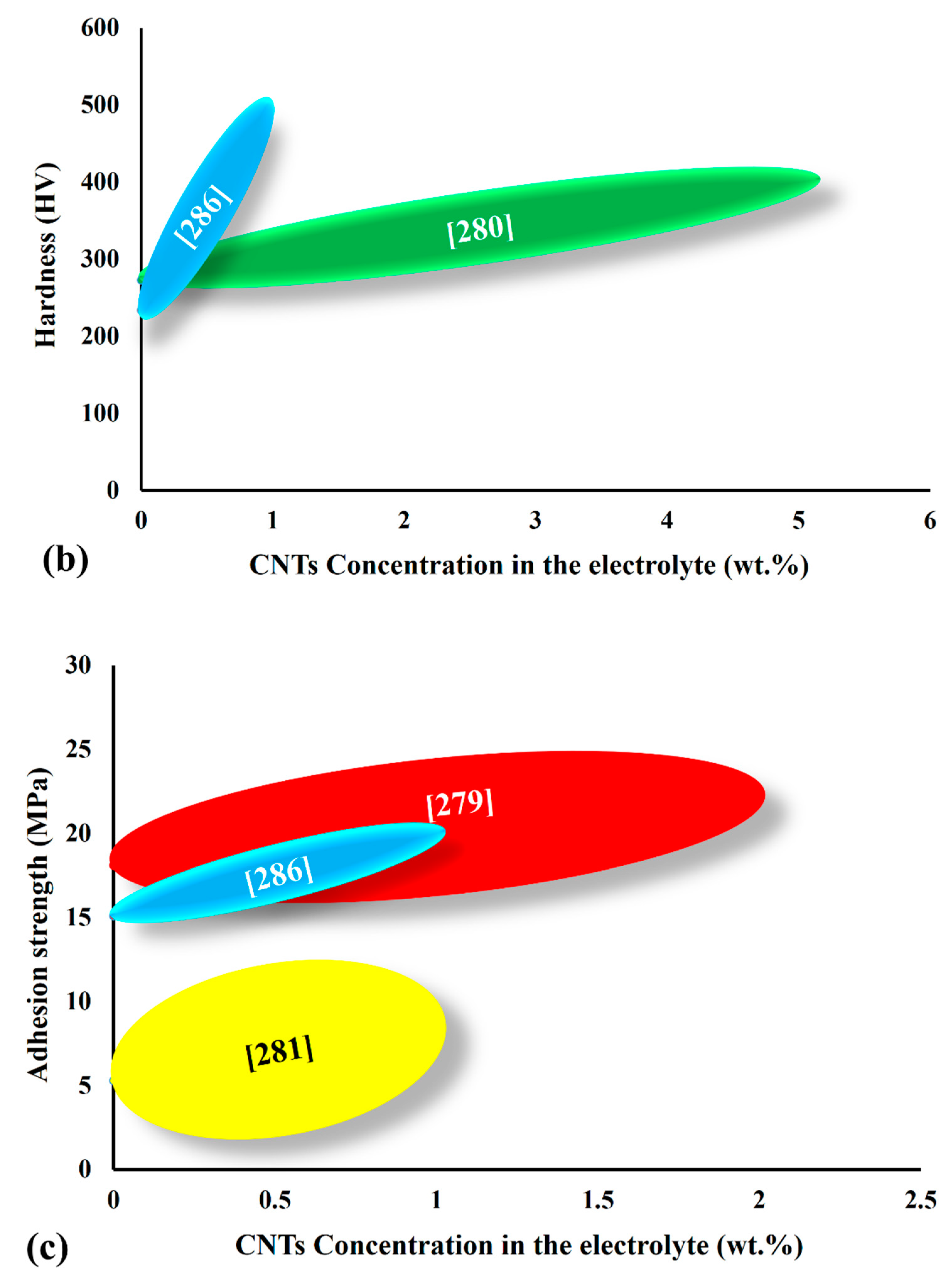
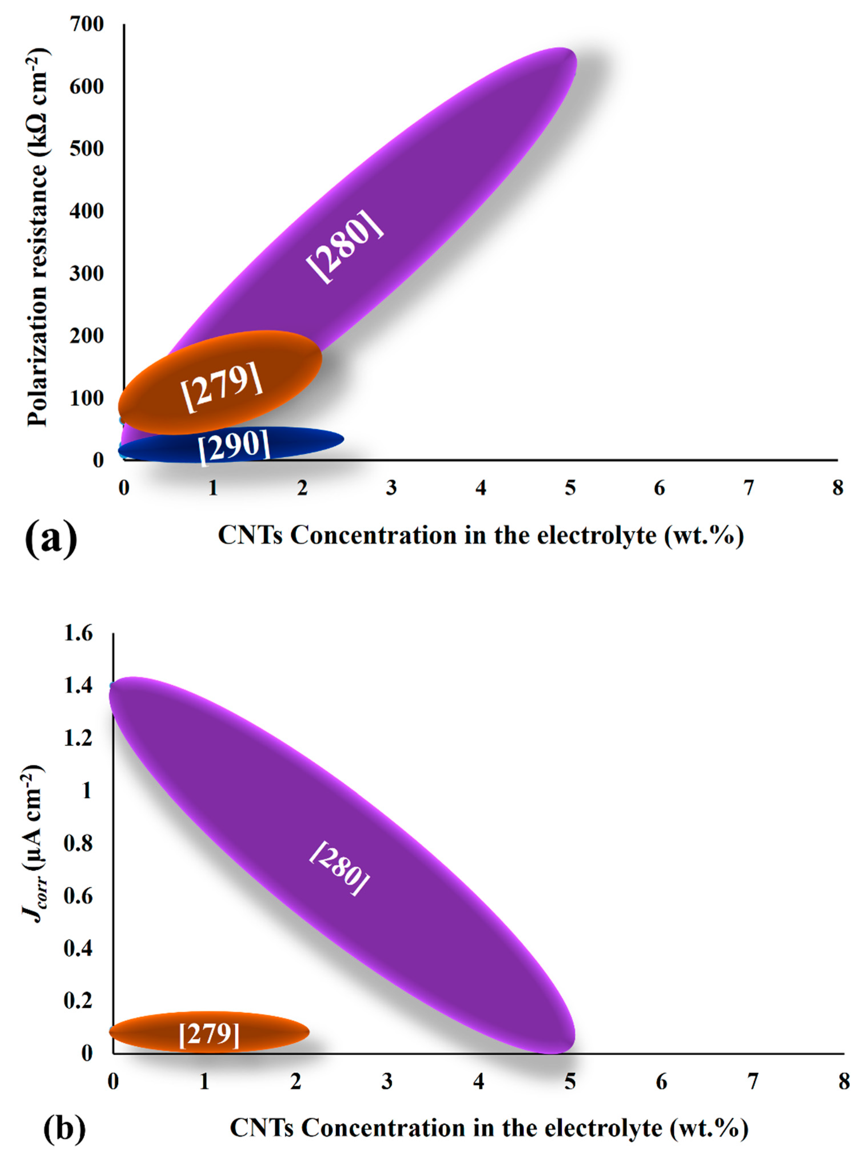
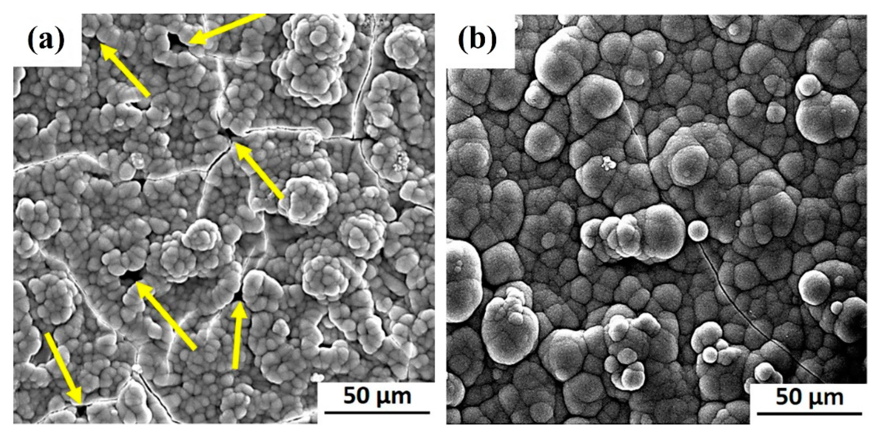
Figure 1. A timeline for the development of electrochemically deposited hydroxyap-atite (HAp) coatings.
Coatings 11 00110 g001
Figure 2. The schematic illustration of thermal spraying.
Coatings 11 00110 g002
Figure 3. A schematic illustration of physical vapor deposition (PVD) process for deposition of HAp coatings.
Coatings 11 00110 g003
Figure 4. Schematic illustration of the spin-coating process for fabrication of HAp coating.
Coatings 11 00110 g004
Figure 5. A schematic illustration of the conventional set-up for electrophoretic deposition (EPD) of HAp coatings.
Coatings 11 00110 g005
Figure 6. Schematic illustration of the proposed model for movement and deposition of the ions during the electrodeposition process.
Coatings 11 00110 g006
Figure 7. Electrical control of, and connections to, an electrodeposition cell: (a) Potentiostatic control, where the cathode is connected as the working electrode of a potentiostat, which controls the working electrode potential with respect to a reference electrode and monitors this potential, E and the current, I flowing through the WE. It is useful to monitor the cell voltage, Ecell using a digital voltmeter, DVM. (b) galvanostatic control of the current, I, where the cathode and anode are directly connected to the negative and positive terminals, respectively, of a DC power supply. It is useful to incorporate a reference electrode in the cell to facilitate measurement of the potential, E between the WE and RE, using a high impedance DVM.
Coatings 11 00110 g007
Figure 8. 3-D surface topography images of the HAp layers electrodeposited using DC at various times: (A) 60, (B) 75, (C) 90, (D) 105, and (E) 120 min [113].
Coatings 11 00110 g008
Figure 9. The top-view SEM images of HAp electrodeposited over a range of ultrasonic power: (a) 0 W, (b) 20 W, (c) 60 W, and (d) 100 W. Reprinted with permission from [175]. Copyright 2017 Elsevier.
Coatings 11 00110 g009
Figure 10. (a) The elastic modulus, (b) hardness, and (c) adhesion strength of the HAp-carbon nanotubes (CNTs) composite coatings as a function of CNTs loading in the electrolyte.
Coatings 11 00110 g010aCoatings 11 00110 g010b
Figure 11. (a) Rp and (b) jcorr of HAp-CNTs composite coatings as a function of CNTs loadings in the electrolyte.
Coatings 11 00110 g011
Figure 12. The surface morphology of formed apatite layer over the surface of (a) HAp and (b) HAp-100 µg mL−1 graphene oxide (GO) coatings during the immersion in simulated body fluid (SBF). Yellow arrows highlight the discontinuity throughout the surface of the apatite layer formed over the pure HAp. Reprinted with permission from [264]. Copyright 2018 Elsevier.
Coatings 11 00110 g012
Table 1. Some examples of implants based on their constituent material.
Implant MaterialExamplesRef.
MetallicTi and its alloys such as Ti6Al4V, NiTi, Co-Cr alloy, stainless steel and Mg[2]
CeramicCalcium phosphates, zirconia, silicon, and alumina[3]
PolymerPolyester, polyethylene terephthalate (PET), polytetrafluoroethylene (PTFE)
and polyurethane (PU)
[1]
CompositeCalcium phosphates/collagen and carbon-fiber/polyetheretherketone (PEEK)[4]
NaturalBone, tissue, and skin[6]
Table 2. The advantages and limitations of the techniques used for fabrication of HAp biocoatings [95,96,97,98,99].
Deposition TechniqueThicknessAdvantagesLimitations
Thermal spraying (plasma spraying)30–200 µm for thermal spraying and less than 20 µm for plasma sprayingHigh deposition rate, low cost, improved corrosion resistance, rapid bone healing, and low risk of degradation of coating.Relatively poor adhesion to the substrate, formation of amorphous structure, coarse grains, lack of uniformity, inability to fabricate composite coating and crack formation.
PVD (sputter coating)0.5–3.0 µmHigh adhesion, dense coating, and uniformity in thickness.Expensive, line of sight technique, time consuming, low deposition rate, amorphous coatings produced.
Sol-gel0.5–2.0 µm Ability to coat substrates with complex geometries, low temperature, high purity, uniform coating, moderate adhesion, and excellent corrosion resistance.Requires precise control of reaction environment, pilot-scale production; high cost of precursors, porous structure, poor tribological properties and a need for post-treatments.
EPD0.1–2.0 mmUniform thickness, fast deposition, simple procedure, low cost, ability to coat substrates with complex geometries, and possibility for the incorporation of reinforcing agents.Requires post-sintering; deposit has a crack-containing microstructure.
ED0.05–0.5 mmLow temperature, uniform coatings, rapid coating process, and possibility to incorporate reinforcing agents.Poor adhesion, difficulty in controlling electrolyte parameters, and residual stress in deposits.
Table 3. Applied current density/potential, current/potential type, and deposition mode during the electrodeposition of pure HAp coatings.
Applied Current Density/PotentialCurrent/Potential TypeOptimum Value in Terms of Surface PropertiesDeposition ModeRef.
5–20 mA cm−2Pulsed current 10 mA cm−2 Galvanostatic[11]
−1.4 to −2.2 VConstant potential−1.8 VPotentiostatic[123]
0.2–15 mA cm−2Direct current 10 mA cm−2 Galvanostatic[142]
−1200 to −1600 mVDirect voltage−1400 mVPotentiostatic[143]
0.5–3 mA cm−2Pulsed current 0.5 mA cm−2 Galvanostatic[110]
−1.3 to −1.7 VPulsed potential−1.5 VPotentiostatic[144]
20–40 mA cm−2Pulsed current40 mA cm−2 Galvanostatic[132]
1.5–5 mA cm−2 Pulsed current5 mA cm−2Galvanostatic[122]
1.5–15 mA cm−2Pulsed currentNot reportedGalvanostatic [145]
Table 4. Electrolyte composition and role of incorporated during electrodeposition of HAp coatings.
Electrolyte CompositionConcentration (M)ObjectiveRef.
CaCl2·2H2O0.15Calcium precursor [11]
NH4H2PO40.1Phosphate precursor
NaCl1Improve ion conductivity of the electrolyte
CaCl20.167Calcium precursor[151]
NH4H2PO40.1Phosphate precursor
NaCl 0.1Increase of electrolyte conductivity
Ca(NO3)20.042Calcium precursor[10]
(NH4)2HPO40.025Phosphate precursor
NaNO30.1Improvement of ionic strength
H2O22000 (ppm)H2 evolution suppression
Ca(NO3)20.042Calcium precursor[127]
(NH4)2HPO40.025Phosphate precursor
NaNO30.1Improvement of ionic strength
Ca(NO3)2·4H2O0.042Calcium precursor[101,102,140,152]
(NH4)2HPO40.025Phosphate precursor
Ca(NO3)2·4H2O0.0006Calcium precursor[123]
(NH4)2HPO40.00036Phosphate precursor
NaNO30.1Improve conductivity of the electrolyte
Ca(NO3)20.1Calcium precursor[129]
(NH4)3HPO40.06Phosphate precursor
H2O210 mL/LSuppress H2 evolution
Ca(NO4)20.1Calcium precursor[142]
NH4H2PO40.05Phosphate precursor
Ca(NO3)2·4H2O0.00175Calcium precursor[153]
(NH4)2HPO40.00105Phosphate precursor
Ca(NO3)2·4H2O0.042Calcium precursor[100,105]
(NH4)2HPO40.025Phosphate precursor
NaNO30.1Improvement of ionic strength
CaCl20.040Calcium precursor[154]
KH2PO40.024Phosphate precursor
H2O20–4.0 mL/LH2 evolution suppression
Ca(NO3)2·4H2O0.42Calcium precursor[143]
KH2PO40.25Phosphate precursor
Ca(NO3)2·4H2O0.042Calcium precursor[107]
NH4H2PO40.025Phosphate precursor
H2O22000–3000 ppmFavoring HAp formation
NaOH2000–4000 ppmFavoring HAp formation
Ca(NO3)20.1Calcium precursor[130,141]
NH4H2PO40.06Phosphate precursor
H2O210 mL/LFavoring HAp formation
Ca(NO3)2·4H2O0.015Calcium precursor[144]
NH4H2PO40.009Phosphate precursor
NaNO30.003Improve electrolyte conductivity
Ca(NO3)20.042Calcium precursor[132,155]
NH4H2PO40.025Phosphate precursor
NaNO30.1Improvement of ionic strength
CaCl2·2H2O0.5Calcium precursor [156]
(NH4)2HPO40.3Phosphate precursor
Ca(NO3)2·4H2O0.0084Calcium precursor [157]
NH4H2PO40.005Phosphate precursor
NaNO30.1Improvement of ionic strength
H2O20.0588H2 evolution suppression
Ca(NO3)2·4H2O0.042Calcium precursor [158]
NH4H2PO40.025Phosphate precursor
Table 5. The reported electrodeposition time range and the optimum time for obtaining favorable physicochemical features in the HAp coatings.
Electrodeposition Time Range (min)Optimum Time in Terms of Physicochemical Features (min)Ref.
5–30 30[127]
60–180180[159]
15–6060[107]
15–180180[169]
30–180180[135]
60–120120[113]
Table 6. Pre-treatments and claimed achievement(s).
Type of Pre-TreatmentSubstrateApplying ProtocolOutstanding Achievement(s)Ref.
AnodizingTi6Al4VAnodizing is carried out in a solution containing 1 M (NH4)2SO4 + 0.5 wt.% NH4F at room temperature under potential ramp from OCP to 25 V for 2700 s.Improved bonding strength[10]
Anodizing followed by alkali solutionTi6Al4VAnodizing is carried out in a solution containing 1 M H2SO4 at constant cell voltage of 100 V for 2 min. The anodized samples soaked in a NaOH solution.No comparison of pre-treatment processes was made.[182]
Etching TiSubstrates are etched in a solution containing 25, 50, 75, and 97% H2SO4 at 60 °C for 30 min.HAp deposited on etched substrates with 50% and 75% concentrations yields superior adhesion strength to the substrate.[183]
Anodizing followed by soaking in a alkali solutionTi6Al4VAnodizing is carried out in a solution containing H2SO4 under constant anodic voltage of 100 V for 2 min. The anodized substrates are immersed in a 5 M NaOH solution at 60 °C for 10 min.Enhanced tribo-electrochemical performance.[192]
Borate passivation Stainless steel (316 L)Passivation is carried out potensiostatically in a 0.4 M borate buffer solution (pH 9.3) at 640 mV vs. SCE for 1–3 h.Enhanced corrosion protection performance.[143]
HELCDEB irradiation Ti6Al4VHELCDEB irradiation is performed using high energy beam by 700 keV DC accelerator which generates a beam current of 0.5–6.0 mA. Improved adhesion strength along with corrosion behavior.[156]
Anodizing followed by post-annealing TiAnodizing is carried out in a solution containing NH4F with/without modifiers such as 10% (NH4F in H2O): 90% glycerol or polyethylene glycerol under two various potential control, e.g., pulsed potential (20/−4 V) and constant potential of 20 V for 30–150 min. The anodized layer is post-annealed at 450 °C for 30 min.Increased adhesion strength. Anodizing for 90 min yields the highest strength.[191]
NaOH or H2O2 treatment Commercially pure TiNaOH treatment includes immersing the substrates into a 5 M NaOH solution at 60 °C for 24 h, followed by annealing at 600 °C for 1 h.
H2O2 treatment carried out by soaking the substrates into a 5 M H2O2 solution at 60 °C for 24 h.
Enhanced biocompatibility for NaOH-treated samples with HAp top layer. [168]
GrindingCommercially pure Ti Grinding to #1000The highest adhesion strength is obtained for “grinding + etching + grit blast + alkali” treatment. The superior biological performance of pre-treated samples compared to as-deposited ones. [187]
Grinding + etching Grinding to #1000, followed by etching in HNO3/HF
Grinding + etching + grit blast Grinding to #1000, followed by etching in HNO3/HF, then grit blast
Grinding + etching + grit blast + alkali treatment Grinding to #1000, followed by etching in HNO3/HF, then grit blast, finally soaking in NaOH.
Grinding + etching + grit blast + H2O2 treatmentGrinding to #1000, followed by etching in HNO3/HF, then grit blast, finally soaking in H2O2.
Mechanical polishing NiTiMechanical polishing of the substrates by #80-600 papers. Improved corrosion resistance for HAp coatings electrodeposited on mechanically polished substrate.[184]
Mechanical polishing + Etching + post treatmentSoaking the polished substrates in to a solution containing HF:HNO3:H2O with ratio of 1:4:5 for 4 min, followed by immersion in boiling water for 20 min.
Heat treatment NiTi The substrates are heat-treated at 470 °C for 30 min.Increased corrosion protection as well as less Ni ion release.[122]
Heat treatmentTi6Al4V The substrates are heat-treated at 400–1000 °C for 60 min.Enhanced surface roughness for substrates heat-treated at 800 and 1000 °C.[199]
Alkali treatmentCommercially pure Ti Soaking the substrates in a 5 M NaOH solution at 60 °C for 5 min.Enhanced bonding strength to the substrate. [188]
Table 7. The various types of post-treatments, process conditions and major objectives/outcomes.
Type of Post-TreatmentConditionsObjective(s)/Outcome(s)Ref.
Alkali treatmentImmersion in 1M NaOH solution for 1 h.Conversion of other calcium phosphate (CaP) phases to HAp.[108]
Alkali treatmentImmersion in 1M NaOH solution at 80 °C for 2 h. Conversion of dihydrogen phosphate or brushite (DCPD) phase to HAp. A uniform surface morphology is obtained.[129]
Alkali treatmentImmersion in 1 M NaOH solution at 80 °C for 2 h. Conversion of DCPD phase to HAp. [142]
Alkali treatmentImmersion in 1 M NaOH solution at 80 °C for 1 h. Conversion of DCPD phase to HAp. [135]
Alkali treatmentImmersion in 0.25 M NaOH solution at 80 °C for 4 h.Conversion of DCPD phase to HAp. Higher bioactivity and biocompatibility are attained. [105]
Alkali treatmentImmersion in 0.1 M NaOH solution at 25 °C for 72 h.Conversion of DCPD phase to HAp. A slight increment in nano-hardness and elastic modulus is reported. [107]
Alkali treatmentImmersion in 1M NaOH solution at 80 °C for 2 h. Conversion of DCPD and tricalcium phosphate (TCP) phases to HAp. [130]
Alkali treatmentImmersion in 0.25 M NaOH solution at 80 °C for 4 h. Conversion of DCPD and OCP phases to HAp. Evolution of needle-like morphology which is beneficial for bone growth. A crucial improvement in corrosion behavior is obtained after alkali treatment. [132]
Alkali treatmentImmersion in 0.25 M NaOH solution at 80 °C for 4 h. Conversion of DCPD phase to HAp. Evolution of needle-like morphology which is beneficial for bone growth. A crucial improvement in corrosion behavior and in-vitro bioactivity is achieved after alkali treatment.[155]
Alkali treatmentImmersion in 1 M NaOH solution at 80 °C for 2 h. Conversion of DCPD phase to HAp. A significant increase in corrosion performance of coatings is attained after alkali treatment.[25]
Alkali treatmentImmersion in 1 M NaOH solution at 80 °C for 2 h. Conversion of DCPD phase to HAp.[200]
Alkali treatmentImmersion in 0.1 M NaOH solution at 95–100 °C for 24 h.Conversion of DCPD phase to HAp.[155]
Immersion in 0.1 M NaOH solution at 25 °C for 72 h.
Heat treatmentHeating the as-deposited coatings according to the following condition: Heating at 120 °C for 1 h to water evaporation, then heating at 400–1000 °C for 1 h.A remarkable promotion in crystallinity of the as-deposited coatings.[199]
Heat treatmentHeating the as-deposited coatings at 600 °C for 60 min.The coatings were dehydrogenized after heat treatment. [183]
Heat treatmentAnnealing the as-deposited coatings at 550 °C for 2 h.Enhancement in stability and bonding strength between HAp/substrate. [101]
Electron-beam treatment Irradiating the electrons under vacuum level of 10−7 Torr with excitation energy ranging from several tens of eV to hundreds of eV at a current density of 10–100 nA cm−2.A noticeable decrement in living cells number with electron-beam treatment is addressed.[168]
Low energy electron irradiation Irradiating the electrons under vacuum level of 10−7 Torr with excitation energy ranging from several tens of eV to 100 eV at an incident charge of 0–300 µC cm−2.A suitable substrate for cell attachment is obtained for post-treated surface to θ = 30°.[187]
Hydrothermal, followed by annealingHydrothermally treating the as-deposited coating in autoclave at 200 °C for 3 h, followed by sintering at 700 °C for 2 h. The effect of post treatments is not clearly mentioned[185]
Table 8. The potential minerals that can be doped into HAp as well as their precursors and benefits.
Type of Substituted MineralPrecursor(s) in ElectrolyteBiological Features and Benefits for Synthetic HApRef.
SrSrCl2·6H2O or Sr(NO3)2·6H2OSr is an essential trace element in human body that stimulates bone formation, growth, and healing since it can satisfactorily prevent the osteoclast activity and resorbing as well as osteoclastic differentiation. Sr stimulates the proliferation and differentiation of osteoblasts. It also inhibits inflammation and osteoporosis, promoting bone remolding. There would be a lower risk of failure in postmenopausal osteoporotic patients if a controlled dose of Sr incorporates. When incorporating into HAp structure, Sr2+ substitutes for Ca2+ yielding the superior bioactivity and biocompatibility.[188,203,204,205,211,214,215,216,217,218]
MgMgCl2·6H2O or Mg(NO3)2·6H2OAs one of the most abundant elements in human body (0.72 and 1.23 wt.% Mg exits in bone and dentin, respectively), Mg plays a vital role in skeletal metabolism and stimulates the bone growth through interacting with osteoblast integrin that serves as a cell-adhesion receptor. [203,204,205,212,219,220]
ZnZnCl2 or Zn(NO3)2There is a trace amount of Zn (0.012–0.025 wt.%) in the human bone. Apart from its antibacterial and antimicrobial effect, Zn positively contributes to cell division, bone formation and mineralization, bonding strength of the implanted material and enzymes, together with hormone adjustment.[186,203,205,221,222,223,224]
MnMn(NO3)2Mn2+ ions stimulate the interactions between Mn-substituted HAp-coated implant/host tissues since these ions can enhance ligand-binding affinity of integrins. The integrins serve as cell adhesion receptor, which bind to extracellular, soluble, and cell-surface ligands, therefore they can affect pathological processes. Mn is a primary trace element required for bone development. Its close radius to that of Ca provides a facilitated platform for entering Mn to osteoblast via Ca2+ channel. Mn2+ determines the bone metabolism due to its capability for controlling bone resorption and osteoblast differentiation.[188,189,225,226,227,228]
AgAgNO3The incorporated Ag+ can be substituted with Ca2+ ions in the HAp structure. Such a substitution yields a significant increase in the antibacterial performance of HAp that inhibits post-implantation infections. For antibacterial applications, Ag has numerous advantages over other antibacterial agents, such as Cu and Zn since it offers stronger performance against a variety of bacteria including gram-positive (Bacillus subtilis and Staphylococcus aureus) and gram-negative (Escherichia coli and Pseudomonas aeruginosa).[207,229,230,231]
SiNa2SiO3·9H2OSilicon is a primary element involved in the early stages of soft tissue development and bone mineralization. The Si-substituted HAp exhibits higher biocompatibility including cell activity, attachment, and growth. [206,232]
FNaFF is a an important trace element in human body, where about 90% of existing F in body locates in hard tissues such as bones and dental enamel. F-substituted HAp offers lower solubility, improved crystallization, cell attachment, mineralization, apatite formation, alkaline phosphate (ALP) activity, protein adsorption, and bonding strength.[208,209,211,213,233,234,235]
CuCu(NO3)2Cu is of fundamental trace elements for mammals as it has a key role in cross-linking of elastin-collagen in bones and stimulating the enzymes. Cu2+ ions are best known for their strong antibacterial performance, which cause low cytotoxicity if its content is kept below a defined threshold. The incorporation of secondary bioactive dopant is essential to prevent adverse effects.[186,214,236,237]
CeCe(NO)3·6H2OThe embedded Ce3+ ions may promote antimicrobial efficiency of HAp, however they can cause cytotoxicity. It is better to dope this ion with a secondary substituent mineral.[210,238]
EuEu(NO3)3·5H2OEu3+ ion can mimic the Ca2+ and determine the bone remolding cycle.[210,239]
Table 9. The reported mechanical properties of mineral-substituted HAp coatings as a function of type, concentration of doped minerals and operational factors.
Coating CompositionMineral(s) Concentration in Electrolyte (M)Studied VariableCharacterization Method (Test Condition)Highest Reported Adhesion Strength (MPa)% Increase Rate Compared to Pure HApRef.
Mn-doped HAp3 × 10−4 Mn(NO3)2 Incorporation of mineralPull-out (crosshead speed of 0.001 cm min−1)14 ± 4107.5[189]
(Sr,Mn)-co-doped HAp1 × 10−2 Sr(NO3)2 Incorporation of minerals Pull-out (crosshead speed of 1 mm min−1)15 ± 519.8[188]
1 × 10⁻² Mn(NO3)2
(Sr,Cu)-co-doped HAp2.1 × 10−3 Sr(NO3)2 Incorporation of minerals Pull-out (crosshead speed of 1 mm min−1) 15 ± 623.4[214]
2.1 × 10⁻³ Cu(NO3)2
(Zn,Cu)-co-doped HAp2 × 10−3 Zn(NO3)2·6H2OIncorporation of minerals Pull-out (crosshead speed of 1 mm min−1) 9 ± 316.1[186]
2 × 10⁻³ Cu(NO3)2·3H2O
Ag-doped HAp on anodized Ti1 × 10−4 AgNO3Incorporation of mineralPull-out (crosshead speed of 1 mm min−1) 15.9 ± 0.6−7.0[207]
F-doped HAp0.001–0.016 NaFIncorporation of mineral Pull-out (crosshead speed of 10 mm min−1)21.543.3[208]
(Sr,Mn,Zn)-co-doped HAp0.042 SrCl2·6H2ODuty cyclePull-out18.3 ± 0.7-[203]
0.042 MgCl2·6H2O
0.042 ZnCl2
(Sr,Mn,Zn)-co-doped HAp0.042 SrCl2·6H2OHELCDEB pre-treatmentPull-out22.1 ± 1.18.3 (compared to the same coating deposited on an untreated substrate)[205]
0.042 MgCl2·6H2O
0.042 ZnCl2
Table 10. The corrosion parameters of electrodeposited mineral(s)-doped HAp coatings. Ecorr, jcorr, and Rp are corrosion potential, corrosion current density, and polarization resistance, respectively. These parameters are obtained from potentiostatic polarization assay. Rcoat is the coating area resistance obtained by fitting the EIS data via an appropriate equivalent electrical circuit (EEC). The polarization resistance, Rp is stated in some papers.
Coating TypeStudied VariableMediumHighest Ecorr (V)Lowest jcorr (µA cm−2)Highest Rp (Ω)Highest Rcoat (Ω cm2)Ref.
(Ag,Zn,Mg,Sr)-co-doped HApIncorporation of minerals SBF, pH = 7.40 at 37 °C---85[204]
Mn-doped HApIncorporation of mineral SBF, at 37 ± 0.5 °C−0.5693.19--[189]
F-doped HApIncorporation of mineralSBF, pH = 7.40 at 37 °C−1.290.283--[209]
(Sr,Mn)-co-doped HApIncorporation of minerals SBF, at 36.5 ± 0.5 °C −0.8654.02--[188]
(Sr,Cu)-co-doped HApIncorporation of minerals SBF, pH = 7.40 at 37 °C−0.8572.45--[214]
(Zn,Cu)-co-doped HApIncorporation of minerals SBF, at 37 ± 0.5 °C−0.4431.66--[186]
(Ce,Eu)-co-doped HApIncorporation of minerals Ringer’s solution, pH = 7.40 at 37 ± 1 °C−0.558--2349[210]
(Sr,F)-co-doped HApIncorporation of minerals SBF, at 37 ± 0.5 °C−0.8051.66--[211]
Ag-doped HApIncorporation of mineral SBF, at 37 °C−0.265---[207]
Si-doped HAp Incorporation and various concentrations of mineralSBF−1.523-2608[206]
Mg-doped HAp Incorporation and various concentrations of mineralSBF, pH = 7.40 at 37 ± 0.5 °C−0.1100.0537250,000-[212]
(Sr,Mg,Zn)-co-doped HApDuty cycleSBF 0.2250.52-6,460,000[203]
(Sr,Mn,Zn)-co-doped HApHELCDEB pre-treatmentSBF, pH = 7.40 at 37 °C−0.090.08--[205]
F-doped HApType of current controlSBF, pH = 7.40 at 37 °C−1.512.51--[213]
Table 11. The biological characteristics of the electrodeposited mineral(s)-substituted HAp coatings.
Coating CompositionType of Cultured CellAssay Type% Highest Cell Viability Highest Reported Absorbance; Cell Viability/Proliferation (by Measuring Optical Density)Highest Reported ALP Activity (U/g)Apatite Formation AbilityRef.
(Ag,Zn,Mg,Sr)-co-doped HApMG63 osteoblast-like cellsWST-8 assay900.65 ± 0.1--[204]
Mn-doped HApMouse calvarial cells (MC3T3-E1)MTT assay-0.55 ± 0.05-[189]
F-doped HApMouse preosteogenic
cell line MC3T3-E1 cells
CCK-8 assay-1.4 ± 0.5--[209]
(Sr,Mn)-co-doped HApMouse skull cell line MC3T3-E1MTT assay-0.8 ± 0.1160 ± 40-[188]
(Sr,Cu)-co-doped HApMC3T3-E1 osteoblast cellsMTT assay-1.0 ± 0.2130 ± 25[214]
(Zn,Cu)-co-doped HApMouse skull cell line MC3T3-E1MTT assay-0.7 ± 0.5--[186]
(Ce,Eu)-co-doped HAp MG63 osteoblast-like cells MTT assay95---[210]
(Sr,F)-co-doped HAp Mouse calvaria cells (MC3T3-E1) MTT assay -1.0 ± 0.1--[213]
Ag-doped HAp Mouse calvaria cells (MC3T3-E1) MTT assay -0.5 ± 0.1-[207]
Si-doped HApMG63 cells MTT assay 95---[206]
(Sr,Mg,Zn)-co-doped HApMG63 osteoblast-like cellsModified MTT assay95- -[203]
Publisher’s Note: MDPI stays neutral with regard to jurisdictional claims in published maps and institutional affiliations.

