Article

Electrochemical Deposition of Hydroxyapatite on Stainless Steel Coated with Tantalum/Tantalum Nitride Using Simulated Body Fluid as an Electrolytic Medium

1
Departamento de Ingeniería Química, Escuela Politécnica Nacional, Quito 17-01-2759, Ecuador
2
Escuela de Ciencias Químicas, Pontificia Universidad Católica del Ecuador, Av. 12 de Octubre 1076, Apartado, Quito 17-01-2184, Ecuador
3
Departamento de Física, Facultad de Ciencias, Universidad Central de Venezuela, Caracas 1050, Venezuela
4
School of Physical Sciences and Nanotechnology, Yachay Tech University, Urcuqui 100650, Ecuador
5
Instituto Venezolano de Investigaciones Científicas, Centro de Ingeniería Materiales y Nanotecnología, Caracas 1020-A, Venezuela
*
Authors to whom correspondence should be addressed.
Submission received: 26 January 2022 / Revised: 2 March 2022 / Accepted: 15 March 2022 / Published: 24 March 2022

Abstract

:
In the present work the electrochemical deposition of hydroxyapatite using simulated body fluid (SBF) as an electrolytic medium was carried out on Ta and Ta/TaN coatings on BIOLINE stainless steel SS316LVM (SS). The electrochemical deposition performed on each substrate for 3000 and 6000 s, at different potentials were determined from cyclic voltammetry. The best conditions found were −1.4 V for bare SS and −1.7 V for Ta/TaN coating. The structural characterization was carried out by SEM, FTIR, XRD, and contact angle measurements. The electrochemical characterization was done by electrochemical impedance (EIS), which allowed us to know the capacitive and resistive character of the substrates. The substrate (Ta/TaN)/SS at −1.7 V 6000 s presented the largest formation of a nonstoichiometric hydroxyapatite with a uniform distribution on the substrate, implying that Ta–OH is formed on the tantalum metallic surface, due to formation of the passivation layer of tantalum oxide. These groups attract Ca2+ ions and PO43− ions absorbed on the surface will form the precursors of the apatite crystals that finally transform to hydroxyapatite. The electrodeposition of HAp the double layer Ta/TaN resulted in a more uniform and denser layer than SS alone.

1. Introduction

The fundamental requirements of metals for implants are concerned with biocompatibility, resistance to corrosion, and wear to prevent release of potentially dangerous ions in the body. The most widely used metals for this purpose are stainless steel and titanium and its alloys; however, these materials, especially steel, do not present a good osseointegration in orthopedics and dentistry implants [1]. On the other side, these alloys have been proven to behave poorly in friction, since wear particles were often detected in the tissues and organs associated with titanium implants. This affects the type of bonding with the bone tissue and determines the stability of the biomaterial. The severe environment of body fluids can cause corrosion in the implants, affecting the chemical stability and mechanical integration of the biomaterial, which can result in implant failure and/or degradation [2,3,4].
The mechanical and chemical characteristics of the implant surface affect the cellular attachment and determine the chemical stability and reactivity of the biomaterial. The severe body can cause corrosion on the biomaterials [2,4]. Therefore, altering the chemical stability and mechanical integrity of the implant that could result in a catastrophic failure. The chemical carboxyl groups (-COOH), hydroxyl (-OH), amine (-NH2), and metil (-CH3), formed around the biomaterial, affect the absorption of proteins and interaction cel-protein [3]. The deposition of different coatings could prevent corrosion, improve wear resistance, and increase biocompatibility. Tantalum and tantalum nitride have also shown excellent biocompatibility and effective integration to the bone [5,6,7]. Tantalum show excellent anticorrosive properties, due to the natural formation of Ta2O5, which forms a passivation layer and has an excellent biocompatibility [5,7,8]. TaN is well-known for his high wear resistance, good mechanical an anticorrosive property, and biocompatibility [6,9,10]; this has also been reported for the bilayer Ta/TaN [11].
The main criteria for biocompatibility is concerned with the need to minimize the release of any degradation, wear particles, or corrosion products and maximize the rate and efficiency of bone adaptation. Additionally, the enhancement of bioactivity, to increase bone bonding, is important [12]. The surface treatment of implant metals is also important, in order to protect the material from the biological environment (improving wear and corrosion resistance) and enhance bioactivity through the deposition of hydroxyapatite (HAp); favoring osseointegration is the aim of this study. Hydroxyapatite coatings, deposited by different methods, have been shown to not only provide a mechanism to enhance osseointegration [8,13,14,15], but function to seal the interface from wear particles and macrophage-associated periprosthetic osteolysis [16]. Among the principal methods to form HAp on different substrates, the biomimetic method has been widely used to also monitor the bioactivity of different substrates [17,18,19], recently the effect of a magnetic field on HAp deposition, using the biomimetic method, has been studied by Uribe et al. [20].
Electrochemical deposition of HAp has unique advantages, due to the formation of a uniform coating, independent of the sample morphology, easiness of processing, and control of parameters. The electrochemical deposition of HAp on Ti and Ti-based alloys has been widely studied by different authors changing electrochemical potential and electrolyte composition and concentration [21,22,23,24,25,26,27]; additionally, deposition on steel has been reported [28], and different electrolyte concentrations were studied. The optimal conditions were determined for a potential range from 0 to −1.6 V/SCE and temperature of 70 °C [29]. Furthermore, the electrochemical deposition of HAp has been studied on biodegradable materials under different synthesis parameters [30]. Ban et al. [31] used a combined electrochemical–hydrothermal method for deposition of HAp on different metals (Ti, Ni, Zn, Fe, and steel) using a similar solution to simulated body fluid (SBF). They obtained the formation of very large crystals on all the substrates at high temperature (100–200 °C), but different compounds, apart from HAp, were also formed. Recently, fine reviews were published on the performances of the electrodeposited HAp coating on different substrates (Ti, NiTi, 316SS, graphene oxide, and chitosan), in terms of electrodeposition parameters that affect topography, chemical component, surface roughness, and degree of crystallinity. These properties were fairly correlated with biocompatibility, osteo-inductivity, corrosion resistance efficiency, and adhesive strength [32,33]. Few studies have used simulated body fluid (SBF) as an electrolyte solution for the electrodeposition of HAp [34,35,36,37]. Peng et al. [34] used a modified SBF on Ti, applying periodic pulsed potentials. Ca/P ratio of 1.65 and pore sizes in the range of a few nano meters to 1 μm was obtained.
In spite of the large number of published works on the processing of titanium and titanium alloys, the electrochemical deposition of HAp on Ta and/or TaN, has not been published as far as we know. Furthermore, studies using simulated body fluid (SBF) as an electrolyte on these substrates are very scarce. Therefore, in the present work, we study the electrochemical deposition of hydroxyapatite on a new generation of metal coatings, with multifunctional purposes, formed by multilayer thin films of Ta and Ta/TaN on BIOLINE stainless steel SS316LVM (SS) as substrate, using SBF as an electrolyte solution.

2. Materials and Methods

2.1. Ta and Ta/TaN Coating Deposition

BIOLINE stainless steel SS316LVM (SS) from Sandvik (SANDVIK BIOLINE 316LVM: Austenitic Stainless Steel for Implants substrates) [38] was used and coated with thin films of Ta and TaN by RF sputtering. Prior to coating, the substrates were prepared by grinding and polishing to final finishing of 0.05 μm and, finally, ultrasonic cleaning with distilled water and acetone was used and dried under compressed air. The physical vapor deposition (PVD) coating process is described in [39]. In summary, Ta thin film deposition was carried out for 30 min, under Ar atmosphere, with a pressure of 6.5 Pa. TaN thin films were deposited using Ar/N2 atmosphere with a N2 partial pressure of 2%. The bilayer Ta/TaN was deposited in two steps: first the Ta thin film deposition, following the procedure described above; then, without breaking the vacuum, an Ar flow of 50 sccm and nitrogen flow of 0.2 sccm were kept constant, while the pressure in the chamber was controlled at 6.5 Pa (48.5 mTorr). The thickness of the Ta and TaN thin films was 300 nm each and 600 nm for the bilayer.

2.2. Microstructural Characterization

The materials were characterized by X-ray diffraction, with Cu Kα radiation, using a PANalytical X-Pert Pro MPD diffractometer (PANalytical, Malvern, United Kingdom), operating at 45 kV and 30 mA and a step of 0.02°/s. Additionally, grazing incidence X-ray diffraction was performed in a PANalytical X-Pert MRD θ-2θ, operating at 45 kV and 20 mA, with a 7° offset angle and estimated penetration depth of 250 nm. High resolution scanning electron microscopy (HRSEM) was carried out in a FEI Inspec F50 and Phenom ProX desktop SEM, with Fourier transform infrared spectroscopy (FTIR), the latter using a Perkin Elmer Spectrum 100 spectrophotometer and a wavenumber range of 4000–500 cm−1. In addition, hydrophilicity tests by contact angle were carried out on the substrates that showed a better formation of HAp films.

2.3. Preparation of Simulated Body Fluid (SBF)

The simulated body fluid (1.5 SBF) is a supersaturated solution was prepared at 37 ± 1 °C, at pH 7.1 and in a water bath, according to a typical procedure proposed by Kakubo et al. [17]. This is an acellular solution with inorganic ions concentrations, similar to blood plasma, using the following salts: (g/L) NaCl: 11.994; KCl: 0.336; CaCl2·2H2O: 0.438; MgCl2·6H2O: 0.458; NaHCO3:0.525; K2HPO4·3H2O: 0.347; and Na2SO4: 0.107 [40]. This solution is used for evaluating in vitro bioactivity and apatite coating under biomimetic conditions. The preparation steps consist of a very careful cleaning of the container, where the solution is going to be prepared and stored. The chemicals must be added to ultra-pure water at 37 ± 1 °C one-by-one, in the order listed above, until each one is completely dissolved. The pH must be carefully adjusted.

2.4. Impedance Characterization

Electrochemical impedance spectroscopy (EIS) was carried out for each of the materials, coated and uncoated (etched SS, Ta/SS, and (Ta/TaN)/SS), over a frequency range of 100 Kz to 0.1 Hz, employing an excitation signal of 10 mV peak-to-peak, using a BioLogic SP-50 potentiostat.

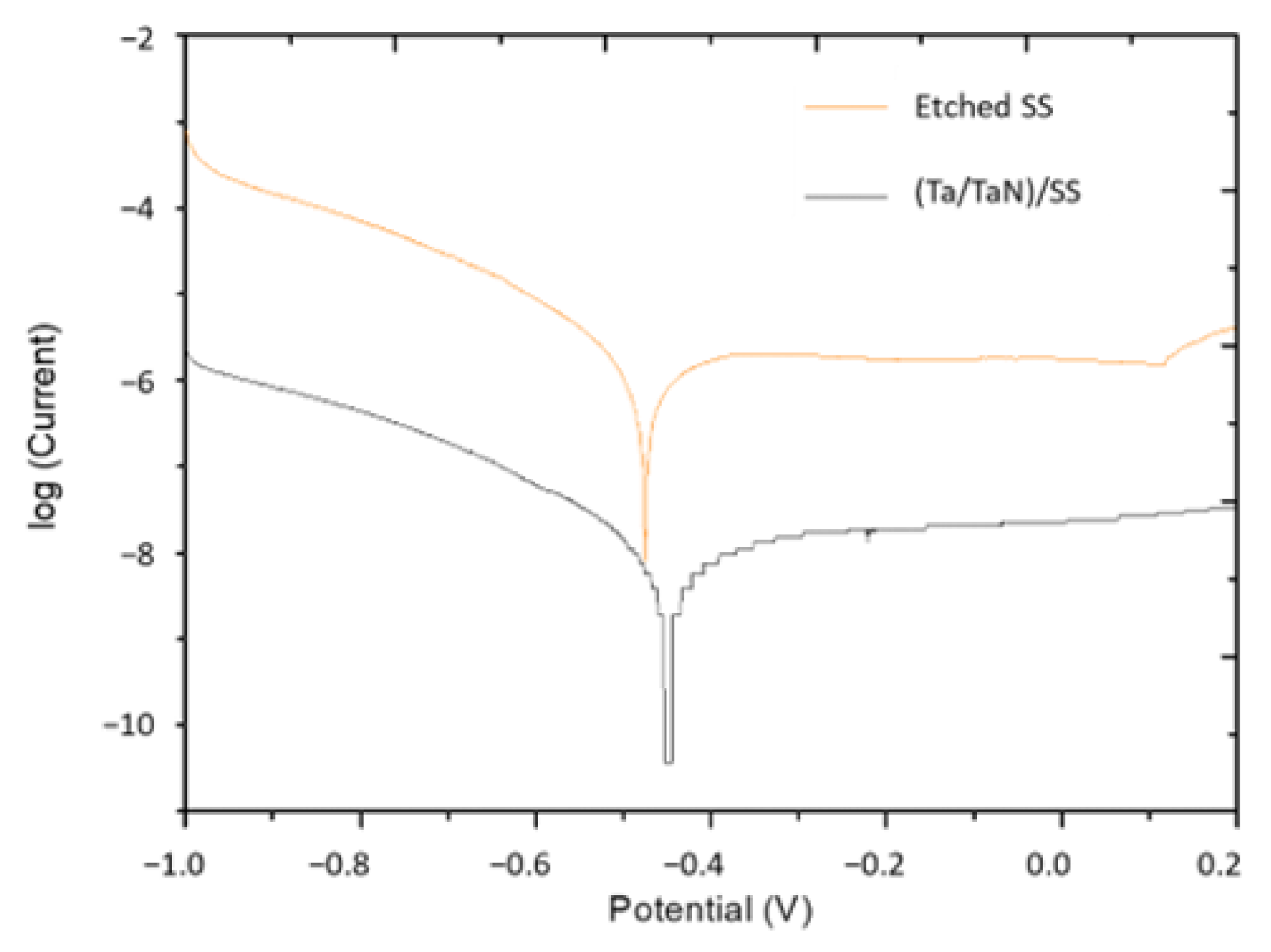
2.5. Potentiodynamic Polarization

Potentiodynamic polarization was carried out at a scan rate of 0.3 mV/s (0.05 mV/min), 20 mV/min, using a large cell, with a volume of 1400 cm3; the working electrode was the test specimen, the counter electrode was a Pt mesh, and the reference electrode was Ag/AgCl. The electrolyte solution was SBF pH de 7.2 at 37 °C.

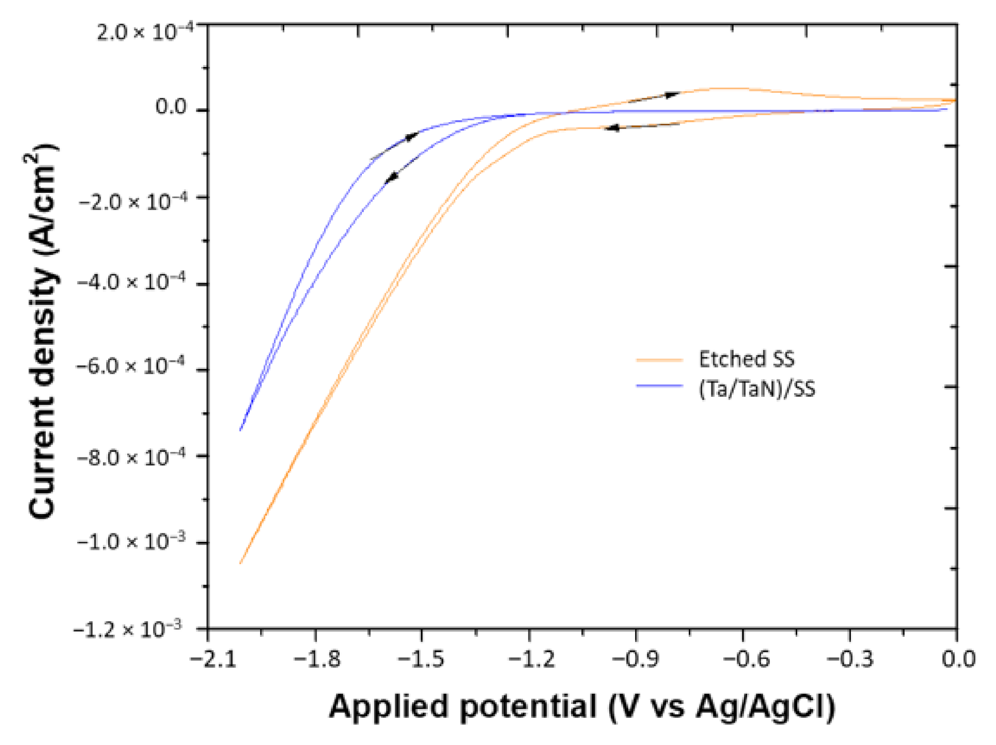
2.6. Electrochemical Deposition

Electrochemical deposition was performed using a simulated body fluid solution, with electrolytes at 37 °C, pH 7.2–7.4, and a CHI660C electrochemical potentiostat, with a three-electrode configuration. The working electrode was the test specimen with an exposed area of 7.54 mm2. The reference electrode was Ag/AgCl and counter electrode was a large area stainless steel spiral. A 1.5 SBF solution was employed [40], with ion concentrations that nearly reproduce the composition of blood plasma at 37 °C and pH = 7.4. The chronoamperograms were carried out at −1.2 and −1.4 V for periods of 3000 and 6000 s.

3. Results and Discussion

The capacitive characteristics of the different bare substrates (SS, Ta/SS, and Ta/TaN/ SS) were analyzed by electrochemical impedance spectroscopy and polarization curves, before the electrochemical deposition of HAp, in order to obtain the best conditions for the HAp electrochemical coating deposition through a cyclic voltammetry test. These results are described below.

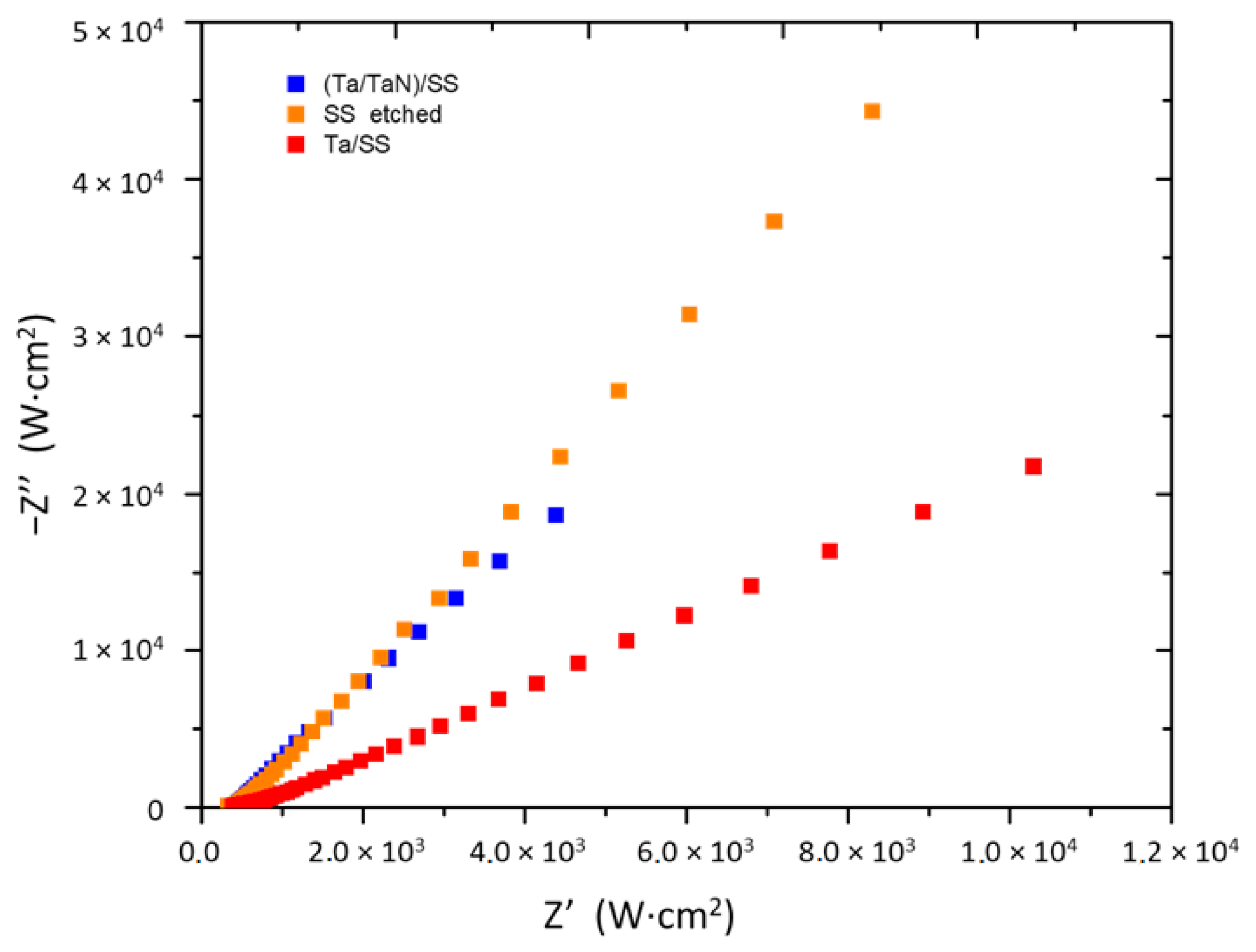
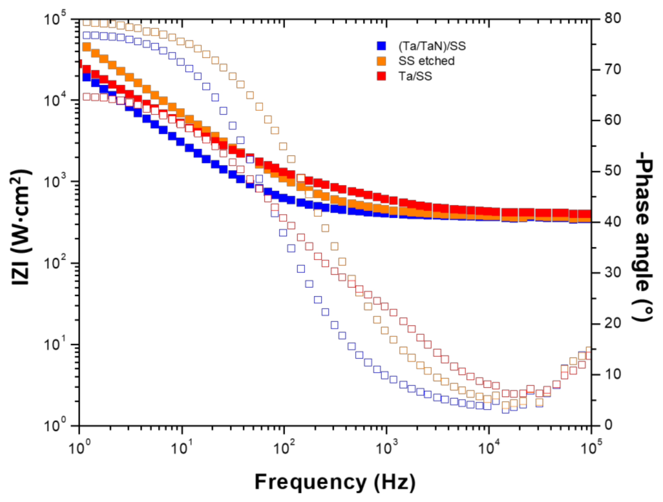
3.1. Characterization by Electrochemical Impedance Spectroscopy

Electrochemical impedance spectroscopy (EIS) characterization has a tremendous importance in the study of materials. Analysis of the system response contains information about the interface, its structure, and the reactions taking place there. The data obtained is presented in the Nyquist diagrams that relate the imaginary component of the impedance (Z”) vs. the real component (Z’), as well as the base diagrams that plot the modulus of impedance |Z (m)| and phase angle φ vs. log of frequency [41]. The slopes obtained from the Nyquist diagrams (Figure 1) show the capacitive behavior of the different substrates in the following order: etched stainless steel (etched SS), Ta coated stainless steel (Ta/SS), and stainless steel coated with the double layer ((Ta/TaN)/SS). The Bode diagrams (Figure 2) support this result, indicating that the etched SS is the most resistive material. In the range of low and medium frequency, log Z keeps a constant angle near 90° characteristic and capacitive behavior and decreases for high frequencies [17]. Furthermore, this substrate shows a higher Log Z vs. frequency in the linear range and plateau of the phase angle than the coated material. This result is attributed to the isolating effect of the coating [8]. The Ta film on SS would not be an effective protective coating, due to the delamination observed. According to Jara et al. [39] and Gladzuk et al. [42], α-Ta film deposited on SS316LVM presents delamination, due to the very high strain at the interface. The delamination problem could be overcome by the deposition of an underlayer of TaN [43,44].
The polarization plots obtained for the etched SS and double layer (Ta/TaN)/SS substrates are presented in Figure 3. The cathodic current density can be attributed to the reduction of the oxygen dissolved in the solution, while the anodic branch does not show the typical passivation value. However, the Tafel relation is in agreement with the anodic polarization of the alloys in the presence of calcium and seric proteins [45].
The cyclic voltammetry of the different substrates for the etched SS and double layer on steel (Ta/TaN)/SS, taken in a SBF solution at 37 °C, pH de 7.2–7.4, are shown in Figure 4. The electrodeposition process of HAp on the substates surface occurs at potentials between −0.5 and −1.8 V, through a combination of several equilibrium chemical reactions [29,46,47,48]:
O 2 + 2 H 2 O + 4 e   4 OH
H 2 PO 4   HPO 4 2 + H +
2 H 2 O + 4 e = H 2 + 2 OH
Ca 2 + HPO 4 2 + 2 H 2 O =   CaHPO 4 ·   2 H 2 O
Figure 1. Nyquist diagram of stainless steel substrates of the following types: etched SS, Ta/SS coating, and (Ta/TaN)/SS.
Coatings 12 00440 g001
Figure 2. Bode diagram of stainless steel substrates of the following types: etched SS, Ta/SS coating, and (Ta/TaN)/SS.
Coatings 12 00440 g002
Currents at powers less negative than −0.5 V are associated with equilibrium (1) and equilibrium (2), with those currents at potentials between −0.5 and −1.5 V, while an increase in the current at potentials more negative than −1.5 V are associated with balance (3). Electrochemically-generated OH ions in equilibrium (3) and HPO42− ions in equilibrium (2) react with the Ca2+ ions present in the electrolyte solution, equilibrium (4), to form the HAp coating on the substrate surface, where the formation of OH in the reaction medium favors the equilibrium (2), towards the production of HPO42−. Therefore, on those substrates that catalyze the electroreduction of H2O, a higher yield of HAp could be achieved; however, the production of H2 gas could mechanically destabilize the HAp coating, resulting in detachment from the substrate surface. In our case, the growing process of HAP on etched SS is between the −0.3 and −1.6 V potentials, with −1.3 and −1.8 V for the (Ta/TaN)/SS. According to this, the etched SS substrate would be more efficient as an electrocatalyst for the generation of OH ions from O2 and H2O and, therefore, would favor the formation of HAp on its surface. However, a greater potential window for the evolution of H2 was obtained for the (Ta/TaN)/SS, which could delay the mechanical destabilization of the coating, due to the production of this gas at higher potentials. Table 1 presents the potentials selected for the HAp growth on the different substrates by the chronoamperometry method, using deposition times of 3000 and 6000 s, according to the results shown by cyclic voltammetry.
Figure 3. Polarization curves of the Bode diagram of etched SS and (Ta/TaN)/SS.
Coatings 12 00440 g003
Figure 4. Cyclic voltammetry of etched SS and (Ta/TaN)/SS in SBF solution, at 37 °C, pH de 7.2–7.4.
Coatings 12 00440 g004
Table 1. Applied potentials for chronoamperometry.
SamplePotential 1 (V)Potential 2 (V)
Etched SS−1.2−1.4
(Ta/TaN)/SS−1.4−1.7

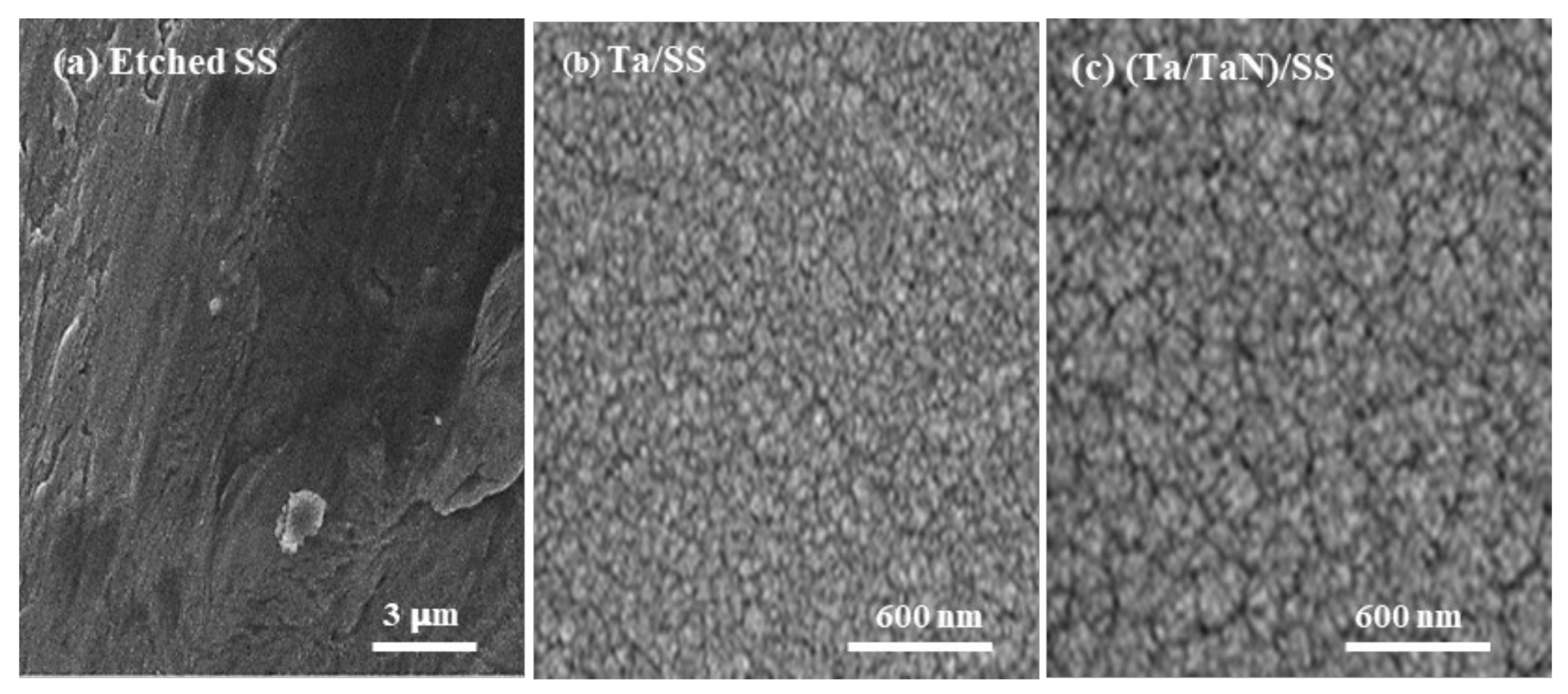
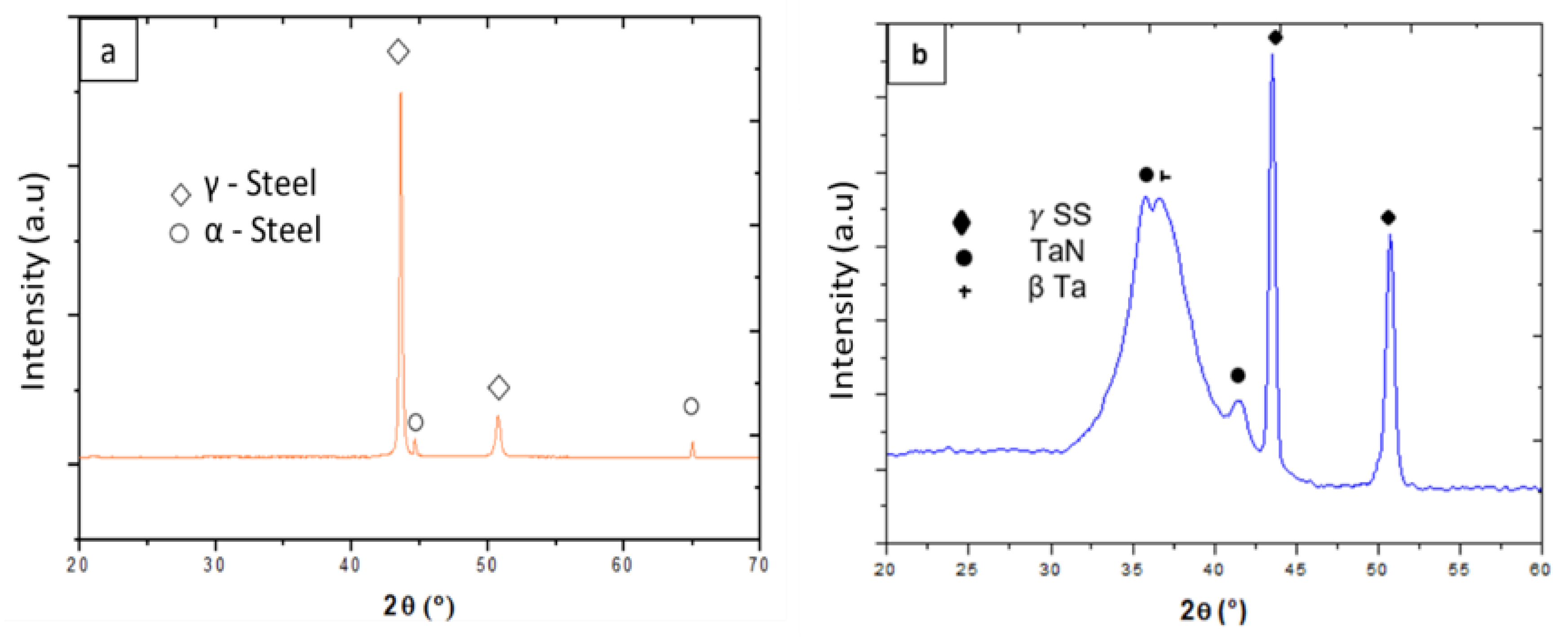
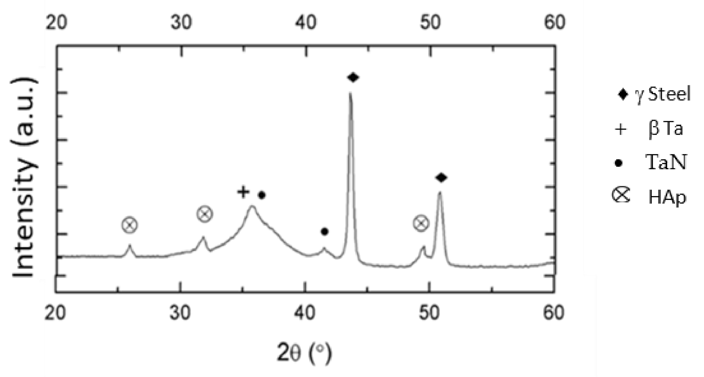
3.2. Characterization of Substrates

Figure 5 shows SEM images of the different surface-modified stainless steel substrates. Figure 5a shows the sample after etching with NaOH. A sodium chromate layer can be formed after a reaction with NaOH, which could favor the formation of phosphates on the surface [49,50]. Figure 5b shows the microstructure of the tantalum film deposited on SS, evidencing a column-like growth, as previously reported by Jara et al. [39]. The grain size of the Ta layer formed on the steel’s surface is about 50 nm. Figure 5c show the deposition of the bilayer Ta/TaN layers on SS, where the microstructure of the Ta film that was deposited on TaN on SS can be observed. A change in the grain size can be observed, which is attributed to the influence of the TaN underlayer.
Figure 6a corresponds to the diffractogram of the SS substrate, showing a mixture of gamma (γ) austenite at 44.1 and 50.8 2θ° [JCPDS 33-0945] and (α) phase at 44.8 and 66.1 2θ° [JCPDS 35–1375]. Figure 6b is the grazing angle XRD of (Ta/TaN)/SS sample, apart from the SS phases, the formation of β-tantalum center at 37.5 2θ° [JCPDS 25-1280] and TaN at 35.8 and 41.6 2θ° can be observed.

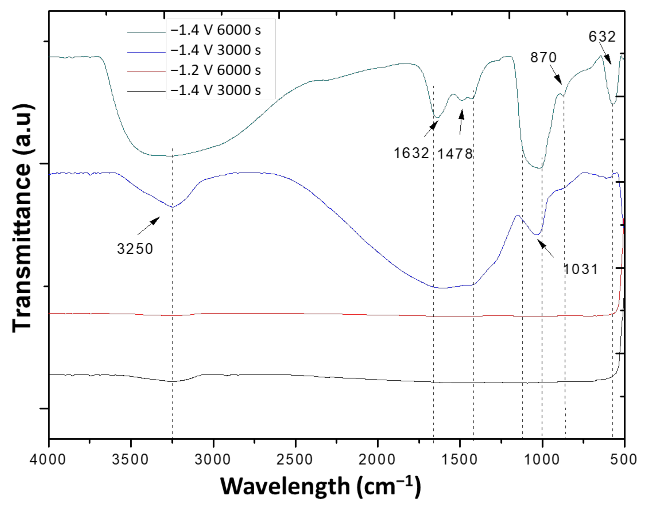
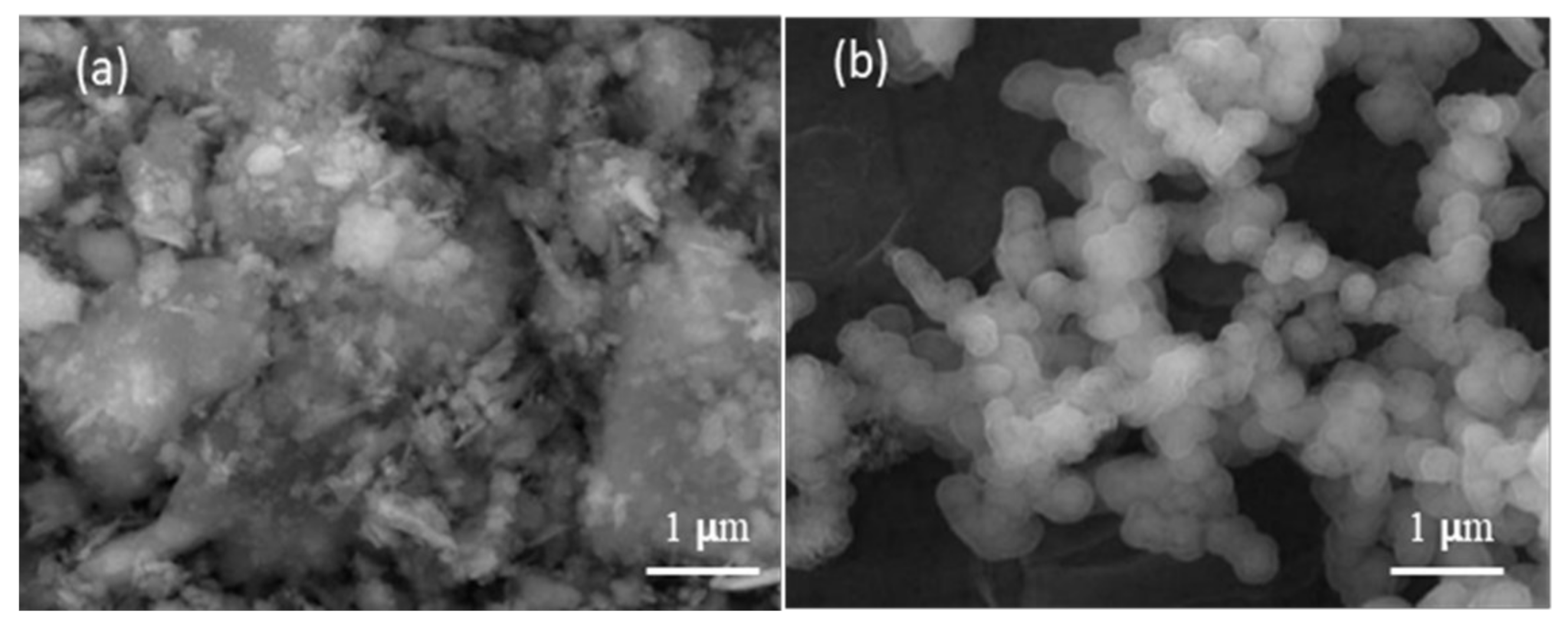
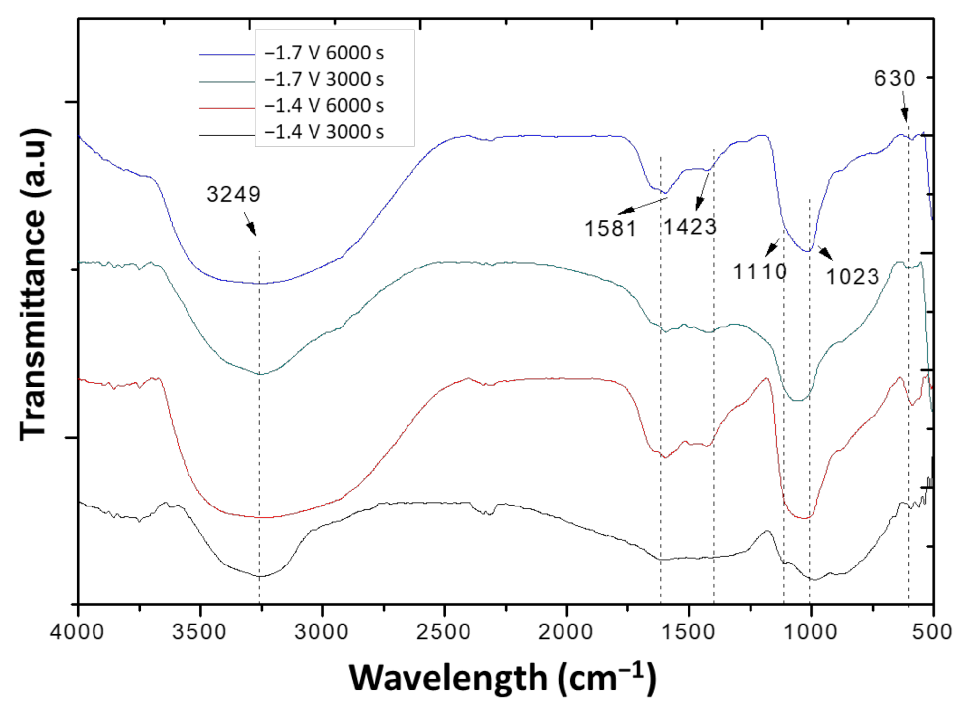
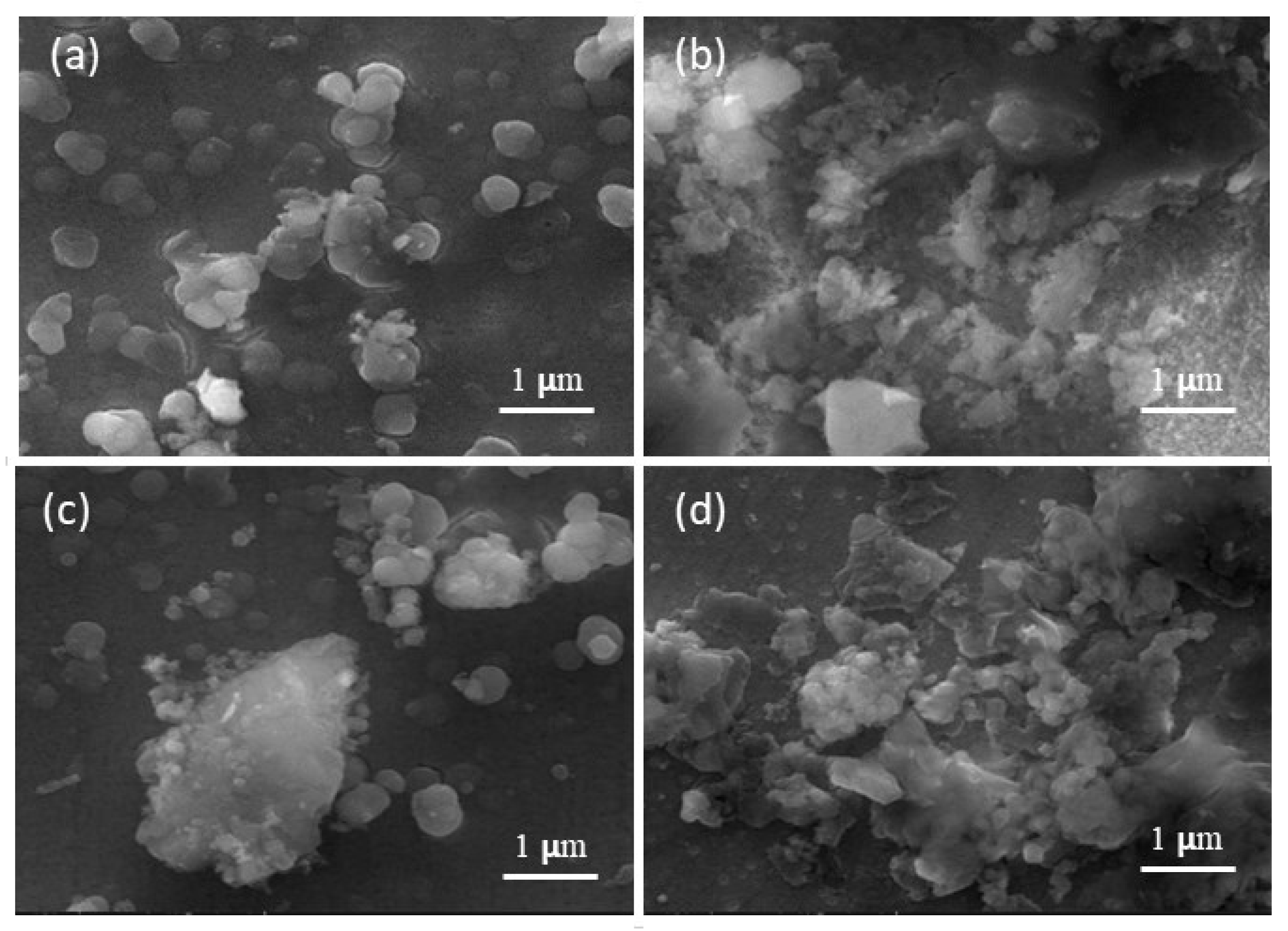
3.3. Characterization of Layers Formed on the Different Substrates by Electrodeposition

The FTIR results of the layer formed on the SS by electrodeposition is shown in Figure 7. When a potential of −1.4 V for 6000 s was applied, the presence of the characteristic PO4−3 groups corresponding to HAp were observed between 1110 and 1020 cm−1, OH at 630 cm−1, and HPO42− at 870 cm−1. For the same potential, −1.4 V and a lower period of 3000 s, the formation of a phosphate is seen at 1031 cm−1. For lower potentials (−1.2 V), the structures did not form. This could be due to the low generation of OH ions that could contribute to the displacement of equilibrium, disfavoring the formation of HAp. The SEM images of the electrodeposited layer of HAp on etched SS, when a potential of −1.4 V was applied for 3000 and 6000 s, are shown in Figure 8. The formation of HAp is obtained with the characteristic spherical morphology after 6000 s, consistent with the FTIR results (Figure 7).
When the electrodeposition was carried out on the bilayer, (Ta/TaN)/SS, the formation of HAp is favored after 6000 s for both potentials (Figure 9). For the latter condition, the characteristic phosphate PO4−3 bands of HAp are observed in the region 1110–1020 cm−1 and HPO42− at 865–877 cm−1 [51].
The SEM images of the layer electrodeposited on (Ta/TaN)/SS (Figure 10) show similar morphologies for both potentials (−1.4 V and −1.7 V), with a formation of agglomerates and larger formation of the product for longer periods (6000 s); this is in good agreement with the measured mass deposition (Table 2). The Ca/P was determined by EDS, indicating that, for 6000 s, this ratio corresponded to non-stoichiometric HAp (Table 2).
The morphology of HAp electrochemically-deposited depends on many different parameters, such as pH, temperature, time, current density, distance between working and counter electrodes, topography and roughness of the substrates, and composition of the electrolyte. Cell adhesion is affected by the morphology and composition of the layer. However, the morphology obtained in the present work is similar to the reported morphology reported by electrochemically using SBF as electrolyte with similar voltages than the used in the present work [34,37].
The Ca/P ratio determined from EDS and the average mass formed is shown in Table 2 for the different potentials and time applied on substrates SS and (Ta/TaN)/SS. The best condition was −1.7 V for 6000 s, with the highest deposition and a Ca/P ratio of 1.65.
The chronoamperometric results, chosen in the region of nucleation and growth of HAp, determined from cyclic voltammetry, indicated that higher potential and longer periods of deposition favor the formation of a uniform Hap layer. It has been reported that high current density and relatively high overpotential results in high depositional resistance and, therefore, small HAp crystals are obtained. Low overpotential helps the ionic aggregation caused by the slow charge transfer process near the cathode; as a result, high crystal growth was observed [52]. The deposition of HAp on the double layer material for the higher potential and longer periods on Ta/TaN/SS was more homogeneous than on steel, and a Ca/P ratio characteristic of nonstoichiometric HAp was obtained. This behavior is in good agreement with the Nyquist diagrams (Figure 1), which shows that the double layer Ta/TaN presents higher electrochemical resistance, suggesting a protective coating for the corrosion processes. Hydroxyapatite coatings have been grown on Ta samples by plasma electrolytic oxidation, using calcium and phosphorus-containing electrolytic solutions [53]. These authors evaluated the influence of polarization voltage and treatment time. They reported that the HAp coatings produced, using voltages of 500 V, for times over 300 s, were denser, composed of more than 80% crystalline HAp.
The electrochemical formation of HAp films, using SBF as an electrolyte, was a successful method, with the formation of a homogenous film in a very short time, compared to more conventional methods of immersion Uribe et al. [20].
The grazing angle XRD of the hydroxyapatite layer, formed on the double layer Ta/TaN/SS, is presented in Figure 11. The diffractogram presents the (002), (211), and (213) reflexions of HAp and those corresponding to the double layer: nanocrystalline β-Ta phase and TaN, as well as the steel substrate austenitic γ phase. For the deposition of HAp on steel lower intensity, the HAp reflexions were obtained, indicating that lower amount of coating.

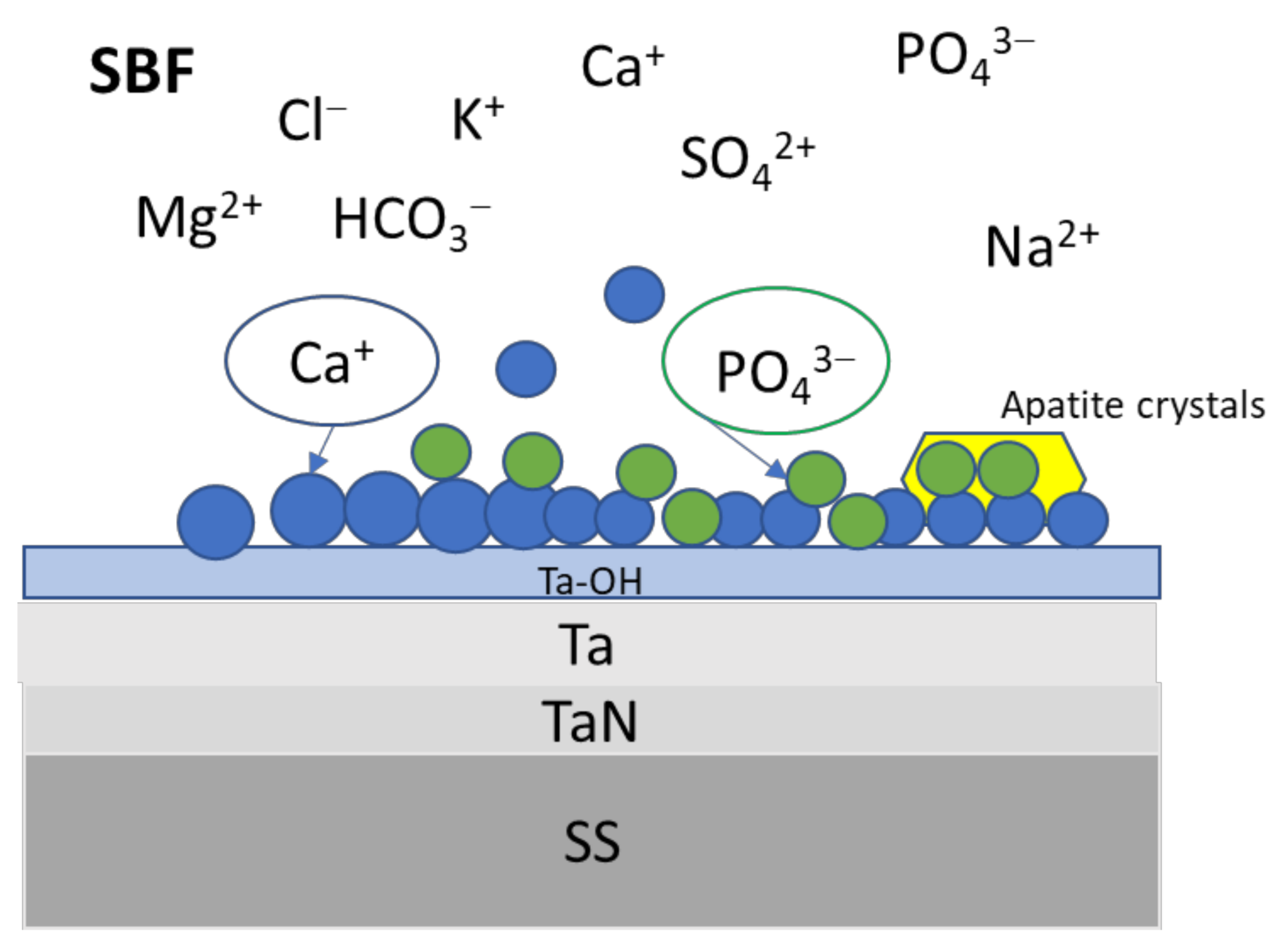
3.4. Mechanism of Formation of Hydroxyapatite

During the process of the formation of apatite on the different surfaces, an amorphous precursor phase of calcium phosphate is formed [54,55,56,57,58]. The primary and secondary nucleation is induced by the ion charges at the surfaces and under conditions of supersaturation of the SBF solution. Electrostatic interactions, van der Waals, and hydration forces are present, being the electrostatic interaction, and the main factor present at the initial nucleation, due to the negative electric charges present at the surface [23,24,25,54,55,56]. During electrodeposition, the electrostatic migration of OH groups adsorb to the surface and react at the interface, conforming the Ca2+ nucleation sites. As a result, hydrogen phosphate and phosphate increase in concentration, as proposed in Equation (2), while Ca2+ ions interact with phosphate groups to form hydroxyapatite (Equation (4)). Experimental results suggest that more complex routes could be followed before the formation of hydroxyapatite crystals. The scheme presented in Figure 12 is a proposal of the formation mechanism. Many parameters, such as substrate topography, roughness, crystal structure, surface energies, and morphology, affect the formation of the apatite phase. Moreover, in the double layer, Ta/TaN/SS, the formation of the Ta–OH group on the external layer tantalum surface is obtained, due to formation of the passivation layer of tantalum oxide [8]. These Ta–OH groups will attract Ca2+ ions, and a non-stoichiometric tantalum calcium hydroxide could form. In the next stage the phosphate ions bind to the calcium ions, and the nucleation of the amorphous phase takes place [36,54,55,56,57,58]. The Ca2+ and PO43− ions absorbed on the surface will form the precursors of the apatite crystals that, finally, transform to hydroxyapatite. In the SBF, other ions are also present that form part of the layer, as has been shown in the EDS results.
One problem of electrochemical deposition of HAp is the formation of bubbles, since these could hamper the nucleation and growth of HAp, as well as adhesion to the substrate. Therefore, the control of bubbles, during electrochemical deposition, is extremely important to obtain a very homogeneous coating, the parameters temperature, pH, deposition time, and current density should be controlled very carefully [23,25,28].
Figure 12. Scheme of HAp mechanism of formation.
Coatings 12 00440 g012

3.5. Contact Angle Measurements

Contact angle measurements are shown in Table 3 for both substrates, before and after HAp deposition. A hydrophilic characteristic is obtained for all the surfaces, but a large increase in the hydrophilic character after HAp deposition was especially observed for (Ta/TaN)/SS [59,60]. The latter is a condition desirable for cellular adhesion [61], corroborating the beneficial effect of these coatings on SS, protecting against corrosion and promoting the HAp deposition.

4. Conclusions

The formation of apatite was successfully obtained by electrochemical deposition on steel and Ta/TaN double coating on steel, using simulated body fluid as an electrolyte. The deposition on Ta/TaN was much more effective than on etched steel. The chronoamperometric results, chosen in the region of nucleation and growth of HAp, determined from cyclic voltammetry, indicated that higher potential and longer periods of deposition favor the formation of a uniform HAp layer on both substrates. Furthermore, the deposition on the double layer material Ta/TaN/SS is more homogeneous and with a Ca/P ratio characteristic of nonstoichiometric HAp. The electrochemical method resulted in the formation of a homogenous film in a very short time, compared to more conventional biomimetic methods of immersion. Furthermore, an increase in the hydrophilic character after HAp deposition was particularly observed for Ta/TaN/SS material. This condition is desirable for cellular adhesion.
Additionally, it was found that the double layer Ta/TaN has bioactive behavior and protects against corrosion in the presence of the simulated body fluid aggressive electrolyte, implying that it could be a potential coating for metallic implants.

Author Contributions

Conceptualization, R.U. and G.G.; methodology. R.U., A.U., O.B., L.F., A.J. and G.G.; validation, R.U., O.B. and G.G.; formal analysis, A.U., A.J., R.U. and G.G.; investigation, R.U., A.U., O.B., L.F., A.J. and G.G.; resources, O.B. and R.U.; data curation, R.U., L.F. and G.G.; writing—original draft preparation, A.U. and R.U.; writing—review and editing, R.U. and G.G.; visualization, R.U., A.U., O.B., L.F. and G.G.; supervision, R.U., O.B., L.F. and G.G.; project administration, O.B. and R.U.; funding acquisition, O.B. and R.U. All authors have read and agreed to the published version of the manuscript.

Funding

This research was funded by the Project EPN-PIMI 17-05: “Desarrollo de sistemas de hidroxiapatita dopados con óxidos metálicos biocompatibles para aplicaciones biomédicas”, Escuela Politécnica Nacional, Quito-Ecuador. The payment of the publication was financed by Vicerectorado de Investigación, Innovación y Vinculación de la EPN.

Institutional Review Board Statement

Not applicable.

Informed Consent Statement

Not applicable.

Data Availability Statement

Not applicable.

Acknowledgments

The authors would like to thank Vicerrectorado de Investigación, Innovación y Vinculación, Escuela Politécnica Nacional, Escuela de Ciencias Químicas, Pontificia Universidad Católica del Ecuador, and Universidad Yachay Tech del Ecuador. The authors thank P. Espinoza and N. Fretty for the laboratory facilities.

Conflicts of Interest

The authors declare no conflict of interest.

References

  1. Ratner, B.; Hoffman, A.; Schoen, F.; Lemons, J. Biomaterials Science: An Introduction to Materials in Medicine; Academic Press: San Diego, CA, USA, 1996; pp. 527–573. [Google Scholar]
  2. Bergmann, C.; Stumpf, A. Dental Ceramics. Topics in Mining, Metallurgy and Materials Engineering; Springer Science & Business Media: Berlin/Heidelberg, Germany, 2013; pp. 9–14. [Google Scholar]
  3. Dos Santos, V.; Brandalise, R.; Savaris, M. Engineering of Biomaterials. Topics in Mining. In Metallurgy and Materials Engineering; Springer: Berlin/Heidelberg, Germany, 2017; pp. 5–86. [Google Scholar]
  4. Rodil, S.E. Modificación Superficial De Biomateriales Metálicos. Rev. Latin Am. Metal. Mater. 2009, 29, 67–83. [Google Scholar]
  5. Levine, B.R.; Sporer, S.; Poggie, R.A.; Della Valle, C.J.; Jacobs, J.J. Experimental and clinical performance of porous tantalum in orthopedic surgery. Biomaterials 2006, 27, 4671–4681. [Google Scholar] [CrossRef] [PubMed]
  6. Leng, Y.; Sun, H.; Yang, P.; Chen, J.; Wang, J.; Wan, G.; Huang, N.; Tian, X.; Wang, L.; Chu, P. Biomedical properties of tantalum nitride films synthesized by reactive magnetron sputtering. Thin Solid Films 2001, 398–399, 471–475. [Google Scholar] [CrossRef]
  7. Matsuno, H.; Yokoyama, A.; Watari, F.; Uo, M.; Kawasaki, T. Biocompatibility and osteogenesis of refractory metal implants, titanium, hafnium, niobium, tantalum and rhenium. Biomaterials 2001, 22, 1253–1262. [Google Scholar] [CrossRef]
  8. Miyazaki, T.; Kim, H.-M.; Kokubo, T.; Ohtsuki, C.; Kato, H.; Nakamura, T. Mechanism of bonelike apatite formation on bioactive tantalum metal in a simulated body fluid. Biomaterials 2002, 23, 827–832. [Google Scholar] [CrossRef]
  9. Yang, Y.H.; Chen, D.J.; Wu, F.B. Microstructure, hardness, and wear resistance of sputtering TaN coating by controlling RF input power. Surf. Coat. Technol. 2016, 303, 32–40. [Google Scholar] [CrossRef]
  10. Flores, J.F.; Schorr, M.; Olaya, J.J. Corrosion protection of steels by PVD TaN thin films. Anti-Corros. Methods Mater. 2006, 53, 88–94. [Google Scholar] [CrossRef]
  11. Ma, G.; Lin, G.; Gong, S.; Liu, X.; Sun, G.; Wu, H. Mechanical and corrosive characteristics of Ta/TaN multilayer coatings. Vacuum 2013, 89, 244–248. [Google Scholar] [CrossRef]
  12. Fathyunes, L.; Khalil-Allafi, J.; Sheykholeslami, S.O.R.; Moosavifar, M. Biocompatibility assessment of graphene oxide-hydroxyapatite coating applied on TiO2 nanotubes by ultrasound-assisted pulse electrodeposition. Mater. Sci. Eng. C 2018, 87, 10–21. [Google Scholar] [CrossRef]
  13. Rigo, E.; Boschi, A.; Yoshimoto, M.; Allegrini, S.; Konig, B.; Carbonari, M. Evaluation in vitro and in vivo of biomimetic hydroxyapatite coated on titanium dental implants. Mater. Sci. Eng. C 2004, 24, 647–651. [Google Scholar] [CrossRef]
  14. Lee, H.; Jeong, Y.; Park, S.; Jeong, S.; Kim, H.; Cho, C. Surface properties and cell response of fluoridated hydroxyapatite/TiO2 coated on Ti substrate. Curr. Appl. Phys. 2008, 9, 528–533. [Google Scholar] [CrossRef]
  15. Kim, H.-W.; Koh, Y.-H.; Li, L.-H.; Lee, S.; Kim, H.-E. Hydroxyapatite coating on titanium substrate with titania buffer layer processed by sol–gel method. Biomaterials 2003, 25, 2533–2538. [Google Scholar] [CrossRef]
  16. Williams, D.F. On the mechanisms of biocompatibility. Biomaterials 2008, 29, 2941–2953. [Google Scholar] [CrossRef]
  17. Kokubo, T.; Takadama, H. How useful is SBF in predicting in vivo bone bioactivity? Biomaterials 2006, 27, 2907–2915. [Google Scholar] [CrossRef]
  18. Wei, X.; Carl, L.; Jukka, L.; Hakan, E. Biomimetic Hydroxyapatite Deposition on Titanium Oxide Surfaces for Biomedical Application. In Advances in Biomimetics; Marko, C., Ed.; InTech: London, UK, 2011; ISBN 978-953-307-191-6. Available online: http://www.intechopen.com/books/advances-inbiomimetics/biomimetic-hydroxyapatite-deposition-on-titanium-oxide-surfaces-for-biomedical-application (accessed on 20 February 2022).
  19. Forsgren, J.; Svahn, F.; Jarmar, T.; Engqvist, H. Formation and adhesion of biomimetic hydroxyapatite deposited on titanium substrates. Acta Biomater. 2007, 3, 980–984. [Google Scholar] [CrossRef]
  20. Uribe, R.; Uvillús, A.; Bonilla, O.; Lascano, L.; González, G. Effect of Low Magnetic Field on Biomimetic Deposition of Hydroxyapatite on Titanium and BIOLINE Stainless Steel. Coatings 2021, 11, 1484. [Google Scholar] [CrossRef]
  21. Chakraborty, R.; Saha, P. A comparative study on surface morphology and electrochemical behaviour of hydroxyapatite-calcium hydrogen phosphate composite coating synthesized in-situ through electro chemical process under various deposition conditions. Surf. Interfaces 2018, 12, 160–167. [Google Scholar] [CrossRef]
  22. San, H.; Hu, J.; Zhang, Y.; Han, J.; Tang, S. Formation and in vitro mineralization of electrochemically deposited coatings prepared on micro-arc oxidized titanium alloy. J. Appl. Electrochem. 2019, 49, 485–501. [Google Scholar] [CrossRef]
  23. Mokabber, T.; Lu, L.; van Rijn, P.; Vakis, A.; Pei, Y. Crystal growth mechanism of calcium phosphate coatings on titanium by electrochemical deposition. Surf. Coatings Technol. 2018, 334, 526–535. [Google Scholar] [CrossRef]
  24. Katić, J.; Metikoš-Huković, M.; Škapin, S.; Petravić, M.; Varašanec, M. The potential-assisted deposition as valuable tool for producing functional apatite coatings on metallic materials. Electrochim. Acta 2014, 127, 173–179. [Google Scholar] [CrossRef]
  25. Abdel-Aal, E.; Dietrich, D.; Steinhaeuser, S.; Wielage, B. Electrocrystallization of nanocrystallite calcium phosphate coatings on titanium substrate at different current densities. Surf. Coat. Technol. 2008, 202, 5895–5900. [Google Scholar] [CrossRef]
  26. Schmidt, R.; Hoffmann, V.; Helth, A.; Gostin, P.F.; Calin, M.; Eckert, J.; Gebert, A. Electrochemical deposition of hydroxyapatite on beta-Ti-40Nb. Surf. Coat. Technol. 2016, 294, 186–193. [Google Scholar] [CrossRef]
  27. Chakraborty, R.; Sengupta, S.; Saha, P.; Das, K.; Das, S. Synthesis of calcium hydrogen phosphate and hydroxyapatite coating on SS316 substrate through pulsed electrodeposition. Mater. Sci. Eng. C 2016, 69, 875–883. [Google Scholar] [CrossRef] [PubMed]
  28. Li, T.-T.; Ling, L.; Lin, M.-C.; Jiang, Q.; Lin, Q.; Lin, J.-H.; Lou, C.-W. Properties and Mechanism of Hydroxyapatite Coating Prepared by Electrodeposition on a Braid for Biodegradable Bone Scaffolds. Nanomaterials 2019, 9, 679. [Google Scholar] [CrossRef] [PubMed] [Green Version]
  29. Thanh, D.T.M.; Nam, P.T.; Phuong, N.T.; Que, L.X.; Van Anh, N.; Hoang, T.; Lam, T.D. Controlling the electrodeposition, morphology and structure of hydroxyapatite coating on 316L stainless steel. Mater. Sci. Eng. C 2013, 33, 2037–2045. [Google Scholar] [CrossRef]
  30. Chakraborty, R.; Seesala, V.S.; Manna, J.S.; Saha, P.; Dhara, S. Synthesis, characterization and cytocompatibility assessment of hydroxyapatite-polypyrrole composite coating synthesized through pulsed reverse electrochemical deposition. Mater. Sci. Eng. C 2018, 94, 597–607. [Google Scholar] [CrossRef] [PubMed]
  31. Ban, S.; Matsuo, K.; Mizutani, N.; Hasegawa, J. Hydrothermal-Electrochemical Deposition of Calcium Phosphates on Various Metals. Dent. Mater. J. 1999, 18, 259–270. [Google Scholar] [CrossRef] [PubMed]
  32. Li, T.-T.; Ling, L.; Lin, M.-C.; Peng, H.-K.; Ren, H.-T.; Lou, C.-W.; Lin, J.-H. Recent advances in multifunctional hydroxyapatite coating by electrochemical deposition. J. Mater. Sci. 2020, 55, 6352–6374. [Google Scholar] [CrossRef]
  33. Zielinski, A.; Bartmanski, M. Electrodeposited Biocoatings, Their Properties and Fabrication Technologies: A Review. Coatings 2020, 10, 782. [Google Scholar] [CrossRef]
  34. Peng, P.; Kumar, S.; Voelcker, N.; Szili, E.; Smart, R.; Griesser, H. Thin calcium phosphate coatings on titanium by electro-chemical deposition in modified simulated body fluid. J. Biomed. Mater. Res. 2016, 76, 347–355. [Google Scholar]
  35. Ban, S.; Maruno, S. Deposition of Calcium Phosphate on Titanium by Electrochemical Process in Simulated Body Fluid. Jpn. J. Appl. Phys. 1993, 32, L1577–L1580. [Google Scholar] [CrossRef]
  36. Zhao, X.; Yang, L.; Zuo, Y.; Xiong, J. Hydroxyapatite Coatings on Titanium Prepared by Electrodeposition in a Modified Simulated Body Fluid. Chin. J. Chem. Eng. 2009, 17, 667–671. [Google Scholar] [CrossRef]
  37. Liu, S.; Li, H.; Zhang, L.; Yin, X.; Guo, Y. In simulated body fluid performance of polymorphic apatite coatings synthesized by pulsed electrodeposition. Mater. Sci. Eng. C 2017, 79, 100–107. [Google Scholar] [CrossRef] [PubMed]
  38. Sandvik. SANDVIK BIOLINE 316LVM: Austenitic Stainless Steel for Implants. Alloy. Dig. 2000, 49, SS-784. [Google Scholar] [CrossRef]
  39. Jara, A.; Fraisse, B.; Flaud, V.; Fréty, N.; Gonzalez, G. Thin film deposition of Ta, TaN and Ta/TaN bi-layer on Ti and SS316-LVM substrates by RF sputtering. Surf. Coat. Technol. 2017, 309, 887–896. [Google Scholar] [CrossRef]
  40. Oyane, A.; Kim, H.-M.; Furuya, T.; Kokubo, T.; Miyazaki, T.; Nakamura, T. Preparation and assessment of revised simulated body fluids. J. Biomed. Mater. Res. 2003, 65, 188–195. [Google Scholar] [CrossRef] [PubMed]
  41. Pérez, C.A. Evaluación Mediante Técnicas Electroquímicas del Comportamiento de Recubrimientos Alquídicos Utilizados en la Protección Anticorrosiva del Acero. Bachelor’s Thesis, Escuela Politecnica Nacional, Quito, Ecuador, 2012. [Google Scholar]
  42. Gladczuk, L.; Patel, A.; Paur, C.S.; Sosnowski, M. Tantalum films for protective coatings of steel. Thin Solid Films 2004, 467, 150–157. [Google Scholar] [CrossRef]
  43. Bernoulli, D.; Müller, U.; Schwarzenberger, M.; Hauert, R.; Spolenak, R. Magnetron sputter deposited tantalum and tantalum nitride thin films: An analysis of phase, hardness and composition. Thin Solid Films 2013, 548, 157–161. [Google Scholar] [CrossRef]
  44. Gladczuk, L.; Patel, A.; Demaree, J.D.; Sosnowski, M. Sputter deposition of bcc tantalum films with TaN underlayers for protection of steel. Thin Solid Films 2005, 476, 295–302. [Google Scholar] [CrossRef]
  45. Cheng, X.; Roscoe, S.G. Corrosion behavior of titanium in the presence of calcium phosphate and serum proteins. Biomaterials 2005, 26, 7350–7356. [Google Scholar] [CrossRef]
  46. Kuo, M.; Yen, S. The process of electrochemical deposited hydroxyapatite coatings on biomedical titanium at room temperature. Mater. Sci. Eng. C 2002, 20, 153–160. [Google Scholar] [CrossRef]
  47. Zhang, Y.-Y.; Tao, J.; Pang, Y.-C.; Wang, W.; Wang, T. Electrochemical deposition of hydroxyapatite coatings on titanium. Trans. Nonferrous Met. Soc. China 2006, 16, 633–637. [Google Scholar] [CrossRef]
  48. Nam, P.T.; Lam, T.D.; Huong, H.T.; Phuong, N.T.; Trang, N.T.T.; Hoang, T.; Huong, N.T.T.; Thang, L.B.; Drouet, C.; Grossin, D.; et al. Electrodeposition and Characterization of Hydroxyapatite on TiN/316LSS. J. Nanosci. Nanotechnol. 2015, 15, 9991–10001. [Google Scholar] [CrossRef]
  49. Kadhim, M.J.; Abdulateef, N.E.; Abdulkareem, M.H. Evaluation of Surface Roughness of 316L Stainless Steel Substrate on Nanohydroxyapatite by Electrophoretic Deposition. Al-Nahrain J. Eng. Sci. 2018, 21, 28–35. [Google Scholar] [CrossRef] [Green Version]
  50. Huynh, V.; Ngo, N.K.; Golden, T.D. Surface Activation and Pretreatments for Biocompatible Metals and Alloys Used in Biomedical Applications. Int. J. Biomater. 2019, 2019, 3806504. [Google Scholar] [CrossRef]
  51. Berzina-Cimdina, L.; Borodajenko, N. Research of Calcium Phosphates Using Fourier Transform Infrared Spectroscopy. Infrared Spectrosc. Mater. Sci. Eng. Technol. 2012, 12, 251–263. [Google Scholar]
  52. Budevski, E.; Staikov, G.; Lorenz, W. Electrocrystallization: Nucleation and growth phenomena. Electrochim. Acta 2000, 45, 2559–2574. [Google Scholar] [CrossRef]
  53. Antonio, R.F.; Rangel, E.C.; Mas, B.A.; Duek, E.A.; Cruz, N.C. Growth of hydroxyapatite coatings on tantalum by plasma electrolytic oxidation in a single step. Surf. Coat. Technol. 2019, 357, 698–705. [Google Scholar] [CrossRef]
  54. Beniash, E.; Metzler, R.A.; Lam, R.S.; Gilbert, P. Transient amorphous calcium phosphate in forming enamel. J. Struct. Biol. 2009, 166, 133–143. [Google Scholar] [CrossRef] [Green Version]
  55. Xie, Y.; Liu, X.; Chu, P.K.; Ding, C. Nucleation and growth of calcium–phosphate on Ca-implanted titanium surface. Surf. Sci. 2006, 600, 651–656. [Google Scholar] [CrossRef]
  56. Dey, A.; Bomans, P.; Muller, F.; Will, J.; Frederik, P.; De With, G.; Sommerdijk, N. The role of prenucleation clusters in surface-induced calcium phosphate crystallization. Nat. Mater. 2010, 9, 1010–1014. [Google Scholar] [CrossRef]
  57. Gopi, D.; Indira, J.; Kavitha, L. A comparative study on the direct and pulsed current electrodeposition of hydroxyapatite coatings on surgical grade stainless steel. Surf. Coat. Technol. 2012, 206, 2859–2869. [Google Scholar] [CrossRef]
  58. Manso, M.; Jiménez, C.; Morant, C.; Herrero, P.; Martínez-Duart, J.M. Apatite films produced by electrodeposition: Characterization by TEM and AFM. Surf. Interface Anal. 2001, 31, 1104–1109. [Google Scholar] [CrossRef]
  59. Strnad, G.; Chirila, N.; Petrovan, C.; Russu, O. Contact Angle Measurement on Medical Implant Titanium Based Biomaterials. Procedia Technol. 2016, 22, 946–953. [Google Scholar] [CrossRef] [Green Version]
  60. Minagar, S.; Berndt, C.C.; Wen, C. Fabrication and Characterization of Nanoporous Niobia, and Nanotubular Tantala, Titania and Zirconia via Anodization. J. Funct. Biomater. 2015, 6, 153–170. [Google Scholar] [CrossRef] [Green Version]
  61. Menzies, K.L.; Jones, L. The Impact of Contact Angle on the Biocompatibility of Biomaterials. Optom. Vis. Sci. 2010, 87, 387–399. [Google Scholar] [CrossRef]
Figure 5. SEM images of substrates: (a) etched SS; (b) Ta/ SS [26], and (c) (Ta/TaN)/ SS [26]. Reprinted with permission from [26], 2017 Elsevier.
Coatings 12 00440 g005
Figure 6. XRD of: (a) SS sample and (b) (Ta/TaN)/SS.
Coatings 12 00440 g006
Figure 7. FTIR spectra electrodeposited layer on etched SS at different potentials and time.
Coatings 12 00440 g007
Figure 8. SEM images of electrodeposited layers on SS at −1.4 V: (a) 3000 and (b) 6000 s.
Coatings 12 00440 g008
Figure 9. FTIR spectra of HAp deposition on (Ta/TaN)/SS at different potential and time.
Coatings 12 00440 g009
Figure 10. SEM images of HAp deposition on (Ta/TaN)/SS: (a) −1.4 V 3000 s, (b) −1.4 V 6000 s, (c) −1.7 V 3000 s, and (d) −1.7 V 6000 s.
Coatings 12 00440 g010
Figure 11. Grazing angle XRD of HAp deposited on Ta/TaN/SS.
Coatings 12 00440 g011
Table 2. The ratio of Ca/P and mass of HAp, formed at the different conditions of potential and time, on substrates of SS and (Ta/TaN)/SS.
SubstratePotential
(V)
Time
(s)
Ca/PMass of Deposition of HAp (mg/mm2)
SS−1.2030001.200.056
−1.2060001.270.064
−1.4030001.410.105
−1.4060001.610.170
(Ta/TaN)/SS−1.4030001.250.021
−1.4060001.550.286
−1.7030001.620.267
−1.7060001.650.288
Table 3. Contact angle measurements on the different substrates with HAp deposition.
SubstrateBefore
HAp Deposition
Applied Potentials (V)After
HAp Deposition
Angle (°)6000 sAngle (°)
Ta/TaN/SS48.65 ± 0.93 Coatings 12 00440 i001−1.731.00 ± 1.53 Coatings 12 00440 i002
SS46.73 ± 1.00 Coatings 12 00440 i003−1.440.35 ± 1.29 Coatings 12 00440 i004
Publisher’s Note: MDPI stays neutral with regard to jurisdictional claims in published maps and institutional affiliations.

