Review

Strategies to Enhance Biomedical Device Performance and Safety: A Comprehensive Review

by
Julia Sánchez-Bodón
1,
Maria Diaz-Galbarriatu
1,
Leyre Pérez-Álvarez
1,2,
Isabel Moreno-Benítez
3,* and
José Luis Vilas-Vilela
1,2,*
1
Macromolecular Chemistry Group (LABQUIMAC), Department of Physical Chemistry, Faculty of Science and Technology, University of the Basque Country (UPV/EHU), Barrio Sarriena s/n, 48940 Leioa, Spain
2
BCMaterials, Basque Center for Materials, Applications and Nanostructures, UPV/EHU, Science Park, 48940 Leioa, Spain
3
Macromolecular Chemistry Group (LABQUIMAC), Department of Organic and Inorganic Chemistry, Faculty of Science and Technology, University of the Basque Country (UPV/EHU), Barrio Sarriena s/n, 48940 Leioa, Spain
*
Authors to whom correspondence should be addressed.
Submission received: 25 October 2023 / Revised: 14 November 2023 / Accepted: 19 November 2023 / Published: 21 November 2023
(This article belongs to the Special Issue Advanced Coatings for Biomedical Applications)

Abstract

:
This paper reviews different approaches to obtain biomaterials with tailored functionalities and explains their significant characteristics that influence their bioactivity. The main goal of this discussion underscores the significance of surface properties in materials, with a particular emphasis on their role in facilitating cell adhesion in order to obtain good biocompatibility and biointegration, while preventing adverse effects, such as bacterial contamination and inflammation processes. Consequently, it is essential to design strategies and interventions that avoid bacterial infections, reducing inflammation and enhancing compatibility systems. Within this review, we elucidate the most prevalent techniques employed for surface modification, notably emphasizing surface chemical composition and coatings. In the case of surface chemical composition, we delve into four commonly applied approaches: hydrolysis, aminolysis, oxidation, and plasma treatment. On the other hand, coatings can be categorized based on their material composition, encompassing ceramic-based and polymer-based coatings. Both types of coatings have demonstrated efficacy in preventing bacterial contamination, promoting cell adhesion and improving biological properties of the surface. Furthermore, the addition of biological agents such as drugs, proteins, peptides, metallic ions plays a pivotal role in manifesting the prevention of bacterial infection, inflammatory responses, and coagulation mechanism.

1. Introduction

In 1987, the European Society for Biomaterials coined the term “biomaterial”, defining it as a non-biological material used in medical devices with the specific purpose of interacting with biological systems [1]. Over time, this definition of biomaterial has evolved, adapting to various contexts. Currently, biomaterials are described as materials that actively interact with biological system to assess, treat, promote healing or even replace any tissue or body function [2,3].
The main characteristic of a biomaterial is its biocompatibility, which refers to the ability of the material to elicit an appropriate response from the host in a specific situation [4,5,6]. Again, the interpretation of biocompatibility varies based on the required performance or function of the material. Chen et al. [7] defined biocompatibility as a factor that can be assessed through parameters such as cell viability, tissue response, tumor formation, genetic integrity, immune reaction, and blood clotting potential. Acknowledging this wide spectrum of considerations, the Food and Drug Administration (FDA) agency has stipulated that to consider a material biocompatible [8], it must not cause harm to the patient. Consequently, evaluating the biocompatibility of a medical device involves considering not only the biological compatibility of the materials used, but also other factors such as its design, including geometry, electric control, and mechanical performance [7,9]. A comprehensive assessment of these aspects ensures the safety and efficacy of the medical device in its intended application, prioritizing patient well-being.
Beside biocompatibility, as described by Reinwald and collaborators, every biomaterial device must fulfil some functional requirements: safety, which is the most crucial aspect of a medical device; durability, in order to minimize the number of surgical interventions; and bio-functionality, as the biomaterial should be functionally optimized for its intended purpose, ensuring seamless performance without any interferences that could compromise its efficacy [10]. Biodegradable biomaterials naturally break down over time, potentially eliminating the need for device removal. Thus, in certain applications, biodegradability can offer significant benefits by enhancing biocompatibility and reducing negative immune responses in the patient.
Regarding toxicology, biomaterials can be categorized based on their different types of responses [2]. These categories include: (I) toxic biomaterials, which can lead to cell death or damage in the surrounding and contiguous tissues; (II) non-toxic and biologically inactive biomaterials, which refers to materials that do not elicit toxic responses but, instead, trigger the formation of fibrous tissue with varying thickness at the implant site; (III) non-toxic and biologically active biomaterials, which provide a formation of a strong bonding at the interface zone between the implant and surrounding tissues; and (IV) non-toxic and biodegradable, because as the biomaterial degrades, the surrounding tissue replaces the implant [2,11,12].

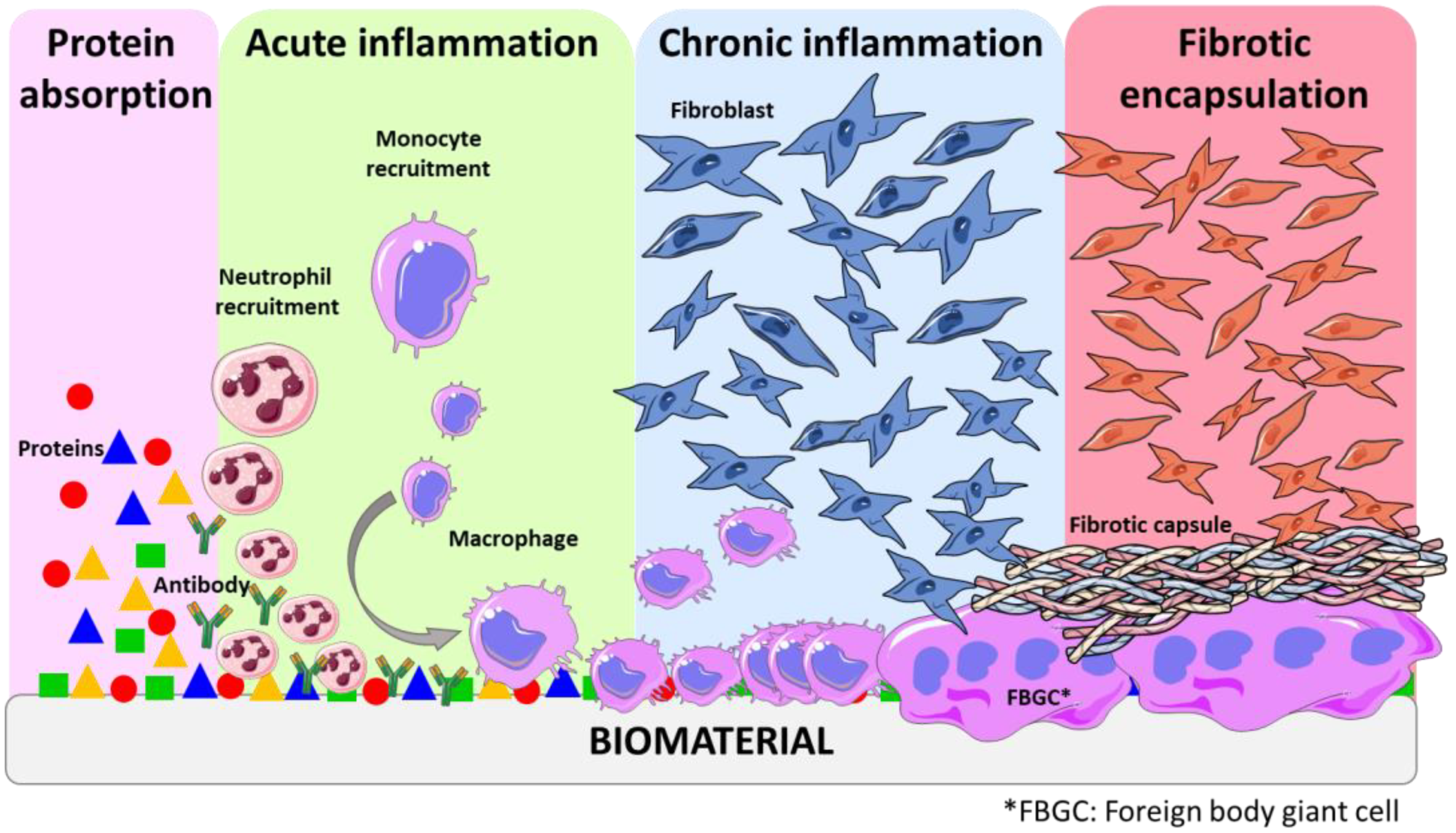
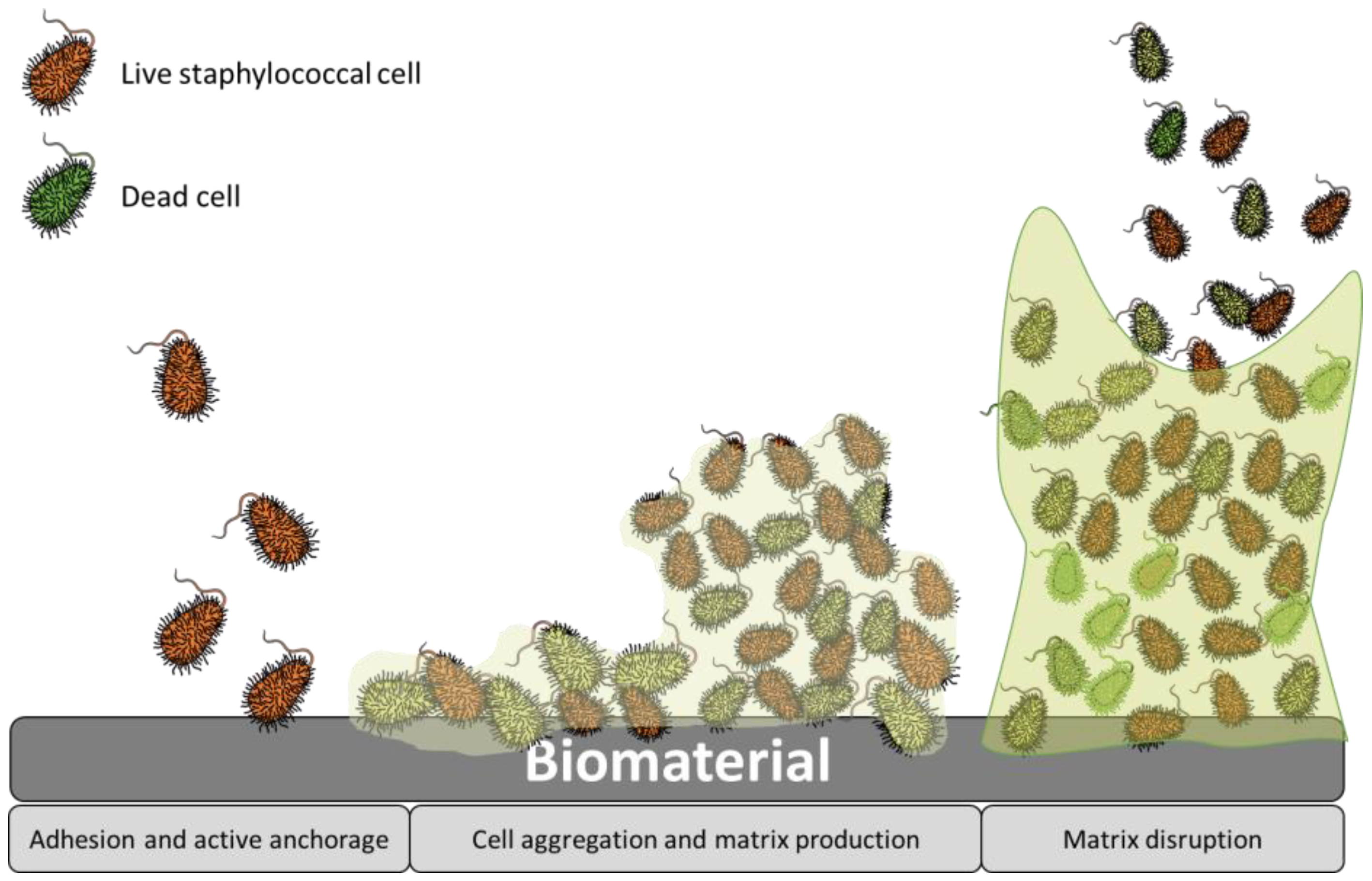
2. Biomedical Device Related Complications

Biomedical implants encompassing prosthetics, catheters, and an array of other devices, have undoubtedly revolutionized modern medicine, significantly improving the quality of life for countless patients. However, their integration with the human body does come with an inherent risk, which is an increased susceptibility to infections [13,14,15]. In fact, implant-related infections and the lack of biointegration represent the most prevalent and severe complications associated with the utilization of biomaterials. Infections can lead to various complications, ranging from localized discomfort to systemic health issues, potentially needing additional medical interventions and compromising patient outcomes [16].
When any biomaterial is implanted in the body, it induces a response from the host tissue, known as the host response [17]. This response occurs regardless of the method used to introduce the biomaterial, whether by injection or through surgery. The presence of a foreign biomaterial disrupts the local host tissue environment [17]. The magnitude of the host response depends on the extent to which the normal state of the equilibrium, known as homeostasis, is disturbed by the injury caused during implantation. This disruption, along with the introduction of the foreign object, determines the biocompatibility of the material. While numerous biomaterials and medical devices have been successfully implanted in humans, there is currently no material that can completely evade the highly efficient surveillance system of the human body. The host response is initiated by the adsorption of proteins on the surface of the material, leading to the formation of a dense collagenous capsule around the implant [11,18]. This encapsulation impedes further interaction of the implant with the surrounding tissue, a process often referred to as biofouling [17,19].
The various stages of foreign body response (FBR) constitute a dynamic process involving multiple intricate events. These stages include injury, blood–material interactions, provisional matrix formation, acute inflammation, chronic inflammation, granular tissue development, and fibrous capsule development (Figure 1) [17]. Blood is often the first body fluid to come into contact with implanted devices. Blood compatibility or hemocompatibility refers to a material’s ability to regulate the thrombotic and inflammatory responses induced by the foreign surface upon contact with blood. This attribute is an essential requirement for materials designed for blood–contact applications [20]. Such interactions between blood and medical devices trigger a complex series of events, including protein adsorption, platelet adhesion and activation, coagulation and thrombosis. The rapid absorption of plasma protein into the surface of biomaterial represents the initial event in blood–material interaction. This adsorption results in activated proteins that can catalyze, mediate, or moderate subsequent biological response to the biomaterial [19]. Surface-induced thrombosis is the main problem impeding the development of long-term blood contacting devices. Thrombus formation on device surfaces is a consequence of two key factors: platelet-mediated reactions and coagulation of blood plasma [20].
Throughout this process, a complex interactions of inflammatory cells, mitogens, chemo-attractants, cytokines, and other bioactive agents also play a key role in orchestrating the response [11]. Understanding each of these events is essential as they contribute significantly to the overall outcome of FBR. The delicate interaction between the immune system and the foreign material leads to the formation of provisional matrices, which triggers acute inflammation. This initial inflammatory response paves the way for chronic inflammation and subsequent granular tissue development. Ultimately, a fibrous capsule is formed to completely cover the foreign material, isolating and protecting the surrounding tissue from potential harm [21]. It is noteworthy that this late state of FBR is also influenced by the surface properties of the biomaterial. Some studies confirmed that all material classes elicited a comparable inflammatory response, suggesting that the material’s chemical composition plays a secondary role in this process. However, the roughness of the surface has great impact on the FBR—in fact, switching from a flat surface to a microstructured surface using the same material resulted in a notable decrease in the FBR [22,23].
Over the years, the concept of a “race to the surface” has been proposed to describe the competition between host cells and contaminating bacteria for occupying biomaterial surfaces [24]. The successful integration of biomaterials into host tissues is crucial for the effectiveness of many implants. Moreover, most studies conclude that rapid integration is also essential for preventing bacterial adhesion and colonization. In the particular case of orthopedics, the healing of bone tissue around the implant leads to the apposition of bone, facilitating the integration of the implant into the bone tissue, a process known as osseointegration [25]. In vitro studies with osteosarcoma cells demonstrate that pre-colonizing bacteria significantly alter and compromise host cell adhesion to material surfaces. It is important to note that if bacterial adhesion occurs before tissue repair takes place, the defense mechanism of the host may not be able to prevent surface colonization and subsequent biofilm formation [26].
The primary focus of this review will be orthopedic implants, as the prosthetics that remain in the body are particularly susceptible to thrombosis, inflammation, and infections, presenting significant challenges. In fact, these complications associated with implants frequently lead to device failure, requiring replacement in some cases, and can even result in chronic diseases [27,28]. Identifying and diagnosing orthopedic implant infections and inflammation, including determining the infectious agent and its antimicrobial sensitivity, pose significant difficulties. Moreover, treating these infections can be complicated due to various factors, such as antimicrobial resistance, tolerance, and/or persistence. Although the most widely recognized bacterial defense mechanism against antibiotics is resistance, which is based on the release of hydrolases to break down antibiotics and eject the antibiotics from cytosol, persistence stands as another fundamental mechanism that causes antibiotic treatment failure [29]. In contrast to resistant cells, persistent cells are genetically susceptible to antibiotics, yet they exhibit phenotypic tolerance, allowing them to endure antibiotic exposure. This phenomenon seems to be an ancestral trait, inherited from predecessor cells, as it is commonly observed in a variety of bacterial species, encompassing both Gram-negative and Gram-positive bacteria. During exposure to antibiotics, these species tend to develop a persister subpopulation as part of their adaptive survival strategy [30]. Besides Staphylococcus aureus being a common bacteria around orthopedic implant infection, it is essential to recognize that many other pathogens can also be responsible for causing such infections [15,31].
Implant infections are complex processes involving interactions among the pathogens, biomaterial, and the response of the host immune system. In the absence of foreign bodies, opportunistic pathogens are typically cleared by the defenses of the immune system. However, as commented previously, in the case of implant-associated infections, the biomaterial triggers a localized tissue response, leading to acute and chronic inflammation, foreign body reaction, granulation tissue formation, and eventual fibrous encapsulation. This unique environment creates a niche of immune depression, known as a locus minoris resistentiae, which makes the implant more susceptible to microbial colonization and infection. Furthermore, the biomaterial serves as a substrate for bacterial adhesion and biofilm formation [15]. Bacterial adhesion is the initial step in biomaterial-related infections and serves as a foundation for subsequent implant colonization. Once attached, pathogens form micro-colonies and develop protective biofilms, allowing them to persist in the hostile host environment. Thus, adhesion and biofilm formation are critical processes that enable pathogens to establish and maintain infections in implant sites. Understanding these complex interactions is essential for developing effective strategies to prevent and treat implant-associated infections [32,33].
Bacterial adhesion is a multi-stage process that can be divided into two main phases (Figure 2). The first stage involves the primary unspecific reversible attachment, while the second stage comprises specific irreversible attachment. When bacteria initially adhere to abiotic surfaces, such as those found in implants, the attachment is typically unspecific [15]. However, when they attach to living tissues, it involves specific interactions facilitated by lectins or adhesins. When a bare material surface comes into contact with physiological fluids such as blood and interstitial fluids, it rapidly becomes covered by extracellular matrix (ECM) proteins and immune components within nanoseconds [34,35]. This process is influenced by the surface chemistry and wettability of the implant surface. Hence, adhesins play a crucial role as the primary mechanism for bacterial attachment to the implant surface within the body. Both Staphylococcus aureus and Staphylococcus epidermis possess multiple mechanism for attachment and biofilm formation, significantly contributing to their virulence in chronic implant infections. The process of biofilm formation encompasses several stages (Figure 2): (I) adhesion, which is the initial stage; (II) micro-colony, where bacterial cells form aggregations and extracellular polymeric substances (EPS) are produced; (III) macro-colony formation, which undergoes further remodeling and maturation, resulting in the development of macro-colonies that appear as towers within the biofilm structure; and (IV) biofilm dispersal, which is the final stage, wherein some bacteria revert to a planktonic lifestyle, potentially colonizing new areas and initiating the biofilm formation process elsewhere [15].
Biological responses and bacterial adhesion are intricate processes influenced by numerous factors, but it is widely accepted that these responses are significantly affected by the surface properties [36]. In fact, various surface characteristics including chemistry, topography, surface free energy, elasticity, and charge play essential roles in modulating protein and cell interactions, and, consequently, host response.
Regarding surface topography and roughness, they play a crucial role in determining the biological responses to foreign materials and bacterial adhesion. Extensive research has shown that surfaces with micro- and nanoscale structures significantly impact various cells and bacteria behaviors. Surface patterning serves as a key determinant influencing both the contact area and the adhesion force between bacteria, proteins or cells, and the substrate. Indeed, these surface features can modulate cell orientation, morphology, adhesion, proliferation, and even regulate cellular functions and gene expression [37]. For instance, Yang et al. [38] compared the adhesion of both Gram-positive and Gram-negative bacteria on different patterned surfaces (Figure 3). Factors such as the geometry, size, and the height of the patterned surface impact on the interaction of bacterial and surfaces. Nanostructures with a high aspect-ratio, such as nanopillars and nanospikes, exhibited exceptional bactericidal activity. Indeed, when bacterial attachment occurs, the cell membrane of the bacteria lies within these nanostructured patterns cavities until the membrane breaks. On the other hand, both nanotubes and nano ripples have demonstrated efficacy in diminishing bacterial adhesion. Furthermore, enhanced bacterial reduction is obtained with larger diameters for nanotubes and reduced contact within the structure array for both nanotubes and nano ripples. Similarly microscale patterned surfaces including microwells, sub micro pillars, micro pillars, and micro protrusions present significant bacterial growth and colonization inhibition [39]. In fact, they trap bacteria within deep valleys, shielding them from the shear force of fluid, while a smooth surface facilitates the movement of attached bacteria, thereby increasing the probability of bacterial adhesion [38].
As commented, bacteria are not the only compounds that are influenced by the topography. Surface texturing serves as a strategic approach to modulate protein uptake on surfaces as well. This technique requires precise control over total protein adsorption levels, influencing the ratio of various proteins, spatial distribution, protein conformation, and surface binding affinity. The impact of nanoscale topographies on protein adsorption is particularly significant when the surface features align with the dimensions of the proteins. Conversely, interactions with topographies significantly larger than dimensions of proteins, such as micrometer-scale patterning, are generally perceived by proteins as a flat surface [40]. Moreover, smooth and flat implant surfaces have shown to induce the adhesion of foreign body giant cell (FBGC), which provoke the fibrotic capsule formation [22].
Concerning roughness, under static culture conditions, bacteria exhibit preference for smoother surfaces when the average roughness (Ra) value is low, ranging between 0.23 and 6.13 nm. Conversely, as these values increase within the range of 6–30 nm, bacteria tend to adhere to rougher surfaces [38,41]. This roughness adaptability was studied by Mu et al. [42], who prepared quartz surfaces with different roughness and treated with Salmonella enterica culture. The impact of the surface roughness on bacterial adhesion is evident from the findings illustrated in Figure 4. When the roughness is low (root-mean-square (RMS) > 10 nm), isolated microcolonies form, hosting a relatively sparse population of adherent bacteria with a low overall areal density. Progressing to intermediate roughness values (RMS between 10 and 40 nm), a substantial increase in adherent bacteria is observed, replacing isolated microclines with loosely connected bacterial monolayers. Additionally, the bacteria exhibited a more pronounced deformation/flattening ratio on these surfaces, suggesting a heightened attraction between bacteria and the surfaces. Conversely, at high roughness values (RMS < 45 nm), the areal density of adhering bacteria is exceeding low, and no microcolonies are observed. Bacteria predominantly exist as individual isolated organisms on these surfaces with a small fraction forming dimeric and trimeric aggregates.
Surface wettability is governed by both roughness and the chemistry of the surface jointly influencing its capability. It must be noted that the water contact angle (WCA) of rough surfaces (known as “apparent” WCA) differs from smooth surfaces (called “intrinsic” WCA). According to the Wenzel model, a rough hydrophilic surface exhibits an apparent WCA value lower than its intrinsic WCA value. Conversely, a rough hydrophobic surface displays an apparent WCA higher that it inherent WCA [43]. Some studies concluded that bacteria prefer to adhere to hydrophobic surfaces rather than hydrophilic ones. However, both superhydrophilic and superhydrophobic surfaces have demonstrated antibacterial behavior [44,45]. In fact, superhydrophobic surfaces, characterized by an apparent WCA exceeding 150°, require the entrapment of air bubbles within nanostructures or microstructures, as outlined by the Cassie and Bexter model [45]. Regarding proteins and macrophages, hydrophobic materials exhibit increased protein adsorption but also enhanced macrophage adhesion [46,47], potentially contributing to the initiation of fibrotic encapsulation. Conversely, in the case of hydrophilic materials, macrophages demonstrate heightened adhesion to positively charged implants in comparison to anionic or nonionic alternatives [22].
To address this critical challenge, extensive research and advancements in material science and implant design are continuously pursued.

3. Strategies for Combating Complications

As commented, several studies concluded that surface characteristic such as topography, wettability, charge, and chemical properties play a key role in proteins, cells, and bacterial adhesion and growth and, consequently, they influence hemo- and biocompatibility [48,49]. This section will delve into the crucial attributes of a surface, providing an in-depth exploration of the key factors necessary for optimal biointegration. It will also expose the desirable properties required to foster a favorable response from the body and establish robust protection against bacteria. Consequently, researches have recognized the significance of modifying these surface properties in implanted biomaterials to achieve enhanced biocompatibility and hemocompatibility and reduce both inflammatory response and bacterial adhesion.
Recent studies exploring the improvements on host tissue response and antibacterial properties of various materials have highlighted the potential of surface modification technologies in limiting and preventing bacterial contamination, as well as to promote proper adhesion of cells and proteins [38,50]. Notably, surface chemical modifications of biomaterial, drug delivery as well as immobilization of bioactive molecules that can directly or indirectly control the activity of components of the immune system have emerged as effective approaches in this regard. Therefore, this section will comprehensively delve into the chemical modifications required to tailored surface properties and it will present surfaces with added specific biological compounds (Figure 5).

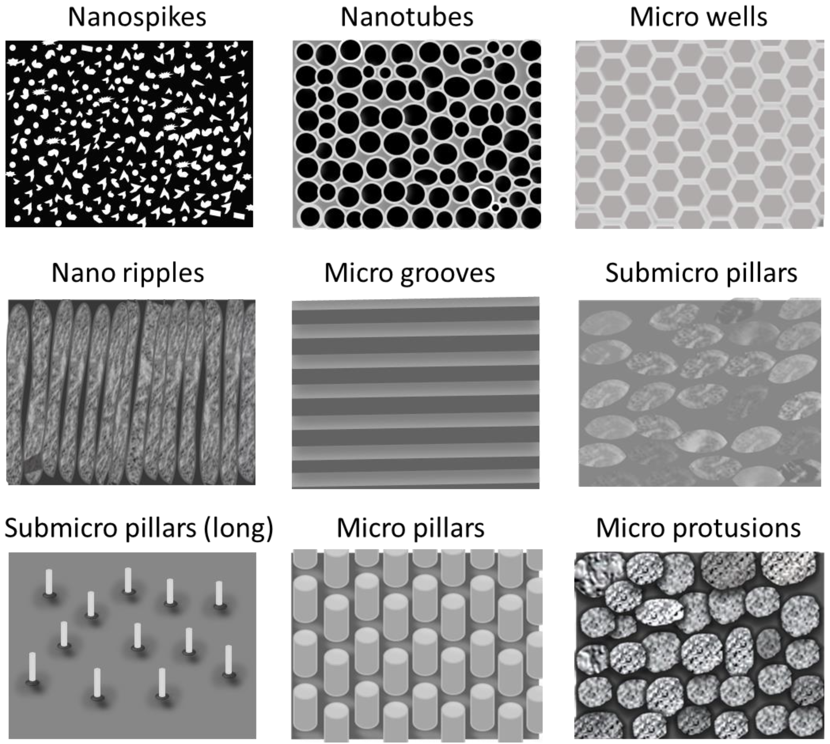
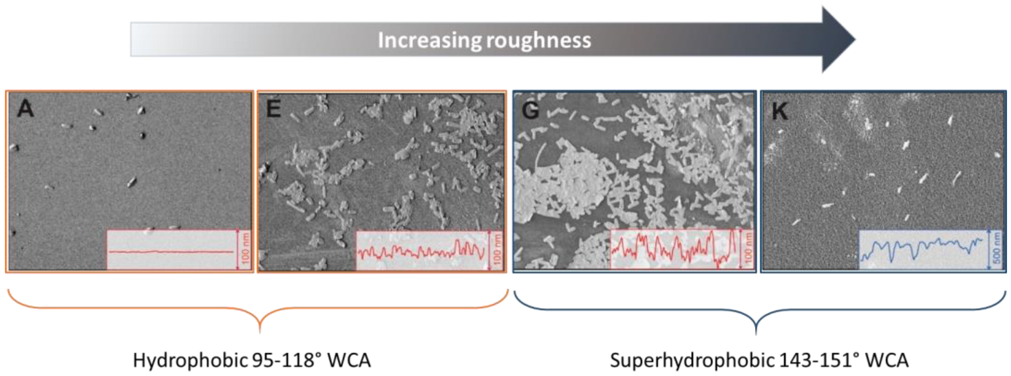
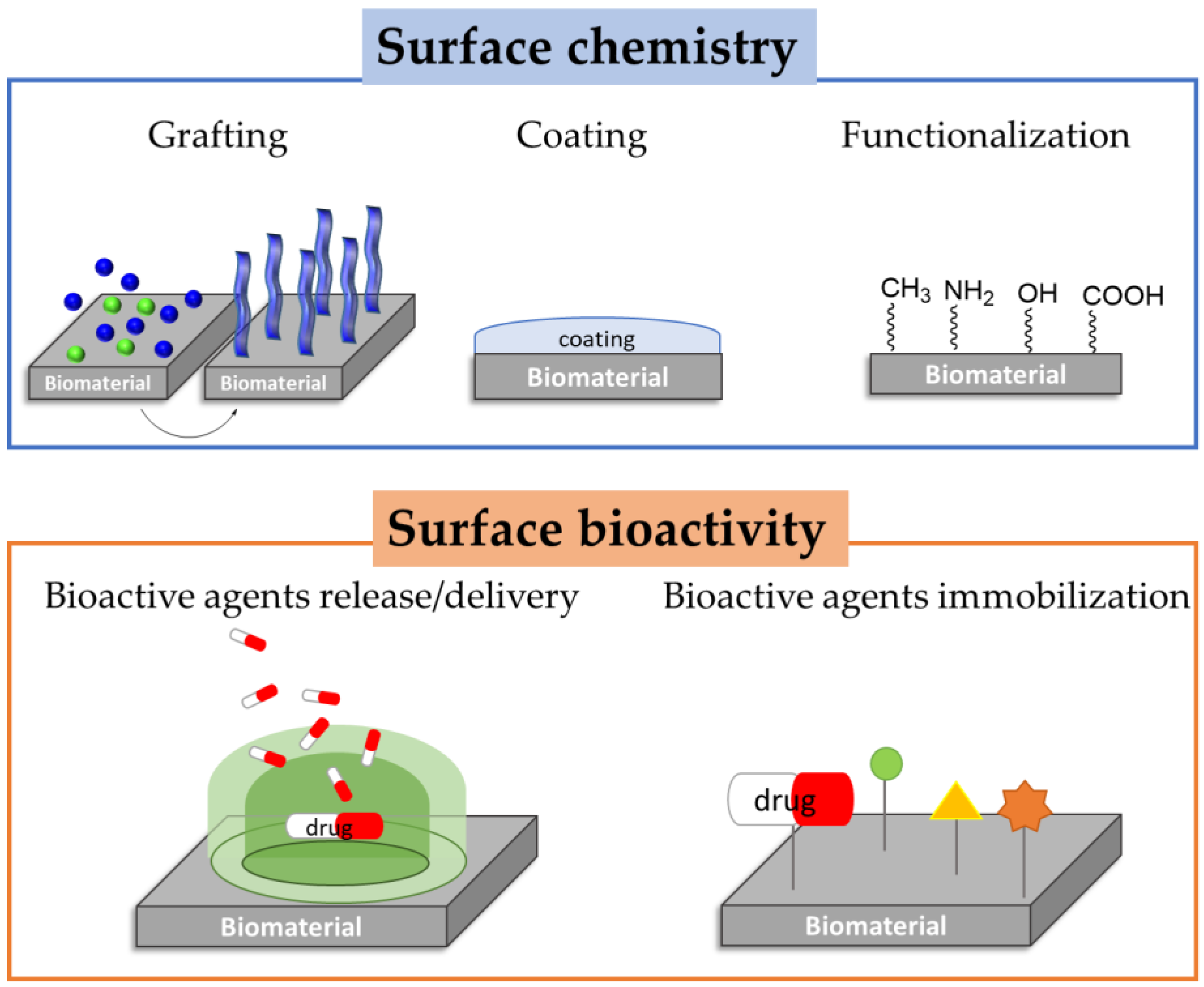
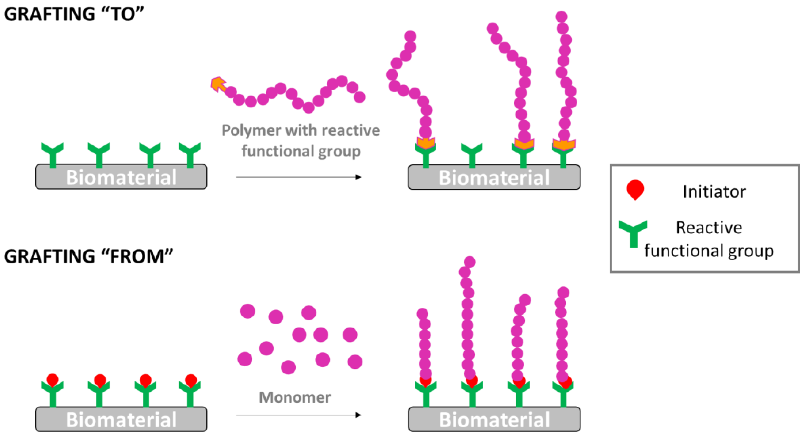
3.1. Surface Modification

Surface modifications encompass a wide range of complexities, ranging from simple alterations or introductions of a single functional group, to more intricate multi-step surface grafting reactions [36,51,52,53]. These grafting strategies often involve a preliminary surface activation step, where reactive functional groups, such as hydroxyl, amines, or carboxylic acids, among others, are introduced, followed by subsequent reaction to covalently link the molecule of interest to the surface [54].
A wide array of chemically based methods can be employed to introduce reactive functional groups onto biomaterial surfaces, effectively “activating” them for subsequent grafting reactions [55]. Interestingly, many of these treatments can also independently alter specific material surface properties, leading to modifications in cell–material interactions as well as bacterial–substrate interactions [56,57]. For instance, techniques that generate polarized hydroxyl [58], carboxyl [59], or amino [60,61] groups arouse changes in hydrophilicity/hydrophobicity and surface charge, influencing protein, cellular and bacterial adhesion [62]. The most commonly used chemically based surface functionalization methods involve surface hydrolysis, aminolysis, oxidation, and plasma treatment (Table 1) [63]. Each of these techniques offers unique advantages and can be tailored to suit specific biomaterial requirements.

3.1.1. Hydrolysis and Aminolysis

Surface hydrolysis via acid or base treatment is a commonly employed method to modify aliphatic polyesters, such as poly(lactic acid) (PLA), poly(glycolic acid) (PGA), or polyethyelene terephthalate (PET), and also metallic substrates, including Ti6Al4V [72,73,74]. However, it is important to note that the mechanism underlying the hydrolysis of polymers and metal substrates are inherently distinct. In polymers, hydrolysis induces random chemical cleavage of the ester bonds on the polymer backbone, generating, consequently, hydroxyl and carboxyl groups at the polymer surface [75,76,77]. On the other hand, in titanium (Ti) and its alloys, the hydrolysis only affects the passivated TiO2 coatings previously generated on the metallic surface, introduced by oxidation hydroxyl groups [36]. In any case, on both type of substrates, acid and alkali chemical treatments are the most used in the industry due to their versatility, simplicity, and effectiveness. A wide range of different treatments and mixtures can be employed, including basic or alkaline solutions (NH4/H2O2, NaOH, KOH, etc.) and acid solutions (HCl, HCl/H2O2, H2SO4/H2O2, H2SO4/HCl, etc.). Nevertheless, there are some concerns associated with this wet chemical technique [76,77,78]. For polymers, it is important to ensure that the concentration and treatment duration of the acidic/alkali solution do not significantly alter the bulk properties of the underlying polymer. Additionally, in both cases, the nonspecific nature of the treatment can lead to irregular surface degradation, potentially affecting the overall surface integrity and properties of the modified material [58,79]. Therefore, careful optimization of the hydrolysis process is essential to achieve desired modifications while preserving the core properties of the substrate. Nonetheless, these newly produced functional groups offer valuable attachment points for covalently linking other molecules to the polymer surface through various conjugation strategies. Although, it should be noted that hydrolysis itself has proved to increase cellular attachment in some polyesters as well as in some Ti alloys, due to the increment of the hydrophilicity and roughness, because of the change in the wettability properties of the surface [80].
Similarly, aminolysis aims to introduce reactive amine groups onto polymer and metallic surfaces. For this modification, polymers such as polyurethane (PU), poly(caprolactone) (PCL), or PLA are submerged onto diamines solutions such as 1,6-hexanediamine or ethylediamine, forming amides and obtaining free amino-end groups onto polymer surfaces [64,81]. Conversely, the introduction of amino groups into metallic substrates is usually more complex and it is generally necessary to use stronger conditions such as plasma. Nevertheless, similar to hydrolysis approaches, aminolysis can cause polymer degradation by increasing polymer roughness and wettability, which can alter subsequent protein, cell and even bacterial–material interactions [60,82].
An example of this was described by Yaseri et al. [64], In this work, the applicability of PCL nanofibers in tissue engineering was analyzed employing surface treatments strategies including hydrolysis and aminolysis. The hydrolysis was performed by using NaOH solution at different concentrations, while aminolysis was conducted using hexamethylenediamine (HMD)/isopropanol solution at different concentrations as well. It was observed that both treatments predominantly influenced the surface properties of PCL nanofibers without compromising their bulk properties. Beside minor morphological changes and a moderate reduction in mechanical performance, a notable enhancement in hydrophilicity was observed in most modified samples. In fact, an increase in hydrophilicity was observed when higher concentration of hydrolysis solutions and longer incubation times were employed. However, aminolysis solution concentrations did not significantly influence the hydrophilicity. It is worth to note that in vitro studies showed that the surface modifications of PCL nanofibers presented non-cytotoxicity as well as provided an ideal substrate for cell attachment, spreading, and proliferation when cultured L929 cells were employed.

3.1.2. Oxidation

The introduction of peroxide groups onto polymers or metallic surfaces for subsequent grafting reactions can be accomplished through various strategies and techniques, such as photo-oxidation by UV light or ozone oxidation. While UV light can discompose hydroperoxide groups onto reactive oxygen and hydroxyl radicals, ozone treatment produces peroxides, carboxyl, and carbonyl groups that can be further employed to initialize surface polymerizations or grafting reactions [83]. However, both approaches can degrade the polymer, thus, it is important to control and minimize significant changes of the bulk properties of the substrates [84]. Conversely, in the case of metals, electrochemical anodic oxidation has stood as the method of choice for over a decade to grow a thick and uniform oxide layer on metal surfaces [85]. This technique has been demonstrated to significantly enhance the biocompatibility of metal implants, as detailed by Huang et al. [66]. They employed an efficient electrochemical anodization treatment, which led to the formation of a nanoporous oxide layer free of aluminum onto Ti-6Al-7Nb surface. By oxidating the surface, they obtained notable improvements in corrosion resistance, as they observed reduction in both corrosion rate and passive current when immersed in simulated blood plasma. Additionally, the presence of the nanoporous oxide layer exhibited a positive impact on cell behavior. Specifically, it enhanced adhesion and proliferation of human bone marrow mesenchymal stem cells, providing significant importance in biomedical applications. It is worth to note that the development of an orderly oxide layer can be tailored by regulating parameters such as the choice of electrolyte, applied current density, electrolyte concentration, electrolyte temperature, stirring rate, and the ratio of cathode-to-anode surface areas [86].

3.1.3. Plasma

Plasma treatment offers a versatile method for introducing functional groups onto inert polymeric and metallic surfaces, both directly and indirectly. The process involves exciting a low-pressure gas, such as ammonia, oxygen or argon, in a chamber through various energy sources such as electric discharge, alternating/direct current, radio-frequency energy, microwaves, or heat [87,88]. This partial ionization of the gas leads to charged molecules that bombard the material surface by modifying its chemical and physical properties. The type of functionality introduced on the surface of the substrate depends on the choice of plasma gas and the operating parameters, such as pressure, power, gas flow rate, and time. For instance, reactive NH3 plasma introduces amines, O2 plasma produces a mixture of OH and COOH functionalities, and argon plasma creates radicals. Similarly to other approaches, these functional groups can be effectively employed in combination with other surface grafting methods. As other approaches, plasma treatments can directly enhance surface hydrophilicity and cellular adhesion, offering advantages in biomedical applications [63,89,90].
In this regard, Ujino et al. [70] employed atmospheric pressure plasma treatment to increase the hydrophilicity of pure Ti surfaces. The main goal of this study was to evaluate the impact of the hydrophilicity surfaces on the initial adhesion of the material to rat bone marrow and its subsequent differentiation into hard tissue. After applying plasma to 30 s, superhydrophilicity was induced on pure Ti surfaces. The results suggested that a notable enhancement in both adhesion and osteogenic differentiation of cells culture was obtained on plasma-treated samples in comparison with untreated disks.
Similarly, Mian Chen et al. [71] developed a fluorinated surface by plasma treatment of Ti surfaces. The experiments involved various fluorine chemical compositions applied as coatings. In vitro antibacterial studies were evaluated using Staphylococcus aureus and cell compatibility was studied employing MC3T3-E1 cells. The results suggested that both fluorocarbon coatings and metal fluorides coatings provided hydrophilicity with a nano-scaled roughness. Interestingly, the coating consisting of metal fluorides exhibited excellent bactericidal properties and demonstrated exceptional cytocompatibility. It has to be noted that antibacterial activity was attributed to the presence of metal fluorides and the release of fluoride ions.

3.2. Coatings

Polymeric and ceramic coatings have become interesting subjects for biomedical applications (Table 2). These coatings provide a number of valuable advantageous properties attributed to the underlying material, such as enhanced biocompatibility, improved mechanical robustness, increased wear and corrosion resistance, and enhanced functional capabilities.

3.2.1. Ceramic Coatings

Ceramic coatings are thin layers of ceramic materials that are applied to the surface of various substrates, such as metal, glass, or ceramics to enhance their properties or provide specific functionalities. These coatings are commonly used in a wide range of industrial and technological applications due to their unique combination of properties, which can include high temperature resistance, wear resistance, corrosion resistance, electrical insulation, thermal insulation, and biocompatibility.
Among ceramic coatings, calcium phosphates (CaPs) are the most commonly employed due to their remarkable similarity to bone tissue. Indeed, they represent highly promising materials in the field of bone regeneration, providing compelling substitutes for auto- and allografts in facilitating and reinforcing tissue regeneration within critically-sized bone defects [109]. Their exceptional biocompatibility and biodegradability make them especially well-suited for this purpose, owing to their resemblance to the mineral phase found in natural bone. Numerous studies have been dedicated to the development of CaP ceramic coatings on metallic substrates with the goal of replicating the biological properties of bulk bone tissue and improving the durability and stability of implants.
Biomineral formation and the adhesion of cells and proteins can be effectively regulated by tailoring the surface properties such as roughness and porosity of CaP materials. On the other hand, it should be noted that different phosphates, such as hydroxyapatite (Hap) or tricalcium phosphate (TCP) have different biocompatibility due to differences in crystallinity, solubility, stability, ion release, and mechanical properties.
As commented, among CaPs, HAp deserves special attention, as it constitutes the primary inorganic component of bone tissue. In fact, recent advancements in materials science and processing have enabled the production of hydroxyapatite-based grafts in various forms, satisfying the demand for a wide range of clinical applications. Moreover, these innovations have obtained promising results in both in vitro and in vivo studies [109].
In this context, Chen et al. [100] introduced a method for electrodepositing a nanostructured HAp coating onto Ti surface. To enhance the adhesion between the HAp coating and the Ti surface, they employed chemical etching and oxidation treatments, generating a thin TiO2 layer which served as an interlayer that mitigated thermal stress and prevented the formation of crack in the coating. After electrodepositing HAp, uniform and crack free HAp nanostructured coating was successfully generated onto the Ti surface. Additionally, in vitro MSCs cell culture experiments demonstrated the excellent biocompatibility and bioactivity of HAp-Ti nanostructrured surface. The MSCs exhibited enhanced proliferation on Ti surfaces with HAp coating compared to pristine Ti surfaces.
Similarly, Hui Du et al. [96] fabricated a coating composed of calcium silicate and calcium phosphate onto Mg-Zn-Mn-Ca alloy through a chemical reaction involving NaSiO3 and Ca(NO3)2. In vitro cell studies concluded that osteoblasts exhibited good cell adhesion, high growth rates and proliferation characteristics. These results indicate a significance enhancement in surface cytocompatibility attributed to the presence of calcium phosphate coating.
Other type of ceramic materials are zirconia-based coatings (ZrO2). This type of material can withstand high temperatures and elevated stresses. Its uses span across various domains, including dental implants and the application of protective coatings on metallic implants to enhance their resistance to corrosion. ZrO2 ceramics offer a multitude of advantages, encompassing robust mechanical strength, chemical stability, biocompatibility, and superior wear resistance. Additionally, zirconia stabilized with yttria (YSZ) has gained prominence as a dental implant material. YSZ coatings exhibit superior hardness and scratch resistance when compared to HAp coatings. Furthermore, Saravan et al. [110] revealed that YSZ-coated Ti substrates exhibit enhanced hemocompatibility, stimulating blood platelets to develop pseudopods.
Bioactive glasses (BGs) constitute another type of promising ceramic material, mainly due to their osteoinductive and bioresorbable properties, which make them suitable materials for bone tissue engineering applications. Ideally, bioactive implants used in clinical applications should exhibit similar properties as the host tissue, while establishing robust interfacial connections with both hard and soft tissues. Owing to the inorganic composition and mechanical attributes of the bioactive glasses, which closely mimic those of “hard” bone tissue, there has been considerable interest in their application in bone and teeth-based implants. However, their inadequate mechanical properties considerably hinder their use in load-bearing situations [111,112]. Indeed, the majority of BGs present lower fracture toughness when compared to natural load-bearing cortical bone, with BGs ranging between 0.2 and 0.6 MPa, while cortical bone registers a range from 2 to 12 MPa in terms of fracture toughness [113]. Therefore, the use of a BG coating emerges as a viable strategy to not only bolster the osseointegration of metallic implants but also mitigate the inherent brittleness of BGs. BG and glass-ceramic coatings on metallic implants can be produced by different techniques, including thermal spraying, radiofrequency magnetron sputtering (RF-MS) deposition, pulser lasered deposition (PLD), sol gel coating, and electrophoretic deposition (EPD) [114].
In this context, Bargavi et al. [115] presented a thin film coating based on zirconia incorporated on a BG matrix and deposited onto commercially pure Ti (Cp-Ti) substrates. The incorporation of Zr, in different concentrations, aimed to enhance the mechanical stability of the coating. Hemocompatibilty studies revealed excellent compatibility, with a favorable hemolysis rate of less than 2%. Furthermore, in vitro cytocompatibility assays employing MG-63 osteoblast cell lines demonstrated a noteworthy enhancement in cell viability. Additionally, according to antibacterial assays, when Bg-Zr composites with high contents of Zr were used to coat Cp-Ti substrates, reduced biofilm formation was observed presumably due to the increase in surface roughness. Consequently, surface modification of Cp-Ti implant materials using BG-Zr coating exhibited improved bioactivity and enhanced osseointegration, making this type of coating suitable for orthopedic applications.

3.2.2. Polymer Coatings

Another type of coatings with a crucial role in biomedical applications are polymeric coatings. In fact, these types of coatings are commonly used to improve the performance, biocompatibility and functionality of biomedical devices, Furthermore, while providing these specific benefits, these type of polymers can improve interactions with tissues and biological fluids [33,116]. In this context, it is worth noting the widely employed strategy of covering a surface with a polymer with antifouling properties.
Another widely employed strategy to alter surface characteristics involves the passivation of biomaterials through surface coatings with antifouling behavior. For instance, polyethyelene glycol (PEG), poly(hydroxyethyl methacrylate) (PHEMA), and phosphatidylcholine polymers [117,118,119], among others constitute prominent examples of polymeric materials employed in this regard. These coatings exhibit robust steric repulsion and instigate hydration forces that effectively avoid protein and bacterial adsorption. By employing these mechanisms, these coatings protect the material form of the host immune system and, consequently, limit leukocyte adhesion and the host inflammatory response. Moreover, these highly hydrophilic coatings offer a straightforward solution for repelling bacteria and enhancing a more favorable host–material interaction.
In this context, Ungureanu and coworkers electrodeposited a composite coating based on Polypirrole (PPy) and Polyethyelene glycol (PEG) onto Ti alloy [120]. Three different concentrations of PEG were employed, specifically 0.5%, 2%, and 4%. When testing antibacterial properties of the coatings, the best effect was found for the coating with 2% PEG concentration, which has hydrophilic character and minor roughness. Such results are in concordance with the mechanism of biomaterial–bacteria interaction, which involves as factors affecting bacterial adhesion and growth an initial physicochemical interaction stage, where roughness and wettability are factors that can regulate bacterial adhesion and biofilm deposition.
In recent years, the layer-by-layer (LBL) methodology has gained widespread popularity as a versatile and effective technique for depositing polymeric materials on a surface This innovative method involves the sequential deposition of cationic and anionic polyelectrolyte layers, which can be firmly bonded through ionic interactions to create a thin, precisely controlled coating film. This process relies on the positive and negative charges that each polymer acquired under specific pH conditions. The LBL approach offers exceptional flexibility, allowing the creation of coating with diverse functionalities and tailored properties [121]. By varying the types and sequences of polyelectrolytes used, it is possible to achieve specific surface characteristics, such as charge, hydrophilicity, or bioactivity. This technique has found application in various fields, including biomedical engineering, drug delivery systems and surface modification of medical implants [122,123,124,125]. Its ability to produce thin and uniform coatings with controlled release capabilities has opened new avenues for enhancing biocompatibility and functionality of biomaterials.
The successful coating of PET films by positively charged chitosan (CHI) and negatively charged hyaluronic acid (HA) described by Alvarez et al. [74] constitute an example of this methodology. A layer-by-layer technique was employed to introduce each polymer onto the surface, fabricating a nanometer-scale thickness coating and producing a potentially antifouling surface by electrostatic interactions. While CHI contributed to contact-killing properties, hydrophilicity provided by HA facilitated bacteria repellence through a steric effect generated by water absorption.
Various strategies have been proposed to develop coatings that exhibit enhanced physical and chemical resistance. However, these approaches are often constrained by the type of bonding between the coating and the substrate, as the coatings discussed so far are physically placed on top of the material, without any stable, covalent interaction between the two systems. A promising alternative method involves grafting, where covalent immobilization of compound takes place to create a resilient film on the material surface. Currently, two main grafting methods are employed: “grafting to” and “grafting from” [126].
In the “grafting to” method, polymer chains already preformed are attached to the surface, providing a means to modify the properties of the coating. Conversely, in the case of the “grafting from” methodology, monomers are bonded to the surface where subsequent polymerization takes place (Figure 6). This last approach offers greater control over the structure of the coating and properties. By employing grafting techniques, it is possible to obtain coatings with tailored functionalities, such as improved resistance to wear, corrosion, and environmental degradation. Additionally, covalently immobilizing the polymers ensures better adhesion and durability, resulting in coatings that can withstand harsh conditions over extended periods [127].
In this context, Huh et al. [128] performed different experiments using oxygen plasma glow discharge onto PET samples. As a consequence of the plasma, the texture of PET surfaces was enhanced, resulting in the formation of peroxides on its surfaces. These peroxides were employed as catalysts for the grafting and polymerization of acrylic acid in order to introduce carboxylic acids onto the surfaces. Subsequently, neutral and quaternized chitosan were coupled with the introduced carboxyl groups, leading to chitosan-grafted PET and quaternized chitosan-grafted PET. To assess the antibacterial activity of the modified PET textures, a shake flask method was employed. After shaking for 6 h, it was observed that PET with covalently grafted chitosan and quaternized chitosan showed significant inhibition of bacterial growth. Even after PET texture laundering, the inhibition of bacterial growth remained in the range 48–58%, demonstrating the durability and effectiveness of the chitosan grafted PET textures against washing.

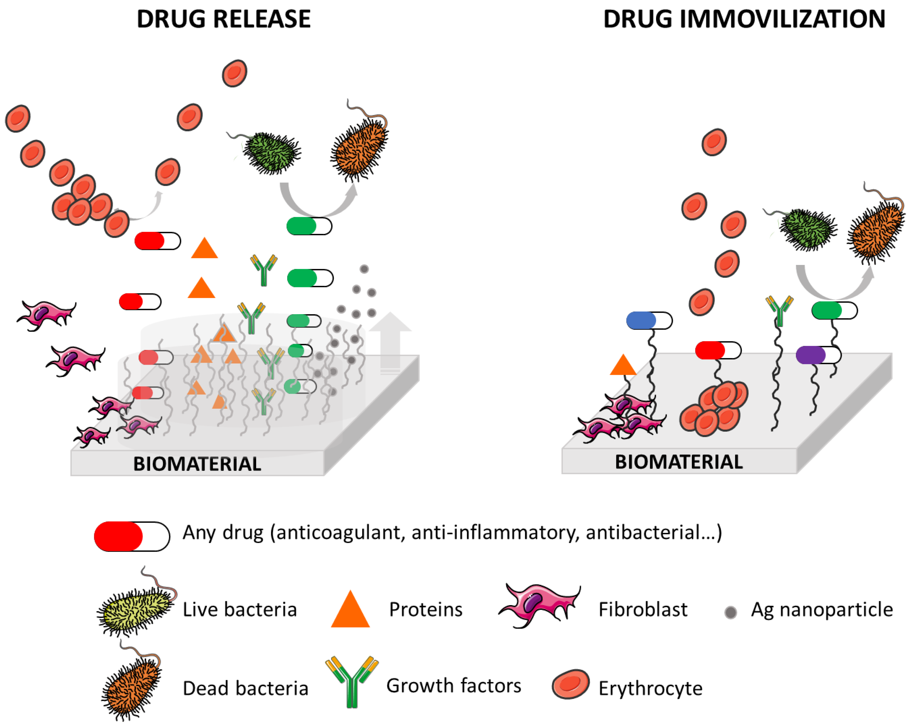
3.3. Targeted Drug Delivery

A promising strategy to develop active surfaces involves the controlled release of various active agents, such as drugs, growth factors, proteins, peptides, nucleic acids, and even silver nanoparticles [54]. This controlled release occurs from a variety of platforms, including hydrogels and nanogels, polymer multilayers and cyclodextrines and enables the desired surface response [129,130,131]. These structures, which are built upon polymers and proteins, serve as remarkably versatile reservoirs capable of releasing bioactive molecules (Figure 7).
As commented before, an alternative and versatile approach to creating coatings with self-controlled active agent release ability is through the use of multilayer systems. While multilayered coatings may have lower drug loading capacity compared to hydrogels, they present excellent control in chemical composition, structure, thickness, homogeneity, and responsiveness. To achieve effective loading and sustained release of active compounds, it is essential to have intermediate strength bonds or interactions between polymers and the active agents. Similar to hydrogels, the release of active agents from multilayer coatings typically occurs through diffusion and multilayer degradation processes.
Implant-associated infections in orthopedic surgeries represent a critical concern due to their potential to impede bone healing, induce implant failure, and even escalate to osteomyelitis. The concept of drug-eluting implants, designed for localized antibiotic delivery at surgical sites holds promise in mitigating these infections. In the study presented by Li et al. [132], vancomycin, an antibiotic, was encapsulated within a PEG based hydrogel film. This hydrogel was covalently bind to Ti implants and subsequently enveloped by a PEG-(poly(lactic-co-caprolactone) (PEG-PLC) membrane. Additionally, crosslinked starch was incorporated into the hydrogel due to its porous microstructure, which effectively curbed hydrogel swelling and consequently regulated drug release. The release kinetics of vancomycin were found to be controllable, dependent on both the drug loading and the thickness of the coating. Notably, the vancomycin-loaded Ti samples exhibited a sustained drug release profile, with no initial burst release. In fact, in vitro experiments demonstrated continuous drug release for nearly 3 weeks, while in vivo testing extended this period to over 4 weeks. Furthermore, a rabbit model subjected to Staphylococcus aureus infections exhibited a significant reduction in the inflammatory response and demonstrated robust antimicrobial property when implants containing 4 mg of vancomycin were used. Therefore, this approach holds promise as an effective strategy for the treatment and prevention of localized bone infections.
Similarly, Karakurt et al. [133] presented two different strategies for creating a combined saccharide coating onto PLLA with the aim to develop antibacterial biomaterial surfaces. Initially, PLLA samples were exposed to low-pressure plasma treatment and were then reacted with acrylic acid solution to obtain COOH and OH reactive functional groups. Subsequently, a “grafting from” approach was employed to create polyacrilic acid (PAA) brushes on PLLA surface. Afterward, chitosan was introduced to the surface by either covalently carbodiimide coupling reactions or by direct coating method with electrostatic interactions. Following this, lomefloxacin-containing chondroitin sulfate saccharide was coated onto the previously prepared surface, resulting in a polyelectrolyte complex (PEC). The coatings with the PEC formation between CS and ChS exhibited enhanced antibacterial activity against bacterial strains compared to individual coatings. Furthermore, these interactions increased the amount of lomefloxacin adhered to the film coatings and extended the drug release profile. Finally, the zone of inhibition test confirmed that the CS-ChS coating showed a contact killing mechanism, whereas drug-loaded films demonstrated a dual killing mechanism, encompassing both contact and release-based antibacterial actions.
Another example regarding antibacterial properties is described by Chen et al. [134] They successfully developed a cost-effective strategy to obtain antibacterial 3D-printed PLA disks. They employed the direct adsorption of two antibiotic agents, ampicillin and vancomycin, onto the PLA disk surfaces. They observed the maximum adsorption capacities of ampicillin and vancomycin on the PLA disk surfaces to be approximately 75 mg/g of PLA and 65 mg/g of PLA, respectively. As they varied the concentration of the antibiotic agents in the aqueous solution, they noted a corresponding decrease in the amount of antibiotic agents absorbed on the sample surfaces. When they employed an antibiotic agent concentration of 50 mg/mL in the aqueous solution for absorption onto the samples, they achieved stable drug release profiles. These profiles consistently maintained antibiotic agent concentration in the buffer solution above the minimum inhibitory concentration (MIC90) for Staphylococcus aureus. Furthermore, the drug release kinetics of the antibiotic agents from the samples closely followed the Korsmeyer–Peppas model. The bioactivity of ampicillin and vancomycin, when suitably absorbed onto the sample surfaces, remained effective for at least 28 days. In practical terms, the PLA disk with directly absorbed antibiotic agents reduced the relative optical density of Staphylococcus aureus in a solution with a concentration of 106 colony-forming units per milliliter (CFU/mL) to 40%, compared to a solution with only Staphylococcus aureus under the same conditions.

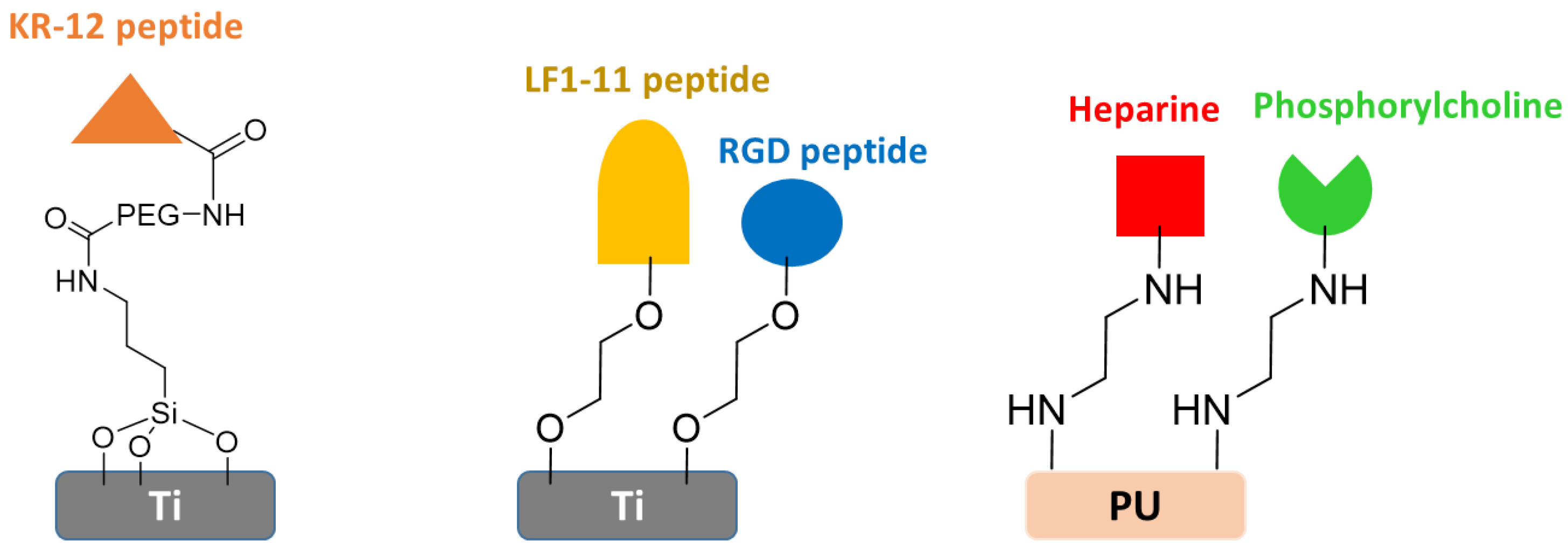
3.4. Drug Immobilization Approach

In this approach, the biomolecule or bioactive agent is effectively anchored to the surface of the material through covalent immobilization. Concerning antibacterial behavior, bactericidal agents such as low molecular antibiotics, bacteriophages, cationic antimicrobial peptides, lysozyme, or quaternary ammonium polymers can cause bacterial death upon contact (Figure 7). Typically, the death of bacteria occurs by either the disruption of the bacterial membrane or from specific interactions of the immobilized agent with target biomolecules on the bacterial surface. Similarly, anti-inflammatory and anticoagulant properties can be obtained by immobilizing agents with the corresponding activity. However, the mechanisms behind these actions are intricate and depends on the specific agent involved. For instance, anti-inflammatory biomolecules such as glycosaminoglycans (GAGs), including heparin (HEP), chondroitin sulfate (CS), or hyaluronic acid (HA), which have demonstrated significant anti-inflammatory potency in numerous experimental studies and clinical trials, play a different mechanism to induce anti-inflammatory response.
It is noteworthy that in order to carry out the immobilization of the active agents, it is necessary to previously modify or activate the material surface. For this purpose, it is common to use the aforementioned strategies such as grafting and functionalization, since they allow the introduction of suitable functional groups to carry out the conjugation reactions such as amidation, esterification, or even click reactions widely used in this regard.
In this context, the covalent immobilization of antimicrobial peptides onto Ti surfaces has indeed been a well-established approach to prevent bacterial adhesion and biofilm formation. However, uncertainty remains regarding the necessity of using a spacer to bind the peptide onto the surface in order to promote antibacterial adhesion, while maintaining excellent biocompatibility. In this sense, the antibacterial properties and the inflammatory response elicited by non-functionalized Ti substrates and PEG covered Ti surfaces were investigated in a study carried out by Nie and coworkers [135]. Both surfaces were subsequently covalently functionalized with KR-12, a derived peptide from LL-37, a substance known for its bactericidal and bacteriostatic properties in solution. For this purpose, Ti surfaces were initially activated with NaOH alkali solution to introduce OH functional groups on the surface and then silanized with (2-aminoethylamino)propyltrimethoxysilane. Alternatively, for PEG conjugation, 1-ethyl-3-(3-dimethylaminopropyl)carbodiimide (EDC) and N-hydroxysuccinimide (NHS) coupling agents were employed. Finally, KR-12 peptide was immobilized onto silanizated Ti surfaces and PEG covered Ti surfaces using, again, EDC and NHS (Figure 8). Authors reported that the introduction of KR-12 profoundly affected bacterial adhesion. Indeed, a significant decrease of bacterial adhesion was achieved on both surfaces. In comparative terms, PEGylated surfaces demonstrated a marked enhancement in antimicrobial efficacy, resulting in a notable reduction in vitro adhesion and biofilm formation of Staphylococcus epidermis compared to non-PEGylated Ti surfaces. Furthermore, both PEGylated and non-PEGylated Ti surfaces exhibited a significant decrease in TNF-α and IL-1β secretion, leading to macrophages remaining in an inactive rounded state. Therefore, this study confirmed the increase in the effectiveness of the same active agent by the use of a spacer to attach it to a surface.
On the other hand, Andras Heijink and coworkers proposed to enhance the cellular adhesion of Ti implant surfaces by functionalizing them with the Arg-Gly-Aps tripeptide (RGD) [136]. Their objective was to facilitate the attachment of osteoblasts, a key step in achieving improved implant fixation. This study encompassed a comprehensive examination of the histomorphometric and mechanical performance of Ti implants, exploring two different approaches for RGD immobilization: one involving self-assembled monolayers of phosphonates (RGD/SAMPS) and the other employing the more conventional thiolate-gold interface (RGD/thiolate-gold). The results suggested that RGD/SAMP-coated implants exhibited a substantially greater affinity for bone growth and superior implant fixation compared to their RGD/thiolate-gold-coated surfaces.
In this context, Hoyos-Nogues and collaborators employed a dual peptide approach and PEG coating strategy onto commercially pure Ti samples [137]. They presented a method for the development of a trifunctional coating designed to repel bacterial contamination, kill adhering bacteria, and promote osteoblast adhesion. For this purpose, the functionalization of Ti surfaces was carried out through the electrodeposition of an antifouling PEG layer, followed by the binding of a peptide platform, which contained RGD and LF1-11, which provided both cell-adhesive and bactericidal properties (Figure 8). As the results suggested, the deposition of the PEG coating and the immobilization of the biomolecules did not alter the morphology and topography of Ti samples. Additionally, PET-coated and peptide immobilized samples demonstrated an efficacy in preventing protein adsorption and hindered the attachment of osteoblast cells. However, the introduction of cell adhesive domains rescued osteoblast adhesion, resulting in significantly higher levels of cell attachment and spreading when compared to control samples. Regarding antibacterial properties, the presence of PEG layers led to a substantial reduction in bacterial attachment on the surface, which was further improved when the bactericidal peptide was introduced, reaching levels below 0.2%. As commented on the Introduction Section, the balance between the risk of infection and the optimal osseointegration of a biomaterial is often described as “the race for the surface”, in which contaminating bacteria and host tissue cells compete to colonize the implant. In this study, a multifunctional coating for Ti surfaces was successfully developed, since it not only promoted the attachment and spreading of osteoblast cells, but also effectively inhibited bacterial colonization.
Regarding anticoagulant behavior, Tan and coworkers [138] grafted heparin (HEP) and phosphorylcholine groups (PC) onto a polyurethane (PU) surface in order to enhance biocompatibility and impart anticoagulant properties. After the surface grafting sites of PU were amplified with the primary amine groups of polyethylenimine (PEI), heparin was covalently anchored to the surface through an amidation reaction. Simultaneously, PC groups were covalently immobilized on the PU-PEI surface through the reaction between the amino group and the aldehyde group of phosphorylcholine glyceraldehyde (PCGA) (Figure 8). The resulted PU-HEP and PU-PC composite films exhibited a significant reduction in platelet adhesion, underscoring the efficacy in minimizing thrombotic events. Importantly, these materials exhibited exceptional antithrombogenicity and blood compatibility, rendering them versatile candidates with potential applications in many fields, including artificial blood vessels, artificial heart valve prothesis, or heart stents. Furthermore, these modifications significantly enhanced the hydrophilicity and hemocompatibility. These results suggested that the PU-HEP and PU-PC composite films are promising candidates for blood contacting tissue engineering.
Similarly, Ozaltin and coworkers employed direct current air plasma treatment onto PET in order to create and oxidative layer to bind the marine-derived anticoagulant sulphated polysaccharide, fucoidan [139]. To optimize the chemical bonding behavior and, consequently, the anticoagulant performance, this immobilization process was meticulously conducted at various pH values from 3 to 7, concluding that pH 5 was optimal. Under these conditions, the immobilized fucoidan exhibited exceptional anticoagulant activity, consistently surpassing the crucial threshold of 100 s. This remarkable performance serves as clear evidence of its complete suitability for PET devices designed for direct contact with blood.

4. Conclusions and Future Trends

Biomedical devices play a crucial role in modern medicine; in fact, their use has contributed to significantly improving the quality of life of some patients and even, in some cases, life expectancy. For this reason, great efforts have been devoted to developing new materials that can be used in the design of these medical devices. In any case, a multidisciplinary approach based on the principles of materials science, engineering, biomechanics, molecular biology, pharmaceuticals, and ongoing comprehensive clinical monitoring is mandatory in the design of such implants and prostheses. As a result of this intense multidisciplinary research, first generation biomaterials, generally inert materials whose only function was to replace organs or tissues, have given way to second generation materials which, in addition to performing their function, minimize or even cancel the associated drawbacks, such as the risk of infection related to bacterial adhesion, thrombus formation, or inflammatory response.
Research in this field is currently focused on two priority directions. On the one hand, the development of new smart implants based on third or even fourth generation materials, with biomimetic properties capable of, after binding to damaged tissue, generating signals to stimulate its growth and even disappearing, bio-absorbing once its function has been performed.
On the other hand, 3D printing, currently in full swing, is a revolutionary technique with a profound impact on the manufacturing of medical implants mainly due to its great precision even with very complex structures. Everything indicates, therefore, that combining 3D printing with smarts biomaterials represents the future not only in the development of medical devices but also in the medical industry in general.

Author Contributions

Conceptualization, J.S.-B. and I.M.-B.; writing—original draft preparation, J.S.-B. and M.D.-G.; writing—review and editing, J.S.-B. and I.M.-B.; supervision, J.L.V.-V.; project administration, L.P.-Á.; funding acquisition, J.L.V.-V. and L.P.-Á. All authors have read and agreed to the published version of the manuscript.

Funding

The authors acknowledge the Basque Government for Grupos Consolidados grant IT1756-22 and ELKARTEK program KK-2021/00025.

Institutional Review Board Statement

Not applicable.

Informed Consent Statement

Not applicable.

Data Availability Statement

Data are contained within the article.

Conflicts of Interest

The authors declare no conflict of interest.

References

  1. Williams, D.F. On the nature of biomaterials. Biomaterials 2009, 30, 5897–5909. [Google Scholar] [CrossRef]
  2. Festas, A.J.; Ramos, A.; Davim, J.P. Medical devices biomaterials—A review. Proc. Inst. Mech. Eng. Part L J. Mater. Des. Appl. 2020, 234, 218–228. [Google Scholar] [CrossRef]
  3. Crawford, L.; Wyatt, M.; Bryers, J.; Ratner, B. Biocompatibility Evolves: Phenomenology to Toxicology to Regeneration. Adv. Healthc. Mater. 2021, 10, e2002153. [Google Scholar] [CrossRef]
  4. Black, J. Biological Performance of Materials: Fundamentals of Biocompatibility, 4th ed.; Routledge: London, UK, 2005; ISBN 9781420057843. [Google Scholar]
  5. Parisi, L.; Galli, C.; Neri, A.; Toffoli, A.; Calciolari, E.; Manfredi, E.; Lumetti, S.; Macaluso, G.M.; Rivara, F.; Macaluso, C. Aptamers improve the bioactivity of biomaterials. Aptamers 2017, 1, 3–12. [Google Scholar]
  6. Williams, D.F. On the mechanisms of biocompatibility. Biomaterials 2008, 29, 2941–2953. [Google Scholar] [CrossRef]
  7. Chen, Q.; Thouas, G.A. Metallic implant biomaterials. Mater. Sci. Eng. R Rep. 2015, 87, 1–57. [Google Scholar] [CrossRef]
  8. Cber, C. Use of the International Standard ISO 10993-1; FDA Guidance Document; FDA: Silver Spring, ML, USA, 2023; Docket No. FDA-2013-D-0350; pp. 1–66. [Google Scholar]
  9. Ghasemi-Mobarakeh, L.; Kolahreez, D.; Ramakrishna, S.; Williams, D. Key terminology in biomaterials and biocompatibility. Curr. Opin. Biomed. Eng. 2019, 10, 45–50. [Google Scholar] [CrossRef]
  10. Reinwald, Y.; Shakesheff, K.; Howdle, S. Biomedical Devices; Wiley: Chichester, UK, 2011; ISBN 9780470390849. [Google Scholar]
  11. Velnar, T.; Bunc, G.; Klobucar, R.; Gradisnik, L. Biomaterials and host versus graft response: A short review. Bosn. J. Basic Med. Sci. 2016, 16, 82–90. [Google Scholar] [CrossRef]
  12. Chalovich, J.M.; Eisenberg, E. Synthetic biodegradable functional polymers for tissue engineering: A brief review. Sci. China Chem. 2014, 57, 490–500. [Google Scholar]
  13. Agrawal, R.; Kumar, A.; Mohammed, M.K.A.; Singh, S. Biomaterial types, properties, medical applications, and other factors: A recent review. J. Zhejiang Univ. Sci. A, 2023; ahead of print. [Google Scholar] [CrossRef]
  14. Schierholz, J.M.; Beuth, J. Implant infections: A haven for opportunistic bacteria. J. Hosp. Infect. 2001, 49, 87–93. [Google Scholar] [CrossRef]
  15. Arciola, C.R.; Campoccia, D.; Montanaro, L. Implant infections: Adhesion, biofilm formation and immune evasion. Nat. Rev. Microbiol. 2018, 16, 397–409. [Google Scholar] [CrossRef]
  16. Campoccia, D.; Montanaro, L.; Renata, C. The significance of infection related to orthopedic devices and issues of antibiotic resistance. Biomaterials 2006, 27, 2331–2339. [Google Scholar] [CrossRef]
  17. Chandorkar, Y.; Ravikumar, K.; Basu, B. The Foreign Body Response Demystified. ACS Biomater. Sci. Eng. 2019, 5, 19–44. [Google Scholar] [CrossRef]
  18. Sheikh, Z.; Brooks, P.J.; Barzilay, O.; Fine, N.; Glogauer, M. Macrophages, foreign body giant cells and their response to implantable biomaterials. Materials 2015, 8, 5671–5701. [Google Scholar] [CrossRef]
  19. Zhou, G.; Groth, T. Host Responses to Biomaterials and Anti-Inflammatory Design—A Brief Review. Macromol. Biosci. 2018, 18, e1800112. [Google Scholar] [CrossRef]
  20. Franz, S.; Rammelt, S.; Scharnweber, D.; Simon, J.C. Immune responses to implants—A review of the implications for the design of immunomodulatory biomaterials. Biomaterials 2011, 32, 6692–6709. [Google Scholar] [CrossRef]
  21. Zhang, H.; Wu, X.; Wang, G.; Liu, P.; Qin, S.; Xu, K.; Tong, D.; Ding, H.; Tang, H.; Ji, F. Macrophage polarization, inflammatory signaling, and NF-κB activation in response to chemically modified titanium surfaces. Colloids Surf. B Biointerfaces 2018, 166, 269–276. [Google Scholar] [CrossRef]
  22. Hernandez, J.L.; Park, J.; Yao, S.; Blakney, A.K.; Nguyen, H.V.; Katz, B.H.; Jensen, J.T.; Woodrow, K.A. Effect of tissue microenvironment on fibrous capsule formation to biomaterial-coated implants. Biomaterials 2021, 273, 120806. [Google Scholar] [CrossRef]
  23. Klopfleisch, R.; Jung, F. The pathology of the foreign body reaction against biomaterials. J. Biomed. Mater. Res.-Part A 2017, 105, 927–940. [Google Scholar] [CrossRef]
  24. Subbiahdoss, G.; Kuijer, R.; Grijpma, D.W.; van der Mei, H.C.; Busscher, H.J. Microbial biofilm growth vs. tissue integration: “The race for the surface” experimentally studied. Acta Biomater. 2009, 5, 1399–1404. [Google Scholar] [CrossRef]
  25. López-Valverde, A.; López-Valverde, N.; Flores-Fraile, J. The unknown process osseointegration. Biology 2020, 9, 168. [Google Scholar] [CrossRef]
  26. Campoccia, D.; Montanaro, L.; Renata, C. Biomaterials A review of the biomaterials technologies for infection-resistant surfaces. Biomaterials 2013, 34, 8533–8554. [Google Scholar] [CrossRef]
  27. Jiao, J.; Zhang, S.; Qu, X.; Yue, B. Recent Advances in Research on Antibacterial Metals and Alloys as Implant Materials. Front. Cell. Infect. Microbiol. 2021, 11, 693939. [Google Scholar] [CrossRef]
  28. Ordikhani, F.; Zustiak, S.P.; Simchi, A. Surface Modifications of Titanium Implants by Multilayer Bioactive Coatings with Drug Delivery Potential: Antimicrobial, Biological, and Drug Release Studies. Jom 2016, 68, 1100–1108. [Google Scholar] [CrossRef]
  29. Yan, J.; Bassler, B.L. Surviving as a Community: Antibiotic Tolerance and Persistence in Bacterial Biofilms Minireview. Cell Host Microbe 2019, 26, 15–21. [Google Scholar] [CrossRef]
  30. Jung, S.; Ryu, C.; Kim, J. Bacterial persistence: Fundamentals and clinical importance. J. Microbiol. 2019, 57, 829–835. [Google Scholar] [CrossRef]
  31. Harris, L.G.; Tosatti, S.; Wieland, M.; Textor, M.; Richards, R.G. Staphylococcus aureus adhesion to titanium oxide surfaces coated with non-functionalized and peptide-functionalized poly(L-lysine)-grafted-poly(ethylene glycol) copolymers. Biomaterials 2004, 25, 4135–4148. [Google Scholar] [CrossRef]
  32. Benčina, M.; Mavrič, T.; Junkar, I.; Bajt, A.; Krajnović, A.; Lakota, K.; Žigon, P.; Sodin-Šemrl, S.; Kralj-Iglič, V.; Iglič, A. The Importance of Antibacterial Surfaces in Biomedical Applications. Adv. Biomembr. Lipid Self-Assembly 2018, 28, 115–165. [Google Scholar] [CrossRef]
  33. Del Olmo, J.A.; Ruiz-Rubio, L.; Pérez-Alvarez, L.; Sáez-Martínez, V.; Vilas-Vilela, J.L. Antibacterial coatings for improving the performance of biomaterials. Coatings 2020, 10, 139. [Google Scholar] [CrossRef]
  34. Roach, P.; Eglin, D.; Rohde, K.; Perry, C.C. Modern biomaterials: A review—Bulk properties and implications of surface modifications. J. Mater. Sci. Mater. Med. 2007, 18, 1263–1277. [Google Scholar] [CrossRef]
  35. Bandzerewicz, A.; Gadomska-Gajadhur, A. Into the Tissues: Extracellular Matrix and Its Artificial Substitutes: Cell Signalling Mechanisms. Cells 2022, 11, 914. [Google Scholar] [CrossRef]
  36. Chouirfa, H.; Bouloussa, H.; Migonney, V.; Falentin-Daudré, C. Review of titanium surface modification techniques and coatings for antibacterial applications. Acta Biomater. 2019, 83, 37–54. [Google Scholar] [CrossRef]
  37. Wu, S.; Zhang, B.; Liu, Y.; Suo, X.; Li, H. Influence of surface topography on bacterial adhesion: A review (Review). Biointerphases 2018, 13, 060801. [Google Scholar] [CrossRef]
  38. Yang, K.; Shi, J.; Wang, L.; Chen, Y.; Liang, C.; Yang, L.; Wang, L.N. Bacterial anti-adhesion surface design: Surface patterning, roughness and wettability: A review. J. Mater. Sci. Technol. 2022, 99, 82–100. [Google Scholar] [CrossRef]
  39. Francone, A.; Merino, S.; Retolaza, A.; Ramiro, J.; Alves, S.A.; de Castro, J.V.; Neves, N.M.; Arana, A.; Marimon, J.M.; Torres, C.M.S.; et al. Impact of surface topography on the bacterial attachment to micro- and nano-patterned polymer films. Surf. Interfaces 2021, 27, 101494. [Google Scholar] [CrossRef]
  40. Luong-Van, E.; Rodriguez, I.; Low, H.Y.; Elmouelhi, N.; Lowenhaupt, B.; Natarajan, S.; Lim, C.T.; Prajapati, R.; Vyakarnam, M.; Cooper, K. Review: Micro-and nanostructured surface engineering for biomedical applications. J. Mater. Res. 2013, 28, 165–174. [Google Scholar] [CrossRef]
  41. Crawford, R.J.; Webb, H.K.; Truong, V.K.; Hasan, J.; Ivanova, E.P. Surface topographical factors influencing bacterial attachment. Adv. Colloid Interface Sci. 2012, 179–182, 142–149. [Google Scholar] [CrossRef]
  42. Mu, M.; Liu, S.; DeFlorio, W.; Hao, L.; Wang, X.; Salazar, K.S.; Taylor, M.; Castillo, A.; Cisneros-Zevallos, L.; Oh, J.K.; et al. Influence of Surface Roughness, Nanostructure, and Wetting on Bacterial Adhesion. Langmuir 2023, 39, 5426–5439. [Google Scholar] [CrossRef]
  43. Ubuo, E.E.; Udoetok, I.A.; Tyowua, A.T.; Ekwere, I.O.; Al-Shehri, H.S. The direct cause of amplified wettability: Roughness or surface chemistry? J. Compos. Sci. 2021, 5, 213. [Google Scholar] [CrossRef]
  44. Ellinas, K.; Kefallinou, D.; Stamatakis, K.; Gogolides, E.; Tserepi, A. Is There a Threshold in the Antibacterial Action of Superhydrophobic Surfaces? ACS Appl. Mater. Interfaces 2017, 8, 39781–39789. [Google Scholar] [CrossRef]
  45. Zhang, X.; Wang, L.; Levänen, E. Superhydrophobic surfaces for the reduction of bacterial adhesion. RSC Adv. 2013, 3, 12003–12020. [Google Scholar] [CrossRef]
  46. Mariani, E.; Lisignoli, G.; Maria, R.; Pulsatelli, L. Biomaterials: Foreign Bodies or Tuners for the Immune Response? Int. J. Mol 2019, 20, 636–678. [Google Scholar] [CrossRef]
  47. Anderson, J.M. Exploiting the inflammatory response on biomaterials research and development. J. Mater. Sci. Mater. Med. 2015, 26, 121. [Google Scholar] [CrossRef]
  48. Li, W.; Thian, E.S.; Wang, M.; Wang, Z.; Ren, L. Surface Design for Antibacterial Materials: From Fundamentals to Advanced Strategies. Adv. Sci. 2021, 8, 2100368. [Google Scholar] [CrossRef]
  49. Manivasagam, V.K.; Sabino, R.M.; Kantam, P.; Popat, K.C. Surface modification strategies to improve titanium hemocompatibility: A comprehensive review. Mater. Adv. 2021, 2, 5824–5842. [Google Scholar] [CrossRef]
  50. Andrade del Olmo, J.; Alonso, J.M.; Ronco-Campaña, A.; Sáez-Martínez, V.; Pérez-González, R.; Rothnie, A.J.; Tighe, B.J.; Vilas-Vilela, J.L.; Pérez-Álvarez, L. Effectiveness of physicochemical techniques on the activation of Ti6Al4V surface with improved biocompatibility and antibacterial properties. Surf. Coatings Technol. 2022, 447, 128821. [Google Scholar] [CrossRef]
  51. Damodaran, V.B.; Bhatnagar, D.; Leszczak, V.; Popat, K.C. Titania nanostructures: A biomedical perspective. RSC Adv. 2015, 5, 37149–37171. [Google Scholar] [CrossRef]
  52. Ruckenstein, E.; Li, Z.F. Surface modification and functionalization through the self-assembled monolayer and graft polymerization. Adv. Colloid Interface Sci. 2005, 113, 43–63. [Google Scholar] [CrossRef]
  53. Roh, S.; Jang, Y.; Yoo, J.; Seong, H. Surface Modification Strategies for Biomedical Applications: Enhancing Cell—Biomaterial Interfaces and Biochip Performances. BioChip J. 2023, 17, 174–191. [Google Scholar] [CrossRef]
  54. Sánchez-Bodón, J.; Andrade-Del Olmo, J.; Alonso, J.M.; Moreno-Benítez, I.; Vilas-Vilela, J.L.; Pérez-Alvarez, L. Bioactive Coatings on Titanium: A Review on Hydroxylation, Self-Assembled Monolayers (SAMs) and Surface Modification Strategies. Polymers 2022, 14, 165. [Google Scholar] [CrossRef]
  55. Liu, X.; Chu, P.K.; Ding, C. Surface modification of titanium, titanium alloys, and related materials for biomedical applications. Mater. Sci. Eng. R Rep. 2004, 47, 49–121. [Google Scholar] [CrossRef]
  56. Stewart, C.; Akhavan, B.; Wise, S.G.; Bilek, M.M.M. A review of biomimetic surface functionalization for bone-integrating orthopedic implants: Mechanisms, current approaches, and future directions. Prog. Mater. Sci. 2019, 106, 100588. [Google Scholar] [CrossRef]
  57. Civantos, A.; Martínez-Campos, E.; Ramos, V.; Elvira, C.; Gallardo, A.; Abarrategi, A. Titanium Coatings and Surface Modifications: Toward Clinically Useful Bioactive Implants. ACS Biomater. Sci. Eng. 2017, 3, 1245–1261. [Google Scholar] [CrossRef]
  58. Liu, Y.; He, T.; Gao, C. Surface modification of poly (ethylene terephthalate) via hydrolysis and layer-by-layer assembly of chitosan and chondroitin sulfate to construct cytocompatible layer for human endothelial cells. Colloids Surf. B Biointerfaces 2005, 46, 117–126. [Google Scholar] [CrossRef]
  59. Shiu, H.T.; Goss, B.; Lutton, C.; Crawford, R.; Xiao, Y. Controlling whole blood activation and resultant clot properties by carboxyl and alkyl functional groups on material surfaces: A possible therapeutic approach for enhancing bone healing. J. Mater. Chem. B 2014, 2, 3009–3021. [Google Scholar] [CrossRef]
  60. Jeznach, O.; Kołbuk, D.; Marzec, M.; Bernasik, A.; Sajkiewicz, P. Aminolysis as a surface functionalization method of aliphatic polyester nonwovens: Impact on material properties and biological response. RSC Adv. 2022, 12, 11303–11317. [Google Scholar] [CrossRef]
  61. Delaittre, G.; Greiner, A.M.; Pauloehrl, T.; Bastmeyer, M.; Barner-Kowollik, C. Chemical approaches to synthetic polymer surface biofunctionalization for targeted cell adhesion using small binding motifs. Soft Matter 2012, 8, 7323–7347. [Google Scholar] [CrossRef]
  62. Drobota, M.; Ursache, S.; Aflori, M. Surface Functionalities of Polymers for Biomaterial Applications. Polymers 2022, 14, 2307. [Google Scholar] [CrossRef]
  63. Pelleg, J. Surface Treatment. In Structural Integrity; Springer: Berlin/Heidelberg, Germany, 2022; Volume 22, pp. 431–478. ISBN 9780323266987. [Google Scholar]
  64. Yaseri, R.; Fadaie, M.; Mirzaei, E.; Samadian, H.; Ebrahiminezhad, A. Surface modification of polycaprolactone nanofibers through hydrolysis and aminolysis: A comparative study on structural characteristics, mechanical properties, and cellular performance. Sci. Rep. 2023, 13, 9434. [Google Scholar] [CrossRef]
  65. Yang, Y.; Zhang, H.; Komasa, S.; Morimoto, Y.; Sekino, T.; Kawazoe, T.; Okazaki, J. UV/ozone irradiation manipulates immune response for antibacterial activity and bone regeneration on titanium. Mater. Sci. Eng. C 2021, 129, 112377. [Google Scholar] [CrossRef]
  66. Huang, H.H.; Wu, C.P.; Sun, Y.S.; Lee, T.H. Improvements in the corrosion resistance and biocompatibility of biomedical Ti-6Al-7Nb alloy using an electrochemical anodization treatment. Thin Solid Films 2013, 528, 157–162. [Google Scholar] [CrossRef]
  67. Huang, M.S.; Wu, C.Y.; Ou, K.L.; Huang, B.H.; Chang, T.H.; Endo, K.; Cho, Y.C.; Lin, H.Y.; Liu, C.M. Preparation of a biofunctionalized surface on titanium for biomedical applications: Surface properties, wettability variations, and biocompatibility characteristics. Appl. Sci. 2020, 10, 1438–1449. [Google Scholar] [CrossRef]
  68. Shiau, D.K.; Yang, C.H.; Sun, Y.S.; Wu, M.F.; Pan, H.; Huang, H.H. Enhancing the blood response and antibacterial adhesion of titanium surface through oxygen plasma immersion ion implantation treatment. Surf. Coat. Technol. 2019, 365, 173–178. [Google Scholar] [CrossRef]
  69. Morozov, I.A.; Kamenetskikh, A.S.; Beliaev, A.Y.; Scherban, M.G.; Lemkina, L.M.; Eroshenko, D.V.; Korobov, V.P. The Effect of Damage of a Plasma-Treated Polyurethane Surface on Bacterial Adhesion. Biophys. Russ. Fed. 2019, 64, 410–415. [Google Scholar] [CrossRef]
  70. Ujino, D.; Nishizaki, H.; Higuchi, S.; Komasa, S.; Okazaki, J. Effect of plasma treatment of titanium surface on biocompatibility. Appl. Sci. 2019, 9, 2257. [Google Scholar] [CrossRef]
  71. Chen, M.; Wang, X.Q.; Zhang, E.L.; Wan, Y.Z.; Hu, J. Antibacterial ability and biocompatibility of fluorinated titanium by plasma-based surface modification. Rare Met. 2022, 41, 689–699. [Google Scholar] [CrossRef]
  72. Sánchez-Bodón, J.; Ruiz-Rubio, L.; Hernáez-Laviña, E.; Vilas-Vilela, J.L.; Moreno-Benítez, M.I. Poly(L-lactide)-based anti-inflammatory responsive surfaces for surgical implants. Polymers 2021, 13, 34. [Google Scholar] [CrossRef]
  73. del Olmo, J.A.; Alonso, J.M.; Martínez, V.S.; Ruiz-Rubio, L.; González, R.P.; Vilas-Vilela, J.L.; Pérez-Álvarez, L. Biocompatible hyaluronic acid-divinyl sulfone injectable hydrogels for sustained drug release with enhanced antibacterial properties against Staphylococcus aureus. Mater. Sci. Eng. C 2021, 125, 112102. [Google Scholar] [CrossRef]
  74. Del Hoyo-Gallego, S.; Pérez-Álvarez, L.; Gómez-Galván, F.; Lizundia, E.; Kuritka, I.; Sedlarik, V.; Laza, J.M.; Vila-Vilela, J.L. Construction of antibacterial poly(ethylene terephthalate) films via layer by layer assembly of chitosan and hyaluronic acid. Carbohydr. Polym. 2016, 143, 35–43. [Google Scholar] [CrossRef]
  75. Da Silva, D.; Kaduri, M.; Poley, M.; Adir, O.; Krinsky, N.; Shainsky-Roitman, J.; Schroeder, A. Biocompatibility, biodegradation and excretion of polylactic acid (PLA) in medical implants and theranostic systems. Chem. Eng. J. 2018, 340, 9–14. [Google Scholar] [CrossRef]
  76. Donelli, I.; Freddi, G.; Nierstrasz, V.A.; Taddei, P. Surface structure and properties of poly-(ethylene terephthalate) hydrolyzed by alkali and cutinase. Polym. Degrad. Stab. 2010, 95, 1542–1550. [Google Scholar] [CrossRef]
  77. Pérez-Álvarez, L.; Ruiz-Rubio, L.; Moreno, I.; Vilas-Vilela, J.L. Characterization and optimization of the alkaline hydrolysis of polyacrylonitrile membranes. Polymers 2019, 11, 1843. [Google Scholar] [CrossRef]
  78. Guo, C.; Xiang, M.; Dong, Y. Surface modification of poly (lactic acid) with an improved alkali-acid hydrolysis method. Mater. Lett. 2015, 140, 144–147. [Google Scholar] [CrossRef]
  79. Pérez-Álvarez, L.; Lizundia, E.; Ruiz-rubio, L.; Benito, V.; Moreno, I.; Vilas-vilela, J.L. Hydrolysis of poly (L-lactide)/ZnO nanocomposites with antimicrobial activity. J. Appl. Pharm. Sci. 2019, 136, 47786. [Google Scholar] [CrossRef]
  80. Valverde, A.; Pérez-Álvarez, L.; Ruiz-Rubio, L.; Pacha Olivenza, M.A.; García Blanco, M.B.; Díaz-Fuentes, M.; Vilas-Vilela, J.L. Antibacterial hyaluronic acid/chitosan multilayers onto smooth and micropatterned titanium surfaces. Carbohydr. Polym. 2019, 207, 824–833. [Google Scholar] [CrossRef]
  81. Carmagnola, I.; Chiono, V.; Abrigo, M.; Ranzato, E.; Martinotti, S.; Ciardelli, G. Tailored functionalization of poly(L-lactic acid) substrates at the nanoscale to enhance cell response. J. Biomater. Sci. Polym. Ed. 2019, 30, 526–546. [Google Scholar] [CrossRef]
  82. Zhu, Y.; Mao, Z.; Gao, C. Aminolysis-based surface modification of polyesters for biomedical applications. RSC Adv. 2013, 3, 2509–2519. [Google Scholar] [CrossRef]
  83. Diamanti, M.V.; del Curto, B.; Pedeferri, M. Anodic oxidation of titanium: From technical aspects to biomedical applications. J. Appl. Biomater. Biomech. 2011, 9, 55–69. [Google Scholar] [CrossRef]
  84. Celina, M.C. Review of polymer oxidation and its relationship with materials performance and lifetime prediction. Polym. Degrad. Stab. 2013, 98, 2419–2429. [Google Scholar] [CrossRef]
  85. Huynh, V.; Ngo, N.K.; Golden, T.D. Surface Activation and Pretreatments for Biocompatible Metals and Alloys Used in Biomedical Applications. Int. J. Biomater. 2019, 2019, 3806504. [Google Scholar] [CrossRef]
  86. Minagar, S.; Berndt, C.C.; Wang, J.; Ivanova, E.; Wen, C. A review of the application of anodization for the fabrication of nanotubes on metal implant surfaces. Acta Biomater. 2012, 8, 2875–2888. [Google Scholar] [CrossRef]
  87. Mai-Prochnow, A.; Murphy, A.B.; McLean, K.M.; Kong, M.G.; Ostrikov, K. Atmospheric pressure plasmas: Infection control and bacterial responses. Int. J. Antimicrob. Agents 2014, 43, 508–517. [Google Scholar] [CrossRef]
  88. Cools, P.; De Geyter, N.; Vanderleyden, E.; Dubruel, P.; Morent, R. Surface analysis of titanium cleaning and activation processes: Non-thermal plasma versus other techniques. Plasma Chem. Plasma Process. 2014, 34, 917–932. [Google Scholar] [CrossRef]
  89. Paredes, V.; Salvagni, E.; Rodriguez, E.; Gil, F.J.; Manero, J.M. Assessment and comparison of surface chemical composition and oxide layer modification upon two different activation methods on a cocrmo alloy. J. Mater. Sci. Mater. Med. 2014, 25, 311–320. [Google Scholar] [CrossRef]
  90. De Geyter, N.; Morent, R.; Desmet, T.; Trentesaux, M.; Gengembre, L.; Dubruel, P.; Leys, C.; Payen, E. Plasma modification of polylactic acid in a medium pressure DBD. Surf. Coatings Technol. 2010, 204, 3272–3279. [Google Scholar] [CrossRef]
  91. Cunningham, B.W.; Hu, N.; Zorn, C.M.; McAfee, P.C. Bioactive titanium calcium phosphate coating for disc arthroplasty: Analysis of 58 vertebral end plates after 6- to 12-month implantation. Spine J. 2009, 9, 836–845. [Google Scholar] [CrossRef]
  92. Katić, J.; Krivačić, S.; Petrović, Ž.; Mikić, D.; Marciuš, M. Titanium Implant Alloy Modified by Electrochemically Deposited Functional Bioactive Calcium Phosphate Coatings. Coatings 2023, 13, 640. [Google Scholar] [CrossRef]
  93. Farjam, P.; Luckabauer, M.; de Vries, E.G.; Rangel, V.R.; Hekman, E.E.G.; Verkerke, G.J.; Rouwkema, J. Bioactive calcium phosphate coatings applied to flexible poly(carbonate urethane) foils. Surf. Coat. Technol. 2023, 470, 129838. [Google Scholar] [CrossRef]
  94. Heimann, R.B. Osseoconductive and corrosion-inhibiting plasma-sprayed calcium phosphate coatings for metallic medical implants. Metals 2017, 7, 468. [Google Scholar] [CrossRef]
  95. Yang, F.; Wolke, J.G.C.; Jansen, J.A. Biomimetic calcium phosphate coating on electrospun poly(ε-caprolactone) scaffolds for bone tissue engineering. Chem. Eng. J. 2008, 137, 154–161. [Google Scholar] [CrossRef]
  96. Du, H.; Wei, Z.; Wang, H.; Zhang, E.; Zuo, L.; Du, L. Surface microstructure and cell compatibility of calcium silicate and calcium phosphate composite coatings on Mg-Zn-Mn-Ca alloys for biomedical application. Colloids Surf. B Biointerfaces 2011, 83, 96–102. [Google Scholar] [CrossRef]
  97. Lin, B.; Zhong, M.; Zheng, C.; Cao, L.; Wang, D.; Wang, L.; Liang, J.; Cao, B. Preparation and characterization of dopamine-induced biomimetic hydroxyapatite coatings on the AZ31 magnesium alloy. Surf. Coat. Technol. 2015, 281, 82–88. [Google Scholar] [CrossRef]
  98. Deplaine, H.; Lebourg, M.; Ripalda, P.; Vidaurre, A.; Sanz-Ramos, P.; Mora, G.; Prõsper, F.; Ochoa, I.; Doblaré, M.; Gõmez Ribelles, J.L.; et al. Biomimetic hydroxyapatite coating on pore walls improves osteointegration of poly(L-lactic acid) scaffolds. J. Biomed. Mater. Res.-Part B Appl. Biomater. 2013, 101 B, 173–186. [Google Scholar] [CrossRef]
  99. Rigo, E.C.S.; Boschi, A.O.; Yoshimoto, M.; Allegrini, S.; Konig, B.; Carbonari, M.J. Evaluation in vitro and in vivo of biomimetic hydroxyapatite coated on titanium dental implants. Mater. Sci. Eng. C 2004, 24, 647–651. [Google Scholar] [CrossRef]
  100. Chen, F.; Lam, W.M.; Lin, C.J.; Qiu, G.X.; Wu, Z.H.; Luk, K.D.K.; Lu, W.W. Biocompatibility of Electrophoretical Deposition of Nanostructured Hydroxyapatite Coating on Roughen Titanium Surface: In Vitro Evaluation Using Mesenchymal Stem Cells. J. Biomed. Mater. Res. 2006, 82B, 183–191. [Google Scholar] [CrossRef]
  101. Yuan, H.; De Bruijn, J.D.; Zhang, X.; Van Blitterswijk, C.A.; De Groot, K. Bone induction by porous glass ceramic made from Bioglass® (45S5). J. Biomed. Mater. Res. 2001, 58, 270–276. [Google Scholar] [CrossRef]
  102. El-Fiqi, A.; Kim, J.H.; Kim, H.W. Osteoinductive fibrous scaffolds of biopolymer/mesoporous bioactive glass nanocarriers with excellent bioactivity and long-term delivery of osteogenic drug. ACS Appl. Mater. Interfaces 2015, 7, 1140–1152. [Google Scholar] [CrossRef]
  103. D’Almeida, M.; Attik, N.; Amalric, J.; Brunon, C.; Renaud, F.; Abouelleil, H.; Toury, B.; Grosgogeat, B. Chitosan coating as an antibacterial surface for biomedical applications. PLoS ONE 2017, 12, e0189537. [Google Scholar] [CrossRef]
  104. Palla-Rubio, B.; Araújo-Gomes, N.; Fernández-Gutiérrez, M.; Rojo, L.; Suay, J.; Gurruchaga, M.; Goñi, I. Synthesis and characterization of silica-chitosan hybrid materials as antibacterial coatings for titanium implants. Carbohydr. Polym. 2019, 203, 331–341. [Google Scholar] [CrossRef]
  105. Xing, C.M.; Meng, F.N.; Quan, M.; Ding, K.; Dang, Y.; Gong, Y.K. Quantitative fabrication, performance optimization and comparison of PEG and zwitterionic polymer antifouling coatings. Acta Biomater. 2017, 59, 129–138. [Google Scholar] [CrossRef]
  106. Ao, H.; Zong, J.; Nie, Y.; Wan, Y.; Zheng, X. An in vivo study on the effect of coating stability on osteointegration performance of collagen/hyaluronic acid multilayer modified titanium implants. Bioact. Mater. 2018, 3, 97–101. [Google Scholar] [CrossRef]
  107. Guarise, C.; Maglio, M.; Sartori, M.; Galesso, D.; Barbera, C.; Pavan, M.; Martini, L.; Giavaresi, G.; Sambri, V.; Fini, M. Titanium implant coating based on dopamine-functionalized sulphated hyaluronic acid: In vivo assessment of biocompatibility and antibacterial efficacy. Mater. Sci. Eng. C 2021, 128, 112286. [Google Scholar] [CrossRef]
  108. Guarise, C.; Barbera, C.; Pavan, M.; Pluda, S.; Celestre, M.; Galesso, D. Dopamine-functionalized sulphated hyaluronic acid as a titanium implant coating enhances biofilm prevention and promotes osseointegration. Biofouling 2018, 34, 719–730. [Google Scholar] [CrossRef]
  109. Fiume, E.; Magnaterra, G.; Rahdar, A.; Vern, E. Hydroxyapatite for Biomedical Applications: A Short Overview. Ceramics 2021, 4, 542–563. [Google Scholar] [CrossRef]
  110. Kaliaraj, G.S.; Bavanilathamuthiah, M.; Kirubaharan, K.; Ramachandran, D.; Dharini, T.; Viswanathan, K.; Vishwakarma, V. Bio-inspired YSZ coated titanium by EB-PVD for biomedical applications. Surf. Coat. Technol. 2016, 307, 227–235. [Google Scholar] [CrossRef]
  111. Sergi, R.; Bellucci, D.; Cannillo, V. A review of bioactive glass/natural polymer composites: State of the art. Materials 2020, 13, 5560. [Google Scholar] [CrossRef]
  112. Montazerian, M.; Hosseinzadeh, F.; Migneco, C.; Fook, M.V.L.; Baino, F. Bioceramic coatings on metallic implants: An overview. Ceram. Int. 2022, 48, 8987–9005. [Google Scholar] [CrossRef]
  113. Kaur, G.; Kumar, V.; Baino, F.; Mauro, J.C.; Pickrell, G.; Evans, I.; Bretcanu, O. Mechanical properties of bioactive glasses, ceramics, glass-ceramics and composites: State-of-the-art review and future challenges. Mater. Sci. Eng. C 2019, 104, 109895. [Google Scholar] [CrossRef]
  114. Sergi, R.; Bellucci, D.; Cannillo, V. A comprehensive review of bioactive glass coatings: State of the art, challenges and future perspectives. Coatings 2020, 10, 757. [Google Scholar] [CrossRef]
  115. Bargavi, P.; Chitra, S.; Durgalakshmi, D.; Radha, G.; Balakumar, S. Zirconia reinforced bio-active glass coating by spray pyrolysis: Structure, surface topography, in-vitro biological evaluation and antibacterial activities. Mater. Today Commun. 2020, 25, 101253. [Google Scholar] [CrossRef]
  116. Song, J.; Winkeljann, B.; Lieleg, O. Biopolymer-Based Coatings: Promising Strategies to Improve the Biocompatibility and Functionality of Materials Used in Biomedical Engineering. Adv. Mater. Interfaces 2020, 7, 2000850. [Google Scholar] [CrossRef]
  117. Ishihara, K. Blood-Compatible Surfaces with Phosphorylcholine-Based Polymers for Cardiovascular Medical Devices. Langmuir 2019, 35, 1778–1787. [Google Scholar] [CrossRef]
  118. Peng, L.; Chang, L.; Si, M.; Lin, J.; Wei, Y.; Wang, S.; Liu, H.; Han, B.; Jiang, L. Hydrogel-Coated Dental Device with Adhesion-Inhibiting and Colony-Suppressing Properties. ACS Appl. Mater. Interfaces 2020, 12, 9718–9725. [Google Scholar] [CrossRef]
  119. Zare, M.; Bigham, A.; Zare, M.; Luo, H.; Ghomi, E.R. pHEMA: An Overview for Biomedical Applications. Int. J. Mol. Sci. 2021, 22, 6376–6384. [Google Scholar] [CrossRef]
  120. Ungureanu, C.; Pirvu, C.; Mindroiu, M.; Demetrescu, I. Antibacterial polymeric coating based on polypyrrole and polyethylene glycol on a new alloy TiAlZr. Prog. Org. Coat. 2012, 75, 349–355. [Google Scholar] [CrossRef]
  121. Tang, J.; Yan, D.; Chen, L.; Shen, Z.; Wang, B.; Weng, S.; Wu, Z.; Xie, Z.; Fang, K.; Hong, C.; et al. Enhancement of local bone formation on titanium implants in osteoporotic rats by biomimetic multilayered structures containing parathyroid hormone (PTH)-related protein. Biomed. Mater. 2020, 15, 045011. [Google Scholar] [CrossRef]
  122. Wang, B.; Ren, K.; Chang, H.; Wang, J.; Ji, J. Construction of Degradable Multilayer Films for Enhanced Antibacterial Properties. ACS Appl. Mater. Interfaces 2013, 5, 4136–4143. [Google Scholar] [CrossRef]
  123. Wu, C.; Shao, X.; Lin, X.; Gao, W.; Fang, Y.; Wang, J. Surface modification of titanium with collagen/hyaluronic acid and bone morphogenetic protein 2/7 heterodimer promotes osteoblastic differentiation. Dent. Mater. J. 2020, 39, 1072–1079. [Google Scholar] [CrossRef]
  124. Jacob, J.; Haponiuk, J.T.; Thomas, S.; Gopi, S. Biopolymer based nanomaterials in drug delivery systems: A review. Mater. Today Chem. 2018, 9, 43–55. [Google Scholar] [CrossRef]
  125. Criado-Gonzalez, M.; Mijangos, C.; Hernández, R. Polyelectrolyte multilayer films based on natural polymers: From fundamentals to Bio-Applications. Polymers 2021, 13, 2254. [Google Scholar] [CrossRef]
  126. Helary, G.; Migonney, V. Bioactive Polymer Coatings to Improve Bone Repair; Woodhead Publishing Limited: Sawston, UK, 2009; ISBN 9781845693855. [Google Scholar]
  127. Helwig, N.E.; Hong, S.; Hsiao-wecksler, E.T. Grafting on Solid Surfaces: “Grafting to” and “Grafting from” Methods. In Polymer Surfaces and Interfaces; Springer: Berlin/Heidelberg, Germany, 2008; pp. 215–231. ISBN 9783540738640. [Google Scholar]
  128. Huh, M.W.; Kang, I.K.; Lee, D.H.; Kim, W.S.; Lee, D.H.; Park, L.S.; Min, K.E.; Seo, K.H. Surface characterization and antibacterial activity of chitosan-grafted poly(ethylene terephthalate) prepared by plasma glow discharge. J. Appl. Polym. Sci. 2001, 81, 2769–2778. [Google Scholar] [CrossRef]
  129. Li, J.; Mooney, D.J. Designing hydrogels for controlled drug delivery. Nat. Rev. Mater. 2016, 1, 16071. [Google Scholar] [CrossRef]
  130. Zafar, M.S.; Fareed, M.A.; Riaz, S.; Latif, M.; Habib, S.R.; Khurshid, Z. Customized therapeutic surface coatings for dental implants. Coatings 2020, 10, 568. [Google Scholar] [CrossRef]
  131. Li, X.; He, L.; Li, N.; He, D. Curcumin loaded hydrogel with anti-inflammatory activity to promote cartilage regeneration in immunocompetent animals. J. Biomater. Sci. Polym. Ed. 2023, 34, 200–216. [Google Scholar] [CrossRef]
  132. Li, D.; Lv, P.; Fan, L.; Huang, Y.; Yang, F.; Mei, X.; Wu, D. The immobilization of antibiotic-loaded polymeric coatings on osteoarticular Ti implants for the prevention of bone infections. Biomater. Sci. 2017, 5, 2337–2346. [Google Scholar] [CrossRef]
  133. Karakurt, I.; Ozaltin, K.; Pištěková, H.; Vesela, D.; Michael-Lindhard, J.; Humpolícek, P.; Mozetič, M.; Lehocky, M. Effect of Saccharides Coating on Antibacterial Potential and Drug Loading and Releasing Capability of Plasma Treated Polylactic Acid Films. Int. J. Mol. Sci 2022, 23, 8821. [Google Scholar] [CrossRef]
  134. Chen, C.; Yao, Y.; Tang, H.; Lin, T.; Chen, D.W.; Cheng, K. Long-term antibacterial performances of biodegradable polylactic acid materials with direct absorption of antibiotic agents. RSC Adv. 2018, 8, 16223–16231. [Google Scholar] [CrossRef]
  135. Nie, B.; Long, T.; Li, H.; Wang, X.; Yue, B. A comparative analysis of antibacterial properties and inflammatory responses for the KR-12 peptide on titanium and PEGylated titanium surfaces. RSC Adv. 2017, 7, 34321–34330. [Google Scholar] [CrossRef]
  136. Heijink, A.; Schwartz, J.; Zobitz, M.E.; Nicole Crowder, K.; Lutz, G.E.; Sibonga, J.D. Self-assembled monolayer films of phosphonates for bonding RGD to titanium. Clin. Orthop. Relat. Res. 2008, 466, 977–984. [Google Scholar] [CrossRef]
  137. Hoyos-Nogués, M.; Buxadera-Palomero, J.; Ginebra, M.P.; Manero, J.M.; Gil, F.J.; Mas-Moruno, C. All-in-one trifunctional strategy: A cell adhesive, bacteriostatic and bactericidal coating for titanium implants. Colloids Surf. B Biointerfaces 2018, 169, 30–40. [Google Scholar] [CrossRef]
  138. Tan, M.; Feng, Y.; Wang, H.; Zhang, L.; Khan, M.; Guo, J. Immobilized Bioactive Agents onto Polyurethane Surface with Heparin and Phosphorylcholine Group Immobilized Bioactive Agents onto Polyurethane Surface with Heparin and Phosphorylcholine Group. Macromol. Res. 2013, 21, 541–549. [Google Scholar] [CrossRef]
  139. Ozaltin, K.; Lehocky, M.; Humpolicek, P.; Pelkova, J.; Di Martino, A.; Karakurt, I.; Saha, P. Anticoagulant polyethylene terephthalate surface by plasma-mediated fucoidan immobilization. Polymers 2019, 11, 750. [Google Scholar] [CrossRef]
Figure 1. Schematic representation of some stages of the host tissue response.
Coatings 13 01981 g001
Figure 2. Stages of staphylococcal biofilm formation.
Coatings 13 01981 g002
Figure 3. Schematic representation of the different patterned surfaces.
Coatings 13 01981 g003
Figure 4. Adapted SEM micrographs displaying bacterial adhesion trends on hydrophobically modified quartz surfaces roughness for Salmonella. Adapted with permission from Influence of Surface Roughness, Nanostructure, and Wetting on Bacterial Adhesion. Copyright 2023 American Chemical Society.
Coatings 13 01981 g004
Figure 5. Schematic representation of biopassive and bioactive surfaces.
Coatings 13 01981 g005
Figure 6. Representative scheme of grafting “to” and grafting “from” strategies.
Coatings 13 01981 g006
Figure 7. Representative illustration of drug delivery and drug immobilization approaches.
Coatings 13 01981 g007
Figure 8. Schematic representation of different biological compound immobilization onto titanium (Ti) and polyurethane (PU) surfaces.
Coatings 13 01981 g008
Table 1. Description of different techniques used for surface modification in metal, ceramic, and polymers materials.
TechniqueSurfaceTreatmentAdvantageRef. *
Hydrolysis and aminolysisPCL nanofibersNaOH solution and
ethylendiamine/isopropanol solution
Improved cytocompatibility Heightened cell attachment, spreading, and proliferation[64]
Ti6Al4VAcidic and alkalyne piranhaExcellent biocompatibility, cell proliferation and excellent hemocompatibility
Enhanced antibiofilm activity
[50]
OxidationTitaniumUltraviolet (UV)/ozoneImproved antibacterial activity and bone regeneration[65]
Ti6Al7NbElectrochemical anodizationEnhanced adhesion and proliferation of human bone marrow mesenchymal stem cells[66]
PlasmaTitaniumPlasma polymerization with allylamineIncreased cell adhesion capability[67]
TitaniumOxygen plasma immersionPromoted blood clotting and enhanced resistance to bacterial adhesion[68]
PolyurethanePlasma immersion of nitrogen ionsDecreased bacterial adhesion: both Gram-positive (Staphylococcus) and Gram-negative (Escherichia coli) bacteria decreased[69]
TitaniumAtmospheric pressure plasma (APP)Provide both adhesion and osteogenic differentiation of cells culture[70]
TitaniumPlasma fluoride ion releaseBactericidal properties[71]
* Ref.: References.
Table 2. Classification of most used ceramic and polymer coatings in the field of biomaterials.
CoatingApproach/MaterialAdvantage/ActivityRef.
CeramicCalcium phosphateSuperior osseintegration rate
Corrosion resistance
Boosted cell adhesion
[91,92,93,94,95,96]
HydroxyapatiteFavourated cell adhesion and proliferation
Enhanced osteoconductivity
Improved osteointegration
[97,98,99,100]
Bioactive glasses (BGs)Excellente osteoconductivity and osteoinductivity properties[101,102]
PolymerChitosan
Collagen
Hyaluronic acid
PEG
Antibacterial and antifouling properties
Improved osteogenesis
Enhanced biofilm prevention
[74,103,104,105,106,107,108]
Disclaimer/Publisher’s Note: The statements, opinions and data contained in all publications are solely those of the individual author(s) and contributor(s) and not of MDPI and/or the editor(s). MDPI and/or the editor(s) disclaim responsibility for any injury to people or property resulting from any ideas, methods, instructions or products referred to in the content.

