Review

A Short Overview of Recent Developments on Antimicrobial Coatings Based on Phytosynthesized Metal Nanoparticles

by
Irina Fierascu
1,
Ioana Catalina Fierascu
2,
Cristina Elena Dinu-Pirvu
2,*,
Radu Claudiu Fierascu
1,*,
Valentina Anuta
2,
Bruno Stefan Velescu
2,
Mariana Jinga
2 and
Viorel Jinga
2
1
National Institute for Research & Development in Chemistry and Petrochemistry–ICECHIM Bucharest, Emerging Nanotechnologies Group, 202 Spl. Independentei, 060021 Bucharest, Romania
2
University of Medicine and Pharmacy “Carol Davila”, 37 Dionisie Lupu Str., 030167 Bucharest, Romania
*
Authors to whom correspondence should be addressed.
Submission received: 27 September 2019 / Revised: 21 November 2019 / Accepted: 21 November 2019 / Published: 23 November 2019

Abstract

:
The phytosynthesis of metallic nanoparticles represents an exciting new area of research, with promising perspectives, gaining in the last decades an increasing importance. Nanotechnology represents an important tool and an efficient option for obtaining particles with controlled morphology and shapes, phytosynthesized nanoparticles (NPs) being a good alternative to remove hazardous reagents. Due to the practical applications of the phytosynthesized nanoparticles, which are mainly associated with their antimicrobial potential, the abundance of scientific literature in this domain is given by researches in the phytosynthesis of metallic nanoparticles (3654 articles) and the evaluation of their antimicrobial properties (2338 papers). The application of phytosynthesized nanoparticles as antimicrobial coatings represented the subject of only 446 works, which lead us to the subject of this review paper. Application of antimicrobial coatings containing phytosynthesized nanoparticles for the development of antimicrobial textiles, other biomedical applications, protection of food (including fruits and vegetables), as well as for other types of applications based on their antimicrobial potential are covered by the present review.

1. Introduction

The phytosynthesis of metallic nanoparticles can be defined, in its largest sense, as the application of natural extracts for obtaining metal or metal oxides’ nanoparticles. The phytoconstituents of the extracts act both as reducing and capping agent [1,2,3]. Several review papers describe the mechanisms and synthesis and potential application of such nanoparticles, most authors assigning the main role in the phytosynthesis to phenolic compounds, flavonoids, terpenoids, or other biomolecules present in the extract [4,5,6]. The practical applications of the phytosynthesized nanoparticles (NPs) are mainly associated with their antimicrobial potential [1,2,3,4,6]; the NPs can also find application on other areas (such as catalysis [7] or pest control [8]). The area of nanomaterials’ phytosynthesis have gained in the last decades an increasing importance (as proved by the scientific studies published on this area—Figure 1A, source—Scopus database). The search was performed using the keywords “nanoparticles extract” and “nanoparticles phytosynthesis” (9888 results). The use of multiple keywords was necessary as the term “phytosynthesis” is not adopted by all the authors, many using a description such as “synthesis of nanoparticles using (plant name) extract…”. Out of these published articles, 3654 presented the phytosynthesis of metallic nanoparticles (Figure 1B). The selection was performed by using the option “search within the results” using “metal” as supplementary keywords. The evaluation of their antimicrobial properties was the subject of 2338 papers (Figure 1C) (selected using the supplementary keyword “antimicrobial”). Finally, the selection of the articles on the topic of the present research was performed by using the supplementary keyword “coating” and the validation of the results through manual inspection (reading the entire article, in order to remove possible false-positive returns). After validation and removing the review papers, a number of 446 works were selected for inclusion in the present review (Figure 1D). Finally, in the review paper only articles presenting the development of antimicrobial coatings were inserted (not studies only proposing the potential use of NPs). Scopus database was selected as a more exhaustive database (compared with other databases from the main flux of scientific research, such as ScienceDirect, SpringerLink, or PubMed).
When discussing the application of phytosynthesized nanoparticles as antimicrobial coatings, three main categories can be distinguished: application for food conservation and related industries; application for development of antimicrobial textile; biomedical applications (Figure 2).
The literature provides several review papers dealing with more general aspects concerning those applications: Long et al. [9] exhaustively described the applications of active packaging to control fungal spoilage, including the application of metallic nanoparticles (such as silver, copper, gold, platinum, TiO2, ZnO, MgO); Zambrano-Zaragoza et al. [10] reviewed the general topic of nanosystems in edible coatings (with a focus on the strategies involving organic matrixes); Xing et al. [11] reviewed the recent advances in the application of nanoparticles for the conservation of fruits and vegetables, without discriminating on the synthesis methods applied for obtaining the NPs; silver nanoparticles (AgNPs) were reviewed for the role in the prevention of prosthetic joint infection [12], application in dentistry [13], and other biomedical applications [14,15], the authors incorporating in their reviews mostly classical methods for AgNPs synthesis; transition metal and metal oxides nanoparticles were also reviewed by Bottagisio et al. [16] for their applications in the prevention of orthopedic infections, drawing attention to the lack of relevant studies and clinical trials regarding the potential toxic effects of the NPs coatings; Selvaraj and Rajendran [17] and Joshi and Roy [18] reviewed the application of NPs for antimicrobial textiles; the mechanisms of microbial toxicity of nanoparticles was presented by Eduok and Coulon [19], while the potential risks associated with the application of AgNPs for the ecosystems were detailed by Colman et al [20].
All the above-mentioned reviews present important data and can constitute an important starting point for researchers working in the area of nanotechnology. However, a review regarding the application of phytosynthesized nanoparticles for the development of antimicrobial coatings was not published to this date, up to our knowledge. The current review article tries to fill this void, covering the research in this area published starting with 2009 (Figure 1D). Scientific literature data were selected considering only original research papers, and the potential application, as described by the authors. As our aim was to present the best routes for incorporating phytosynthesized NPs in antimicrobial coatings, articles dealing only with the simple estimation of the antimicrobial potential of metal nanoparticles, those describing the obtaining of nanoparticles by synthetic chemicals (such as commercial available fatty acids or phenolic compounds similar to the natural phytoconstituents) or research regarding the synthesis using microorganisms (such as yeasts or bacteria, for example), were not included in the present review. Application of antimicrobial coatings containing phytosynthesized nanoparticles for the development of antimicrobial textiles, other biomedical applications, protection of food (including fruits and vegetables), as well as for other types of applications based on their antimicrobial potential are covered by the present review. In addition, four tables containing the main data regarding the antimicrobial coatings (including details on the phytosynthesis process) are provided, for quick reference. The review is concluded with a chapter containing the main conclusions of the literature study and the future perspectives, as they emerge from the present work.

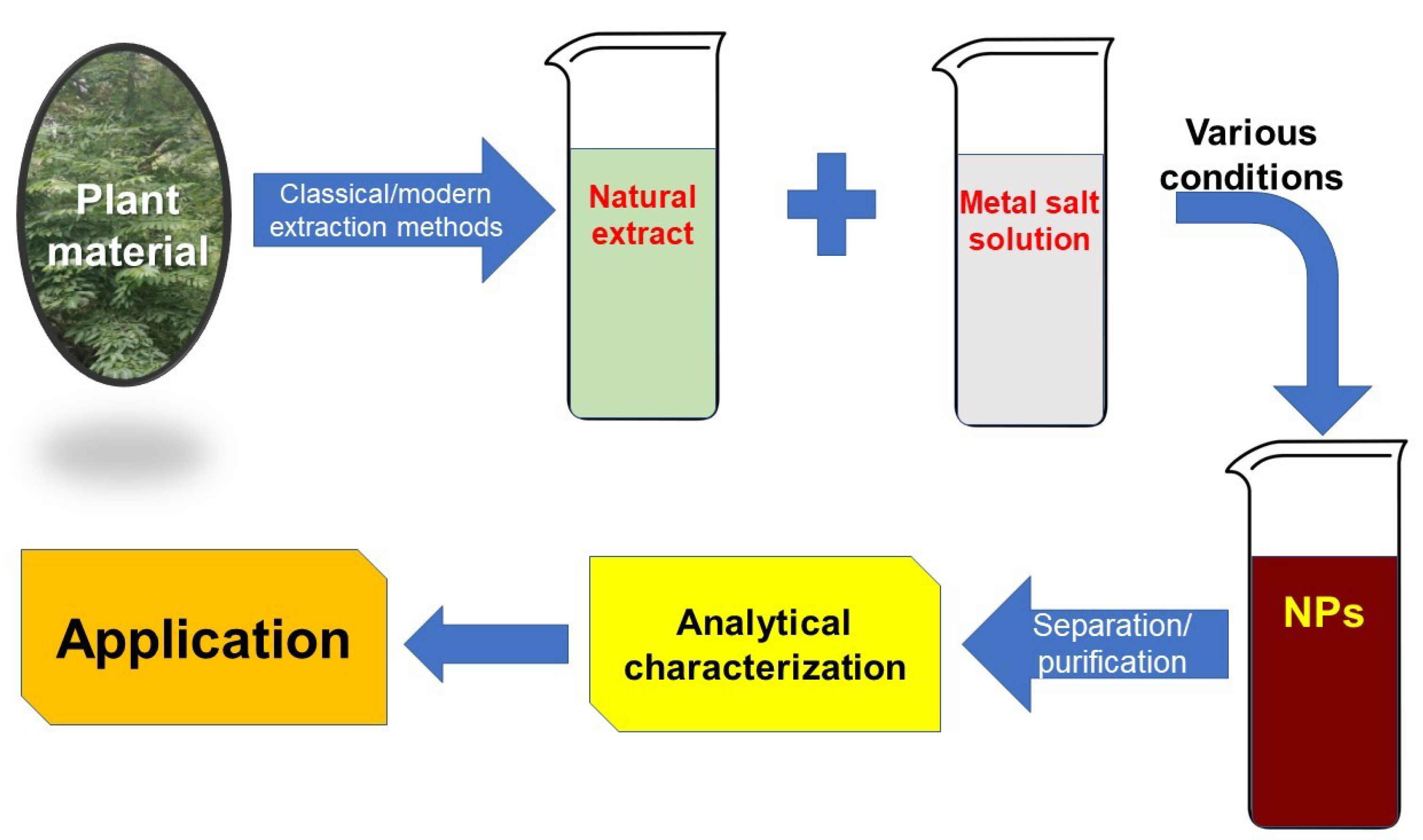
2. General Considerations Regarding Nanoparticle Phytosynthesis

Metal nanoparticles, obtained either by destructive or constructive methods, are known to have different properties, the bulk properties of the particles being substituted by that of “quantum dots” [21]. In order to achieve the nanoparticle form, two approaches are presented in the literature: the top-down approach (including several destructive methods, as mechanical milling, nanolithography, laser ablation, sputtering or thermal decomposition) and a bottom-up approach (including methods such as sol-gel method, spinning, chemical vapor deposition, pyrolysis, ionizing radiation assisted synthesis, and biosynthesis) [22]. Biosynthesis, a green and environmentally friendly approach [23], covers the use of bacteria, fungi or plant extracts, along with the precursors, for bioreduction and capping purposes. The biosynthesized nanoparticles possess unique and enhanced properties that make them useful in biomedical applications [22].
Of particular interest to the current review, phytosynthesis can be defined as the use of plant extracts for the synthesis of the metallic/metal oxide nanoparticles [1,2,3,24]. The general procedure for the phytosynthesis of metallic nanoparticles involves the obtaining of the natural extracts (using different classical or advanced extraction techniques, such as classical temperature extraction, Soxhlet extraction, microwave-assisted extraction, ultrasounds-assisted extraction, accelerated solvent extraction, etc.) followed by their mixing with metallic salt solutions, under various conditions. As it was shown in the literature, [1,2,3,24], the extracts’ phytoconstituents (phenolic compounds, flavonoids, etc.) act as both reducing and capping agents, leading to the obtaining of nanoparticles with enhanced properties (compared with other “green” methods) [3]. The process was presented by our group in several published papers [1,2,3], being schematically presented in Figure 3.
The final products of the phytosynthesis process depend on several factors that can be divided into two categories [23] that can influence the size and morphology of the phytosynthesized nanoparticles, hence their potential application:
  • related to the extract: the extraction procedure, the solvent used for the extraction, the part of the plant used, the pH of the solution, etc.
  • related to the process: temperature, metal salt concentration, extract concentration, reaction time, presence of light radiation, synthesis time, etc.
The separation of the nanoparticles from the solution can be achieved by several methods, the most encountered methods being (ultra)centrifugation and dialysis [25]. Confirmation of the phytosynthesis process can be monitored by UV-Vis spectrometry, as most metallic nanoparticles present specific absorbance peaks in the UV or visible region, their optical properties being dependent on the size, shape, and distribution [1,2]. The advantage of using this technique is that most nanoparticles present specific adsorption bands (for example silver—370~450 nm, gold—500~600 nm, copper—550~650 nm, zinc—370~450 nm, lead—300~400 nm, etc.) and the analysis can be usually performed on the extract/nanoparticle mixture, offering at the same time information regarding the size of the obtained nanoparticles [1]. The main disadvantage of the technique is that the results can be strongly influenced by the extract matrix, leading to erroneous results [3].
According to the literature studies, as well as the authors’ opinion, whenever possible, the NPs’ phytosynthesis should be confirmed by other techniques, especially using methods that would provide information related to the size, morphology and structure of the obtained particles (such as, for example, X-ray diffraction, X-Ray photoelectron spectroscopy, electron microscopy—scanning SEM/transmission TEM, preferably with the EDX accessory, zeta potential, etc.). The precise determination of NP sizes and morphologies should represent an important chapter in any study regarding the (phyto)synthesis of metallic nanoparticles, as those characteristics greatly influence the potential applications (as will be presented in the following chapters).

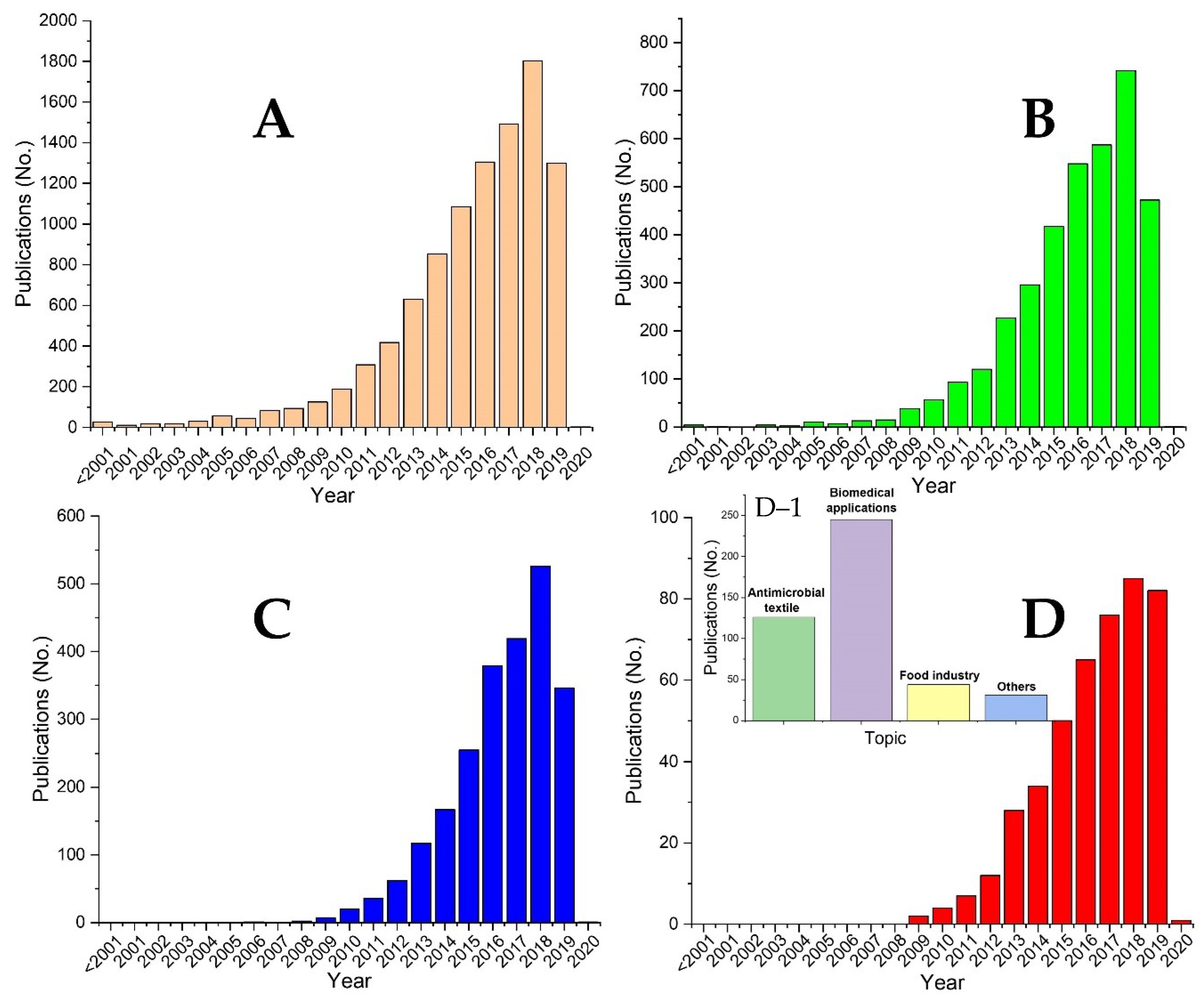
3. Antimicrobial Textiles

Textile materials (especially those based on natural fibers, either vegetal or protein) find application in a wide variety of industries, such as automotive, constructions, sport textile and shoe lining, hygiene and health products, or non-implantable medical products [26,27]. In all these applications, the fibers are exposed to pathogenic microorganisms. Due to their structural characteristics (such as porous structure, moisture retaining capability or the presence of nutrients), the fibers in general, and natural fibers in particular, are considered a very good medium for proliferation of the microorganisms, causing, among other things, bad odors, allergies, infections, or even the degradation of the fiber itself [26,27]. For combating these possible problems, several strategies were applied for the development of antimicrobial textiles, including the use of nitro compounds, metal complexes, or quaternary ammonium salts [27,28,29]. Considering the antimicrobial potential of the phytosynthesized NPs, it is only logical that these materials could be applied as coatings for the development of antimicrobial textiles (Table 1).
Tripathi et al. [30] phytosynthesized silver nanoparticles (AgNPs) using aqueous neem (Azadirachta indica A. Juss., 1830) extract, obtaining nanoparticles with a mostly spherical morphology and dimensions between 50 and 100 nm. The coating of cotton was achieved using three methods (namely centrifuging the cotton disks with the solution containing nanoparticles, in situ coating process simultaneous with the phytosynthesis, and injection with a solution containing purified NPs, respectively). The authors obtained inhibition zones between 7.5 and 9 mm against Escherichia coli, compared with the positive control (penicillin, 8 mm). The authors also studied the retention of the NPs on the cotton disks, by repeatedly washing the disks with distilled water. The reduction in inhibition zone after the third wash (from 8.5 mm after first and second wash to 8 mm) was assigned by the authors to the washing of active neem principles, not of the AgNPs, as no UV-Vis peaks specific to the AgNPs were observed in the washings.
Ravindra et al. [31] used neelagiri (Eucalyptus citriodora Hook.) and marri (Ficus benghalensis L. 1753) extracts to obtain AgNPs with an in situ approach, the phytosynthesis reaction being performed directly on the cotton fibers. The variation of the NPs size was achieved by using different concentration extracts (2%, 4%, and 6%, respectively), the smallest dimensions being recorded for 2% extracts. The antimicrobial potential was tested against E. coli, the results demonstrating superior properties for the materials containing smaller dimensions NPs. The authors categorized the materials as “good antibacterial products”, in reference to the standards in force.
Yang and Li [32] obtained AgNPs using aqueous mango (Mangifera indica Linn) peel extract and studied the effect of extract concentration, silver salt concentration, incubation temperature, and reaction time on the sizes of the obtained NPs. The authors coated non-woven fabrics by an ex situ approach (dipping in the optimized NP solution) and evaluated the antimicrobial potential of the coated materials against Escherichia coli, Staphylococcus aureus, and Bacillus subtilis. The coated material exhibited inhibition zones of 13 mm, 14.5 mm, and 11 mm, respectively, in a modified disk diffusion assay.
Prathna et al. [33] used two different extracts (neem–Azadirachta indica A. Juss., 1830 and lemon–Citrus lemon (L.) Burm.f.) for the phytosynthesis of AgNPs. The coating of cotton was achieved using two methods (in situ, by immersing the cotton sample in the phytosynthesis medium, followed by drying the textile, and, ex situ, by immersion of the cotton in the solution containing phytosynthesized NPs. The authors also studied the influence of a natural binder on the properties of the coated cotton (0.25% starch) and the retention of the NPs, by evaluating the coated cotton after a ten-washing cycle. The results of the cited study revealed a contribution of the binder to the material’s stability (increasing the coating efficiency up to 80%), as well as the superior properties of the cotton phytosynthesized NPs coated, compared with the ones obtained by a chemical reduction method (in terms of mechanical properties and laundering durability), accompanied by an up to 99% microbial reduction (tested against Escherichia coli and Staphylococcus aureus), calculated using an uncoated textile as reference.
Velmurugan et al. [34] phytosynthesized AgNPs using an aqueous annual fleabane (Erigeron annuus (L.) Pers.) flowers extract. The authors compared the antimicrobial properties of cotton and tanned leather samples coated with phytosynthesized AgNPs, commercial AgNPs, silver ions, and AgNPs/extract against the odor causing bacteria Brevibacterium linens and Staphylococcus epidermidis. The best results in inhibition zones and minimum bactericidal concentrations (calculated according to “AATCC 100-Antimicrobial Fabric Test”) were obtained for AgNPs coated cotton (2 mm/63.63%) and AgNPs/extract coated leather (5 mm/30.27%) against B. linens, respectively, for AgNPs/extract coated cotton (1.2 mm/45.45%) and AgNPs/extract coated leather (1 mm/46.63%) against S. epidermidis.
Gowri et al. [35] phytosynthesized TiO2NPs using an aqueous extract of True Aloe (Aloe vera (L.) Burm.f.) gel used for coating of cotton fabric by a direct application system. The coated fabrics were evaluated in terms of antimicrobial properties by the agar diffusion method, against Gram-positive, Gram-negative bacteria, and fungal strains. The results obtained demonstrated significant antibacterial effect against S. aureus (superior to the effect on the Gram-negative E. coli—45 mm inhibition zone, compared with 36 mm) and antifungal effect (inhibition zones of 20 mm against C. albicans, respectively, and 15 mm against A. niger).
Kashid et al. [36] obtained AgNPs using a Chinese chaste tree (Vitex negundo Linn) aqueous extract. The NPs were coated following an in situ procedure over cotton fibers. The antimicrobial potential of the coated fibers was evaluated against Escherichia coli, Klebsiella pneumoniae, Staphylococcus aureus, and Candida albicans, exhibiting superior results, compared with silver nitrate impregnated cotton in a disk diffusion assay (mm inhibition zones—7.72/3.7/4.47/8.37, respectively 6.71/2.52/2.12/2.11) demonstrating that the antimicrobial effect can be attributed to the silver in nanoparticle state. The biocompatibility tests (performed over mouse normal fibroblast cell lines) revealed the fact that the AgNPs-coated cotton is safe and biocompatible (over 80% viability, very close to the untreated cells).
Das and Rebecca [37] applied the phytosynthesis procedure (using a balloon plant leaves extract) for obtaining spherical and rod-shaped zinc oxide nanoparticles (ZnONPs) with dimensions under 80 nm and applied them for the ex situ coating of cotton cloth. The obtained results showed antimicrobial activity only against Gram-negative bacteria (E. coli) (inhibition zone diameter 13–15 mm), finding a direct correlation between the cotton-ZnONPs contact time and the antimicrobial effect. In the live/dead bacterial fluorescence viability assay, the authors observed a nearly equal proportion of live and dead cells after 15 min of E. coli incubation with 50 μg/mL ZnONPs.
Following the same “green” principles, Saha et al. [38] phytosynthesized ZnONPs using guava (Psidium guajava L.) extract and three methods for the NPs formation: sonication, wet-chemical, and hydrothermal. The antimicrobial properties of the coated material (cotton) were tested by an agar well diffusion method, agar disk diffusion method, and percentage reduction test (AATCC 100), against E. coli and S. aureus. As the results on in vitro experiments carried out on ZnONPs showed clearly superior antimicrobial results for the nanoparticles obtained using the hydrothermal method (assigned by the authors to their regular spherical morphology and lower dimensions), those NPs were incorporated in a ZnONPs/chitosan coating applied to cotton fabric. The coating showed superior antimicrobial results to uncoated, respectively chitosan-coated cotton (28.6 mm against E. coli and 30.3 mm against S. aureus, 96%, respectively 99% bacterial reduction percentage). In our opinion, the difference between the two cited studies regarding the antimicrobial potential of ZnONPs (especially against S. aureus) is mainly due to the differences in terms of particle dimensions, that allows the penetration of the thick peptidoglycan layer of the Gram-positive bacteria.
Rajaboopathi and Thambidurai [39] obtained AgNPs using as reducing and capping agent extract of brown seaweed (Padina gymnospora (Kützing) Sonder) obtained by a multi-step procedure. The nanoparticles were coated on cotton by a pad-dry-cure method, using citric acid as crosslinking agent. The material showed inhibition zone diameter superior to the positive control used (amikacin) for both Gram-positive and Gram-negative bacteria (S. aureus, respectively E. coli)—21 and 19 mm, compared with 17 mm for the positive control. The antimicrobial effect was only slightly affected by the repeated washing of the fabric (15.4 mm, respectively 14.2 mm after 10 washing cycles), suggesting the strong fixation of the AgNPs to the fibers.
Sharma et al. [40] optimized the AgNPs phytosynthesis process using onion (Allium cepa L.) aqueous extract (considering the volume of extract and reaction temperature), obtaining nanoparticles with dimensions as low as 36 nm. The obtained NPs were coated on mercerized cotton and the antimicrobial properties of the materials were studied against Gram-positive and Gram-negative bacteria, using ampicillin as positive control. The inhibition zones and % efficacy of the tested materials were up to 19.23 mm and 99.27% against Gram-negative bacteria, respectively 19.96 mm and 102.88% against Gram-positive bacteria, superior to the positive control. The accelerated laundering test (50 cycles) suggested that AgNPs coated cotton fabric still possessed more than 90% efficiency against tested microorganism.
Copper nanoparticles (CuONPs) were phytosynthesized by Sathiyavimal et al. [41] using an aqueous common wireweed (Sida acuta Burm.f.) extract. The nanoparticles (rod-shaped, with dimensions around 50 nm) were coated over cotton fabrics by the dipping technique. The bactericidal efficacy tests showed clear inhibition zones around coated cotton fabrics for all pathogens tested (Escherichia coli, Proteus vulgaris, and Staphylococcus aureus).
Hiremath et al. [42] used phytosynthesized Fe3O4 (obtained using lemongrass—Cymbopogon schoenanthus (L.) Spreng. extract) for coating over cotton roll, gauze roll and cotton cloth, using the dipping and ultrasonication method. Selected materials (coated cotton cloth) were tested for antimicrobial properties against E. coli using the agar well and disk diffusion methods, showing a significant inhibition of growth (similar to the positive control, ampicillin).
Khatami et al. [43] obtained silver nanoparticles using Prosopis fracta J.F.Macbr. seeds extract and zinc oxide nanoparticles using coffee grounds aqueous extract. The individual nanoparticles and a mixture of them were coated on cotton bandages by soaking the fabrics in the solutions containing the NPs. The evaluation of the antimicrobial properties of the bandages was made against Acinetobacter baumannii and Pseudomonas aeruginosa, at concentrations of 1.56 mg/L for AgNPs, 12.5 mg/L for ZnONPs and 3.12 mg/L for AgNPs + ZnONPs against A. baumannii, respectively 3.12 mg/L for AgNPs, 12.5 for ZnONPs and 3.12 for AgNPs + ZnONPs against P. aeruginosa (minimum inhibitory concentrations determined by macro dilution method). The antimicrobial bandages exhibited clear inhibition zones, with the best results obtained for AgNPs bandages.
Sivaranjana et al. [44] applied the in situ approach to obtain AgNPs coated cotton fabrics using candle bush (Cassia alata L.) leaves extract. The procedure involved the diffusion of the extract into the fabric, followed by that immersed in different concentrations silver nitrate solutions. The first indicator of the NPs formation was given by the change of color of the fabric (to black). The best antibacterial effect against E. coli was recorded for a 5 mM concentration of AgNPs (15.81 mm). The antibacterial activity recorded a slight decrease after 15 washings (to 12.9 mm). For the tests regarding the bacterial inhibition in water, the authors performed the diffusion assay under stable sewage water conditions, obtaining an inhibition zone of 38 cm for the fabric coated with AgNPs at a 5 mM concentration.
Phytosynthesis of copper nanoparticles (CuNPs) by heart-leaved moonseed (Tinospora cordifolia Thunb.) Miers) extract was achieved by Sharma et al. [45], and the selected NPs were used for the coating of mercerized cotton fabrics by dipping. The antimicrobial assays were carried out using both Gram-positive and Gram-negative bacteria (S. aureus and E. coli). The coated materials demonstrated a superior effect on the Gram-positive bacteria (21.99 mm and 101% efficacy for 175 mg/L CuNP content, superior to the positive control ampicillin), compared with the Gram-negative bacteria (11 mm and 74% efficacy at 300 mg/L concentration). After 50 laundry rounds, a decrement of the entrapment of NPs was observed (37.7 mg/L for Gram-positive bacteria and 29 mg/L for Gram-negative bacteria).
Turakhia et al. [46] used ginger (Zingiber officinale Roscoe) root extract to phytosynthesize iron nanoparticles (FeNP). The NPs were coated over surgical cotton by dip coating, and the final coated material was tested in terms of antimicrobial activity against Bacillus subtilis, Staphylococcus aureus, and Escherichia coli in an agar well diffusion assay. The assays showed inhibition zones of 9, 12, and 14 mm, respectively, 24 h after coating. The estimation of the antimicrobial potential after 30 days revealed a decrease of the inhibition zones (5, 11, and 6 mm, respectively), which the authors assign to the development of resistance in the microbial cultures studied.
The use of phytosynthesized gold nanoparticles (AuNPs) for the development of antimicrobial textiles was presented by Ganesan and Prabu [47]. The authors obtained the nanoparticles by the use of calamus (Acorus calamus L., 1753) rhizomes aqueous extracts, varying the extraction temperature and the metallic salt concentration, spherical nanoparticles with dimensions under 100 nm being observed for the application of lower metallic source concentration (0.001 M). The coated fabric presented superior antibacterial activity against S. aureus and E. coli, compared with the neat extract coated cotton and uncoated cotton, significantly increased after 48 hours, compared to 24 h (63.9/50.5% against S. aureus, respectively 80.3/58% against E. coli).
As emerging from the literature study, the field of antimicrobial textiles incorporating phytosynthesized nanoparticles represents an area of interest, with great potential for future development. However, several drawbacks in the current approach must be highlighted: first of all, in order to be successfully used in daily applications, the textiles need to satisfy several requirements (besides their antimicrobial properties), such as durability to laundering, dry cleaning, and pressing. More than that, the support fibers should not be affected (in terms of quality and appearance) by the coating. The reviewed studies do not present such a complete approach; thus, the results can only be classified as laboratory research, not as ready for industrial implementation. For proposing the use of this solution at industrial scale, further research is also necessary in terms of coating procedures (as most literature data present the coating by a dipping technique, a method that does not ensure a good stability of the nanoparticles on the support fibers). Secondly, the potential of phytosynthesized nanoparticles for the development of antimicrobial textiles should be studied considering other natural (protein) and synthetic fibers. For the synthetic fibers, the incorporation can be achieved in the polymer melt or in the polymer solution before spinning, a solution that can not be achieved for natural fibers, offering a superior stability. For the natural fibers, the stability can be increased either by chemical bonding the nanoparticles on the surface of the fiber or by their treatment with functionalized coatings [48].
The existence of dedicated standards for the evaluation of the antimicrobial properties of the textiles (AATCC 100 Antimicrobial Test Method for Textile/Fabrics) should represent an advantage for the authors working in this area. The use of a single standard method would allow a better comparison of the obtained results with available literature data. Finally, the optimization of the nanoparticle phytosynthesis (considering at least the parameters presented in chapter 2) should be performed. As presented in Table 1, E. coli represents a widely used strain in the antimicrobial assays; however, due to the multiple variables of the studies (size/morphology of the nanoparticles, different phytoconstituents present in extracts), a comparison of the results is very hard to be performed. A good example is represented by the papers of Das [37] and Saha [38]. Although at first sight these papers could be compared (having the same target strains and the same type of nanoparticles), the authors obtained very different morphologies for the phytosynthesized ZnO nanoparticles and, consequently, different antimicrobial results. The same discussion can be made for the AgNPs. Slight modifications of the phytosynthesis protocols can lead to different types of nanoparticles (in terms of morphology and sizes), thus influencing the antimicrobial potential.

4. Biomedical Applications of Antimicrobial Coatings Based on Phytosynthesized NPs

Besides the antimicrobial textile, presented in the previous chapter, the antimicrobial coatings find applications in a wide variety of implantable devices intended for medical uses (Table 2). Either used in dentistry [49], for orthopedic implants [50,51] or for other medical devices [52], antimicrobial coatings based on metallic nanoparticles plays an important role in medicine, constituting at the same time an important research area. As the field of antimicrobial textiles used for wound dressing was already presented in Section 3, the presented chapter will focus on the use of phytosynthesized nanoparticles in other biomedical applications.
Carboxymethyl chitosan (CMC) represents a natural polymer derivative that finds application in several biomedical fields (such as tissue engineering, bioimaging, drug or enzyme delivery or cosmetics, against skin aging) [53]. The coating of CMC with phytosynthesized Fe3O4 nanoparticles was achieved by Narendhar et al. [54] using an ex situ approach. The materials were evaluated for their antimicrobial properties against a series of pathogens (by monitoring the optical density of the bacterial cultures), the best results being obtained against Pseudomonas aeruginosa. Sripriya et al. [55] used poly(allylamine hydrochloride) and dextran sulfate to develop a polyelectrolyte multilayer (PEM) thin film using a layer-by-layer assembly method, over which they coated AgNPs phytosynthesized using an aqueous Spade Flower (Hybanthus enneaspermus (L.) F.Muell.) extract. The NPs showed excellent antimicrobial activity against the tested lines (Escherichia coli, Proteus vulgaris, Bacillus cereus, Staphylococcus aureus—7/6/5.5/6.5 mm inhibition zones), and the toxicity increased between 41% and 76.47%, when the NPs were used combined with amoxiclav. More than that, the AgNPs coated PEM films favored drug loading, presenting rupture when irradiated with laser light, allowing the authors to propose the coated films for the remote activated drug delivery.
Anghel et al. [56] phytosynthesized Fe3O4 nanoparticles using an aqueous extract of true cinnamon tree (Cinnamomum verum J.Presl) and used the obtained NPs for the coating of gastrostomy tubes, by the matrix assisted pulsed laser evaporation technique. The authors evaluated the interaction of the coated materials with the eukaryotic cells (Human endothelial cells) and prokaryotic cells (S. aureus and E. coli). Their results suggested good biocompatibility of the obtained coatings, as well as very good antibiofilm inhibition properties (2-fold to 4-fold inhibition of the biofilm formation, compared to the control un-coated gastronomy tubes).
Jyoti and Singh [57] developed an antimicrobial coating based on phytosynthesized AgNPs obtained using aqueous extract of chutro (Berberis asiatica DC.) leaves. The coating was deposited on glass surfaces and the bactericidal effect of the coated material was studied against Staphylococcus epidermidis and Staphylococcus aureus antibiotic resistant strains. The coated glass exhibited a bactericidal effect (calculated as the difference between the logarithm of the number of colony forming units developed on the un-coated class and the logarithm of the number of colony forming units developed on the coated class, divided by the logarithm of the CFU for the uncoated sample) of 3.15–3.36 (6 to 18 h contact time) against S. epidermidis, respectively 3.20–3.60 against S. aureus.
Lozoya-Rodríguez et al. [58] developed an antimicrobial coating comprised of AgNP and hydroxyapatite deposited on a Ti6Al4V alloy substrate (217.33 μm coating) for hip applications. The AgNPs were phytosynthesized using prickly pear cactus (Opuntia ficus-indica) extract and the antimicrobial coating was applied on the metallic alloy by plasma spray coating. The coating was evaluated in terms of biocompatibility (by single-cell gel electrophoresis assay and Tali image cytometry) and antimicrobial potential (against E. coli, S. aureus, and P. aeruginosa), showing no DNA damage, respectively an antibacterial efficiency of 99.99% (evaluated according the JIS Z 2801:2000 standard—Antimicrobial products—Test for antimicrobial activity and efficacy).
Srivastava et al. [59] coated tasar nanofibrous mats with in situ generated AgNPs using an aqueous dandelion extract. As the intended use of the developed material i`s for skin tissue engineering, the authors evaluated its physical, mechanical, antimicrobial, and biological properties. The coated materials exhibited good mechanical strength, 70% water uptake capability and 2300 g/m2/day water vapor permeability. The material also presented very good biocompatibility and inhibition areas between 8 and 14 mm against the studied lines—E. coli, S. aureus, P. aeruginosa and S. epidermidis (the best results being obtained against E. coli) in the antimicrobial assay performed according to a standard method (AATCC 30 Antifungal Assessment and Mildew Resistance Test).
A particularly interesting case is represented by the gold nanoparticles. Although AuNPs have multiple biomedical applications (including in photodynamic therapy, photothermal therapy, X-ray imaging, drug delivery, or sensing) [60], their use in antimicrobial applications is limited by their weak influence on the bacterial growth [61].
Phytosynthesized AuNPs were used by Emmanuel et al. [61] blended with azithromycin and clarithromycin against a series of oral pathogens (Micrococcus luteus, Bacillus subtilis, Staphylococcus aureus, Streptococcus mutans, Lactobacillus acidophilus, Escherichia coli, Pseudomonas aeruginosa, Saccharomyces cerevisiae and Candida albicans). After establishing the minimal inhibitory concentration, the authors evaluated their synergistic antimicrobial activity with azithromycin and clarithromycin at a 1:1 ratio (final concentration 100 μg/mL), obtaining larger inhibition zones, compared with pure antibiotics. The authors assign the increase in antimicrobial effect to AuNPs drug carrier potential.
The biomedical applications presented in the current chapter suggest several opportunities for future research areas: for example, the incorporation of NPs in chitosan (presented in Table 2 for magnetite nanoparticles) can be extended to a wide variety of antimicrobial nanoparticles, as can also be the use for different nanoparticles for antimicrobial films designed for medical devices. In addition, development of hydroxyapatite/NPs composites (as presented in Table 2—[58] and by our group [62]) could lead to the development of “green” coatings for implantable devices. Another surprising aspect emerging from Table 2 is represented by the relatively large dimensions of the nanoparticles presented by the authors for biomedical applications (compared to the ones used for the development of antimicrobial textiles). This, in turn, could represent an opportunity for the synthesis and optimization of smaller dimensions nanoparticles. Related to the nanoparticles characteristics, a lack of reliable analytical results in the cited articles can also be noticed. Few authors present the clear determination of size and morphology of NPs, a significant drawback when trying to compare different studies.

5. Antimicrobial Coatings for Increasing the Quality of Food

With the increase of human population, the reduction of food wastes is considered a viable alternative strategy for a sustainable development [63]. At the European Union level, it was estimated (2018), that, for the entire food chain, up to 290 kg of food per person per year are wasted [63]. Similar values (over 300 kg per person per year) were estimated for the United States [64]. One major contributor to these losses is represented by the food spoilage, induced by a wide range of microorganisms [65]. As all over the world there is an increase interest in combating the food spoilage, several groups studied the application of the nanoparticles’ antimicrobial potential for the preservation of fruits, vegetables, and other foods. The literature data are mainly focused on the incorporation of phytosynthesized silver nanoparticles in antimicrobial coatings. Some examples of antimicrobial coatings used for increasing the quality of food are presented in Table 3.
Gudadhe et al. [66] developed incorporated phytosynthesized silver nanoparticles (AgNPs) in agar, obtaining a film with application in increasing the shelf-life of fruits. The film was obtained by mixing the NPs with agar powder and glycerol, and the coating was performed by dipping the fruits (apple and lime) in the solution for 10 seconds. The film was proven efficient in terms of antimicrobial effect against the studied lines (E. coli and S. aureus), with maximum zone of inhibition of 15 nm and 14 nm, respectively, superior to gentamycin and ampicillin (used as positive controls). The film also had a positive effect in terms of weight loss and (minimum weight loss, compared with uncoated fruits or fruits coated with agar film) and soluble protein content. A very important aspect highlighted by the authors was that the low silver content of the film (nanograms) made the coating safe in terms of cytotoxic and genotoxic effect in humans.
Muthulakshmi et al. [67] phytosynthesized copper nanoparticles (CuNPs) using the aqueous extract of country almond (Terminalia catappa L.). The nanoparticles were generated in the cellulose matrix in a two-step procedure: first, the extract was diffused in the cellulose, followed by the in situ phytosynthesis of CuNPs from the copper salt solution. The antimicrobial properties of the coating were studied against E. coli, and the results showed a direct correlation between the inhibition zone diameter and the concentration of copper sulfate precursor (from 2 mm for the 5 mM concentration to 12 mm for the 250 mM concentration). Considering the superior tensile strength of the composite, compared with conventional packaging polymers, the authors suggested the potential use of the developed material for antimicrobial packaging.
Basumatary et al. [68] developed the agar/AgNPs films with antimicrobial properties (estimated by the agar diffusion technique against Aeromonas hydrophilla). Not only did the developed coating exhibit antimicrobial properties against the studied line (increasing with the concentration of AgNPs, quantitative determination not provided by the authors), but the authors stated that the reddish-brown color could also prevent UV penetration of the packaging, leading to the retention of color, flavor, and nutritional value of the packaged materials.
The coatings designed for food packaging by Vishnuvarthanan and Rajeswari [69], composed of pectin–laponite–phytosynthesized AgNPs coated on polypropylene (by meter bar technique), were tested in terms of O2 transmission and antimicrobial activity, against Escherichia coli and Staphylococcus aureus. The authors observed the reduction of oxygen transmission rate (from 1853.32 cc/m2 day atm. for the uncoated film, to 1509.71 cc/m2 day atm.) and of the water vapor transmission rate (from 9.62 to 7.79 g/m2/day), while testing of the coated film antimicrobial properties revealed an inhibition diameter of 21 mm against E. coli, respectively 15 mm, against S. aureus. The same group of authors [70] incorporated the AgNPs obtained by the same method in a carrageenan/AgNP/laponite nanocomposite coated over polypropylene, observing a reduction of the oxygen transmission rate to 735.35 cc/m2 day atm and of the water vapor transmission rate to 2.02 g/m2/day. The authors also report a strong antimicrobial activity against E. coli and S. aureus of the obtained coating (no measurement of the inhibition zone provided by the authors), compared with the neat carrageenan polypropylene film and Laponite/carrageenan films that presented no zone of inhibition.
Kowsalya et al. [71] obtained coatings comprised of phytosynthesized AgNPs and poly(vinyl alcohol) for increasing the shelf life of fruits (lemon and strawberries). The incorporation of AgNPs in the polymer matrix was performed by electrospinning, while the coating material was tested against Bacillus subtilis, Staphylococcus aureus, Escherichia coli, and Pseudomonas aeruginosa. The coatings led to inhibition zones very close to those observed for the positive control gentamycin (17.3/21.47/17.33/15.20 mm, respectively 18.23/23.60/18.07/16.53 mm for the positive control), while also extending the shelf life of treated fruits (the coated fruits being acceptable in terms of color, appearance, texture and weight) up to ten days, compared to the uncoated fruits (that were qualified as unacceptable after the third storage day).
Mathew et al. [72] developed a biodegradable antimicrobial coating for increasing the shelf life of chicken meat products, by incorporating phytosynthesized AgNPs in poly(vinyl alcohol)/montmorillonite composites. The material exhibited antimicrobial potential against Salmonella typhimurium and Staphylococcus aureus (complete inhibition, respectively log(CFU/mL) = 3.6, compared with the values obtained for the films without nanoparticles—log(CFU/mL) = 9.3, respectively 10), while pouches fabricated from the proposed nanocomposite inhibited the bacterial growth on chicken sausage samples (as proved by the superior bacterial growth observed by the authors in the plate count assay for normal polyethylene pouches compared with the pouches made of the proposed film).
Kadam et al. [73] also developed an antimicrobial coating based on phytosynthesized AgNPs and chitosan and tested it in terms of antimicrobial potential, film thickness, moisture content, water vapor permeability, tensile strength, extensibility, color, and opacity, all important parameters for packaging and coating food or other associated products. The antimicrobial coating exhibited bactericidal effect against Escherichia coli, Pseudomonas aeruginosa, Bacillus subtilis, and Staphylococcus aureus (inhibition zones—approx. 18.5/16.8/15.4/13 mm, compared with the undefined positive control—21/21/21.5/23.4 mm; growth inhibition–approx. 52.5/54.5/61.5/56.8%, compared with the undefined positive control—90/92/91/92%). The difference in terms of inhibition zones (superior in the case of Gram-negative bacteria) was assigned by the authors to the structural and chemical composition differences between the bacterial cell membranes.
As presented in Table 3, literature data are scarce in the field of phytosynthesized metallic nanoparticles application for increasing the quality of food (such as, for example, increasing the shelf-life of products). This can be explained, on one hand, by the difficulties in achieving the coatings with the required general properties (in terms of color, transparency/opacity, thickness, water vapor permeability, solubility, mechanical properties, etc.) when using phytosynthesized nanoparticles. On the other hand, a mandatory condition for food additives is to be recognized as safe to use (for example, at the level of the European Union by inclusion in the flavorings list, or by the inclusion in the Generally Recognized as Safe US list) [74]. This is also a concern for the development of antimicrobial polymeric/NPs packaging systems (as the migration of the nanoparticles to the packed products represents a possibility that must be considered (and should be evaluated even in the case of published studies). As the field of nanoparticles’ toxicity represents a very complex and debated area of research (the nanoparticles toxicity depending on a series of factors, such as the dose and exposure time, aggregation and concentration, particle nature, size, shape, crystal structure, functionalization. and surface area, etc.) [75,76], the application of phytosynthesized nanoparticles for antimicrobial food coatings and packaging can be considered in its pioneering years.

6. Other Applications of NPs-Based Antimicrobial Coatings

Besides the previously presented aspects, the phytosynthesized nanoparticles can be used for several other applications, from environmental applications to the development of antimicrobial paints (Table 4). Those miscellaneous applications will be briefly presented in the following paragraphs.
Manjumeena et al. [77] developed an anticorrosive and antimicrobial coating by using phytosynthesized AgNPs as additive of an epoxy resin (diglycidyl ether of bisphenol-A). The NPs were obtained using extracts of cannonball tree (Couroupita guianensis Aubl.) leaves and were amine functionalized through reaction with 3-aminopropyltriethoxysilane in o-xylene. The NPs were mixed into the epoxy resin and the resulting coating was applied on mild steel specimens using bar coater (resulting in a 100 µm coating). Very interestingly, the epoxy containing 1% functionalized AgNPs showed enhanced corrosion properties (determined by potentiodynamic polarization measurements, electrochemical impedance spectroscopic studies and cross scratch testing), compared with the neat epoxy and the resin containing 3% and 5% functionalized AgNPs. The same system (epoxy resin containing 1% functionalized AgNPs coated over mild steel) was evaluated in terms of antimicrobial potential against several pathogens (Pseudomonas aeruginosa, Bacillus subtilis, Escherichia coli, and Candida albicans) in a diffusion assay. The materials showed clear inhibition halos, compared with the mild steel sample coated with neat epoxy resin alone (further details not provided by the authors). The authors propose the application of the developed epoxy coatings for corrosion and fouling protection.
Saravanan et al. [78] used methanolic extract (Soxhlet extraction) of Madras pea pumpkin (Mukia maderasapatna (L.) M. Roem.) leaves to phytosynthesize several types of metallic nanoparticles (AgNPs, CuNPs and PbNPs). The obtained NPs were obtained following an in situ approach on activated carbon (obtained from agricultural biomass). The nanocomposites (nanoparticle coated activated carbon) were tested against four bacterial strains (Staphylococcus aureus, Pseudomonas aeruginosa, Staphylococcus epidermidis, Escherichia coli) and one fungal strain (Candida albicans). The best results were obtained for AgNPs/activated carbon (12/14.1/10.2/8/11.1 mm inhibition zones), superior to the inhibition zones obtained for the standards used—ampicillin (6/5/4/7/4.1 mm) and chloramphenicol (7/6/7.1/6.2/5.2 mm), but promising results were also obtained for the PbNPs/activated carbon composite (8/6.1/3.2/13.3/9.1 mm).
Lateef et al. [79] incorporated phytosynthesized AgNPs (obtained using aqueous extract of kola nut—Cola nitida Schott & Endl. pods) in commercially available emulsion paint. The protective attributes of the nanoparticles were evaluated by inoculation of the control paint and the NPs/paint (at a final concentration of 5 mg/L of silver) with bacteria and fungi isolated from soil (E. coli, P. aeruginosa. A, flavus, A, fumigatus and A, niger) followed by the bacterial/fungal growth evaluation using the pour plate method. The authors observed total obliteration of all tested lines for the AgNPs paints, compared with the abundant growth observed for the control paint (without biocide).
Deyá and Bellotti [80] evaluated the potential of several extracts (cedrón—Aloysia triphylla (L’Hér.) Britton, laurel—Laurelia sempervirens (Ruiz & Pav.)Tul., and ruda—Ruta chalepensis L.) to phytosynthesize silver nanoparticles, which, in turn, would be useful as additives in water-based coatings for indoor use. The authors studied the potential of the extracts to synthesize AgNPs at two different temperatures (60 and 26 °C). After the study, the authors selected the laurel-mediated NPs at 60 °C, due to superior stability and lower dimensions (around 9.8 nm). The formulation of the acrylic water-based paint was obtained by incorporating two different concentration of AgNPs (3.6 and 5.4 mg/100 g of paint) with a high-speed disperser. The best antifungal results (inhibition zone 6 mm, determined using the agar diffusion method, with cylinders samples disposed on plates with rose bengal-based culture medium) were obtained using the paint with a higher concentration of AgNPs, against both fungal lines used (Chaetomium globosum, Alternaria alternate). The hypothesis of the authors was further confirmed by performing a bio-resistance test: for this purpose, the NPs containing paints were applied on glasses immersed in minimum mineral medium, inoculated with spore suspension and incubated for one month. The fungal growth was evaluated at the end of the test according a standard procedure [81]. The results obtained (quantified according the standard specification) were superior (traces of growth for Chaetomium globosum, respectively light growth for Alternaria alternate), compared with the control paint (heavy growth for both species).
Barberia-Roque et al. [82] used three Argentinian native plants (Peruvian pepper—Schinus molle L., southern giant horsetail—Equisetum giganteum L., and yerba mate—Ilex paraguariensis A.St.-Hil.) to phytosynthesize silver nanoparticles. Differences were observed both in terms of morphology and particle dimensions (Table 4), which, in turn, led to major differences in terms of antimicrobial potential. For this reason, the authors selected the AgNPs phytosynthesized using E. giganteum to obtain an antimicrobial coating by incorporating the NPs solution in waterborne paint, at different concentrations (5.8, 10 and 15 mg of silver/100 g of paint). The fungal resistance tests, performed according the standard ASTM D5590, revealed that the paint containing the highest concentration of silver presented the best results (against C. globosum—no growth and A. alternate—light growth, compared with the control paint, without biocides, showed moderate growth with sporulation, respectively heavy growth), while the antibiofilm formation assay performed for the paint sample containing 15 mg of silver/100 g of paint (evaluated against S. aureus and E. coli, pure cultures in the liquid nutrient culture medium) revealed no bacterial growth.
Unlike the previously presented ones, the miscellaneous applications in the present chapter (detailed in Table 4) have a very serious growth potential. Either in epoxy resins or in paints, several drawbacks of other uses can be avoided. Thus, those applications are not required further supplementary research in terms of toxicity (especially due to the low levels of nanomaterials in the final products), several commercial products being already available on the market, incorporating mainly silver, either in its ionic or nanoparticle form (such as those produced by SilverArmor®, BioCote®, SonoTek®, Clariant®, Microban®, Thomson Research Associates Inc.®, Policolor®, and many others). The producers are required, however, to fulfill a series of particular regulations (such as registration and approval by the U.S. Environmental Protection Agency or conforming to the European Biocidal Product Directive) [83].
The phytosynthesized nanoparticles can be easily incorporated in paint/epoxy matrix and do not influence the mechanical and aesthetic properties of the final products. The focus towards different types of nanomaterials, as well as the tuning of the phytosynthesis process towards lower dimensions and more size-controlled particles would also enhance the antimicrobial properties, together with a decrease of the level of metallic nanoparticles in the products.
Application of nanoparticles in other areas, such as water disinfection, would require an increase level of precaution, as any applications in which the nanoparticles could finally end up affecting the human health.

7. Conclusions and Future Perspectives

The phytosynthesis of metallic nanoparticles represents an exciting new area of research, with promising perspectives. However, when dealing with these types of nanoparticles, it should be remembered that the final application of the NPs (in our case the antimicrobial potential) is strongly related to the size and shape of the NPs. Several authors [84,85] described the dependence of the antimicrobial potential of silver nanoparticles, for example, on their morphology. Morones et al. [84] showed that, in the 1–100 nm range, the best antimicrobial properties are obtained for NPs with dimensions under 10 nm, while Pal et al. [85] categorized the antimicrobial silver nanoparticles considering their shape, finding that, at the same size, the most active AgNPs are those triangular, followed by the spherical and, respectively, rod-shaped NPs. Considering those aspects, future works should also cover a complete characterization of the obtained NPs, in terms of size and shape, whenever possible using multiple complementary techniques.
For phytosynthesized nanoparticles, the size and shapes are strongly related to several parameters: characteristics of the extract used (such as extraction procedure [1] or solvent used for extraction [2]), the pH of the solution [86], the temperature at which the phytosynthesis process is carried out [87], concentration of the metallic salt, extract concentration, or reaction time [32,88]. Varying those parameters, the morphology of the NPs can be tuned for the desired application. Regarding the used natural extracts, future works should try to elucidate as much as possible, their composition. Knowing the extract composition would allow the purification of the extracts, that, in turn, could lead to another tool for tuning the properties of the nanoparticles.
Regarding the use of phytosynthesized nanoparticles in reviewed applications, a surprisingly lack of interest in this type of NPs can be noticed. Thus, although the application of nanoparticles is well established in areas such as textile coatings [18,89], food industry [90], or other biomedical [91] and industrial applications [92], representing at the same time a continuously growing market [93], the use of phytosynthesized nanoparticles seems under-explored. Even for the examples presented, the works are focused mainly on silver nanoparticles, which are widely known for their antimicrobial potential. In this respect, there is a tremendous potential for the application of other phytosynthesized nanoparticles in active antimicrobial coatings, in all the presented applications (such as AuNPs). However, future works should always take into consideration the potential toxic effect of the NPs and appropriate assays should be performed.
Finally, it can be noticed that the heterogeneity in using and describing the antimicrobial assays applied; this represents a major drawback when trying to compare the efficiency of different antimicrobial coatings. Standard procedures should be carried out (as several dedicated standards are available), and their presentation should be as detailed as possible, to allow the valid antimicrobial comparisons.

Funding

I.F. and R.C.F. gratefully acknowledge the support of the Romanian National Authority for Scientific Research and Innovation, CNCS/CCCDI–UEFISCDI, project BIOHORTINOV, project code PN-III-P1-1.2-PCCDI-2017-0332, project number 6 PCCDI/2018, within PNCDI III. Publication of this paper was financially supported by “Carol Davila” University of Medicine and Pharmacy through Contract No. 23PFE/17.10.2018 funded by the Ministry of Research and Innovation within PNCDI III, Program 1—Development of the National RD system, Subprogram 1.2—Institutional Performance—RDI excellence funding projects.

Conflicts of Interest

The authors declare no conflict of interest.

References

  1. Fierascu, I.; Georgiev, M.I.; Ortan, A.; Fierascu, R.C.; Avramescu, S.M.; Ionescu, D.; Sutan, A.; Brinzan, A.; Ditu, L.M. Phyto-mediated metallic nanoarchitectures via Melissa officinalis L.: Synthesis, characterization and biological properties. Sci. Rep. 2017, 7, 12428. [Google Scholar] [CrossRef]
  2. Sutan, N.A.; Manolescu, D.S.; Fierascu, I.; Neblea, A.M.; Sutan, C.; Ducu, C.; Soare, L.C.; Negrea, D.; Avramescu, S.M.; Fierascu, R.C. Phytosynthesis of gold and silver nanoparticles enhance in vitro antioxidant and mitostimulatory activity of Aconitum toxicum Reichenb. rhizomes alcoholic extracts. Mater. Sci. Eng. C 2018, 93, 746–758. [Google Scholar] [CrossRef] [PubMed]
  3. Fierascu, R.C.; Fierascu, I.; Lungulescu, E.M.; Nicula, N.; Somoghi, R.; Diţu, L.M.; Ungureanu, C.; Sutan, A.N.; Drăghiceanu, O.A.; Paunescu, A.; et al. Phytosynthesis and radiation-assisted methods for obtaining metal nanoparticles. J. Mater. Sci. 2020, 55, 1915–1932. [Google Scholar] [CrossRef]
  4. Tripathi, R.M.; Chung, S.J. Biogenic nanomaterials: Synthesis, characterization, growth mechanism, and biomedical applications. J. Microbiol. Meth. 2019, 157, 65–80. [Google Scholar] [CrossRef] [PubMed]
  5. Agarwal, H.; Nakara, A.; Shanmugam, V.K. Anti-inflammatory mechanism of various metal and metal oxide nanoparticles synthesized using plant extracts: A review. Biomed. Pharmacother. 2019, 109, 2561–2572. [Google Scholar] [CrossRef]
  6. Some, S.; Kumar Sen, I.; Mandal, A.; Aslan, T.; Ustun, Y.; Yilmaz, E.Ş.; Kati, A.; Demirbas, A.; Mandal, A.K.; Ocsoy, I. Biosynthesis of silver nanoparticles and their versatile antimicrobial properties. Mater. Res. Express 2019, 6, 012001. [Google Scholar] [CrossRef]
  7. Nasrollahzadeh, M.; Ghorbannezhad, F.; Issaabadi, Z.; Sajadi, S.M. Recent developments in the biosynthesis of Cu-based recyclable nanocatalysts using plant extracts and their application in the chemical reactions. Chem. Rec. 2019, 19, 601–643. [Google Scholar] [CrossRef]
  8. Nabi, G.; Qurat-ul-Aain; Khalid, N.R.; Tahir, M.B.; Rafique, M.; Rizwan, M.; Hussain, S.; Iqbal, T.; Majid, A. A review on novel eco-friendly green approach to synthesis TiO2 nanoparticles using different extracts. J. Inorg. Organomet. Polym. Mater. 2018, 28, 1552–1564. [Google Scholar] [CrossRef]
  9. Long, N.N.V.; Joly, C.; Dantigny, P. Active packaging with antifungal activities. Int. J. Food Microbiol. 2016, 220, 73–90. [Google Scholar] [CrossRef]
  10. Zambrano-Zaragoza, M.L.; González-Reza, R.; Mendoza-Muñoz, N.; Miranda-Linares, V.; Bernal-Couoh, T.F.; Mendoza-Elvira, S.; Quintanar-Guerrero, D. Nanosystems in edible coatings: A novel strategy for food preservation. Int. J. Mol. Sci. 2018, 19, 705. [Google Scholar] [CrossRef]
  11. Xing, Y.; Li, W.; Wang, Q.; Li, X.; Xu, Q.; Guo, X.; Bi, X.; Liu, X.; Shui, Y.; Lin, H.; et al. Antimicrobial nanoparticles incorporated in edible coatings and films for the preservation of fruits and vegetables. Molecules 2019, 24, 1695. [Google Scholar] [CrossRef] [PubMed]
  12. Gallo, J.; Panacek, A.; Prucek, R.; Kriegova, E.; Hradilova, S.; Hobza, M.; Holinka, M. Silver Nanocoating technology in the prevention of prosthetic joint infection. Materials 2016, 9, 337. [Google Scholar] [CrossRef] [PubMed]
  13. Noronha, V.T.; Paula, A.J.; Durán, G.; Galembeck, A.; Cogo-Müller, K.; Franz-Montan, M.; Durán, N. Silver nanoparticles in dentistry. Dent. Mater. 2017, 33, 1110–1126. [Google Scholar] [CrossRef] [PubMed]
  14. Burdusel, A.C.; Gherasim, O.; Grumezescu, A.M.; Mogoanta, L.; Ficai, A.; Andronescu, E. Biomedical applications of silver nanoparticles: An up-to-date overview. Nanomaterials 2018, 8, 681. [Google Scholar] [CrossRef] [PubMed]
  15. Lee, S.H.; Jun, B.H. Silver nanoparticles: Synthesis and application for nanomedicine. Int. J. Mol. Sci. 2019, 20, 865. [Google Scholar] [CrossRef] [PubMed]
  16. Bottagisio, M.; Lovati, A.B.; Galbusera, F.; Drago, L.; Banfi, G. A Precautionary approach to guide the use of transition metal-based nanotechnology to prevent orthopedic infections. Materials 2019, 12, 314. [Google Scholar] [CrossRef]
  17. Selvaraj, A.S.; Rajendran, M. Antimicrobial nanomaterials for wound dressings. In Nanomedicine and Tissue Engineering: State of the Art and Recent Trends; Nandakumar, K., Robin, A., Oluwatobi, S.O., Joshy, K.S., Sabu, T., Eds.; Apple Academic Press: Oakville, ON, Canada, 2016; pp. 435–492. [Google Scholar]
  18. Joshi, M.; Roy, A. Antimicrobial textiles based on metal and metal oxide nano-particles. In Nanomaterials in the Wet Processing of Textiles; ul-Islam, S., Butola, B.S., Eds.; Scrivener Publishing LLC: Beverly, MA, USA, 2018; pp. 71–111. [Google Scholar]
  19. Eduok, S.; Coulon, F. Microbiological toxicity of nanoparticles. In Emerging Nanotechnologies in Food Science. Micro and Nano Technologies; Busquets, R., Ed.; Elsevier: Amsterdam, The Netherlands, 2017; pp. 97–117. [Google Scholar]
  20. Colman, B.P.; Espinasse, B.; Richardson, C.J.; Matson, C.W.; Lowry, G.W.; Hunt, D.E.; Wiesner, M.R.; Bernhardt, E.S. Emerging contaminant or an old toxin in disguise? Silver nanoparticle impacts on ecosystems. Environ. Sci. Technol. 2014, 48, 5229–5236. [Google Scholar] [CrossRef]
  21. Salavati-Niasari, M.; Davar, F.; Mir, N. Synthesis and characterization of metallic copper nanoparticles via thermal decomposition. Polyhedron 2008, 27, 3514–3518. [Google Scholar] [CrossRef]
  22. Ealias, A.M.; Saravanakumar, M.P. A review on the classification, characterization, synthesis of nanoparticles and their application. IOP Conf. Ser. Mater. Sci. Eng. 2017, 263, 032019. [Google Scholar]
  23. Kuppusamy, P.; Yusoff, M.M.; Maniam, G.P.; Govindan, N. Biosynthesis of metallic nanoparticles using plant derivatives and their new avenues in pharmacological applications—An updated report. Saudi Pharm. J. 2016, 24, 473–484. [Google Scholar] [CrossRef]
  24. Fierascu, R.C.; Ortan, A.; Avramescu, S.M.; Fierascu, I. Phyto-nanocatalysts: Green synthesis, characterization, and applications. Molecules 2019, 24, 3418. [Google Scholar] [CrossRef]
  25. Moldovan, B.; Sincari, V.; Perde-Schrepler, M.; David, L. Biosynthesis of silver nanoparticles using Ligustrum ovalifolium fruits and their cytotoxic effects. Nanomaterials 2018, 8, 627. [Google Scholar] [CrossRef] [PubMed]
  26. Coradi, M.L.; Zanetti, M.; Valério, A.; de Oliveira, D.; da Silva, A.; de Souza, S.M.A.G.U.; de Souza, A.A.U. Production of antimicrobial textiles by cotton fabric functionalization and pectinolytic enzyme immobilization. Mater. Chem. Phys. 2018, 208, 28–34. [Google Scholar] [CrossRef]
  27. Shahid-ul-Islam; Butola, B. S. Recent advances in chitosan polysaccharide and its derivatives in antimicrobial modification of textile materials. Int. J. Biol. Macromol. 2019, 121, 905–912. [Google Scholar] [CrossRef] [PubMed]
  28. Gao, Y.; Cranston, R. Recent advances in antimicrobial treatments of textiles. Text. Res. J. 2008, 78, 60–72. [Google Scholar]
  29. Lin, J.; Winkelman, C.; Worley, S.D.; Broughton, R.M.; Williams, J.F. Antimicrobial treatment of nylon. J. Appl. Polym. Sci. 2001, 81, 943–947. [Google Scholar] [CrossRef]
  30. Tripathi, A.; Chandrasekaran, N.; Raichur, A.M.; Mukherjee, A. Antibacterial applications of silver nanoparticles synthesized by aqueous extract of Azadirachta indica (Neem) leaves. J. Biomed. Nanotechnol. 2009, 5, 93–98. [Google Scholar] [CrossRef]
  31. Ravindra, S.; Murali Mohan, Y.; Narayana Reddy, N.; Mohana Raju, K. Fabrication of antibacterial cotton fibres loaded with silver nanoparticles via “Green Approach”. Colloids Sur. A Physicochem. Eng. Asp. 2010, 367, 31–40. [Google Scholar] [CrossRef]
  32. Yang, N.; Li, W.H. Mango peel extract mediated novel route for synthesis of silver nanoparticles and antibacterial application of silver nanoparticles loaded onto non-woven fabrics. Ind. Crop. Prod. 2013, 48, 81–88. [Google Scholar] [CrossRef]
  33. Prathna, T.C.; Raichur, A.M.; Chandrasekaran, N.; Mukherjee, A. Process Development for functionalization of cotton with silver nanoparticles synthesized by bio-based approaches. Curr. Nanosci. 2013, 9, 479–488. [Google Scholar] [CrossRef]
  34. Velmurugan, P.; Cho, M.; Lee, S.M.; Park, J.H.; Bae, S.; Oh, B.T. Antimicrobial fabrication of cotton fabric and leather using green-synthesized nanosilver. Carbohydr. Polym. 2014, 106, 319–325. [Google Scholar] [CrossRef] [PubMed]
  35. Gowri, S.; Rajiv Gandhi, R.; Senthil, S.; Suresh, J.; Sundrarajan, M. Enhancing antimicrobial activity of biotemplated TiO2 nanoparticles using aloe vera plant extract. J. Bionanosci. 2016, 10, 181–190. [Google Scholar] [CrossRef]
  36. Kashid, S.B.; Lakkakula, J.R.; Chauhan, D.S.; Srivastava, R.; Raut, R.W. Biocompatible antimicrobial cotton fibres for healthcare industries: A biogenic approach for synthesis of bio-organic-coated silver nanoparticles. IET Nanobiotechnol. 2017, 11, 1046–1051. [Google Scholar] [CrossRef] [PubMed]
  37. Das, M.P.; Rebecca, L.J. Evaluation of antibacterial efficacy of biogenic zinc oxide nanoparticles on cotton fabrics. J. Pharm. Sci. Res. 2017, 9, 2553–2557. [Google Scholar]
  38. Saha, R.; Karthik, S.; Kumar, P.M.R.S.A.; Suriyaprabha, R.; Rajendran, V. Psidium guajava leaf extract-mediated synthesis of ZnO nanoparticles under different processing parameters for hydrophobic and antibacterial finishing over cotton fabrics. Prog. Org. Coat. 2018, 124, 80–91. [Google Scholar] [CrossRef]
  39. Rajaboopathi, S.; Thambidurai, S. Evaluation of UPF and antibacterial activity of cotton fabric coated with colloidal seaweed extract functionalized silver nanoparticles. J. Photochem. Photobiol. B 2018, 183, 75–87. [Google Scholar] [CrossRef]
  40. Sharma, P.; Pant, S.; Rai, S.; Yadav, R.B.; Sharma, S.; Dave, V. Green synthesis and characterization of silver nanoparticles by Allium cepa L. to produce silver nano-coated fabric and their antimicrobial evaluation. Appl. Organometal. Chem. 2018, 32, e4146. [Google Scholar] [CrossRef]
  41. Sathiyavimal, S.; Vasantharaj, S.; Bharathi, D.; Saravanan, M.; Manikandan, E.; Kumar, S.S.; Pugazhendhi, A. Biogenesis of copper oxide nanoparticles (CuONPs) using Sida acuta and their incorporation over cotton fabrics to prevent the pathogenicity of gram negative and gram positive bacteria. J. Photochem. Photobiol. B 2018, 188, 126–134. [Google Scholar] [CrossRef]
  42. Hiremath, L.; Kumar, N.S.; Sukanya, P. Development of antimicrobial smart textiles fabricated with magnetite nano particles obtained through green synthesis. Mater. Today Proc. 2018, 5, 21030–21039. [Google Scholar] [CrossRef]
  43. Khatami, M.; Varma, R.S.; Zafarnia, N.; Yaghoobi, H.; Sarani, M.; Kumar, V.G. Applications of green synthesized Ag, ZnO and Ag/ZnO nanoparticles for making clinical antimicrobial wound-healing bandages. Sustain. Chem. Pharm. 2018, 10, 9–15. [Google Scholar] [CrossRef]
  44. Sivaranjana, P.; Nagarajan, E.; Rajini, N.; Ayrilmis, N.; Rajulu, A.V.; Siengchin, S. Preparation and characterization studies of modified cellulosic textile fabric composite with in situ-generated AgNPs coating. J. Ind. Text. 2019, in press. [Google Scholar] [CrossRef]
  45. Sharma, P.; Pant, S.; Dave, V.; Tak, K.; Sadhu, V.; Reddy, K.R. Green synthesis and characterization of copper nanoparticles by Tinospora cardifolia to produce nature-friendly copper nano-coated fabric and their antimicrobial evaluation. J. Microbiol. Meth. 2019, 160, 107–116. [Google Scholar] [CrossRef] [PubMed]
  46. Turakhia, B.; Chikkala, S.; Shah, S. Novelty of bioengineered iron nanoparticles in nanocoated surgical cotton: a green chemistry. Adv. Pharmacol. Sci. 2019, 2019, 9825969. [Google Scholar] [CrossRef] [PubMed] [Green Version]
  47. Ganesan, R.M.; Prabu, H.G. Synthesis of gold nanoparticles using herbal Acorus calamus rhizome extract and coating on cotton fabric for antibacterial and UV blocking applications. Arab. J. Chem. 2015, in press. [Google Scholar] [CrossRef] [Green Version]
  48. Wagener, S.; Dommershausen, N.; Jungnickel, H.; Laux, P.; Mitrano, D.; Nowack, B.; Schneider, G.; Luch, A. Textile functionalization and its effects on the release of silver nanoparticles into artificial sweat. Environ. Sci. Technol. 2016, 50, 5927–5934. [Google Scholar] [CrossRef]
  49. Parnia, F.; Yazdani, J.; Javaherzadeh, V.; Maleki Dizaj, S. Overview of nanoparticle coating of dental implants for enhanced osseointegration and antimicrobial purposes. J. Pharm. Pharm. Sci. 2017, 20, 148–160. [Google Scholar] [CrossRef]
  50. Orapiriyakul, W.; Young, P.S.; Damiati, L.; Tsimbouri, P.M. Antibacterial surface modification of titanium implants in orthopaedics. J. Tissue Eng. 2018, 9. [Google Scholar] [CrossRef] [Green Version]
  51. Pan, C.; Zhou, Z.; Yu, X. Coatings as the useful drug delivery system for the prevention of implant-related infections. J. Orthop. Surg. Res. 2018, 13, 220. [Google Scholar] [CrossRef] [Green Version]
  52. Cyphert, E.L.; von Recum, H.A. Emerging technologies for long-term antimicrobial device coatings: Advantages and limitations. Exp. Biol. Med. 2017, 242, 788–798. [Google Scholar] [CrossRef]
  53. Shariatinia, Z. Carboxymethyl chitosan: Properties and biomedical applications. Int. J. Biol. Macromol. 2018, 120, 1406–1419. [Google Scholar] [CrossRef]
  54. Narendhar, C.; Abey, T.; Jatin, B.; Kumar, M.M.S.; Regitha, M. Facile Synthesis of carboxy methyl chitosan coated iron oxide nanoparticles and their antimicrobial activity. In Proceedings of the International Conference on Advanced Nanomaterials & Emerging Engineering Technologies, Chennai, India, 24–26 July 2013; pp. 290–292. [Google Scholar] [CrossRef]
  55. Sripriya, J.; Anandhakumar, S.; Achiraman, S.; Antony, J.J.; Siva, D.; Raichur, A.M. Laser receptive polyelectrolyte thin films doped with biosynthesized silver nanoparticles for antibacterial coatings and drug delivery applications. Int. J. Pharmaceut. 2013, 457, 206–213. [Google Scholar] [CrossRef] [PubMed]
  56. Anghel, A.G.; Grumezescu, A.M.; Chirea, M.; Grumezescu, V.; Socol, G.; Iordache, F.; Oprea, A.E.; Anghel, I.; Holban, A.M. MAPLE Fabricated Fe3O4@Cinnamomum verum antimicrobial surfaces for improved gastrostomy tubes. Molecules 2014, 19, 8981–8994. [Google Scholar] [CrossRef] [PubMed] [Green Version]
  57. Jyoti, K.; Singh, A. Evaluation of antibacterial activity from phytosynthesized silver nanoparticles against medical devices infected with Staphylococcus spp. J. Taibah Univ. Med. Sci. 2017, 12, 47–54. [Google Scholar] [CrossRef] [PubMed]
  58. Lozoya-Rodríguez, D.A.; de Lima, R.; Fraceto, L.F.; Pérez, A.L.; Domínguez, M.B.; Batres, R.G.; Rojas, A.R.; Carmona, V.O. Development of HA/Ag-NPs composite coating from green process for hip applications. Molecules 2017, 22, 1291. [Google Scholar] [CrossRef] [Green Version]
  59. Srivastava, C.M.; Purwar, R.; Gupta, A.P. Enhanced potential of biomimetic, silver nanoparticles functionalized Antheraea mylitta (tasar) silk fibroin nanofibrous mats for skin tissue engineering. Int. J. Biol. Macromol. 2019, 130, 437–453. [Google Scholar] [CrossRef]
  60. Elahi, N.; Kamali, M.; Baghersad, M.H. Recent biomedical applications of gold nanoparticles: A review. Talanta 2018, 184, 537–556. [Google Scholar] [CrossRef]
  61. Emmanuel, R.; Saravanan, M.; Ovais, M.; Padmavathy, S.; Shinwari, Z.K.; Prakash, P. Antimicrobial efficacy of drug blended biosynthesized colloidal gold nanoparticles from Justicia glauca against oral pathogens: A nanoantibiotic approach. Microb. Pathog. 2017, 113, 295–302. [Google Scholar] [CrossRef]
  62. Fierascu, I.; Fierascu, R.C.; Somoghi, R.; Ion, R.M.; Moanta, A.; Avramescu, S.M.; Damian, C.M.; Ditu, L.M. Tuned apatitic materials: Synthesis, characterization and potential antimicrobial applications. Appl. Surf. Sci. 2018, 438, 127–135. [Google Scholar] [CrossRef]
  63. De Laurentiis, V.; Corrado, S.; Sala, S. Quantifying household waste of fresh fruit and vegetables in the EU. Waste Manag. 2018, 77, 238–251. [Google Scholar] [CrossRef]
  64. Barth, M.; Hankinson, T.R.; Zhuang, H.; Breidt, F. Microbiological spoilage of fruits and vegetables. In Compendium of the Microbiological Spoilage of Foods and Beverages; Sperber, W.H., Doyle, M.P., Eds.; Springer: New York, NY, USA, 2009; pp. 135–183. [Google Scholar]
  65. Petruzzi, L.; Corbo, M.R.; Sinigaglia, M.; Bevilacqua, A. Microbial spoilage of foods. In The Microbiological Quality of Food; Bevilacqua, A., Corbo, M.R., Sinigaglia, M., Eds.; Woodhead Publishing: Duxford, UK, 2017; pp. 1–21. [Google Scholar]
  66. Gudadhe, J.A.; Yadav, A.; Gade, A.; Marcato, P.D.; Durán, N.; Rai, M. Preparation of an agar-silver nanoparticles (A-AgNp) film for increasing the shelf-life of fruits. IET Nanobiotechnol. 2014, 8, 190–195. [Google Scholar] [CrossRef]
  67. Muthulakshmi, L.; Rajini, N.; Nellaiah, H.; Kathiresan, T.; Jawaid, M.; Varada Rajulu, A. Preparation and properties of cellulose nanocomposite films with in situ generated copper nanoparticles using Terminalia catappa leaf extract. Int. J. Biol. Macromol. 2017, 95, 1064–1071. [Google Scholar] [CrossRef] [PubMed]
  68. Basumatary, K.; Daimary, P.; Das, S.K.; Thapa, M.; Singh, M.; Mukherjee, A.; Kumar, S. Lagerstroemia speciosa fruit-mediated synthesis of silver nanoparticles and its application as filler in agar based nanocomposite films for antimicrobial food packaging. Food Packag. Shelf Life 2018, 17, 99–106. [Google Scholar] [CrossRef]
  69. Vishnuvarthanan, M.; Rajeswari, N. Food packaging: Pectin–laponite–Ag nanoparticle bionanocomposite coated on polypropylene shows low O2 transmission, low Ag migration and high antimicrobial activity. Environ. Chem. Lett. 2019, 17, 439–445. [Google Scholar] [CrossRef]
  70. Vishnuvarthanan, M.; Rajeswari, N. Preparation and characterization of carrageenan/silver nanoparticles/Laponite nanocomposite coating on oxygen plasma surface modified polypropylene for food packaging. J. Food Sci. Technol. 2019, 56, 2545–2552. [Google Scholar] [CrossRef] [PubMed]
  71. Kowsalya, E.; Mosa Christas, K.; Balashanmugam, P.; Tamil Selvi, A.; Jaquline Chinna Rani, I. Biocompatible silver nanoparticles/poly(vinyl alcohol) electrospun nanofibers for potential antimicrobial food packaging applications. Food Packag. Shelf Life 2019, 21, 100379. [Google Scholar]
  72. Mathew, S.; Snigdha, S.; Mathew, J.; Radhakrishnan, E.K. Biodegradable and active nanocomposite pouches reinforced with silver nanoparticles for improved packaging of chicken sausages. Food Packag. Shelf Life 2019, 19, 155–166. [Google Scholar] [CrossRef]
  73. Kadam, D.; Momin, B.; Palamthodi, S.; Lele, S.S. Physicochemical and functional properties of chitosan-based nanocomposite films incorporated with biogenic silver nanoparticles. Carbohydr. Polym. 2019, 211, 124–132. [Google Scholar] [CrossRef]
  74. Valdés, A.; Ramos, M.; Beltrán, A.; Jiménez, A.; Garrigós, M.C. State of the art of antimicrobial edible coatings for food packaging applications. Coatings 2017, 7, 56. [Google Scholar] [CrossRef] [Green Version]
  75. Jeevanandam, J.; Barhoum, A.; Chan, Y.S.; Dufresne, A.; Danquah, M.K. Review on nanoparticles and nanostructured materials: History, sources, toxicity and regulations. Beilstein J. Nanotechnol. 2018, 9, 1050–1074. [Google Scholar] [CrossRef] [Green Version]
  76. Ajdary, M.; Moosavi, M.A.; Rahmati, M.; Falahati, M.; Mahboubi, M.; Mandegary, A.; Jangjoo, S.; Mohammadinejad, R.; Varma, R.S. Health concerns of various nanoparticles: A review of their in vitro and in vivo toxicity. Nanomaterials 2018, 8, 634. [Google Scholar] [CrossRef] [Green Version]
  77. Manjumeena, R.; Venkatesan, R.; Duraibabu, D.; Sudha, J.; Rajendran, N.; Kalaichelvan, P.T. Green nanosilver as reinforcing eco-friendly additive to epoxy coating for augmented anticorrosive and antimicrobial behavior. Silicon 2016, 8, 277–298. [Google Scholar] [CrossRef]
  78. Saravanan, A.; Senthil Kumar, P.; Karthiga Devi, G.; Arumugam, T. Synthesis and characterization of metallic nanoparticles impregnated onto activated carbon using leaf extract of Mukia maderasapatna: Evaluation of antimicrobial activities. Microb. Pathog. 2016, 97, 198–203. [Google Scholar] [CrossRef] [PubMed]
  79. Lateef, A.; Azeez, M.A.; Asafa, T.B.; Yekeen, T.A.; Akinboro, A.; Oladipo, I.C.; Azeez, L.; Ajibade, S.E.; Ojo, S.A.; Gueguim-Kana, E.B.; et al. Biogenic synthesis of silver nanoparticles using a pod extract of Cola nitida: Antibacterial and antioxidant activities and application as a paint additive. J. Taibah Univ. Sci. 2016, 10, 551–562. [Google Scholar] [CrossRef] [Green Version]
  80. Deyá, C.; Bellotti, N. Biosynthesized silver nanoparticles to control fungal infections in indoor environments. Adv. Nat. Sci. Nanosci. Nanotechnol. 2017, 8, 025005. [Google Scholar] [CrossRef] [Green Version]
  81. ASTM D5590—Standard Test Method for Determining the Resistance of Paint Films and Related Coatings to Fungal Defacement by Accelerated Four-Week Agar Plate Assay; ASTM International: West Conshohocken, PA, USA, 2017.
  82. Barberia-Roque, L.; Gámez-Espinosa, E.; Viera, M.; Bellotti, N. Assessment of three plant extracts to obtain silver nanoparticles as alternative additives to control biodeterioration of coatings. Int. Biodeter. Biodegr. 2019, 141, 52–61. [Google Scholar] [CrossRef]
  83. Kähkönen, E.; Hirvonen, T.; Nordström, K. New biocide active substances: Needs and challenges in the EU as viewed by industry. J. Bus. Chem. 2010, 7, 69–79. [Google Scholar]
  84. Morones, J.R.; Elechiguerra, J.L.; Camacho, A.; Holt, K.; Kouri, J.B.; Ramírez, J.T.; Yacaman, M.J. The bactericidal effect of silver nanoparticles. Nanotechnology 2005, 16, 2346–2353. [Google Scholar] [CrossRef] [Green Version]
  85. Pal, S.; Tak, Y.K.; Song, J.M. Does the antibacterial activity of silver nanoparticles depend on the shape of the nanoparticle? A study of the gram-negative bacterium Escherichia coli. Appl. Environ. Microbiol. 2007, 73, 1712–1720. [Google Scholar]
  86. Singh, P.; Kim, Y.J.; Zhang, D.; Yang, D.C. Biological synthesis of nanoparticles from plants and microorganisms. Trends Biotechnol. 2016, 34, 588–599. [Google Scholar] [CrossRef]
  87. Makarov, V.V.; Love, A.J.; Sinitsyna, O.V.; Makarova, S.S.; Yaminsky, I.V.; Taliansky, M.E.; Kalinina, N.O. “Green” Nanotechnologies: Synthesis of metal nanoparticles using plants. Acta Naturae 2014, 6, 35–44. [Google Scholar] [CrossRef] [Green Version]
  88. Mittal, A.K.; Chisti, Y.; Banerjee, U.C. Synthesis of metallic nanoparticles using plant extracts. Biotechnol. Adv. 2013, 31, 346–356. [Google Scholar] [CrossRef] [PubMed]
  89. Hassan, M.M. Antimicrobial coatings for textiles. In Handbook of Antimicrobial Coatings; Tiwari, A., Ed.; Elsevier: Amsterdam, The Netherlands, 2018; pp. 321–355. [Google Scholar]
  90. Carrillo-Inungaray, M.L.; Trejo-Ramirez, J.A.; Reyes-Munguia, A.; Carranza-Alvarez, C. Use of nanoparticles in the food industry: advances and perspectives. In Impact of Nanoscience in the Food Industry. Handbook of Food Bioengineering; Grumezescu, A.M., Holban, A.M., Eds.; Academic Press: London, UK, 2018; pp. 419–444. [Google Scholar]
  91. Khanna, L.; Verma, N.K.; Tripathi, S.K. Burgeoning tool of biomedical applications—Superparamagnetic nanoparticles. J. Alloy Compd. 2018, 752, 332–353. [Google Scholar] [CrossRef]
  92. Olajire, A.A. Recent advances on organic coating system technologies for corrosion protection of offshore metallic structures. J. Mol. Liq. 2018, 269, 572–606. [Google Scholar] [CrossRef]
  93. Gressmann, A.; Mondragon, Y.B.; Bacigalupo, D. Critical Review of the Relevance and Reliability of Data Sources, Methods, Parameters and Determining Factors to Produce Market Studies on Manufactured Nanomaterials on the EU Market; European Chemicals Agency: Helsinki, Finland, 2018; Available online: https://euon.echa.europa.eu/documents/23168237/24095696/170718_critical_review_of_market_studies_nanomaterials_final_report_en.pdf/ec77f39e-0918-5984-d7b1-654e3b1f14da (accessed on 26 October 2019).
Figure 1. Published scientific papers on the phytosynthesis of nanoparticles (A), phytosynthesis of metal nanoparticles (B), antimicrobial applications of phytosynthesized nanoparticles (C) and phytosynthesized metal nanoparticles for antimicrobial coatings (D), with particular applications (D–1). Source—Scopus database, results based on data accessed on 13 August 2019.
Coatings 09 00787 g001
Figure 2. Application of phytosynthesized nanoparticles in various areas, as will be detailed in the present review.
Coatings 09 00787 g002
Figure 3. General procedure of the phytosynthesis process: in the mixing stage (extract + metal salt solution), the phytoconstituents of the extract as both as reducing and capping agents.
Coatings 09 00787 g003
Table 1. Examples of antimicrobial coatings based on phytosynthesized nanoparticles—textile coatings.
ApplicationSupport MaterialAntimicrobial AssayStrainsNPsPlant Extract UsedNP CharacteristicsRef.
Textile coatingCottonDisk diffusion methodEscherichia coliAgNPsAqueous extract of Azadirachta indica A. Juss., 1830 leavesSpherical, 50–100 nm[30]
Textile coatingCottonDisk diffusion method; Textile Fabrics—Determination of the Antibacterial Activity—Agar Diffusion Plate Test standard SNV 195920-1992Escherichia coliAgNPsEucalyptus citriodora Hook. and Ficus benghalensis L. 1753 leaves aqueous extractsSpherical, average diameters~21 nm[31]
Textile coatingNon-woven fabricDisk diffusion methodEscherichia coli, Staphylococcus aureus, Bacillus subtilisAgNPsAqueous Mangifera indica Linn peel extractQuasi-spherical, 7–37 nm average sizes[32]
Textile coatingCottonImmersion of coated textile in microbial culture solutionsEscherichia coli, Staphylococcus aureusAgNPsAqueous extracts of Azadirachta indica A. Juss., 1830 and Citrus lemon (L.) Burm.f.Under 50 nm[33]
Textile coatingCotton, tanned leatherDisk diffusion method, Brain Heart Infusion broth; Determination of minimum bactericidal concentrations, standard AATCC 100Brevibacterium linens, Staphylococcus epidermidisAgNPsAqueous Erigeron annuus (L.) Pers. flowers extract.Spherical, hexagonal, 10–20 nm[34]
Textile coatingCottonDisk diffusion methodEscherichia coli, Staphylococcus aureus, Candida albicans, Aspergillus nigerTiO2NPsAloe vera (L.) Burm.f. extractSpherical, 40 nm[35]
Textile coatingCottonDisk diffusion method;Escherichia coli, Klebsiella pneumoniae, Staphylococcus aureus, Candida albicansAgNPsAqueous extract of Vitex negundo LinnSpherical, 50 nm[36]
Textile coatingCottonDisk diffusion method, live/dead bacterial fluorescence viability assay (propidium iodide and fluorescein diacetate dyes)Escherichia coli, Staphylococcus aureusZnONPsCardiospermum halicacabum L. leaves aqueous extractSpherical, rod shaped, 30–80 nm[37]
Textile coatingCottonAgar well diffusion method, disk diffusion method, determination of minimum bactericidal concentrations, standard AATCC 100Escherichia coli, Staphylococcus aureusZnONPsPsidium guajava L. extractIrregular, spherical, 12–45 nm[38]
Textile coatingCottonAgar well diffusion methodEscherichia coli, Staphylococcus aureusAgNPsExtract of Padina gymnospora (Kützing) Sonder powderSpherical, 2–20 nm[39]
Textile coatingCottonDisk diffusion method, determination of IC50, bactericidal efficiencyEscherichia coli, Staphylococcus aureusAgNPsAllium cepa L., 1753 aqueous extractSpherical, 36–98 nm[40]
Textile coatingCottonDisk diffusion methodEscherichia coli, Proteus vulgaris, Staphylococcus aureusCuONPsAqueous Sida acuta Burm.f.extractNanorods, 50 nm[41]
Textile coatingCottonAgar well diffusion method, disk diffusion methodEscherichia coliFe3O4NPsAqueous Cymbopogon schoenanthus (L.) Spreng. leaves extractIrregular, under 100 nm[42]
Textile coatingCottonDisk diffusion methodAcinetobacter baumannii, Pseudomonas aeruginosaAgNPs ZnONPs AgNPs + ZnONPsAqueous Prosopis farcta J.F.Macbr. seed (AgNPs) and coffee extract (ZnONPs)Spherical, 5–35 nm (AgNPs), 5–40 nm (ZnONPs)[43]
Textile coatingCottonDisk diffusion method, disk diffusion method under stable sewage water conditionsEscherichia coliAgNPsCassia alata L. leaves extractSpherical, 20–119 nm[44]
Textile coatingCottonDisk diffusion method, bactericidal efficiencyEscherichia coli, Staphylococcus aureusCuNPsAqueous extract of Tinospora cordifolia (Thunb.) Miers leavesSpherical, 63.3 nm[45]
Textile coatingCottonAgar well diffusion methodBacillus subtilis, Staphylococcus aureus, Escherichia coliFeNPsZingiber officinale Roscoe root extract56.2 nm[46]
Textile coatingCottonDetermination of minimum bactericidal concentrations, standard AATCC 100Escherichia coli, Staphylococcus aureusAuNPsAcorus calamus L., 1753 rhizomes aqueous extracts obtained at different temperaturesSpherical, from under 100 nm up to 500 nm[47]
Where: AgNPs—silver nanoparticles, AuNPs—gold nanoparticles; CuNPs—copper nanoparticles, CuONPs—CuONPs: FeNPs—iron nanoparticles, Fe3O4NPs—Fe3O4 nanoparticles, IC50—the minimum concentration that produced 50% inhibition of bacterial growth; TiO2NPs—TiO2 nanoparticles, ZnONPs—ZnO nanoparticles.
Table 2. Examples of biomedical applications of antimicrobial coatings based on phytosynthesized nanoparticles.
ApplicationSupport MaterialAntimicrobial AssayStrainsNPsPlant Extract UsedNP CharacteristicsRef.
Biomedical applicationsCarboxymethyl chitosanEvaluation of the optical density of bacterial cultures after addition of NPsPseudomonas aeruginosa, E. coli, Enterobacter, Staphylococcus aureus, KlebsiellaFe3O4NPsAqueous extract of Cuminum cyminum L.Under 10 nm[54]
Antimicrobial coating and drug deliveryPolyelectrolyte thin filmDisk diffusion methodEscherichia coli, Proteus vulgaris, Bacillus cereus, Staphylococcus aureusAgNPsAqueous Hybanthus enneaspermus (L.) F.Muell. leaves extractSpherical, 80 nm[55]
Antimicrobial coating Gastronomy tubesEvaluation of adherence and biofilm formation using a static model for monospecific biofilms on coated glass slidesEscherichia coli, Staphylococcus aureusFe3O4NPsCinnamomum verum J.Presl aqueous extract9.4 nm[56]
Antimicrobial coatingGlassEvaluation of bactericidal effect of coated glass slides in contact with liquid films containing bacteriaStaphylococcus epidermidis and Staphylococcus aureusAgNPsAqueous extract of Berberis asiatica DC. leavesSpherical, 15–35 nm[57]
Antimicrobial coatingTi6Al4V alloyJIS Z 2801:2000 standard (Antimicrobial products—Test for antimicrobial activity and efficacy)Escherichia coli, Staphylococcus aureus, Pseudomonas aeruginosaAgNPs/hydroxyapatiteOpuntia ficus-indica extractNot determined[58]
Antimicrobial coatingTasar fibroin nanofibrous matsDisk diffusion method, standard AATCC 30 (Antifungal Assessment and Mildew Resistance Test)Staphylococcus aureus, Pseudomonas aeruginosa, Escherichia coli, Staphylococcus epidermidisAgNPsAqueous Tridax procumbens L. leaves extractNearly spherical, 20–50 nm[59]
Antimicrobial agents (drug delivery)Drug (azithromycin and clarithromycin) conjugated AuNPsWell diffusion methodMicrococcus luteus, Bacillus subtilis, Staphylococcus aureus, Streptococcus mutans, Lactobacillus acidophilus, Escherichia coli, Pseudomonas aeruginosa, Saccharomyces cerevisiae and Candida albicansAuNPsAqueous Justicia glauca Heyne ex Wall. leaves extractHexagonal, spherical, nanoprism shaped, average size 32.5 nm [61]
Where: AgNPs—silver nanoparticles, AuNPs—gold nanoparticles; Fe3O4NPs—Fe3O4 nanoparticles.
Table 3. Examples of antimicrobial coatings based on phytosynthesized nanoparticles used for increasing the quality of food.
ApplicationSupport MaterialAntimicrobial AssayStrainsNPsPlant Extract UsedNP CharacteristicsRef.
Increasing shelf-life of fruitsAgarDisk diffusion methodEscherichia coli, Staphylococcus aureusAgNPsOcimum sanctum L. leaves aqueous extractSpherical and quasi-spherical, 50–200 nm, average size 95 nm[66]
Antimicrobial packagingCelluloseWell diffusion methodEscherichia coliCuNPsTerminalia catappa L. leaves aqueous extractSpherical, 10–60 nm[67]
Food packagingAgarDisk diffusion methodAeromonas hydrophillaAgNPsAqueous fruit extract of Lagerstroemia speciose (L.) Pers.Hexagonal, 32–62 nm[68]
Food packagingPolypropyleneDisk diffusion methodEscherichia coli, Staphylococcus aureusAgNPs (in pectin-laponite nanocomposite)Aqueous Digitalis purpurea L. extractSpherical, 25 nm[69]
Food packagingPolypropyleneDisk diffusion methodEscherichia coli, Staphylococcus aureusAgNPs (in carrageenan-laponite nanocomposite)Aqueous Digitalis purpurea L. extractSpherical, 25 nm[70]
Increasing shelf-life of fruitsPVADisk diffusion methodBacillus cereus, Staphylococcus aureus, Escherichia coli, Pseudomonas aeruginosa.AgNPsVitis vinifera L. fruits peels extractSpherical, quasi-spherical, 10–50 nm, average size 30 nm[71]
Extending the shelf life of chicken meat productsPVAMicrodilution method, according CLSI guidelines M26-A (Methods for determining bactericidal activity of antimicrobial agents. Approved guideline); Standard aerobic plate count assaySalmonella typhimurium, Staphylococcus aureusAgNPsZingiber officinale Rosc. rhizomes extractNot determined[72]
Active packaging and coatingChitosanDisk diffusion method; growth inhibition assayEscherichia coli, Pseudomonas aeruginosa, Bacillus subtilis, Staphylococcus aureusAgNPsAqueous Nigela sativa L. seedcake extractPredominant spherical, triangular, pentagonal, and hexagonal observed, 2–15 nm[73]
Where: AgNPs—silver nanoparticles; CLSI—Clinical and Laboratory Standards Institute; CuNPs—copper nanoparticles; PVA—Poly(vinyl alcohol)
Table 4. The use of antimicrobial coatings based on phytosynthesized nanoparticles for other applications.
ApplicationSupport MaterialAntimicrobial AssayStrainsNPsPlant Extract UsedNP CharacteristicsRef.
Antimicrobial epoxy resinEpoxy resin–DGEBADiffusion method (modified resin coated over mild steel)Pseudomonas aeruginosa, Bacillus subtilis, Escherichia coli, Candida albicansAgNPsAqueous Couroupita guianensis Aubl. leaves extractSpherical, 5–15 nm[77]
Water disinfectionActivated carbonWell diffusion methodStaphylococcus aureus, Staphylococcus epidermidis, Pseudomonas aeruginosa, Escherichia coli, Candida albicansAgNPs
CuNPs
PbNPs
Methanolic extract of Mukia maderasapatna (L.) M.Roem. leavesNot determined[78]
Antimicrobial paintEmulsion paintBacterial/fungal growth evaluation using the pour plate methodEscherichia coli, Pseudomonas aeruginosa. Aspergillus flavus, Aspergillus fumigatus and Aspergillus nigerAgNPsAqueous extract of Cola nitida Schott & Endl. podsSpherical, 12–80 nm[79]
Antimicrobial paintAcrylic water-based paintAgar diffusion method (cylindric samples); bio-resistance test, according ASTM D 5590 (Standard Test Method for Determining the Resistance of Paint Films and Related Coatings to Fungal Defacement by Accelerated Four-Week Agar Plate AssayChaetomium globosum, Alternaria alternateAgNPsAloysia triphylla (L’Hér.) Britton, Laurelia sempervirens (Ruiz & Pav.)Tul., and Ruta chalepensis L. extractsQuasi-spherical, average size 9.8 nm (laurel)[80]
Antimicrobial paintWaterborne paintDisk diffusion method; Fungal resistance test (ASTM D 5590); antibacterial biofilm testsEscherichia coli, Staphylococcus aureus, Alternaria alternate, Chaetomium globosumAgNPsAqueous extracts of Schinus molle L. and Equisetum giganteum L. leaves, aqueous extract of Ilex paraguariensis A.St.-Hil.Quasi-spherical, 20.0 ± 1.0 nm (E. giganteum), 12.0 ± 0.6 nm (S. molle), 41.0 ± 2.0 nm (I. paraguariensis)[82]
Where: AgNPs—silver nanoparticles; CuNPs—copper nanoparticles, DGEBA—diglycidyl ethers of bisphenol-A; PbNPs—lead nanoparticles.

