
Low-Molecular-Weight Gels as Smart Materials for the Enhancement of Antioxidants Activity

1
Department of Food Technology, Institute of Food Science and Nutrition, Faculty of Chemical and Food Technology, Slovak University of Technology in Bratislava, Radlinského 9, 812 37 Bratislava, Slovakia
2
Dipartimento di Chimica Giacomo Ciamician, Università di Bologna, Via Selmi, 2, 40126 Bologna, Italy
*
Authors to whom correspondence should be addressed.
Cosmetics 2023, 10(2), 38; https://0-doi-org.brum.beds.ac.uk/10.3390/cosmetics10020038
Submission received: 24 January 2023
/
Revised: 10 February 2023
/
Accepted: 18 February 2023
/
Published: 21 February 2023
(This article belongs to the Special Issue Active Substances and Bioavailability in Cosmetics)
Abstract
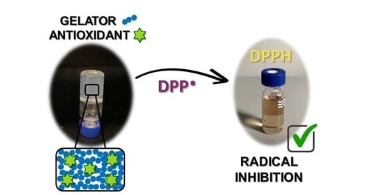
:Antioxidants are important substances used in the cosmetic and pharmaceutical fields that are able to block free radicals. These compounds can be incorporated into formulations for many reasons, such as release over time or preservation of the formulation activity and applicability. In the present study, a low-molecular-weight gel made with Boc-L-DOPA(Bn)2-OH was studied as suitable material to host antioxidants and improve their activity. The solvent change (DMSO/H2O) in combination with temperature was the technological procedure for the preparation of the gel. Two different antioxidants were tested: (1) α-tocopherol and (2) postbiotics. The antioxidant activity of α-tocopherol and of the postbiotics in the gel, measured by the (2,2-diphenyl-1-picryl-hydrazyl radical (DPPH) assay, showed higher values than those in the pure solvent. The antioxidant activity of the gel with 0.8 w/v% of gelator and α-tocopherol in the concentration range of 5–100 µM was 2.7–1.1 times higher on average than in the pure solvent. In the case of both postbiotics, the biggest difference was observed at 30% of postbiotics in the gel with 0.5% of a gelator, when the antioxidant activity was 4.4 to 4.7 times higher than that in the pure solvent.
1. Introduction
Low-molecular-weight gelators (LMWGs) are small molecules that can self-assemble inside a solvent, leading to the formation of a physical gel [1]. The formation of this class of gels occurs thanks to non-covalent interactions, including hydrogen bonding and π-π stacking, and it leads to the formation of reversible materials as opposed to other classes of materials, such as the polymeric ones [2,3]. These gels are characterized by several benefits in the cosmetic industry (encapsulation of substances and better penetration through the skin [4] and the release of fragrances [5]), in the food industry (transfer of substances to targeted places without their disintegration) [6], in medicine for the controlled drug delivery [7], as well as in the environmental field [8,9,10,11] and the biomedical one [12]. A variety of bioactive molecules, such as antibiotics or antioxidants, can be encapsulated in the gel matrix through additional non-covalent interactions to form smart and highly functionalized gels [13,14].
The number of papers on the encapsulation of antioxidants in gels is mainly limited to polymeric systems [15,16,17,18,19]. The encapsulation often resulted in a similar behavior of the antioxidant, sometimes with a preservation of its activity and sometimes with a small improvement. The same studies of encapsulation of antioxidants inside low-molecular-weight (LMW) gels are still very limited, which makes this field of research very interesting and appealing.
Salazar-Bautista et al. [20] reported the preparation of gel nanoparticles suitable for the controlled delivery of antioxidants. They observed that depending on the structure of the antioxidant, their encapsulation inside LMW gels could be involved in the formation of supramolecular interactions (H bondings and π-π stacking) with the fibrous three-dimensional network of L amino acid-based gelators. Wei et al. [21] reported a gel functionalized with ferulic acid with quite a high antioxidant efficiency and even improved cutaneous wound healing.
Antioxidants are important substances for in vivo applications in cosmetics as well as in pharmaceutics and can be incorporated into gels [22] or other formulations to be released over time, but they also may improve and preserve the gel activity and applicability.
The occurrence of injury is closely linked to the generation of free radicals and, thus, oxidative stress [16]. It is well known that free radicals damage fatty acids, deoxyribonucleic acid, proteins, and other important bio compounds not only in the body but also on/in the skin [23]. Antioxidants are important compounds able to block the formation of these dangerous free radicals. As fatty acids are part of cell membranes, free radicals become dangerous for any cell and destroy the functions of the cells they attack. This mechanism explains the formation of atherosclerosis (hardening of blood vessels), rheumatic diseases, premature skin aging, skin barrier disruption, skin inflammation, diabetes, and many other diseases that many of us have in our “daily order” [24,25].
α-tocopherol is a primary antioxidant that can break and block the chain reaction in the initiation phase of oxidative stress by donating an electron to stabilize the existing free radical, and its effect has been widely studied [26]. It is the most active biological form among the eight forms of vitamin E. For this reason and it may be dispensed by oral as well as by topical application [27], as dermal penetration is extremely effective [28].
Postbiotic complexes can be obtained by fermentation of suitable matrices with living probiotics, followed by cell rupture, that produces a complex mixture containing only parts of its DNA, exopolysaccharide complexes, peptides, organic acids, cell wall, and membrane, as well as many other active by-products. The composition depends on the specific matrix and the specific probiotic strain [29]. Postbiotics have been recently used as new antioxidants in food-based products, cosmetic formulations, and pharmaceutical preparations. Although the contribution of probiotic bacteria ingested through food and nutritional supplements in the intestinal microbiota has already been studied by researchers in several respects, the protective effects of postbiotics as by-products of probiotics against oxidative stress caused by free radicals on skin exposure have not yet been evaluated enough. The current cosmetic industry offers them in the formulations such as creams, serums, masks, or gels [30]. However, according to ISO 17516:2014, their further development in cosmetics in order to meet the microbiological safety limits of cosmetics is questionable. Therefore, when it comes to cosmetics, products contain extracts, lysates, or products of the fermentation process made from bacteria (referred to as postbiotics) rather than living cultures (probiotics). The antioxidant activity of postbiotics has been documented by Feng and Wang [31] or Ácsová et al. [32].
In this work, the antioxidant activity of two types of antioxidants, both in solutions and in supramolecular gels, were compared. First, α-tocopherol was chosen as a model molecule because it is a widely studied antioxidant. Then, after the positive results obtained in gels with this simple system, we expanded our study to postbiotics, which are a novel and more complex class of antioxidants. In particular, postbiotics obtained by lysing of living Gram-positive bacterial species Lactococcus lactis and Bifidobacterium longum were evaluated.
The supramolecular gels were prepared by self-assembly of Boc-L-DOPA(Bn)2-OH. This gelator was chosen because of its easy synthesis in a multigram scale, its ability to form gels under several conditions (also hosting inorganic crystals [33] or small molecules [5]), and its biocompatibility [34].
The optimal experimental parameters, such as the concentration of Boc-L-DOPA(Bn)2-OH and of the antioxidant substances, the conditions to prepare the desired gels, and the experimental conditions to perform the DPPH test on the resulting gel matrices, were identified and optimized. For both types of antioxidants, significative enhancement in the antioxidant activity inside the supramolecular LMW gels was found.
2. Materials and Methods
2.1. Materials
2.1.1. Chemicals
2,2-diphenyl-1-picryl-hydrazyl radical (•DPPH, ≥99%) was purchased from Sigma-Aldrich (Taufkirchen, Germany), α-tocopherol (>96%) from Sigma-Aldrich (Germany), and L-DOPA was purchased from TCI. Ethanol, Methanol, Milli-Q Water (Millipore, resistivity = 18.2 mΩ cm), and Dimethyl Sulfoxide (DMSO) were purchased from Sigma-Aldrich (Germany).
2.1.2. Postbiotics
(1) ProRenew Complex CLR as well as (2) Repair Complex CLR are yellowish-white mixtures obtained by lysing of living Gram-positive bacterial species Lactococcus lactis and Bifidobacterium longum, respectively. The method of lysis was chosen on the basis of empirical evidence, which is obtained by cell biological research on skin cells at CLR—Chemisches Laboratorium Dr. Kurt Richter GmbH (Berlin, Germany). These lysates (postbiotics) were fermented under special conditions of catabolic repression. No fractions were isolated after cell disintegration, thus ensuring the presence of all components in the complexes in their natural distribution—fragments such as DNA, metabolites, cytoplasmic compounds, and cell wall materials. In addition, after disintegration, stabilizing preservatives were added to the mixtures of postbiotics in an acidic, aqueous buffer medium to prevent the growth of any microorganisms. ProRenew Complex CLR and Repair Complex CLR consist of 98% of Lactococcus Ferment Lysate and Bifida Ferment Lysate, respectively.
2.2. Methods
2.2.1. Solutions of Tested Substances
Solutions of α-tocopherol were prepared in ethanol in the concentration range from 5 to 100 µM.
Both postbiotics (1) ProRenew Complex CLR and (2) Repair Complex were prepared as aqueous solutions in the concentration range from 30 to 80% v/v.
2.2.2. Synthesis and Characterization of Boc-L-DOPA(Bn)2-OH
2.2.3. Preparation of Gels with α-Tocopherol
Boc-L-DOPA(Bn)2-OH was dissolved in DMSO to a final concentration of 0.8 or 1.0% w/v, and the mixture was stirred in a water bath at 55 °C for 5 min to allow complete dissolution. Moreover, α-tocopherol was dissolved in ethanol for the preparation of stock solutions (1 mM), and subsequently, this was added to the mixture of Boc-L-DOPA(Bn)2-OH in DMSO in specific volumes. Finally, water was added to DMSO in a ratio of 3:7, 2:8, or 1:1, respectively, and the mixture was allowed to rest at room temperature for 16 h to allow gel formation. The composition of gels with incorporated α-tocopherol is shown in Supporting Information (Table S1).
2.2.4. Preparation of Gels with Postbiotics
Boc-L-DOPA(Bn)2-OH was dissolved in DMSO to a final concentration of 0.5% w/v, and the mixture was stirred in a water bath at 55 °C for 5 min to allow complete dissolution. Moreover, postbiotic (1) or postbiotic (2) was heated to a temperature of about 35 °C and subsequently added to the mixture of Boc-L-DOPA(Bn)2-OH with DMSO, using different amounts depending on the different concentrations of postbiotics in the final product. The gels were allowed to rest for at least 4 h before analyses. The composition of gels with incorporated postbiotics in the different concentrations is shown in Table S2.
2.2.5. Optical Microscopy
Gels were deposited after formation on microscope slides while still wet. The time required for the acquisition was very short compared to the time needed for the solvent (DMSO/H2O) to evaporate, thus ensuring that the sample was still wet and hydrated and avoiding drying artifacts. The optical microscope images were recorded using a Nikon (Minato, Japan) 13 ECLIPSE Ti2 Inverted Research Microscope was used to collect the images. Nikon (Minato, Japan) 13 ECLIPSE Ti2 Inverted Research Microscope with a 10× magnifier.
2.2.6. Rheological Properties
Rheological properties of the gels were analyzed 16 h after their preparation with an Anton Paar Rheometer MCR 102. Experiments were performed at a fixed temperature of 23 °C (controlled by an integrated Peltier system). Time sweep experiments were performed using the parallel plate geometry (PP25) at a constant shear strain (γ) of 0.05% and a constant angular frequency (ω) of 10 rad/s, collecting 1 point every 20 s. Each gel sample (3 mL) was prepared in a Petri glass (diameter = 30 mm), and the experiment was started immediately after the addition of the trigger, setting a gap of 4.0 mm. Oscillatory amplitude sweep experiments (γ: 0.01–100%) were performed at a constant angular frequency of 10 rad/s. The gel samples were prepared as described and tested directly in Sterilin Cups®, which fit in the rheometer, setting a fixed gap value of 2.1 mm.
2.2.7. Determination of DPPH Radical Scavenging Activity (in Gel/in Solution)
The antioxidant potential of tested substances was investigated for the scavenging activity of DPPH radicals [32] with slight modification for determination in a cuvette spectrophotometer. Both postbiotics were evaluated (i) as a 30, 50, 60, 70, and 80% (v/v) aqueous solution or (ii) as a gel with incorporated postbiotics in the same concentration as solutions. An amount of 2 mL of the solution/gel with postbiotics in a specific concentration was reacted with 667 µL of DPPH radical solution (0.3 mM in methanol). After 30 min of reaction in the dark, mixtures of gels with DPPH radicals were centrifuged (4400 rpm/6 min/20 °C) due to the hard spectrophotometric measurement of non-transparent gels. An amount of 1 mL of supernatant was transferred into the quartz cuvette, and the absorbance was measured at 517 nm against DMSO with a Cary 300 UV-vis double-beam spectrophotometer.
2.2.8. Statistical Analysis
Every assay was performed in triplicates, and data are given as the average with standard deviation (SD). Statistical data were obtained by one-way analysis of variance (ANOVA) followed by Bonferroni’s test for comparison of gels with the solution form of the test substance. Differences were considered significant at p < 0.05(*).
3. Results and Discussion
3.1. LMW Gels with α-Tocopherol
In this work, Boc-L-DOPA(Bn)2-OH (Figure 1) was used for the formation of gels containing two classes of antioxidants: α-tocopherol and postbiotics. This specific gelator was chosen as it may be easily prepared in a multigram scale starting from commercially available L-Dopa and can form robust gels under several conditions in very low concentrations, even with the addition of different species [5,33].
The first antioxidant studied in this work is α-tocopherol, one form of vitamin E commonly used in the food, cosmetic, and pharmaceutical industries [36].
The first goal was its solubilization and incorporation inside DMSO/H2O gels of Boc-L-DOPA(Bn)2-OH. For this aim, three major requirements had to be satisfied: the gels had to be mixed by a uniform process to create homogeneous matrices and avoid the formation of precipitates; the gel should form in a short time, ranging from some hours to max 1 day, to avoid longer experimental time; and, finally, a gel should form for any antioxidant concentration that we decided to test.
Due to the high lipophilicity of α-tocopherol, it is advisable to prepare a lipophilic solution of the antioxidant, which is then incorporated into the gel [20,37]. Following this approach, in the study by Patel et al. [37], gels of 12-hydroxystearic acid with α-tocopherol were formed at high gelator concentration (5% w/v) by dissolving the antioxidant in the oil phase at high temperature. Thus, we tried to dissolve α-tocopherol directly in DMSO and prepare the DMSO/H2O gels starting from this solvent, but no gel formed under these conditions. Therefore, a premix stock solution of α-tocopherol in ethanol (1 mM) was prepared, which facilitated its incorporation into DMSO/H2O (7/3) gel and did not hinder the gel formation.
The minimum gelator concentration to obtain a gel under these conditions and for all the EtOH solutions of α-tocopherol (concentrations ranging from 5 µM to 100 µM) was 0.8% w/v, forming stable and reproducible gels, so this concentration was chosen for further characterizations (Table S1). The gels were transparent at the beginning of the gelation process and became whitish overnight, suggesting that fiber formation and entanglement continue overnight (Figure S1). This hypothesis was confirmed by the time sweep analysis (Figure S2), where it is possible to see that even after a few minutes, G’ is already higher than G’’, and its value continues to increase for hours before reaching a plateau. Amplitude sweep measurements were used to determine the mechanical properties of the gels and to check the effect of antioxidants addition on the gel network. The presence of α-tocopherol did not cause big changes in the rheological properties of gels in the range of concentration of α-tocopherol tested, as reported in Figure S3. The morphology of the gels shows a network of long entangled fibers in the presence of both the lowest (5 µM) and the highest (100 µM) concentration of α-tocopherol (Figure S4).
Gels with 1% w/v of Boc-L-DOPA(Bn)2-OH were also prepared in the same conditions to compare the antioxidant activity of α-tocopherol in gels with different concentrations of gelator (0.8% and 1.0% w/v). The aim was to determine whether the gelator content affects the overall antioxidant activity of the gel proportionally or not. These samples formed in a few hours as the gels with 0.8% w/v (Figure S1).
The antioxidant ability of the two sets of gels containing α-tocopherol was evaluated by testing their scavenging efficacy of 1,1-diphenyl-2-picrylhydrazyl (DPPH) free radical, which is a compound able to donate an electron or hydrogen atom to neutralize radicals (Figure S5) [38]. Prior to the analysis of the ability to inhibit DPPH, the gel was disrupted due to its strength by gentle mixing with DPPH radical solution and then incubated in the dark for 30 min.
The antioxidant activity of α-tocopherol in ethanol solutions (black bars) and in gels in both concentrations (bars with dots or lines) was compared (Figure 2). In any case, the percentage of DPPH inhibition increased with the increase of α-tocopherol concentration.
Remarkably, the antioxidant activity of gels with α-tocopherol exceeded the activity of the pure solutions in every concentration analyzed, and was generally higher with gels with 1% w/v of Boc-L-DOPA(Bn)2-OH than with 0.8% w/v, even though the difference between the two sets is very small. For example, the antioxidant potential of α-tocopherol in 5 µM concentration was 2.7 times higher in 0.8% w/v gels (22.4% of inhibited DPPH radical) than in a solution (8.3% of inhibited DPPH radical) and 3.5 times higher in 1.0% w/v gels (29.4% of inhibited DPPH radical) than in a solution.
As the gel formation usually occurs in 16 h, antioxidant activity measurement was also feasible in the form of a “pre-gel” solution (before gel formation). The aim was to compare the antioxidant activity before and after gel formation, analyzing the set of gels containing the gelator in 0.8% w/v concentration (Figure 3). In the case of the pre-gel solutions, the antioxidant activity value was slightly lower than that of gels, meaning that the formation of the fibers of the gel network slightly improves the antioxidant activity of α-tocopherol.
The higher values of the gel scavenging effect after its formation could be due to the binding of α-tocopherol with hydrophilic and hydrophobic groups of the fibers [39]. This may increase the number of α-tocopherol binding sites with DPPH radical during the scavenging process, as recently reported by Zhang et al. [40], who suggested that the interactions among one molecule of α-tocopherol and DPPH radical by the partial reduction in radical and the simultaneous formation of a stable oxidation product has been mostly completed. This effect may also be enhanced by the presence of a protic solvent such as water, which can donate a hydrogen atom to the formed radical from α-tocopheryl quinone, then forming a fully new product. The naturally occurring tetrahydropyran in α-tocopherol is unbound, and a functional -OH group and p-benzoquinone are formed from its oxygen and benzene, respectively.
To better understand the role of the solvent in the α-tocopherol antioxidant activity, we compared the antioxidant activity of three gels with 1% w/v of Boc-L-DOPA(Bn)2-OH and different ratios of DMSO to H2O (8:2, 7:3 and 1:1) (Figure 4). The fact that the antioxidant activity of α-tocopherol is influenced by the solvent was also reported by Müller et al. and by Iwatsuki et al. [41,42], who compared the antioxidant activity of α-tocopherol in apolar solvent (hexane) and in polar protic ones (methanol), observing a decrease in antioxidant activity in protic solvents.
3.2. LMW Gel with Postbiotics (PB)
As Averina et al. mentioned [43], many postbiotics (PB) have naturally scavenged free radicals by producing their own antioxidant enzymes, such as superoxide dismutase. The presence of this enzyme has been reported in several cell-free postbiotic extracts [44]. Metabolites of postbiotics as parts of cell walls containing exopolysaccharides, in most cases uronic acid polysaccharides, could significantly support the inhibition activity of postbiotics against free radicals [45]. As Abbasi et al. claims [46], it is important to design and develop new effective delivery systems of postbiotics for reaching their beneficial action in targeted tissue in the human body as well as on the skin. Most recently, PB has been recognized as a valuable raw material in the development of a new type of gels for the adjuvant treatment of teeth periodontitis [47].
Following the procedure previously established for the α-tocopherol containing gels, gels containing our samples of PB, Repair (1), and ProRenew (2) were prepared. In this case, the addition of ethanol was completely avoided, as PB is available in an aqueous medium and is able to trigger gel formation when added to gelator solutions of Boc-L-DOPA(Bn)2-OH in DMSO. The minimum gelator concentration to obtain a gel under these conditions is 0.5% w/v, so this concentration was chosen for further characterization (Table S2). Although these PB solutions may trigger the gel formation when added to gelator solutions in H2O/NaOH, in this work, we focused our attention on the gels obtained from DMSO to maintain the same conditions as the α-tocopherol gels. Furthermore, PB should now be regarded as an interesting additive, acting, at the same time, as bioactive substances entrapped in the gel network, antioxidants, and trigger for LMW gel formation in different conditions (from organic solvent to water).
The final pH values of both PB complexes were evaluated, and their value was determined to be 4.13 for Repair (1) and 4.06 for ProRenew (2). With both complexes, the gels with 30–80% v/v of PB (PB-gels) were prepared (Figure S6), and the gelation time was shorter (about 10 min) than in the case of α-tocopherol (Figures S7 and S8).
With the smallest amount of PB (30%), we could observe the formation of long entangled fibers with the microscope, while gels with an increased amount of PB showed only shorter fibers and less ordered networks (Figure S9).
By measuring the viscoelastic properties of the gels, we recorded a slight decrease in the gel stiffness when increasing the amount of PB in gels (Figures S10 and S11). Gels with the ProRenew Complex were less strong than gels with the Repair ones. This behavior demonstrates once again how Boc-L-DOPA(Bn)2-OH is a robust gelator, able to form gels under several conditions and host other species (α-tocopherol and postbiotics in different concentrations).
The evaluation of the PB gels’ antioxidant activity was obtained by testing their scavenging efficacy of 1,1-diphenyl-2-picrylhydrazyl (DPPH) free radicals, as previously reported. Unlike the α-tocopherol gels, PB-gels show a thixotropic behavior, reforming after mixing with DPPH radical solution. Therefore, after the reaction incubation (30 min), they were centrifuged before analyzing via a UV-VIS spectrophotometer.
In Figure 5, grey and white bars without any filler are the two postbiotic solutions in DMSO without a gelator. Bars filled with dots and lines are the PB gels. In all cases, the antioxidant activity of the gels was higher than that of the PB solutions.
Additionally, the antioxidant activity between gels formed with two gelator concentrations (0.5 and 1.0% w/v) was compared, as it was for α-tocopherol. In this case, the result was opposite to the previous one, as the antioxidant activity of 1% gels with PB did not lead to a two-fold increase in antioxidant activity but to a decreased activity compared to gels with a lower concentration of gelator (0.5% w/v) (Figure 6).
4. Conclusions
In this work, we reported a library of supramolecular gels made with Boc-L-DOPA(Bn)2-OH in DMSO/H2O. These gels are able to host two classes of antioxidants in different concentrations: α-tocopherol and two postbiotics (Repair and ProRenew) obtained from two different microorganisms. A wide range of conditions, including (i) antioxidant concentration, (ii) gelator concentration, and (iii) solvent ratio, was monitored and analyzed to obtain a gel with the best antioxidant activity without affecting the rheological properties of the gels.
Therefore, we used Boc-L-DOPA(Bn)2-OH, which is a robust gelator, able to form gels under several conditions and to host both classes of antioxidants efficiently. Besides the formation of strong materials, we demonstrated that the antioxidant activity was not only maintained but even significantly enhanced by inserting the antioxidants in the supramolecular gels.
This interesting result could pave the way for the preparation of many other gels containing antioxidants of different types. For this reason, we believe that LMW gels could be successful in the cosmetics and pharmaceutical industries as materials that are able to host antioxidants and even increase their activity.
Supplementary Materials
The following supporting information can be downloaded at: https://0-www-mdpi-com.brum.beds.ac.uk/article/10.3390/cosmetics10020038/s1, and contain—Table S1: Composition of LMW gels with α-tocopherol; Figure S1: Gels of Boc-L-Dopa(Bn)2-OH with α-tocopherol; Figure S2: Time sweep analysis of the gel of Boc-L-Dopa(Bn)2-OH 0.8% w/v (DMSO/H2O 70/30) with 5 µM concentration of α-tocopherol; Figure S3: Amplitude sweep of gels 0.8% w/v of Boc-L-DOPA(Bn)2-OH in DMSO:H2O = 7:3 with incorporated α-tocopherol in a specific concentration; Figure S4: Optical microscope images of gels (0.8% w/v) of Boc-L-DOPA(Bn)2-OH in DMSO:H2O = 7:3 with incorporated of α-tocopherol; Figure S5: 2,2-diphenyl-1-picrylhydrazyl (DPPH) reaction mechanism; Table S2: Composition of LMW gels with postbiotics (PB); Figure S6: Gels of Boc-L-Dopa(Bn)2-OH with postbiotics; Figure S7: Time sweep analysis of the gel of Boc-L-Dopa(Bn)2-OH 0.5% w/v with 30% of Repair complex; Figure S8: Time sweep analysis of the gel of Boc-L-Dopa(Bn)2-OH 0.5% w/v with 30% of ProRenew complex; Figure S9: Optical microscope images showing gel fibers when postbiotics ProRenew Complex or Repair Complex is used to trigger gelation; Figure S10: Amplitude sweep of gels 0.5% of Boc-L-DOPA(Bn)2-OH in DMSO:Postbiotics (Repair Complex) in specific concentrations; Figure S11: Amplitude sweep of gels 0.5% of Boc-L-DOPA(Bn)2-OH in DMSO:Postbiotics (ProRenew Complex) in specific concentrations.
Author Contributions
Conceptualization, A.Á.T. and D.G.; Methodology, A.Á.T. and D.G.; Validation, A.Á.T.; Formal Analysis, A.Á.T. and D.G.; Investigation, A.Á.T. and D.G.; Resources, A.Á.T. and C.T.; Data Curation, C.T.; Writing—Original Draft Preparation, A.Á.T., D.G., S.M., and C.T.; Writing—Review and Editing, A.Á.T., D.G., S.M., and C.T.; Supervision, S.M. and C.T.; Project Administration, A.Á.T. and D.G.; Funding Acquisition, S.M. All authors have read and agreed to the published version of the manuscript.
Funding
This research was funded by the Scientific grant agency of the Ministry of Education, Science, Research and Sports of the Slovak Republic and the Slovak Academy of Sciences VEGA, VEGA 2/0136/20.
Institutional Review Board Statement
Not applicable.
Informed Consent Statement
Not applicable.
Data Availability Statement
Not applicable.
Acknowledgments
We thank CLR—Chemisches Laboratorium Kurt Richter GmbH (Berlin, Germany) for the provision of postbiotics.
Conflicts of Interest
The authors declare no conflict of interest.
References
- Draper, E.R.; Adams, D.J. Low-Molecular-Weight Gels: The State of the Art. Chem 2017, 3, 390–410. [Google Scholar] [CrossRef] [Green Version]
- Wang, J.; Liu, K.; Xing, R.; Yan, X. Peptide self-assembly: Thermodynamics and kinetics. Chem. Soc. Rev. 2016, 45, 5589–5604. [Google Scholar] [CrossRef]
- Hanabusa, K.; Suzuki, M. Development of low-molecular-weight gelators and polymer-based gelators. Polym. J. 2014, 46, 776–782. [Google Scholar] [CrossRef]
- Dastidar, P.; Roy, R.; Parveen, R.; Sarkar, K. Supramolecular Synthon Approach in Designing Molecular Gels for Advanced Therapeutics. Adv. Ther. 2019, 2, 1800061. [Google Scholar] [CrossRef] [Green Version]
- Nicastro, G.; Black, L.M.; Ravarino, P.; Agostino, S.; Faccio, D.; Tomasini, C.; Giuri, D. Controlled Hydrolysis of Odorants Schiff Bases in Low-Molecular-Weight Gels. Int. J. Mol. Sci. 2022, 23, 3105. [Google Scholar] [CrossRef]
- Du, H.; Liu, J.; Pan, B.; Yang, H.-Y.; Liu, G.-B.; Lu, K. Fabrication of the low molecular weight peptide-based hydrogels and analysis of gelation behaviors. Food Hydrocoll. 2022, 131, 107751. [Google Scholar] [CrossRef]
- Oliveira, C.B.P.; Gomes, V.; Ferreira, P.M.T.; Martins, J.A.; Jervis, P.J. Peptide-Based Supramolecular Hydrogels as Drug Delivery Agents: Recent Advances. Gels 2022, 8, 706. [Google Scholar] [CrossRef]
- Okesola, B.O.; Smith, D.K. Applying low-molecular weight supramolecular gelators in an environmental setting-self-assembled gels as smart materials for pollutant removal. Chem. Soc. Rev. 2016, 45, 4226–4251. [Google Scholar] [CrossRef] [Green Version]
- Giuri, D.; D’Agostino, S.; Ravarino, P.; Faccio, D.; Falini, G.; Tomasini, C. Water Remediation from Pollutants Agents by the Use of an Environmentally Friendly Supramolecular Hydrogel. ChemNanoMat 2022, 8, e202200093. [Google Scholar] [CrossRef]
- Giuri, D.; Zanna, N.; Tomasini, C. Low Molecular Weight Gelators Based on Functionalized l-Dopa Promote Organogels Formation. Gels 2019, 5, 27. [Google Scholar] [CrossRef] [PubMed] [Green Version]
- Guidetti, G.; Giuri, D.; Zanna, N.; Calvaresi, M.; Montalti, M.; Tomasini, C. Biocompatible and Light-Penetrating Hydrogels for Water Decontamination. ACS Omega 2018, 3, 8122–8128. [Google Scholar] [CrossRef]
- Skilling, K.J.; Citossi, F.; Bradshaw, T.D.; Ashford, M.; Kellam, B.; Marlow, M. Insights into low molecular mass organic gelators: A focus on drug delivery and tissue engineering applications. Soft Matter 2014, 10, 237–256. [Google Scholar] [CrossRef]
- Jeong, S.; Lee, S.; Oh, I. Development of Antioxidant-Fortified Oleogel and Its Application as a Solid Fat Replacer to Muffin. Foods 2021, 10, 3059. [Google Scholar] [CrossRef] [PubMed]
- Zhao, Q.; Zhao, Y.; Lu, Z.; Tang, Y. Amino Acid-Modified Conjugated Oligomer Self-Assembly Hydrogel for Efficient Capture and Specific Killing of Antibiotic-Resistant Bacteria. ACS Appl. Mater. Interfaces 2019, 11, 16320–16327. [Google Scholar] [CrossRef]
- Hadian, M.; Rajaei, A.; Mohsenifar, A.; Tabatabaei, M. Encapsulation of Rosmarinus officinalis essential oils in chitosan-benzoic acid nanogel with enhanced antibacterial activity in beef cutlet against Salmonella typhimurium during refrigerated storage. Lwt 2017, 84, 394–401. [Google Scholar] [CrossRef]
- Barreira, J.C.M.; Rodrigues, S.; Carvalho, A.M.; Ferreira, I.C.F.R. Development of hydrosoluble gels with Crataegus monogyna extracts for topical application: Evaluation of antioxidant activity of the final formulations. Ind. Crops Prod. 2013, 42, 175–180. [Google Scholar] [CrossRef]
- Maqsoudlou, A.; Assadpour, E.; Mohebodini, H.; Jafari, S.M. Improving the efficiency of natural antioxidant compounds via different nanocarriers. Adv. Colloid Interface Sci. 2020, 278, 102122. [Google Scholar] [CrossRef] [PubMed]
- Villalva, M.; Jaime, L.; Arranz, E.; Zhao, Z.; Corredig, M.; Reglero, G.; Santoyo, S. Nanoemulsions and acidified milk gels as a strategy for improving stability and antioxidant activity of yarrow phenolic compounds after gastrointestinal digestion. Food Res. Int. 2020, 130, 108922. [Google Scholar] [CrossRef]
- Lee, S.H.; Chow, P.S.; Yagnik, C.K. Developing Eco-Friendly Skin Care Formulations with Microemulsions of Essential Oil. Cosmetics 2022, 9, 30. [Google Scholar] [CrossRef]
- Salazar-Bautista, S.C.; Chebil, A.; Pickaert, G.; Gaucher, C.; Jamart-Gregoire, B.; Durand, A.; Leonard, M. Encapsulation and release of hydrophobic molecules from particles of gelled triglyceride with aminoacid-based low-molecular weight gelators. Colloids Surfaces A Physicochem. Eng. Asp. 2017, 514, 11–20. [Google Scholar] [CrossRef]
- Wei, Q.; Duan, J.; Ma, G.; Zhang, W.; Wang, Q.; Hu, Z. Enzymatic crosslinking to fabricate antioxidant peptide-based supramolecular hydrogel for improving cutaneous wound healing. J. Mater. Chem. B 2019, 7, 2220–2225. [Google Scholar] [CrossRef]
- Butkeviciute, A.; Ramanauskiene, K.; Janulis, V. Formulation of Gels and Emulgels with Malus domestica Borkh: Apple Extracts and Their Biopharmaceutical Evaluation In Vitro. Antioxidants 2022, 11, 373. [Google Scholar] [CrossRef]
- Lutchmanen Kolanthan, V.; Brown, A.; Soobramaney, V.; Philibert, E.G.; Francois Newton, V.; Hosenally, M.; Sokeechand, B.N.; Petkar, G.; Moga, A.; Andres, P.; et al. Clinical Evaluation of Indian Sandalwood Oil and Its Protective Effect on the Skin against the Detrimental Effect of Exposome. Cosmetics 2022, 9, 35. [Google Scholar] [CrossRef]
- Wortzman, M.; Nelson, D.B. A comprehensive topical antioxidant inhibits oxidative stress induced by blue light exposure and cigarette smoke in human skin tissue. J. Cosmet. Dermatol. 2021, 20, 1160–1165. [Google Scholar] [CrossRef]
- Shahzad, A.; Hussain, S.; Anwar, N.; Karim, A.; Aeman, U.; Iqbal, M.J. An overview of free Radicals & antioxidants and its Deletenous actions. Front. Chem. Sci. 2021, 2, 147–164. [Google Scholar] [CrossRef]
- Temova Rakuša, Ž.; Roškar, R. Quality Control of Vitamins A and E and Coenzyme Q10 in Commercial Anti-Ageing Cosmetic Products. Cosmetics 2021, 8, 61. [Google Scholar] [CrossRef]
- Akhtar, N.; Akhtar, N. Development of stable tocopherol succinate-loaded ethosomes to enhance transdermal permeation: In vitro and in vivo characterizations. J. Cosmet. Dermatol. 2022, 21, 4942–4955. [Google Scholar] [CrossRef] [PubMed]
- Sharma, G.N.; Gupta, G.; Sharma, P. A comprehensive review of free radicals, antioxidants and their relationship with human ailments. Crit. Rev. Eukaryot. Gene Expr. 2018, 28, 139–154. [Google Scholar] [CrossRef]
- Chaudhari, A.; Dwivedi, M.K. Chapter 1—The concept of probiotics, prebiotics, postbiotics, synbiotics, nutribiotics, and pharmabiotics. In Probiotics in the Prevention and Management of Human Diseases; Dwivedi, M.K., Amaresan, N., Sankaranarayanan, A., Kemp, E.H., Eds.; Academic Press: Cambridge, MA, USA, 2022; pp. 1–11. ISBN 978-0-12-823733-5. [Google Scholar]
- Zawistowska-Rojek, A.; Zaręba, T.; Tyski, S. Microbiological Testing of Probiotic Preparations. Int. J. Environ. Res. Public Health 2022, 19, 5701. [Google Scholar] [CrossRef] [PubMed]
- Feng, T.; Wang, J. Oxidative stress tolerance and antioxidant capacity of lactic acid bacteria as probiotic: A systematic review. Gut Microbes 2020, 12, 1801944. [Google Scholar] [CrossRef]
- Ácsová, A.; Hojerová, J.; Martiniaková, S. Efficacy of postbiotics against free radicals and UV radiation. Chem. Pap. 2022, 76, 2357–2364. [Google Scholar] [CrossRef]
- Giuri, D.; Jurković, L.; Fermani, S.; Kralj, D.; Falini, G.; Tomasini, C. Supramolecular Hydrogels with Properties Tunable by Calcium Ions: A Bio-Inspired Chemical System. ACS Appl. Bio Mater. 2019, 2, 5819–5828. [Google Scholar] [CrossRef] [PubMed]
- Di Filippo, M.F.; Giuri, D.; Marchiori, G.; Maglio, M.; Pagani, S.; Fini, M.; Tomasini, C.; Panzavolta, S. Self-assembling of fibers inside an injectable calcium phosphate bone cement: A feasibility study. Mater. Today Chem. 2022, 24, 100991. [Google Scholar] [CrossRef]
- Gaucher, A.; Dutot, L.; Barbeau, O.; Hamchaoui, W.; Wakselman, M.; Mazaleyrat, J.P. Synthesis of terminally protected (S)-β3-H-DOPA by Arndt-Eistert homologation: An approach to crowned β-peptides. Tetrahedron Asymmetry 2005, 16, 857–864. [Google Scholar] [CrossRef]
- Tangpromphan, P.; Duangsrisai, S.; Jaree, A. Development of separation method for Alpha-Tocopherol and Gamma-Oryzanol extracted from rice bran oil using Three-Zone simulated moving bed process. Sep. Purif. Technol. 2021, 272, 118930. [Google Scholar] [CrossRef]
- Patel, A.R.; Remijn, C.; Heussen, P.C.M.; Den Adel, R.; Velikov, K.P. Novel low-molecular-weight-gelator-based microcapsules with controllable morphology and temperature responsiveness. ChemPhysChem 2013, 14, 305–310. [Google Scholar] [CrossRef]
- Sadeer, N.B.; Montesano, D.; Albrizio, S.; Zengin, G.; Mahomoodally, M.F. The versatility of antioxidant assays in food science and safety—Chemistry, applications, strengths, and limitations. Antioxidants 2020, 9, 709. [Google Scholar] [CrossRef] [PubMed]
- Nikiforidis, C.V.; Scholten, E. Self-assemblies of lecithin and α-tocopherol as gelators of lipid material. RSC Adv. 2014, 4, 2466–2473. [Google Scholar] [CrossRef] [Green Version]
- Zhang, L.; Zhang, T.; Chang, M.; Lu, M.; Liu, R.; Jin, Q.; Wang, X. Effects of interaction between α-tocopherol, oryzanol, and phytosterol on the antiradical activity against DPPH radical. LWT 2019, 112, 108206. [Google Scholar] [CrossRef]
- Müller, L.; Theile, K.; Böhm, V. In vitro antioxidant activity of tocopherols and tocotrienols and comparison of vitamin E concentration and lipophilic antioxidant capacity in human plasma. Mol. Nutr. Food Res. 2010, 54, 731–742. [Google Scholar] [CrossRef]
- Iwatsuki, M.; Tsuchiya, J.; Komuro, E.; Yamamoto, Y.; Niki, E. Effects of solvents and media on the antioxidant activity of α-tocopherol. Biochim. Biophys. Acta—Gen. Subj. 1994, 1200, 19–26. [Google Scholar] [CrossRef] [PubMed]
- Averina, O.V.; Poluektova, E.U.; Marsova, M.V.; Danilenko, V.N. Biomarkers and Utility of the Antioxidant Potential of Probiotic Lactobacilli and Bifidobacteria as Representatives of the Human Gut Microbiota. Biomedicines 2021, 9, 1340. [Google Scholar] [CrossRef] [PubMed]
- Bourebaba, Y.; Marycz, K.; Mularczyk, M.; Bourebaba, L. Postbiotics as potential new therapeutic agents for metabolic disorders management. Biomed. Pharmacother. 2022, 153, 113138. [Google Scholar] [CrossRef] [PubMed]
- Ahire, J.J.; Jakkamsetty, C.; Kashikar, M.S.; Lakshmi, S.G.; Madempudi, R.S. In Vitro Evaluation of Probiotic Properties of Lactobacillus plantarum UBLP40 Isolated from Traditional Indigenous Fermented Food. Probiotics Antimicrob. Proteins 2021, 13, 1413–1424. [Google Scholar] [CrossRef] [PubMed]
- Abbasi, A.; Hajipour, N.; Hasannezhad, P.; Baghbanzadeh, A.; Aghebati-Maleki, L. Potential in vivo delivery routes of postbiotics. Crit. Rev. Food Sci. Nutr. 2022, 62, 3345–3369. [Google Scholar] [CrossRef]
- Butera, A.; Gallo, S.; Pascadopoli, M.; Taccardi, D.; Scribante, A. Home Oral Care of Periodontal Patients Using Antimicrobial Gel with Postbiotics, Lactoferrin, and Aloe Barbadensis Leaf Juice Powder vs. Conventional Chlorhexidine Gel: A Split-Mouth Randomized Clinical Trial. Antibiotics 2022, 11, 118. [Google Scholar] [CrossRef]
Figure 1.
Chemical structure of the gelator.

Figure 2.
DPPH radical inhibition (%) by α-tocopherol in EtOH solution and in gels with 0.8% or 1% w/v of Boc-L-DOPA(Bn)2-OH, in DMSO:H2O (7:3 v/v) (mean value ± SD, n = 3). The stars (*) above the columns indicate a statistically significant difference (p < 0.05) between α-tocopherol in a specific concentration in solution vs. gel.

Figure 3.
DPPH radical inhibition (%) by α-tocopherol in gels and in pre-gel solutions (0.8% w/v) of Boc-L-DOPA(Bn)2-OH in DMSO:H2O (7:3 v/v) (mean value ± SD, n = 3). The stars (*) above the columns indicate a statistically significant difference (p < 0.05) between α-tocopherol in a specific concentration in gel vs. in pre-gel solution after breaking the gel.

Figure 4.
DPPH radical inhibition (%) by α-tocopherol in gel with 1% w/v of Boc-L-DOPA(Bn)2-OH and different ratios of DMSO/H2O (mean value ± SD, n = 3).
Figure 4.
DPPH radical inhibition (%) by α-tocopherol in gel with 1% w/v of Boc-L-DOPA(Bn)2-OH and different ratios of DMSO/H2O (mean value ± SD, n = 3).

Figure 5.
DPPH radical inhibition (%) by POSTBIOTICS in solution and in gel with 0.5% of Boc-L-DOPA(Bn)2-OH (mean value ± SD, n = 3). Negative control presents solution of L-DOPA in DMSO. The stars (*) above the columns indicate a statistically significant difference (p < 0.05) between specific postbiotics in solution vs. in gel at a given concentration.

Figure 6.
DPPH radical inhibition (%) by POSTBIOTICS in gels with different amounts of Boc-L-DOPA(Bn)2-OH (0.5% and 1.0% w/v) (mean value ± SD, n = 3). Negative control presents solution of L-DOPA in DMSO.
Figure 6.
DPPH radical inhibition (%) by POSTBIOTICS in gels with different amounts of Boc-L-DOPA(Bn)2-OH (0.5% and 1.0% w/v) (mean value ± SD, n = 3). Negative control presents solution of L-DOPA in DMSO.

Disclaimer/Publisher’s Note: The statements, opinions and data contained in all publications are solely those of the individual author(s) and contributor(s) and not of MDPI and/or the editor(s). MDPI and/or the editor(s) disclaim responsibility for any injury to people or property resulting from any ideas, methods, instructions or products referred to in the content. |
© 2023 by the authors. Licensee MDPI, Basel, Switzerland. This article is an open access article distributed under the terms and conditions of the Creative Commons Attribution (CC BY) license (https://creativecommons.org/licenses/by/4.0/).
Share and Cite
MDPI and ACS Style
Toronyi, A.Á.; Giuri, D.; Martiniakova, S.; Tomasini, C. Low-Molecular-Weight Gels as Smart Materials for the Enhancement of Antioxidants Activity. Cosmetics 2023, 10, 38. https://0-doi-org.brum.beds.ac.uk/10.3390/cosmetics10020038
AMA Style
Toronyi AÁ, Giuri D, Martiniakova S, Tomasini C. Low-Molecular-Weight Gels as Smart Materials for the Enhancement of Antioxidants Activity. Cosmetics. 2023; 10(2):38. https://0-doi-org.brum.beds.ac.uk/10.3390/cosmetics10020038
Chicago/Turabian StyleToronyi, Aneta Ácsová, Demetra Giuri, Silvia Martiniakova, and Claudia Tomasini. 2023. "Low-Molecular-Weight Gels as Smart Materials for the Enhancement of Antioxidants Activity" Cosmetics 10, no. 2: 38. https://0-doi-org.brum.beds.ac.uk/10.3390/cosmetics10020038
Note that from the first issue of 2016, this journal uses article numbers instead of page numbers. See further details here.