Article

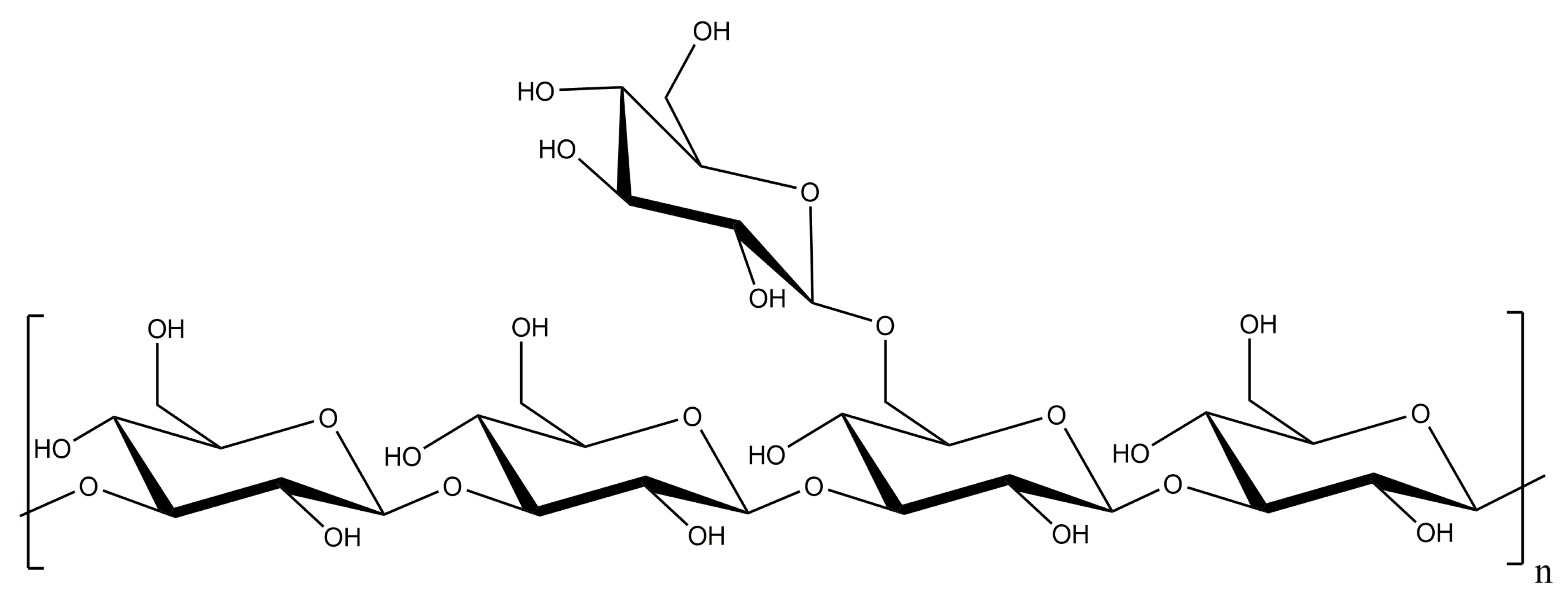
Laminarin Effects, a β-(1,3)-Glucan, on Skin Cell Inflammation and Oxidation

1
I3MTO, EA 4708, Université d’Orléans, CEDEX 2, 45067 Orléans, France
2
Département de Rhumatologie, Centre Hospitalier Régional d’Orléans, 45067 Orléans, France
3
Plateforme Recherche Innovation Médicale Mutualisée d’Orléans, Centre Hospitalier Régional d’Orléans, 14 Avenue de l’Hôpital, 45100 Orléans, France
4
GLYcoDiag, Bâtiment Physique-Chimie, Rue de Chartres, BP6759, CEDEX 2, 45067 Orléans, France
5
Institut de Chimie Organique et Analytique, UMR CNRS 7311, Université d’Orléans, Rue de Chartres, BP6759, CEDEX 2, 45067 Orléans, France
*
Authors to whom correspondence should be addressed.
Submission received: 30 July 2020 / Revised: 10 August 2020 / Accepted: 12 August 2020 / Published: 16 August 2020
(This article belongs to the Special Issue Feature Papers in Cosmetics in 2020)

Abstract

:
Laminarin, a β-(1,3)-glucan from the seaweed Laminaria digitata, is a polysaccharide which provides anti-inflammatory and anti-oxidative properties. Its influence on both human dermal fibroblasts adult (HDFa) and normal human epidermal keratinocytes (NHEK) has not been established yet. Herein, laminarin effects were examined on skin cells’ mitochondrial and antioxidant activities. Cytokines, hyaluronic acid, and procollagen type I secretions and interaction mechanisms were explored after a maximum of 72 h treatment with laminarin. Our results demonstrated a decrease in mitochondrial activities with 72 h treatment with laminarin from 500 µg.mL−1 for NHEK cells and from 100 µg.mL−1 for HDFa cells without cytotoxicity. No variation of hyaluronic acid or type I procollagen was observed for all laminarin concentrations, while an antioxidant effect was found against reactive oxygen species (ROS) from 1 µg.mL−1 for HDFa cells in both H2O2 and UVA radiation conditions, and from 10 µg.mL−1 and 1 µg.mL−1 for NHEK cells in both H2O2 and UVA radiation conditions, respectively. Laminarin treatment modulated both cells surface glycosylation and cytokine secretions of skin cells. Overall, our data suggest a positive effect of β-(1,3)-glucan on skin cells on oxidative stress and inflammation induced by environmental factors. Of note, these effects are through the modulation of glycan and receptors interactions at the skin cells surface.

1. Introduction

For years, research has focused on β-glucans, polysaccharides that are widely present in the cell walls of bacteria, algae, fungi, yeasts, or cereals [1,2,3], for their physico-chemical properties such as thickening, stabilizing, emulsification, and gelation [4]. Interestingly, β-glucans structure such as linear β-(1→3)- or β-(1→6)-chain core, molecular weight, length, or the number and degree of branching side-chain influence their solubility and conformation in aqueous media (single helix, triple helix, random coil conformation) [5]. These structural characteristics confer β-glucans biological properties, such as anti-cancer [6,7], anti-oxidative [8], and anti-inflammatory effects [9]. In immune cells, these mechanisms were achieved through interaction with β-glucans specifics receptors, such as CR3 and/or Dectin-1 [10,11]. Although the Dectin-1 receptor is expressed on immune cells, it is also expressed on cutaneous cells surface, such as fibroblasts and keratinocytes [12,13]. Hence, β-glucans are used in cosmetics [1] for their antioxidant and moisturizing properties [14,15], but also as a texturizing agent in cosmetic creams [16].
Among marine β-glucans, herein we focus on laminarin, a storage polysaccharide found in specific algae. Laminarin has a low molecular weight of 5 kDa and consists of an assembly of β-(1,3)-glucose units with branched β-(1,6)-glucose side chain [17,18,19]. It has been isolated from several brown seaweed species, such as Eisenia bicyclis, Saccharina longicruris, Laminaria digitata, Laminaria hyperborean, and Laminaria japonica [20,21]. Several studies demonstrated that laminarin provides a wide array of biological activities such as anti-tumor [22], anti-apoptotic [23], anti-inflammatory [24,25], and anti-oxidative activities [18]. These properties can be increased by several sulfated modifications on its polymeric osidic structure [26]. Laminarin presents an inherently low viscosity and high solubility in organic and aqueous solvents that facilitate processing and make laminarin very attractive for cosmetics. Herein, we investigated the influence of laminarin on skin cell viability, oxidative stress, and on extracellular matrices secretion. We have performed, for the first time, a GLYcoPROFILE® and NeoPROFILE on both human dermal fibroblasts (HDFa) and normal human epidermal keratinocytes (NHEK), in the presence of laminarin, to highlight all possible laminarin interactions with glycans moieties and carbohydrate receptors on skin cells surface. In addition, we evaluated the cytokine profile of skin cells exposed to different concentrations of laminarin to assess interaction with diverse receptors on the skin cell surface.

2. Materials and Methods

2.1. Cell Culture

Primary Human dermal fibroblasts adult (HDFa; C-013-5C, Invitrogen, Carlsbad, CA, USA) and spontaneously immortalized human keratinocyte cell line (HaCaT; 300493, Cell Lines Service, Eppelheim, Germany) were cultured in Dulbecco’s modified Eagle’s medium (DMEM; D6546, Sigma-Aldrich) supplemented with 10% Fetal Bovine Serum (F9665, Sigma-Aldrich), 2% L-Glutamine (17-605E, Lonza, Basel, Switzerland) and 1% Pen/Strep Amphotericin B (17-745E, Lonza, Basel, Switzerland) at 37 °C in a controlled humidified 5% CO2 atmosphere. Normal human epidermal keratinocytes (NHEK; 00192627, Lonza, USA) were cultured in a KGM-Gold bullet kit medium, including supplements (Kit KGM-Gold; 00192151, Lonza) at 37 °C in a controlled humidified 5% CO2 atmosphere. Cells were seeded in 75 cm2 tissue flasks at 3.75 × 10⁵ cells, 7.5 × 10⁵ cells, and 2.62 × 10⁵ cells for HDFa, HaCaT, and NHEK cells, respectively. Cells were passaged when their confluence was at 80%. Media were changed three times a week.

2.2. Cell Treatment

All experiment groups were treated with laminarin (L9634-1G, Sigma-Aldrich, St. Louis, MO, USA). The concentration ranges used were 1, 10, 100, 500, and 1000 µg.mL−1. All experiments were compared to untreated cells (0 µg.mL−1).

2.3. Cell Viability Assay

HDFa, HaCaT, and NHEK cells were seeded at 8 × 10³, 10 × 103, and 14 × 103 cells per well, respectively, in 96-well micro-plates and maintained for 24 h (h) at 37 °C, 5% CO2. The culture supernatant was replaced with a fresh medium (200 µL) with or without laminarin (Figure 1) at different concentrations (1, 10, 100, 500, 1000 µg.mL−1) and cells were cultivated for 24 h, 48 h and 72 h. Cell viability was assessed by MTT (3-(4,5-181dimethylthiazole-2-yl)-2,5-diphenyl tetrazolium bromide) assay (M2128-1G, Sigma, St. Louis, MO, USA). This method was first described in 1983 by Dr. Mosmann [27]. Briefly, after 24 h, 48 h, or 72 h of culture, 10% (w/v) of MTT at 5 mg.mL−1 were added into media, and cells were further incubated for 4 h. Then, the culture medium was removed, and 100 µL of lysis buffer (0.5% Sodium dodecyl sulfate (SDS; L3771, Sigma, Tokyo Japan), Hydrochloric acid (HCl; 1.09911.0001, Merk, Kenilworth, NJ, USA) 5 M, Isopropanol (I9516, Sigma, Steinheim, Germany) in sufficient quantity) was added into each well. The formazan precipitates in the supernatant were measured by absorbance at 540 nm and 620 nm using a multi-plate reader (Multiskan GO Microplate Spectrophotometer, Thermo Scientific, Sunnyvale, CA, USA). The percentages of cell viability were calculated with the range of cells.

2.4. Cell Cytotoxicity Assay

Cell cytotoxicity was assessed by LDH cytotoxicity assay kit (88953, Thermo Scientific, Sunnyvale, CA, USA). Briefly, after 24 h, 48 h, or 72 h of culture with laminarin (1, 10, 100, 500, 1000 µg.mL−1), 10 µL of lysis buffer provided by the kit was added into wells corresponding to the 100% cell lysis control, and 10 µL of sterile ultrapure water was added to all other wells. After 45 min at 37 °C, 5% CO2, 50 µL of supernatant was transferred to another 96-well microplate with 50 µL of reaction mix provided by the kit. Next, 50 µL of stop solution was added into each well after 30 min incubation at room temperature to stop the reaction. The absorbance of the formazan precipitate, if LDH was present in the supernatant, was measured at 490 nm and 680 nm using a microplate reader (Multiskan GO Microplate Spectrophotometer, Thermo Scientific, Sunnyvale, CA, USA). The percentage of cytotoxicity (% of cytotoxicity) was calculated using the following formula: % of cytotoxicity = ((A treated cells LDH activity − A spontaneous LDH activity)/(A maximum LDH activity − A spontaneous LDH activity)) × 100 (A = absorbance).

2.5. Hyaluronic Acid (HA) Dosage

HDFa, HaCaT, and NHEK cells were seeded at 7.5 × 10⁴ cells per well in a 24-well culture plate and maintained for 24 h at 37 °C, 5% CO2 in a humid atmosphere. Then, cells were cultured in fresh serum-free DMEM media (0.5 mL) with or without laminarin (1, 10 and 100 µg.mL−1) for 72 h. Cell free supernatants were collected, and the level of secreted hyaluronic acid (HA) was determined by an ELISA assay kit (K-1200; Echelon Bioscience Inc, USA) according to the manufacturer’s recommendations. Briefly, a standard range was prepared from the HA provided by the kit (0, 50, 100, 100, 200, 200, 400, 800, 800, 1600 ng.mL−1). Then, 100 µL of standard and samples was added to the wells of an incubation plate and 100 µL of diluent was added to the wells corresponding to the zero (blank). Finally, 150 µL of diluent was added to all wells. To detect HA, 50 µL of a solution called “working HA detector” was added to all wells except in the control well (blank). After a 1-h incubation at 37 °C, 100 µL of controls and samples were transferred into a detection plate and incubated at 4 °C for 30 min. The wells were washed 4 times with a wash buffer, and 100 µL of the working enzyme was added into each well. After a 30 min incubation, each well was washed, and 100 µL of substrate solution was added to all wells. Finally, 50 µL of stop solution was added after 45 min of incubation. The absorbance of the formed product was measured at 405 nm using a microplate reader (Multiskan GO Microplate Spectrophotometer, Thermo Scientific, Sunnyvale, CA, USA).

2.6. Type I Procollagen Dosage

HDFa cells were seeded at 7.5 × 10⁴ cells per well in a 24-well culture plate and maintained for 24 h at 37 °C, 5% CO2 in a humid atmosphere. Cells were then cultured in fresh serum-free DMEM media (0.5 mL) with or without laminarin (1, 10 and 100 µg.mL−1) for 72 h. Cell free-supernatants were collected, and type I procollagen was quantified in the conditioned media using EIA kit (TaKara Bio Inc., Otsu, Japan) according to the ‘manufacturer’s instruction. Briefly, a standard range was produced from the PIP (0, 10, 20, 20, 40, 80, 160, 320 and 640 ng.mL−1). Then, 100 µL of anti-PIP-POD antibodies and 20 µL of samples or standards were put into the wells of the microplate provided by the kit. After 3 h of incubation at 37 °C, the wells were washed 4 times with a rinsing solution, and 100 µL of the substrate solution was added to all wells. The absorbance of the formed product is measured at 450 nm using a microplate reader (Multiskan GO Microplate Spectrophotometer, Thermo Scientific, Sunnyvale, CA, USA).

2.7. Intracellular Reactive Oxygen Species (ROS) Assay

ROS intracellular level was evaluated using a 2′,7′-Dichrorofluorescin diacetate (DCFH2-DA, Sigma-Aldrich, St. Louis, MO, USA) probe. Intracellular ROS oxidizes the DCFH2-DA to a fluorescent compound 2′, 7′7′—dichlorofluorescein (DFC). Briefly, HDFa cells were seeded at 1.5 × 10⁴ cells per well. HaCaT and NHEK cells were seeded at 2.0 × 10⁴ cells per well in 96-well micro-plate. All cells were maintained for 24 h at 37 °C, 5% CO2 in a humid atmosphere. The culture medium was then replaced with a fresh one containing 0.012 mg.mL−1 of DCFH2-DA probe for 45 min. Cells were washed with PBS and fresh culture medium supplemented with different laminarin concentrations (1, 10 and 100 µg.mL−1) were added and cells were placed in oxidative stress condition (H2O2, 0.5 mM) or not for 4 h. Concerning UVA radiation (365 nm, 0.42 J.s−1.cm², Bioblok Scientific, Fischer), the cells were first exposed for 15 min in PBS and a fresh culture medium counting DCFH2-DA probe was added during 45 min. Then, cells were washed with PBS and incubated for 4 h before fluorescence reading. The DCFH2-DA fluorescence intensity was measured with a microplate reader at λex = 485 nm and λem = 520 nm (Fluostar OPTIMA, BMG LABTECH, France).

2.8. Effect of Laminarin with Cells—Glycans Interaction Study

HDFa and NHEK GLYcoPROFILEs were performed with the LEctPROFILE® plates obtained from GLYcoDiag (Orléans, France) [28,29,30,31,32]. Interaction with thirteen lectins were also explored. Table 1 summarizes the lectins used in this study with their specificities toward carbohydrate structures.
The assessment of interactions of lectins with glycans on cell surfaces were achieved when cells grew up to 80–90% confluence in 75 cm3 culture flask after incubation for 72 h with laminarin (100 µg.mL−1). Then, cells were washed with PBS and harvested with a Trypsin/EDTA solution [28,32]. After washing and centrifugation, the cells were suspended in PBS and labeled with carboxyfluorescein diacetate succinimidylester (CFDA-SE, Sigma-Aldrich, St. Louis, MO, USA) in PBS. Next, 100 μL of labeled cells (about 2 × 10⁵ cells) were added in each well of the LEctPROFILE® plates and incubated 2 h at 37 °C under gentle agitation. After washing with PBS, fluorescence intensity was measured using a microplate reader (λex = 485 nm, λem = 530 nm, Fluostar OPTIMA, BMG LABTECH, France). In parallel, a calibration curve was achieved with the labeled cells solution to determine the number of cells stayed in interactions with lectins.

2.9. Effect of Laminarin with Cells—Carbohydrate Receptor Study

Laminarin interactions with carbohydrate recognition receptors expressed at the surface of both HDFa and NHEK cells were measured and achieved with fluoresceinylated neoglycoproteins (i.e., Chitobiose-BSA, α-Galactose-BSA, β-Glucose-BSA, α-Mannose-6-Phosphate-BSA, α-Rhamnose-BSA, α-Fucose-BSA for HDFa but without α-Fucose-BSA for NHEK) according to GLYcoDiag technology (NeoPROFILE). To analyze the interactions of these fluorescent neoglycoproteins with cell surfaces over 72 h stimulation with laminarin (10 µg.mL−1 and 100 µg.mL−1), cells were first grown to confluence (80–90%) in 96-well plates. Once the confluence reaches, cells were washed several times with PBS and then incubated with fluorescent neoglycoproteins in the presence or absence of laminarin (10 µg.mL−1 and 100 µg.mL−1). After 4 h of incubation at 4 °C, wells were gently rinsed with PBS, and new PBS was added for the fluorescence readout (λex = 485 nm, λem = 530 nm, Fluostar OPTIMA, BMG LABTECH, France). The level of neoglycoproteins stayed in interaction with cells was compared with cells cultured without laminarin stimulation.

2.10. Cytokine Secretions Study

Cells were seeded at 1.0 × 10⁴ cells per well for HDFa cells and 1.9 × 10⁴ cells per well for both HaCaT and NHEK cells in a 24-well culture plate and maintained at 37 °C, 5% CO2 in a humid atmosphere. When cells reached approximately 80% confluence, the culture medium was replaced with a fresh one (500 µL) with or without different laminarin concentrations (1, 10 and 100 µg.mL−1) in inflammatory condition, induced by lipopolysaccharides (LPS, 10 µg.mL−1). The cells were stimulated for 24 h for HDFa cells, and 48 h for both HaCaT and NHEK cells. Both IL-6 and IL-8 cytokines levels were determined by an ELISA assay kit (Peprotech, Rocky Hill, CT, USA) according to the manufacturer recommended protocol.

2.11. Statistic Analysis

Results were compared to control (condition without laminarin) using GraphPad Prism software (Prism6, GraphPad Software Inc., San Diego, CA, USA) using a non-parametric Kruskal–Wallis test (Dunn’s multiple comparisons test). Data are expressed as mean ± SD of three independent experiments, except for GLYcoPROFILE and NeoPROFILE, data are expressed as mean ± SD of two independent experiments. A p-value < 0.05 was considered statistically significant.

3. Results

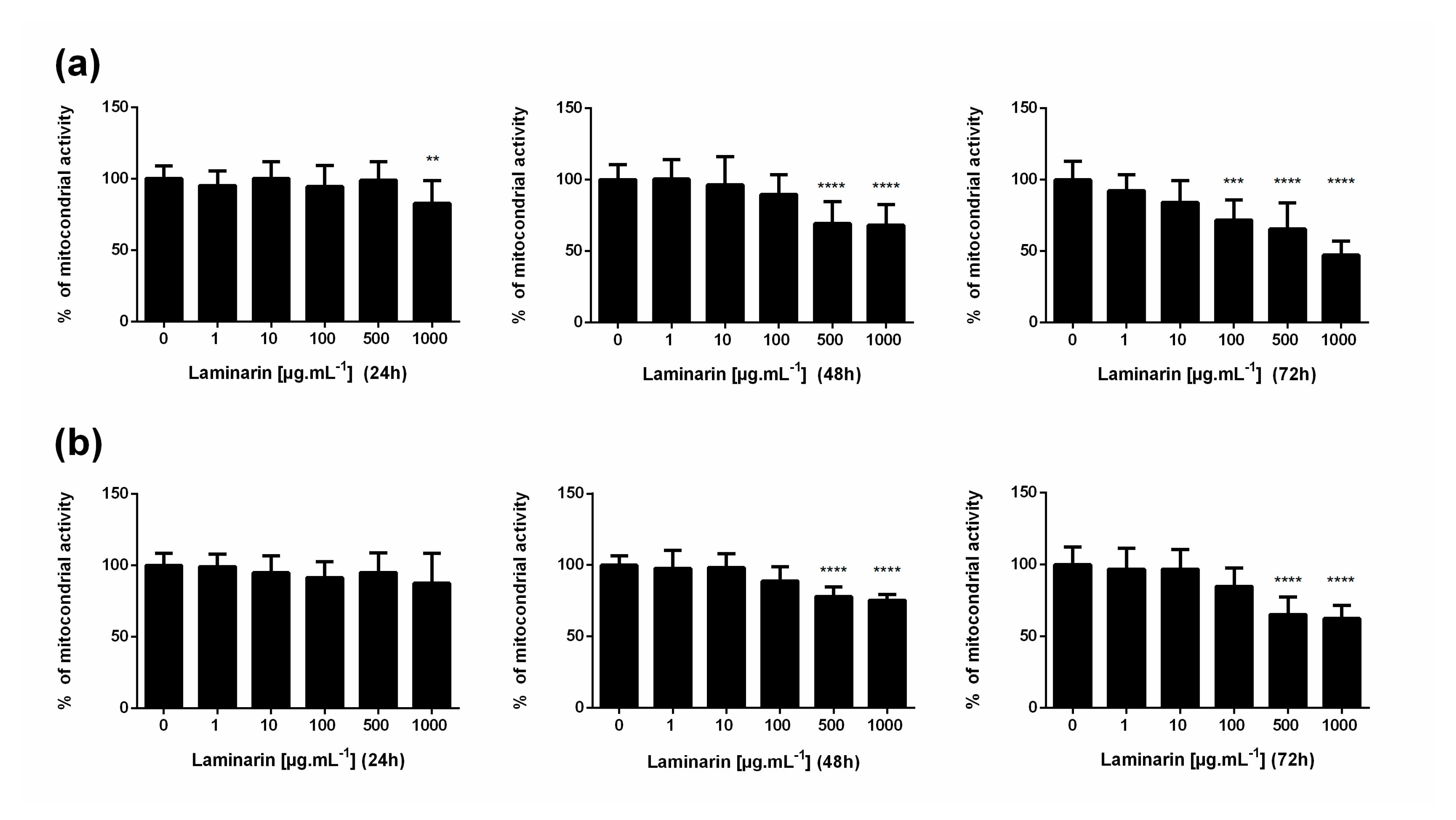
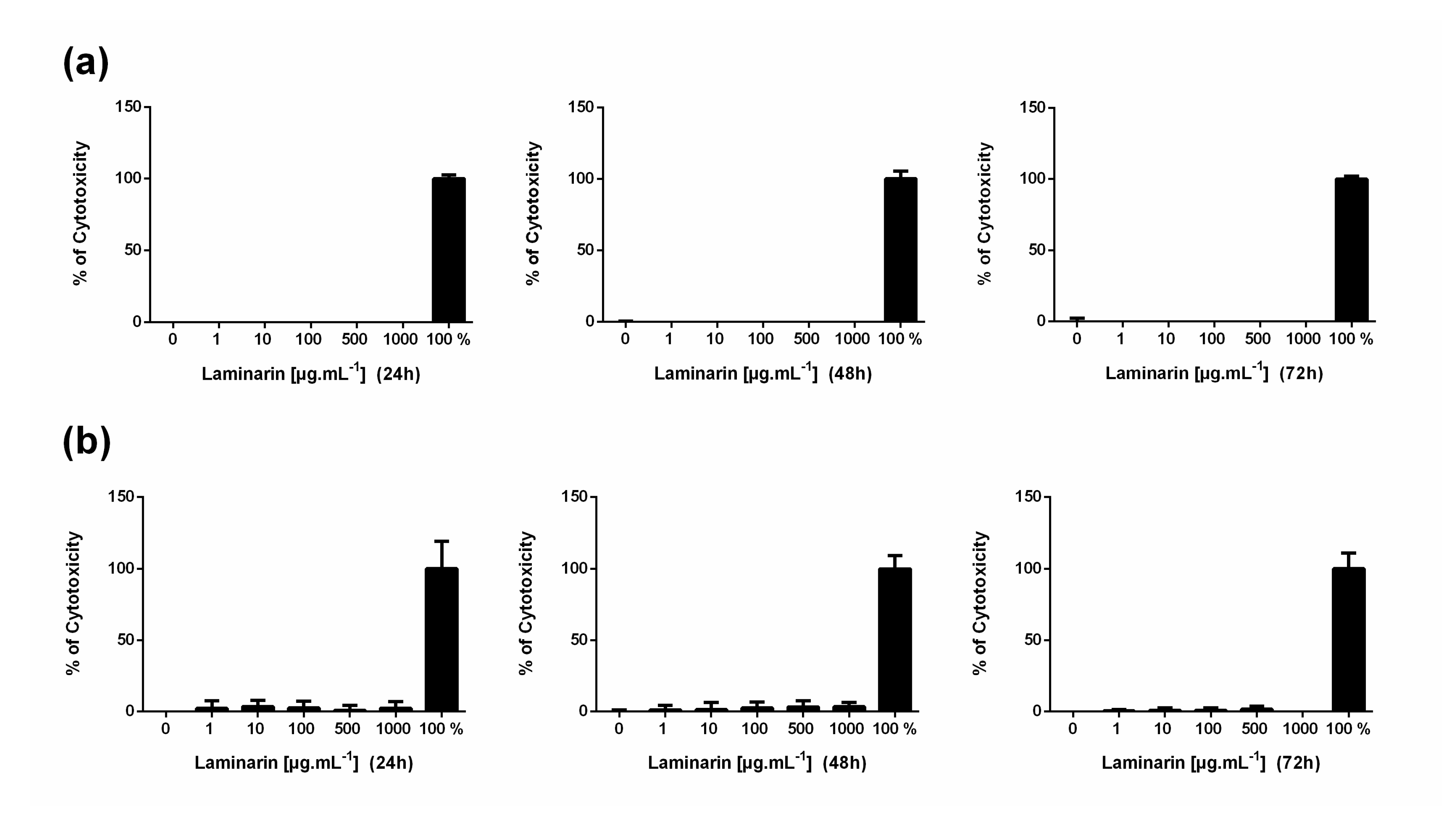
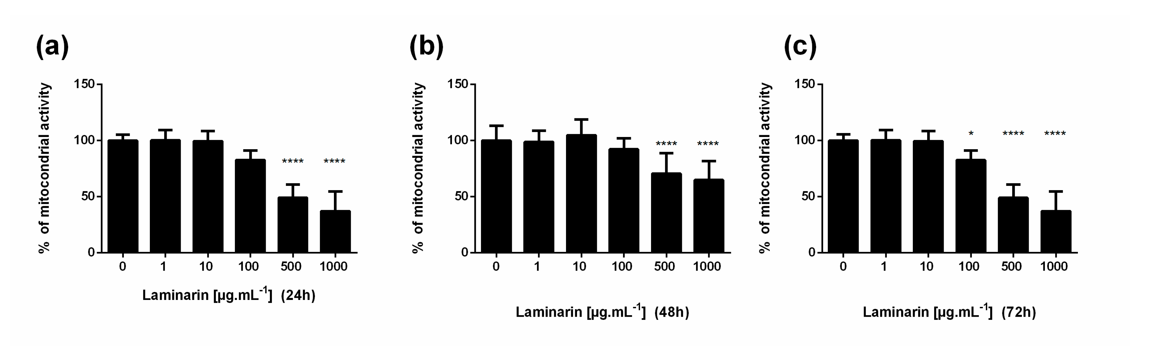
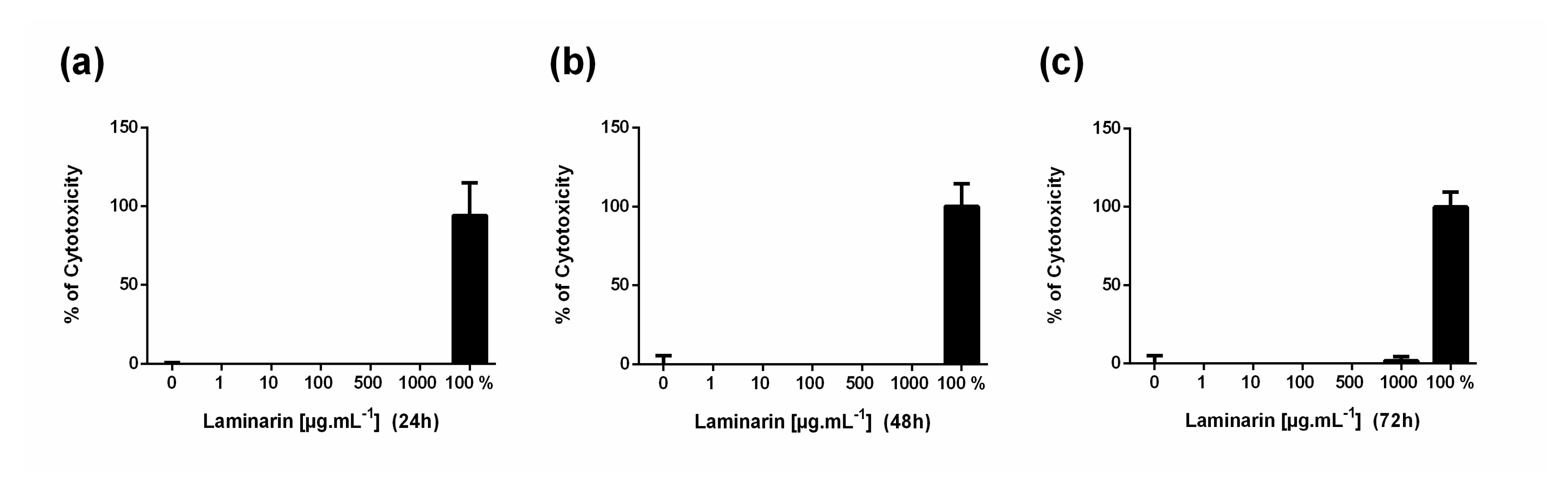
3.1. High Concentration of Laminarin Reduces Cellular Metabolic Activity without Cytolytic Activity

In preliminary experiments, the effect of laminarin was evaluated at several concentrations (1, 10, 100, 500, and 1000 μg.mL−1) in HDFa, NHEK, and HaCaT cells at 24 h, 48 h, and 72 h. For this purpose, mitochondrial activity was analyzed using an MTT assay, and an LDH dosage measured cell cytotoxicity. Laminarin significantly reduced mitochondrial activity for HDFa cells at 1000 µg.mL−1 after 24 h, from 500 µg.mL−1 after 48 h and from 100 µg.mL−1 after 72 h of stimulation (Figure 2a). For the NHEK cells (Figure 2b), cellular metabolic activity decreased later than fibroblasts at 500 µg.mL−1 after an incubation time of 48 h. Similar results were obtained for HaCaT cells (Figure A1). However, laminarin did not induce cellular damage as reflected by LDH measurement. (Figure 3 and Figure A2).
Based on MTT assays results reflecting cellular metabolic activity, we next focused our experiments with conditions using only 0, 1, 10, and 100 µg.mL−1 laminarin concentrations.

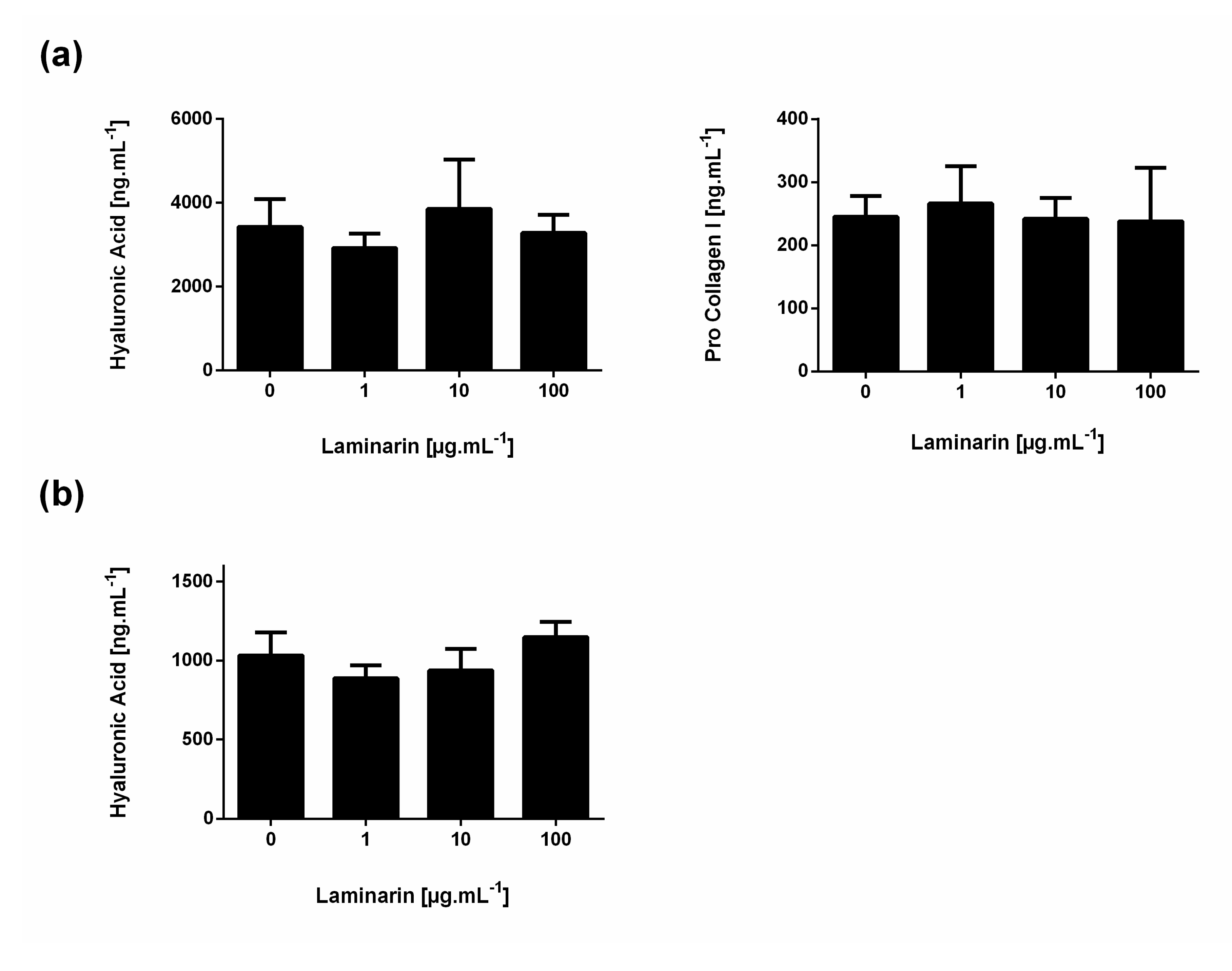
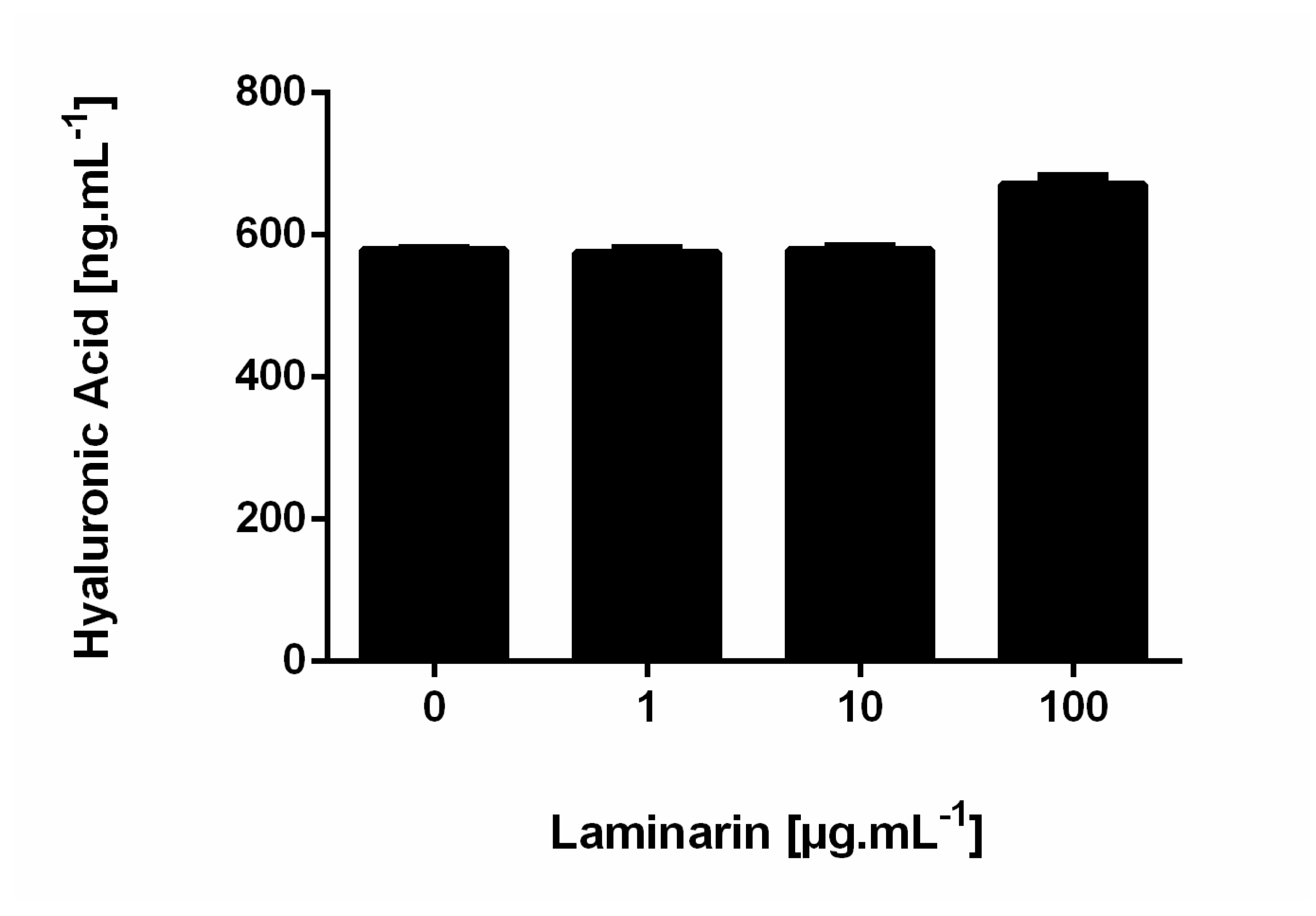
3.2. Influence of Laminarin on Matrix Deposition

The skin extracellular matrix is in constant renewal, and its homeostasis was monitored by both collagen and hyaluronic acid deposition and degradation. We investigated laminarin’s influence on both type-I procollagen and hyaluronic acid deposition by HDFa cells and hyaluronic acid deposition by NHEK and HaCaT cells. Results showed no variation in both hyaluronic acid and type-I procollagen deposition between the different laminarin concentrations on HDFa, NHEK cells (Figure 4) and HaCaT cells (Figure A3).

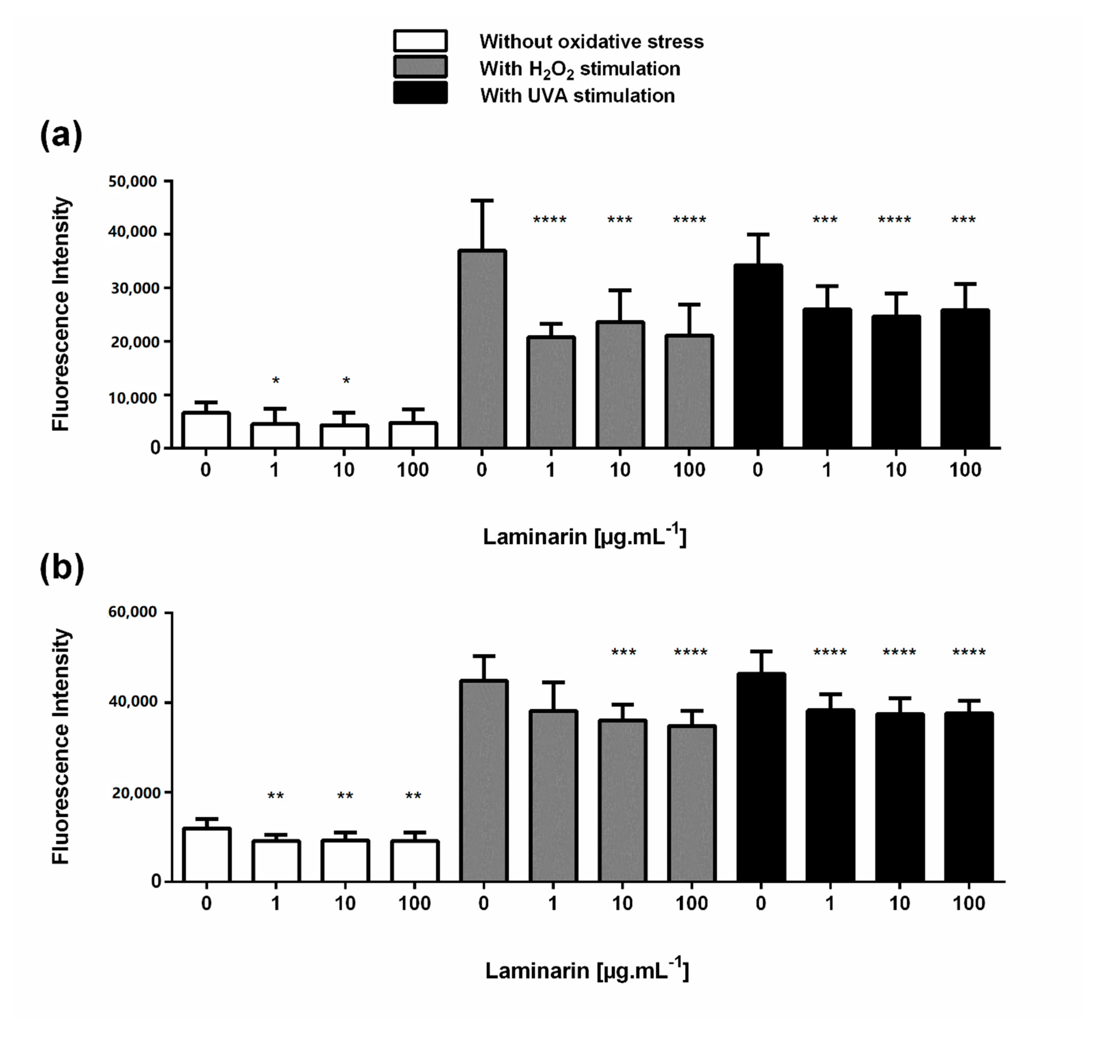
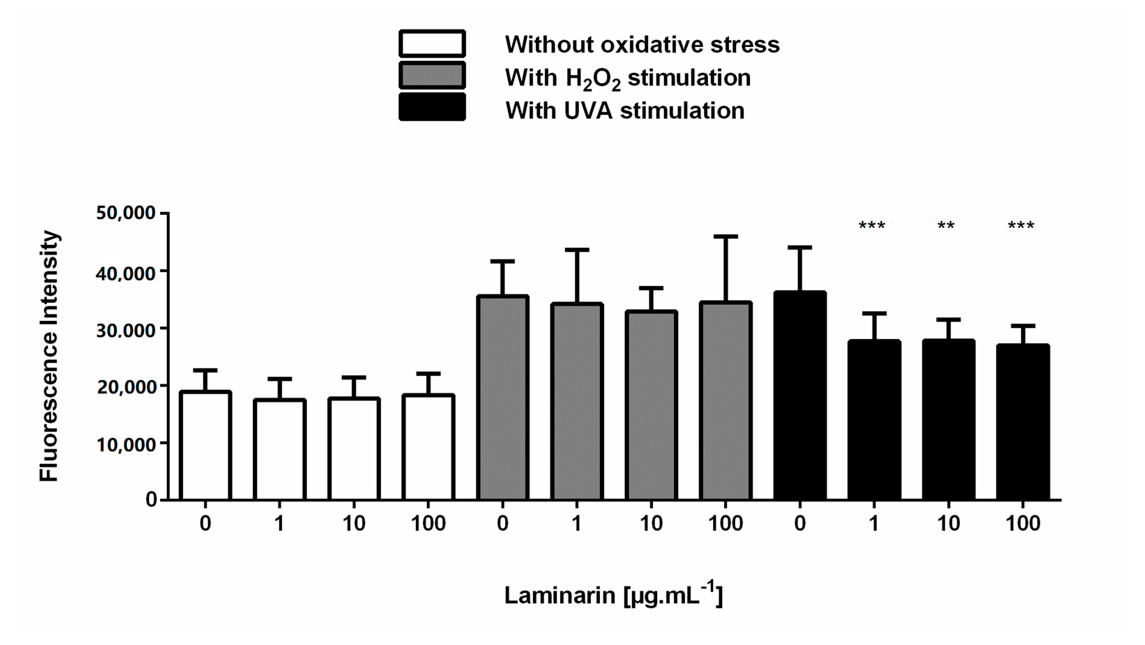
3.3. Laminarin Reduced Oxidative Stress on Skin Cells

The effects of laminarin on ROS production in H2O2 and UVA-stimulated skin cells have been tested during 4 h. Results showed no pro-oxidative effect of laminarin on the two cell types studied compared with non-treated cells (Figure 5). Interestingly, laminarin appears to significantly reduce basal ROS levels at 1 and 10 µg.mL−1 in HDFa cells and from 1 µg.mL−1 in NHEK cells (Figure 5). We found the same results for HaCaT cells (Figure A4). H2O2 and UVA radiations induced an increase in intracellular ROS production in HDFa, NHEK, and HaCaT cells compared with non-stimulated cells (Figure 5 and Figure A4). The increase in intracellular ROS levels under H2O2 treatment was significantly reduced by laminarin from 1 µg.mL−1 in HDFa cells (Figure 5a) and from 10 µg.mL−1 in NHEK cells (Figure 5b). However, laminarin had no significant effect on HaCaT cells (Figure A4). Regarding UVA radiations, laminarin significantly decreased intracellular ROS levels from 1 µg.mL−1 for both HDFa and NHEK cells (Figure 5, right panel), as well as in HaCaT cells (Figure A4).

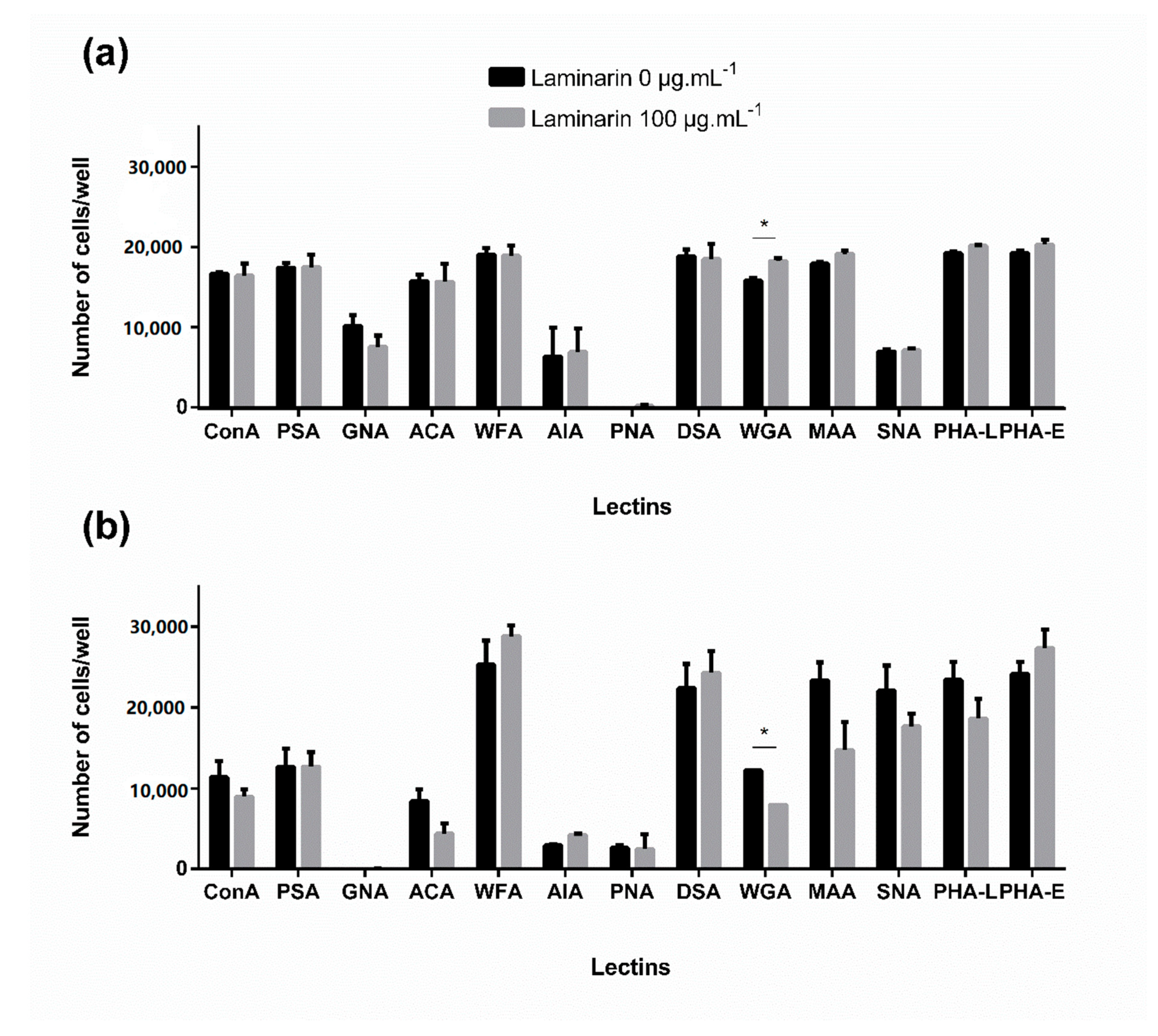
3.4. Effect of Laminarin on Cells—Glycan Interaction Study

Carbohydrate-binding proteins, called lectins, have extensively been used in the past decade as tools to study glycobiology processes [32,33]. GLYcoPROFILEs, a lectin array, of both fibroblasts and keratinocytes were achieved with the help of GLYcoDiag technology (LEctPROFILE® plates). Using this technology, we studied the effect of laminarin (100 µg. mL−1, 72 h of incubation) on the expression of lectins and their accessibility.
The GLYcoPROFILE of HDFa cells was slightly modified in the presence of laminarin (Figure 6a). A significant increase in the interaction with the lectin WGA (Wheat Germ Agglutinin), which recognizes GlcNAc and NeuAc motifs, was observed. These results suggest that laminarin induced an overexpression or promoted accessibility of glycans owned GlcNAc and NeuAc moieties (WGA interaction, p < 0.05) at the cell surface. For the other lectins, some trends were observed, such as a slight increase in the MAA (Maackia Amurensis Agglutinin), PHA-L, and PHA-Electins (Phaseolus Vulgaris Agglutinin), and a weak decrease in interaction with GNA (Galanthus Nivalis Agglutinin) (Figure 6a). However, no conclusion can be drawn on these changes.
The GLYcoPROFILE carried out with NHEKs cells (Figure 6b) showed more variations in terms of fluorescence intensity on the panel of lectin studied compared to the HDFa GLYcoPROFILE. Unlike HDFa cells, NHEK GLYcoPROFILE displayed only a statistically decreased in the interaction with WGA (WGA interaction, p < 0.05). Regarding the other lectins, results show a slight decrease in interaction with lectins that recognized respectively mannose (i.e., Con-A), galactose and GalNAc (i.e., ACA), sialic acid residues (i.e., MAA, SNA), complex glycans (i.e., PHA-L) and a low increase in interactions with WFA, AIA, DSA and PHA-E that recognized GalNAc, galactose, GlcNAc, and complex glycans, respectively. Given the information obtained on NHEK cells with the GLYcoPROFILE, the different interactions took in considerations with monosaccharides specificities of lectins need to be shade regarding the specific glycans specificities recognized by each lectin used (i.e., WGAs only recognize terminal GlcNAc moieties, while DSA has more affinity for sequences of GlcNAc and/or lactosamine units chains; WFA better recognize GalNAcα1-6Gal motif, whereas ACA recognizes Galβ1-3GalNAc motif).

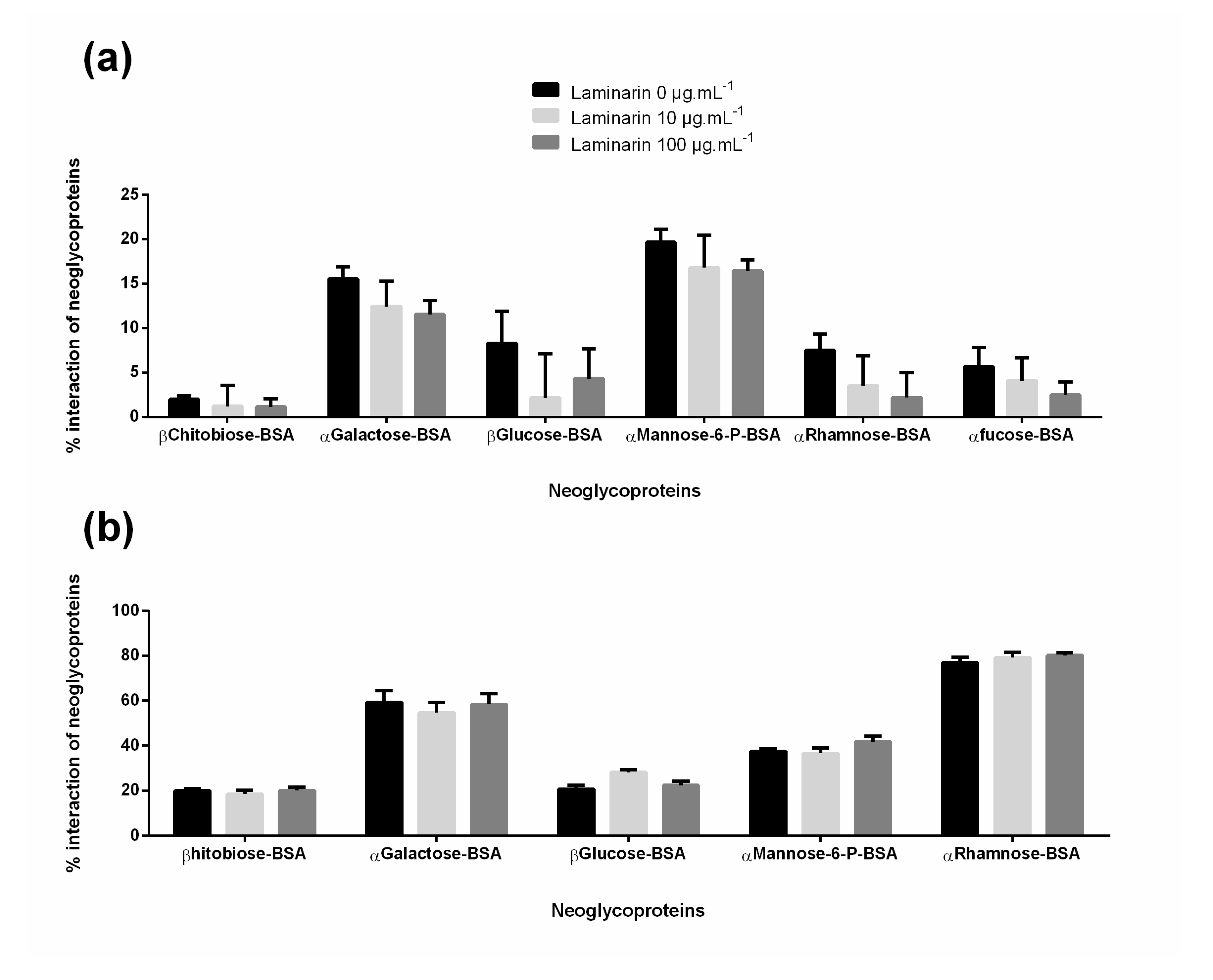
3.5. Effect of Laminarin with Cells—Carbohydrates Receptors Study

Laminarin effects on the expression and/or accessibility of cells glycans receptors were analyzed by NeoPROFILE (GLYcoDiag technology). The NeoPROFILE allows neoglycoproteins specific interactions with a cell surface to be quantified. The experiment was performed after a 72 h cell stimulation with laminarin at 0 µg.mL−1, 10 µg.mL−1, and 100 µg.mL−1. In both HDFa and NHEK NeoPROFILE, no significant differences were observed (Figure 7a,b). However, the results obtained for HDFa reflect an adhesion loss of all neoglycoprotein in a dose-dependent manner compared with the untreated cells (Figure 7a).

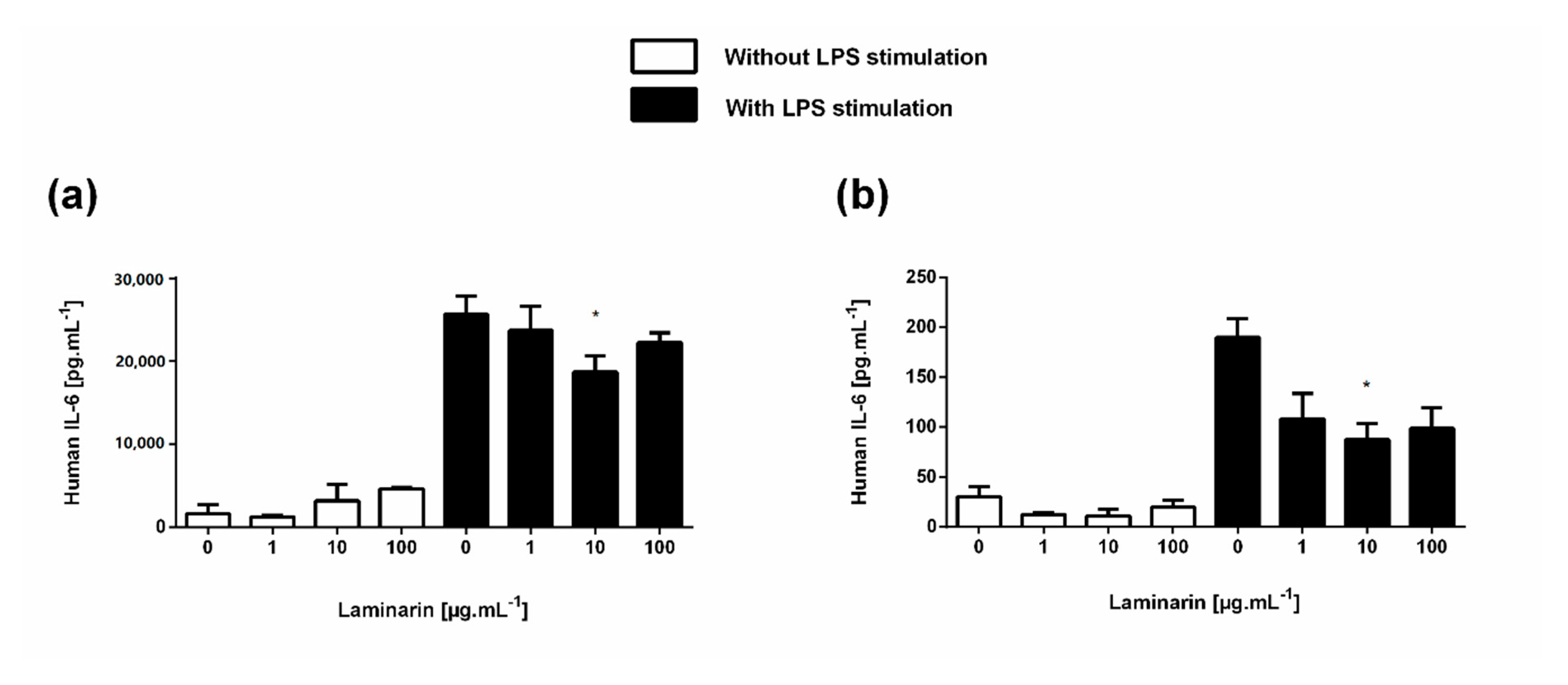
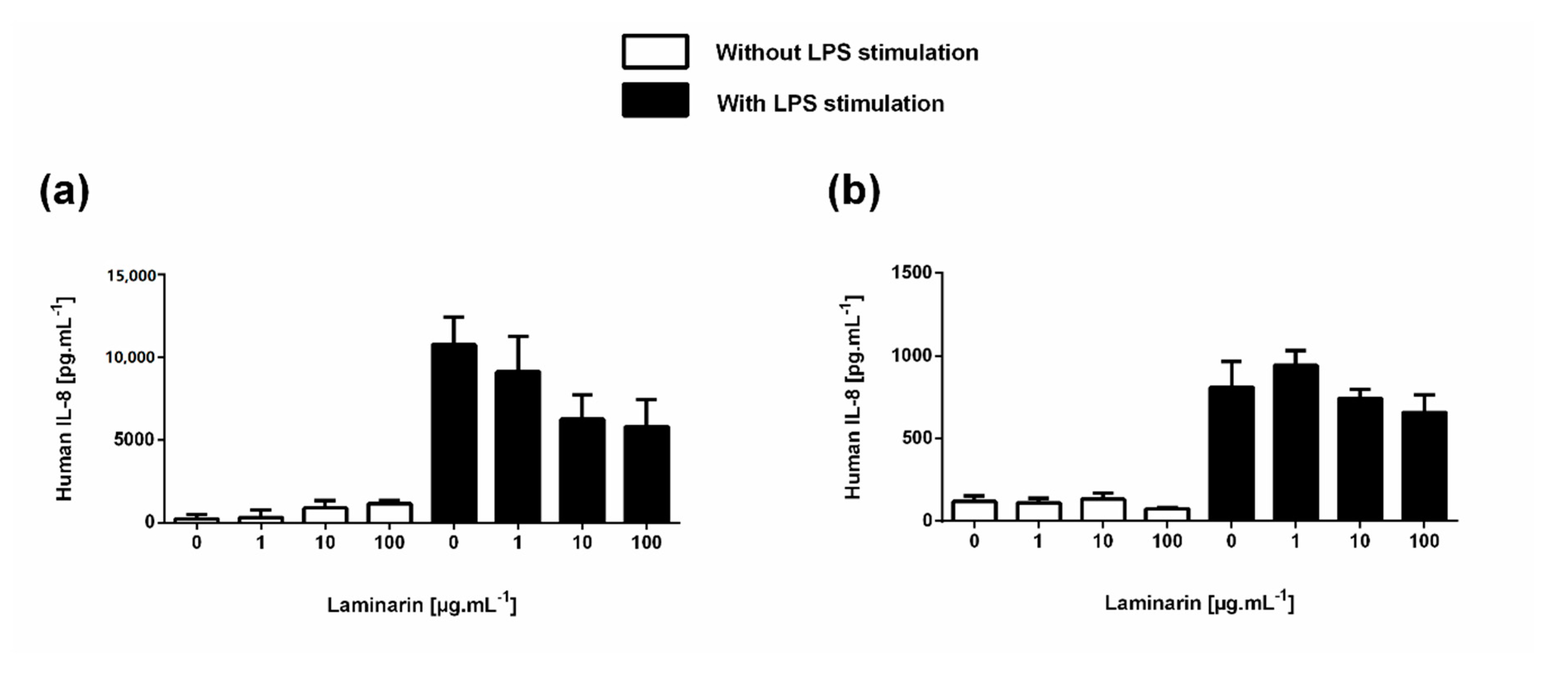
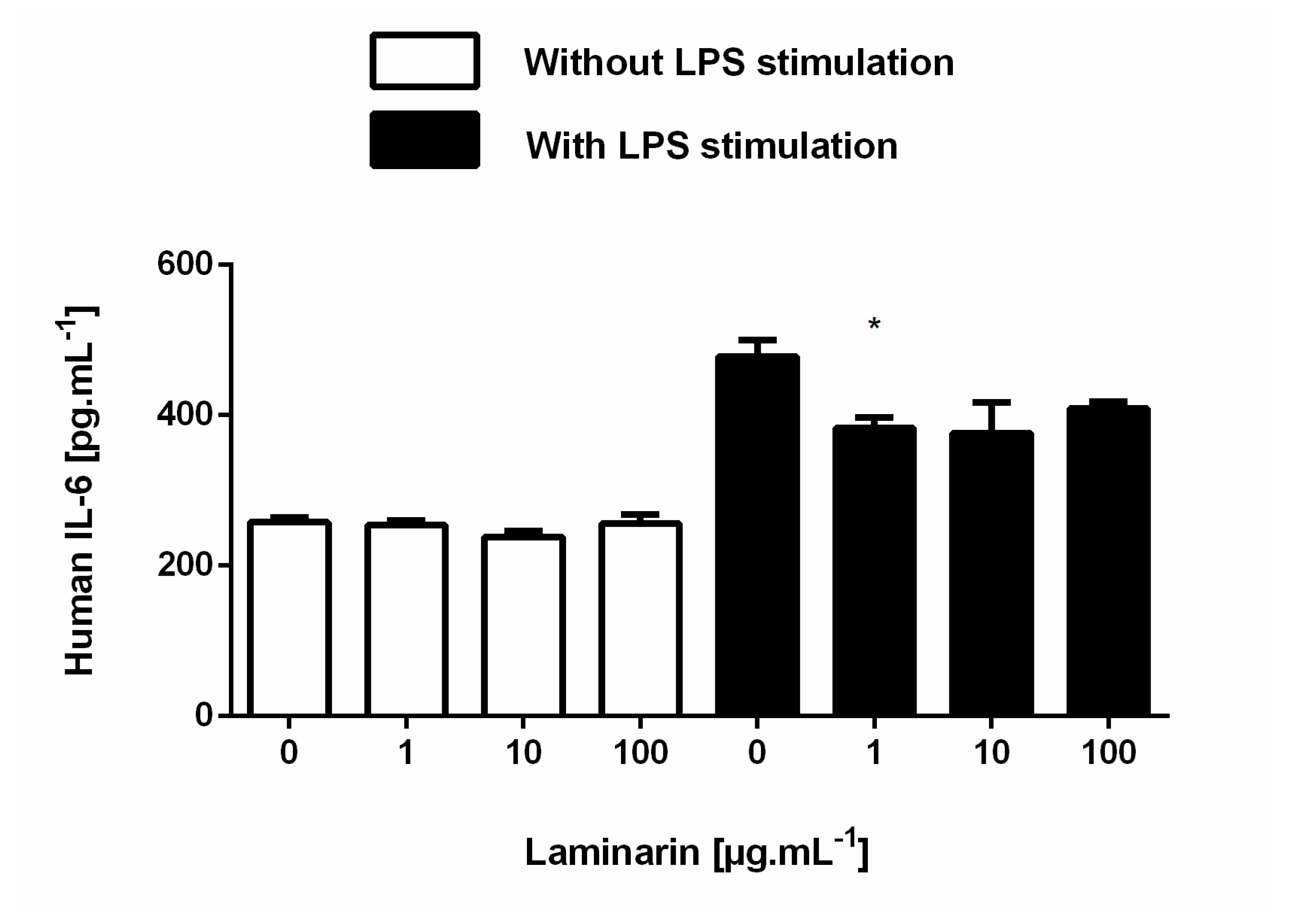
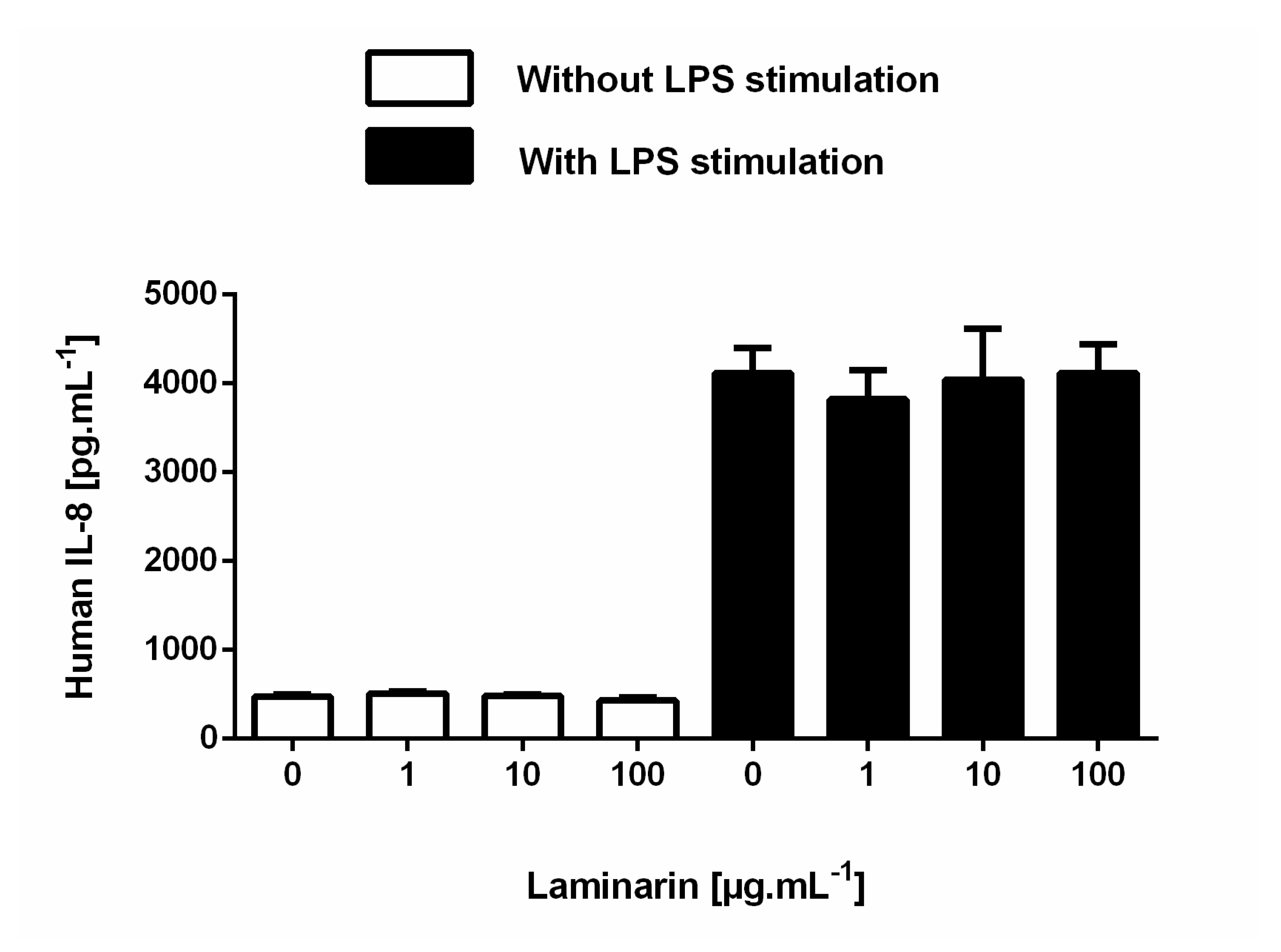
3.6. Laminarin Reduced IL-6 Secretion in Inflammatory Skin Cells

ELISA assay was performed to evaluate IL-6 and IL-8 cytokine secretion by cells. LPS (10 µg.mL−1) was employed to induce inflammatory stress. As expected, we observed that LPS treatment induced an increase in IL-6 and IL-8 secretion by both HDFa and NHEK cells compared to untreated cells (Figure 8 and Figure 9). In inflammatory conditions, laminarin significantly reduces IL-6 secretion at 10 µg.mL−1 in HDFa (27% off) and NHEK (54% off) cells (Figure 8a,b). Comparable results were obtained for HaCaT (Figure A5). Concerning IL-8 cytokine, no significant result was obtained. However, laminarin treatment tends to reduce IL-8 secretion only in HDFa cells in a dose-dependent manner (Figure 9a). Regarding NHEK and HaCaT cells, laminarin did not modified IL-8 secretion (Figure 9b and Figure A6).

4. Discussion

β-glucans have been used in cosmetics [1] for their antioxidant and moisturizing properties [14,15], but also as a texturizing agent in cosmetic creams [16]. Interestingly, inflammation and oxidative stress are often involved in extrinsic skin aging [34,35,36], leading to matrix extracellular disorganization and alteration of tissue function [34]. When too high a quantity of ROS is produced and antioxidant enzymes cannot reduce this excess, ROS can react with different cell components, such as membranes, proteins, lipids, and nucleic acids, and can induce cell damage [36,37]. Natural compounds are used in cosmetics in order to maintain skin cells’ integrity and protect cells from oxidative stress and inflammation. Herein, we investigated the effect of laminarin, a β-glucan from the brown seaweed Laminaria digitata, on both human keratinocytes and fibroblast cells.
First, we used HDFa, NHEK primary cells in these study, and HaCaT cell line, which is an immortalized human keratinocytes line [38]. HaCaT cells express various epidermal differentiation markers making them a cell line widely used as an alternative for NHEKs [39,40]. We have chosen to use this model along with NHEK for various reasons, as (1) donor variability of human keratinocytes, short culture lifetime, variations between passages can make interpretation of experimental data complicated; (2) Colombo et al. demonstrated that HaCaT cells are a reliable in vitro cell culture model to study inflammatory/repair responses [41].
Regarding laminarin effects on cells, our results showed that laminarin induced a significant decrease in cell metabolic activity (mitochondrial activity) in a time- and concentration-dependent manner from 100 µg.mL−1 for HDFa and 500 µg.mL−1 for NHEK cells. Interestingly, no-cytotoxic effect of laminarin on both HDFa and NHEK cells was observed. These results show that cell viability is not affected by laminarin. The tetrazolium salt MTT is reduced to purple formazan crystal by mitochondrial dehydrogenases in metabolically active cells [27,42], mostly succinate dehydrogenase (Electron Transport Chain Complex II) [43,44,45,46]. However, this modification in metabolic activity observed in laminarin conditions is still compatible with cell viability [42,47]. Based on these results, we can hypothesize that laminarin affects cell proliferation. This hypothesis is supported by the literature. Firstly, Park et al. show that laminarin stops the cell cycle in the sub-G2 and G2-M phase [48]. Secondly, Martinez-Diez et al. showed that alteration in the biogenesis of mammalian mitochondria leads to an alteration of cellular proliferation cycle (S-G2-M phase) [42]. Therefore, it is important to explore cell cycles by flow cytometry in order, to verify whether the decrease in HDFa and NHEK cell metabolic activity was associated with decreases in cell proliferation, as reported by the study cited above.
Regarding the extracellular matrix compounds, the results indicate no variation in the secretion of both HA and type-I procollagen for both HDFa and NHEK cells in the presence of laminarin compared to the control. Both HA and type-I collagen are fundamental components of the skin extracellular matrix (ECM) [49,50], where HA represents more than 50% of the HA reservoir of the body [51]. HA is also synthesized by keratinocytes in the epidermis [50]. HA and type-I collagen are responsible for skin hydration, elasticity, and resistance [52,53]. However, a previous study demonstrated that laminarin from the brown seaweed Saccharina longicruris, increased type-I collagen deposition in a dose-dependent manner by fibroblasts after 35 days of treatment [54]. The difference between our findings and those of Ayoub et al.’s study can be explained by the shorter stimulation time used (72 h) in our study, and by the structural difference of the two laminarin. Laminarin from Laminaria digitata is a linear β-(1,3)-glucan with branched glucose linked to the main chain by β-(1,6)-glucose bonds and has a molecular weight of about 5000 Dalton (Da) [55], while that from Saccharina longicruris displays the same structure but has a smaller molecular weight (2900–32000 Da) and some glucose linked to the main chain by β-(1,2)-linkages [56]. Our results show that laminarin from Laminaria digitata has no deleterious effect on the secretion of both hyaluronic acid and type I collagen, but no effect on the integrity of the extracellular matrix preservation was observed.
Oxidative stress was evaluated with both H2O2 and UVA radiation stimulation with or without the different laminarin concentrations. It is essential to present data from the literature on the effect of laminarin on oxidative stress first. It has been previously reported that laminarin exerts anti-oxidative activity [18,57,58]. As example, oligosaccharides derived from Laminaria japonica show high hydroxyl radical scavenging activity at the concentration of 100 µg.mL−1 [59]. Moreover, previous studies demonstrated that laminarin decreases oxidative stress and lipid peroxidation in rats [58], and significantly decreases ROS levels in aged pig oocytes [18]. Interestingly, in 2011, Cheng et al. reported that treatment with polysaccharides extract from laminarin purchased from a local market in Shenyang city considerably increased the antioxidant levels of superoxide dismutase, glutathione peroxidase and catalase in aseptic models [58]. In line with the results found in the literature, our findings show that under oxidative stress conditions, laminarin from Laminaria digitata decrease ROS level significantly. More specifically, we observed a significant decrease in ROS level from 1 µg.mL−1 in HDFa cells in H2O2 conditions and, to a lesser extent, in UVA radiation condition. In NHEK cells, the results show a very slight decrease in ROS level in both conditions. In skin aging, ROS secretion can lead to different cellular damages, such as lipid [60], protein [61], and DNA [62] modifications. Considering our results and previous observations, laminarin appears to be a promising component with potential anti-aging effects. To be able to ensure that laminarin would have a real cosmetic interest, it would be necessary to better characterize the laminarin from Laminaria digitata in terms of solubility, dependence on pH and oxygen partial pressure, and ability to act as a reducing agent towards electron donors of physiological importance, but also in terms of half-life time in blood, bioavailability, or biodistribution. Previous studies provide some elements to answer these questions. Laminarin appear soluble in water or organic solvent, a feature that depends mainly on the level of branching of these molecules [63,64]. For example, low branched laminarin is soluble only in hot water and highly branched laminarin in both cold and hot water. In 1982, Hoffman et al. reported that laminarin possesses blood anticoagulant activity upon a structural modification—sulphation [65]. However, further studies will need to answer these questions. In particular, ex vivo studies on skin explants would be necessary to study its penetration capacity and its bioavailability.
Our data demonstrated that incubation with laminarin at 100 µg.mL−1 slightly increases the interaction with the lectin WGA, a lectin that recognizes GlcNAc and NeuAc residues in HDFa. In the case of NHEK, mirror results were obtained; only the interaction with WGA lectin decreased significantly. These results suggest that the stimulation of cells by laminarin induces modifications and/or modulations on glycans accessibilities at the skin cell surface. According to the literature, it is well-known that glycans play an essential part in the modulation of cell communication [66]. Hence, we can hypothesize that laminarin treatment would modify cell–cell communications and the effect of laminarin on the cell glycosylation machinery could be addressed via the study of the glycan structure to show better expression and/or accessibility (i.e., glycans with terminal GlcNAc moieties).
Regarding neoglycoprotein interactions, no significant result was obtained. On HDFa cells, we observed a general trend of slight decrease in all neoglycoproteins interactions, but these observations did not reach statistical significance. For HDFa and based on previous studies, we assume that (i) laminarin could induced modifications on receptors accessibilities at the skin cell surface, and/or (ii) laminarin molecules stay (trough non-specific interaction) linked (through Dectin-1 or other β-glucan binding protein) at the cell surface thus covering the glycan binding motifs [12].
Herein, we observed that laminarin influences oxidative response and glycan profiles on skin cells. It is well known that oxidative stress promotes pro-inflammatory cytokines secretion [67]. Moreover, glycans in immune cells are involved in molecular processes that regulate cell activation and fine-tune the inflammatory response [68]. Cytokine secretion (IL-6 and IL-8) was evaluated by ELISA assay in inflammatory conditions (10 µg.mL−1 of LPS). We observed a significant decrease in IL-6 secretion at 10 µg.mL−1 of laminarin in both HDFa and NHEK cells. In HaCaT cells, this decrease that reaches significance was observed at 1 µg.mL−1 of laminarin. For IL-8 secretion, no significant result was obtained. However, we can note a decreasing trend from 10 µg.mL−1 of laminarin. Previous studies established that laminarin acted as an immunomodulatory mediator [19] and carried out an anti-inflammatory activity with decreased levels of both IL-6 and IL-8 cytokines secretions in an animal model [25]. Our results are in accordance with those of the literature, even if the decrease in the secretion of these cytokines is slight.
Regarding IL-6, its role is complex. Indeed, IL-6 has both pro- and anti-inflammatory roles depending on the environmental conditions [67,69] and is involved in various biological mechanisms as immune response, metabolism, or tumorigenesis notably in epithelial cancer [70,71,72]. Interestingly, the role of IL-6 in cancer has been quite controversial. Dual roles for IL-6 in both tumor-promoting and -suppressive activities have been reported [73,74]. It has been shown that IL-6 can activate three regeneration-promoting transcription factors as YAP, Notch, and STAT3, which are also involved in stem cell activation [75]. In skin context, IL-6 plays a role in wound repair [76]. A recent review published in 2020 points out that IL-6 signaling deregulation can lead to either fibrosis or a healing failure [77]. Taken as a whole, these data show that IL-6 regulation is complex and can lead to various biological responses, either beneficial or harmful. As previously mentioned, our results show that laminarin modulates IL-6 secretion at 10 µg.mL−1. However, further investigations are warranted to understand the importance of this finding and, in particular, on the effect of laminarin in skin healing/fibrosis.
In the present study, we showed that high concentrations of laminarin lead to a decrease in metabolic activity in dermal fibroblasts and keratinocytes. We also observe a beneficial effect of low doses of laminarin against oxidative stress, and the potential anti-inflammatory role in dermal fibroblasts and keratinocytes. This major effect of laminarin in metabolic activity is of utmost importance and elevates its potential utility for dermal application. However, further tests are required to determine the optimum concentration of active compound availability in the dermis or epidermis.

5. Conclusions

To conclude, high concentrations of laminarin should not be considered for cosmetic use. However, a low concentration (10 µg.mL−1) of laminarin provides antioxidant protection, but also modulates IL-6 secretions by cutaneous cells under inflammatory conditions. These two properties make the laminarin from Laminaria digitata attractive in the context of skin aging.

Author Contributions

Conceptualization, H.O., H.T., B.R., L.L., E.L., R.D. and A.C.; Data curation, H.O., H.T., B.R., L.L., E.L., R.D. and A.C.; Formal analysis, H.O., H.T., B.R., L.L., R.D. and A.C.; Funding acquisition, H.T., L.L., R.D. and A.C.; Investigation, H.O., B.R. and A.C.; Methodology, H.O., H.T., B.R., L.L., R.D. and A.C.; Project administration, H.T., L.L., E.L., R.D. and A.C.; Resources, H.T., L.L., R.D. and A.C.; Supervision, H.T., R.D. and A.C.; Validation, H.T., L.L., R.D. and A.C.; Writing—original draft, H.O.; Writing—review and editing, H.T., R.D. and A.C. All authors have read and agreed to the published version of the manuscript.

Funding

This research was supported by Cosmetosciences, a global training and research program dedicated to the cosmetic industry. Located in the heart of the cosmetic valley, this program led by University of Orléans is funded by the Région Centre-Val de Loire.

Acknowledgments

This research was supported by Cosmetosciences, a global training and research program dedicated to the cosmetic industry.

Conflicts of Interest

The authors declare no conflict of interest. The funders had no role in the design of the study; in the collection, analyses, or interpretation of data; in the writing of the manuscript, or in the decision to publish the results.

Appendix A

Figure A1. Evaluation of cell metabolic activity by the MTT assay. HaCaT were stimulate with 1, 10, 100, 500 and 1000 µg.mL−1 laminarin for (a) 24 h, (b) and (c) 72 h, and cell viability was performed using a tetrasodium salt (MTT). Data are expressed as mean ± SD of three independent experiments. * p ≤ 0.05, **** p ≤ 0.0001 compared with 0 µg.mL−1 (control group).
Cosmetics 07 00066 g0a1
Figure A2. Evaluation of cell cytotoxicity by the LDH cytotoxicity assay. HaCaT cells were stimulated with 1, 10, 100, 500, and 1000 µg.mL−1 laminarin for 24 h (a), 48 h (b) and 72 h (c) and cell cytotoxicity was performed using a tetrasodium salt (INT). Data are expressed as mean ± SD of three independent experiments.
Cosmetics 07 00066 g0a2
Figure A3. Quantitative analysis of hyaluronic acid (HA) by ELISA assay. HaCaT cells were treated with 1, 10 and 100 µg.mL−1 laminarin for 72 h. An ELISA assay was performed to quantified HA on cell culture medium supernatant. Data are expressed as mean ± SD of three independent experiments.
Cosmetics 07 00066 g0a3
Figure A4. Effect of laminarin on oxidative stress. HaCaT cells were treated with 1, 10 and 100 µg.mL−1 laminarin for 4 h. ROS production was induced by 0.5 mM of H2O2 or by 365 nm, 0.42 J.s−1.cm² of UVA radiation. Free radical scavenging activity was evaluated using a DCFH-DA probe. Data are expressed as mean ± SD of three independent experiments. ** p ≤ 0.01, *** p ≤ 0.001 compared with 0 µg.mL−1 (control group).
Cosmetics 07 00066 g0a4
Figure A5. Quantitative analysis of IL-6 cytokine with an ELISA kit. HaCaT cells were stimulate with 1, 10 and 100 µg.mL−1 laminarin for 48 h in absence or in presence of LPS at 10 µg.mL−1. An ELISA assay was performed to quantify the level of IL-6 cytokine secretion on the cell culture medium supernatant. Data are expressed as mean ± SD of two independent experiments. * p ≤ 0.05 compared with 0 µg.mL−1 (control group).
Cosmetics 07 00066 g0a5
Figure A6. Quantitative analysis of IL-8 cytokine with an ELISA kit. HaCaT cells were stimulate with 1, 10 and 100 µg.mL−1 laminarin for 48 h in absence or in presence of LPS at 10 µg.mL−1. An ELISA assay was performed to quantify the level of IL-8 cytokine secretion on the cell culture medium supernatant. Data are expressed as mean ± SD of two independent experiments.
Cosmetics 07 00066 g0a6

References

  1. Ruocco, N.; Costantini, S.; Guariniello, S.; Costantini, M. Polysaccharides from the marine environment with pharmacological, cosmeceutical and nutraceutical potential. Molecules 2016, 21, 551. [Google Scholar] [CrossRef]
  2. Ou, B.; Huang, D.; Hampsch-Woodill, M.; Flanagan, J.A.; Deemer, E.K. Analysis of antioxidant activities of common vegetables employing oxygen radical absorbance capacity (ORAC) and ferric reducing antioxidant power (FRAP) assays: A comparative study. J. Agric. Food Chem. 2002, 50, 3122–3128. [Google Scholar] [CrossRef] [PubMed]
  3. Ferrières, V.; Legentil, L.; Sylla, B.; Descroix, K.; Nugier-Chauvin, C.; Daniellou, R. Contribution of Biocatalysis to the Synthesis of β-(1, 3)-Glucans. Biol. Chem. Beta Glucan 2013, 2, 102. [Google Scholar]
  4. Zhu, F.; Du, B.; Xu, B. A critical review on production and industrial applications of beta-glucans. Food Hydrocoll. 2016, 52, 275–288. [Google Scholar] [CrossRef]
  5. Zeković, D.B.; Kwiatkowski, S.; Vrvić, M.M.; Jakovljević, D.; Moran, C.A. Natural and modified (1→3)-β-D-glucans in health promotion and disease alleviation. Crit. Rev. Biotechnol. 2005, 25, 205–230. [Google Scholar] [CrossRef] [PubMed]
  6. Sylla, B.; Legentil, L.; Saraswat-Ohri, S.; Vashishta, A.; Daniellou, R.; Wang, H.-W.; Vetvicka, V.; Ferrières, V. Oligo-β-(1→3)-glucans: Impact of thio-bridges on immunostimulating activities and the development of cancer stem cells. J. Med. Chem. 2014, 57, 8280–8292. [Google Scholar] [CrossRef]
  7. Chen, X.; Zhang, L.; Cheung, P.C.K. Immunopotentiation and anti-tumor activity of carboxymethylated-sulfated β-(1→3)-d-glucan from Poria cocos. Int. Immunopharmacol. 2010, 10, 398–405. [Google Scholar] [CrossRef]
  8. Toklu, H.Z.; Sener, G.; Jahovic, N.; Uslu, B.; Arbak, S.; Yeğen, B.C. Beta-glucan protects against burn-induced oxidative organ damage in rats. Int. Immunopharmacol. 2006, 6, 156–169. [Google Scholar] [CrossRef]
  9. Kim, H.-D.; Cho, H.-R.; Moon, S.-B.; Shin, H.-D.; Yang, K.-J.; Park, B.-R.; Jang, H.-J.; Kim, L.-S.; Lee, H.-S.; Ku, S.-K. Effects of beta-glucan from Aureobasidium pullulans on acute inflammation in mice. Arch. Pharm. Res. 2007, 30, 323–328. [Google Scholar] [CrossRef]
  10. Vetvicka, V.; Thornton, B.P.; Ross, G.D. Soluble beta-glucan polysaccharide binding to the lectin site of neutrophil or natural killer cell complement receptor type 3 (CD11b/CD18) generates a primed state of the receptor capable of mediating cytotoxicity of iC3b-opsonized target cells. J. Clin. Investig. 1996, 98, 50–61. [Google Scholar] [CrossRef] [Green Version]
  11. Brown, G.D.; Taylor, P.R.; Reid, D.M.; Willment, J.A.; Williams, D.L.; Martinez-Pomares, L.; Wong, S.Y.C.; Gordon, S. Dectin-1 is a major beta-glucan receptor on macrophages. J. Exp. Med. 2002, 196, 407–412. [Google Scholar] [CrossRef] [PubMed] [Green Version]
  12. Kougias, P.; Wei, D.; Rice, P.J.; Ensley, H.E.; Kalbfleisch, J.; Williams, D.L.; Browder, I.W. Normal human fibroblasts express pattern recognition receptors for fungal (1→3)-beta-D-glucans. Infect. Immun. 2001, 69, 3933–3938. [Google Scholar] [CrossRef] [PubMed] [Green Version]
  13. Van den Berg, L.M.; Zijlstra-Willems, E.M.; Richters, C.D.; Ulrich, M.M.W.; Geijtenbeek, T.B.H. Dectin-1 activation induces proliferation and migration of human keratinocytes enhancing wound re-epithelialization. Cell. Immunol. 2014, 289, 49–54. [Google Scholar] [CrossRef] [PubMed]
  14. Du, B.; Bian, Z.; Xu, B. Skin Health Promotion Effects of Natural Beta-Glucan Derived from Cereals and Microorganisms: A Review. Phytother. Res. 2014, 28, 159–166. [Google Scholar] [CrossRef]
  15. Gautier, S.; Xhauflaire-Uhoda, E.; Gonry, P.; Piérard, G.E. Chitin–glucan, a natural cell scaffold for skin moisturization and rejuvenation. Int. J. Cosmet. Sci. 2008, 30, 459–469. [Google Scholar] [CrossRef]
  16. Wang, Y.; Viennet, C.; Jeudy, A.; Fanian, F.; He, L.; Humbert, P. Assessment of the efficacy of a new complex antisensitive skin cream. J. Cosmet. Derm. 2018, 17, 1101–1107. [Google Scholar] [CrossRef]
  17. Choi, J.-I.; Kim, H.-J.; Kim, J.-H.; Lee, J.-W. Enhanced Biological Activities of Laminarin Degraded by Gamma-Ray Irradiation. J. Food Biochem. 2012, 36, 465–469. [Google Scholar] [CrossRef]
  18. Yao, X.; Jiang, H.; Liang, S.; Shen, X.; Gao, Q.; Xu, Y.N.; Kim, N.-H. Laminarin enhances the quality of aged pig oocytes by reducing oxidative stress. J. Reprod. Dev. 2018, 64, 489–494. [Google Scholar] [CrossRef]
  19. Smith, A.J.; Graves, B.; Child, R.; Rice, P.J.; Ma, Z.; Lowman, D.W.; Ensley, H.E.; Ryter, K.T.; Evans, J.T.; Williams, D.L. Immunoregulatory Activity of the Natural Product Laminarin Varies Widely as a Result of Its Physical Properties. J. Immunol. 2018, 200, 788–799. [Google Scholar] [CrossRef] [Green Version]
  20. Kadam, S.U.; Tiwari, B.K.; O’Donnell, C.P. Extraction, structure and biofunctional activities of laminarin from brown algae. Int. J. Food Sci. Technol. 2015, 50, 24–31. [Google Scholar] [CrossRef]
  21. Bonfim-Mendonça, P.D.S.; Capoci, I.R.G.; Tobaldini-Valerio, F.K.; Negri, M.; Svidzinski, T.I.E. Overview of β-Glucans from Laminaria spp.: Immunomodulation Properties and Applications on Biologic Models. Int. J. Mol. Sci. 2017, 18, 1629. [Google Scholar] [CrossRef] [Green Version]
  22. Song, K.; Xu, L.; Zhang, W.; Cai, Y.; Jang, B.; Oh, J.; Jin, J.-O. Laminarin promotes anti-cancer immunity by the maturation of dendritic cells. Oncotarget 2017, 8, 38554–38567. [Google Scholar] [CrossRef] [PubMed] [Green Version]
  23. Kim, K.-H.; Kim, Y.-W.; Kim, H.B.; Lee, B.J.; Lee, D.S. Anti-apoptotic activity of laminarin polysaccharides and their enzymatically hydrolyzed oligosaccharides from Laminaria japonica. Biotechnol. Lett. 2006, 28, 439–446. [Google Scholar] [CrossRef] [PubMed]
  24. Neyrinck, A.M.; Mouson, A.; Delzenne, N.M. Dietary supplementation with laminarin, a fermentable marine beta (1-3) glucan, protects against hepatotoxicity induced by LPS in rat by modulating immune response in the hepatic tissue. Int. Immunopharmacol. 2007, 7, 1497–1506. [Google Scholar] [CrossRef] [PubMed]
  25. O’Shea, C.J.; O’Doherty, J.V.; Callanan, J.J.; Doyle, D.; Thornton, K.; Sweeney, T. The effect of algal polysaccharides laminarin and fucoidan on colonic pathology, cytokine gene expression and Enterobacteriaceae in a dextran sodium sulfate-challenged porcine model. J. Nutr. Sci. 2016, 5. [Google Scholar] [CrossRef] [Green Version]
  26. Ji, C.-F.; Ji, Y.-B.; Meng, D.-Y. Sulfated modification and anti-tumor activity of laminarin. Exp. Ther. Med. 2013, 6, 1259–1264. [Google Scholar] [CrossRef] [Green Version]
  27. Mosmann, T. Rapid colorimetric assay for cellular growth and survival: Application to proliferation and cytotoxicity assays. J. Immunol. Methods 1983, 65, 55–63. [Google Scholar] [CrossRef]
  28. Landemarre, L.; Cancellieri, P.; Duverger, E. Cell surface lectin array: Parameters affecting cell glycan signature. Glycoconj. J. 2013, 30, 195–203. [Google Scholar] [CrossRef]
  29. Morello, E.; Mallet, A.; Konto-Ghiorghi, Y.; Chaze, T.; Mistou, M.-Y.; Oliva, G.; Oliveira, L.; Di Guilmi, A.-M.; Trieu-Cuot, P.; Dramsi, S. Evidence for the sialylation of PilA, the PI-2a pilus-associated adhesin of Streptococcus agalactiae strain NEM316. PLoS ONE 2015, 10, e0138103. [Google Scholar] [CrossRef] [Green Version]
  30. Hassani, Z.; Saleh, A.; Turpault, S.; Khiati, S.; Morelle, W.; Vignon, J.; Hugnot, J.-P.; Uro-Coste, E.; Legrand, P.; Delaforge, M. Phostine PST3. 1a targets MGAT5 and inhibits glioblastoma-initiating cell invasiveness and proliferation. Mol. Cancer Res. 2017, 15, 1376–1387. [Google Scholar] [CrossRef] [Green Version]
  31. Cutolo, G.; Reise, F.; Schuler, M.; Nehmé, R.; Despras, G.; Brekalo, J.; Morin, P.; Renard, P.-Y.; Lindhorst, T.K.; Tatibouët, A. Bifunctional mannoside–glucosinolate glycoconjugates as enzymatically triggered isothiocyanates and FimH ligands. Org. Biomol. Chem. 2018, 16, 4900–4913. [Google Scholar] [CrossRef] [PubMed]
  32. Fagot, D.; Pham, D.M.; Laboureau, J.; Planel, E.; Guerin, L.; Nègre, C.; Donovan, M.; Bernard, B.A. Crocin, a natural molecule with potentially beneficial effects against skin ageing. Int. J. Cosmet. Sci. 2018, 40, 388–400. [Google Scholar] [CrossRef] [PubMed]
  33. Wu, A.M.; Lisowska, E.; Duk, M.; Yang, Z. Lectins as tools in glycoconjugate research. Glycoconj. J. 2009, 26, 899–913. [Google Scholar] [CrossRef] [PubMed]
  34. Zhang, S.; Duan, E. Fighting against Skin Aging: The Way from Bench to Bedside. Cell Transplant. 2018, 27, 729–738. [Google Scholar] [CrossRef] [PubMed]
  35. Pittayapruek, P.; Meephansan, J.; Prapapan, O.; Komine, M.; Ohtsuki, M. Role of Matrix Metalloproteinases in Photoaging and Photocarcinogenesis. Int. J. Mol. Sci. 2016, 17, 868. [Google Scholar] [CrossRef] [PubMed] [Green Version]
  36. Cezar, T.L.C.; Martinez, R.M.; Da Rocha, C.; Melo, C.P.B.; Vale, D.L.; Borghi, S.M.; Fattori, V.; Vignoli, J.A.; Camilios-Neto, D.; Baracat, M.M.; et al. Treatment with maresin 1, a docosahexaenoic acid-derived pro-resolution lipid, protects skin from inflammation and oxidative stress caused by UVB irradiation. Sci. Rep. 2019, 9, 3062. [Google Scholar] [CrossRef]
  37. Puizina-Ivić, N. Skin aging. Acta Dermatovenerol. Alp. Panon. Et Adriat. 2008, 17, 47–54. [Google Scholar]
  38. Boukamp, P.; Petrussevska, R.T.; Breitkreutz, D.; Hornung, J.; Markham, A.; Fusenig, N.E. Normal keratinization in a spontaneously immortalized aneuploid human keratinocyte cell line. J. Cell Biol. 1988, 106, 761–771. [Google Scholar] [CrossRef] [Green Version]
  39. Park, M.; Woo, S.-Y.; Cho, K.-A.; Cho, M.-S.; Lee, K.H. PD-L1 produced by HaCaT cells under polyinosinic-polycytidylic acid stimulation inhibits melanin production by B16F10 cells. PLoS ONE 2020, 15, e0233448. [Google Scholar] [CrossRef]
  40. Lehmann, B. HaCaT cell line as a model system for vitamin D3 metabolism in human skin. J. Investig. Dermatol. 1997, 108, 78–82. [Google Scholar] [CrossRef] [Green Version]
  41. Colombo, I.; Sangiovanni, E.; Maggio, R.; Mattozzi, C.; Zava, S.; Corbett, Y.; Fumagalli, M.; Carlino, C.; Corsetto, P.A.; Scaccabarozzi, D.; et al. HaCaT Cells as a Reliable In Vitro Differentiation Model to Dissect the Inflammatory/Repair Response of Human Keratinocytes. Mediat. Inflamm. 2017, 2017, 7435621. [Google Scholar] [CrossRef] [PubMed]
  42. Martínez-Diez, M.; Santamaría, G.; Ortega, A.D.; Cuezva, J.M. Biogenesis and dynamics of mitochondria during the cell cycle: Significance of 3’UTRs. PLoS ONE 2006, 1, e107. [Google Scholar] [CrossRef] [PubMed] [Green Version]
  43. Garn, H.; Krause, H.; Enzmann, V.; Drössler, K. An improved MTT assay using the electron-coupling agent menadione. J. Immunol. Methods 1994, 168, 253–256. [Google Scholar] [CrossRef]
  44. Thom, S.M.; Horobin, R.W.; Seidler, E.; Barer, M.R. Factors affecting the selection and use of tetrazolium salts as cytochemical indicators of microbial viability and activity. J. Appl. Bacteriol. 1993, 74, 433–443. [Google Scholar] [CrossRef]
  45. Shoemaker, M.; Cohen, I.; Campbell, M. Reduction of MTT by aqueous herbal extracts in the absence of cells. J. Ethnopharmacol. 2004, 93, 381–384. [Google Scholar] [CrossRef]
  46. Peng, L.; Wang, B.; Ren, P. Reduction of MTT by flavonoids in the absence of cells. Colloids Surf. B Biointerfaces 2005, 45, 108–111. [Google Scholar] [CrossRef]
  47. Morgan, D.M.L. Tetrazolium (MTT) Assay for Cellular Viability and Activity. In Polyamine Protocols; Morgan, D.M.L., Ed.; Humana Press: Totowa, NJ, USA, 1998; pp. 179–184, Methods in Molecular BiologyTM; ISBN 978-1-59259-565-5. [Google Scholar]
  48. Park, H.-K.; Kim, I.-H.; Kim, J.; Nam, T.-J. Induction of apoptosis and the regulation of ErbB signaling by laminarin in HT-29 human colon cancer cells. Int. J. Mol. Med. 2013, 32, 291–295. [Google Scholar] [CrossRef] [Green Version]
  49. Uitto, J. Collagen polymorphism: Isolation and partial characterization of α1(I)-trimer molecules in normal human skin. Arch. Biochem. Biophys. 1979, 192, 371–379. [Google Scholar] [CrossRef]
  50. Stern, R.; Maibach, H.I. Hyaluronan in skin: Aspects of aging and its pharmacologic modulation. Clin. Dermatol. 2008, 26, 106–122. [Google Scholar] [CrossRef]
  51. Meyer, L.J.M.; Stern, R. Age-Dependent Changes of Hyaluronan in Human Skin. J. Investig. Dermatol. 1994, 102, 385–389. [Google Scholar] [CrossRef] [Green Version]
  52. Voigt, J.; Driver, V.R. Hyaluronic acid derivatives and their healing effect on burns, epithelial surgical wounds, and chronic wounds: A systematic review and meta-analysis of randomized controlled trials. Wound Repair Regen 2012, 20, 317–331. [Google Scholar] [CrossRef] [PubMed]
  53. Kwansa, A.L.; De Vita, R.; Freeman, J.W. Tensile mechanical properties of collagen type I and its enzymatic crosslinks. Biophys. Chem. 2016, 214, 1–10. [Google Scholar] [CrossRef] [PubMed]
  54. Ayoub, A.; Pereira, J.M.; Rioux, L.-E.; Turgeon, S.L.; Beaulieu, M.; Moulin, V.J. Role of seaweed laminaran from Saccharina longicruris on matrix deposition during dermal tissue-engineered production. Int. J. Biol. Macromol. 2015, 75, 13–20. [Google Scholar] [CrossRef] [PubMed]
  55. Read, S.M.; Currie, G.; Bacic, A. Analysis of the structural heterogeneity of laminarin by electrospray-ionisation-mass spectrometry. Carbohydr. Res. 1996, 281, 187–201. [Google Scholar] [CrossRef]
  56. Rioux, L.-E.; Turgeon, S.L.; Beaulieu, M. Structural characterization of laminaran and galactofucan extracted from the brown seaweed Saccharina longicruris. Phytochemistry 2010, 71, 1586–1595. [Google Scholar] [CrossRef]
  57. Moroney, N.C.; O’Grady, M.N.; Lordan, S.; Stanton, C.; Kerry, J.P. Seaweed polysaccharides (laminarin and fucoidan) as functional ingredients in pork meat: An evaluation of anti-oxidative potential, thermal stability and bioaccessibility. Mar. Drugs 2015, 13, 2447–2464. [Google Scholar] [CrossRef] [Green Version]
  58. Cheng, D.; Liang, B.; Li, M.; Jin, M. Influence of laminarin polysaccahrides on oxidative damage. Int. J. Biol. Macromol. 2011, 48, 63–66. [Google Scholar] [CrossRef]
  59. Wu, S.-J. Preparation and antioxidant activity of the oligosaccharides derived from Laminaria japonica. Carbohydr. Polym. 2014, 106, 22–24. [Google Scholar] [CrossRef]
  60. Ramachandran, S.; Rajendra Prasad, N.; Karthikeyan, S. Sesamol inhibits UVB-induced ROS generation and subsequent oxidative damage in cultured human skin dermal fibroblasts. Arch. Dermatol. Res. 2010, 302, 733–744. [Google Scholar] [CrossRef]
  61. Karran, P.; Brem, R. Protein oxidation, UVA and human DNA repair. DNA Repair 2016, 44, 178–185. [Google Scholar] [CrossRef] [Green Version]
  62. Karthikeyan, R.; Kanimozhi, G.; Prasad, N.R.; Agilan, B.; Ganesan, M.; Srithar, G. Alpha pinene modulates UVA-induced oxidative stress, DNA damage and apoptosis in human skin epidermal keratinocytes. Life Sci. 2018, 212, 150–158. [Google Scholar] [CrossRef] [PubMed]
  63. Custódio, C.A.; Reis, R.L.; Mano, J.F. Photo-Cross-Linked Laminarin-Based Hydrogels for Biomedical Applications. Biomacromolecules 2016, 17, 1602–1609. [Google Scholar] [CrossRef] [PubMed]
  64. Miao, H.Q.; Ishai-Michaeli, R.; Peretz, T.; Vlodavsky, I. Laminarin sulfate mimics the effects of heparin on smooth muscle cell proliferation and basic fibroblast growth factor-receptor binding and mitogenic activity. J. Cell. Physiol. 1995, 164, 482–490. [Google Scholar] [CrossRef] [PubMed]
  65. Hoffman, J.; Larm, O.; Larsson, K.; Andersson, L.O.; Holmer, E.; Söderström, G. Studies on the blood-anticoagulant activity of sulphated polysaccharides with different uronic acid content. Carbohydr. Polym. 1982, 2, 115–121. [Google Scholar] [CrossRef]
  66. Tsoni, S.V.; Brown, G.D. β-Glucans and Dectin-1. Ann. N. Y. Acad. Sci. 2008, 1143, 45–60. [Google Scholar] [CrossRef]
  67. Pillai, S.; Oresajo, C.; Hayward, J. Ultraviolet radiation and skin aging: Roles of reactive oxygen species, inflammation and protease activation, and strategies for prevention of inflammation-induced matrix degradation—A review. Int. J. Cosmet. Sci. 2005, 27, 17–34. [Google Scholar] [CrossRef]
  68. Rabinovich, G.A.; Toscano, M.A. Turning′sweet′on immunity: Galectin–glycan interactions in immune tolerance and inflammation. Nat. Rev. Immunol. 2009, 9, 338–352. [Google Scholar] [CrossRef]
  69. Kobayashi, M.; Yoshiki, R.; Sakabe, J.; Kabashima, K.; Nakamura, M.; Tokura, Y. Expression of toll-like receptor 2, NOD2 and dectin-1 and stimulatory effects of their ligands and histamine in normal human keratinocytes. Br. J. Dermatol. 2009, 160, 297–304. [Google Scholar] [CrossRef]
  70. Schafer, Z.T.; Brugge, J.S. IL-6 involvement in epithelial cancers. J. Clin. Investig. 2007, 117, 3660–3663. [Google Scholar] [CrossRef] [Green Version]
  71. Kishimoto, T. INTERLEUKIN-6: From Basic Science to Medicine—40 Years in Immunology. Annu. Rev. Immunol. 2005, 23, 1–21. [Google Scholar] [CrossRef] [Green Version]
  72. Wallenius, V.; Wallenius, K.; Ahrén, B.; Rudling, M.; Carlsten, H.; Dickson, S.L.; Ohlsson, C.; Jansson, J.-O. Interleukin-6-deficient mice develop mature-onset obesity. Nat. Med. 2002, 8, 75–79. [Google Scholar] [CrossRef]
  73. Chiu, J.J.; Sgagias, M.K.; Cowan, K.H. Interleukin 6 acts as a paracrine growth factor in human mammary carcinoma cell lines. Clin. Cancer Res. 1996, 2, 215–221. [Google Scholar] [PubMed]
  74. Conze, D.; Weiss, L.; Regen, P.S.; Bhushan, A.; Weaver, D.; Johnson, P.; Rincón, M. Autocrine production of interleukin 6 causes multidrug resistance in breast cancer cells. Cancer Res. 2001, 61, 8851–8858. [Google Scholar] [PubMed]
  75. Taniguchi, K.; Wu, L.; Grivennikov, S.I.; De Jong, P.R.; Lian, I.; Yu, F.-X.; Wang, K.; Ho, S.B.; Boland, B.S.; Chang, J.T.; et al. A gp130-Src-YAP module links inflammation to epithelial regeneration. Nature 2015, 519, 57–62. [Google Scholar] [CrossRef] [PubMed] [Green Version]
  76. Arranz-Valsero, I.; Soriano-Romaní, L.; García-Posadas, L.; López-García, A.; Diebold, Y. IL-6 as a corneal wound healing mediator in an in vitro scratch assay. Exp. Eye Res. 2014, 125, 183–192. [Google Scholar] [CrossRef] [PubMed]
  77. Johnson, B.Z.; Stevenson, A.W.; Prêle, C.M.; Fear, M.W.; Wood, F.M. The Role of IL-6 in Skin Fibrosis and Cutaneous Wound Healing. Biomedicines 2020, 8, 101. [Google Scholar] [CrossRef]
Figure 1. General Structure of Laminarin.
Cosmetics 07 00066 g001
Figure 2. Evaluation of cellular metabolic activity by the MTT assay. HDFa (a) and NHEK cells (b) were stimulated with 1, 10, 100, 500 and 1000 µg.mL−1 laminarin for 24 h (left), 48 h (middle) and 72 h (right) and cellular metabolic activity was performed using a tetrasodium salt (MTT). Data are expressed as mean ± SD of three independent experiments. ** p ≤ 0.01, *** p ≤ 0.001, **** p ≤ 0.0001 compared with 0 µg.mL−1 (control group).
Cosmetics 07 00066 g002
Figure 3. Evaluation of cell cytotoxicity by the LDH cytotoxicity assay. HDFa (a) and NHEK cells (b) were stimulated with 1, 10, 100, 500 and 1000 µg.mL−1 laminarin for 24 h (left), 48 h (middle) and 72 h (right) and cell cytotoxicity was performed using a tetrasodium salt (INT). Data are expressed as mean ± SD of three independent experiments.
Cosmetics 07 00066 g003
Figure 4. Quantitative analysis of hyaluronic acid (HA) and procollagen I by ELISA assay. (a) HDFa and (b) NHEK cells were treated with 1, 10 and 100 µg.mL−1 laminarin for 72 h. An ELISA assay was performed to quantified HA (left) and procollagen I (right) on cell culture medium supernatant. Data are expressed as mean ± SD of three independent experiments.
Cosmetics 07 00066 g004
Figure 5. Effect of laminarin on oxidative stress. (a) HDFa and (b) NHEK cells were treated with 1, 10 and 100 µg.mL−1 of laminarin for 4 h. ROS production was induced by 0.5 mM of H2O2 or by 365 nm, 0.42 J.s−1.cm² of UVA radiation. Free radical scavenging activity was evaluated using a DCFH-DA probe. Data are expressed as mean ± SD of three independent experiments. * p ≤ 0.05, ** p ≤ 0.01, *** p ≤ 0.001, **** p ≤ 0.0001 compared with 0 µg.mL−1 (control group).
Cosmetics 07 00066 g005
Figure 6. Effect of laminarin on lectin binding skin cell surface (GLYcoPROFILE). (a) HDFa and (b) NHEK cells were stimulated in the absence (black bar) or with 100 μg.mL−1 (dark grey bar) of laminarin (stimulation 72 h before the analysis). Data are expressed as mean ± SD of two independent experiments. For the abbreviations of lectins, see Table 1. * p ≤ 0.05.
Cosmetics 07 00066 g006
Figure 7. Neoglycoprotein interaction (NeoPROFILE) with (a) HDFa (neoglycoproteins: β-Chitobiose-BSA, α-Galactose-BSA, β-Glucose-BSA, α-Mannose-6-Phosphate-BSA, α-Rhamnose-BSA, α-Fucose-BSA) and with (b) NHEK (neoglycoproteins: β-Chitobiose-BSA, α-Galactose-BSA, β-Glucose-BSA,α-Mannose-6-Phosphate-BSA, α-Rhamnose-BSA) after stimulation with laminarin for a stimulation time of 72 h before the analysis; black laminarin at 0 μg.mL−1, grey laminarin at 10 μg.mL−1, dark grey laminarin at 100 μg.mL−1. Data are expressed as mean ± SD of two independent experiments.
Cosmetics 07 00066 g007
Figure 8. Quantitative analysis of IL-6 cytokine with an ELISA kit. (a) HDFa and (b) NHEK cells were stimulated with 1, 10 and 100 µg.mL−1 laminarin for 24 h (HDFa cells) and 48 h (NHEK cells) in the absence or in presence of LPS at 10 µg.mL−1. An ELISA assay was performed to quantify the level of IL-6 cytokine secreted in cell culture medium supernatant. Data are expressed as mean ± SD of two independent experiments. * p ≤ 0.05, compared with 0 µg.mL−1 (control group).
Cosmetics 07 00066 g008
Figure 9. Quantitative analysis of IL-8 cytokine with an ELISA kit. (a) HDFa and (b) NHEK cells were stimulated with 1, 10 and 100 µg.mL−1 laminarin for 24 h (HDFa cells) and 48 h (NHEK cells) in the absence or in the presence of LPS at 10 µg.mL−1. An ELISA assay was performed to quantify the level of IL-8 cytokine secreted in cell culture medium supernatant. Data are expressed as mean ± SD of two independent experiments.
Cosmetics 07 00066 g009
Table 1. Lectins Specificities.
Short NameCommon NameGlycan Structures Specificities
ConAConcanavalin AgglutininManα6(Manα3)Man
PSAPisum Sativum AgglutininManα6(Manα3)Man
GNAGalanthus Nivalis AgglutininManα6(Manα3)Man
ACAAmaranthus Caudatus AgglutininGalβ3GalNAc
WFAWisteria Floribunda AgglutininGalNAcα6Gal > GalNAcα3GalNAc > GalNAc
PNAPeanut AgglutininGalβ3GalNAc
AIAAutocarpus Intergrifolia AgglutininGalβ3GalNAc
DSADatura Stramonium AgglutininGlcNAcβ4GlcNAc
WGAWheat Germ AgglutininGlcNAcGlcNAcβ4
MAAMaackia Amurensis AgglutininNeu5Ac3Gal4GalNAc
SNASambucus Nigra AgglutininNeu5Ac6Gal/GalNAc
PHA-LPhaseolus Vulgaris AgglutininComplex glycans
PHA-EPhaseolus Vulgaris AgglutininComplex glycans

