
Application of Niosomes in Cosmetics: A Systematic Review

1
Department of Pharmaceutical Technology, Faculty of Pharmacy, University of Malaya, Kuala Lumpur 50603, Malaysia
2
School of Pharmacy, Management and Science University, Shah Alam 40100, Malaysia
*
Authors to whom correspondence should be addressed.
Cosmetics 2022, 9(6), 127; https://0-doi-org.brum.beds.ac.uk/10.3390/cosmetics9060127
Submission received: 25 October 2022
/
Revised: 21 November 2022
/
Accepted: 22 November 2022
/
Published: 25 November 2022
(This article belongs to the Special Issue Feature Papers in Cosmetics in 2022)
Abstract
:A rising volume of the literature acknowledges the significance of nanotechnology in the cosmetics industry, particularly with the invention and use of techniques based on niosomes to generate unique formulations with both medicinal and aesthetic benefits. The current study’s objectives are to undertake a comprehensive review of the previously published data on the use and applications of niosomes in cosmetics and to give a succinct summary of that data. Preferred Reporting standards for Systematic Reviews and Meta-Analyses (PRISMA) guidelines were used in the design of the current review. The core concept and keywords were derived from the research question using the SPIDER tool. The main steps of this review included: design of the research question, preliminary research, search strategy, searching the database, exclusion and inclusion criteria, approval by authors, title and abstract screening, reporting of the number of data selected, full text download and reading, manual research (Google Scholar, Scopus, and WoS), data extraction and quality assessment, double data checking, and manuscript writing, revision, and submission. After thorough data analysis, it was discovered that a cosmetic product’s aesthetic impact significantly improved when it was created utilising niosomes technology. The majority of cosmeceutical niosomes’ skin and hair products demonstrated an enhanced therapeutic and cosmeceutical effect. These discoveries may contribute to the treatment of skin conditions under the umbrella of cosmeceutical niosomes.
1. Introduction
The term “cosmetics” refers to a broad range of products that may be applied topically as well as taken internally. In most cases, they are used to provide a pleasant odour to the area of the body to which they are administered or to modify or correct the look of that area. As a result of their advantages, such as increased skin penetration, bioavailability, surface adherence, and prolonged release properties, niosomes have received substantial attention as a carrier system for cosmeceutical active ingredients [1,2,3,4,5,6,7,8,9,10,11,12,13,14]. The overwhelming majority of cosmeceutical active ingredients have insufficient ability to permeate through the layers of skin and exhibit minimal chemical or physical stability. With the help of niosomes and other recent advancements in nanotechnology, it is now feasible to load a range of medications onto nano-like particles, enabling efficient targeted medication administration and enhancing the chemical and physical stability of those cosmeceutical and pharmaceutical products. Niosomes are more stable and less expensive to produce; their sizes range from 100 to 200 nanometres [15]. Materials that are both biodegradable and biocompatible are used to make niosomes [15]. Because of its distinct structure, the niosome is a classic example of a new drug delivery system that can transport amphiphilic, lipophilic, and hydrophilic molecules [16]. There is evidence that niosomes have been utilised as anti-ageing, antiwrinkle, bleaching, anti-alopecia, and moisturising cosmetics [17]. Niosomes revealed lower toxicity, allowing for regulated delivery and the release of loaded active compounds with beneficial properties to give a moisturising and tanning effect to the skin [1,8]. L’Oréal was the first firm to develop a skincare product incorporating niosomes in the 1970s as oil-in-water anti-ageing emulsion [2,8,18]. The product was released in the cosmetic industry by Lâncome (one of the French company’s brands) in 1986 under the name Niosôme [2,3,8,14,18,19]. Meanwhile, several bioactive substances derived from plant materials have piqued the interest of cosmetic researchers; these molecules have medicinal and cosmeceutical effects including antioxidant properties and anti-ageing [1,8]. Niosomes are an effective compound carrier because they can encapsulate water-soluble, lipid-soluble, and amphiphilic active ingredients without requiring chemical modification [2,20,21]. Niosomes have the potential to be employed in the manufacturing of a wide range of cosmetics, such as anti-ageing effect, antioxidant and free radical inhibitory effect, whitening, anti-scarring, and other capabilities. Based on our preliminary search, there is no single review that systematically focuses on the applications of niosomes in cosmetics. The vast majority of previously published information describes the general applications of niosomes in both the pharmaceutical and cosmeceutical fields together. As a result, the purpose of this study is to provide a concise summary of the previously published data on the application of niosomes in cosmetics and systematically conduct a comprehensive analysis of that data. Moreover, the current review was designed using PRISMA guidelines. The research question was developed using the SPIDER tool, from which the key concept and keywords were created. The Scopus and Web of Science (WoS) databases were used for the database search.
2. Methodology
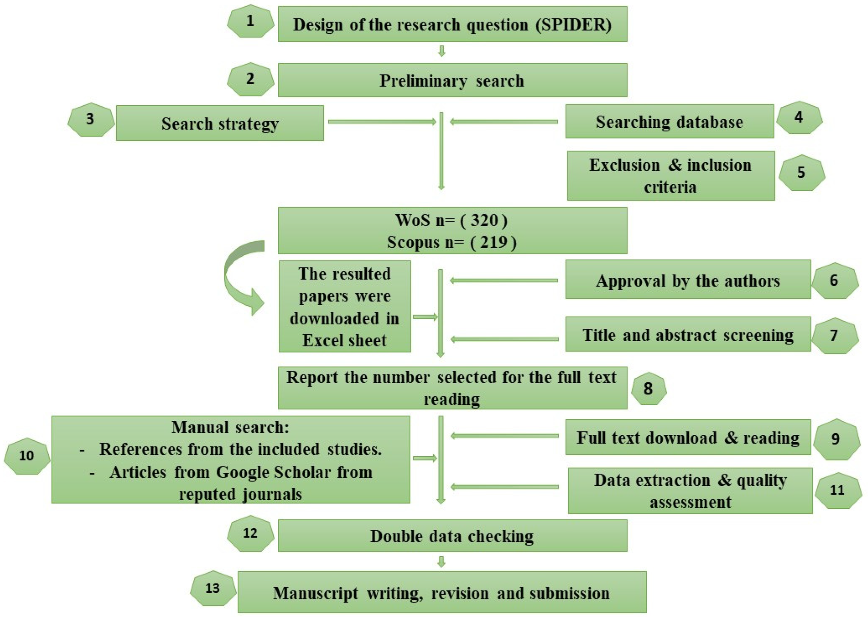
The protocol for the current systematic literature review was designed, and it was slightly modified from the PRISMA (Figure 1) guidelines [22]. The main steps of this review included: design of the research question using the SPIDER tool [23], preliminary research, search strategy, searching the database, exclusion and inclusion criteria, approval by authors, title and abstract screening, reporting of the number of data selected, full text download and reading, manual research (Google Scholar, Scopus, and WoS), data extraction and quality assessment, double data checking, and manuscript writing, revision, and submission.
The research question was developed based on the SPIDER tool (Table 1). The developed question was: “What are the cosmeceutical applications of niosomes or nonionic surfactants among other nanoparticles in Scopus and Web of Science (WoS) databases”? This question reflected the sample, phenomenon of interest, design, evaluation, and research type as shown in Table 1.
To confirm that the proposed topic had not been studied previously, preliminary investigation was conducted. The proposed topic was typically discussed based on the findings of the preliminary investigation. Numerous papers were identified about the applications of niosomes in general (in all fields: medicine, pharmacy, and cosmetics). However, we were unable to locate a single source that specifically mentions the use of niosomes in the cosmetics field. Based on the created research question, four key concepts were identified to search in the Scopus and WoS databases, as shown in Table 2.
The search using the key concepts was conducted in the WoS and Scopus databases. The results of both searches are shown in Table 3.
Inclusion and exclusion criteria were designed and approved by all authors. The inclusion criteria included the type of study, type of database, type of intervention, and type of outcome measures. Exclusion criteria included the time of the published data, for example, we excluded the data published in 2017 in the Scopus database. Other exclusion criteria applied through the title and abstract screening. A full record of the data obtained from the database were downloaded as a spreadsheet. The title and abstract matching of the downloaded spreadsheet file was then checked. In the subsequent stage, the matching data were chosen after the irrelevant titles and abstracts were eliminated.
The selected documents were reported, with the data divided by the number of the authors for the full text download reading, summarising, and writing. The selected papers were downloaded, reviewed for quality assessment, and then summarised, revised, and rewritten by the authors.
3. Niosomes Structure, Types, and Methods of Preparation
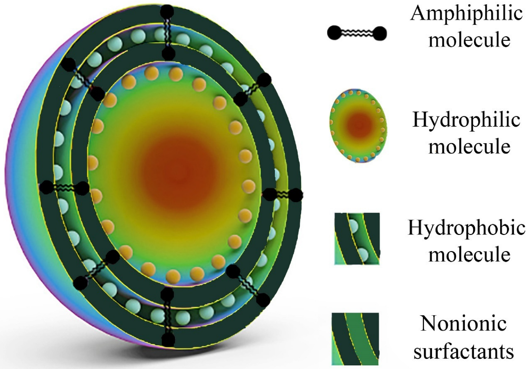
The distinctive form, composition, chemistry, and small size of niosomes make it possible for them to effectively stabilise and deliver the drug molecule to the desired site (Figure 2). Due to their hydrophilic nature and hydrophobic activity, niosomes are an ideal vesicle for encasing lipophilic, hydrophilic, and amphiphilic compounds [3]. Niosomes come in three different varieties depending on the size of the particles: large unilamellar vesicles (LUVs), small unilamellar vesicles (SUVs), and multilamellar vesicles (MLVs). The most crucial component in the formation of niosomes is nonionic surfactants. Numerous research has examined the role, benefits, and uses of nonionic surfactants to form niosomes for delivery of molecules, targeting a specific site or tissue, and for the production of different cosmetic products. The production of niosomes is significantly influenced by the structure of nonionic surfactants. As a result, they are comprised of one or more lamellae, which are bilayers with hydrophilic and hydrophobic components that are joined by ester, ether, or amide bonds [3].
A bilayer-inducing substance, typically cholesterol, is one of the components employed in the development of niosomes. The primary job of cholesterol is to keep the membrane of niosomes stable. Therefore, it was reported that the use of cholesterol may enhance the drug loading of the encapsulated drugs [3]. Another ingredient which may be used in the preparation of niosomes is charge inducer agents. These types of materials are utilised to prevent the aggregation of the niosome particles and to deliver the drug to a specific area of the body using the zeta potential parameter [3]. Examples of negative charge inducer agents are dicetyl phosphate (DCP) and phosphatidic acid, while examples of positive charge inducer agents are stearyl amine (SA) and stearyl pyridinium chloride.
Niosomes can be prepared in a variety of ways [1], with all methods of preparation summarised in Table 4.
Despite being a straightforward approach, organic solvents must be used to dissolve surfactant and cholesterol in thin film hydration. In a round-bottomed flask, surfactants and cholesterol are dissolved, and the organic solvent is then evaporated to leave the flask’s bottom with a thin coating. Multilamellar vesicles are created by adding water medium and swelling the film from the round bottom flask’s wall for a certain period of time while gently stirring it continuously. These vesicles are then treated to create unilamellar vesicles [1].
The ether injection method includes gently injecting drugs and surfactants into an aqueous phase while heating the mixture above the organic solvent’s boiling point [1]. This approach generates LUVs, which may then be processed to minimise their size [1].
In the reverse phase evaporation approach, surfactants are dispersed in an organic mixture of chloroform and ether before an aqueous drug solution is added. After homogenising the two immiscible phases, niosomes mixture should be formed when the solvents are evaporated under decreased pressure. This process purports to produce niosomes with high EE and large particle sizes. The reverse phase technique has been discovered to encapsulate large hydrophilic molecules with a higher encapsulation than earlier techniques [1].
The transmembrane pH gradient technique uses surfactants and cholesterol that have been dissolved in an organic solvent to create an emulsion containing niosomes. This organic solvent is then evaporated when a rotary evaporator is used to generate a thin film on the flask’s wall. Vortex is used to mix the added citric acid solution (pH 4) and to hydrate the formed film. Following that, the resulting droplets are frozen and thawed before being sonicated. Aqueous drug mixture is then added, and the process is vortexed [1]. To form multilamellar vesicles, this solution’s pH is raised to 7, then it is heated to 60 °C [1].
Using the emulsion method, surfactant and cholesterol are mixed with an organic phase and added to an aqueous phase containing the active ingredient to create an oil in water emulsion; as soon as the organic solvent has evaporated, niosomal suspension should be created [1].
Organic solvents are not required for the lipid injection method. A heated, highly agitated aqueous phase containing dissolved drug molecules is combined with a nonionic surfactant and a bilayer-inducing substance (cholesterol) to obtain a niosomal suspension [1].
Enzymes can be used to create niosomes from a mixed micellar solution. When dicetyl phosphate and other lipids are added, esterase, for instance, can be used to break the ester bonds of polyethylene stearyl derivatives, producing breakdown products such as cholesterol and polyoxyethylene that can be utilised to create multilamellar niosomes [1].
As a one-step process, the bubble approach does not require the use of organic solvents. Nonionic surfactant and a bilayer-inducing agent (cholesterol) are added to a buffer solution at 70 °C, mixed for 15 s with a high shear homogeniser, and then nitrogen gas is cycled through the mixture to create LUVs [1].
With a limited size distribution, the innovative microfluidisation method produces smaller unilamellar particles. A surfactant and drug combination is forced through a chamber under pressure at a rate of approximately 100 mL/min. To make niosomes, the fluid is then sent through a cooling circle in order to remove the heat created by microfluidisation [1].
Proniosomes can be produced by coating a water-soluble carrier material and then combining it with surfactants to produce niosomes. In order to create a niosomal solution, the water-soluble carriers are typically coated with a thin surfactant coating and rehydrated in a hot aqueous environment with churning. Due to the physical instability issues connected with niosomes, such as aggregation, fusion, and leakage, proniosomes are created as a dry powder. Proniosomes produced by this procedure are stable during storage and transportation, with the dry powder also making it possible for patients to receive convenient unit dosages. The coacervation phase separation process may also be used to generate proniosomes. A mixture of the nonionic surfactant, cholesterol, active ingredient, and phosphatidylcholine is mixed in 100% ethanolic liquid and placed in a wide mouth glass tube. The open end of this tube is covered with a gasket and heated in a water bath for 5 min at 70 °C. Aqueous phase is then added and reheated on a water bath until a transparent liquid is created. The transparent liquid should be cooled to room temperature to obtain proniosomal gel [1].
Reverse phase evaporation does not require organic solvents, which may be difficult to remove and are potentially hazardous. This method is also flexible and scalable for creating huge quantities of niosomes. However, massive unilamellar niosomes with diameters ranging from 100 to 500 nm are generated [1]. This approach can be used with sonication or extrusion to make smaller niosomes.
4. Niosomes in Cosmetics: How They Work on the Skin
To understand the work of niosomes on the skin, the interactions of niosomes with the stratum corneum (SC) were studied using microscopic approaches. The existence of vesicular components is prevalent on or around the surface, but this concentration gradually diminishes on the inner SC region. The niosomes are assumed to have fused and mingled with the native SC lipids at this point. Although some images of vesicular parts in even inner sections of the SC have been reported, it is unclear if these actually reflect entire niosomes that have travelled from the surface of the skin or whether there has been uncontrolled vesicle renewal as the SC gets more hydrated. Due to a modified thermodynamic activity gradient or the impact of “released” particles on the SC barrier function, these vesicles may be more permeable to skin when the active substances contained in niosomes merge with the SC. For instance, niosomes were substantially more successful in delivering enoxacin than liposomes or a simple active component solution. Even larger molecules can be delivered to skin employing niosomes if the barrier function is significantly lower than that of undamaged skin [9].
5. Niosomes as a Delivery System
Niosomes are loaded with resveratrol, skin-compatible oleins utilised to enhance the penetration processes through the skin. Negatively charged niosomes with 200 nm size were formed. Compared to the control, the results demonstrated significant accumulation and poor transdermal delivery of resveratrol, with superior behaviour for cutaneous delivery of resveratrol [1,24]. This effect was particularly notable for niosomes, suggesting cutaneous resveratrol targeting by niosomes [24].
Resveratrol, curcumin, and alpha-tocopherol were used as antioxidant models. Tavano et al. (2014) developed niosomal formulations containing antioxidant molecules as adjuvant and in tandem to test the influence of encapsulation on the physicochemical and antioxidant characteristics of the vehicles. Tween60 was utilised as a surfactant because it could create vesicular networks in the absence of a membrane component. Tween60 has an optimum critical packing parameter and satisfies the configuration of the amphiphiles’ hydrophobic and hydrophilic sections, resulting in the formation of spherical aggregates. Because tween60 has one portion of hydrophilic nature and the other portion of lipophilic nature, the generated particles provide a significantly conductive environment for weakly water-soluble compounds in aqueous solution, demonstrating that they are more effective than other nonionic surfactants at hydrolysing water-insoluble molecules. Tween60, a Polysorbates component, is said to improve percutaneous penetration due to its hydrocarbon chains which are highly pliable and non-bulky. Results showed that the antioxidants combinations resulted in a significantly bigger niosome with a diameter of 471–565 nm. However, due to a synergistic antioxidant effect with improved skin penetration activity for cosmeceutical uses, the compositions boosted the capacity to eliminate free radicals [13,25,26]. This study revealed that chemicals extracted from plants may be employed successfully in niosome preparation for cosmeceutical purposes without affecting the bioactivity [25]. However, size is a crucial factor, and downsizing niosomes to nanosize by modifying manufacturing processes may boost their activity in a variety of cosmeceutical applications [25,27].
Mangosteen extract has antioxidant, antimicrobial, and anti-inflammatory properties. The produced alpha-mangostin niosomes had a particle size of about 213 ± 26.47 nm and an excellent polydispersity index (PDI). Following that, mangostin niosomal cream and gel were made as a skin moisturiser. When the mangostin niosomes contained 2.5–5.0% mangostin, the particle size (mean) was around 600–700 nm. Additionally, studies on the skin’s absorption of mangostin niosomes from cream and serum formulations were conducted in contrast to the ethanolic solution of mangostin. It was found that 10–40% of mangostin in the niosomes was released over the course of 24 h. Mangostin encapsulated in niosomes produces a product that is both affordable and chemically stable since they are resistant to oxidation or hydrolysis during storage [28].
6. Niosomes for the Delivery of Antioxidant and Whitening Ingredients
Ellagic acid (EA) works extremely well as an antioxidant. However, due to its poor solubility and permeability, its use is limited. Studies have been conducted on the transdermal administration of EA niosome particles made with span60 and tween60. It was documented that compared to the EA solution, the distribution of the niosomal formulation in the individual epidermis and dermis tissue may increase skin penetration. Turmeric contains curcuminoids, which are significant bioactive compounds with antioxidant, anti-inflammatory, and anti-cancer properties encapsulated in niosomes to increase skin penetration with an encapsulation of 83% [1,24]. An in vitro release investigation revealed that the nonionic surfactants considerably improved curcuminoids’ permeability compared to the control solution. Curcuminoids’ characteristics were increased by encapsulation in nanoemulsions for skin delivery [1]. Similarly, enhanced antioxidant activity and improved skin tone were obtained by incorporating curcumin and resveratrol in niosomal formulations. Curcumin, one of several phytocompounds recently produced using niosome methods in the 91 nm size range, has demonstrated an improved skin protective effect. Cosmeceutical niosomes contain bioactive chemicals derived from plants, are more stable, and improve skin look over time. The ability of bioactive substances to permeate the skin may be decreased as these niosomes become larger, reducing their ability to have aesthetic and healing effects. The types of solubility and cosmeceutical uses of natural-based and phytochemical compounds define the delivery method for cosmeceutical niosomes development [27]. Fruit blackberries Rosaceae, sometimes referred to as Rubus spp. L., has potential uses in cosmetics because of its antioxidant and sun-protective qualities. The fabricated niosomes containing blackberry extract obtained an adequate entrapment efficiency of 10–40% [29]. Additionally, the zeta potential of the niosome dispersion was 27.1 ± 5.85 mV, demonstrating the particles’ high physical stability. As a result of the antioxidant activity of the polyphenols in the extract, niosomes might be thought of as a viable delivery method for bioactive substances [29]. The antioxidant epigallocatechin gallate (EGCG) has anti-ageing, anti-inflammatory, and anti-cancer activities. When administered orally, however, its efficiency is limited due to enzymatic liver metabolism and gastrointestinal breakdown. Conversely, the SC layer creates a skin barrier that inhibits exogenous compounds from entering the skin and hinders the penetration of external molecules into the inner skin layer. The drug-loaded niosomes resulted in a biphasic drug release pattern, prolonged release, and a steady drug level, resulting in an improved antioxidant effect. Biphasic release patterns are preferable for topical drug delivery because the first fast release enhances drug permeation through the skin layers, whereas the succeeding controlled release extends the medication’s duration of action in the dermis to maintain a therapeutic response and minimises the number of reapplications [30]. The niosome carrier has a sizable impact on both better topical penetration of active compounds and greater drug retention in human skin, both of which are advantages for skin preparations [30,31]. Endocytosis is the principal method by which cells absorb chemicals and macromolecules [12,30]. In contrast to passive transport, which demands no energy intake, endocytosis niosomes consume energy. They are an energy-dependent system with a point of saturation that exhibits a dose-dependent and time-dependent pattern. Therefore, it is likely that cell surface proteins play a role in niosome endocytosis. Even if the drug has a limited permeability into the cells, niosomes have the power to increase the cellular uptake of the active ingredients that are encapsulated. Therefore, these particles have the capability to be employed as a topical medication vehicle for a range of bioactive substances in the pharmaceutical and cosmetic sectors [30]. A collagen hydrolysate taken from a jellyfish (Rhopilema hispidum) was used in a niosome formulation that was manufactured and tested utilising coacervation and sonication procedures. The collagen hydrolysate showed promise as an antioxidant and an antibacterial agent. According to the study, optimised niosome formulations have strong antioxidant properties, including 34.6% metal chelating activity and 65.9% DPPH scavenging activity, which points to a prospective application for the collagen from the jellyfish niosomal systems as antioxidants in the cosmetic field [32].
Physic nut (Jatropha curcas Linn.) has been studied for its skin-glowing and anti-ageing cosmetic advantages. Pure physic nut oil demonstrated advantages in cosmetics, particularly when the oil was encapsulated in niosomes, according to the results. The initial steps in the intricacy of glowing skin and oxidative stress are in vitro tyrosinase suppression and scavenging of free radicals’ ability. These two processes were shown to be more prevalent in encapsulated pure oil than in crude oil that had been filtered, crude oil with no filtration, and unencapsulated oil. This could be due to the oil being trapped inside niosomes, which increases its stability [33]. In addition to having a high concentration of bioactive compounds that offer antioxidants and medicinal benefits, kenaf seeds oil (KSO), kenaf seeds extract (KSE), and kenaf leaves extract (KLE) were found to have a prospective skin-whitening effect. This suggests that they could be a source in the pharmaceutical and cosmetic industry segments. However, their utility in skincare and cosmetic products is constrained by oxidation, which causes unfavourable responses. Encapsulation technology can be used on kenaf derivatives to produce innovative, very potent cosmetic formulations. Antioxidant-rich KSO, KSE, and KLE can be encased into niosomes to circumvent these restrictions and enhance functionality. Enhancing stability and regulating the release of active substances or unsaturated fatty acids are both made possible by this process [18].
In order to increase the efficiency of skin whitening, niosomes were said to be able to increase the skin’s permeability and stability of phenylethyl resorcinol. Research also looked at the possibilities of adding arbutin into niosomes, and found that it improved skin permeability and whitening performance. Compared to its solution form, ellagic acid that has been encapsulated in a niosome exhibits improved skin penetration [5].
7. Niosomes for the Delivery of Anti-Scarring Ingredients
Meanwhile, the effectiveness of a topical gel made of elastic niosome particles packed with papain vs. typical nanomaterials has been evaluated. Comparing elastic fabricated niosome particles to typical nanomaterials revealed improved transdermal absorption, increased papain penetration, and decreased scarring. Elastic fabricated niosome particles were able to improve the chemical stability and dermal penetration of gallic acid, suggesting that they might be ideal carriers for skin anti-ageing compounds [1]. Similarly, black tea extract, commonly used as a skin antioxidant, was successfully encapsulated in niosomes using the film hydration method. According to in vitro penetration assays, gallic acid and caffeine in tea leaf extract were discharged from niosomal particles and entered the dermis effectively [2]. An in vivo comparative experiment was conducted to check the penetration rate of papain gel through the skin of a rabbit when formulated as elastic niosomes and nanospheres for the treatment of the skin scars. The elastic niosomes containing papain were fabricated through utilising both the thin film hydration technique and sonication. The nonionic surfactant (tween61), the bilayer inducing agent (cholesterol), sodium cholate (an edge activator), and a mix of chloroform and methanol (as the solvent) were the material used in the fabrication process. Superior chemical stability for three months and higher accumulate amounts and fluxes were seen in the papain elastic niosomes compared to other formulations of non-elastic niosomes and nanospheres of the same drug. Histological analysis revealed that the elastic papain niosomes significantly reduced the amount of collagen fibres and height of the treated scars [34].
8. Niosomes for the Delivery of Anti-Ageing Effect Ingredients
Manosroi et al. (2012) looked at the ageing-fight benefits of niosomes made from rice bran. It was a comparative study between the entrapped rice bran niosomes and the unentrapped rice bran particles. The formulations improved skin qualities such as dryness, hyperpigmentation, thickness and ruggedness, and skin elasticity, in addition to preventing the development of aged skin from the destruction of skin collagen. Moreover, the formulation containing both unentrapped and entrapped compounds have prominent efficacy compared to formulation that contained only entrapped compounds. The synergistic skin anti-ageing benefits of the unentrapped particles and entrapped niosomes of rice are mostly responsible for this. When included in a texture similar to gel or cream, which has shown to be more stable than the unentrapped particles, niosomes can extend the shelf life of the molecules they are holding. According to the study, niosomes formed of rice bran offer synergistic advantages for delaying skin ageing in cosmetic applications [20]. To assess the in vitro effectiveness of UV protection and anti-ageing, morin niosomes made of sorbitan monostearate (span40) and polyoxyethylene sorbitan monopalmitate (tween40) were created using the thin film hydration technique. The flavonoid morin has substantial antityrosinase and radical scavenging activities, but its weak water solubility has restricted its use. The spherical, unilamellar niosomes that had been tuned had the size of 6.13 ± 0.40 μm, zeta potentials of −0.81 ± 0.32 mV, entrapment efficiency of 89.35 ± 2.80%, and stability of three months at −4 °C. The prolonged release throughout 8 h was shown by the kinetic release patterns of the prepared morin loaded in niosomes. Besides exhibiting sun protection and delaying skin ageing, it includes antityrosinase effects and antioxidant and scavenging activity, suggesting its beneficial use in pharmaceutical and cosmetic products [35]. For further slowing of skin ageing activity to be used in the pharmaceutical and cosmetic industries, Chaikul et al. (2019) examined gallic acid encapsulated in cationic cetyltrimethylammonium bromide (CTAB) niosomes [36]. Because gallic acid, also known as 3,4,5-trihydroxybenzoic acid, is a hydrophilic molecule, applying it topically may have an impact on how well it permeates the skin [10]. Gallic acid was found to be loaded into cationic CTAB niosomes at a rate of 10.94 ± 0.78% and free gallic acid particles at a rate of 4.48 ± 2.10%, showing that cationic niosome loading is larger than that of free gallic acid particles loading due to the electrostatic attraction between gallic acid and niosomal cationic components. Furthermore, when comparing the free gallic acid particles to the gallic acid loaded in cationic niosomes, gallic acid loaded in cationic CTAB niosomes demonstrated a greater delay in skin ageing activity, greater physicochemical stability, and a superior in vitro release pattern [36]. Because of its antioxidant content of -oryzanol and anthocyanin, purple glutinous rice, commonly known as oryza sativa Linn., has been proven to exhibit free radical scavenging effect. The synergistic efficacy of the purple glutinous rice extract to prevent skin ageing may be influenced by all of these constituents. Manosroi et al. (2020) generated the extract packed with niosomes and the cream containing the niosomes. The extract’s solubility in different solvents and its chemical stability in varied conditions were both improved after being loaded in niosomes. In relation to the before application, cream with niosomes loaded with purple glutinous rice extract showed noticeably larger improvements in moisture retention, pigmentation index, decreasing skin stiffness, and enhancing elasticity of the skin, with no skin irritation equivalent to that of commercial products. Due to its good in vitro and in vivo delay skin ageing effects, the cream prepared with purple glutinous rice extract niosomes can be manufactured as a novel topical cosmeceutical product [37].
9. Application on the Hair
Nanotechnology is used in cosmeceuticals that target hair. Hair care products are designed to make hair shafts look bright, glossy, healthier, and silky. Innovative carriers, such as niosomes, leave the hair feeling non-greasy and silky, while also healing damaged hair, restoring the natural tone, and making it appear shinier [13].
Niosomes, like other nanoparticles vehicles, have been shown to deliver active ingredients to the hair follicles, with promising outcomes for different types of molecules such as the vasodilator minoxidil, polypeptides, cyclosporine, and interferon-α [9].
DAV (known as deer antler velvet) powder extract has the potential to boost the skin and hair cell development. DAV encapsulated niosomes showed a loading capacity of 51.62 ± 9.63 and 50.13 ± 9.35, respectively. The niosomes featured nanometre-sized vesicles with a limited size distribution and a negative zeta potential. Thereafter, the loaded niosomes were employed to make niosomes-loaded microspicules serum. In addition to increasing the moisturiser effect of skin and decreasing the erythema, the niosome serum microspicules formulation provided significantly higher cumulative in vitro skin permeation study at every time interval (1–24 h) and, contrasted with other formulations, accumulation into the deepest layer of the skin encourages hair to grow without causing skin problems. The authors concluded that a fusion of these enhancing techniques, employing niosome nanotechnology and minimally invasive skin microspicules, was fabricated and tested to bypass the skin’s natural barriers and deliver different types of DAV extract through the skin and hair [38]. Delivery of a hydrophobic agent for the treatment of deep hair follicle diseases was accomplished by manufacturing a multilamellar niosome made from cholesterol, poloxamer 407 polymer, and glyceryl monooleate, and free from phospholipids. It was concluded that the manufactured multilamellar niosomes were a promising substitute for cutaneous and dermal active ingredients delivery [39]. For the hair loss, pumpkin seed oil was used to prepare a niosome containing cholesterol and tween20 with the aid of the ether injection method. This study’s findings included improved skin penetration and niosome accumulation in hair roots as paralleled to the pumpkin seed oil normal mixture. Strong suppression of IL-6 cell line activity in RAW 264.7 and significant inhibition of 5′-reductaseactivity in DU-145 cells was provided as proof for the outcomes mentioned [40].
10. Miscellaneous Applications of the Cosmeceutical Niosomes
Caffeine-loaded niosomes for cellulite therapy were developed and tested. Compared to the market-available product Cellu Destock®, a histology study revealed that the gel contained niosomes was significantly decreased the size and thickness of the fatty tissue of the skin in rats. Furthermore, the niosomal group had a greater caffeine penetration, showing that the niosomes containing caffeine increased the drug permeation through the skin and the deep fatty layer. This is a possible technique for developing a caffeine-based topical anti-cellulite drug as niosomes with enhanced topical administration [1]. Vitamin C, commonly known as ascorbic acid, is an antioxidant found in many cosmetics and beauty items. Due to the very low stability of ascorbic acid in cosmetic products, the stabilised ascorbic acid derivative, magnesium ascorbyl phosphate (MAP), was created as vesicular carriers: ethosomes and niosomes. Because of their low hydrophilic–lipophilic balances (HLBs) (about 4.7 and 4.3, respectively), span60 and span80 were chosen as nonionic surfactants for the synthesis of niosomes. Nonionic surfactants with high HLB values inhibit bilayer formation, whereas span60 and span80 promote the development of sturdy, stiff, and undamaged niosomes with high entrapment efficiency. Formulations containing span60 had a much higher encapsulation efficiency than those containing span80 at the same surfactant and cholesterol ratio. Span80 has a lower ability to create tighter, less porous niosomes than span60, leading to leaky rigid bilayers with bigger particle sizes and lower encapsulation effectiveness. The adjusted niosomes formulation has a higher entrapment efficiency at 86.82 ± 4.52% compared to ethosomal formulation of 83.43 ± 2.23%. While much less MAP was absorbed into the skin after 8 h in both the ethosomal and niosomal gels than in their respective formulations, this allowed for regulated and efficient drug administration throughout the skin layers. After clinical testing of the MAP ethosomal and niosomal gels for the treatment of melasma, the authors proposed combining the two formulations to potentially provide faster and longer-lasting outcomes. The ethosomes responded more quickly and the niosomes had a longer-lasting impact [41]. Garca-Manrique et al. (2016) used a full factorial design to examine how the modified ether injection method’s experimental parameters affected the mean size and distribution of sizes of the fabricated liposomes and niosome particles. With the model equations discovered, more stable liposomes and niosome particles of the desired sizes were synthesised effectively, with encapsulation efficiencies for certain hydrophobic substances exceeding 73.9% in all cases (Sudan Red 7B and vitamin D3). The most important variables found by ANOVA were the organic/aqueous phase volume ratio, (final aqueous-phase) phospholipid content, and sonication amplitude. These findings provide fresh understanding of the operation and outcomes of the variables involved in the EIM’s creation of niosomes. Meanwhile, these specialised soft niosomes might be used for diagnostic or therapeutic purposes in the food, cosmetic, pharmaceutical, or medical industries [42]. Because of its widespread usage as a food ingredient, supercritical carbon dioxide (scCO2) was employed as the solvent (scRPE technique) to create niosomes with polyglycerol fatty acid ester (PG ester)-type. These surfactants are generated from natural sources and are not damaging to the human body or the environment. Organic solvents must be avoided in cosmetics because customers prefer cosmetics created from safer components, particularly organic ingredients. The scRPE method was used to create niosomes with ethanolic co-solvent. Decaglycerol distearate (DG2S) and decaglycerol diisostearate (DG2IS) were used in this process to create niosomes. Decaglycerol tristearate (DG3S), which has a low HLB number, created a gel-like mixture, while decaglycerol monostearate (DG1S), which has a high HLB number, formed a solution of spherical particles. Because of the increased membrane fluidity of DG2IS niosomes, they exhibit better trapping efficiency and dispersion stabilities than DG2S niosomes. The molecule’s branching hydrophobic components cause this membrane fluidity. The results of this study provide important knowledge about the environmentally acceptable way for producing niosomes utilising nonionic surfactants produced from natural sources for cosmetic and medicinal uses [43]. The development of niosomes to improve skin irritation behaviour and bioavailability of tazarotene resulted in prolonged drug penetration across the skin and enhances drug retention within the skin compared to plain drug gel and tazarotene-marketed formulation [11].
Acitretin niosomes developed by thin-film hydration using span60 and cholesterol produced an entrapment efficiency of 90.3%. When the formulated niosomes were dispersed in hydroxypropyl methylcellulose gel matrix to produce niosome-based gel, it showed an enhanced ex vivo permeation profile up to 30 h and significant drug deposition in the viable epidermal–dermal layers compared to free acitretin gel with higher in vivo anti-psoriatic activity, negligible skin irritation, and better skin tolerability [11]. Azelaic acid (AA) has been shown to possess antimicrobial and anti-cancer activities. The hydrophilicity and lipophilicity of AA can be modified to AA β-cyclodextrin complex (AACD) and diethyl azelate (DA), respectively [21]. AA, AACD, and DA were entrapped in liposomes and niosomes by the chloroform film method with sonication. These encapsulated systems showed nanosize characteristics with good physical stability. The authors also concluded that AA and its derivatives were safe for topical use for pharmaceutical and cosmetic applications when entrapped in nanovesicles because of no toxicity to normal cell lines and no allergy on rabbit skin [44]. Bromelain is widely used in a variety of fields, including medicine, health, food, and cosmetics, due to its powerful proteolytic action [45]. Novel elastic vesicles have been developed to deeply and easily penetrate through the skin; among them, elastic niosomes are the most recent novel elastic nanovesicles which have been developed [1,31,45]. This newly designed elastic niosomal formulation was employed to include protease enzymes (papain and bromelain), with the goal of treating keloids and hypertrophic scars. Blank and enzyme-loaded elastic niosomes are smaller than non-elastic niosomes. Bromelain-loaded niosomes have lower particle sizes than control particles, which was ascribed to charge interaction between bromelain and the vesicular membrane [45]. KT2 and RT2 are antimicrobial peptides, from crocodile (Crocodylus siamensis) leukocyte. KT2 and RT2 provide strong efficacy to kill both Gram-positive bacteria and Gram-negative bacteria, besides preventing biofilm formation. Niosomes are encapsulated with KT2 and RT2 for better stability. KT2 and RT2 were successfully encapsulated in the niosomes with corresponding encapsulation rates of 70.4% and 59.6%, respectively. Peptides’ varying niosome encapsulation efficacy might account for differences in how well they dissolve in PBS. Lysine (K) and arginine (R), two distinct types of basic amino acids that are present in KT2 and RT2, respectively, may be a critical factor in peptide solubility. The uncoated niosomes were then coated with hydroxypropyl methylcellulose phthalate (HPMCP), resulting in larger sizes. The peptide-encapsulated niosomes possess a high-enough electrostatic stability, and following coating with HPMCP, their electrostatics may be enhanced. This property of niosomes is significant for long-term preservation and usage, particularly in cosmetic and aesthetic product formulation and medicinal drug delivery [46]. Vitamin B12 niosomes were developed and characterised because of their synergistic effect with antibiotics. The produced vitamin B12 niosomal formulations could be useful in the food, cosmetics, and pharmaceutical industries, according to the author [47]. Formulation development and evaluation for catechins tea nanoparticles such as niosomes were reported due to their application in cosmetics. The low bioavailability and poor skin penetration of catechins, which may be improved by the formulation of nanotechnology, were the second key driver behind the invention of catechins tea niosomal particles [48]. Different types of essential oils, including tea tree, lavender, sandalwood, rosemary, bergamot, rose, chamomile, ylang-ylang, jasmine, lemon, orange, basil, eucalyptus, turmeric, lemongrass, peppermint, patchouli, frankincense, citronella, cinnamon, geranium, and cedarwood, were used to prepare niosomes and other types of nanoparticles to improve skin conditions [49]. A catalytic application of niosomes was reported when the thin film hydration method was used to prepare copper niosomes made from span20, span40, span80, and cholesterol in the mixture of chloroform and methanol as the mixture solvent. The study showed a promised catalytic activity of the prepared niosomes [50]. As a result of their tiny size and rapid penetration, cosmeceutical niosomes have been used to treat skin diseases, including herpes labialis, when formed into a lipstick, lip balm, and lip rouge containing acyclovir using the thin film hydration technique [51] All lipstick, lip balm, and lip rouge niosomes containing acyclovir were prepared using span60, span80, and cholesterol in different ratios. According to the findings of an experiment that compared lipstick, lip balm, and lip rouge for in vitro release, lip rouge was shown to be the most effective formulation in terms of physicochemical qualities and drug release. It will have a favourable aesthetic application as well as an increased therapeutic impact in the management of recurrent herpes labialis since it is a cosmetic formulation with an appealing appearance, colour, and flavour [51]. These findings could be useful for the treatment of skin diseases that fall under the umbrella of niosomes being employed in cosmetic applications. Laslau et al. (2020) found that niosomes containing ascorbic acid had an entrapment efficacy of 30–50%, with span80 formulations outperforming tween80 [52]. Tween80’s increased hydrophilicity may favour ascorbic acid retention on the vesicle’s surface rather than encapsulation within it, increasing the amount of unencapsulated medication and so reducing the value of entrapment efficiency. Improved skin permeation was seen when the ellagic acid prepared as niosomes contained a mixture of span60 and tween60. The study highlighted the significant effect of particle size of the produced niosomes on the skin rate penetration [53].
11. Future Prospects of Niosomes Application in Cosmetics
The current study showed the significant effect of prepared cosmeceutical niosomes compared to other type of formulations. This finding may have some influence on and provide a forecast for the potential use of niosomes in cosmetics in the near future. This proven truth arises from the fact that niosomes technology is required for the preparation of cosmeceutical products to show a better effect on the skin and hair due to its small size and high penetration rate, which was reported from the reviewed data. Better cosmeceutical results raise product quality, which in turn boosts sales of these items and results in a more effective marketing approach. Therefore, based on the reviewed data, it is recommended to prepare the cosmeceutical products using niosomes technology.
12. Conclusions
The purpose of the current systematic study was to identify the applications of niosomes in the cosmetics industry. The findings provided a substantial response to the above-designed research question. After careful data evaluation, a significant improvement in the aesthetic impact was reported in nearly all of the articles when the cosmetic product was made employing niosomes technology. Furthermore, the comparative research included in the current analysis demonstrated that niosomes technology in cosmetic products outperforms conventional technology and even other types of nanotechnology. The majority of cosmeceutical niosomes skin and hair products demonstrated an enhanced therapeutic and cosmeceutical effect. These discoveries may contribute to the treatment of skin conditions under the umbrella of cosmeceutical niosomes. The summarised data showed that the research studies conducted for the preparation of cosmetics are minimal compared to the significant effects that they revealed. Therefore, it is highly recommended to conduct more research and develop more cosmetic products based on niosomes technology.
Author Contributions
Conceptualization, R.T.W. and S.M.M.; methodology, R.T.W. and S.M.M.; software, S.M.M. and T.J.A.; validation, R.T.W., S.M.M. and T.J.A.; formal analysis, S.M.M. and T.J.A.; investigation, S.M.M. and T.J.A.; resources, R.T.W. and S.M.M.; data curation, S.M.M. and T.J.A.; writing—original draft preparation, S.M.M. and T.J.A.; writing—review and editing, R.T.W., S.M.M. and T.J.A.; visualization, R.T.W., S.M.M. and T.J.A.; supervision, R.T.W.; project administration, R.T.W., S.M.M. and T.J.A. All authors have read and agreed to the published version of the manuscript.
Funding
This research received no external funding.
Institutional Review Board Statement
Not applicable.
Informed Consent Statement
Not applicable.
Data Availability Statement
Not applicable.
Conflicts of Interest
The authors declare no conflict of interest.
References
- Chen, S.; Hanning, S.; Falconer, J.; Locke, M.; Wen, J. Recent advances in non-ionic surfactant vesicles (niosomes): Fabrication, characterization, pharmaceutical and cosmetic applications. Eur. J. Pharm. Biopharm. 2019, 144, 18–39. [Google Scholar] [CrossRef] [Green Version]
- Cerqueira-Coutinho, C.; dos Santos, E.P.; Mansur, C.R.E. Niosomes as nano-delivery systems in the pharmaceutical field. Crit. Rev. Ther. Drug Carr. Syst. 2016, 33, 195–212. [Google Scholar] [CrossRef]
- Saraswathi, T.S.; Mothilal, M.; Jaganathan, M.K. Niosomes as an emerging formulation tool for drug delivery—A review. Int. J. Appl. Pharm. 2019, 11, 7–15. Available online: https://www.scopus.com/inward/record.uri?eid=2-s2.0-85065293053&doi=10.22159%2Fijap.2019v11i2.30534&partnerID=40&md5=48a0fd10ea0f63087a3fe273a81a8d55 (accessed on 20 September 2022).
- Umbarkar, M.G. Niosome as a novel pharmaceutical drug delivery: A brief review highlighting formulation, types, composition and application. Indian J. Pharm. Educ. Res. 2021, 55, s11–s28. [Google Scholar] [CrossRef]
- Hatem, S.; El Hoffy, N.M.; Elezaby, R.S.; Nasr, M.; Kamel, A.O.; Elkheshen, S.A. Background and different treatment modalities for melasma: Conventional and nanotechnology-based approaches. J. Drug Deliv. Sci. Technol. 2020, 60, 101984. [Google Scholar] [CrossRef]
- Van Tran, V.; Moon, J.Y.; Lee, Y.C. Liposomes for delivery of antioxidants in cosmeceuticals: Challenges and development strategies. J. Control. Release 2019, 300, 114–140. [Google Scholar] [CrossRef] [PubMed]
- Costa, R.; Santos, L. Delivery systems for cosmetics—From manufacturing to the skin of natural antioxidants. Powder Technol. 2017, 322, 402–416. [Google Scholar] [CrossRef]
- Bilal, M.; Iqbal, H.M.N. New insights on unique features and role of nanostructured materials in cosmetics. Cosmetics 2020, 7, 24. [Google Scholar] [CrossRef] [Green Version]
- Wu, X.; Guy, R.H. Applications of nanoparticles in topical drug delivery and in cosmetics. J. Drug Deliv. Sci. Technol. 2009, 19, 371–384. [Google Scholar] [CrossRef] [Green Version]
- Alsabeelah, N.; Arshad, M.F.; Hashmi, S.; Khan, R.A.; Khan, S. Nanocosmeceuticals for the management of ageing: Rigors and Vigors. J. Drug Deliv. Sci. Technol. 2021, 63, 102448. [Google Scholar] [CrossRef]
- Desai, J.; Mallya, R. A review on novel topical formulations of vitamins. J. Rep. Pharm. Sci. 2021, 10, 159–170. [Google Scholar]
- Mohanty, D.; Jhansi, M.; Bakshi, V.; Haque, A.; Swapna, S.; Sahoo, C.K.; Upadhyay, A.K. Niosomes: A Novel Trend in Drug Delivery. Res. J. Pharm. Technol. 2018, 11, 5205–5211. [Google Scholar] [CrossRef]
- Abu Hajleh, M.N.; Abu-Huwaij, R.; AL-Samydai, A.; Al-Halaseh, L.K.; Al-Dujaili, E.A. The revolution of cosmeceuticals delivery by using nanotechnology: A narrative review of advantages and side effects. J. Cosmet. Dermatol. 2021, 20, 3818–3828. [Google Scholar] [CrossRef] [PubMed]
- Saraf, S.; Kaur, C.D.; Gupta, A.; Verma, N. Skin targeting approaches in cosmetics. Indian J. Pharm. Educ. Res. 2019, 53, 577–594. [Google Scholar] [CrossRef] [Green Version]
- Mohanty, S.; Badhei, L.; Pal, A.; Panda, P. Novel cosmeceutical formulations: A better approach to photoprotection. Int. J. Appl. Pharm. 2022, 14, 9–17. Available online: https://0-www-scopus-com.brum.beds.ac.uk/inward/record.uri?eid=2-s2.0-85134419033&doi=10.22159%2Fijap.2022v14i4.44602&partnerID=40&md5=afa27a1e0bc83819077a649f4a6d9586 (accessed on 20 September 2022). [CrossRef]
- Kheilnezhad, B.; Hadjizadeh, A. Factors affecting the penetration of niosome into the skin, their laboratory measurements and dependency to the niosome composition: A review. Curr. Drug Deliv. 2021, 18, 555–569. Available online: https://0-www-scopus-com.brum.beds.ac.uk/inward/record.uri?eid=2-s2.0-85113665243&doi=10.2174%2F1567201817999200820161438&partnerID=40&md5=515c513aaee0f4489501215d455f3abd (accessed on 20 September 2022). [CrossRef]
- Zhou, H.; Luo, D.; Chen, D.; Tan, X.; Bai, X.; Liu, Z.; Yang, X.; Liu, W. Current advances of nanocarrier technology-based active cosmetic ingredients for beauty applications. Clin. Cosmet. Investig. Dermatol. 2021, 14, 867–887. Available online: https://0-www-scopus-com.brum.beds.ac.uk/inward/record.uri?eid=2-s2.0-85111013452&doi=10.2147%2FCCID.S313429&partnerID=40&md5=798349bea501dbbb3a166d2febb4b14b (accessed on 20 September 2022). [CrossRef]
- Chu, C.C.; Chew, S.C.; Nyam, K.L. Recent advances in encapsulation technologies of kenaf (Hibiscus cannabinus) leaves and seeds for cosmeceutical application. Food Bioprod. Process. 2021, 127, 99–113. [Google Scholar] [CrossRef]
- Dhapte-Pawar, V.; Kadam, S.; Saptarsi, S.; Kenjale, P.P. Nanocosmeceuticals: Facets and aspects. Future Sci. OA 2020, 6, FSO613. [Google Scholar] [CrossRef] [PubMed]
- Manosroi, A.; Chutoprapat, R.; Abe, M.; Manosroi, W.; Manosroi, J. Anti-aging efficacy of topical formulations containing niosomes entrapped with rice bran bioactive compounds. Pharm. Biol. 2012, 50, 208–224. [Google Scholar] [CrossRef] [Green Version]
- Santos, A.C.; Rodrigues, D.; Sequeira, J.A.D.; Pereira, I.; Simões, A.; Costa, D.; Peixoto, D.; Costa, G.; Veiga, F. Nanotechnological breakthroughs in the development of topical phytocompounds-based formulations. Int. J. Pharm. 2019, 572, 118787. [Google Scholar] [CrossRef] [PubMed]
- Sarkis-Onofre, R.; Catalá-López, F.; Aromataris, E.; Lockwood, C. How to properly use the PRISMA Statement. Syst. Rev. 2021, 10, 117. Available online: https://0-systematicreviewsjournal-biomedcentral-com.brum.beds.ac.uk/articles/10.1186/s13643-021-01671-z#citeas (accessed on 20 September 2022). [CrossRef]
- Rehman, Y. Difference between Quantitative and Qualitative Research Question-PICO vs. SPIDER. Am. Acad. Sci. Res. J. Eng. Technol. Sci. 2021, 77, 188–199. [Google Scholar]
- Aljuffali, I.; Hsu, C.Y.; Lin, Y.K.; Fang, J.Y. Cutaneous Delivery of Natural Antioxidants: The Enhancement Approaches. Curr. Pharm. Des. 2015, 21, 2745–2757. [Google Scholar] [CrossRef] [PubMed]
- Tavano, L.; Muzzalupo, R.; Picci, N.; De Cindio, B. Co-encapsulation of lipophilic antioxidants into niosomal carriers: Percutaneous permeation studies for cosmeceutical applications. Colloids Surf. B Biointerfaces 2014, 114, 144–149. [Google Scholar] [CrossRef] [PubMed]
- Jain, S.; Patel, N.; Shah, M.K.; Khatri, P.; Vora, N. Recent Advances in Lipid-Based Vesicles and Particulate Carriers for Topical and Transdermal Application. J. Pharm. Sci. 2017, 106, 423–445. [Google Scholar] [CrossRef]
- Ganesan, P.; Choi, D.K. Current application of phytocompound-based nanocosmeceuticals for beauty and skin therapy. Int. J. Nanomed. 2016, 11, 1987–2007. [Google Scholar] [CrossRef] [Green Version]
- Limphapayom, W.; Loylerd, K.; Leabwan, N.; Sukhasem, S. Encapsulation of alpha-mangostin in cosmetic production by using nanotechnology. Acta Hortic. 2017, 1186, 189–191. Available online: https://0-www-scopus-com.brum.beds.ac.uk/inward/record.uri?eid=2-s2.0-85039036183&doi=10.17660%2FActaHortic.2017.1186.29&partnerID=40&md5=1239f36f687973f91c20e59e20d5f782 (accessed on 20 September 2022). [CrossRef]
- D’Angelo, R.W.; Gonçalves, M.M.; Fachi, M.M.; Vilhena RD, O.; Pontarolo, R.; Maluf, D.F. UPLC–QToF-MS Characterization of Blackberry Extracts of Cultivars ‘Tupy’, ‘Guarani’, and ‘Xavante’: Development of Extract-Loaded Niosomes. Rev. Bras. Farmacogn. 2020, 30, 519–527. [Google Scholar] [CrossRef]
- Li, D.; Martini, N.; Wu, Z.; Chen, S.; Falconer, J.R.; Locke, M.; Zhang, Z.; Wen, J. Niosomal Nanocarriers for Enhanced Dermal Delivery of Epigallocatechin Gallate for Protection against Oxidative Stress of the Skin. Pharmaceutics 2022, 14, 726. [Google Scholar] [CrossRef]
- Manosroi, A.; Jantrawut, P.; Khositsuntiwong, N.; Manosroi, W.; Manosroi, J. Novel elastic nanovesicles for cosmeceutical and pharmaceutical applications. Chiang Mai J. Sci. 2009, 36, 168–178. [Google Scholar]
- Ab Aziz, N.A.; Salim, N.; Saari, N.; Md Yusoff, F.; Zarei, M. Jellyfish collagen hydrolysate-loaded niosome for topical application: Formulation development, antioxidant and antibacterial activities. J. Sustain. Sci. Manag. 2022, 17, 1–17. Available online: https://0-www-scopus-com.brum.beds.ac.uk/inward/record.uri?eid=2-s2.0-85130431516&doi=10.46754%2Fjssm.2022.02.001&partnerID=40&md5=b9a36ce342e312d730939e4f7aed0e83 (accessed on 20 September 2022). [CrossRef]
- Manosroi, A.; Boonpisuttinant, K.; Winitchai, S.; Manosroi, W.; Manosroi, J. Free Radical Scavenging and Tyrosinase Inhibition Activity of Physic Nut (Jatropha curcas Linn.) Seed Oil Entrapped in Niosomes. Curr. Nanosci. 2011, 7, 825–829. [Google Scholar] [CrossRef]
- Manosroi, A.; Chankhampan, C.; Manosroi, W.; Manosroi, J. Transdermal absorption enhancement of papain loaded in elastic niosomes incorporated in gel for scar treatment. Eur. J. Pharm. Sci. 2013, 48, 474–483. [Google Scholar] [CrossRef] [PubMed]
- Mohamadi, N.; Soltanian, S.; Moeinzadeh, M.R.M.; Ohadi, M.; Sharifi, F.; Pardakhty, A.; Sharififar, F. Characteristics and in vitro anti skin aging activity and UV radiation protection of morin loaded in niosomes. J. Cosmet. Dermatol. 2022, 21, 81–82. [Google Scholar] [CrossRef]
- Chaikul, P.; Khat-udomkiri, N.; Iangthanarat, K.; Manosroi, J.; Manosroi, A. Characteristics and in vitro anti-skin aging activity of gallic acid loaded in cationic CTAB niosome. Eur. J. Pharm. Sci. 2019, 131, 39–49. [Google Scholar] [CrossRef]
- Manosroi, J.; Chankhampan, C.; Kitdamrongtham, W.; Zhang, J.; Abe, M.; Akihisa, T.; Manosroi, W.; Manosroi, A. In vivo anti-ageing activity of cream containing niosomes loaded with purple glutinous rice (Oryza sativa Linn.) extract. Int. J. Cosmet. Sci. 2020, 42, 622–631. [Google Scholar] [CrossRef]
- Tansathien, K.; Chareanputtakhun, P.; Ngawhirunpat, T.; Opanasopit, P.; Rangsimawong, W. Hair growth promoting effect of bioactive extract from deer antler velvet-loaded niosomes and microspicules serum. Int. J. Pharm. 2021, 597, 120352. [Google Scholar] [CrossRef] [PubMed]
- Wu, T.; Zhu, C.; Wang, X.; Kong, Q.; Guo, T.; He, Z.; He, Y.; Ruan, S.; Ruan, H.; Pei, L.; et al. Cholesterol and phospholipid-free multilamellar niosomes regulate transdermal permeation of a hydrophobic agent potentially administrated for treating diseases in deep hair follicles. J. Pharm. Sci. 2022, 111, 1785–1797. [Google Scholar] [CrossRef] [PubMed]
- Teeranachaideekul, V.; Parichatikanond, W.; Junyaprasert, V.B.; Morakul, B. Pumpkin Seed Oil-Loaded Niosomes for Topical Application: 5α-Reductase Inhibitory, Anti-Inflammatory, and In Vivo Anti-Hair Loss Effects. Pharmaceuticals 2022, 15, 930. [Google Scholar] [CrossRef]
- Kandil, S.M.; Soliman, I.I.; Diab, H.M.; Bedair, N.I.; Mahrous, M.H.; Abdou, E.M. Magnesium ascorbyl phosphate vesicular carriers for topical delivery; preparation, in-vitro and ex-vivo evaluation, factorial optimization and clinical assessment in melasma patients. Drug Deliv. 2022, 29, 534–547. [Google Scholar] [CrossRef] [PubMed]
- García-Manrique, P.; Matos, M.; Gutiérrez, G.; Estupiñán, O.R.; Blanco-López, M.C.; Pazos, C. Using Factorial Experimental Design to Prepare Size-Tuned Nanovesicles. Ind. Eng. Chem. Res. 2016, 55, 9164–9175. [Google Scholar] [CrossRef] [Green Version]
- Yamaguchi, S.; Sakai, K.; Sakai, H. Preparation and properties of niosomes prepared with polyglycerol fatty acid esters using the supercritical carbon dioxide reverse phase evaporation method. In Engineering Sciences and Fundamentals 2016-Core Programming Area at the 2016 AIChE Annual Meeting; AIChE: New York, NY, USA, 2016; pp. 454–464. [Google Scholar]
- Panyosak, A.; Manosroi, J.; Rojanasakul, Y.; Manosroi, A. Safety assessment of azelaic acid and its derivatives entrapped in nanovesicles. Hum. Exp. Toxicol. 2009, 28, 387–392. [Google Scholar] [CrossRef] [PubMed]
- Ataide, J.A.; Gérios, E.F.; Mazzola, P.G.; Souto, E.B. Bromelain-loaded nanoparticles: A comprehensive review of the state of the art. Adv. Colloid Interface Sci. 2018, 254, 48–55. [Google Scholar] [CrossRef] [PubMed]
- Theansungnoen, T.; Daduang, J.; Priprem, A.; Boonsiri, P.; Daduang, S.; Klaynongsruang, S. ormulation and Evaluation of Niosomes Encapsulated with KT2 and RT2: Antimicrobial and Anticancer Peptides Derived from Crocodile Leukocyte Extract. Int. J. Pharm. Sci. Res. 2020, 11, 623–630. [Google Scholar]
- Marchianò, V.; Matos, M.; Serrano, E.; Álvarez, J.R.; Marcet, I.; Blanco-López, M.C.; Gutiérrez, G. Lyophilised nanovesicles loaded with vitamin B12. J. Mol. Liq. 2022, 365, 120129. Available online: https://0-www-scopus-com.brum.beds.ac.uk/inward/record.uri?eid=2-s2.0-85137166831&doi=10.1016%2Fj.molliq.2022.120129&partnerID=40&md5=6efff44d6e7e399aba7d4408fda3c3ae (accessed on 20 September 2022). [CrossRef]
- Aljuffali, I.A.; Lin, C.H.; Yang, S.C.; Alalaiwe, A.; Fang, J.Y. Nanoencapsulation of Tea Catechins for Enhancing Skin Absorption and Therapeutic Efficacy. AAPS PharmSciTech 2022, 23, 187. Available online: https://0-www-scopus-com.brum.beds.ac.uk/inward/record.uri?eid=2-s2.0-85133617338&doi=10.1208%2Fs12249-022-02344-3&partnerID=40&md5=927ece3f97963d5259f25082ee9bc6d6 (accessed on 20 September 2022). [CrossRef]
- Kashyap, N.; Kumari, A.; Raina, N.; Zakir, F.; Gupta, M. Prospects of essential oil loaded nanosystems for skincare. Phytomed. Plus 2022, 2, 100198. Available online: https://0-www-scopus-com.brum.beds.ac.uk/inward/record.uri?eid=2-s2.0-85125604896&doi=10.1016%2Fj.phyplu.2021.100198&partnerID=40&md5=e2e316086441a3ae8420bab19d487e01 (accessed on 20 September 2022). [CrossRef]
- Mandal, S.; De, S. Copper nanoparticles in membrane mimicking vesicles-synthesis and catalytic activity. J. Indian Chem. Soc. 2021, 98, 100061. Available online: https://0-www-scopus-com.brum.beds.ac.uk/inward/record.uri?eid=2-s2.0-85114205011&doi=10.1016%2Fj.jics.2021.100061&partnerID=40&md5=29e0e518989f59b6109379e215968ba3 (accessed on 20 September 2022). [CrossRef]
- Rajalakshmi, S.V.; Vinaya, O.G. Formulation development, evaluation and optimization of medicated lip rouge containing niosomal acyclovir for the management of recurrent herpes labialis. Int. J. Appl. Pharm. 2017, 9, 21–27. Available online: https://0-www-scopus-com.brum.beds.ac.uk/inward/record.uri?eid=2-s2.0-85034428692&doi=10.22159%2Fijap.2017v9i6.19349&partnerID=40&md5=7364ccabef7a74e100b88de5ff010188 (accessed on 20 September 2022).
- Laslau, C.; Gologan, D.; Ott, C.; Balanuca, B.; Stan, R. Niosomes loaded with ascorbic acid—Influence of surfactants on stability and morphology. UPB Sci. Bull. Ser. B Chem. Mater. Sci. 2020, 82, 101–112. [Google Scholar]
- Junyaprasert, V.B.; Singhsa, P.; Suksiriworapong, J.; Chantasart, D. Physicochemical properties and skin permeation of Span60/Tween 60 niosomes of ellagic acid. Int. J. Pharm. 2012, 423, 303–311. [Google Scholar] [CrossRef]
Figure 1.
PRISMA tool and steps involved in this review.

Figure 2.
General structure of niosomes.


Table 1.
Design of the research question for the current systematic literature review.
| Letter | S | PI | D | E | R |
|---|---|---|---|---|---|
| Definition | Sample | Phenomenon of interest | Design | Evaluation | Research type |
| Reflection in the above-created research question | Niosomes | Cosmeceutical | Nanoparticles | Applications | Qualitative review of research papers in Scopus and WoS |
Table 2.
Key concepts identified for use in the databases.
| Concept 1 | Concept 2 | Concept 3 | Concept 4 |
|---|---|---|---|
| Application of niosomes in cosmetics | Niosomes in cosmetics | Nonionic surfactants in cosmetics | Cosmeceutical niosomes |
Table 3.
Database search, keywords used, number of data found, and number included.
| Concept | Keyword | Number of Data Found | Total Inclusion | |
|---|---|---|---|---|
| WoS | Scopus | |||
| 1 | Application of niosomes in cosmetics | 37 | 38 | 46 |
| 2 | Niosomes in cosmetics | 58 | 64 | |
| 3 | Nonionic surfactants in cosmetics | 214 | 92 | |
| 4 | Cosmeceutical niosomes | 11 | 25 | |
| Total | 320 | 219 | ||
Table 4.
Methods of niosomes preparation.
| No | Name of the Method | References |
|---|---|---|
| 1 | Thin film hydration | [1,7] |
| 2 | Ether injection | [1,7] |
| 3 | Reverse phase evaporation | [1,7] |
| 4 | Trans-membrane pH gradient drug uptake process | [1,7] |
| 5 | Emulsion method | [1,7] |
| 6 | Lipid injection | [1,7] |
| 7 | Micelle solution and enzyme | [1,7] |
| 8 | Bubble method | [1,7] |
| 9 | Microfluidisation method | [1,7] |
| 10 | Formation of niosomes from proniosomes | [1,7] |
| 11 | Supercritical reverse phase evaporation | [1,7] |
Publisher’s Note: MDPI stays neutral with regard to jurisdictional claims in published maps and institutional affiliations. |
© 2022 by the authors. Licensee MDPI, Basel, Switzerland. This article is an open access article distributed under the terms and conditions of the Creative Commons Attribution (CC BY) license (https://creativecommons.org/licenses/by/4.0/).
Share and Cite
MDPI and ACS Style
Mawazi, S.M.; Ann, T.J.; Widodo, R.T. Application of Niosomes in Cosmetics: A Systematic Review. Cosmetics 2022, 9, 127. https://0-doi-org.brum.beds.ac.uk/10.3390/cosmetics9060127
AMA Style
Mawazi SM, Ann TJ, Widodo RT. Application of Niosomes in Cosmetics: A Systematic Review. Cosmetics. 2022; 9(6):127. https://0-doi-org.brum.beds.ac.uk/10.3390/cosmetics9060127
Chicago/Turabian StyleMawazi, Saeid Mezail, Tong Jo Ann, and Riyanto Teguh Widodo. 2022. "Application of Niosomes in Cosmetics: A Systematic Review" Cosmetics 9, no. 6: 127. https://0-doi-org.brum.beds.ac.uk/10.3390/cosmetics9060127
Note that from the first issue of 2016, this journal uses article numbers instead of page numbers. See further details here.